The American Brachytherapy Society consensus guidelines ... · The American Brachytherapy Society...
14
The American Brachytherapy Society consensus guidelines for plaque brachytherapy of uveal melanoma and retinoblastoma The American Brachytherapy Society - Ophthalmic Oncology Task Force ABSTRACT PURPOSE: To present the American Brachytherapy Society (ABS) guidelines for plaque brachy- therapy of choroidal melanoma and retinoblastoma. METHODS AND MATERIALS: An international multicenter Ophthalmic Oncology Task Force (OOTF) was assembled to include 47 radiation oncologists, medical physicists, and ophthalmic on- cologists from 10 countries. The ABS-OOTF produced collaborative guidelines, based on their eye cancerespecific clinical experience and knowledge of the literature. This work was reviewed and approved by the ABS Board of Directors as well as within the journal’s peer-reivew process. RESULTS: The ABS-OOTF reached consensus that ophthalmic plaque radiation therapy is best performed in subspecialty brachytherapy centers. Quality assurance, methods of plaque construc- tion, and dosimetry should be consistent with the 2012 joint guidelines of the American Association of Physicists in Medicine and ABS. Implantation of plaque sources should be performed by subspecialty-trained surgeons. Although there exist select restrictions related to tumor size and location, the ABS-OOTF agreed that most melanomas of the iris, ciliary body, and choroid could be treated with plaque brachytherapy. The ABS-OOTF reached consensus that tumors with gross orbital extension and blind painful eyes and those with no light perception vision are unsuitable for brachytherapy. In contrast, only select retinoblastomas are eligible for plaque brachytherapy. Prescription doses, dose rates, treatment durations, and clinical methods are described. CONCLUSIONS: Plaque brachytherapy is an effective eye and vision-sparing method to treat patients with intraocular tumors. Practitioners are encouraged to use ABS-OOTF guidelines to enhance their practice. Ó 2013 American Brachytherapy Society. Published by Elsevier Inc. All rights reserved. Keywords: Plaque; Brachytherapy; Radiation; Guidelines; Methods; ABS; Consensus; Melanoma; Retinoblastoma Introduction Brachytherapy has been used to treat intraocular tumors since 1930 (1). Subsequent reports described 60 Co, 106 Ru, 125 I, 103 Pd, 90 Sr, and 131 Cs plaque sources (2e12). Modern plaques currently include assemblies of gold shells with low-energy photon seeds ( 125 I, 103 Pd, and 131 Cs) or solid beta ( 106 Ru and 90 Sr) plaques (13). Despite the international use of ophthalmic brachytherapy for both uveal melanoma and retinoblastoma (Rb), there exist no prospective random- ized or case-matched clinical trials comparing the clinical effectiveness or side effects related to these radionuclides. The sole standardized clinical trial for choroidal melanoma, The Collaborative Ocular Melanoma Study (COMS), was restricted to the use of 125 I plaques (14, 15). In 1985, the COMS provided the first standardized methods for multicenter tumor diagnosis, plaque construction, and 125 I plaque dosimetry (14). Then, the COMS conducted a 12-year study that demonstrated the relative equivalence of 125 I plaque compared with enucleation (removal of the eye) for the prevention of metastatic melanoma for a specific cohort of select medium-sized coroidal melanoma (15). An unintended consequence was that the method of using 125 I seeds in COMS-shaped gold carrier plaques was established as the most common plaque method in North America (16e18). Similarly, Lommatzsch et al. have established a long tradition of using 106 Ru plaque therapy in Europe (19e25). The guidelines defined herein will exclude general aspects recently published by the American Association of Physicists in Medicine (AAPM) and the American Brachytherapy Society (ABS) (13, 26). The AAPM Task Group 129 (TG-129) has recently provided medical Received 9 September 2013; received in revised form 5 November 2013; accepted 21 November 2013. Corresponding author. Paul T. Finger, MD, The New York Eye Cancer Center, Suite 5B, 115 East 61st Street, New York City, NY 10065. Tel.: þ1-212-832-8170; fax: þ1-212-888-4030. E-mail address: paulfi[email protected] 1538-4721/$ - see front matter Ó 2013 American Brachytherapy Society. Published by Elsevier Inc. All rights reserved. http://dx.doi.org/10.1016/j.brachy.2013.11.008 Brachytherapy - (2013) -
Transcript of The American Brachytherapy Society consensus guidelines ... · The American Brachytherapy Society...
Brachytherapy - (2013) -
The American Brachytherapy Society consensus guidelinesfor plaque brachytherapy of uveal melanoma and retinoblastoma
The American Brachytherapy Society - Ophthalmic Oncology Task Force
ABSTRACT PURPOSE: To present the American Brachyth
Received 9 Septe
2013; accepted 21 No
Corresponding aut
Center, Suite 5B, 11
Tel.: þ1-212-832-817
E-mail address: pa
1538-4721/$ - see fro
http://dx.doi.org/10
erapy Society (ABS) guidelines for plaque brachy-therapy of choroidal melanoma and retinoblastoma.METHODS AND MATERIALS: An international multicenter Ophthalmic Oncology Task Force(OOTF) was assembled to include 47 radiation oncologists, medical physicists, and ophthalmic on-cologists from 10 countries. The ABS-OOTF produced collaborative guidelines, based on their eyecancerespecific clinical experience and knowledge of the literature. This work was reviewed andapproved by the ABS Board of Directors as well as within the journal’s peer-reivew process.RESULTS: The ABS-OOTF reached consensus that ophthalmic plaque radiation therapy is bestperformed in subspecialty brachytherapy centers. Quality assurance, methods of plaque construc-tion, and dosimetry should be consistent with the 2012 joint guidelines of the American Associationof Physicists in Medicine and ABS. Implantation of plaque sources should be performed bysubspecialty-trained surgeons. Although there exist select restrictions related to tumor size andlocation, the ABS-OOTF agreed that most melanomas of the iris, ciliary body, and choroid couldbe treated with plaque brachytherapy. The ABS-OOTF reached consensus that tumors with grossorbital extension and blind painful eyes and those with no light perception vision are unsuitablefor brachytherapy. In contrast, only select retinoblastomas are eligible for plaque brachytherapy.Prescription doses, dose rates, treatment durations, and clinical methods are described.CONCLUSIONS: Plaque brachytherapy is an effective eye and vision-sparing method to treatpatients with intraocular tumors. Practitioners are encouraged to use ABS-OOTF guidelines toenhance their practice. � 2013 American Brachytherapy Society. Published by Elsevier Inc. Allrights reserved.
Keywords: Plaque; Brachytherapy; Radiation; Guidelines; Methods; ABS; Consensus; Melanoma; Retinoblastoma
Introduction
Brachytherapy has been used to treat intraocular tumorssince 1930 (1). Subsequent reports described 60Co, 106Ru,125I, 103Pd, 90Sr, and 131Cs plaque sources (2e12). Modernplaques currently include assemblies of gold shells withlow-energy photon seeds (125I, 103Pd, and 131Cs) or solidbeta (106Ru and 90Sr) plaques (13). Despite the internationaluse of ophthalmic brachytherapy for both uveal melanomaand retinoblastoma (Rb), there exist no prospective random-ized or case-matched clinical trials comparing the clinicaleffectiveness or side effects related to these radionuclides.The sole standardized clinical trial for choroidal melanoma,
mber 2013; received in revised form 5 November
vember 2013.
hor. Paul T. Finger, MD, The New York Eye Cancer
5 East 61st Street, New York City, NY 10065.
0; fax: þ1-212-888-4030.
nt matter � 2013 American Brachytherapy Society. Publis
.1016/j.brachy.2013.11.008
The Collaborative Ocular Melanoma Study (COMS), wasrestricted to the use of 125I plaques (14, 15).
In1985, theCOMSprovided thefirst standardizedmethodsfor multicenter tumor diagnosis, plaque construction, and 125Iplaque dosimetry (14). Then, the COMS conducted a 12-yearstudy that demonstrated the relative equivalence of 125I plaquecompared with enucleation (removal of the eye) for theprevention of metastatic melanoma for a specific cohort ofselect medium-sized coroidal melanoma (15). An unintendedconsequence was that the method of using 125I seeds inCOMS-shaped gold carrier plaques was established as themost common plaque method in North America (16e18).Similarly, Lommatzsch et al. have established a long traditionof using 106Ru plaque therapy in Europe (19e25).
The guidelines defined herein will exclude generalaspects recently published by the American Associationof Physicists in Medicine (AAPM) and the AmericanBrachytherapy Society (ABS) (13, 26). The AAPM TaskGroup 129 (TG-129) has recently provided medical
hed by Elsevier Inc. All rights reserved.
Table 1
American Brachytherapy Society Ophthalmic Oncology Task Force levels
of consensus
Level 1: Uniform panel consensus, evidence primarily from the published
literature.
Level 2: Uniform panel consensus, based on clinical experience.
Level 3: No uniform panel consensus or specific recommendation.
2 The American Brachytherapy Society - Ophthalmic Oncology Task Force / Brachytherapy - (2013) -
physics guidelines in two publications. The first comparedthe currently available methods of plaque treatment plan-ning and contrasted the patterns of intraocular dose depo-sition of 103Pd and 125I plaques for an average-sizedhypothetical intraocular tumor located at a variety of posi-tions within the eye (26). Therein, comparative dosimetryrevealed that the lower energy photons from 103Pd irradia-tion were more rapidly absorbed within the target volume(hypothetical tumor and 2-mm margin) with less irradia-tion to most normal ocular structures (26). The secondAAPM TG-129 report was published with the ABS andoffers preferred methods for dose calculation, plaquehandling, and quality assurance (13). This same AAPMreport also includes an appendix describing current clinicalcontroversies and applications.
Herein, we supplement the aforementioned work with anABS-sanctioned study of clinical eye plaque brachytherapy.A panel of eye cancer specialists was assembled to broadlyreflect current multicenter international practice patterns.Thus, the ABS Ophthalmic Oncology Task Force (ABS-OOTF) includes a total of 47 ophthalmic oncologists,medical physicists, and radiation oncologists from Canada,Finland, France, Germany, India, Japan, United Kingdom,the United States, Russia, and Sweden. Charged with devel-oping modern guidelines for the use of plaque brachyther-apy for uveal melanoma and Rb, consensus methods andindications for treatment are presented.
Methods and materials
Formation of the committee
This study involved a reviewof the literature. This includedbut was not limited to searching PubMed for the followingterms: brachytherapy, choroid, iris, ciliary body, orbit, mela-noma, retinoblastoma, 125I, 103Pd, 106Ru, 90Sr, 60Co, 131Cs,radionuclide, plaque, slotted, notched, proton beam, heliumion, cyberknife, gamma knife, stereotactic radiosurgery,intensity-modulated radiation therapy, extrascleral extension,COMS, dose, dose rate, and side effects. This reviewwas sup-plemented by the participating authors’ general workingknowledge of the literature.
In addition, internet-based surveys (SurveyMonkey,Palo Alto, CA, USA) of the subjects explored herein weresent to the participating eye cancer specialists. The resultsof the literature review and survey were adapted to theBrachytherapy journal’s instructions for authors by thecorresponding author (PTF). Then, every ABS-OOTFmember was allowed at least one opportunity to reviewand comment. Based on this feedback, the report was edi-ted and returned to at least one representative from eachcenter for a second review. As possible, all commentsand suggestions were included in this report. In addition,the report was submitted to the ABS for additionalreview and approval before submission to the journal,Brachytherapy.
Many important recommendations of the ABS-OOTFwere graded using levels of consensus modified from the2003 ABS levels of Nag et al. (27) (Table 1).
ABS-OOTF’s recommended methodsThe ABS-OOTF recommends that plaque procedures
should be performed in specialized medical centers withexpertise in ophthalmic brachytherapy (Level 1 Consensus).Such centers should include a team composed of asubspecialty-trained plaque surgeon, a radiation oncologist,and a medical physicist experienced in plaque brachyther-apy. Furthermore, it was agreed that these centers read andbecome familiar with the 2011 and 2012 published eye pla-que dosimetry, construction, and quality assurance guide-lines published by the TG-129 and ABS (13, 26). Inaddition, each program should have written quality assur-ance guidelines functionally in place at their institutions.The results of the ABS-OOTF review of the literature, ourclinical experience, and collective judgment are as follows.
Case selection
The diagnosis of uveal melanoma and Rb is complex.However, modern methods have greatly improved the accu-racy of clinical diagnosis. Although patient history andphysical examination (slit lamp and ophthalmoscopy) areindispensible, state of the art ophthalmic oncology servicesalso use high- and low-frequency ultrasound imaging,photography, intraocular angiography, fundus autofluores-cence imaging, optical coherence tomography, CT, MRI,positron emission tomography/CT, and biopsy (28e36).In addition, wide-field fundus photography (RetCam;Clarity Medical Systems, Pleasanton, CS) has become in-dispensible for the diagnosis, staging, and monitoring theeffects of Rb treatment. Although beyond the scope of thiswork, multimodality ophthalmic imaging plays an increas-ingly integral role in tumor diagnosis and follow-up.Although the initial diagnosis, follow-up for tumor control,and intraocular side effects are best revealed by theophthalmic oncologist, these results should be periodicallyexamined and reported by each brachytherapy center.
Uveal melanoma
Indications for the use of plaque therapy have expandedsince 2003 ABS guidance (Table 2) (27). Reports nowinclude brachytherapy for most uveal melanomas. This in-cludes iris, ciliary body, choroidal, subfoveal, juxtapapillary,
Table 2
Changes in general guidelines for the treatment of uveal melanoma
2003 ABS recommendations Current ABS recommendations
Clinical diagnosis of uveal melanoma is adequate for treatment.
Histopathologic verification is not required.
Small melanomas may be treated if there is evidence of growth.
Clinical diagnosis of uveal melanoma is adequate for treatment.
Histopathologic verification is not required.
Small melanomas can be treated at the eye cancer specialist’s discretion.
COMS medium and large uveal melanomas can be treated, after
counseling about likely vision outcomes.
AJCC T1, T2, T3, and T4aed uveal melanoma patients can be treated,
after counseling about likely vision, eye retention, and local control
outcomes.
Patients with peripapillary melanomas have poorer vision and local control
outcomes and should be accordingly counseled.
Patients with peripapillary and subfoveal and those with exudative retinal
detachments typically have poorer resultant vision and local control
outcomes. They should be accordingly counseled.
Patients with gross extrascleral extension, ring melanoma, and tumor
involvement of half of the ciliary body are not suitable for plaque
therapy.
Tumors with T4e extraocular extension,a basal diameters that exceed the
limits of brachytherapy, blind painful eyes, and those with no light
perception vision are not suitable for plaque therapy.
ABS 5 American Brachytherapy Society; COMS 5 Collaborative Ocular Melanoma Study; AJCC 5 American Joint Commission on Cancer.a 106Ru and 90Sr plaques are less accommodating for nodular extrascleral extension.
3The American Brachytherapy Society - Ophthalmic Oncology Task Force / Brachytherapy - (2013) -
and circumpapillary melanomas (37e46). The reportedliterature also includes treatment of small and large tumorsas well as those with limited extrascleral extension (47e53).
The ABS-OOTF agreed to adopt the, 7th edition, Amer-ican Joint Committee on Cancer (AJCC) eye cancer stagingsystem for uveal melanoma for many reasons. Some exam-ples include the COMS small, medium, and large cate-gories only applied to choriodal melanomas withoutextrascleral extension; the AJCC uveal melanoma T-stagingsystem has been shown to predict metastasis in more than7000 cases; and the use of tumor, node, and metastasis stag-ing brings ophthalmic oncology into the mainstream ofgeneral oncology (54e56). Clearly, universal staging pro-motes multicenter cooperation and data analysis.
Therefore, rather than describing a specific range ofuveal melanoma sizes or locations, the ABS-OOTF recom-mends (Level 2 Consensus) that brachytherapy exclusioncriteria include tumors with gross (T4e orO5 mm) extraoc-ular extension and blind painful eyes and those with nolight perception vision. The ABS-OOTF recognizes thatthere will be instances in which alternative treatments areunacceptable, and patient preference for brachytherapymust be considered.
Special circumstances: uveal melanoma
1. There exists a controversy (Level 3 Consensus) abouttreatment of certain uveal melanomas. For example, inthe diagnosis of ‘‘small’’ AJCC T1 uveal melanomas,the ABS-OOTF recommends (Level 2 Consensus) thatin the absence of thickness $2 mm, subretinal exuda-tive fluid, and superficial orange pigment lipofuscintumors, patients could be offered the alternativeof ‘‘observation’’ for evidence of change (within6 months), typically for documented growth beforeintervention (52, 57e59). This is particularly appli-cable for tumors near the fovea and optic nerve, ormonocular patients in which treatment is likely tocause radiation-related vision morbidity (60e62).
Patients should also be counseled concerning the asyet unquantified, albeit small risk of metastasis relatedto ‘‘observation as treatment.’’
2. Ocular melanosis, the Nevus of Ota, and even naturalpigmentation can darken the uvea and can preventsuccessful intraoperative tumor transillumination.This (in turn) makes definition of the target volumeand plaque placement particularly difficult (63).These cases typically require experience and skillsin scleral depression, focal transscleral transillumina-tion (fiber optic or HeNe), and intraoperative ultra-sound imaging to confirm proper plaque placement.
3. Select centers routinely biopsy uveal melanomas forpathologic, genetic, and molecular biologic analyses(64, 65). However, patients must be counseled thatstudies of the ocular and metastatic risks of biopsyhave been small, limited in follow-up, single center,and thus did not reach Level 2 Consensus (66).
4. Brachytherapy for tumors near, touching, or sur-rounding the optic disc is also controversial (37).As seen within the eye, the optic disc diameter is typi-cally 1.8 mm. However, as the optic nerve exits theeye into the orbit, it is surrounded by additional com-ponents such as the optic nerve sheath and widens to5e6 mm (67). Thus, if a round plaque is perfectlyplaced against the retrobulbar optic nerve sheath, itsposterior extent will be offset at least 1.5 mm fromthe edge of the optic disc. Therefore, the orbital opticnerve size prevents standard plaque positioning as tocover the tumor and safety margin. In the past, 4-mmnotches were placed in plaques to compensate. How-ever, 4-mm notches cannot overcome the 5- to 6-mmoptic nerve sheath obstruction to allow proper plaquepositioning. In that brachytherapy for juxtapapillarytumors has been associated with higher rates of fail-ure of local control, some centers have used laser toextend the treatment zone, whereas others have usedexternal beam radiation therapy (EBRT) (e.g., pro-tons) (68, 69).
4 The American Brachytherapy Society - Ophthalmic Oncology Task Force / Brachytherapy - (2013) -
In 2005, slotted plaques were devised with 8-mm open-ings (37, 70). In contrast to a notch, a slot allows the opticnerve sheath to enter the plaque carrier, thus more posteri-orly locate the seed sources and move the target volumeinto a normalized position (surrounding the choroidalmelanoma). It is important to note that plaque slots makedosimetry more complex. In these cases, medical physicistsmust locate seed sources to both ‘‘fill-in’’ the gap createdby the slot and cover the target volume (71). Slotted pla-ques can be made by cutting standard size plaque shellsor by special request from a local source (e.g., TrachselDental Studio, Rochester, MN, USA).
However, the ABS-OOTF also recognizes that the penum-bra at the edge of beta (106Ru and 90Sr) plaques is relativelysharp compared with the low-energy gamma of 125I and103Pd plaques (13, 26, 72, 73). Thus, tumor tissue withinthe slot is likely to receive less radiation with slotted 106Ruand 90Sr plaques comparedwith 125I and 103Pd slotted plaquesin treatment of juxtapapillary and circumpapillary tumors.
Uveal melanoma metastasis
The ABS-OOTF recommends (Level 2 Consensus) thatall patients with uveal melanoma should be evaluated formetastatic disease before treatment (74). However, stagingmethods vary throughout the world. They range from rela-tively nonspecific hematologic surveys, chest X-rays, andultrasonographic or radiographic imaging of the abdomen(MRI or CT) to total body positron emission tomography/CT (33, 74, 75). The ABS-OOTF notes a trend towardgreater use of abdominal ultrasound screening in Europeand Russia. However, all regimens focus on the liver as pri-mary or sentinel organ at risk. We agree with the COMSthat early detection of metastatic melanoma allows foradjunctive systemic therapy (76). A statistically significantcomparison of the efficacy of each form of metastatic sur-vey has not been performed.
The ABS-OOTF recommends (Level 2 Consensus) thatthe presence of metastatic disease from uveal melanomais not an absolute contraindication for brachytherapy. Forexample, there exist ocular situations in which brachyther-apy may limit or prevent vision loss from tumor-associatedretinal detachment or when tumor growth will soon causesecondary angle closure glaucoma. In addition, brachyther-apy of the primary tumor may allow the patient to enter sys-temic treatment trial in which a small proportion willsurvive. The ABS-OOTF does not recommend brachyther-apy for patients whose death is imminent or those whocannot tolerate surgery.
Retinoblastoma
Brachytherapy is less commonly used as a primary treat-ment for Rb (23, 77, 78). More frequently, radioactive pla-ques are used secondarily, after local treatment failure(after cryotherapy, chemotherapy [systemic or ophthalmic
artery perfusion], focal therapy [e.g., laser or cryotherapy],EBRT, or a combination thereof (79)). For example, a spe-cific indication for plaque treatment may be found whenthere is residual macular Rb that failed control with chemo-reduction with subsequent focal therapy. Also in caseswhen focal therapy would surely affect the patients poten-tial for vision.
The ABS-OOTF recommends (Level 2 Consensus) thatideal tumors for primary brachytherapy are located anteriorto the equator and in unilaterally affected children. For sec-ondary treatment, residual or recurrent tumors are treatedirrespective of location. Exceptions include anteriorsegment involvement (typically an indication for enucle-ation) and juxtapapillary location (there exists no reportsof slotted plaque therapy for Rb). There exists a worldwideconsensus to avoid EBRTwhen possible. For example, non-plaque brachytherapy implants have been used for orbitalrecurrence of Rb (80, 81).
Systemic evaluations for Rb vary widely but typicallyconsist of orbital and intracranial MRI imaging. Dueionizing radiations oncogenic impact on children withRB1 mutations, CT imaging is used only when MRI isnot available (82). In high-risk patients, imaging is coupledwith lumbar puncture and bone marrow aspiration biopsy.
Determinations of metastatic risk are typically based onclinical and histopathologic staging of the enucleated eye(83, 84). However, fewer eyes are being enucleated becauseof chemoreduction with focal therapy consolidation and therecent use of ophthalmic arterial chemotherapy for intraoc-ular disease. Both these techniques likely result in down-staging, in which histopathologic markers for metastasismay disappear, leaving only clinical staging (84e86).
Therefore, before plaque therapy, the ABS-OOTF rec-ommends (Level 2 Consensus) that children with risk of ex-traocular Rb undergo systemic staging.
Plaque treatment planningCommunication between the radiation oncologist,
ophthalmic oncologist, and medical physicist is critical forany successful brachytherapy program (Level 2 Consensus).To facilitate this communication, a treatment form andfundus diagram should be available to all participating spe-cialists. It should be made part of the radiation oncologymedical record and should be available to the surgeon inthe operating room.
1. The treatment form contains demographic identifyinginformation about the patient, laterality of the involvedeye, the largest basal dimension of the tumor, whentreatment is scheduled, and contact information forthe treatment by eye cancer specialists. Each tumorshould be staged according to the latest AJCC or equiv-alent Union for International Cancer Control (UICC)staging system (currently the 7th edition) (87, 88).
2. The fundus diagram should be created as to demon-strate the tumors clock hour orientation within the
5The American Brachytherapy Society - Ophthalmic Oncology Task Force / Brachytherapy - (2013) -
eye, its longitudinal and transverse diameters, and itslargest basal diameter. It should include measure-ments from the tumor to the fovea, optic nerve, lens,and opposite eye wall. This information is typicallyderived from judgments correlating the ophthalmicexamination, ultrasound findings, and photographicimages. The ABS-OOTF agreed (Level 2 Consensus)that neither CT nor MRI currently offers superior tu-mor measurements.
The medical physicist transfers this information to acomputerized treatment planning system. Although des-cribed by the joint AAPM/ABS TG-129 report, this processalso requires a determination of the radionuclide, prescrip-tion dose, and dose rate. For those centers using radioactiveseeds, there must also be seed selection and orientation.The ABS-OOTF recommends that all centers perform pre-implant treatment planning with documentation of doses tocritical structures (26). The ABS-OOTF also recommendsthat each plaque dosimetry plan undergo independent veri-fication by a qualified medical physicist. The methods ofpreplanning, dose calculation, plaque design, plaquehandling, and quality assurance are recently described inthe TG-129 reports (13, 26).
Radionuclide selection
The ABS-OOTF found that 125I and 103Pd plaques areused by three or more centers in North America, 125I or106Ru in Europe, solely 106Ru in Japan, and both 106Ru or90Sr sources in Russia. Russian 90Sr plaques are currentlyused for uveal melanoma up to 2.5 mm in height and Rbup to 3 mm (10).
In that normal ocular tissue, side effects are dose related(Level 1 Consensus); the ABS-OOTF suggests that each cen-ter should engage in an intraocular dose distribution compar-ison (tumor apex, tumor base, lens, fovea, optic nerve, andopposite eye wall) of locally available radionuclide sourcesbefore radiation source selection. We also agree (Level 1Consensus) that each radionuclide offers different energies,intraocular dose distributions, and requirements for handling(Table 3). TheABS-OOTF recommends (Level 2Consensus)the goal of treatment to be delivery of a curative dose to thetumor while offering the least possible radiation to normalocular structures.
Dose prescription
In the survey of customs and practice of the ABS-OOTFcenters, there exists significant variation in radionuclidecharacteristics, selection, and prescription dose. We recog-nize the significant differences in dose distribution patternsand a lack of internationally accepted dosimetry standardsfor each radionuclide. Furthermore, the ABS-OOTF couldfind no prospective randomized or case-matched studiescomparing the efficacy or side effects of available plaqueradionuclide techniques. Therefore, specific ABS-OOTF
recommendations concerning the relative risks and benefitsof each technique were considered beyond the scope of thisreport.
The ABS-OOTF guidelines offer an overview of the com-mittee’s current practices and published results (6, 20,21e24, 49, 50, 52, 89). Dose prescriptions for uveal mela-noma typically range from 70 to 100 Gy to the tumors apex.Two ABS-OOTF centers report using a minimum 106Rudose to the sclera and one center continues to use theCOMS-mandated minimum 85 Gy of 125I to 5 axial intraoc-ular millimeters. Depending on the ABS-OOTF center, evenhigher tumor apex and minimum scleral ‘‘base’’ doses havebeen used for both 106Ru and 90Sr plaques.
The ABS-OOTF recommends (Level 1 Consensus) thatthe tumor apex or point of maximal thickness remains theprescription point. However, the prescription isodose lineshould encompass the entire tumor. In this, it may affectlocal control; dose rates should not be less than the COMShistorical standard of 0.60 Gy/h for 125I or that publishedfor 103Pd plaques (90). Dose modifications may be appro-priate to account for different tumor sizes, implant dura-tions, threshold doses to critical normal ocular structures,and the use of alternate radionuclide sources.
Plaque selection
ABS-OOTF centers using 106Ru plaques (Bebig, Eckertand Ziegler Corp., Berlin, Germany) typically restrict tumorapical height less than a mean of 6 mm and rarely usecommercially available 106Ru plaques larger than 20 mmin diameter. In contrast, centers using 125I or 103Pd plaquesdo not as closely restrict their treatments based on tumorthickness. These patients with tumors greater than 12 mmin apical height or 20 mm in base are advised of theirguarded prognosis for retaining useful vision and are coun-seled regarding alternative therapies. The largest commer-cially available gold COMS-type plaque (Trachsel DentalStudio) is 22 mm in diameter.
The ABS-OOTF recommends (Level 1 Consensus) thattumor diameters should not exceed the diameter of the plan-ning target volume to prevent geographic miss. Thus,plaque apertures should exceed the largest tumor diameteras to create a tumor-free margin of safety to preventgeographic miss. That said, centers that use 106Ru plaquesmust adjust for the 1-mm rim of silver designed to surroundthe periphery of the source aperture or ‘‘window.’’ For smalltumors, particularly those treated with 106Ru plaques, dura-tions may be as short as 3 days. However, in the survey ofABS-OOTF centers, brachytherapy for uveal melanomatreatment durations typically range from 5 to 7 days.
Rb brachytherapy practice patternsEligible Rbs are typically less than 15 mm in base and
no more than 10 mm in thickness (23, 77e79, 91, 92).Some describe Group B (International Classification) as be-ing the most commonly applicable stage. The ABS-OOTF
Table 3
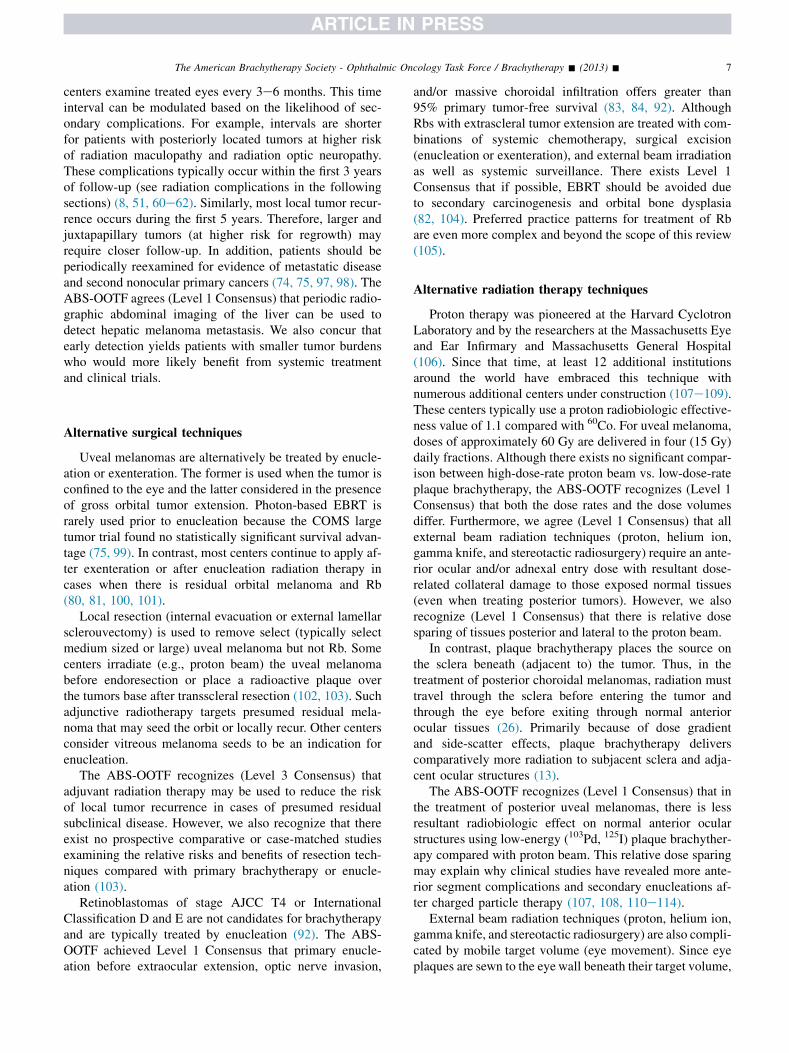
Radiological characteristics of radionuclides used for episcleral brachytherapy
Emitters Half-lifea Mean photon energy (keV)a Water TVL (mm)b Pb TVL (mm)c
Photon125I 59.4 d 28.4 55 0.059103Pd 16.99 d 20.7 30 0.026131Cs 9.69 d 30.4 62 0.070
Emitters Half-lifea End point beta energy (MeV)a CSDA range in water (mm)d
Beta106Ru/106Rh 371.8 d 3.541e 1790Sr 28.8 y 0.546f 1.9
Photon emissions less than 5 keV were removed from calculations of mean energy and tenth value layers (TVLs). Pb 5 lead; CSDA 5 continuous slow
down approximation.a http://www.nndc.bnl.gov/chart/.b http://physics.nist.gov/PhysRefData/XrayMassCoef/ComTab/water.html.c http://physics.nist.gov/PhysRefData/XrayMassCoef/ElemTab/z82.html.d Handbook of Radioactivity Analysis, edited by M. F. L’Annunziata (2003): http://books.google.com/books?id5OfqdTC6deZkC&pg5PA19&lpg5
PA19&dq5betaþparticleþrangeþinþair&source5bl&ots5D7gm8TeI3a&sig5zmcdrOUS15NVqqfDl oPfOvhRCA&hl5en&ei5yN7MSfvZDprNlQfnqtXQCQ&
sa5X&oi5book result&resnum58&ct5result#v5onepage&q&f5false http://www.alpharubicon.com/basicnbc/article16radiological71.htm.e http://www.nndc.bnl.gov/chart/decaysearchdirect.jsp/nuc5106Rh&unc5nds.f http://www.nndc.bnl.gov/chart/decaysearchdirect.jsp?nuc5l06Rh&unc-nds.
6 The American Brachytherapy Society - Ophthalmic Oncology Task Force / Brachytherapy - (2013) -
recommends (Level 2 Consensus) that vitreous seedingshould be absent or within 2 mm of the tumor surface.Either low-energy 103Pd, 125I (for thicker tumors), or106Ru plaques (for thinner tumors) has been used. Usinglow-energy plaques, a solitary Rb is typically treated witha dose of 40e50 Gy to the tumor apex over 3e5 days. De-pending on the ABS-OOTF center, typically higher tumorapex doses have been used for both 106Ru and 90Sr plaques.
Murphree (78) noted that a history of or synchronoustreatment with chemotherapy potentiates radiation-relatedintraocular vasculopathy (retinopathy and optic neuropa-thy). In these cases, they advocated reduced apical 125I pre-scription doses of 20e25 Gy or allowing several monthsbetween chemotherapy and brachytherapy (78).
Plaque surgery
Survey of ABS-OOTF centers suggests that brachyther-apy using both low-energy photon-emitting sources (103Pdand 125I) and beta-emitting 103Ru have been performed asoutpatient procedures. However, centers must comply withlocal government regulations. The surgeries should be per-formed under either general or regional anesthesia, by asubspecialty-trained surgeon, thus experienced in plaqueinsertion. Ocular muscles should be relocated if they inter-fere with plaque position. This includes both rectus and ob-lique muscles.
Typically localized by transpupillary or transocular illu-mination of the globe, the tumor base shadows its subja-cent sclera. The edges of the shadow are marked on thesclera with tissue dye. An additional 2e3 mm ‘‘freemargin’’ is typically measured and marked around the tu-mor base. Some centers directly sew the plaque over themarked target, whereas others preplace sutures using‘‘dummy’’ plaques. The ABS-OOTF defines ‘‘normal pla-que position’’ (Level 1 Consensus) that the target volume
includes the tumors base and safety margin. The ABS-OOTF survey found that compared with 103Pd and 125I pla-ques, larger physical safety margins are typically used with106Ru.
Extra care must be taken in transilluminating thicker(e.g., O5-mm thick) uveal melanomas. Here, the tumorcan cast eccentric shadows, thus yielding false tumor basediameters. Small posterior and amelanotic tumors can alsobe a challenge to mark. Here, two techniques are helpfulincluding: posterior point source illumination (e.g., fiberoptic or HeNe light sources or scleral depression combinedwith indirect ophthalmoscopy) and/or intraoperativeophthalmic ultrasound verification (93, 94). When this isnot possible (e.g., iris and iridociliary melanoma), high-frequency ultrasound imaging and direct transcorneal visu-alization play a more important role during intraoperativetumor localization (28).
In all cases, the plaque is sutured as to cover the scleral-marked target volume. Then, the extraocular muscles andconjunctiva are reattached as not to disturb brachytherapy.When using plaque with low-energy seeds, the eye is typi-cally covered with a lead patch shield. Typically, after5e7 days, the patient is returned to the operating room,where the plaque is removed under regional or generalanesthesia. The ABS-OOTF agreed (Level 2 Consensus)that displaced muscles should be reattached into their inser-tions after plaque removal. However, one ABS-OOTF cen-ter did not find it necessary to reattach the inferior obliquemuscle. If an amniotic membrane is used to buffer thecornea during brachytherapy, it should be removed beforeconjunctival closure (95, 96).
Follow-up after brachytherapy
After brachytherapy, patients are followed for local con-trol, complications, and systemic disease. Most ABS-OOTF
7The American Brachytherapy Society - Ophthalmic Oncology Task Force / Brachytherapy - (2013) -
centers examine treated eyes every 3e6 months. This timeinterval can be modulated based on the likelihood of sec-ondary complications. For example, intervals are shorterfor patients with posteriorly located tumors at higher riskof radiation maculopathy and radiation optic neuropathy.These complications typically occur within the first 3 yearsof follow-up (see radiation complications in the followingsections) (8, 51, 60e62). Similarly, most local tumor recur-rence occurs during the first 5 years. Therefore, larger andjuxtapapillary tumors (at higher risk for regrowth) mayrequire closer follow-up. In addition, patients should beperiodically reexamined for evidence of metastatic diseaseand second nonocular primary cancers (74, 75, 97, 98). TheABS-OOTF agrees (Level 1 Consensus) that periodic radio-graphic abdominal imaging of the liver can be used todetect hepatic melanoma metastasis. We also concur thatearly detection yields patients with smaller tumor burdenswho would more likely benefit from systemic treatmentand clinical trials.
Alternative surgical techniques
Uveal melanomas are alternatively be treated by enucle-ation or exenteration. The former is used when the tumor isconfined to the eye and the latter considered in the presenceof gross orbital tumor extension. Photon-based EBRT israrely used prior to enucleation because the COMS largetumor trial found no statistically significant survival advan-tage (75, 99). In contrast, most centers continue to apply af-ter exenteration or after enucleation radiation therapy incases when there is residual orbital melanoma and Rb(80, 81, 100, 101).
Local resection (internal evacuation or external lamellarsclerouvectomy) is used to remove select (typically selectmedium sized or large) uveal melanoma but not Rb. Somecenters irradiate (e.g., proton beam) the uveal melanomabefore endoresection or place a radioactive plaque overthe tumors base after transscleral resection (102, 103). Suchadjunctive radiotherapy targets presumed residual mela-noma that may seed the orbit or locally recur. Other centersconsider vitreous melanoma seeds to be an indication forenucleation.
The ABS-OOTF recognizes (Level 3 Consensus) thatadjuvant radiation therapy may be used to reduce the riskof local tumor recurrence in cases of presumed residualsubclinical disease. However, we also recognize that thereexist no prospective comparative or case-matched studiesexamining the relative risks and benefits of resection tech-niques compared with primary brachytherapy or enucle-ation (103).
Retinoblastomas of stage AJCC T4 or InternationalClassification D and E are not candidates for brachytherapyand are typically treated by enucleation (92). The ABS-OOTF achieved Level 1 Consensus that primary enucle-ation before extraocular extension, optic nerve invasion,
and/or massive choroidal infiltration offers greater than95% primary tumor-free survival (83, 84, 92). AlthoughRbs with extrascleral tumor extension are treated with com-binations of systemic chemotherapy, surgical excision(enucleation or exenteration), and external beam irradiationas well as systemic surveillance. There exists Level 1Consensus that if possible, EBRT should be avoided dueto secondary carcinogenesis and orbital bone dysplasia(82, 104). Preferred practice patterns for treatment of Rbare even more complex and beyond the scope of this review(105).
Alternative radiation therapy techniques
Proton therapy was pioneered at the Harvard CyclotronLaboratory and by the researchers at the Massachusetts Eyeand Ear Infirmary and Massachusetts General Hospital(106). Since that time, at least 12 additional institutionsaround the world have embraced this technique withnumerous additional centers under construction (107e109).These centers typically use a proton radiobiologic effective-ness value of 1.1 compared with 60Co. For uveal melanoma,doses of approximately 60 Gy are delivered in four (15 Gy)daily fractions. Although there exists no significant compar-ison between high-dose-rate proton beam vs. low-dose-rateplaque brachytherapy, the ABS-OOTF recognizes (Level 1Consensus) that both the dose rates and the dose volumesdiffer. Furthermore, we agree (Level 1 Consensus) that allexternal beam radiation techniques (proton, helium ion,gamma knife, and stereotactic radiosurgery) require an ante-rior ocular and/or adnexal entry dose with resultant dose-related collateral damage to those exposed normal tissues(even when treating posterior tumors). However, we alsorecognize (Level 1 Consensus) that there is relative dosesparing of tissues posterior and lateral to the proton beam.
In contrast, plaque brachytherapy places the source onthe sclera beneath (adjacent to) the tumor. Thus, in thetreatment of posterior choroidal melanomas, radiation musttravel through the sclera before entering the tumor andthrough the eye before exiting through normal anteriorocular tissues (26). Primarily because of dose gradientand side-scatter effects, plaque brachytherapy deliverscomparatively more radiation to subjacent sclera and adja-cent ocular structures (13).
The ABS-OOTF recognizes (Level 1 Consensus) that inthe treatment of posterior uveal melanomas, there is lessresultant radiobiologic effect on normal anterior ocularstructures using low-energy (103Pd, 125I) plaque brachyther-apy compared with proton beam. This relative dose sparingmay explain why clinical studies have revealed more ante-rior segment complications and secondary enucleations af-ter charged particle therapy (107, 108, 110e114).
External beam radiation techniques (proton, helium ion,gamma knife, and stereotactic radiosurgery) are also compli-cated by mobile target volume (eye movement). Since eyeplaques are sewn to the eyewall beneath their target volume,
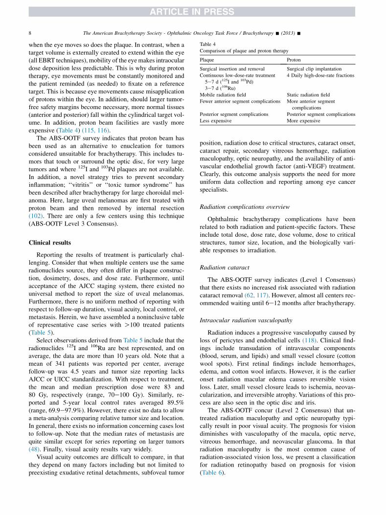
Table 4
Comparison of plaque and proton therapy
Plaque Proton
Surgical insertion and removal Surgical clip implantation
Continuous low-dose-rate treatment 4 Daily high-dose-rate fractions
5e7 d (125I and 103Pd)
3e7 d (106Ru)
Mobile radiation field Static radiation field
Fewer anterior segment complications More anterior segment
complications
Posterior segment complications Posterior segment complications
Less expensive More expensive
8 The American Brachytherapy Society - Ophthalmic Oncology Task Force / Brachytherapy - (2013) -
when the eye moves so does the plaque. In contrast, when atarget volume is externally created to extend within the eye(all EBRT techniques), mobility of the eyemakes intraoculardose deposition less predictable. This is why during protontherapy, eye movements must be constantly monitored andthe patient reminded (as needed) to fixate on a referencetarget. This is because eye movements cause misapplicationof protons within the eye. In addition, should larger tumor-free safety margins become necessary, more normal tissues(anterior and posterior) fall within the cylindrical target vol-ume. In addition, proton beam facilities are vastly moreexpensive (Table 4) (115, 116).
The ABS-OOTF survey indicates that proton beam hasbeen used as an alternative to enucleation for tumorsconsidered unsuitable for brachytherapy. This includes tu-mors that touch or surround the optic disc, for very largetumors and where 125I and 103Pd plaques are not available.In addition, a novel strategy tries to prevent secondaryinflammation; ‘‘vitritis’’ or ‘‘toxic tumor syndrome’’ hasbeen described after brachytherapy for large choroidal mel-anoma. Here, large uveal melanomas are first treated withproton beam and then removed by internal resection(102). There are only a few centers using this technique(ABS-OOTF Level 3 Consensus).
Clinical results
Reporting the results of treatment is particularly chal-lenging. Consider that when multiple centers use the sameradionuclides source, they often differ in plaque construc-tion, dosimetry, doses, and dose rate. Furthermore, untilacceptance of the AJCC staging system, there existed nouniversal method to report the size of uveal melanomas.Furthermore, there is no uniform method of reporting withrespect to follow-up duration, visual acuity, local control, ormetastasis. Herein, we have assembled a noninclusive tableof representative case series with O100 treated patients(Table 5).
Select observations derived from Table 5 include that theradionuclides 125I and 106Ru are best represented, and onaverage, the data are more than 10 years old. Note that amean of 341 patients was reported per center, averagefollow-up was 4.5 years and tumor size reporting lacksAJCC or UICC standardization. With respect to treatment,the mean and median prescription dose were 83 and80 Gy, respectively (range, 70e100 Gy). Similarly, re-ported and 5-year local control rates averaged 89.5%(range, 69.9e97.9%). However, there exist no data to allowa meta-analysis comparing relative tumor size and location.In general, there exists no information concerning cases lostto follow-up. Note that the median rates of metastasis arequite similar except for series reporting on larger tumors(48). Finally, visual acuity results vary widely.
Visual acuity outcomes are difficult to compare, in thatthey depend on many factors including but not limited topreexisting exudative retinal detachments, subfoveal tumor
position, radiation dose to critical structures, cataract onset,cataract repair, secondary vitreous hemorrhage, radiationmaculopathy, optic neuropathy, and the availability of anti-vascular endothelial growth factor (anti-VEGF) treatment.Clearly, this outcome analysis supports the need for moreuniform data collection and reporting among eye cancerspecialists.
Radiation complications overview
Ophthalmic brachytherapy complications have beenrelated to both radiation and patient-specific factors. Theseinclude total dose, dose rate, dose volume, dose to criticalstructures, tumor size, location, and the biologically vari-able responses to irradiation.
Radiation cataract
The ABS-OOTF survey indicates (Level 1 Consensus)that there exists no increased risk associated with radiationcataract removal (62, 117). However, almost all centers rec-ommended waiting until 6e12 months after brachytherapy.
Intraocular radiation vasculopathy
Radiation induces a progressive vasculopathy caused byloss of pericytes and endothelial cells (118). Clinical find-ings include transudation of intravascular components(blood, serum, and lipids) and small vessel closure (cottonwool spots). First retinal findings include hemorrhages,edema, and cotton wool infarcts. However, it is the earlieronset radiation macular edema causes reversible visionloss. Later, small vessel closure leads to ischemia, neovas-cularization, and irreversible atrophy. Variations of this pro-cess are also seen in the optic disc and iris.
The ABS-OOTF concur (Level 2 Consensus) that un-treated radiation maculopathy and optic neuropathy typi-cally result in poor visual acuity. The prognosis for visiondiminishes with vasculopathy of the macula, optic nerve,vitreous hemorrhage, and neovascular glaucoma. In thatradiation maculopathy is the most common cause ofradiation-associated vision loss, we present a classificationfor radiation retinopathy based on prognosis for vision(Table 6).
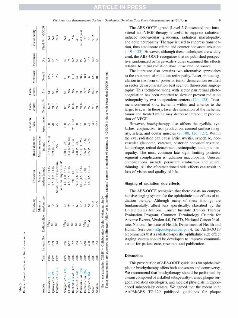
Table
5
Review
ofuveal
melanomaclinical
case
series
Author
Year
Patients
No.
Radionuclide
Follow
-up
Thickness
Basal
diameter
Radiation
dose
Local
control
Local
control
Metastasis
Metastasis
Visual
acuity
Meanor
median,mo
Meanor
median(range)
Meanormedian
(range)
Apex,mean
Overall,%
5y
Overall
5y
Final
%,O
20/200
Lommatzsch
(3)
1987
309
106Ru
80
3.7
(1.2e11.8)
9.7
(4.5e21.5)
100
69.9
84
12.9
NA
NA
Quivey
etal.(90)
1996
239
125I
36
5.5
(1.9e11.0)
10.9
(4e18)
70
91.7
82
7.5
12
NA
Fontenesiet
al.(17)
1993
144
125I
46
Small,n5
15;medium,
n5
84;large,
n5
45
NA
75
97.7
94.4
2.7
271.3
Seregardet
al.(24)
1997
266
106Ru
43
4.4
(1.0e13.1)
10.0
(3e23)
100
83
82
11
14
NA
COMS(16)
2006
657
125I
96
4.8
(2.5e10.0)
11.4
(upto
16)
85
NA
NA
99
63
Bechrakiset
al.(128)
2002
152
125I
30.1
9.0
�1.1
14.6
�2.4
98�
18
88.8
NA
11.1
NA
5.6
Shieldset
al.(48)
2002
354
125I
60
9.0
(9.8e16)
14.0
(5e21)
80
91
91
24
24
43
Puusaariet
al.(49)
2003
97
125I
43.2
10.7
(4.5e16.8)
16.1
(7.3e25)
87
94.8
94
28.9
35
42at
1year
Dam
atoet
al.(89)
2005
458
106Ru
47
3.2
(0.7e7.0)
10.6
(5e16.6)
80
97
97.9
8.1
NA
57
Finger
etal.(6)
2009
400
103Pd
51
3.8
(1.5e12.3)
10.5
(5e19.9)
73
97
NA
67.3
79
Mean
2000
308
53.2
84.8
90.1
89.3
12.2
14.8
53.2
Median
2002
354
46.5
82.5
91.7
91
10
12
57
NA
5notavailable;COMS5
Collaborative
OcularMelanomaStudy.
Tumormeasurements
areexpressed
inmillimeters,follow
-upin
months,patients’number
innumber
ofpatients,%
!20/200in
those
withbetterthan
20/200vision.
9The American Brachytherapy Society - Ophthalmic Oncology Task Force / Brachytherapy - (2013) -
The ABS-OOTF agreed (Level 2 Consensus) that intra-vitreal anti-VEGF therapy is useful to suppress radiation-induced neovascular glaucoma, radiation maculopathy,and optic neuropathy. Therapy is used to suppress transuda-tion, thus ameliorate edema and counter neovascularization(119e123). However, although these techniques are widelyused, the ABS-OOTF recognizes that no published prospec-tive randomized or large-scale studies examined the effectsrelative to initial radiation dose, dose rate, or source.
The literature also contains two alternative approachesto the treatment of radiation retinopathy. Laser photocoag-ulation in the form of posterior tumor demarcation resultedin sector devascularization best seen on fluorescein angiog-raphy. This technique along with sector pan retinal photo-coagulation has been reported to slow or prevent radiationretinopathy by two independent centers (124, 125). Treat-ment converted slow ischemia within and anterior to thetarget to scar. In theory, laser devitalization of the ischemictumor and treated retina may decrease intraocular produc-tion of VEGF.
However, brachytherapy also affects the eyelids, eye-lashes, conjunctiva, tear production, corneal surface integ-rity, sclera, and ocular muscles (8, 100, 126, 127). Withinthe eye, radiation can cause iritis, uveitis, synechiae, neo-vascular glaucoma, cataract, posterior neovascularization,hemorrhage, retinal detachment, retinopathy, and optic neu-ropathy. The most common late sight limiting posteriorsegment complication is radiation maculopathy. Unusualcomplications include persistent strabismus and scleralthinning. All the aforementioned side effects can result inloss of vision and quality of life.
Staging of radiation side effects
The ABS-OOTF recognize that there exists no compre-hensive staging system for the ophthalmic side effects of ra-diation therapy. Although many of these findings arefundamentally, albeit less specifically, classified by theUnited States National Cancer Institute (Cancer TherapyEvaluation Program, Common Terminology Criteria forAdverse Events, Version 4.0, DCTD, National Cancer Insti-tute, National Institute of Health, Department of Health andHuman Services (http://ctep.cancer.gov)), the ABS-OOTFrecommends that a radiation-specific ophthalmic side effectstaging system should be developed to improve communi-cation for patient care, research, and publication.
Discussion
This presentation ofABS-OOTFguidelines for ophthalmicplaque brachytherapy offers both consensus and controversy.We recommend that brachytherapy should be performed bya team composed of a skilled subspecialty-trained plaque sur-geon, radiation oncologists, and medical physicists in experi-enced subspecialty centers. We agreed that the recent jointAAPM/ABS TG-129 published guidelines for plaque
Table 6
Classification for radiation retinopathy
Stage Sign Symptom Location Best viewed by Risk of vision loss
1 Cotton wool spots None Extramacular Ophthalmoscopy Mild
Retinal hemorrhages None Extramacular Ophthalmoscopy Mild
Retinal microaneurysms None Extramacular Ophthalmoscopy/FA Mild
Exudate None Extramacular Ophthalmoscopy Mild
Uveal effusion None Extramacular Ophthalmoscopy/OCT Mild
Chorioretinal atrophy None Extramacular Ophthalmoscopy Mild
Choroidopathy None Extramacular ICG Mild
Retinal ischemia (!5 DA) None Extramacular FA Mild
2 Above findings None Macular All Moderate
3 Any combination of the above plus
Retinal neovascularization Vision loss Extramacular FA Severe
Macular edemadnew onset Vision loss Macular FA/OCT Severe
4 Any combination of the above plus
Vitreous hemorrhage Vision loss Vitreous Ophthalmoscopy Severe
Retinal ischemia ($5 DA) Vision loss Both FA Severe
FA 5 fluorescein angiography; OCT 5 optical coherence tomography; ICG 5 indocyanine green angiography; DA 5 disc areas.
Vision loss must be related to associated sign(s). This table is modified and updated from an original classification (124).
10 The American Brachytherapy Society - Ophthalmic Oncology Task Force / Brachytherapy - (2013) -
construction, dosimetry, and quality assurance should be readand widely used at active centers (13, 26). We also concurredthat many radionuclide sources can be used, but only 125I,103Pd, and 106Ruare used in three ormoreABS-OOTFcenters.Although there exist tumor thickness restrictions for 106Ru and90Sr, taller tumors can be treated with 125I or 103Pd techniques(7, 11, 13, 72).
Overall, the ABS-OOTF expanded general indicationsfor uveal melanoma patient selection (Table 2). Fianlly,we found that plaque brachytherapy is not commonly usedfor Rb. However, indications include: small anterior tumorsin unilateral cases, for salvage after chemoreduction withsubsequent alternative therapies and in select cases inwhich macular laser will likely cause loss of vision.
The ABS-OOTF recommends that the eye cancer com-munity use universal AJCCeUICC staging to define tumorsize, location, and associated variables (87, 88). This wouldenable multicenter communication, comparative analysis,and patient education. This in turn, would allow for collec-tion of numbers large enough to reach statistical signifi-cance. The ABS-OOTF recommends the development ofa site-specific staging system for complications afterophthalmic radiation therapy. This would facilitate scienti-fic comparisons between treatments, help predictophthalmic side effects, and improve informed consent.
Unanswered questions
However, the ABS-OOTF acknowledges the myriad un-answered questions that challenge ophthalmic plaquebrachytherapy researchers. Select questions offered by theABS-OOTF include: What are the radiobiological differ-ences between continuous low-dose-rate plaque brachyther-apy in comparison with fractionated high-dose-rate protonbeam irradiation? What is the ‘‘correct’’ apical prescriptiondose and dose rate required for treatment of uveal mela-noma, and how do we accommodate for the steep dose
gradient within the tumor? For example, should there bea dose deescalation study or a thickness-based sliding scalein treatment of uveal melanoma? Can there be internationalstandards for dosimetry to determine the relative efficacy ofphotons, electrons, and protons? Is there a role for radiationsensitizers during plaque therapy? Should the presence ofintravitreal melanoma seeds affect case selection? What isthe role and best timing for the use of anti-VEGF agents intreatment of radiation maculopathy and optic neuropathy?Are there differences in the efficacy of anti-VEGF agentsrelated to radionuclide, radiation dose, and dose rate? Donotched and slotted plaques address geographic miss inthe treatment of juxtapapillary and circumpapillary tumors?With regard to Rb, are there oncogenic risks of plaquebrachytherapy? What are the optimal parameters for tumorsize selection and radiation dose (if used before or afterchemotherapy)? The ABS-OOTF hopes future research willanswer some of these questions.
Summary
Currently, plaque brachytherapy offers an eye and visionsparing alternative to enucleation annually for thousands ofpatients’ worldwide. Herein, we present the current ABSguidelines for patient selection, informed consent, andmethods of treatment. We encourage all centers to use theseguidelines to formulate their treatment patterns and report-ing policies. However, we realize that such guidelines aredynamic and will need to be modified as to conform to everevolving clinical evidence.
Conclusions
The ABS-OOTF, comprised 47 eye cancer specialistsfrom 10 countries, present our current guidelines andmethods of plaque brachytherapy for uveal melanoma and
United Kingdom - Liverpool University MedicalCenter, LiverpoolBertil DamatoeOphthalmic Oncology
11The American Brachytherapy Society - Ophthalmic Oncology Task Force / Brachytherapy - (2013) -
Rb. We point out what is currently accepted as known, un-known, and a need for standardization, staging as well asfuture research.
R Doug ErringtoneRadiation OncologyPhilip MayleseMedical PhysicsHelen MayleseMedical Physics
United States - Emory Eye CancerEmory University Medical Center Atlanta, GeorgiaChris BergstromeOphthalmic Oncology
Acknowledgments
The research was supported (in part) by The Eye CancerFoundation, Inc. (http://eyecancerfoundation.net) and TheAmerican Brachytherapy Society.
The ABS e OOTF Committee
Canada - Princess Margaret HospitalSick Kids Hospital e Toronto, OntarioE. Rand SimpsoneOphthalmic OncologyBrenda GallieeOphthalmic OncologyNormand LaperrierreeRadiation OncologyAkbar Beiki-ArdakanieMedical Physics
Finland - Helsinki University Central HospitalUniversity of Helsinki, HelsinkiTero Kivel€aeOphthalmic OncologyVirpi RaivioeOphthalmic OncologyJorma HeikkoneneMedical Physics
France - The Curie Institute, ParisLaurence DesjardinseOphthalmic OncologyRemi DendaleeRadiation OncologyAlexandro MazaleMedical Physics
Germany - University of Duisburg-Essen, EssenNorbert BornfeldeOphthalmic OncologyWolfgang SauerweineRadiation OncologyDirk Fl€uehseMedical PhysicsLorenzo BruallaeMedical Physics
India e Centre for Sight Superspecialty EyeHospital, HyderabadSantosh G. HonavareOphthalmic OncologyVijay Anand P. ReddyeRadiation Oncology
Japan e National Cancer Center Hospital, TokyoShigenobu SuzukieOphthalmic OncologyNaoya MurakamieRadiation Oncology
Russia - Helmholtz Research Institute of EyeDiseases, MoscowSvetlana SaakyaneOphthalmic Oncology, RadiologyVladimir ValskiyeOphthalmic Oncology, RadiologyAnush AmiryaneOphthalmic Oncology, Radiology
Sweden - St. Erik’s Eye Hospital, StockholmStefan SeregardeOphthalmic OncologyCharlotta All-ErikssoneOphthalmic OncologyLars HjelmqvisteOphthalmic OncologyG€oran LundelleRadiation OncologyGeorges SinclaireRadiation OncologyMarie LundelleMedical Physics
Hans GrossniklauseOphthalmic OncologyIan CrockereRadiation OncologyElizabeth ButkereMedical Physics
United States - University of Tennessee e MemphisMethodist University HospitalSt. Jude’s Children’s Research HospitalMatthew WilsoneOphthalmic OncologyBarrett HaikeOphthalmic OncologyHolger GeischeneRadiation OncologyPradeep PatraeMedical Physics
United States - Tufts University Medical Center,Boston, MassJay DukereOphthalmic OncologyJohn MignanoeRadiation OncologyMark RivardeMedical Physics
United States - The New York Eye Cancer Center,New York CityBeth Israel Comprehensive Cancer CenterThe New York Eye and Ear InfirmaryPaul T. Finger, ABS-OOTFChaireOphthalmicOncologyEkaterina SemenovaeOphthalmic OncologyWalter ChoieRadiation OncologyNina I. KalacheMedical Physics
References
[1] Moore R. Choroidal sarcoma treated by the intraocular insertion of
radon seeds. Br J Ophthalmol 1930;14:145e156.[2] Stallard HB. Radiotherapy for malignant melanoma of the choroid.
Br J Ophthalmol 1966;50:147e155.
[3] Lommatzsch PK. Results after beta-irradiation (106Ru/106Rh) of
choroidal melanomas. Twenty years’ experience. Am J Clin Oncol
1987;10:146e151.
[4] Packer S, Rotman M. Radiotherapy of choroidal melanoma with
iodine-125. Ophthalmology 1980;87:582e590.
[5] Sealy R, le Roux PL, Rapley F, et al. The treatment of ophthalmic
tumours with low-energy sources. Br J Radiol 1976;49:551e554.
[6] Finger PT, Chin KJ, Duvall G, et al. Palladium-103 ophthalmic pla-
que radiation therapy for choroidal melanoma: 400 treated patients.
Ophthalmology 2009;116:790e796.
[7] Rivard MJ, Melhus CS, Sioshansi S, et al. The impact of prescription
depth, dose rate, plaque size, and source loading on the central axis us-
ing 103Pd, 125I, and 131Cs. Brachytherapy 2008;7:327e335.[8] Finger PT. Radiation therapy for choroidal melanoma. Surv Oph-
thalmol 1997;42:215e232.
[9] Leonard KL, Gagne NL, Mignano JE, et al. A 17-year retrospective
study of institutional results for eye plaque brachytherapy of uveal
12 The American Brachytherapy Society - Ophthalmic Oncology Task Force / Brachytherapy - (2013) -
melanoma using (125)I, (103)Pd, and (131)Cs and historical
perspective. Brachytherapy 2011;10:331e339.
[10] Vakulenko MP, Dedenkov AN, Brovkina AF, et al. Results of beta-
therapy of choroidal melanoma. Med Radiol (Mosk) 1980;25:
73e74.[11] Brovkina AF, Zarubei GD, Val’skii VV. Criteria for assessing the ef-
ficacy of brachytherapy of uveal melanomas, complications of ther-
apy and there prevention. Vestn Oftalmol 1997;113:14e16.
[12] Murakami N, Suzuki S, Ito Y, et al. 106Ruthenium plaque therapy
(RPT) for retinoblastoma. Int J Radiat Oncol Biol Phys 2012;84:
59e65.
[13] Chiu-Tsao ST, Astrahan MA, Finger PT, et al. Dosimetry of 125I and103Pd COMS eye plaques for intraocular tumors: Report of Task
Group 129 by the AAPM and ABS. Med Phys 2012;39:6161e6184.
[14] Collaborative Ocular Melanoma Study Group. Ch 12: Radiation
therapy. In: National Technical Information Service (NTIS), editor.
COMS manual of procedures. Springfield, VA; 1995. PB95-179693.
[15] Collaborative Ocular Melanoma Study Group. The COMS random-
ized trial of iodine 125 brachytherapy for choroidal melanoma: V.
Twelve-year mortality rates and prognostic factors: COMS report
No. 28. Arch Ophthalmol 2006;124:1684e1693.
[16] Earle J, Kline RW, Robertson DM. Selection of iodine 125 for the
Collaborative Ocular Melanoma Study. Arch Ophthalmol 1987;
105:763e764.
[17] Fontanesi J, Meyer D, Xu S, et al. Treatment of choroidal mela-
noma with I-125 plaque. Int J Radiat Oncol Biol Phys 1993;26:
619e623.
[18] Packer S, Stoller S, Lesser ML, et al. Long-term results of iodine
125 irradiation of uveal melanoma. Ophthalmology 1992;99:
767e773.
[19] Bergman L, Nilsson B, Lundell G, et al. Ruthenium brachytherapy
for uveal melanoma, 1979-2003: Survival and functional outcomes
in the Swedish population. Ophthalmology 2005;112:834e840.
[20] Summanen P, Immonen I, Kivel€a T, et al. Visual outcome of eyes
with malignant melanoma of the uvea after ruthenium plaque radio-
therapy. Ophthalmic Surg Lasers 1995;26:449e460.
[21] Damato B, Patel I, Campbell IR, et al. Local tumor control after106Ru brachytherapy of choroidal melanoma. Int J Radiat Oncol
Biol Phys 2005;63:385e391.
[22] Foerster MH, Bornfeld N, Wessing A, et al. Treatment of malignant
melanomas of the uvea with 106-ruthenium applicators. Report on
the first 100 Essen cases. Klin Monbl Augenheilkd 1984;185:
490e494.
[23] Schueler AO, Fl€uehs D, Anastassiou G, et al. Beta-ray brachyther-
apy of retinoblastoma: Feasibility of a new small-sized ruthenium-
106 plaque. Ophthalmic Res 2006;38:8e12.[24] Seregard S, aft Trampe E, Lax I, et al. Results following episcleral
ruthenium plaque radiotherapy for posterior uveal melanoma. The
Swedish experience. Acta Ophthalmol Scand 1997;75:11e16.[25] Lommatzsch PK, Werschnik C, Schuster E. Long-term follow-up of
Ru-106/Rh-106 brachytherapy for posterior uveal melanoma.
Graefes Arch Clin Exp Ophthalmol 2000;238:129e137.
[26] Rivard MJ, Chiu-Tsao S-T, Finger PT, et al. Comparison of dose
calculation methods for brachytherapy of intraocular tumors. Med
Phys 2011;38:306e316.
[27] Nag S, Quivey JM, Earle JD, et al. The American Brachytherapy
Society recommendations for brachytherapy of uveal melanomas.
Int J Radiat Oncol Biol Phys 2003;56:544e555.
[28] Finger PT, Reddy S, Chin K. High-frequency ultrasound character-
istics of 24 iris and iridociliary melanomas: Before and after plaque
brachytherapy. Arch Ophthalmol 2007;125:1051e1058.
[29] Romani A, Baldeschi L, Genovesi-Ebert F, et al. Sensitivity and
specificity of ultrasonography, fluorescein videoangiography, indoc-
yanine green videoangiography, magnetic resonance and radioim-
munoscintigraphy in the diagnosis of primary choroidal malignant
melanoma. Ophthalmologica 1998;212:44e46.
[30] Finger PT, Garcia JP Jr, Pro MJ, et al. ‘‘C-scan’’ ultrasound imaging
of optic nerve extension of retinoblastoma. Br J Ophthalmol 2005;
89:1225e1226.
[31] Marigo FA, Finger PT, McCormick SA, et al. Iris and ciliary body
melanomas: Ultrasound biomicroscopy with histopathologic corre-
lation. Arch Ophthalmol 2000;118:1515e1521.
[32] Chin K, Finger PT. Autofluorescence characteristics of suspicious
choroidal nevi. Optometry 2009;80:126e130.
[33] Freton A, Chin KJ, Raut R, et al. Initial PET/CT staging for
choroidal melanoma: AJCC correlation and second nonocular pri-
maries in 333 patients. Eur J Ophthalmol 2012;22:236e243.
[34] Shields CL, Kaliki S, Rojanaporn D, et al. Enhanced depth imaging
optical coherence tomography of small choroidal melanoma: Com-
parison with choroidal nevus. Arch Ophthalmol 2012;130:850e856.
[35] Lommatzsch PK, Ballin RE, Helm W. Fluorescein angiography in
the follow-up study of choroidal melanoma after 106Ru/106Rh pla-
que therapy. Retina 1987;7:148e155.
[36] Rootman DB, Gonzalez E, Mallipatna A, et al. Hand-held high-
resolution spectral domain optical coherence tomography in retino-
blastoma: Clinical and morphologic considerations. Br J Ophthalmol
2013;97:59e65.
[37] Finger PT, Chin KJ, Tena LB. A five-year study of slotted plaque
radiation therapy for choroidal melanoma: Near, touching or sur-
rounding the optic nerve. Ophthalmology 2012;119:415e422.
[38] Finger PT. Plaque radiation therapy for malignant melanoma of the
iris and ciliary body. Am J Ophthalmol 2001;132:328e335.
[39] Yousef YA, Finger PT. Lack of radiation maculopathy after
palladium-103 plaque radiotherapy for iris melanoma. Int J Radiat
Oncol Biol Phys 2012;83:1107e1112.
[40] Shields C, Naseripour M, Shields J, et al. Custom-designed plaque
radiotherapy for nonresectable iris melanoma in 38 patients: Tumor
control and ocular complications. Am J Ophthalmol 2003;135:
648e656.
[41] Petousis V, Finger PT, Milman T. Multifocal iris melanoma treated
with total anterior segment palladium-103 plaque radiation therapy.
Graefes Arch Clin Exp Ophthalmol 2011;249:937e940.
[42] Fernandes BF, Krema H, Fulda E, et al. Management of iris mela-
nomas with 125I plaque radiotherapy. Am J Ophthalmol 2010;149:
70e76.
[43] Krema H, Simpson ER, Pavlin CJ, et al. Management of ciliary
body melanoma with iodine-125 plaque brachytherapy. Can J Oph-
thalmol 2009;44:395e400.[44] Lumbroso-Le Rouic L, Charif Chefchaouni M, Levy C, et al. 125I
plaque brachytherapy for anterior uveal melanomas. Eye (Lond)
2004;18:911e916.
[45] Brovkina AF, Zarubei GD, Fishkin IuG. Validation of the use of
brachytherapy in uveal melanomas of juxtapapillary localization.
Vestn Oftalmol 1991;107:41e44.
[46] Newman H, Chin KJ, Finger PT. Subfoveal choroidal melanoma:
Pretreatment characteristics and response to plaque radiation ther-
apy. Arch Ophthalmol 2011;129:892e898.
[47] Gray ME, Correa ZM, Augsburger JJ, et al. Ciliary body melanoma
with limited nodular extrascleral extension and diffuse iris-angle
infiltration treated by whole anterior segment plaque radiotherapy.
Int Ophthalmol 2007;27:273e276.
[48] Shields CL, Naseripour M, Cater J, et al. Plaque radiotherapy for
large posterior uveal melanomas (O or 58-mm thick) in 354
consecutive patients. Ophthalmology 2002;109:1838e1849.
[49] Puusaari I, Heikkonen J, Summanen P, et al. Iodine brachytherapy
as an alternative to enucleation for large uveal melanomas. Ophthal-
mology 2003;110:2223e2234.
[50] Puusaari I, Heikkonen J, Kivel€a T. Ocular complications after iodine
brachytherapy for large uveal melanomas. Ophthalmology 2004;
111:1768e1777.
[51] Puusaari I, Heikkonen J, Kivel€a T. Effect of radiation dose on ocular
complications after iodine brachytherapy for large uveal melanoma:
13The American Brachytherapy Society - Ophthalmic Oncology Task Force / Brachytherapy - (2013) -
Empirical data and simulation of collimating plaques. Invest Oph-
thalmol Vis Sci 2004;45:3425e3434.
[52] Semenova E, Finger PT. Palladium-103 radiation therapy for small
choroidal melanoma. Ophthalmology 2013;120:2353e2357.
[53] Semenova E, Finger P. Palladium-103 plaque radiation therapy for
AJCC T3 and T4 sized choroidal melanoma. JAMA Ophthalmol
2013. Epubhead of print: November 28, 2013. http://dx.doi.org/10.
1001/jamaophthalmol.2013.5677.
[54] Kujala E, Damato B, Coupland SE, et al. Staging of ciliary body
and choroidal melanomas based on anatomic extent. J Clin Oncol
2013;31:2825e2831.
[55] Finger PT. Do you speak ocular tumor? Ophthalmology 2003;110:
13e14.
[56] Kujala E, Tuomaala S, Eskelin S, et al. Mortality after uveal and
conjunctival melanoma: Which tumour is more deadly? Acta Oph-
thalmol 2009;87:149e153.[57] Augsburger JJ, Vrabec TR. Impact of delayed treatment in growing
posterior uveal melanomas. Arch Ophthalmol 1993;111:
1382e1386.
[58] Sobrin L, Schiffman JC, Markoe AM, et al. Outcomes of iodine 125
plaque radiotherapy after initial observation of suspected small
choroidal melanomas: A pilot study. Ophthalmology 2005;112:
1777e1783.[59] Murray TG, Sobrin L. The case for observational management of
suspected small choroidal melanoma. Arch Ophthalmol 2006;124:
1342e1344.
[60] Finger PT. Tumour location affects the incidence of cataract and
retinopathy after ophthalmic plaque radiation therapy. Br J Ophthal-
mol 2000;84:1068e1070.
[61] Finger PT, Chin KJ, Yu GP. Risk factors for radiation maculopathy
after ophthalmic plaque radiation for choroidal melanoma. Am J
Ophthalmol 2010;149:608e615.
[62] Finger PT, Chin KJ, Yu GP, et al. Risk factors for cataract after
palladium-103 ophthalmic plaque radiation therapy. Int J Radiat
Oncol Biol Phys 2010;80:800e806.
[63] Mashayekhi A, Kaliki S, Walker B, et al. Metastasis from uveal
melanoma associated with congenital ocular melanocytosis: A
matched study. Ophthalmology 2013;120:1465e1468.[64] Onken MD, Worley LA, Char DH, et al. Collaborative Ocular
Oncology Group report number 1: Prospective validation of a
multi-gene prognostic assay in uveal melanoma. Ophthalmology
2012;119:1596e1603.[65] Harbour JW. The genetics of uveal melanoma: An emerging frame-
work for targeted therapy. Pigment Cell Melanoma Res 2012;25:
171e181.
[66] McCannel TA, Chang MY, Burgess BL. Multi-year follow-up of
fine-needle aspiration biopsy in choroidal melanoma. Ophthal-
mology 2012;119:606e610.
[67] Garcia JP Jr, Garcia PT, Rosen RB, et al. A 3-dimensional ultra-
sound C-scan imaging technique for optic nerve measurements.
Ophthalmology 2004;111:1238e1243.
[68] Sagoo MS, Shields CL, Mashayekhi A, et al. Plaque radiotherapy
for juxtapapillary choroidal melanoma: Tumor control in 650
consecutive cases. Ophthalmology 2011;118:402e407.
[69] Houston SK 3rd, Markoe AM, Boldt HC, et al. Juxtapapillary uveal
melanomas: Patient outcomes after treatment with proton irradiation
for peripapillary and parapapillary melanomas. Arch Ophthalmol
2011;129:1218e1220.
[70] Garcia JP Jr, Garcia PM, Rosen RB, et al. Optic nerve measure-
ments by 3D ultrasound-based coronal ‘‘C-scan’’ imaging.
Ophthalmic Surg Lasers Imaging 2005;36:142e146.
[71] Finger PT. Finger’s ‘‘slotted’’ eye plaque for radiation therapy:
Treatment of juxtapapillary and circumpapillary intraocular tu-
mours. Br J Ophthalmol 2007;91:891e894.[72] Brualla L, Sempau J, Zaragoza FJ, et al. Accurate estimation of
dose distributions inside an eye irradiated with 106Ru plaques.
Strahlenther Onkol 2013;189:68e73.
[73] Lommatzsch PK, Lommatzsch R. Treatment of juxtapapillary mel-
anomas. Br J Ophthalmol 1991;75:715e717.
[74] Freton A, Pavlick A, Finger PT. Systemic evaluation and manage-
ment of patients with uveal melanoma. In: Schachat AP, Ryan SJ,
editors. Retina. Vol. III, Tumors of the retina, choroid and vitreous.
London, New York, Oxford, St. Louis, Sydney, Toronto: Elsevier;
2013. p. 2313e2315.
[75] Kivel€a T, Eskelin S, Kujala E. Metastatic uveal melanoma. Int Oph-
thalmol Clin Winter 2006;46:133e149.[76] Diener-West M, Reynolds SM, Agugliaro DJ, et al. Screening for
metastasis from choroidal melanoma: The Collaborative Ocular
Melanoma Study Group Report 23. J Clin Oncol 2004;22:
2438e2444.
[77] Merchant TE, Gould CJ, Wilson MW, et al. Episcleral plaque
brachytherapy for retinoblastoma. Pediatr Blood Cancer 2004;43:
134e139.[78] Finger P, Murphree A. Ophthalmic brachytherapy: Treatment of
choroidal melanoma and retinoblastoma. In: Peyman G,
Meffert S, Conway M, Chou F, editors. Vitreoretinal surgical tech-
niques. London, UK: Martin Dunitz; 2006. p. 452e468.[79] Shields JA, Shields CL, De Potter P, et al. Plaque radiotherapy for
residual or recurrent retinoblastoma in 91 cases. J Pediatr Ophthal-
mol Strabismus 1994;31:242e245.[80] Stannard C, Maree G, Munro R, et al. Iodine-125 orbital brachyther-
apy with a prosthetic implant in situ. Strahlenther Onkol 2011;187:
322e327.
[81] Sealy R, Stannard C, Shackleton D. Improved cosmesis in retino-
blastoma patients treated with iodine-125 orbital irradiation.
Ophthalmic Paediatr Genet 1987;8:95e99.
[82] Bunin GR, Felice MA, Davidson W, et al. Medical radiation expo-
sure and risk of retinoblastoma resulting from new germline RB1
mutation. Int J Cancer 2011;128:2393e2404.
[83] Finger PT, Harbour JW, Karcioglu ZA. Risk factors for metastasis in
retinoblastoma. Surv Ophthalmol 2002;47:1e16.[84] Sastre X, Chantada GL, Doz F, et al. Proceedings of the consensus
meetings from the International Retinoblastoma Staging Working
Group on the pathology guidelines for the examination of enucle-
ated eyes and evaluation of prognostic risk factors in retinoblas-
toma. Arch Pathol Lab Med 2009;133:1199e1202.
[85] Abramson DH, Dunkel IJ, Brodie SE, et al. Superselective
ophthalmic artery chemotherapy as primary treatment for retino-
blastoma (chemosurgery). Ophthalmology 2010;117:1623e1629.[86] Dimaras H, Kimani K, Dimba EA, et al. Retinoblastoma. Lancet
2012;379:1436e1446.
[87] Uveal Melanoma. In: Edge SE, Byrd DR, Compton CC, et al, edi-
tors. AJCC cancer staging manual 7th edition. 7th ed. New York;
London: Springer; 2009. p. 547e559.
[88] Retinoblastoma. In: Edge S, Byrd DR, Compton CC, et al, editors.
The AJCC cancer staging manual. 7th ed. New York, NY: Springer;
2009. p. 561e568.
[89] Damato B, Patel I, Campbell IR, et al. Visual acuity after
ruthenium-106 brachytherapy of choroidal melanomas. Int J Radiat
Oncol Biol Phys 2005;63:392e400.[90] Quivey JM, Augsburger J, Snelling L, et al. 125I plaque therapy for
uveal melanoma. Analysis of the impact of time and dose factors on
local control. Cancer 1996;77:2356e2362.
[91] Kiratli H, Bilgic S, Atahan IL. Plaque radiotherapy in the manage-
ment of retinoblastoma. Turk J Pediatr 1998;40:393e397.
[92] Temming P, Lohmann D, Bornfeld N, et al. Current concepts for
diagnosis and treatment of retinoblastoma in Germany: Aiming
for safe tumor control and vision preservation. Klin Padiatr 2012;
224:339e347.
[93] Harbour JW, Murray TG, Byrne SF, et al. Intraoperative echo-
graphic localization of iodine 125 episcleral radioactive plaques
for posterior uveal melanoma. Retina 1996;16:129e134.
[94] Chang MY, Kamrava M, Demanes DJ, et al. Intraoperative
ultrasonography-guided positioning of iodine 125 plaque
14 The American Brachytherapy Society - Ophthalmic Oncology Task Force / Brachytherapy - (2013) -
brachytherapy in the treatment of choroidal melanoma. Ophthal-
mology 2012;119:1073e1077.
[95] Finger PT. Finger’s amniotic membrane buffer technique: Protecting
the cornea during radiation plaque therapy. Arch Ophthalmol 2008;
126:531e534.[96] Semenova E, Finger PT. Amniotic membrane corneal buffering dur-
ing plaque radiation therapy for anterior uveal melanoma.
Ophthalmic Surg Lasers Imaging Retina 2013;44:477e482.
[97] Kujala E, Makitie T, Kivel€a T. Very long-term prognosis of patients
with malignant uveal melanoma. Invest Ophthalmol Vis Sci 2003;
44:4651e4659.
[98] Chin K, Finger PT, Kurli M, et al. Second cancers discovered by
(18)FDG PET/CT imaging for choroidal melanoma. Optometry
2007;78:396e401.
[99] Hawkins BSCollaborative Ocular Melanoma Study Group. The
Collaborative Ocular Melanoma Study (COMS) randomized trial
of pre-enucleation radiation of large choroidal melanoma: IV.
Ten-year mortality findings and prognostic factors. COMS report
number 24. Am J Ophthalmol 2004;138:936e951.
[100] Finger PT. Radiation therapy for orbital tumors: Concepts, current
use, and ophthalmic radiation side effects. Surv Ophthalmol 2009;
54:545e568.
[101] Finger PT, Tena LB, Semenova E, et al. Extrascleral extension
of choroidal melanoma: Post-enucleation high-dose-rate interstitial
brachytherapyof theorbit.Brachytherapy 2013; [Epub aheadof print].
[102] Bechrakis NE, Hocht S, Martus P, et al. Endoresection following
proton beam irradiation of large uveal melanomas. Ophthalmologe
2004;101:370e376.
[103] Damato BE, Paul J, Foulds WS. Risk factors for residual and recur-
rent uveal melanoma after trans-scleral local resection. Br J Oph-
thalmol 1996;80:102e108.[104] Chan HS, DeBoer G, Thiessen JJ, et al. Combining cyclosporin with
chemotherapy controls intraocular retinoblastoma without requiring
radiation. Clin Cancer Res 1996;2:1499e1508.[105] Desjardins L, Chefchaouni MC, Lumbroso L, et al. Functional results
of retinoblastoma treatment with local treatment used in isolation or
associated with chemotherapy. J Fr Ophtalmol 2005;28:725e731.
[106] Gragoudas ES, Goitein M, Verhey L, et al. Proton beam irradiation
of uveal melanomas. Results of 5 1/2-year study. Arch Ophthalmol
1982;100:928e934.
[107] D’Hermies F, Meyer A, Morel X, et al. Neovascular glaucoma
following proton-beam therapy. Case report. J Fr Ophtalmol
2001;24:95e101.
[108] Egger E, Zografos L, Schalenbourg A, et al. Eye retention after pro-
ton beam radiotherapy for uveal melanoma. Int J Radiat Oncol Biol
Phys 2003;55:867e880.[109] Damato B, Kacperek A, Chopra M, et al. Proton beam radiotherapy
of choroidal melanoma: The Liverpool-Clatterbridge experience. Int
J Radiat Oncol Biol Phys 2005;62:1405e1411.[110] Kim MK, Char DH, Castro JL, et al. Neovascular glaucoma after
helium ion irradiation for uveal melanoma. Ophthalmology 1986;
93:189e193.
[111] Hungerford JL, Foss AJ, Whelahan I, et al. Side effects of photon
and proton radiotherapy for ocular melanoma. Front Radiat Ther
Oncol 1997;30:287e293.
[112] Cassoux N, Cayette S, Plancher C, et al. Choroidal melanoma: Does
endoresection prevent neovascular glaucoma in patient treated with
proton beam irradiation? Retina 2013;33:1441e1447.
[113] Wilson MW, Hungerford JL. Comparison of episcleral plaque and
proton beam radiation therapy for the treatment of choroidal mela-
noma. Ophthalmology 1999;106:1579e1587.[114] Boyd SR, Gittos A, Richter M, et al. Proton beam therapy and iris
neovascularisation in uveal melanoma. Eye (Lond) 2006;20:
832e836.[115] Nakagawa Y, Yoshihara H, Kageji T, et al. Cost analysis of radio-
therapy, carbon ion therapy, proton therapy and BNCT in Japan.
Appl Radiat Isot 2009;67:S80eS83.
[116] Anonymous. Proton therapy appears to be less cost-effective. Can-
cer Discov 2013;3:OF2.
[117] Osman IM, Abouzeid H, Balmer A, et al. Modern cataract surgery
for radiation-induced cataracts in retinoblastoma. Br J Ophthalmol
2011;95:227e230.[118] Archer DB, Amoaku WM, Gardiner TA. Radiation retinopathyd
clinical, histopathological, ultrastructural and experimental correla-
tions. Eye (Lond) 1991;5:239e251.[119] Finger PT. Anti-VEGF bevacizumab (Avastin) for radiation optic
neuropathy. Am J Ophthalmol 2007;143:335e338.
[120] Arriola-Villalobos P, Donate-Lopez J, Calvo-Gonzalez C, et al. In-
travitreal bevacizumab (Avastin) for radiation retinopathy neovascu-
larization. Acta Ophthalmol 2008;86:115e116.
[121] Finger PT. Radiation retinopathy is treatable with anti-vascular
endothelial growth factor bevacizumab (Avastin). Int J Radiat Oncol
Biol Phys 2008;70:974e977.[122] Finger PT, Chin KJ. Intravitreous ranibizumab (Lucentis) for radia-
tion maculopathy. Arch Ophthalmol 2010;128:249e252.
[123] Finger PT, Chin KJ. High-dose (2.0 mg) intravitreal ranibizumab for
recalcitrant radiation retinopathy. Eur J Ophthalmol 2013;23:
850e856.
[124] Finger PT, Kurli M. Laser photocoagulation for radiation retinop-
athy after ophthalmic plaque radiation therapy. Br J Ophthalmol
2005;89:730e738.
[125] Materin MA, Bianciotto CG, Wu C, et al. Sector laser photoco-
agulation for the prevention of macular edema after plaque radio-
therapy for uveal melanoma: A pilot study. Retina 2012;32:
1601e1607.
[126] Radin PP, Lumbroso-Le Rouic L, Levy-Gabriel C, et al. Scleral ne-
crosis after radiation therapy for uveal melanomas: Report of 23
cases. Graefes Arch Clin Exp Ophthalmol 2008;246:1731e1736.[127] Barman M, Finger PT, Milman T. Scleral patch grafts in the man-
agement of uveal and ocular surface tumors. Ophthalmology
2012;119:2631e2636.[128] Bechrakis NE, Bornfeld N, Zoller I, et al. Iodine-125 plaque brachy-
therapy versus transscleral tumor resection in the treatment of large
uveal melanomas. Ophthalmology 2002;109:1855e1861.


![Volume-8 | Issue-5 | May-2019 | PRINT ISSN No. 2250 - 1991 ... · brachytherapy are as per the American Brachytherapy Society (ABS) consensus guidelines for sarcoma brachytherapy[7]](https://static.fdocuments.us/doc/165x107/5ee1cc87ad6a402d666c8c3a/volume-8-issue-5-may-2019-print-issn-no-2250-1991-brachytherapy-are.jpg)