THE 2011 Friedman Brain Institute - Icahn School of...
40
THIRD ANNUAL NEUROSCIENCE RETREAT Friedman Brain Institute THE 2011 photo by Zhenyu Yue and the NEUROSCIENCE TRAINING AREA Mount Sinai
Transcript of THE 2011 Friedman Brain Institute - Icahn School of...
THIRD ANNUAL NEUROSCIENCE RETREAT
Friedman Brain InstituteTHE 2011
photo by Zhenyu Yue
and the NEUROSCIENCE TRAINING AREA
MountSinai

FRIEDMAN BRAIN INSTITUTE
Page 2
CONTENTS
..........................................................................................Neuroscience Retreat Schedule 3.........................................................................................Abstracts (Talks and Posters) 4-39
Sarah Ann Anderson and ......................................................................Christopher Bailey 4Dhananjay Bambah-Mukku and Erik B. Bloss ................................................................... 5Hannah Brautigam and ........................................................Carla Micaela Santos Brosch 6Camilla Butti and ...............................................................................................Ina Caesar 7
................................................................................Michael Chary and Dipesh Chaudhury 8..................................................................................Daniel Christoffel and Paula Croxson 9..............................................................................Marshall Crumiller and Andrew Dacks 10
Karen Dietz and .......................................................................................Valentina Dilda 11Jian Feng and .......................................................................................Allyson Friedman 12Xiaosi Gu and ............................................................................................Jeffery Haines 13Marylens Hernandez and ........................................................................Terrell Holloway 14Eugene Hone and ....................................................................................Georgia Hodes 15Soong Ho Kim and Mohsen Hosseinkhani ...................................................................... 16Kuangfu Hsiao and .....................................................................................Jimmy Huynh 17Brian Iacoviello and .....................................................................................Carmen Inda 18Fumiko Isoda and ................................................................................................ Ying Jin 19
........................................................................................Esther Kim and Yayoi Kinoshita 20Mitsumasa Kurita and ..................................................................... Lenard Lachenmayer 21Rachel Lane and .................................................................................... Quincey LaPlant 22Xianting Li and ....................................................................................................... Jia Liu 23Simona Loreti and ............................................................................... Bridget Marcellino 24Michelle Mazei-Robison and ....................................................................... Jose Moreno 25Steven Mortillo and .................................................................................... Linda Nguyen 26 Jessica Nikitczuk and ........................................................................... Yoshinori Ohnishi 27Olga Ossipova and .............................................................................Danae Papapetrou 28Shekhar Patil and ......................................................................................... James Reilly 29Justin Riceberg and .....................................................................................A.J. Robison 30Shireen Saxena and .............................................................................. Bryan Sepulveda 31John W. Steele and ....................................................................................... Sarah Stern 32Akinobu Suzuki and ................................................................................... Victoria Swiss 33Henrietta Szutorisz and ................................................................................. Neha Uppal 34Tess Veuthey and ............................................................................................. Jing Wang 35Jessica Walsh and ........................................................................................ Muzhou Wu 36Gang Wu and ............................................................................................... Youping Xiao 37
......................................................................................Zhengshan Zhao and Yun Zhong 38.......................................................................................................................Yana Zorina 39
photos by Sam Gandy
FRIEDMAN BRAIN INSTITUTE
Page 3
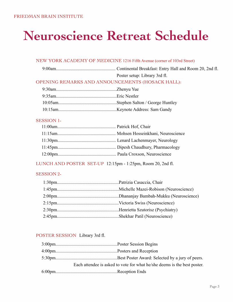
Neuroscience Retreat Schedule
NEW YORK ACADEMY OF MEDICINE 1216 Fifth Avenue (corner of 103rd Street)
9:00am.................................................... Continental Breakfast: Entry Hall and Room 20, 2nd fl. Poster setup: Library 3rd fl.
9:30am.....................................................Zhenyu Yue9:35am.....................................................Eric Nestler10:05am...................................................Stephen Salton / George Huntley10:15am...................................................Keynote Address: Sam Gandy
SESSION 1- 11:00am................................................... Patrick Hof, Chair11:15am................................................... Mohsen Hosseinkhani, Neuroscience11:30pm................................................... Lenard Lachenmayer, Neurology 11:45pm................................................... Dipesh Chaudhury, Pharmacology 12:00pm................................................... Paula Croxson, Neuroscience
LUNCH AND POSTER SET-UP
SESSION 2-
1:30pm......................................................Patrizia Casaccia, Chair1:45pm......................................................Michelle Mazei-Robison (Neuroscience)2:00pm......................................................Dhananjay Bambah-Mukku (Neuroscience)2:15pm......................................................Victoria Swiss (Neuroscience)2:30pm......................................................Henrietta Szutorisz (Psychiatry)2:45pm......................................................Shekhar Patil (Neuroscience)
POSTER SESSION
3:00pm......................................................Poster Session Begins4:00pm......................................................Posters and Reception5:30pm......................................................Best Poster Award: Selected by a jury of peers.
6:00pm......................................................Reception Ends
12:15pm - 1:25pm, Room 20, 2nd fl.
Each attendee is asked to vote for what he/she deems is the best poster.
Library 3rd fl.
OPENING REMARKS AND ANNOUNCEMENTS (HOSACK HALL):

FRIEDMAN BRAIN INSTITUTE
Page 4
Abstracts1
2
Molecular Disturbance of the Amygdala Prodynorphin System in Drug Abuse and Mood Disorders
Sarah Ann R. Anderson, Pernilla Fagergren, Michael Bannon, Michelle Jacobs, Yasmin Hurd
The comorbidity rate between mood disorders and drug addiction is markedly high and confounds the therapeutic strategies currently used for substance dependence. Given this prevalence, the main aim of this study is to investigate the role of the Prodynorphin (PDYN) system in the amygdala relevant to negative mood states seen in drug addiction.
The mRNA expression levels of PDYN and its receptor, the Kappa Opioid Receptor (KOR) was examined in the post-mortem human amygdala of heroin subjects and in a separate subjects major depressive disorder. In situ hybridization histochemistry was used with riboprobes against the PDYN and KOR genes. Single nucleotide polymorphic genotyping was used to identify allelic variants within these genes that are related to phenotype and/or mRNA expression.
Our results demonstrated that heroin abusers and major depressive subjects have significantly reduced PDYN mRNA expression in the peri-amygdaloid cortex (PAC) nucleus of the amygdala, similar to our previous observation in subjects diagnosed with mood disorders. Furthermore, polymorphisms of the PDYN and KOR genes are significantly correlated with decreased PDYN mRNA expression levels in the amygdala in both populations.
Taken together, our data suggests that there is shared dysregulation of the amygdala PDYN/KOR system amongst drug abusers and those with major depressive disorder. This implicates a role for this system in the high comorbidity of drug addiction with depression, suicide and other mood disorders.
Support: DA15446
CB1 Receptor Imaging in Posttraumatic Stress Disorder
Christopher Bailey1, Mark Normandin2, Shannan Henry2, Shireen Saxena1, Marc Potenza2, Henry Huang2, Richard Carson2, Rachel Yehuda1, Alexander Neumeister1
1Psychiatry, MSSM, New York, NY2Diagnostic Radiology & Psychiatry, Yale SOM, New Haven, CT
Posttraumatic stress disorder (PTSD) is a disabling clinical syndrome characterized by recurrent intrusive memories of a traumatic event, repeated avoidance of reminders of the trauma, and high levels of arousal and anxiety. We propose a PTSD model where the maladaptive neurobehavioral trauma response results from impaired endocannabinoid (eCB) signaling associated with upregulation of CB1 receptors in a PTSD circuit, involving the amygdala, anterior and posterior cingulate, caudate, hippocampus, pallidum, and putamen, as well as insufficient glucocorticoid signaling. Using the novel CB1 receptor radioligand [11C]OMAR and positron emission tomography (PET) on a HRRT PET scanner, we determined CB1 receptor expression in 16 medication-free PTSD patients (8F, Age,ys 30.0±8.5, range 20-44) and 16 individually-matched healthy control subjects (8F, Age, ys 30.6±7.5, 20-45). We found elevated CB1 binding in the PTSD group relative to the healthy controls in the PTSD circuit (F(1,28)=12,p<.0017) and decreased serum cortisol levels in PTSD compared to controls (p<.0227). Independent of diagnosis, we found significantly higher CB1 binding in women relative to men. This study revealed dysfunction within the eCB and glucocorticoid systems in PTSD. In addition, we found evidence for gender disparity in CB1 receptor function. These findings could provide the basis for novel evidence-based treatments that aim to modulate impaired eCB and glucocorticoid signaling.
Supported by NIAAA (3RL1AA017540-04S1).

page 5
3
4
A BDNF dependent auto-regulatory positive feedback loop is required for memory consolidation.
Dhananjay Bambah-Mukku, Dillon Y. Chen and Cristina M. Alberini
Department of Neuroscience
The process by which newly learned information becomes a long lasting memory is termed memory consolidation, which depends on an initial phase of transcription and translation. A wealth of literature suggests that de novo protein synthesis and the evolutionarily conserved CREB-C/EBP (cAMP Response Element Binding protein–CCAAT/Enhancer Binding Protein) pathway are critically required for memory consolidation. However, the temporal evolution of the hippocampal gene expression changes underlying consolidation remain largely undetermined.
Using Inhibitory Avoidance (IA) in rats, we find that a rapid wave of protein synthesis in the hippocampus during the first few minutes after training is critical for memory consolidation. Specifically, this protein synthesis is mediated, in part, by BDNF and mTOR signaling. We find that this rapid BDNF signaling recruits the CREB-C/EBP pathway and leads to long term biochemical changes in the dorsal hippocampus lasting ~20hrs including the phosphorylation of CaMKIIα, Synapsin1 and Cofilin. Strikingly, the memory impairment caused by blocking the induction of C/EBPβ in the hippocampus following IA training is rescued by the co-administration of BDNF, which is itself a downstream target of C/EBPβ. This effect is temporally restricted as BDNF no longer rescues the deficit if administered 4 days after the C/EBPβ knockdown. Our data indicates that a rapid wave of BDNF dependent protein synthesis and subsequently a C/EBPβ-dependent gene expression cascade are required for memory consolidation. Moreover, BDNF recruits the CREB-C/EBP pathway in an auto-regulatory positive feed-back loop to mediate memory consolidation which may be a candidate mechanism underlying memory persistence.
Funding: NIMH R01MH063635 (CMA), NIMH R01MH074736 (CMA), NIH F31 MH816213 (DYC)
Reduced experience-dependent dendritic spine plasticity in aging prefrontal cortical neurons
Erik B. Bloss, Bill Janssen, Dan Ohm, Frank Yuk, Shannon Wadsworth, Karl Saardi, Bruce McEwen, and John Morrison
Cognitive functions that require the prefrontal cortex are highly sensitive to aging in humans, non-human primates, and rodents, although the neurobiological correlates of this vulnerability remain largely unknown. Dendritic spines represent a major site of structural plasticity in the adult brain, and recent reports have demonstrated altered dendritic spine number and morphology in aging prefrontal cortical neurons. However, no study to date has directly examined whether aging alters the capacity for experience-dependent spine plasticity in prefrontal cortex. To address this possibility, we used young, middle-aged, and aged rats in a behavioral stress paradigm known to produce spine remodeling in prefrontal cortical neurons. In young rats, stress resulted in dendritic spine loss and alterations of spine morphology; in contrast, spines from middle-aged and aged animals were remarkably stable and failed to show evidence of remodeling. The loss of stress-induced spine plasticity observed in aging rats occurred alongside robust age-related reductions in spine density and shifts in existing spine morphology. Taken together, the data presented here provide the first evidence that experience-dependent spine plasticity is altered by aging in prefrontal cortex, and support a model in which dendritic spines become progressively less plastic in the aging brain.

page 6
PS1∆8 mutation results in motor deficits and region specific cell loss
Hannah Brautigam1, Dara L. Dickstein1, Sam Gandy3, Patrick R. Hof1, Michelle E. Ehrlich2
1Department of Neuroscience, 2Neurology, Pediatrics, 3Neurology, Psychiatry, Mount Sinai School of Medicine and James J. Peters VA Medical Center, Bronx, NY, USA
A presenilin 1 (PS1) missense mutation, L271V, results in a gene that lacks exon 8 (PS1∆8), and causes early onset familial Alzheimer’s disease in a Tasmanian family. The pathogenesis of this mutation is controversial because: (1) exon 8 contains an aspartate critical for PS1 action, and (2) the mutation violates current concepts about γ-secretase structure. We sought to model this disease in transgenic mice, both alone and in combination with a mutation in APP (Dutch APPE693Q) that causes accumulation of amyloid beta oligomers but no plaques. We examined wildtype (wt), Dutch, PS1∆8, and Dutch/PS1∆8 mice for performance on the rotarod motor task at 6 months of age and subsequently performed regional cell and neuronal counts and density determinations at 18 months of age using isotropic fractionator. We found that expression of the PS1∆8 mutation was associated with decreases in hippocampal cell density (p = 0.022) and cerebellar neuronal density (p = 0.017). Expression of the Dutch/PS1∆8 mutation was associated with decreased hippocampal cells (p = 0.01) and deficits on the rotarod motor task (p = 0.065) when compared to wt littermates. These data suggest that PS1∆8 may act in the mouse as a dominant negative and play a role in neurogenesis.
Funding NIA
5
6 Frontoinsular Cortex in Familial Dysautonomia: a clinicopathologic exploration of the role of von Economo neurons in interoception
M Santos1, N Uppal1, C Butti1, B Wicinski1, T Wisniewski2, F B Axelrod2, D Zagzag2, L Norcliffe-Kaufman2, H Kaufman2, P R Hof1
1Department of Neuroscience, Mount Sinai School of Medicine2Department of Neurology, New York University School of Medicine
Familial dysautonomia (FD) affects the development and survival of neurons in the autonomous nervous system. Among other debilitating features consistently exacerbated by emotional states, FD patients present a dysregulation of cardiovascular reflexes and impairments in language, cognition and social skills.
The FI, implicated in interoception (the perception of bodily physiological condition) may be affected in FD patients and is one of the selected cortical regions containing Von Economo neurons (VENs). These large bipolar neurons are consistently affected in neuropsychiatric disorders where social conduct is disturbed such as frontotemporal dementia, early-onset schizophrenia and autism. We hypothesized to find abnormalities in VENs’ numbers, distribution and/or cellular morphology in FD patients in comparison to control subjects.
In the FI of FD patients, our preliminary results show an increased ratio of VENs to pyramidal neurons and a decreased density of pyramidal neurons. Moreover, VENs were observed in unusual cortical areas (orbitofrontal cortex) and presented atypical morphological features. Additional cases will allow us to further explore these trends. As such, our study explores the cortical underpinnings of autonomic dysfunction in order to shed some light on its etiopathogeny and on potential therapeutic strategies.

page 7
7
8
The insular cortex: a comparative perspective
Camilla Butti1, Alanna G. Brake1, Bridget A. Wicinski1, Joy S. Reidenberg2 and Patrick R. Hof1
1Department of Neuroscience and 2Center for Anatomy and Functional Morphology, Mount Sinai School of Medicine
The insular cortex is involved in a variety of viscerosensory, visceromotor, and interoceptive functions. We studied the cytoarchitecture of the insular cortex in uncommon species including a large carnivore, two artiodactyls, two cetaceans, and a sirenian, and compared it with that of human and common laboratory animals. We observed substantial variability in shape, extent, and complexity of gyral and sulcal patterns. Differences in laminar organization, cellular specialization, and the extent of association to the claustrum were observed. The general organization of the insular cortex observed in laboratory animals and human, includes distinct agranular, dysgranular, and granular fields, that are not identifiable in cetaceans, artiodactyls, and sirenians, which all presented agranularity in its entire rostrocaudal extent. A high degree of clustering of layer II was particularly evident in cetaceans and their closest relatives and a pronounced columnar organization associated with the presence of large cellular clusters in layer VI were observed exclusively in the cortex of the manatee. Von Economo neurons were observed in layer V of the insular cortex only in some of the species examined. Structural differences can be considered the result of selective evolutionary pressures and likely constitute the neuroanatomical basis for the functional heterogeneity of this cortical domain.
Supported by the James S. McDonnell Foundation Grant 22002078.
Curcumin Induced A-beta Fibrillation Reduces Neurotoxicity in Transgenic Drosophila
Ina Caesar1, Maria Jonson2, K. Peter R. Nilsson2, Stefan Thor3 and Per Hammarström2
1Department of Neuroscience, Mount Sinai School of Medicine, New York, USA, 2IFM-Department of Chemistry, Linkoping University, Linkoping, Sweden,
3IKE-Department of Clinical and Experimental Medicine, Linkoping University, Linkoping, Sweden
Alzheimer’s disease is pathologically characterized by the presence of extracellular deposits of misfolded and aggregated Amyloid-β (Aβ) peptide and the intraneuronal accumulation of tangles comprised of hyperphosphorylated tau protein. For several years, the natural compound curcumin has been proposed to be a good candidate for enhanced clearance of the toxic amyloids corresponding to the Aβ peptide.
In this study we have studied the potency of curcumin as a drug candidate to alleviate Alzheimer’s disease symptoms in transgenic Drosophila. The longevity as well as the locomotor activity of the different genotypes was measured relative to a control line. To detect amyloid formation we used combined antibody staining and the amyloid specific pFTAA, a luminescent conjugated oligothiophene. Quantification of Aβ produced in Drosophila as well as in vitro fibrillation of synthetic Aβ42 was measured in absence or presence of curcumin. Structure dependent spectra from the pFTAA, for different time points as the aggregation proceeded in the tissue were obtained and indicate accelerated conversion of Aβ42 in curcumin treated flies. The study showed that curcumin accelerated amyloid fibril formation by reducing the pre-fibrillar species of Aβ resulting in a reduced toxicity in Drosophila.

page 8
Network dynamics in psychiatric stress
Michael Chary and Ehud Kaplan
Friedman Brain Institute and Department of Neuroscience Mount Sinai School of Medicine
Psychiatric illnesses are very costly to both society and the individual. Although their biological basis remains largely unknown, prior work suggests that changes in the firing patterns of single neurons in the mesocorticolimbic system could account for the appearance of depressive symptoms after exposure to social defeat, an animal model of psychiatric stress.
To determine whether quantitative measures of network dynamics could detect the impact of psychiatric stress on the brain, we record from small populations of neurons in the nucleus accumbens and medial prefrontal cortex (mPFC) of mice before and after exposure to social defeat.
To demonstrate the use of this systems neuroscience approach to understanding psychiatric illnesses, we present here preliminary data recordings from the mPFC of normal mice, and analysis of such data with severalmethods that characterize network dynamics quantitatively.
Supported by NIH grants EY016371, EY12867, GM71558 and core grant EY01867.
Optogenetic Manipulation of Dopaminergic Neurons in the Brain Reward Circuit Modulates Susceptibility to Social Defeat Stress
Dipesh Chaudhury, Barbara Juarez, Hsing-Chen Tsai, Mary Kay Lobo, Jessica Walsh, Allyson Friedman, Ezekiell Mouzon, Karl Deisseroth, Eric Nestler, Ming-Hu Han
The burst firing of ventral tegmental area (VTA) dopamine neurons encodes natural and drug reward, but their role in mediating stress vulnerability is not fully understood. In a social defeat model of depression, mice exhibiting the susceptible (depressive), but not resilient (non-depressive) phenotype, exhibited consistently increased burst firing in VTA dopamine cells. To understand the relationship between bursting activity and susceptibility to social defeat in freely-behaving mice, we selectively targeted dopamine cells by injecting Cre-identifying viral vector AAV-Channelrhodopsin2 (ChR2), carrying the genes encoding for the light sensitive cation channel, into the VTA of Th-Cre mice. Through in vitro and in vivo electrophysiological recordings, we demonstrated that light activation of ChR2 reliably generated tonic and burst firing patterns in VTA dopamine neurons. By exposing these cells to high frequency light stimulation, in order to mimic burst firing, in AAV-ChR2-injected mice, that had previously undergone 10 days chronic social defeat, we instantly reversed the resilient phenotype. To further investigate the functional relevance of tonic and burst firing on the expression of the susceptible phenotype, we are currently stimulating VTA dopamine cells in ChR2-injected mice exposed to a subthreshold microdefeat paradigm, followed by social interaction and sucrose preference (anhedonia) tests. Our studies will provide direct evidence linking between the firing patterns of VTA dopamine neurons and stress vulnerability.
9
10

page 9
Social Stress and Synaptic Plasticity in the Nucleus Accumbens
Daniel Christoffel
The neurobiological underpinnings of mood and anxiety disorders have been linked to the nucleus accumbens (NAc), a region important in processing the rewarding and emotional salience of stimuli. Using chronic social defeat stress (CSDS), an animal model of mood and anxiety disorders, we investigated if CSDS induces synaptic remodeling and if these synaptic alterations are responsible for the long-lasting behavioral symptoms induced by this form of stress. Previously, we found that NAc MSNs have more stubby spine structures with smaller post-synaptic densities and an increase in the frequency of mEPSCs following social defeat only in a susceptible subpopulation. In parallel to these structural changes, we observed significant increases in IkappaB Kinase (IKK) in the NAc after social defeat, a molecular pathway that has been shown to regulate neuronal morphology. Using viral mediated gene transfer of dominant negative and constitutively active IKK mutants we demonstrate that activation of IKK signaling pathways during social defeat is both necessary and sufficient to induce synaptic alterations and behavioral effects of the stress. Currently we are gaining a more comprehensive image of stress-induced synaptic plasticity via subcellular fractionation to isolate changes in glutamate receptor composition at the synapse, along with ultrastructural analysis of the presynapse, specifically analyzing changes in the number and localization of synaptic vesicles.
In vivo MRI of monkeys with subcortical lesions: the relationship between structure and function
Paula L Croxson1,2, Jill X O’Reilly2, Jerome Sallet2, MaryAnn P Noonan2, Rogier M Mars2, Karla L Miller2, Matthew FS Rushworth2, Mark G Baxter1
1Glickenhaus Laboratory of Neuropsychology, Mount Sinai School of Medicine, 2University of Oxford
Recent advances have allowed us to acquire detailed imaging data from non-human primates in vivo. What value can such data provide when compared with the already high standard of images we can acquire from the human brain? One major contribution is that we can study the effect of targeted brain lesions in specific cortical or subcortical regions in the same subjects, an opportunity that rarely arises in human patients. We acquired high-resolution structural, diffusion and resting-state images from anesthetized rhesus monkeys on two occasions: before and after a subcortical brain lesion. We analyzed the resting-state and diffusion data to assess whether: (i) functional networks are reorganized following damage to subcortical components of these circuits, and (ii) whether structural changes in the white matter reflect these functional changes. Evidence from human studies suggests that structural alterations on a macroscopic level underlie functional changes even in the adult brain. We investigated whether this could also be true in the case of brain damage. In addition to revealing in more detail the relationship between structure and function, these findings could also inform us about the basis of resting-state correlations.
Funding: Wellcome Trust, UK and Medical Research Council, UK
11
12

page 10
Repetition priming in a simple motor circuit
A Dacks and K Weiss
Repetition priming is a basic form of plasticity in which a behavior is performed with increased speed, robustness or accuracy as it is repeated. Although difficult to study in more complex systems, we are studying the organizing principles underlying repetition priming in the feeding circuit of Aplysia californica. As a meal progresses, Aplysia produce more robust motor programs as a result of increased activity of motor neurons driving these movements, a process referred to as “ingestive build-up” which can be induced in the isolated nervous system by triggering repeated feeding motor programs. This build-up in motor neuron activity is not necessarily associated with increased input, as in the case of the B8 motor neuron, which exhibits ingestive build-up without an increase in the activity of its direct input neuron (B40). We therefore sought to determine the mechanism by which B8 increases its activity with successive motor programs during ingestive build-up. We found that after the induction of ingestive build-up results in an increase in B8 excitability that persists for the same duration as ingestive build-up. Repeated B40 activation enhances B8 excitability and can induce ingestive build-up. Furthermore, preliminary results indicate that hyperpolarizing B40 prevents the induction of ingestive build-up. Thus, repetition priming in this simple circuit is mediated by repeated input to a motor neuron which induces a temporary increase in post-synaptic excitability.
How much information can a neural population deliver?
Marshall Crumiller, Bruce Knight and Ehud Kaplan
Information processing in the brain requires interactions of many neuronal networks. Until recently, studiesof network processing have been limited to extrapolations based on the properties of individual neurons. Withthe increasing availability of multicellular recording, we can now record from many neurons simultaneously.Despite the growing need for analyses at the network level, methods of estimating information transmitted bypopulations of cells remain limited to very small number (<9) of neurons.Here we describe a novel method utilizing Fourier series to estimate the amount of information transmittedsimultaneously by a large population of neurons, overcoming many of the obstacles commonly encountered byother methods, most notably the problem of small sampling bias. We demonstrate the application of this methodto the mammalian visual system, using multi-electrode extracellular recordings of both the Lateral GeniculateNucleus and the Primary Visual Cortex of the macaque monkey in response to both full-field pseudo-randomstimuli and natural scenes. We further demonstrate the ability of the method to assess redundancy in anensemble of neurons, and discuss how the functional connectivity of visual neuronal networks may be reflectedin this redundancy.
Supported by NIH grants EY016371, EY12867, GM71558 and core grant EY01867.
13
14

page 11
Alteration of Myelin expression with stress-induced models of depression
Dietz KC, Dietz DM, Nestler EJ, Casaccia P
Department of Neuroscience, Mount Sinai School of Medicine
Neuropsychiatric conditions such as depression and bipolar disorder have been associated with alterations
in white matter, with patients showing marked reductions in volume in limbic areas. Surprisingly little research has been focused on the contributions these changes make to the disorders, nor in understanding the molecular mechanisms of these alterations. Our current research utilizes several animal models of depression to examine this association between depressive behaviors and myelination. As many of these animal models involve physical and psychological stress, we focused on identifying the effects of these stressors on myelin gene
expression in limbic brain regions. Animals subjected to social defeat or isolation stress exhibit depressive-like behaviors, and decreases in myelin gene expression in the ventral striatum. However, there is a differential response to the two types of stress in the prefrontal cortex; where a decrease in these genes is seen with social isolation, but an increase is with a social defeat paradigm. These results suggest complex responses in myelinating cells to different stresses, which are region-specific, and may therefore contribute differently to the
development of depressive behaviors.
Continuous exposure to Galvanic Vestibular Stimulation (GVS): physiological and motor performance.
Dilda Valentina, Morris Tiffany, Hamish MacDougall, Moore Steven.
In the past we have shown that Galvanic vestibular stimulation (GVS) is well tolerated at different peak current levels during intermittent exposure. The present study is aimed to assess tolerance and postural control during 20 minutes of continuous GVS. In our experiment subjects received one of two peak current levels: 3.5 mA (N=12) and 5 mA (N=12), while being tested on a computerized dynamic posturography (CDP) platform (Equitest, Neurocom, OR). The sensory organization test (SOT), limit of stability (LOS), and rhythmic weight shift (RWS) test were performed before, during, and 15 minutes after 20 min continuous GVS exposure. At 3.5 mA 100% of subjects completed 20 min GVS exposure. At 5 mA 67% (8) completed 20 min exposure; 4 subjects asked to interrupt the experiment due to motion sickness symptoms with an average exposure of 11.4 min. At both 3.5 and 5 mA GVS significantly affected postural performance. LOS total distance increased with GVS (p<.01); SOT vestibular indices and composite scores significantly decreased during GVS (p<.05); RWS directional control decreased during GVS (p<.01) at front/back slow, medium, and fast speeds. These results indicate that 3.5 mA and 5mA GVS exposure induce similar postural instability during LOS, SOT and RWS CDP tests. Exposure to the higher (5mA) current induced motion sickness symptoms in 25% of the subjects during motor tasks that directly involved vestibular sensorimotor integration.
15
16

page 12
Long-lasting and Rapid acting Antidepressant Effects of Ih Channel Inhibitors
Allyson K. Friedman, Herbert E Covington, Jessica J Walsh, Barbara Juarez, Dipesh Chaudhury, Vincent Vialou, Eric Nestler, Ming-Hu Han
Major depressive disorder (MDD) is a serious medical illness affecting 15 million Americans. Despite MDD’s prevalence, the few currently mechanistically distinct classes of antidepressants take six to twelve weeks to take full effect and only 50% of patients can achieve full remission. A possible reason for the limitations of current medications is an incomplete understanding of the pathophysiological mechanisms of MDD. Utilizing the social defeat stress model of depression, an increase in the firing rate was found in the ventral tegmental area (VTA) dopamine (DA) neurons in the brain reward circuitry of susceptible mice, but not in the resilient subgroup. We hypothesized that ion channel blockers that inhibit the pathological hyperactivity of VTA DA neurons may act as an antidepressant. Previously we found that chronic social defeat increased Ih current in susceptible mice and that local infusion of Ih inhibitors ZD7288 and DK-AH269 into the VTA normalized depression-like social avoidance. This antidepressant effect occurred within one hour after the infusion, in contrast to traditional slow acting antidepressants. Surprisingly, the antidepressant effect induced by a single-dose infusion of Ih inhibitor DK-AH269 lasted at least two weeks. Importantly, this long-lasting antidepressant effect was repeated with a single-dose i.p. injection. These channels are novel drug targets for the treatment of MDD and may assist in the development of a mechanistically innovative class of antidepressants.
17
18
Drug-induced epigenetics in alternative mRNA splicing
Jian Feng, Li Shen
To study alternative splicing in drug addiction of mouse brain, we used the next-generation sequencing to
quantify gene features in a crucial brain-reward region called Nucleus Accumbens. We performed RNA-seq
experiments to obtain nearly 100 million high-quality short reads from mouse brain RNA samples at 1h, 4h and
24h after the last dose of injection for each condition of cocaine treatment and saline control. Analyzing this total of ~600 million short reads reveals massive changes in six different categories of alternative splicing events, i.e.
exon usage, exon boundary, exon-exon junction, intron, transcripts and intergenic region. We found that the
majority changes reside in alternative exon usage and exon-exon junction gain or loss. The genes involved in
these alternative splicing events are highly enriched with biological functions in neurological disease, psychiatric
disorders, cell growth and cell morphology. We then used ChIP-seq to elucidate the genome-wide chromatin
modifications for four popular histone marks: h3k4me3, h3k36me3, h3k9me2 and Pol II. Using an in-house
developed sliding window approach, we obtained a high-resolution chromatin remodeling map for each histone
mark's differential binding pattern. We found a highly significant association between a histone mark's binding
change and the differential alternative splicing of sequence features in chromosomal neighbors.

page 13
Cognition Emotion Integration in Anterior Insular Cortex
Xiaosi Gu a,b, Xun Liu c, Nicholas T. Van Dam a, Patrick R. Hof b, and Jin Fan a,b,d
a. Department of Psychiatry, Mount Sinai School of Medicine, NY, NYb. Department of Neuroscience, Mount Sinai School of Medicine, NY, NY
c. Key Laboratory of Behavioral Science, Institute of Psychology, Chinese Academy of Sciences, Beijing, Chinad. Department of Psychology, Queens College, The City University of NY, Flushing, NY
Both cognitive and affective processes require mental resources. However, it remains unclear whether these two types of processes work in parallel or in an integrated fashion. In this functional magnetic resonance imaging (fMRI) study, we investigated the functional interaction of these two processes with simultaneous manipulation of task demand and stimulus valence. Eighteen healthy participants viewed photographs showing others’ body-parts in painful or neutral situations while performing tasks of low (body-part judgment) and high cognitive demand (laterality judgment). We found increased reaction times and error rates for painful compared to non-painful stimuli under laterality judgment relative to body-part judgment. fMRI data showed activity in bilateral anterior insula (AI) and somatosensory cortex (SI), but not posterior insula, for main effects of task demand and stimulus valence. Importantly, task demand and stimulus valence showed a significant interaction in AI. These results suggest that cognitive and emotional processes at least partially share common brain networks, and that AI serves as a key node in a brain network subserving cognition emotion integration.
Mechanisms of axonal damage in multiple sclerosis
Jeffery D. Haines, Jin-Young Kim, Patrizia Casaccia
It is becoming increasingly appreciated that there is a neurodegenerative component to multiple sclerosis (MS), which leads to many of the clinical symptoms of the disease including disability, cognitive loss and fatigue. In healthy brain, mitochondria are rapidly transported along neuronal axons where they meet the energy demands of axons. In damaged neurons, however, mitochondrial movement is impaired, resulting in axonal damage. We have previously shown that the histone deacetylase, HDAC1, is translocated from the nucleus to the cytoplasm in demyelinated human brain, animal models of demyelination and following pathological stimuli (e.g., glutamate and TNFα). The translocation of HDAC1 results in altered transport of mitochondria and cargo proteins along the axon, resulting in axonal beading and transection. However, it remains unknown whether the initial phenomenon of HDAC1 export relates to an increased energy demand of the neuron, and how these relate to mitochondrial function. To this end, we are using rat cultured cortical and hippocampal neurons to determine the levels of ATP and reactive oxygen species (ROS) to determine the physiological response of neurons in response to stimuli that induce HDAC1 export. These measures will be correlated with both mitochondrial shape changes (i.e., fission/fusion) and velocity measurements. We will then use cerebral spinal fluid from MS patients to determine its effects on mitochondrial function and neuronal viability. These studies will lay important groundwork for understanding the mechanisms underlying axonal damage and designing therapies to prevent neurodegeneration in MS.
Funded by the National Multiple Sclerosis Society (NMSS) and the NIH.
19
20

page 14
Antagonistic Effects of SHH and BMP4 on HDAC Activity During Differentiation of Oligodendrocyte Progenitor Cells
Marylens Hernandez
Oligodendrocytes are the myelin-forming cells of the CNS. They pro-vide trophic support to neuro-ns and are essential for the saltatory conduction of the nervous impulse. During develop-ment oligo-dendrocytes are originated from bipotential pro-genitor cells that can also differentiate into astrocytes, depending on specific signals. Shh (Sonic Hedgehog) and Bmp4 (Bone morphogenic pro-tein 4) are morphogens widely reported to pro-mote pro-genitor differentiation into oligo-dendrocytes and astrocytes, respectively. Histone deacetylases 1 (Hdac1) and 2 (Hdac2) have been described as essential molecular effectors during oligo-dendrocyte develop-ment and maturation. We therefore hypo-thesized that Shh and Bmp4 could antagonistically regulate Hdac activity. Using pharmacological inhibitors or a silencing appro-ach we show that Hdac1 and Hdac2 inhibition blocks Shh-induced oligo-dendrogliogenesis and favors astrogliogenesis. Finally to define the genes downstream of Hdac activity we utilized affymetrix arrays and the data were further analyzed for genes that were oppositely regulated by Shh and Bmp4, whose expression pro-files were also affected by Hdac inhibition.
Maternal Influenza Viral Infection Causes Schizophrenia-Like Alterations in Behavioral Mouse Models in Adult Offspring
Terrell D. Holloway1, José L. Moreno1, Mitsumasa Kurita1, Javier López1,2, Richard Cadagan3, Luis Martínez-Sobrido3,7, Adolfo García-Sastre3,4,6 and Javier González-Maeso1,2,5
Departments of 1Psychiatry, 2Neurology, 3Microbiology, and 4Medicine.5Friedman Brain Institute, and 6Global Health and Emerging Pathogens Institute. Mount Sinai School of Medicine.
7Department of Microbiology and Immunology. University of Rochester Medical Center.
Epidemiological studies indicate that maternal influenza viral infection increases the risk for schizophrenia in the adult offspring. The serotonin and glutamate systems are suspected in the etiology of schizophrenia, as well as in the mechanism of action of antipsychotic drugs. The effects of hallucinogens, such as psilocybin and mescaline, require the serotonin 5-HT2A receptor, and induce schizophrenia- like psychosis in humans. In addition, metabotropic glutamate receptor mGlu2/3 agonists show promise as a new treatment for schizophrenia. Here, we investigated the level of expression and behavioral function of 5-HT2A and mGlu2 receptors in a mouse model of maternal influenza viral infection. We show that spontaneous locomotor activity is diminished by maternal infection with the mouse-adapted influenza A/WSN/33(H1N1) virus. The behavioral responses to hallucinogens and glutamate antipsychotics are both affected by maternal exposure to influenza virus, with increased head-twitch response to hallucinogens and diminished antipsychotic-like effect of the glutamate agonist. In frontal cortex of mice born to influenza virus-infected mothers, the 5-HT2A receptor is upregulated and the mGlu2 receptor is downregulated, an alteration that may be involved in the behavioral changes observed. Identifying a biochemical alteration that parallels the behavioral changes observed in a mouse model of prenatal viral infection may facilitate targeting therapies for treatment and prevention of schizophrenia.
21
22
page 15
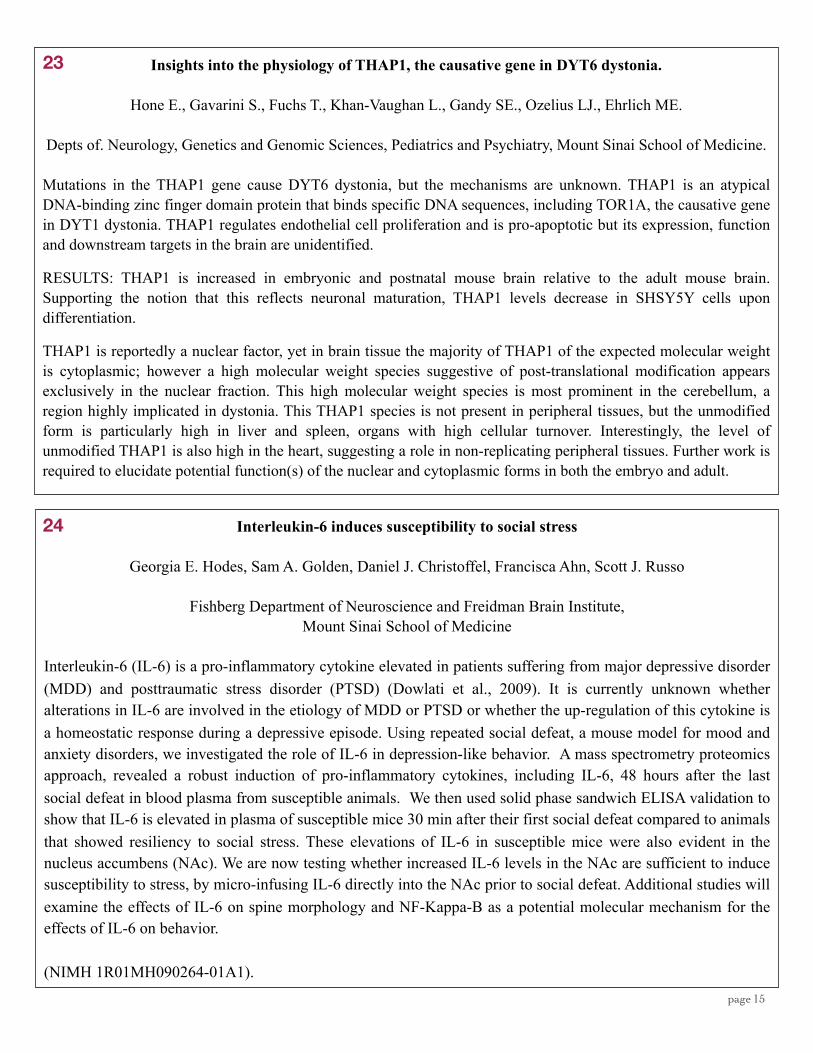
Insights into the physiology of THAP1, the causative gene in DYT6 dystonia.
Hone E., Gavarini S., Fuchs T., Khan-Vaughan L., Gandy SE., Ozelius LJ., Ehrlich ME.
Depts of. Neurology, Genetics and Genomic Sciences, Pediatrics and Psychiatry, Mount Sinai School of Medicine.
Mutations in the THAP1 gene cause DYT6 dystonia, but the mechanisms are unknown. THAP1 is an atypical DNA-binding zinc finger domain protein that binds specific DNA sequences, including TOR1A, the causative gene in DYT1 dystonia. THAP1 regulates endothelial cell proliferation and is pro-apoptotic but its expression, function and downstream targets in the brain are unidentified.
RESULTS: THAP1 is increased in embryonic and postnatal mouse brain relative to the adult mouse brain. Supporting the notion that this reflects neuronal maturation, THAP1 levels decrease in SHSY5Y cells upon differentiation.
THAP1 is reportedly a nuclear factor, yet in brain tissue the majority of THAP1 of the expected molecular weight is cytoplasmic; however a high molecular weight species suggestive of post-translational modification appears exclusively in the nuclear fraction. This high molecular weight species is most prominent in the cerebellum, a region highly implicated in dystonia. This THAP1 species is not present in peripheral tissues, but the unmodified form is particularly high in liver and spleen, organs with high cellular turnover. Interestingly, the level of unmodified THAP1 is also high in the heart, suggesting a role in non-replicating peripheral tissues. Further work is required to elucidate potential function(s) of the nuclear and cytoplasmic forms in both the embryo and adult.
Interleukin-6 induces susceptibility to social stress
Georgia E. Hodes, Sam A. Golden, Daniel J. Christoffel, Francisca Ahn, Scott J. Russo
Fishberg Department of Neuroscience and Freidman Brain Institute, Mount Sinai School of Medicine
Interleukin-6 (IL-6) is a pro-inflammatory cytokine elevated in patients suffering from major depressive disorder (MDD) and posttraumatic stress disorder (PTSD) (Dowlati et al., 2009). It is currently unknown whether alterations in IL-6 are involved in the etiology of MDD or PTSD or whether the up-regulation of this cytokine is a homeostatic response during a depressive episode. Using repeated social defeat, a mouse model for mood and anxiety disorders, we investigated the role of IL-6 in depression-like behavior. A mass spectrometry proteomics approach, revealed a robust induction of pro-inflammatory cytokines, including IL-6, 48 hours after the last social defeat in blood plasma from susceptible animals. We then used solid phase sandwich ELISA validation to show that IL-6 is elevated in plasma of susceptible mice 30 min after their first social defeat compared to animals that showed resiliency to social stress. These elevations of IL-6 in susceptible mice were also evident in the nucleus accumbens (NAc). We are now testing whether increased IL-6 levels in the NAc are sufficient to induce susceptibility to stress, by micro-infusing IL-6 directly into the NAc prior to social defeat. Additional studies will examine the effects of IL-6 on spine morphology and NF-Kappa-B as a potential molecular mechanism for the effects of IL-6 on behavior.
(NIMH 1R01MH090264-01A1).
23
24

page 16
Forebrain striatal-specific expression of mutant huntingtin protein in vivo induces cell-autonomous age-dependent alterations in sensitivity to excitotoxicity and mitochondrial function
Soong Ho Kim*1, Carlos A. Thomas*1, Véronique M. André2, Damian M. Cummings2, Carlos Cepeda2, Michael S. Levine2, Michelle E. Ehrlich1
1Mount Sinai School of Medicine, 2UCLA, *authors contributed equally
Huntington’s disease (HD) is characterized by dysfunction and death of striatal medium spiny neurons (MSNs). To determine the extent of cell-autonomous effects of mutant huntingtin protein (mhtt) on vulnerability to excitotoxic insult in MSNs in vivo, we measured the number of degenerating neurons in response to intrastriatal injection of quinolinic acid (QA) in presymptomatic and symptomatic transgenic (D9-N171-98Q, a.k.a. DE5) mice that express mhtt in MSNs but not in cortex. After QA, the number of degenerating neurons in pre-symptomatic DE5 mice was not significantly different from the number in wild type (WT) controls, suggesting the early, increased vulnerability to excitotoxicity demonstrated in other HD mouse models has a largely non-cell-autonomous component. Conversely, symptomatic DE5 mice showed significantly fewer degenerating neurons relative to WT, implying the resistance to excitotoxicity observed at later ages has a primarily cell-autonomous origin. Interestingly, mitochondrial complex II respiration was enhanced in striatum of symptomatic mice whereas it was reduced in presymptomatic mice, both relative to their age-matched controls. Consistent with the QA data, MSNs from symptomatic mice showed a decreased NMDA currents compared to age-matched controls, suggesting in addition to ageing, there are cell-autonomous mechanisms that mitigate susceptibility to excitotoxicity in the late disease stage.
Fetal stem cell contributes to neural regeneration in injured maternal brain
Mohsen Hosseinkhani, Yuhan Hao, Pranav Parikh, Hongyan Zou
Fetal cells can enter maternal circulation during pregnancy and persist in maternal blood and tissues for decades post-partum. In non-injured maternal brains, few fetal cells were detected. We investigated whether fetal cells can enter into the injured maternal brain during pregnancy. Wild-type virgin female mice were crossed with GFP Tg male mice. At gestational day (gd) 12.5, female mice were undergone traumatic brain injury (TBI). Immunohistochemistry analysis revealed numerous fetal GFP+ cells at the injury site in the maternal brain 4 days after injury (or even 1 week after delivery), but none in the contralateral side or in the control pregnant mice with no brain injury. The present of fetal cells in injured maternal brain was confirmed by PCR using a Y-chromosome and GFP probes. Using flow cytometry, we found numerous GFP+ cells (1.65%) at the injury site in the maternal brain 4 days after injury compared to non-injured maternal brain (0.03%). Besides expressing neural stem cell (formation of neurospheres and expression of Nestin and Sox2) or immature neuronal markers, GFP-positive fetal cells in the maternal brain were found to adopt locations, morphologies, and expression of immunocytochemical markers indicative of neuron-, astrocyte-, and oligodendrocyte-like cell types. Fetal cell-derived neural progenitor cells might represent an new source of stem cells important for cell replacement therapy for CNS injury and disease.
26
25

page 17
Co-translational Modification Regulates Protein Synthesis and Degradation at Hippocampal Synapses
Kuangfu Hsiao, Deanna L. Benson
Fishberg Dept. of Neuroscience, Mount Sinai School of Medicine
Developmental changes in synaptic proteome are locally regulated by both protein degradation and synthesis, both processes are tightly regulated by various post-translational modifications; however, the co-translational modifications and their functions are not well understood. Here, I hypothesis that the a co-translational modification, N-alpha-acetylation (NAA), regulates not only the degradation of synaptic plasticity related proteins, but also involves in protein synthesis at the synapses through interaction with mTOR pathway. We will exam the impacts of perturbation on NAA in synaptic compartment, particularly toward local protein synthesis and degradation, using protein synthesis and degradation reporters. I also hypothesize that the developmental regulation of NAA genes are associated with the dependence of local protein synthesis at labile synapses. I will examine the necessity of NAA genes during synapse maturation.
Oligodendrocyte development requires uhrf1 expression in zebrafish
Jimmy L. Huynh1, Victoria Swiss1, Kirsten Sadler Edepli2, and Patrizia Casaccia1
1Department of Neuroscience, and Genetics and Genomic Sciences, 2Department of Developmental and Regenerative Biology,
Mount Sinai School of Medicine
Our understanding of oligodendrocyte development has progressed tremendously in the past decade, especially in the field of epigenetics. However, while much has been discovered about the role of histone marks in oligodendrogliogenesis, very little is still understood about the role of DNA methylation in this process. A key player facilitating the cross-talk between histone marks and DNA methylation is UHRF1, a multi-domain protein that has been shown to interact with DNMT1, G9a, and HDAC1. During normal oligodendrocyte differentiation, the levels of Uhrf1 are dramatically down-regulated. Conversely, high Uhrf1 levels are found in gliomas. To better understand the role of Uhrf1 in oligodendrocyte development, we have turned to zebrafish as an in vivo model. Utilizing a mutant line of uhrf1, we have shown that loss of this multi-faceted protein results in decreased RNA expression of several myelin-specific genes, as determined by qRT-PCR and in situ hybridization.
Support provided by the Foundation of the Consortium of Multiple Sclerosis Centers and NIH T32 GM007280.
27
28
page 18
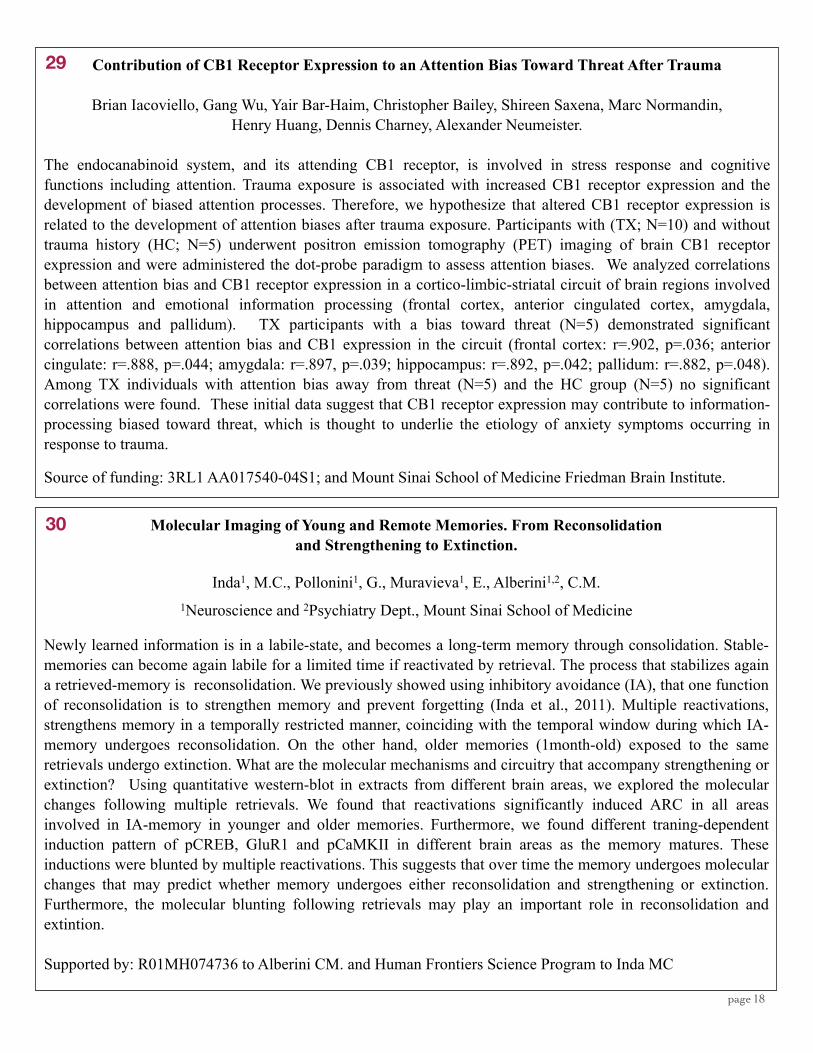
Contribution of CB1 Receptor Expression to an Attention Bias Toward Threat After Trauma
Brian Iacoviello, Gang Wu, Yair Bar-Haim, Christopher Bailey, Shireen Saxena, Marc Normandin, Henry Huang, Dennis Charney, Alexander Neumeister.
The endocanabinoid system, and its attending CB1 receptor, is involved in stress response and cognitive functions including attention. Trauma exposure is associated with increased CB1 receptor expression and the development of biased attention processes. Therefore, we hypothesize that altered CB1 receptor expression is related to the development of attention biases after trauma exposure. Participants with (TX; N=10) and without trauma history (HC; N=5) underwent positron emission tomography (PET) imaging of brain CB1 receptor expression and were administered the dot-probe paradigm to assess attention biases. We analyzed correlations between attention bias and CB1 receptor expression in a cortico-limbic-striatal circuit of brain regions involved in attention and emotional information processing (frontal cortex, anterior cingulated cortex, amygdala, hippocampus and pallidum). TX participants with a bias toward threat (N=5) demonstrated significant correlations between attention bias and CB1 expression in the circuit (frontal cortex: r=.902, p=.036; anterior cingulate: r=.888, p=.044; amygdala: r=.897, p=.039; hippocampus: r=.892, p=.042; pallidum: r=.882, p=.048). Among TX individuals with attention bias away from threat (N=5) and the HC group (N=5) no significant correlations were found. These initial data suggest that CB1 receptor expression may contribute to information-processing biased toward threat, which is thought to underlie the etiology of anxiety symptoms occurring in response to trauma.
Source of funding: 3RL1 AA017540-04S1; and Mount Sinai School of Medicine Friedman Brain Institute.
Molecular Imaging of Young and Remote Memories. From Reconsolidation and Strengthening to Extinction.
Inda1, M.C., Pollonini1, G., Muravieva1, E., Alberini1,2, C.M. 1Neuroscience and 2Psychiatry Dept., Mount Sinai School of Medicine
Newly learned information is in a labile-state, and becomes a long-term memory through consolidation. Stable-memories can become again labile for a limited time if reactivated by retrieval. The process that stabilizes again a retrieved-memory is reconsolidation. We previously showed using inhibitory avoidance (IA), that one function of reconsolidation is to strengthen memory and prevent forgetting (Inda et al., 2011). Multiple reactivations, strengthens memory in a temporally restricted manner, coinciding with the temporal window during which IA-memory undergoes reconsolidation. On the other hand, older memories (1month-old) exposed to the same retrievals undergo extinction. What are the molecular mechanisms and circuitry that accompany strengthening or extinction? Using quantitative western-blot in extracts from different brain areas, we explored the molecular changes following multiple retrievals. We found that reactivations significantly induced ARC in all areas involved in IA-memory in younger and older memories. Furthermore, we found different traning-dependent induction pattern of pCREB, GluR1 and pCaMKII in different brain areas as the memory matures. These inductions were blunted by multiple reactivations. This suggests that over time the memory undergoes molecular changes that may predict whether memory undergoes either reconsolidation and strengthening or extinction. Furthermore, the molecular blunting following retrievals may play an important role in reconsolidation and extintion.
Supported by: R01MH074736 to Alberini CM. and Human Frontiers Science Program to Inda MC
29
30

page 19
Ppar-alpha mediates expression of some, but not all, effects of hypoglycemia on gene expression
Fumiko Isoda, Michal M. Poplawski, Charles V. Mobbs
Department of Neuroscience, Mount Sinai School of Medicine
Peroxisome proliferator-activated receptor α (Ppar-alpha) is a nuclear hormone receptor which plays a major role
in mediating transcriptional responses to fasting by producing a metabolic switch away from glucose utilization
and toward fatty acid oxidation. Since we have observed a similar metabolic response to hypoglycemia,
including the induction of several genes known to be targets for Ppar-alpha, we used qPCR to assess
hypothalamic expression of genes known to be regulated by hypoglycemia, 3 hours after production of
hypoglycemia by a single injection of insulin, in Ppar-alpha knockout and wild type mice. Hypoglycemia
induced Cpt1a (carnitine palmitoyltransferase 1a), Ucp2 (mitochondrial uncoupling protein 2) and Glut4 (glucose
transporter type 4) expression in wild-type but not Ppar-alpha knockout mice. In contrast, hypoglycemia induced
PDK4 (pyruvate dehydrogenase kinase isozyme 4), Glut1 (glucose transporter type 1) and Iκβ (inhibitor of κβ)
expression in both wild type and Ppar-alpha knockout mice. Thus some, but not all, molecular responses to
hypoglycemia are mediated by Ppar-alpha.
31
32 Mining Differential Binding Sites from Chip-seq Data with Biological Replicates
Ying Jin, Li Shen
We present an empirical Bayesian method for mining the differential binding sites from the ChIP-seq data. Most
current approaches do not consider biological replicates or pool the replicates into one combined sample.
However, we observed that the variations between biological replicates are not ignorable. Our approach models
both variations within a single sample and variations between samples (i.e., biological replicates with the same
treatment). To the best of my knowledge, this is the first method that models variations between biological
replicates in the ChIP-seq data analysis. This approach has been applied in studying genome-wide chromatine
changes in the mouse nucleaus accumbens after repeated cocaine administration. Experimental results show that
the more replicates we use the more accurate the detections are. To validate the performance of the proposed and
other statistical models and normalization techniques, we have tested each approach on spiked-in ChIP-seq data
generated by simulation.

page 20
Metabolic memory in diabetic neuropathy: Persistent reprogramming of metabolism associated with persistently reduced association of Ppar-gamma with target genes.
Esther S. Kim, Fumiko Isoda, Charles V. Mobbs
Background: Metabolic memory is the phenomenon whereby diabetic complications progress despite normalization of glucose levels and constitutes a major challenge for treating diabetic neuropathy. However, little is known about the molecular basis of metabolic memory.
Methods: Using an in vitro model, we cultured Schwann cells in chronic (> 2 months) normal (5.6mM) or high (25mM) glucose. We then switched the cells to normal or high glucose for 7 days, then assessed expression of genes that regulate metabolism using qPCR arrays (SABiosciences). We went on to perform chromatin immunoprecipitation (ChIP) to determine the role of transcription factor PPARγ in regulating the gene expression profile seen with high glucose.
Results: Chronic exposure of Schwann cells to high glucose produced persistently increased expression of genes that promote glucose utilization, including hexokinase (Hk2), phosphofructokinase (Pfkl), and persistent inhibition of genes that promote fatty acid metabolism genes, including carnitine palmitoyltransferase (Cpt1b) and the transcriptional factor Ppar-gamma. This profile of gene expression was maintained even after switching to normal glucose for 7 days, and was associated with persistently decreased binding PPARγ to the promoter of several of its target genes.
Conclusions: Chronic glucose produces persistent reprogramming of glucose metabolism associated with persistent reduction in transcriptional activity of Ppar-gamma. Thus Ppar-gamma constitutes a promising target to reverse diabetic complications.
33
34 Role of Nuclear Pore Complex Architecture and Spatial Patterning in Dendritic Arborization
Yayoi Kinoshita, Wendy Yu, Amy Guan, Roxana Mesias*, Deanna L. Benson*, and D. Stave Kohtz
Department of Pathology, *Department of Neuroscience, Mount Sinai School of Medicine
The FG-repeat nucleoporins function in directed transport through the NPC, but also have important roles in chromatin organization and intranuclear gene transport and transcription. Work from our laboratory has revealed architectural diversity among nuclear pore complexes (NPCs) in different cell types. This results from differential spatial distribution of FG-repeat nucleoporins (e.g., NUP62) among NPCs in the nuclear envelope. In some pyramidal neurons, the nuclear distribution of NUP62+ NPCs reflects the distribution of dendritic and axonal processes. The majority of NUP62+ NPCs form a U-shaped ring around the rim of the nucleus, with an NUP62- region observed adjacent to the axonal hillock. Partial knockdown of NUP62 in mature hippocampal neurons in culture results in degeneration of dendritic processes, but not of axons. Partial knockdown of NUP62 in developing hippocampal neurons in culture eventually causes a drastic reduction in dendritic processes accompanied by hyperelongation of axons. The sculpting of dendritic arbors is a key determinant of neuronal connectivity. Alterations in dendritic architecture arise from stress, aging, and neurodegenerative disease, and manifest as declining neuronal function. The selective loss of dendritic processes after knockdown of NUP62 suggests a pivotal role for this FG-repeat nucleoporin in neuronal morphology. NUP62 may also represent an important target when considering the cellular and physiological changes that occur in neurons during stress and aging.

page 21
HDAC2 regulates atypical antipsychotic responses through the modulation of mGlu2 promoter activity
Mitsumasa Kurita, Terrell Holloway, Aintzane García-Bea, José L. Moreno, Alexey Kozlenkov, Mitra Heshmati, Samuel Golden, David M. Dietz, Pamela J. Kenedy, Adrienne Umali, Luis F. Callado, Rachael L. Neve,
Eric J. Nestler, J. Javier Meana, Scott J. Russo, and Javier González-Maeso
Departments of Psychiatry, Neurology, Neuroscience, Pharmacology, Systems Therapeutics andFriedman Brain Institute. Mount Sinai School of Medicine. New York, New York, USA
Department of Pharmacology and CIBER of Mental Health. University of the Basque Country. Bilbao, Spain.Massachusetts Institute of Technology. Cambridge, Massachusetts, USA.
Histone deacetylases (HDACs) are epigenetic regulators which compact chromatin structure and repress gene expression through the removal of acetyl groups from histone tails. Atypical antipsychotics all have in common a high affinity to antagonize serotonin 5-HT2A receptors. Drugs that activate metabotropic glutamate receptor 2 (mGlu2) represent a potential target for new antipsychotic medications. In schizophrenia, clinical studies demonstrate that HDAC inhibitors are efficacious when given in combination with atypical antipsychotics. However, the molecular mechanism that integrates a better response to antipsychotics with changes in chromatin structure remains unclear. Here we show that inhibition of HDAC2 reverses the repressive histone modifications induced at the mGlu2 promoter by chronic atypical antipsychotics, leading to improved efficacy of treatment in mouse models of antipsychotic response. These suggest HDAC2 as a therapeutic target to suppress the atypical antipsychotic-dependent histone modifications responsible for the transcriptional repression of mGlu2 gene, and provide a new approach to treat schizophrenia.
36
35
Latrepirdine (Dimebon™) Enhances Autophagic Activity, reduces α- synuclein and rescues its toxicity
Lenard Lachenmayer, John W. Steele, Shulin Ju, Aryeh Stock, Soong Ho Kim, Dagmar Ringe, Andrew A. Protter, Michelle E. Ehrlich, Gregory Petsko, Sam Gandy, and Zhenyu Yue
Latrepirdine is currently being investigated as a therapeutic agent for the treatment of Alzheimer's disease (AD) and Huntington disease (HD). Outcomes of two clinical AD trials have been decidedly mixed, but proper interpretation of these results is complicated as long as the information regarding the mechanisms of drug action is missing. Here, we investigated the effect of latrepirdine in autophagy and protein degradation using in vitro and in vivo models.
These studies collectively demonstrate that latrepirdine potentiates autophagic activity via the mTOR-pathway in a time- and dose-dependent manner. Moreover, latrepirdine reduces monomeric and oligomeric forms of alpha-synuclein and rescues its toxicity in vitro. Chronic latrepirdine administration enhanced autophagy and reduced endogenous monomeric alpha-synuclein in mouse brains.
Taken together we conclude that laterpirdine increases autophagic activity and facilitates the clearance of alpha-synuclein, highlighting a novel mechanism that may account for the beneficial effects of laterpirdine in AD and a potential strategy for prevention of protein aggregation and neuro¬degeneration associated with synucleinopathy.
page 22
37
38
Diabetes-Associated SorCS1 Regulates Alzheimer’s Amyloid β Metabolism: Evidence for Involvement of SorL1 and the Retromer Complex
Lane RF., Gandy S
SorCS1 and SorL1/SorLA/LR11 belong to the sortilin family of vacuolar protein sorting-10 (Vps10) domain-containing proteins. Both are genetically associated with Alzheimer’s disease (AD), and SORL1 expression is decreased in the brains of patients suffering from AD. SORCS1 is also genetically associated with types 1 and 2 diabetes mellitus (T1DM, T2DM). We have undertaken a study of the possible role(s) for SorCS1 in metabolism of the Alzheimer’s amyloid β peptide (Aβ) and the Aβ precursor protein (APP), to test the hypothesis that Sorcs1 deficiency might be a common genetic risk factor underlying the predisposition to AD that is associated with T2DM. Overexpression of SorCS1cβ-myc in cultured cells caused a reduction (p 0.002) in Aβ generation. Conversely, endogenous murine Aβ40 and Aβ42 levels were increased (Aβ40, p 0.044; Aβ42, p 0.007) in the brains of female Sorcs1 hypomorphic mice, possibly paralleling the sexual dimorphism that is characteristic of the genetic associations of SORCS1 with AD and DM. Since SorL1 directly interacts with Vps35 to modulate APP metabolism, we investigated the possibility that SorCS1cβ-myc interacts with APP, SorL1, and/or Vps35. We readily recovered SorCS1:APP, SorCS1:SorL1, and SorCS1:Vps35 complexes from nontransgenic mouse brain. Notably, total Vps35 protein levels were decreased by 49% (p 0.009) and total SorL1 protein levels were decreased by 29% (p 0.003) in the brains of female Sorcs1 hypomorphic mice. From these data, we propose that dysfunction of SorCS1 may contribute to both the APP/Aβ disturbance underlying AD and the insulin/glucose disturbance underlying DM.
Funded by NIH Grant PO1AG010491, NIDDK Grant DK58037
The CORE issue: cocaine oppositely, selectively and with differing timecourses alters thin dendritic spines in medium spiny neurons of the nucleus accumbens core versus shell.
*Quincey LaPlant1, *Dani Dumitriu1, Yael S. Grossman1, Caroline Dias1, William G Janssen1, John H. Morrison1,2, and Eric J Nestler1
1Fishberg Department of Neuroscience, Mount Sinai School of Medicine, New York, NY 10029
2 Department of Geriatrics and Palliative Care, Mount Sinai School of Medicine, New York, NY 10029
*These authors contributed equally
Numerous studies have found that chronic cocaine increases dendritic spine density in medium spiny neurons in the Nucleus accumbens (NAc). However, recent work has shown that commonly employed spine analysis techniques consistently underestimate spine density, and might even do so with a selective bias against thin spines. Using single cell filling and advanced 3-dimensional imaging and analysis, we assessed several key quantitative and morphologic measures of dendritic spines and dendrites in the core and shell of the NAc following immediate, early, and prolonged withdrawal from chronic cocaine. Depending on the NAc region, our data demonstrate strikingly different cocaine-regulated changes in the density of dendritic spines at all time-points analyzed. At 4 hours post-injection there is no change in total spine density in the NAc core and a significant increase in density in the NAc shell attributable to a selective increase in thin spines. At 24 hours, cocaine reduces the density of dendritic spines in the core and increases it the shell, both modifications due to opposing changes selectively in the density of thin spines. Strikingly, the observed decrease in spine density in the NAc core persists after 28 days of cocaine withdrawal, while the density in the NAc shell returns to baseline. The novel discovery of a divergence in core/shell spine density regulation by cocaine correlates with recently reported electrophysiological data from a similar paradigm and might be a key mediator of the changes in reward circuit that drive continual drug-seeking behavior in animal models of addiction.

page 23
Phosphorylation-dependent 14-3-3 Binding to LRRK2 is Impaired by Common Mutations of Familial Parkinson’s Disease
Xianting Li, Qing Jun Wang, Nina Pan, Sangkyu Lee, Yingming Zhao, Brian T. Chait, and Zhenyu Yue
Recent studies show that mutations in Leucine Rich Repeat Kinase 2 (LRRK2) are the cause of the most common inherited and some sporadic forms of Parkinson’s disease (PD). But the molecular mechanism underlying the pathogenic role of LRRK2 mutations in PD remains unknown. Using affinity purification and mass spectrometric analysis, we identified multiple phosphorylation sites of LRRK2 including S910, S912, S935 and S973. Focusing on the high stoichiometry S935 phosphorylation site, we developed an anti-pS935 specific antibody and showed that S935 is constitutively phosphorylated in various tissues and at different ages in mice. We find that 14-3-3 proteins bind to LRRK2 depending on S935 phosphorylation and this binding can prevent S935 dephosphorylation. Furthermore, we show that protein kinase A (PKA), can cause the phosphorylation of LRRK2 at S935 in vitro and in cell culture, suggesting that PKA is a potential upstream kinase that regulates LRRK2 function. Finally, our study indicates that the common PD-related mutations of LRRK2 (R1441G, Y1699C and G2019S), decrease homeostatic phosphorylation levels of S935 and impair 14-3-3 binding of LRRK2. This study will provide novel insight into the pathogenic mechanism of LRRK2-linked PD.
Supported by the US NIH/NINDS NS061152 (Z.Y.), NS060809 (Z.Y.), RNS055683A (Z.Y.), Michael J. Fox Foundation (Z.Y)
Critical role of histone methylation in oligodendrocyte progenitor differentiation
Jia Liu, Muzhou Wu and Patrizia Casaccia
Department of Neuroscience, Mount Sinai School of Medicine
Failure to remyelinate after demyelinating lesions contribute to the clinical progression detected in multiple sclerosis. Oligodendrocytes are the myelin-forming cells in the central nervous system. We have previously demonstrated that the differentiation of oligodendrocyte progenitor cells (OPC) requires histone deacetylation, to decrease the levels of oligodendrocyte differentiation inhibitors. However histone deacetylation is a transient modification which is followed by histone methylation and DNA methylation. Here we focus our study on the role of histone methylation in OPC differentiation. We show here that OPC differentiating into myelinating oligodendrocytes are characterized by increasing levels of me2K9H3 and me3K9H3. Similar changes were detected in the developing corpus callosum. This was consistent with increased expression of the histone methyltransferases (HMT) responsible for this modification (i.e. G9a), in the oligodendrocyte lineage. Inhibition of H3K9 methylation during a specific temporal window led to decreased expression of myelin genes and decreased the number of mature oligodendrocytes in primary cultures. Furthermore, using immortalized oligodendrocyte cell lines, we co-immunoprecipitated Hdac1 with G9a during differentiation process. Enrichment of me3K9H3 and me2K9H3, detected by ChIP, decreased in the promoter regions of myelin genes, but increased in the promoter regions of differentiation inhibitors. Together, these data suggest a critical role of H3K9 methylation in OPC differentiation and myelination
Supported by Fellowship FG1874-A-1 from National Multiple Sclerosis Society.
39
40

page 24
Role of Yy1 transcription factor during myelination in the peripheral nervous system
Simona Loreti, Ye He, Patrizia Casaccia
Department of Neuroscience, Mount Sinai School of Medicine
Schwann cells are myelinating cells in the peripheral nervous system, whose development and survival is regulated by axonally derived Neuregulin1. We recently identified the transcription factor YY1 as molecular link between Neuregulin1 and the master regulatory gene of peripheral myelination Krox20/Egr2. Phenotypic characterization of mice with conditional ablation of Yy1 showed severe hypomyelination in the sciatic nerve that was associated with decreased Egr2/Krox20 and myelin protein levels. Using gain of function and loss of function experiments we further demonstrated that Yy1 activation of Egr2/Krox20 is dependent on phosphorylation. Recent studies have suggested that Erk1/2 signaling is essential at multiple stages of Schwann cell development and is required for PNS myelination thereby suggesting that Yy1 phosphorylation could be regulated by MAPK cascade. Our goal is to further characterize the kinases regulating YY1 phosphorylation in Schwann cells and to define the functional role of erk-mediated phosphorylation of YY1 during developmental myelination and after injury.
Role of Creb-binding protein (CBP) in Huntington’s disease
Bridget Marcellino, Alex Lublin, and Charles Mobbs
Although age is a major risk factor for most neurodegenerative diseases, the mechanism linking age with these diseases remains unclear. Our laboratory has previously demonstrated that inhibition of CBP significantly accelerates aging and age-related pathology in a C. elegans transgenic model of Alzheimer’s disease. Inhibition of CBP has also been implicated in Huntington’s disease, since the polyQ moiety of the disease-producing allele of the huntingtin gene binds to Creb and leads to its degradation. To assess the functional significance of CBP in Huntington’s disease, we assessed if inhibiting CBP would also accelerate pathology in a C. elegans transgenic model of the disease. As with the model of Alzheimer’s disease, inhibition of CBP accelerated pathology in the C. elegans transgenic model of Huntington’s disease. Conversely, we observed that ablation of the C. elegans homolog of the insulin receptor, which delays the development of pathology in the model of Alzheimer’s disease, also delayed the onset of pathology in the model of Huntington’s disease. These data suggest that CBP plays a key role in mediating effects of aging on both Alzheimer’s disease and Huntington’s disease, possibly through a metabolic mechanism.
41
42

page 25
Morphine-Induced Changes In Ventral Tegmental Area Dopamine Neuronal Morphology are Dependent on mTOR Signaling and Neuronal Activity
M.S. Mazei-Robison, J.W. Koo, C.S. Lansink, A. Friedman, M.H. Han, A.J. Robison, V. Krishnan, S. Kim, M.A. Siuta, A. Galli, K.D. Niswender, R. Appasani, R.L. Neve, P.F. Worley, S.H. Snyder,
J.F. Cheer, S.J. Russo, and E.J. Nestler
We have shown previously that chronic opiate administration decreases the soma size of ventral tegmental area (VTA) dopamine neurons through a neurotrophin-dependent mechanism. Here, we extend these findings by establishing a specific role for mTORC2 (mammalian target of rapamycin complex-2) in mediating opiate action. Chronic morphine increases mTORC1 activity, but decreases mTORC2 activity, in the VTA. The decrease in mTORC2 activity drives the morphological change, since local knock-out of Rictor, a component of mTORC2, decreases dopamine neuron soma size, and overexpression of Rictor in the VTA blocks morphine-induced changes while inhibition of mTORC1 activity by rapamycin does not block the morphine-induced changes. Additionally, the morphine-induced decrease in soma size is contingent upon neuronal activity, as blocking the morphine-induced increase in dopamine neuron firing rate prevents the soma size decrease, whereas mimicking the increased firing rate is sufficient to decrease soma size. Together, these findings demonstrate a novel role for mTORC2 signaling and regulation of neuronal excitability in mediating neuroadaptations to opiate drugs of abuse.
Supported by NIDA: R01 DA14133 (EJN) and F32 DA025381 (MMR)
Oligomeric structure of the 5-HT2A/mGluR2 heterocomplex: A potential molecular target for new antipsychotics.
J.L. Moreno1, T. Holloway1, M. Kurita1, S.C. Sealfon 2, M. Filizola 3, and J, Gonzalez-Maeso1, 2.
Depts. Psychiatry1, Neurology 2, and Structural and Chemical Biology3, Mount Sinai School of Medicine
Monoaminergic neurotransmitters have been the principal focus of schizophrenia research for many decades. Several approaches have also linked the neurotransmitter glutamate to the neurochemical alterations in patients with schizophrenia. Notably, clozapine and other atypical antipsychotics have high affinity for serotonin 5-HT2A receptors (2AR), and metabotropic glutamate receptors 2/3 (mGluR2/3) agonists have shown efficacy in treating schizophrenia. We have identified a functional brain 2AR/mGluR2 complex that may reconcile the monoaminergic and glutamatergic hypothesis of schizophrenia. The next step is to investigate the structure and function of the 2AR/mGluR2 complex, with the ultimate goal of discovering new approaches to the treatment of schizophrenia. We have recently reported that 2AR and mGluR2 form a receptor complex in mouse and human brain, as well as in tissue cultures by co-immunoprecipitation, allosteric binding interaction, bioluminescence resonance energy transfer (BRET) and fluorescence resonance energy transfer (FRET). Our data suggest that the 5HT2A-mGlu2 complex is expressed as a higher order oligomer at the plasma membrane. We also demonstrate that the transmembrane interface TM4-TM4 is necessary and sufficient for 2AR/mGluR2 complex formation. The next goal is to analyze the specific residues and domains involved in heterocomplex formation, and its significance in whole animal models.
Supported by: NIH R01MH084894 and NARSAD
43
44

page 26
45
46
Localization of Synaptic CAMs at Hippocampal CA1 Synapses in Conditional β1-Integrin Knockout and Control Mice.
Steven Mortillo, Alice Elste, Yongchao Ge, Ronald L. Davis, and Deanna Benson.
Integrins comprise a large family of cell adhesion receptors that mediate diverse biological events through cell-cell and cell-extracellular matrix interactions. Recent studies using a postnatal forebrain and excitatory neuron-specific knockout model of β1-integrin in mice have shown that ß1-containing integrins participate in long lasting synapse plasticity and are required for a hippocampal dependent working memory task. Whether ß1-integrins exert these actions directly at synapses is not known. With this in mind, we first asked where ß1-integrins were localized in hippocampal region CA1. Electron microscopic immunogold studies demonstrated that β1-integrin is localized in synapses, where it concentrates in clusters at postsynaptic densities. Labeling for β1-integrin was also found between cells at nonsynaptic junctions.The conditional knockout mice showed no signficant labeling in neurons. We next assessed the influence of synaptic ß1-integrins on other synaptic CAMs, by comparing immunolabeling for N-cadherin, Neuroligins and SynCAMs in control and knockout mice. While we expected to observe a compensatory response, we found that labeling for Neuroligins and SynCAMs was diminished in the knockouts compared to control. These data support the idea that synapse cell adhesion molecules act cooperatively and interdependently.
Supported by NIH Grant NS037731.
Hypothalamic Metabolites Predict Food Intake
Nguyen L, Kleopoulos S, Mobbs CV
To elucidate the molecular mechanisms underlying hypothalamic control of food intake, metabolomics analysis was used to survey metabolite concentrations across a range of pro-hypophagic and pro-hyperphagic dietary conditions with or without estrogen, an anorectic hormone, to assess which metabolic pathways are active during feeding and if concentrations of metabolites can predict feeding behavior. Mice were given either a ketogenic diet (low carbohydrate, high fat, low protein), a high fat diet (moderate carbohydrate, high fat, standard protein) or a control chow diet. To produce profound hypophagia, estrogen with HF diet was used, a robust model for anorexia established by Tritos et al. After 24 hours of administering diet with or without estrogen, food intake, body weight, glucose, and ketone levels were measured and hypothalami collected for metabolomics analysis. Linear regression analysis was used to assess relationships between metabolite concentration and food intake. Notably, lactate positively correlated with food intake, consistent with gene expression data suggesting that shunting of glucose to lactate is associated with increased food intake. Furthermore, estrogen treatment was associated with reduced food intake even after accounting for the relationship between lactate food intake, which fails to support our hypothesis that the anorectic effect of glucose is mediated by increased glucose metabolism. Finally, lysine and lysine metabolites also positively correlated with food intake, suggesting that lysine metabolism may be particularly important in a novel pathway for satiety. These observations demonstrate the utility of metabolomic analysis in supporting hypotheses generated by gene expression data and in generating new hypotheses.

page 27
47
48
Role of N-cadherin in maintaining structural, functional, and behavioral plasticity in mature brain
JS Nikitczuk*, O Bozdagi*, XB Wang*, TR Anderson*, EB Bloss*, R Mesias*, J Mariani*, S Mortillo*, Q Zhou†, DL Benson*, and GW Huntley*.
*Department of Neuroscience, Mount Sinai School of Medicine, NY; †Genentech, CA.
N-cadherin (N-cad) is a synaptically-enriched cell adhesion molecule with well-described functions in establishing synaptic circuitry and plasticity during development. However, its role at synapses in adulthood is unknown. To study this, N-cad-conditional-knockout (cKO) mice were generated in which N-cad was ablated from hippocampal excitatory synapses beginning at ~3-4 postnatal weeks. We verified the synaptic loss of N-cad in adult cKO hippocampus, and found normal basic regional and cellular organization. However, levels of several pre- and postsynaptic molecular markers were diminished. There were no differences across genotypes in overall dendritic spine density or morphological subtype distribution, suggesting that synapse number is preserved. Functionally, we found profound effects in persistence of coordinated long-term-potentiation (LTP) and spine enlargement at mature synapses, with no effects on baseline properties of synaptic transmission or long-term-depression. Behaviorally, a 6-arm radial water maze looked at hippocampal-dependent working and spatial memory and revealed significant impairments in cKO performance. Together, these data reveal novel roles for N-cad in coupling coordinated structural/functional plasticity of mature synapses during LTP, impairments of which likely contribute to the cognitive deficits seen. The data underscore the hypothesis that the role of N-cad at mature synapses is distinct from earlier, broader roles in synapse and spine development.
NIH grants F30MH088058-02, NS037731 and MH075783.
Differential role of ΔFosB and Δ2ΔFosB in response to stress and drugs of abuse
Yoshinori N. Ohnishi, Yoko H. Ohnishi, Ezekiell Mouzon, Vincent Vialou, Quincey Laplant, Eric J. Nestler
Fishberg Dept of Neuroscience, Mount Sinai School of Medicine
The fosB gene produces two distinct mRNAs by alternative splicing. They encode FosB and ΔFosB, respectively. The latter presents two distinct proteins in the brain, one is ΔFosB, C-terminal truncated form of FosB, and the other is Δ2ΔFosB, N-terminal truncated form of ΔFosB. We have reported that ΔFosB accumulates in a region-specific manner in brain after chronic exposure to several types of stress, seizures, or drugs of abuse, and that ΔFosB, acting in the nucleus accumbens (NAc), enhances drug reward and promotes antidepressant-like responses. However, in most cases, these activities were observed under conditions when both ΔFosB and Δ2ΔFosB accumulate. This is because endogenous and transgenic expressing mRNA constructs of ΔFosB may permit Δ2ΔFosB expression and accumulation due to its extraordinary stability similar to ΔFosB.
The goal of the present study was to determine whether the previously reported activities of ΔFosB are dependent on ΔFosB per se or also on Δ2ΔFosB. To accomplish this goal, we injected adeno-associated virus expressing several types of fosB isotype and mutant into NAc, and did a broad behavioral battery. The data show that only ΔFosB has obvious antidepressant and pro-addictive effects, suggesting that Δ2ΔFosB does not complement ΔFosB action. We are now studying the molecular mechanisms underlying these effects.
Supported by NIDA and NIMH.

page 28
49
50
The involvement of noncanonical Wnt signaling and PAR proteins in neural crest specification
Olga Ossipova, Keiji Itoh and Sergei Y. Sokol. Department of Developmental and Regenerative Biology,
Mount Sinai School of Medicine
Neural crest is a population of multipotent progenitor cells that form at the neural plate border and migrate to diverse locations to give rise to many cell types in vertebrate embryos. Whereas the essential role for Wnt/β-catenin signaling in neural crest formation has been well established, the function for β-catenin-independent signaling is less clear. We show that the noncanonical Wnt ligands Wnt11 and Wnt5 are key regulators of neural crest development in embryonic ectoderm explants and in developing Xenopus embryos. Noncanonical Wnt signals alter the localization and the activity of the serine/threonine polarity kinase PAR-1, which itself plays an essential role in neural crest specification. Surprisingly, PAR-1 overexpression rescues neural crest development in embryos, in which non-canonical Wnt signaling has been blocked. These observations are consistent with the hypothesis that the effects of Wnt ligands on neural crest are due to modulation of cell polarity and cell shape. This novel mechanism is of relevance to many human diseases, such as craniosynostosis, Waardenburg and Hirschsprung’s syndromes that are associated with neural crest abnormalities.
Funded by the NIH grant NS40972.
Morphological measures of synaptic plasticity in a mouse model of Phelan McDermid syndrome.
Danae Papapetrou, Joseph D. Buxbaum and Patrick R. Hof
Shank3 is a scaffolding protein of the post-synaptic density which is thought to act as a master organizer, bringing together different glutamate receptors and linking cell signaling to changes in spine shape via its interaction with the actin cytoskeleton. Shank3 is expressed in the hippocampus, cerebellum and cortex and mutations in this gene have been associated with autism spectrum disorders and schizophrenia, while Shank3 haploinsufficiency is thought to underlie the cognitive symptoms seen in patients with Phelan McDermid Syndrome. Human imaging and post-mortem studies have implicated the hippocampus in the pathophysiology of autism. In this study we used a Shank3+/- mouse, with known impairments in hippocampal LTP, to investigate potential changes in dendritic and spine morphology of hippocampal CA1 cells as a potential morphological correlate of the electrophysiological and behavioral symptoms seen in autism. Here we report a reduction in the dendritic length, spine density and percentage of thin spines in the apical dendrites of CA1 cells in the heterozygous mouse. Further experiments showed a decrease in the number of GluR1 containing AMPARs in the stratum radiatum of shank3+/- animals, suggesting that the impairments in LTP maintenance arise from a difficulty in the insertion or tethering of GluR1 receptors to the PSD. These results suggest a possible mechanism by which insufficient Shank3 levels result in a failure to recruit GluR1 to the PSD.

page 29
51
52
Sema7A modulates structural and functional development of cortical sensory maps.
Shekhar Patil, Ioana Carcea, Alfred J. Robinson, Ozlem Bozdagi, Joseph D. Buxbaum, Deanna L. Benson, George W. Huntley.
Department of Neuroscience, Mount Sinai School of Medicine
Many disorders of cognitive, social and perceptual functions are associated with abnormalities of cortical synaptic circuit development and plasticity. Microdeletions in chromosome 15q24 are associated with ASD, and one of the missing genes, SEMA7A, may be highly relevant to the dysfunctions associated with the syndrome. Sema7A is an atypical member of the Semaphorin family: it is membrane-anchored by a GPI-linkage; expressed late in nervous system development; and promotes axon development distinct from the customary Semaphorin-Plexin interactions. Accordingly, we hypothesized that Sema7A functions in fine-tuning of cortical microcircuitry that occurs during early postnatal development. We find that Sema7A is enriched in barrel-centers of thalamocortical areas, with peak expression during establishment of thalamocortical connectivity. With Sema7A ablation, cortical-level whisker-map defects in layer-IV are evident but, strikingly, thalamic and brainstem maps appear normal. Whole-cell recordings from mutant layer-IV neurons demonstrate profound abnormalities in functional thalamocortical synaptic neurotransmission. Gain-of-function approaches suggest that Sema7A may signal through β1-subunit-containing integrin receptor as indicated by phosphorylation of FAK upon Sema7A treatment and its inhibition by echistatin, a β1-specific blocker. Together, our data suggest that Sema7A contributes fundamentally to the establishment and fine-tuning of thalamocortical and local circuits, impairments in which in humans may result in autistic phenotypes.
Emergence and persistence of dendritic protrusions alongside existing shaft synapses.
James E. Reilly, Hugo H. Hanson, Greg R. Phillips
In the early postnatal hippocampus, the first synapses to appear on excitatory pyramidal neurons are formed directly on dendritic shafts. Very few dendritic spines are present at this time. By adulthood, however, the majority of synapses are on dendritic spines. Several models have been proposed to account for the transition from mostly shaft to mostly spinous synapses but none have been demonstrated conclusively. We used live imaging to directly observe synaptic dynamics underlying the shaft-to-spinous synapse transition. Immunofluorescent synaptic labeling of GFP-filled neurons showed that the shaft-to-spinous synapse transition in dissociated culture mirrors that in vivo. Along with electron microscopy, the fluorescent labeling also showed that veritable shaft synapses are abundant in dissociated culture and that shaft synapses are frequently adjacent to spines or other dendritic protrusions, a configuration previously observed in vivo. With live microscopy of GFP-filled dendrites and VAMP2-DsRed-labeled boutons, we recorded the fate of shaft synapses and associated spines and boutons. The live imaging revealed that shaft synapses can persist adjacent to either existing or newly grown spines. However, we never observed shaft synapses transforming themselves into spines. We conclude that repeated iterations of spine outgrowth adjacent to shaft synapses is very likely to be a critical component of the shaft-to-spinous synapse transition during CNS development.
Acknowledgements: This work was supported by NS051238 and an Irma T. Hirschl Award (both to GRP).

page 30
53
54
Reward History Bidirectionally Modulates the Role of Orbitofrontal Cortex in Spatial Tasks
Riceberg, JS; Shapiro, ML
Adapting successfully to new situations depends upon integrating information about the present with memory for outcomes of previous actions in similar situations. Two brain regions involved in this process are the hippocampus (HC) and the orbitofrontal cortex (OFC). The HC is required to form relational memories, exemplified by spatial learning and memory in animals. The orbitofrontal cortex (OFC) helps to form outcome expectancies by representing value of stimuli in the environment, exemplified by reversal learning tasks. The nature of the interaction between these two structures is largely unknown. Here we test rats with OFC or sham lesions in complex spatial reversal learning tasks using a radial maze. We show that OFC contributes to reversal learning not only by inhibiting “perseverative” responses, but also by reducing exploratory errors to irrelevant arms. This finding implicates a critical interaction with hippocampal spatial representations, as errors of exploration may reflect difficulty in assigning value to a specific novel location. Next we show that flexible responding to changing contingencies is dependent on reward history: when reward history includes memory for a single contingency, reward expectancy in OFC improves flexible performance. Conversely, when reward history includes memory for multiple contingencies, reward expectancy in OFC is confused and impairs flexible performance. These findings will be used to explore how OFC and HC together support adaptive behavior by integrating neural coding of reward history and memory for events.
Chronic Cocaine and Stress Engage a Feedback Loop Involving deltaFosB and CaMKII in the Nucleus Accumbens
A.J. Robison, Vincent Vialou, Michelle Mazei-Robison, Said Kourrich, Miles Collins, Mark Thomas, Eric J. Nestler
The transcription factor deltaFosB is stably induced in the nucleus accumbens (NAc) by chronic exposure to stress or to cocaine or other drugs of abuse and mediates sensitized responses to cocaine exposure. Calcium/calmodulin-dependent protein kinase II (CaMKII) is a neuronally-enriched serine/threonine protein kinase whose activity in the NAc is increased by drugs of abuse, including cocaine. Here, we demonstrate that deltaFosB is a potent in vitro substrate (KM 5.7±2 uM) for CaMKIIalpha at multiple sites, including Ser27, a site of phosphorylation previously shown to enhance deltaFosB stability. Moreover, overexpression of constitutively active CaMKII in the mouse NAc increases deltaFosB protein in vivo and regulates cocaine induction of deltaFosB. We also demonstrate that deltaFosB binds at multiple AP-1 consensus sites within the CaMKIIalpha promoter in a manner regulated by cocaine and provide evidence that deltaFosB regulates CaMKII mRNA and protein expression levels in vivo in the D1 but not D2 medium spiny neurons of the NAc. We show that cocaine induces CaMKIIalpha protein in the shell, but not the core, of the NAc, and that deltaFosB is both necessary and sufficient for this induction. In combination, these data suggest that CaMKII and deltaFosB engage in a feedback loop as a mechanism for regulating the brain’s reward circuitry in response to chronic cocaine administration.
Funded by NIDA and NIMH

page 31
PET Imaging Reveals Sex Differences in CB1 Receptor Expression in Humans
Shireen Saxena1, Yiyun Huang2, Marc D. Normandin3, Danielle Piomelli4, Christopher Bailey1, Shannan Henry3, James Murrough1 Marc Potenza2, Rachel Yehuda1, Richard E. Carson2, Alexander Neumeister1
1Psychiatry, Mount Sinai School of Medicine, New York, NY, 2Diagnostic Radiology, 3Psychiatry, Yale University School of Medicine, New Haven, CT
4Pharmacology, University of California, Irvine, Irvine, CA
The endocannbinoid (eCB) system consists primarily of two G-protein coupled receptors: CB1, located mainly in brain regions that constitute a stress circuit, and CB2, expressed peripherally on immune cells. The eCB system has been implicated in a range of disorders including addiction, obesity, psychosis, and anxiety. Relatively few imaging studies have been undertaken to systematically study gender differences in brain function in healthy individuals; however, clarifying the role of gender is important if the eCB system and its attending CB1 receptors will ever be considered as targets for treatment development or prevention. Using the novel CB1 receptor radioligand [11C]OMAR and positron emission tomography (PET) on a HRRT PET scanner, we examined CB1 receptor expression in 16 healthy men and women (8F, Age, ys 30.6±7.5, range 20-45). Within this circuit, we found elevated volume of distribution values, an equivalent of CB1 receptor expression, in women compared to men. Our reported gender differences within the eCB system provide critical and novel information towards a better understanding of this stress system and will have a major impact on study design and the interpretation of psychiatric studies.
Supported by NIAAA (3RL1AA017540-04S1) & NCRR (MO1-RR00071)
55
56 Effects of LRRK2 kinase activity on neuronal differentiation and synaptogenesis
Sepulveda B*, Mesias R*, Li X, Yue Z, Benson DL (*equal contributions)
Mutations in leucine-rich repeat kinase 2 (LRRK2) underlie an autosomal-dominant form of Parkinson’s disease (PD) that is clinically indistinguishable from sporadic PD. While the function of LRRK2 is mostly unknown, it possesses kinase activity, which is increased by the most prevalent mutation, G2019S. Previous work suggests that neurite length is diminished in neurons expressing G2019S, but whether axons or dendrites are differentially affected, whether the effects observed are sustained and whether synapse formation is negatively impacted are not known. To fully understand the impact of LRRK2 on neural differentiation, we assayed and compared several developmental milestones in living and fixed neurons cultured from mice expressing a bacterial artificial chromosome transgene encoding wildtype LRRK2 (LRRK2-Wt), the G2019S mutation (LRRK2-GS), or LRRK2 knockout mice (LRRK2-KO) and non-transgenic mice. Preliminary data show that in young neurons, polarization, dendritic and axonal growth, and synaptogenesis are all impaired by the expression of LRRK2-GS. Simple overexpression cannot account for the negative effects, as the LRRK2-Wt has little impact on the same parameters. Consistent with these findings, LRRK2 deletion promotes axon and dendrite growth. Further work includes testing the same parameters in neurons treated with a specific LRRK2 inhibitor. These findings will help to identify the functional targets of LRRK2 and help to define bioassays directed at therapeutic regulation of LRRK2 activity.
Supported by the Michael J Fox Foundation and NIH training grant 5T32MH087004.

page 32
Latrepirdine (Dimebon™) protects against Aβ toxicity and dynamically regulates intracellular catabolism of APP metabolites via mTOR-dependent autophagy.
John W. Steele, Lenard Lachenmayer, Aryeh Stock, Ralph N. Martins, Michelle E. Ehrlich, Zhenyu Yue, and Sam Gandy
Latrepirdine is a non-specific antihistamine with antagonist affinity for histamine, α-adronergic, serotonergic, and ionotropic glutamate receptors (NMDA and AMPA). Many unrelated plieotropic effects have been reported, including mitochondria protection and regulation of calcium homeostasis, neuroprotective and pro-neurogenic properties, and pro-cognitive effects. Here, we investigated whether latrepirdine protects from APP/Aβ proteotoxicity via regulated degradation of toxic intracellular APP metabolites (i.e. Aβ and β-CTF) using yeast, cell culture, and transgenic mouse Alzheimer’s disease (AD) models.
We found that latrepirdine regulates mTOR-dependent autophagy in a biphasic, time-dependent manner. Both intra- and extra-cellular accumulation of β-CTF and Aβ were dynamically regulated in relation to autophagy activity. Genetic ablation of ATG5 increased accumulation of intracellular APP metabolites and ameliorated latrepirdine-stimulated clearance of APP-CTFs. In yeast over-expressing GFP-Aβ42, induction of TOR-dependent autophagy via rapamycin protected against Aβ42 toxicity. Latrepirdine also protected against Aβ42-related toxicity, but not in GFP-Aβ42/Atg8Δ yeast. Chronic latrepirdine administration enhanced autophagy and appeared to reduce intracellular APP/Aβ accumulation in the hippocampus of TgCRND8 mice. Interestingly, chronic latrepirdine therapy resulted in accumulation multimeric Aβ conformers that promote stable, non-toxic fibril formation in TgCRND8 mouse brain prior to plaque deposition. Taken together with previously published evidence that latrepirdine stimulates APP metabolism in vitro and in vivo, we suggest that latrepirdine rescues APP/Aβ toxicity through degradation of toxic intracellular APP metabolites.
57
58 The role of Insulin-Like Growth Factors in memory enhancement
Sarah Stern1, Dillon Chen1, Gabriella Pollonini1, Cristina Alberini1,2
1 Departments of Neuroscience and 2 Psychiatry, Mount Sinai School of Medicine
We recently found that the insulin-like growth factor II (IGF-II) enhances inhibitory avoidance (IA) memory when injected into the rat hippocampus immediately after training or retrieval (Chen 2011). IGF-II is part of the IGF superfamily, which includes the structurally similar IGF-I and insulin. In this study we asked whether other IGFs may, like IGF-II, enhance memory. Additionally, because IA is a fear-conditioning based task that requires both the hippocampus and amygdala, we investigated whether the different IGFs enhance IA memory when injected into other brain regions including the amygdala and whether they could enhance memory of other tasks, specifically contextual and auditory fear conditioning (CFC and AFC, respectively). We found that IGF-II significantly enhances CFC, but not AFC, when injected into the hippocampus, but has no effect on any type of memory when injected into the amygdala. Insulin transiently enhances CFC and IA memories when injected into the hippocampus and causes only a tendency towards enhancement when injected into the amygdala in all tasks. In contrast, IGF-I showed no effects in any conditions tested. Taken together, these results suggest that IGF-II has the strongest and more persistent effect on memory enhancement for hippocampal-dependent memories, whereas insulin has a milder and transient effect that is also likely mechanistically different than that of IGF-II.
Funding: SS - F31MH090636, DC – F31-MH816213, CA – RO1-MH065635 and RO1-MH074736

page 33
β-adrenergic receptor mediates hippocampal-dependent memory formation through an astrocytic mechanism
Akinobu Suzuki1, Cristina M. Alberini1, 2
1 Departments of Neuroscience and 2 Psychiatry, Mount Sinai School of Medicine
Previous studies have shown that activation of β-adrenergic receptors is required for memory formation. The β-adrenergic receptors are expressed in both neurons and astrocytes, and stimulation of astrocytic β-adrenergic receptors by noradrenaline induces astrocytic glycogenolysis. Recently we found that, in the hippocampus, astrocytic glycogenolysis and the subsequent transport of lactate from astrocytes to neurons are required for inhibitory anoidance (IA) memory consolidation and in vivo LTP as well as the induction of phosphorylated cAMP response element binding protein in ser133 (pCREB) known to be critical for long-term memory formation. From these findings, we hypothesized that β-adrenergic receptors in the hippocampus mediate memory formation through an astrocytic mechanism. To test this hypothesis, we examined the effect of L-lactate on β-adrenergic receptors inhibition using the antagonist propranolol in the hippocampus on IA short-term and long-term memory formation. Injection of propranolol into dorsal hippocampus blocked long-term memory formation but not short-term memory formation. L-lactate injection rescued the propranolol-dependent memory impairments. Furthermore, the phosphorylation of CREB induced by training was blocked by propranolol and rescued by L-lactate. We conclude that β-adrenergic receptors regulate hippocampus-dependent long-term memory formation through an astrocytic mechanism.
Supported by NIMH R01MH063635, R01MH074736
59
60 A switch in E2F family members co-regulates cell cycle exit and oligodendrocyte differentiation
Victoria A. Swiss, Patrizia Casaccia
Department of Neuroscience, Mount Sinai School of Medicine
Oligodendrocytes derive from oligodendrocyte progenitor cells (OPCs) which must exit the cell cycle and initiate a complex transcriptional program of differentiation associated with chromatin compaction. The transcriptional mechanisms linking cell cycle exit to chromatin changes and activation of the transcriptional program of differentiation remain undefined. We have been studying the E2F transcription factors to understand their involvement in orchestrating cell cycle exit and a transcriptional program of OPC differentiation. We first identified co-expressed genes which are enriched in E2F targets and for genes encoding cell cycle and chromatin regulators (e.g. Uhrf1 and H2a.z). Chromatin immunoprecipitation analysis showed that E2F1 and E2F4 family members co-regulate the expression of Uhrf1 and H2a.z by sequential binding to the same promoter site during OPC differentiation. We measured high levels of Uhrf1 and H2A.z in OPCs that were unable to differentiate, after siRNA-mediated silencing of E2f4. Both UHRF1 and H2A.z proteins appear to be critical mediators of an undifferentiated state since were bound to the promoter of the late expressed myelin gene Mog in OPCs, but not in myelin expressing cells. Therefore we conclude that E2F transcription factors act as critical mediators of both cell cycle control and oligodendrocyte progenitor differentiation by regulating the expression of chromatin regulators. Currently we are trying to expand our list of genes which are co-regulated by E2F1 and E2F4 by using ChIP-Seq technology.

page 34
Transgenerational effects of adolescent THC exposure
Henrietta Szutorisz, Jennifer A DiNieri, Yanhua Ren and Yasmin L Hurd
Mount Sinai School of Medicine, Departments of Psychiatry, Pharmacology and Systems Therapeutics, and Neuroscience
The past decade has witnessed environmental changes which not only challenge individuals directly but also raise timely questions about potential consequences across generations. Exposure to drugs during sensitive periods of development is an environmental insult that is known to cause enduring health problems; the effects on later generations of drug abusers are, however, largely unknown. Marijuana (Cannabis sativa) is the illicit drug most frequently abused by teenagers. In this study, we utilized an animal model to examine the transgenerational consequences of exposure to the main psychoactive component of cannabis, Δ 9-tetrahydrocannabinol (THC), during adolescence. Physiological, behavioral and neurobiological characteristics of progeny were monitored between birth and adulthood. Preliminary data indicate that offspring of parents (mated as adults following adolescent THC exposure) show higher body weight and elevated sensitivity to food reward. Animals also tended to display increased motivation to self-administer heroin and enhanced stereotyped behavior as adults. On the molecular level, parental THC exposure was associated with changes in the expression of dopaminergic and glutamatergic receptor genes in the striatum, a key component of the brain circuitry mediating compulsive behaviors and reward sensitivity. These preliminary findings indicate that exposure to THC before mating can alter offspring phenotype. Future studies are aimed at further characterizing behavioral abnormalities and elucidating the underlying neurobiological mechanisms.
Our research is supported by NIH grant DA12030.
61
62 Quantitative characterization of von Economo neurons in the frontoinsular cortex in autism
N. Uppal1, M. Santos1, C. Butti1, B. Wicinski1, H. Heinsen2, C. Schmitz 1,3, P.R. Hof 1
1 Department of Neuroscience, Mount Sinai School of Medicine, USA2Morphological Brain Research Unit, University of Wuerzburg, Germany
3Department of Psychiatry and Neuropsychology, Maastricht University, The [email protected]
The presence of von Economo neurons (VENs) in the frontoinsular cortex (FI) has been linked to a possible role in the integration of bodily feelings, emotional regulation, and goal directed behaviors. They have also been implicated in fast intuitive assessments during complex social situations. Several studies reported a decreased number of VENs in neuropsychiatric diseases in which the “embodied” dimension of social cognition is markedly affected. In this study we evaluated the possible presence of changes in VEN numbers and their relationship with the diagnosis of autism. Using a stereologic approach we quantified VENs and pyramidal neurons in layer V of FI in postmortem brains of four young patients with autism and three comparably aged controls. We also investigated possible autism-related differences in FI layer V volume. Patients with autism consistently had a significantly higher ratio of VENs to pyramidal neurons (p=0.020) than control subjects. This result may reflect the presence of neuronal overgrowth in young patients with autism and may also be related to alterations in migration, cortical lamination, and apoptosis. Higher numbers of VENs in the FI of patients with autism may also underlie a heightened interoception, described in some clinical observations.
Funding: Seaver Foundation
page 35
Recovery from unilateral neglect in rats with lesions of posterior parietal cortex
Tess Veuthey, Kathy Murphy, and Mark Baxter
Baxter Lab, Department of Neuroscience
Unilateral neglect is a common symptom of brain damage, particularly injuries to frontal and posterior parietal areas. In humans, both behavioral and pharmaceutical therapies produce inconsistent results, and lack of recovery from neglect is correlated with poor general functional recovery. In rodent models of neglect using unilateral posterior parietal cortex lesions, animals exhibit neglect but often recover gradually and spontaneously. Studies of motor cortex lesions have indentified inputs from the basal forebrain cholinergic system as crucial for
cortical plasticity and functional motor recovery. We will study (1) how posterior parietal cortex lesions affect rat performance in a 5-choice serial reaction time task, and (2) whether ACh input to surrounding unlesioned parietal areas is necessary for functional recovery. The 5-choice serial reaction time task is analogous to a human clinical test of attention; it requires subjects to respond to flashes of light in 5 distinct, laterally spaced locations.
63
64 PLA2G6 (PARK14) Knock-out Mice Exhibit age-dependent Dopaminergic Terminal Dystrophy and PD-related Behavioral Abnormality
Jing Wang1, Zhengshan Zhao2, Zhongmin Alex Ma2, Zhenyu Yue1,3
1Department of Neurology, 2Department of Geriatrics and Palliative Medicine, 3 Department of Neuroscience. Mount Sinai School of Medicine
PLA2G6, the gene encoding group VI Calcium-independent phospholipase A2 (iPLA2), is known for its
association to the pathogenesis of Infantile Neuroaxonal Dystrophy (INAD) in human patients. Recently, PLA2G6 mutation has also been linked to dystonia-parkinsonism. Here, we investigated the role of PLA2G6 in the CNS using PLA2G6 knock-out mice. Our results showed an age-dependent neuronal loss in several brain regions as well as elevated dopaminergic terminal dystrophy in the striatum after 12 month old. Activated gliosis was observed in striatum and hippocampus. PLA2G6 KO mouse also showed typical Parkinson’s disease
pathology such as accumulation of α-synuclein in brain stem. In behavioral test of open field, our mouse models exhibit both motor and non-motor PD related symptom. Detailed study of PLA2G6 KO mice will assist to understand the causative role of PLA2G6 in PARK14 related pathogenesis of Parkinson’s disease.

page 36
65 Neural Circuit Mechanisms of Behavioral Susceptibility and Resilience to Social Defeat
Jessica Walsh, Allyson Friedman, Mary Kay Lobo, Dipesh Chadhury, Barbara Juarez, Eric Nestler and Ming-Hu Han
There is an urgent need for more effective treatment strategies for major depressive disorder (MDD), as less than 50% of patients achieve full remission with currently available antidepressants. The surprising efficacy of novel depression treatment with deep brain stimulation (DBS) implicates MDD as a neural circuit disorder. Therefore, it has become crucial to understand the neural circuit mechanisms underling MDD, specifically, the projection specific pathways implicated in the disease. Studies have implicated the mesolimbic dopamine (DA) system in the pathophysiology of depression, with DA neurons in the ventral tegmental area (VTA) projecting to the medial prefrontal cortex (mPFC), nucleus accumbens (NAc), and amygdala. We previously found that the firing rate and bursting properties of VTA DA neurons consistently increased in susceptible mice, but not in resilient. However, the exact neural pathway starting from the VTA that mediates susceptibility and resilience has not been elucidated. Utilizing a retrograde dye, known as lumafluors, we are investigating the firing rate of pathway specific DA neurons from the VTA, and found similar baseline firing. Furthermore, we are measuring the firing rate of VTA DA neurons in susceptible and resilient mice. These studies will highly improve our understanding of neural circuit basis of MDD and provide very useful information for MDD DBS treatment.
66 The equilibrium between histone deacetylase 1 (HDAC1) and histone acetytransferase modulates chromatin compaction and nuclear size reduction during oligodendrocyte differentiation
Muzhou Wu1, Rob O'Leary1, Greg Phillips1, Patrizia Casaccia1,2
1Department of Neuroscience, 2 Department of Genetics and Genomics, Mount Sinai School of Medicine
Epigenetic mechanisms regulate oligodendrocyte differentiation from progenitor cells (OPCs). Histone deacetylation plays a critical role in this process and is regulated by the activity of histone deacetylases and histone acetyltransferases. In this study we show that during oligodendrocyte progenitor differentiation, there is a progressive reduction of nuclear size, which is coupled with progressive chromatin compaction and heterochromatin formation, as defined by electron microscopy. The profiles of histone deacetylation (AcH3K9 and AcH3K18) correlate with the nuclear size changes. Using lentiviruses to silence HDAC1, we define the critical role of this enzyme in chromatin compaction. Silencing HDAC1 prevents deacetylation of AcH3K9 and AcH3K18, precludes nuclear size reduction and chromatin compaction and inhibits differentiation. Conversely, silencing histone acetytransferases (i.e. PCAF, Cbp) promotes oligodendrocyte differentiation and up-regulation of myelin basic protein (MBP) gene expression. These findings indicate that the equilibrium between histone deacetylases and histone acetytransferases is critical for inducing the nuclear changes leading to oligodendrocyte differentiation.

page 37
Avoidance of Threat on Anxiety and Depression Levels in PTSD
Gang Wu*, Brian Iacoviello*, Yair Bar-Haim, Alexander Neumeister, Adriana Feder, Christopher Bailey, Shireen Saxena, Dennis Charney
Post-Traumatic Stress Disorder (PTSD) is a widespread and debilitating psychiatric illness and its neurobiology is not completely understood. Cognitive theories of anxiety suggest that early, automatic, and preconscious information-processing attention biases play a central role in the etiology and maintenance of anxiety disorders. However, this important question has not yet been addressed in PTSD. Therefore, we used the dot-probe paradigm, a widely accepted experimental tool for studying attention bias, to investigate attention bias in medication-free, symptomatic patients with PTSD and individually matched, non-traumatized healthy control subjects (HC). Our initial results show that in PTSD (N=14, female=7, age=34.9±10.8 years), the attention bias score is negatively correlated with anxiety scores (HAM-A, r=-0.59, P=0.026; STAI-State, r=-0.59, P=0.026) and depression scores (HAM-D, r=-0.69, P=0.008; BDI-II, r=-0.67, P=0.009). There exists a trend towards significance in the correlation between attention bias score and PTSD severity (CAPS, r=-0.50, P=0.071; CAPS-D, r=-0.53, P=0.051). In contrast, in the HC group (N=14, female=6, age=35.2±8.06), there is no significant correlation between the attention bias score and ratings of anxiety and depression. These initial results suggest that the level of anxiety and depression is correlated with the degree of threat avoidance in PTSD, and that such avoidance may contribute to the etiology and maintenance of PTSD.* Contributed equally
Funding source: Friedman Brian Institute (Fund #: 02850860)
67
68 Thin stripes in area V2 of macaque visual cortex encode physical properties of object surface
Youping Xiao, Yunguo Yu, Ehud Kaplan,
Department of Neuroscience, Mount Sinai School of Medicine
The brain transforms the physical properties of the outside world into perceptual qualities, but the site of this transformation is unknown. It has been controversial whether early visual areas encode physical (such as wavelength) or perceptual (such as color) surface properties. In the current study, we address this issue in thin stripes in macaque visual area V2, which contain hue maps.
Thin stripes were visualized with intrinsic optical imaging, and a multi-shank electrode array was placed in a thin stripe. Neural responses to two types of stimuli were recorded. The first type was full-screen modulation between black and white or between pairs of colors. In the second type the same stimuli were used, but the central region covering the receptive fields of the recorded neurons was not modulated. Perceptually, the central region in the second type appeared to be modulated in the opposite phase of the surround modulation, although physically it was unchanged.
Our results showed that the neural signals in V2 thin stripes were modulated by both types of stimuli. However, the phases of the responses to both stimulus types were the same, suggesting that the responses to the second type of stimuli were not correlated with the perceptual modulation. The results suggest, therefore, that V2 thin stripes encode physical rather than perceptual surface properties.
Support: EY16224, EY16371, NIGMS 1P50-GM071558 and core grant EY12867

page 38
69
70 Genetic Analysis and Structural Basis of Beclin 1 Cellular Function
Yun Zhong1,3*, Xiaohua Li2*, Sarah F. Funderburk1, Ka Hing Che2, Liqiang He1, Nina Pan1, Shiaoching Gong3, Nathaniel Heintz3, Yanxiang Zhao2, and Zhenyu Yue1
1Departments of Neurology and Neuroscience, Mount Sinai School of Medicine, New York 2Department of Applied Biology and Chemical Technology, The Hong Kong Polytechnic University, Hung Hom,
Kowloon, Hong Kong 3The Rockefeller University, New York
(* Both authors contribute equally)
Beclin 1 forms stable and mutually exclusive subcomplexes with either Atg14L or UVRAG, two major activators of VPS34 activity. The Atg14L-containing complex functions in autophagosome formation, whereas the UVRAG-containing complex is involved in autophagosome maturation, lysosome fusion and VPS34-related endocytic trafficking and cytokinesis. The central coiled coil (CC) domain of Beclin 1 acts as an interaction platform for Atg14L, UVRAG and Rubicon to modulate VPS34 activity. CC domains can form oligomeric structures but it is unclear how the interactions modulate Beclin 1-VPS34 activity. Here we present the crystal structure of Beclin 1 CC domain and show that the Beclin 1 dimer interface regulates its interaction with Atg14L or UVRAG. In addition, study in genetic mouse models reveals that Beclin 1 is localized at various organelles including ER, Golgi, mitochondria, and endosomes. Loss of Beclin 1 causes reduced stability of Atg14L, aberrant distribution of VPS34 kinase product PtdIns(3)P, and abnormal morphology of organelles. Our study suggests a critical role of Beclin 1 in controlling autophagy initiation sites and modulating multiple membrane trafficking pathways.
Glial cell accumulation and purkinje cell degeneration contribute to cerebellar atrophy in PLA2G6-knocout mice
Zhengshan Zhao1, Jing Wang 2, Chunying Zhao1, Zhenyu Yue 2, and Zhongmin Alex Ma1
1Division of Experimental Diabetes and Aging, Department of Geriatrics and Palliative Medicine and 2Department of Neurology, Mount Sinai School of Medicine
Infantile neuroaxonal dystrophy (INAD) is an autosomal recessive progressive neurodegenerative disease that is characterized by axonal dystrophy, abnormal iron deposition, and cerebellar atrophy. Recently, the disease gene was mapped to PLA2G6 that encodes group VI Ca2+-independent phospholipase A2 (iPLA2). It has been reported that PLA2G6-knoclout mice recaptured many features of INAD. Here we showed that PLA2G6-knockout mice at the age of older than 13 months developed severe cerebellar atrophy. The weight of cerebella of the PLA2G6-knockout mice was one forth smaller than that of the age-matched wild type mice (n=17, 0.033 g vs. 0.044 g, p<0.0001). Immunohistochemical analysis of calbindin showed the severe degeneration of purkinje cells in the cerebella of the PLA2G6-knoclout mice, which was accompanied by glial cell accumulation identified by immuno-staining of ionized calcium binding adaptor molecule 1 (Iba-1) and glial fibrillary acidic protein (GFAP). Furthermore, we found that cytokine TNF--α expression (mRNA) and levels (protein) in the cerebella of the PLA2G6-knoclout mice were significantly elevated. Since TNF--α released from astrocytes plays a key role in CNS inflammation and neuron degeneration, our results suggest that accumulation of glial cells in the cerebella of the PLA2G6-knoclout mice may release TNF--α that mediates the degeneration of purkinje cells.

page 39
71 Cannabinoid-1 and Interleukin 6 receptors synergistically regulate the re-growth of severed processes in cortical neurons in vitro
Yana Zorina1, Mustafa Siddiq 2, Marie Filbin 2 and Ravi Iyengar1
1 Department of Pharmacology and System Therapeutics, Mount Sinai School of Medicine2 Department of Biological Sciences, Hunter College
Integration of Go/i-coupled cannabinoid-1 receptor (CB1R) and interleukin 6 receptor (IL-6R) signaling results in enhanced activation of STAT3 and CREB transcription factors, leading to synergistic neurite outgrowth in primary cortical neurons. Since STAT3 and CREB have also been implicated in overcoming myelin inhibition in the context of CNS injury, we hypothesized that activation of CB1R and IL-6R may also promote neurite outgrowth under inhibitory conditions. We found that application of HU-210 (CB1R agonist) and IL-6 at low concentrations to P1 cortical neurons does result in synergistic neurite outgrowth on myelin. Additionally, we used microfluidic chambers that allow separate treatment of the soma and the axonal extensions to mimic axonal injury in vitro. After severing of axonal neurites we tested if treatment at the soma or leading edge was capable of inducing new outgrowth. Somal treatment with HU-210 and IL-6 resulted in synergistic re-growth of processes after they were severed, indicating that signals within the cell body can control growth at distal processes. Furthermore, we found that delayed treatment with HU-210 and IL-6 can induce axonal regeneration even when it is applied several hours after axotomy and presentation of myelin. Inhibition of SHP2 phosphatase, which negatively regulates STAT3 signaling, further enhances the regenerative effect of HU-210 and IL-6 treatment.
(Supported by GM54508. YZ is supported by the Pharmacology Training Grant GM062754)

page 40
2011 UPCOMING EVENTS2011 UPCOMING EVENTS2011 UPCOMING EVENTS
July August
September October
November December
Society for NeuroscienceNov. 12-16, 2011 in Washington, DC
Grad School Classes BeginAugust 22, 2011
MD/PhD RetreatSeptember 16-18th, 2011
Grad School Winter PartyDecember 16, 2011
Changes in the Neuroscience MTA since last year’s Retreat include the successful introduction of advanced electives, including the new Topics in Clinical Neuroscience course, co-directed by Jenny Zou and Eric Nestler, which is team-taught by a diverse set of clinical research faculty and physicians, and the development of a new advanced course, Molecular Pathogenesis of Neurological and Psychiatric Disorders, by Patrizia Casaccia. We hope to have an MTA web page summarizing these and other programmatic changes and course offerings on Transmitter, which should be up and running by the time that you read this. In addition, we will also post descriptions of the Neuroscience-specific formats of the Qualifying Exam (Basic Neuroscience Knowledge Exam with no written document) and the Thesis Proposal (written document that conforms to the current NIH NRSA proposal instructions with respect to format and page length).
We are again pleased to report on the many successful NRSA, NSF and other agency grants that our current graduate students garnered during this past year. Keep up the great work! It is a significant and prestigious achievement, particularly in this competitive funding climate, and is a great help to your thesis advisor. It should certainly be a goal of every eligible student to apply for predoctoral grants or fellowships (and this is why we are changing the format of the Neuroscience Thesis Proposal). Finally, we look forward to welcoming a terrific pool of new graduate students matriculating this fall. The talent, diversity, and breadth of our students only continues to rise.
As always, we want to hear from you about what works and what doesn’t work--we remain open to suggestions for improvement!
George Huntley and Stephen Salton photo by the Russo Lab
photo by the Zou Lab
Graduate Program information
Society for Neuroscience workshop “Increasing Women in Neuroscience” Sept 15 & 16, 2011, MSSM, NYC