Testing Gene Therapy Treatment Optionsretinatoday.com/pdfs/0918RT_Cover_Bakall.pdf · Banin E,...
4
40 RETINA TODAY | SEPTEMBER 2018 I nherited retinal diseases (IRDs) are rare, blinding conditions caused by genetic variations. Although effective treatment strategies to arrest or reverse progression of these diseases have been difficult to achieve, this has begun to change as a result of the revolution in understanding of the human genome. Successful identification of the causative genes for IRDs has led to initiation of clinical trials for treatments with the potential to mitigate disease progression and possibly improve vision in patients with IRDs. In December, the US FDA approved the first gene therapy for the treatment of patients with retinal dystrophy caused by biallelic mutations in the RPE65 gene and with viable retinal cells as determined by their treating physician. This approval of voretigene neparvovec-rzyl (Luxturna, Spark Therapeutics) will hopefully facilitate the approval process for other gene therapies using similar technology. This article focuses on the strategies used in several current and upcom- ing clinical trials on gene therapy for IRDs. HOW WE APPROACH THE TREATMENT OF IRD S Specific treatment for IRDs is dependent on the molecu- lar disease mechanism for each condition and the stage of the disease. For recessive genetic diseases caused by loss of gene function, addition of the normal gene can be a sufficient treatment. IRDs with an autosomal dominant inheritance pattern may have a dominant negative effect, and a combined approach, with suppression of expression of the abnormal gene plus addition of a normal gene, may be required. Certain IRDs are caused by mutations that alter the gene splicing and disrupt the coding sequence for the translated protein. Gene editing with the novel technique known as clustered regularly interspaced short palindromic repeats (CRISPR) has great treatment potential. 1 The most common approach for retinal gene therapy, however, is delivery of the normal gene with a viral vector designed not to proliferate or cause cellular damage. Viral Vectors The adeno-associated virus (AAV) has high retinal affin- ity and tolerability and is the most widely used retinal gene therapy vector for genes up to about 5 kb in size. 2 AT A GLANCE s Specific treatment for IRDs is dependent on the molecular disease mechanism for each condition and the stage of the disease. s Successful identification of the causative genes for IRDs has led to initiation of clinical trials for treatments with the potential to mitigate disease progression. s The current standard of care for patients with IRDs is to recommend genetic testing, genetic counseling, and possible treatment or participation in a gene therapy treatment trial. Testing Gene Therapy Treatment Options Surveying the landscape of gene therapy trials for inherited retinal diseases. BY BENJAMIN BAKALL, MD, P H D
-
Upload
duongtuyen -
Category
Documents
-
view
215 -
download
0
Transcript of Testing Gene Therapy Treatment Optionsretinatoday.com/pdfs/0918RT_Cover_Bakall.pdf · Banin E,...
40 RETINA TODAY | SEPTEMBER 2018
Inherited retinal diseases (IRDs) are rare, blinding conditions caused by genetic variations. Although effective treatment strategies to arrest or reverse progression of these diseases have been difficult to achieve, this has begun to change as a result of the revolution in understanding of the human
genome. Successful identification of the causative genes for IRDs has led to initiation of clinical trials for treatments with the potential to mitigate disease progression and possibly improve vision in patients with IRDs.
In December, the US FDA approved the first gene therapy for the treatment of patients with retinal dystrophy caused by biallelic mutations in the RPE65 gene and with viable retinal cells as determined by their treating physician. This approval of voretigene neparvovec-rzyl (Luxturna, Spark Therapeutics) will hopefully facilitate the approval process for other gene therapies using similar technology. This article focuses on the strategies used in several current and upcom-ing clinical trials on gene therapy for IRDs.
HOW WE APPROACH THE TREATMENT OF IRDs Specific treatment for IRDs is dependent on the molecu-
lar disease mechanism for each condition and the stage of the disease. For recessive genetic diseases caused by loss of gene function, addition of the normal gene can be a sufficient treatment. IRDs with an autosomal dominant inheritance pattern may have a dominant negative effect, and a combined approach, with suppression of expression of the abnormal gene plus addition of a normal gene, may be required.
Certain IRDs are caused by mutations that alter the gene splicing and disrupt the coding sequence for the translated
protein. Gene editing with the novel technique known as clustered regularly interspaced short palindromic repeats (CRISPR) has great treatment potential.1
The most common approach for retinal gene therapy, however, is delivery of the normal gene with a viral vector designed not to proliferate or cause cellular damage.
Viral VectorsThe adeno-associated virus (AAV) has high retinal affin-
ity and tolerability and is the most widely used retinal gene therapy vector for genes up to about 5 kb in size.2
AT A GLANCE
s
Specific treatment for IRDs is dependent on the molecular disease mechanism for each condition and the stage of the disease.
s
Successful identification of the causative genes for IRDs has led to initiation of clinical trials for treatments with the potential to mitigate disease progression.
s
The current standard of care for patients with IRDs is to recommend genetic testing, genetic counseling, and possible treatment or participation in a gene therapy treatment trial.
Testing Gene Therapy Treatment Options
Surveying the landscape of gene therapy trials for inherited retinal diseases.
BY BENJAMIN BAKALL, MD, PhD
RARE AND INHERITED RETINAL DISEASES s
SEPTEMBER 2018 | RETINA TODAY 41
Retinal gene therapies for larger genes, up to 9 kb, use the lentiviral equine infectious anemia virus (EAIV) vector.3 Two ongoing trials for rela-tively large genes, including the ABCA4 gene for Stargardt disease and the MYO7A gene for Usher syndrome type 1, use the EAIV vector.
More than 75% of all identified retinal dystrophy genes are relatively
small and can fit into one of these viral vectors.4 To deliver the gene to the retina, the vector is injected either into the vitreous cavity or into the subreti-nal space after surgical vitrectomy to improve transfection of outer retinal cells. Options for delivering larger genes to the retina include a dual AAV vector system, with each vector con-taining half of the gene,5 or attaching
the gene to nanoparticles.IRDs caused by genetic mutations
altering the gene splicing can be treat-ed with allele-specific oligonucleotides that normalize the gene expression. These RNA molecules are injected into the vitreous cavity without any vector. The latter strategy is used in patients with Leber congenital amaurosis (LCA) caused by the CEP290 gene.
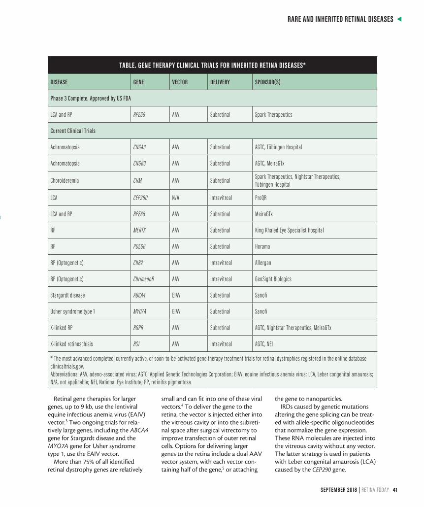
TABLE. GENE THERAPY CLINICAL TRIALS FOR INHERITED RETINA DISEASES*
DISEASE GENE VECTOR DELIVERY SPONSOR(S)
Phase 3 Complete, Approved by US FDA
LCA and RP RPE65 AAV Subretinal Spark Therapeutics
Current Clinical Trials
Achromatopsia CNGA3 AAV Subretinal AGTC, Tübingen Hospital
Achromatopsia CNGB3 AAV Subretinal AGTC, MeiraGTx
Choroideremia CHM AAV Subretinal Spark Therapeutics, Nightstar Therapeutics, Tübingen Hospital
LCA CEP290 N/A Intravitreal ProQR
LCA and RP RPE65 AAV Subretinal MeiraGTx
RP MERTK AAV Subretinal King Khaled Eye Specialist Hospital
RP PDE6B AAV Subretinal Horama
RP (Optogenetic) ChR2 AAV Intravitreal Allergan
RP (Optogenetic) ChrimsonR AAV Intravitreal GenSight Biologics
Stargardt disease ABCA4 EIAV Subretinal Sanofi
Usher syndrome type 1 MYO7A EIAV Subretinal Sanofi
X-linked RP RGPR AAV Subretinal AGTC, Nightstar Therapeutics, MeiraGTx
X-linked retinoschisis RS1 AAV Intravitreal AGTC, NEI
* The most advanced completed, currently active, or soon-to-be-activated gene therapy treatment trials for retinal dystrophies registered in the online database clinicaltrials.gov. Abbreviations: AAV, adeno-associated virus; AGTC, Applied Genetic Technologies Corporation; EIAV, equine infectious anemia virus; LCA, Leber congenital amaurosis; N/A, not applicable; NEI, National Eye Institute; RP, retinitis pigmentosa
s
RARE AND INHERITED RETINAL DISEASES
42 RETINA TODAY | SEPTEMBER 2018
Most retinal gene therapy treat-ments to date require the patient to have viable remaining photoreceptor cells. Optogenetics is a disease gene-independent technology that can be used in conditions with significant pho-toreceptor loss. In this approach, the remaining retinal neurons are convert-ed to photosensitive cells by expression of bacterial rhodopsin genes. In a trial sponsored by Allergan, channelrho-dopsin is being transfected to retinal cells, and in another trial conducted by GenSight Biologics the ChrimsonR photopigment is used in combination with a portable medical device.
There are several completed, ongoing, or upcoming clinical trials involving viral vectors for the treatment of IRDs (Table). The diseases targeted in these trials are discussed briefly below.
RPE65 Gene-Related Retinal DystrophyMutations in the RPE65 gene can
cause LCA and, in some cases, retinitis pigmentosa (RP). The RPE65 protein is an important visual cycle enzyme that contributes to the activation of the chromophore 11-cis-retinal.
After successful treatment of small and large animal models, including rodents and dogs,6 the first human trials of the therapy that eventually became voretigene were initiated in 2007. Multiple competing early-phase clinical trials used variations of AAV vector-based delivery of the RPE65 gene to the subretinal space.7-13
A recent phase 3 trial demonstrated safety and efficacy of the treatment for the primary outcome, a novel standardized multiluminance mobility test of the ambulatory vision in dif-ferent levels of light encountered in daily life.14 In addition, investigators observed significant improvement of light sensitivity and kinetic visual field testing after treatment. The traditional study outcome, visual acuity, which is dependent on cone photoreceptors, was improved, but the change did not reach statistical significance in this rod photoreceptor–mediated condition.
Treatment durability of at least 3 years has recently been reported for vore-tigene.15 Examples of reported visual improvements include patients seeing clouds in the sky for the first time and no longer requiring a white cane for ambulation.
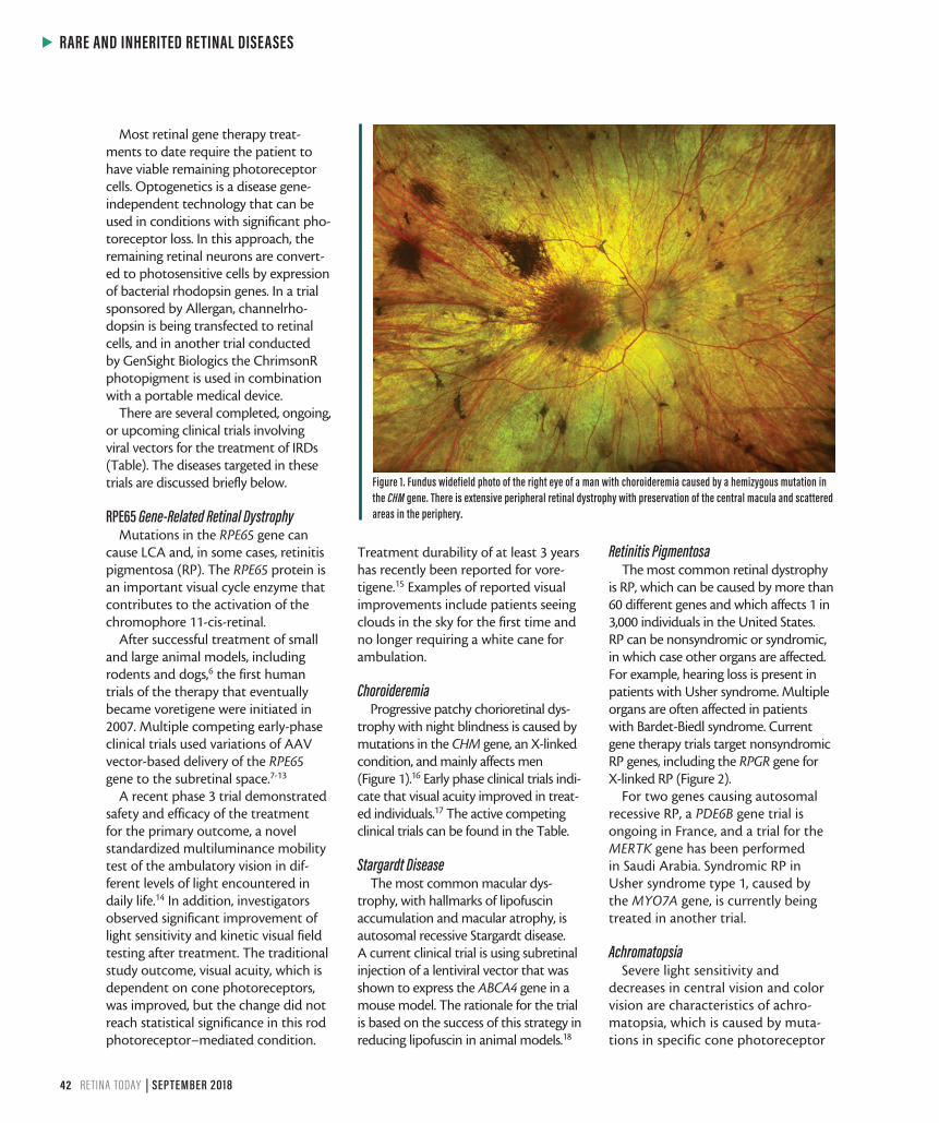
ChoroideremiaProgressive patchy chorioretinal dys-
trophy with night blindness is caused by mutations in the CHM gene, an X-linked condition, and mainly affects men (Figure 1).16 Early phase clinical trials indi-cate that visual acuity improved in treat-ed individuals.17 The active competing clinical trials can be found in the Table.
Stargardt DiseaseThe most common macular dys-
trophy, with hallmarks of lipofuscin accumulation and macular atrophy, is autosomal recessive Stargardt disease. A current clinical trial is using subretinal injection of a lentiviral vector that was shown to express the ABCA4 gene in a mouse model. The rationale for the trial is based on the success of this strategy in reducing lipofuscin in animal models.18
Retinitis PigmentosaThe most common retinal dystrophy
is RP, which can be caused by more than 60 different genes and which affects 1 in 3,000 individuals in the United States. RP can be nonsyndromic or syndromic, in which case other organs are affected. For example, hearing loss is present in patients with Usher syndrome. Multiple organs are often affected in patients with Bardet-Biedl syndrome. Current gene therapy trials target nonsyndromic RP genes, including the RPGR gene for X-linked RP (Figure 2).
For two genes causing autosomal recessive RP, a PDE6B gene trial is ongoing in France, and a trial for the MERTK gene has been performed in Saudi Arabia. Syndromic RP in Usher syndrome type 1, caused by the MYO7A gene, is currently being treated in another trial.
AchromatopsiaSevere light sensitivity and
decreases in central vision and color vision are characteristics of achro-matopsia, which is caused by muta-tions in specific cone photoreceptor
Figure 1. Fundus widefield photo of the right eye of a man with choroideremia caused by a hemizygous mutation in the CHM gene. There is extensive peripheral retinal dystrophy with preservation of the central macula and scattered areas in the periphery.
s
RARE AND INHERITED RETINAL DISEASES
44 RETINA TODAY | SEPTEMBER 2018
genes. Gene therapy targeting the CNGA3 and CNGB3 genes successfully improved vision in large animal mod-els, leading the way to current human clinical trials.19
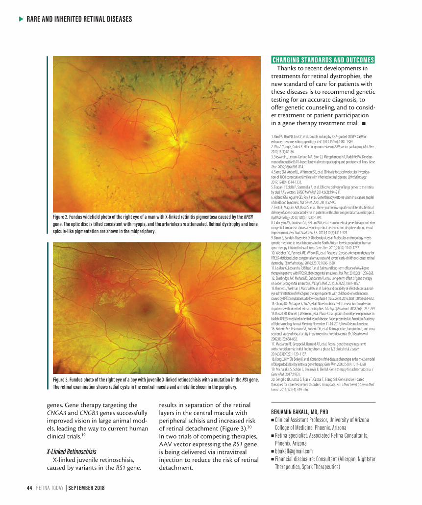
X-Linked RetinoschisisX-linked juvenile retinoschisis,
caused by variants in the RS1 gene,
results in separation of the retinal layers in the central macula with peripheral schisis and increased risk of retinal detachment (Figure 3).20 In two trials of competing therapies, AAV vector expressing the RS1 gene is being delivered via intravitreal injection to reduce the risk of retinal detachment.
CHANGING STANDARDS AND OUTCOMES Thanks to recent developments in
treatments for retinal dystrophies, the new standard of care for patients with these diseases is to recommend genetic testing for an accurate diagnosis, to offer genetic counseling, and to consid-er treatment or patient participation in a gene therapy treatment trial. n
1. Ran FA, Hsu PD, Lin CY, et al. Double nicking by RNA-guided CRISPR Cas9 for enhanced genome editing specificity. Cell. 2013;154(6):1380-1389.2. Wu Z, Yang H, Colosi P. Effect of genome size on AAV vector packaging. Mol Ther. 2010;18(1):80-86.3. Stewart HJ, Leroux-Carlucci MA, Sion CJ, Mitrophanous KA, Radcliffe PA. Develop-ment of inducible EIAV-based lentiviral vector packaging and producer cell lines. Gene Ther. 2009;16(6):805-814.4. Stone EM, Andorf JL, Whitmore SS, et al. Clinically focused molecular investiga-tion of 1000 consecutive families with inherited retinal disease. Ophthalmology. 2017;124(9):1314-1331.5. Trapani I, Colella P, Sommella A, et al. Effective delivery of large genes to the retina by dual AAV vectors. EMBO Mol Med. 2014;6(2):194-211.6. Acland GM, Aguirre GD, Ray J, et al. Gene therapy restores vision in a canine model of childhood blindness. Nat Genet. 2001;28(1):92-95.7. Testa F, Maguire AM, Rossi S, et al. Three-year follow-up after unilateral subretinal delivery of adeno-associated virus in patients with Leber congenital amaurosis type 2. Ophthalmology. 2013;120(6):1283-1291.8. Cideciyan AV, Jacobson SG, Beltran WA, et al. Human retinal gene therapy for Leber congenital amaurosis shows advancing retinal degeneration despite enduring visual improvement. Proc Natl Acad Sci U S A. 2013;110(6):E517-525.9. Banin E, Bandah-Rozenfeld D, Obolensky A, et al. Molecular anthropology meets genetic medicine to treat blindness in the North African Jewish population: human gene therapy initiated in Israel. Hum Gene Ther. 2010;21(12):1749-1757.10. Weleber RG, Pennesi ME, Wilson DJ, et al. Results at 2 years after gene therapy for RPE65-deficient Leber congenital amaurosis and severe early-childhood-onset retinal dystrophy. Ophthalmology. 2016;123(7):1606-1620.11. Le Meur G, Lebranchu P, Billaud F, et al. Safety and long-term efficacy of AAV4 gene therapy in patients with RPE65 Leber congenital amaurosis. Mol Ther. 2018;26(1):256-268.12. Bainbridge JW, Mehat MS, Sundaram V, et al. Long-term effect of gene therapy on Leber’s congenital amaurosis. N Engl J Med. 2015;372(20):1887-1897.13. Bennett J, Wellman J, Marshall KA, et al. Safety and durability of effect of contralateral-eye administration of AAV2 gene therapy in patients with childhood-onset blindness caused by RPE65 mutations: a follow-on phase 1 trial. Lancet. 2016;388(10045):661-672.14. Chung DC, McCague S, Yu ZF, et al. Novel mobility test to assess functional vision in patients with inherited retinal dystrophies. Clin Exp Ophthalmol. 2018;46(3):247-259.15. Russell SR, Bennett J, Wellman J, et al. Phase 3 trial update of voretigene neparvovec in biallelic RPE65-mediated inherited retinal disease. Paper presented at: American Academy of Ophthalmology Annual Meeting; November 11-14, 2017; New Orleans, Louisiana.16. Roberts MF, Fishman GA, Roberts DK, et al. Retrospective, longitudinal, and cross sectional study of visual acuity impairment in choroideraemia. Br J Ophthalmol. 2002;86(6):658-662.17. MacLaren RE, Groppe M, Barnard AR, et al. Retinal gene therapy in patients with choroideremia: initial findings from a phase 1/2 clinical trial. Lancet. 2014;383(9923):1129-1137.18. Kong J, Kim SR, Binley K, et al. Correction of the disease phenotype in the mouse model of Stargardt disease by lentiviral gene therapy. Gene Ther. 2008;15(19):1311-1320.19. Michalakis S, Schön C, Becirovic E, Biel M. Gene therapy for achromatopsia. J Gene Med. 2017;19(3).20. Sengillo JD, Justus S, Tsai YT, Cabral T, Tsang SH. Gene and cell-based therapies for inherited retinal disorders: An update. Am J Med Genet C Semin Med Genet. 2016;172(4):349-366.
BENJAMIN BAKALL, MD, PHDn Clinical Assistant Professor, University of Arizona
College of Medicine, Phoenix, Arizonan Retina specialist, Associated Retina Consultants,
Phoenix, Arizonan [email protected] Financial disclosure: Consultant (Allergan, Nightstar
Therapeutics, Spark Therapeutics)
Figure 3. Fundus photo of the right eye of a boy with juvenile X-linked retinoschisis with a mutation in the RS1 gene. The retinal examination shows radial cysts in the central macula and a metallic sheen in the periphery.
Figure 2. Fundus widefield photo of the right eye of a man with X-linked retinitis pigmentosa caused by the RPGR gene. The optic disc is tilted consistent with myopia, and the arterioles are attenuated. Retinal dystrophy and bone spicule-like pigmentation are shown in the midperiphery.