Lymphoid System Diffuse Lymphoid Tissue –GALT –BALT Lymph node Spleen.
Upload
kathryn-brownCategory
view
217download
2
Tertiary lymphoid organs in renal allografts can beassociated with donor-specific tolerance rather thanrejection
Kathryn Brown, Steven H. Sacks and Wilson Wong
MRC Centre for Transplantation, King’s College London School of Medicine at Guy’s, King’s
and St. Thomas’ Hospitals, London, UK
Tertiary lymphoid organs can form at sites of chronic inflammation. Their presence has
been mainly associated with tissue destruction. In transplantation, there is a dynamic
immune response as in chronic inflammation. Indeed, the presence of tertiary lymphoid
organs has been associated with chronic rejection. In addition to a destructive alloimmune
response, secondary lymphoid organs are also important in transplant tolerance. We
hypothesised that tertiary lymphoid organs may also form during transplantation toler-
ance as this process also requires an active local immune response. If so, their presence
may enhance tolerance, resulting in better graft function rather than chronic rejection.
Using a mouse kidney allograft model of tolerance, we demonstrate the formation of
tertiary lymphoid organs within tolerated allografts. Tertiary lymphoid organs are
supplied by high endothelial venules, and contain T and B cells, macrophages, DC, Foxp31
T cells, donor MHC class II1 cells and recipient cells presenting donor-derived allopeptides.
Formation of tertiary lymphoid organs and the presence of immune cells within them are
associated with superior graft function, suggesting that tertiary lymphoid organs act to
amplify the prevailing immune response, be it a tolerant and beneficial immune response
or the previously described destructive alloimmunity.
Key words: Tertiary lymphoid organs . Tolerance . Transplantation
Introduction
Secondary lymphoid organs are crucial for the adaptive immune
response. Antigen-presenting cells migrate from the site of
inflammation to secondary lymphoid organs that provide a niche
for effective encounters between naı̈ve T cells and their cognate
antigens. In chronic inflammation, however, where antigens are
not cleared, tertiary lymphoid organs (TLO) can develop
ectopically in the periphery (reviewed in [1]). These are
organised structures, resembling lymph nodes, and have been
extensively studied in the context of autoimmune diseases such as
rheumatoid arthritis; chronic infections such as hepatitis C and
malignancy [1].
After organ transplantation, there are continuous low-level,
dynamic interactions between donor alloantigens and the reci-
pient’s immune system, in a situation similar to chronic inflam-
mation. However, as in autoimmune diseases, the antigens
cannot be cleared. As a result, TLO have been observed after
transplantation, for example in cardiac allgorafts undergoing
chronic rejection [2]. TLO within skin allografts were able to
mount an effective alloresponse, leading to rejection and devel-
opment of a memory response [3].
Germinal centres have been found in chronically rejecting rat
aortic, and human heart and kidney grafts [4]. Reports to date
have, in the main, suggested that formation of TLO within
transplanted organs is associated with poorer graft outcome.
SHORT COMMUNICATION
Correspondence: Dr. Wilson Wonge-mail: [email protected]
& 2010 WILEY-VCH Verlag GmbH & Co. KGaA, Weinheim www.eji-journal.eu
Eur. J. Immunol. 2011. 41: 89–96 DOI 10.1002/eji.201040759 Cellular immune response 89

However, the fate of a transplanted organ depends on the balance
between graft destructive alloreactive T cells and graft protective
regulatory T cells [5]. There are examples in humans where the
balance is tipped in favour of regulation, allowing immunosup-
pressive therapy to be reduced, or ceased entirely [6, 7]. In
animal models, this has been described in numerous
situations [8–11]. Tolerance can be viewed as a dynamic inter-
action between donor antigens and recipient T cells [12]
analogous to the situation seen in chronic inflammation. It
involves an active local immune response which may lead to the
development of TLO. Indeed, lymphoid aggregates have been
described in two long-term tolerant human kidney allografts
[13]. The small number of tolerant transplant recipients, and the
sampling error inherent in biopsy samples, makes further inves-
tigation difficult. This prompted us to use a mouse kidney
allograft model where the recipient is tolerant to donor antigens,
resulting in indefinite graft survival in some recipients
and prolonged survival of donor type challenge skin grafts [11],
to see if TLO can be associated with donor-specific unrespon-
siveness.
Results and discussion
Lymphocytic clusters are present within tolerantkidney allografts
We have used a previously described fully allogeneic DBA/2 to
C57BL/6 mouse kidney allograft model in which 40% of
allografts were accepted indefinitely with normal graft function
and histology, whereas a further 40% of grafts were not rejected
acutely but survive up to 105 days with slowly deteriorating graft
function as reflected by blood urea nitrogen (BUN) and
histological features of interstitial fibrosis and tubular atrophy
(IFTA). Since there are no other confounding factors such as
calcineurin inhibitors in this model, the cause of the IFTA is likely
to be chronic rejection [11]. C57BL/6 recipients of DBA/2
kidneys were sacrificed at various time points after transplanta-
tion for histological examination. Syngeneic C57BL/6 kidney
grafts had no cellular infiltrates at day 14. Minimal infiltrates
were seen at day 45 and beyond (Fig. 1A–D). In DBA/2 allografts,
only small amounts of mononuclear cellular infiltrates were seen
at day 8 post-transplant scattered throughout the cortex; these
did not appear to be organised into clusters. At later time points,
clusters of lymphocytic infiltrates could be seen in most sections
(Fig. 1E–H). Their appearance differs from that seen at earlier
time points by being densely packed and having well-defined
margins.
Lymphocytic clusters satisfy the criteria for TLO
The exact definition of TLO has not yet been agreed. However,
the presence of T, B and DC, and high endothelial venules (HEV)
has been suggested. To determine whether the clusters of
lymphocytic infiltrates seen here were TLO, we carried out
histological analysis to investigate whether they fulfil these
criteria.
Constituents of cellular infiltrates resemble that oflymph nodes
Conventional secondary lymphoid tissues contain a variety of
immune cells that are essential constituents for co-ordinated
and effective immune responses. DBA/2 kidney allografts
were therefore examined to identify the cell types present
within the infiltrates. T cells (CD41, CD81 and regu-
latory T cells), B cells, DC and macrophages were all found
in lymphocytic clusters in all tissue sections examined
(Fig. 1I–N).
Clusters of cellular infiltrate contain HEV
Tissue sections were stained for the presence of HEV, sections of
vascular endothelium specialised to allow extravasation of
lymphocytes and normally found only in lymphoid organs, using
the specific marker peripheral lymph node addressin (PNAd).
PNAd1 vessels were seen in kidney allografts from 14 days post-
transplantation, and increased to 0.2970.17 positive vessels per
medium power field (mpf (� 160)) at day 45 post-transplanta-
tion, compared with 0.0870.1 in syngeneic kidney grafts
(Figs. 1O and 2A). This rose to 0.4870.16 positive vessels per
mpf 480 days post-transplantation. At this time point, all but one
kidney allograft had demonstrable PNAd1 vessels. The density of
PNAd1 vessels in conventional lymph nodes is usually much
higher than we have observed here. The reason and functional
relevance of this is unclear. We did not detect PNAd1 vessels in
some of the TLO, which may be at least partially due to their low
densities. Alternatively, PNAd1 vessels may take longer than 80
days to develop.
Put together, these data suggest that the cellular infiltrates
observed in the DBA/2 allografts were TLO and they will be
referred to as such from this point onwards.
Lymphatic neogenesis in kidney allografts
Lymphatic vessels may act to facilitate the trafficking of immune
cells through TLO [14]. We therefore stained DBA/2 kidney
allografts for the presence of podoplanin, a marker of lymphatic
endothelial cells. A higher density of podoplanin1 vessels was
seen from day 45 after transplantation, with an average of
1.6870.74 positive vessels per mpf (compared with 0.5670.15
in syngeneic grafts at the same time point), and increased to
8.6171.94 in the 480 days post-transplantation group (Figs. 1P
and 2B). In all kidney allografts sacrificed 480 days post-
transplantation, at least 60% of all TLO contained a podoplanin1
vessel.
Eur. J. Immunol. 2011. 41: 89–96Kathryn Brown et al.90
& 2010 WILEY-VCH Verlag GmbH & Co. KGaA, Weinheim www.eji-journal.eu

C57BL/6 donor kidneys
A B C D125µm 125µm 125µm 125µm
DBA/2 donor kidneys
E F H125µm 125µm 125µm 125µm
day 8 day 14 day 45 >day 80
G
Days post transplant day day day >daytransplant
J KI 125µm 125µm 125µm
NML
3pxoF8DC4DC
125µm 125µm 125µm
P QO
86DCc11DC91DC
200µm80µm 80µm
TSR
SAPninalpodopdANP
200µm 80µm 80µm
PAS I-Ad Y-Ae
Figure 1. Cellular infiltrates are present within tolerant transplant allografts. Kidneys from syngeneic C57BL/6 or allogeneic DBA/2 micetransplanted into C57BL/6 recipients were harvested at days 8 (A, E), 14 (B, F), 45 (C, G) or 480 (D, H) after transplantation and stained with PAS toidentify area of cellular infiltrates. Sections of a kidney allograft harvested at 95 days post-transplantation were also stained to identify cell typeswithin infiltrates: CD41 (I), CD81 (I), Foxp31 (K), CD191 B cells (L), CD11c1 DC (M) and CD681 macrophages (N). Sections of kidney allograft werestained to identify PNAd (O) and podoplanin1 (P) vessels to distinguish HEV and lymphatic vessels, respectively. PAS-stained sections of long-term(480 days post-transplant) kidney allografts with good (BUN 5 17.1 mmol/L at harvest) (Q) and poor (BUN 5 45.4 mmol/L at harvest) (R) graftfunction, showing cellular infiltrates in tolerant (Q) but not in chronically rejecting (R) kidney. Donor antigens were identified by staining for I-Ad1
donor DC (S); and Y-Ae1 cells expressing recipient MHC class II presenting donor peptide (T). Original magnifications �160 (A–H, I–N); � 250 (O, P,S, T); � 100 (Q, R). PAS, periodic acid Schiff; PNAd, peripheral lymph node addressin and BUN, blood urea nitrogen. A representative of at least threeexperiments is shown.
Eur. J. Immunol. 2011. 41: 89–96 Cellular immune response 91
& 2010 WILEY-VCH Verlag GmbH & Co. KGaA, Weinheim www.eji-journal.eu
Large cross-sectional area of TLO is associated withsuperior graft function
TLO thus far have mostly been associated in humans and
experimental animal models with having a destructive
effect. However, in our model TLO appear to be more pro-
minent in allografts with good function. We therefore investi-
gated the correlation between various attributes of TLO
objectively to see if any features were associated with superior
graft function.
Allograft sections were examined under mpf and the area of
individual TLO measured and expressed as mean area per mpf
(Fig. 2C). A correlation was found between mean TLO area and
graft function as measured by BUN, confirming our initial
subjective observation (r2 5 0.6541, p 5 0.0024, Figs. 1Q and
2D). Almost no TLO were seen in grafts from recipients
with BUN of Z40mmol/L (Fig. 1R), whereas grafts with TLO
area of Z150mm2/mpf were always associated with BUN of
30 mmol/L or less. Therefore, unlike other models, here TLO
formation was associated with a positive rather than a negative
outcome.
To determine whether area of TLO also correspond to graft
function in the tolerant state, we performed sub-group analysis
on recipients that had survived 480 days. Although there was a
trend towards larger TLO areas and better graft function,
(r2 5 0.399, p 5 0.068), it was just below the limit for statistical
significance. One possible explanation for this is that the majority
of the recipients in this sub-group were tolerant and therefore all
had good graft function; many recipients with IFTA do not
survive this long and are therefore under represented.
HEV marker PNAd is associated with superior graftfunction
Although not all of the lymphocytic clusters contained PNAd1
vessels, the presence of PNAd1 vessels within TLO was associated
with better graft function (r2 5 0.521, p 5 0.0281), in those
allografts harvested day 45 post-transplantation onward
(Fig. 2E).
Podoplanin1 vessels are associated with poor graftfunction
Significant amounts of podoplanin1 vessels were observed only
80 or more days after transplantation (Fig. 2B). Therefore, they
were used for analysis here. In contrast to staining for HEV using
PNAd, a strong correlation was seen between the presence of
podoplanin expressing vessels and the poor graft function
(r2 5 0.759, p 5 0.0239) (Fig. 2F).
A higher percentage of TLO containing podoplanin1 vessels
was found to be associated with poor graft function, whereas on
the contrary, a higher percentage of TLO containing PNAd1
vessels was associated with good graft function. These results
suggest that the entry of lymphocytes into the TLO (through
HEV) is beneficial for the allograft, whereas the exit of immune
cells (suggested to be through the newly formed lymphatic
vessels [15]) is detrimental to the allograft. This adds to the
theory that TLO here are not harmful, and may actually be of
benefit, in this model of tolerance. The podoplanin results were in
contrast to the studies by Stuht et al., who found that the
presence of lymph vessels within cellular infiltrates was asso-
ciated with improved graft function [15]. This may be because
their patients had chronic immune damage in their kidneys,
whereas our mouse model is of tolerant kidney grafts, further
supporting the notion that TLO perpetuate and amplify the
prevailing immune response in the local environment, be it
rejection or tolerance.
We have not found any major differences between
the tolerogeneic TLO seen in this model of spontaneous
acceptance, and the TLO found by other groups in
chronic rejection models. However, these structures are likely to
differ in the activity of the cells within them, the balance of
regulatory to effector cells and the cytokines and chemokines
present.
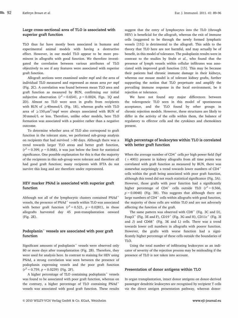
High percentage of leukocytes within TLO is correlatedwith better graft function
When the average number of CD41 cells per high power field (hpf
(� 400)) present in kidney allografts from all time points was
correlated with graft function as measured by BUN, there was
somewhat surprisingly a trend towards lower numbers of CD41
cells within the graft being associated with poor graft function,
although this trend did not reach statistical significance (Fig. 3A).
However, those grafts with poor function had a significantly
higher percentage of CD41 cells outside TLO (r2 5 0.566,
p 5 0.0048) (Fig. 3B). This suggests that although there are
large numbers of CD41 cells within allografts with good function,
the majority of these cells are within TLO and are not adversely
affecting the function of the graft.
The same pattern was observed with CD81 (Fig. 3C and D),
Foxp31 (Fig. 3E and F), CD191 (Fig. 3G and H), CD11c1 (Fig. 3I
and J) and CD681 (Fig. 3K and L) cells. There was a trend
towards lower cell numbers in allografts with poorer function.
However, the grafts with worse function had a signi-
ficantly higher percentage of these cells outside the boundaries of
TLO.
Using the total number of infiltrating leukocytes as an indi-
cator of severity of the rejection process may be misleading if the
presence of TLO is not taken into account.
Presentation of donor antigens within TLO
In organ transplantation, intact donor antigens on donor-derived
passenger dendritic leukocytes are recognised by recipient T cells
via the direct antigen presentation pathway, whereas donor-
Eur. J. Immunol. 2011. 41: 89–96Kathryn Brown et al.92
& 2010 WILEY-VCH Verlag GmbH & Co. KGaA, Weinheim www.eji-journal.eu

derived peptides are presented by recipient antigen-presenting
cells in the context of self-MHC molecules, through the indirect
antigen presentation pathway. Lymphoid organs provide a
physical location for antigen-specific T cells from both antigen
presentation pathways to meet their cognate ligands. We
investigated whether TLO seen here contained antigens for the
direct and indirect pathways.
Sections of kidney allografts were stained for the presence of
the intact donor MHC class II molecule I-Ad. I-Ad1 cells could
clearly be seen within TLO at all time points, revealing the
presence of donor antigen-presenting cells within the TLO
(Fig. 1S).
Sections were also stained with the monoclonal antibody
Y-Ae which recognises the allopeptide Ea52–68 in the
context of I-Ab. It has been previously shown to identify I-Ab1
C57BL/6 cells presenting the relevant peptide derived from I-Ed
MHC class II molecules [16]. Staining was not seen at the earliest
time point analysed (14 days), but was observed from day 45
post-transplantation onwards, and the numbers of Y-Ae1 cells
increased again 480 days post-transplantation (1.6370.64 and
1.25 15
A BninalpodoPdANP
BL/6 d14
BL/6d45
BA/2 d14
BA/2d45
>80
days
0.00
0.25
0.50
0.75
1.00
no
. of
+ve
vess
els
per
mp
f
7BL/6
d14
7BL/6
d45
DBA/2 d14
DBA/2d45
>80
days
0
5
10
no
. of
+ve
vess
els
per
mp
f
500
750
1000
1250
area
C57 C57 D D
DBA/2>8
C57B
C57B DB DB
DBA/2>8
C
D
20
30
40
50
60
70
C57BL/6
d14
C57BL/6
d45
C57BL/6
>80 d
ays
DBA/2 d14
DBA/2d45
DBA/2>8
0 day
s0
250
0 100 200 300 400 500 600 7000
10
TLO area (µm2) per mpf
blo
od
ure
a n
itro
gen
(m
mo
l/L)
E
50
60
F
50
60
% TLO containing PNAd+ vessels
0 10 20 300
10
20
30
40
blo
od
ure
a n
itro
gen
(m
mo
l/L)
% TLO containing podoplanin+ vessels
50 60 70 80 90 100 1100
10
20
30
40
blo
od
ure
a n
itro
gen
(m
mo
l/L)
0
1
2
3
4
5
6
no
. of
Y-A
e+ c
ells
per
hp
f
HG
0
10
20
30
40
50
60
blo
od
ure
a n
itro
gen
(m
mo
l/L)
DBA/2 d14
DBA/2 d45
DBA/2 >8
0 days 0 1 2 3 4 5 6 7
no. of Y-Ae+ cells per hpf
I
40
50
60
10
20
30
0 10 20 30 40 50 60 70 800
%Y-Ae+ cells outside TLOs
blo
od
ure
a n
itro
gen
(m
mo
l/L)
Figure 2. Correlation between graft function and PNAd, podoplanin, TLO area and Y-Ae1 cells. Kidneys from syngeneic C57BL/6 or allogeneic DBA/2mice transplanted into C57BL/6 recipients were harvested at day 14, 45 or 480 days after transplantation and stained for (A) PNAd to show thedevelopment of HEV or (B) podoplanin to show the development of lymphatic vessels. (C) Degree of TLO formation as measured by the average areaper mpf covered by cellular infiltrate. (D) Correlation between average area of infiltrate per mpf and graft function as measured by BUN. (E) Usingonly those samples with significant PNAd staining (day 45 post-transplantation onwards), the percentage of clusters within the kidney allograftcontaining PNAd1 vessel(s) was correlated with BUN. (F) Using only those samples with significant podoplanin staining (480 days post-transplantation group only), the percentage of clusters within the kidney allograft containing podoplanin1 vessel(s) was correlated with BUN.(G) Staining for Y-Ae shows the presence of recipient MHC class II molecules presenting donor peptide. (H) Correlation between total Y-Ae1 cellnumbers and graft function. (I) Correlation between percentage of Y-Ae1 cells outside TLO and graft function. In (E, F, H and I), linear regressionanalyses were used to check for association between the features of TLO and BUN. Non-linear quadratic regression was used for (D) and comparedagainst a linear model. The best fitting one (quadratic) is shown. TLO, tertiary lymphoid organs; PNAd, peripheral lymph node addressin and BUN,blood urea nitrogen. Statistical significance measured using a Mann–Whitney test. ns, non significant. Each data point (solid circle) represents anindividual kidney recipient.
Eur. J. Immunol. 2011. 41: 89–96 Cellular immune response 93
& 2010 WILEY-VCH Verlag GmbH & Co. KGaA, Weinheim www.eji-journal.eu

2.2671 Y-Ae1 cells per hpf, respectively), confirming the
presentation of donor antigen by recipient antigen-presenting
cells (Figs. 1T and 2G). The majority of these cells were found
within TLO.
The presence of donor I-Ad1 cells and Y-Ae1 recipient
cells presenting donor antigen within TLO suggests that
TLO are actively participating in the local immune
response by acting as a site of both direct and indirect allor-
ecognition.
When numbers of Y-Ae1 cells were correlated with graft
function, a non-significant trend towards higher numbers of
Y-Ae1 cells in grafts with poor function was seen (Fig. 2H).
However, a stronger association was found between a high
percentage of Y-Ae1 cells outside TLO and a poor graft function
20
30
40
50
60ea
nitr
oge
n (m
mol
/L)
20
30
40
50
60
ea n
itro
gen
(mm
ol/L
)A Br2=0.566p=0.0048
r2=0.188p=0.160
0 25 50 75 100 125 1500
10
no. of CD4+ cells per hpf
blo
od
ure
0 25 50 75 1000
10
%CD4+ cells outside TLOs
bloo
d ur
e
40
50
60
oge
n (m
mol
/L)C
r2=0.118p=0.275
0 50 100 150 2000
10
20
30
no. of CD8+ cells per hpf
blo
od
urea
nitr
o
50
60
mol
/L)
r
F
0 25 50 75 100 1250
10
20
30
40
%Foxp3+ cells outside TLOs
bloo
d ur
ea n
itro
gen
(mm r2=0.523
p=0.011940
50
60
gen
(mm
ol/L
)D
r2=0.348p=0.043
0 25 50 75 1000
10
20
30
%CD8+ cells outside TLOs
bloo
d ur
ea n
itro
50
60
mol
/L)
r
0 25 50 75 1000
10
20
30
40
no. of Foxp3+ cells per hpf
blo
od
urea
nitr
oge
n (m
m
E
r2=0 263p=0.089
0 25 50 75 1000
10
20
30
40
50
60
blo
od
urea
nitr
oge
n (m
mol
/L)
0 25 50 75 100 1250
10
20
30
40
50
60
bloo
d ur
ea n
itro
gen
(mm
ol/L
)HGr2=0.526p=0.0076
r2=0.079p=0.378
no. of CD19+ cells per hpf
20
30
40
50
60
urea
nitr
oge
n (m
mol
/L)
20
30
40
50
60
urea
nit
roge
n (m
mol
/L)
0 25 50 75 100 125
%CD19+ cells outside TLOs
I
KJ L
r2=0.066p=0.0013
r2=0.055p=0.464
0 25 50 75 100 125 1500
10
no. of CD11c+ cells per hpf
blo
od
u
30 40 50 60 70 80 900
10
%CD11c+ cells outside TLOs
bloo
d u
40
50
60
og
en (
mm
ol/L
)
40
50
60
ogen
(m
mol
/L)
r2=0.554p=0.0055
r2=0.002p=0.886
0 5 10 15 20 250
10
20
30
score per hpf
blo
od
ure
a n
itro
0 25 50 75 100 1250
10
20
30
%CD68 staining outside TLOs
bloo
d ur
ea n
itro
Figure 3. Correlation of graft function with histological parameters. (A, C, E, G, I, K) Numbers of CD4, CD8, Foxp3, CD19 and CD11c1 cells per hpfand CD68 score, respectively, versus BUN, in DBA/2 kidney allografts transplanted into C57BL/6 recipients, harvested 14, 45 and 480 days post-transplantation. (B, D, F, H, J, L) Correlation between the percentage of cells outside of TLO and BUN. In the case of Foxp3, one sample was excludedfrom the analysis due to the paucity of Foxp31 cells (one in entire section) within the section, making statistics unreliable. In all cases, linearregression analyses were used to check for correlation between the numbers or percentages of cells, and BUN. TLO, tertiary lymphoid organs andBUN, blood urea nitrogen. Statistical significance was measured using Mann–Whitney test.
Eur. J. Immunol. 2011. 41: 89–96Kathryn Brown et al.94
& 2010 WILEY-VCH Verlag GmbH & Co. KGaA, Weinheim www.eji-journal.eu

(r2 5 0.419, p 5 0.0315) (Fig. 2I). Hence, again those cells
outside TLO appear to be more damaging to the allograft. It has
been proposed that regulatory T cells act mainly through the
indirect pathway [17], and TLO may therefore promote this
interaction.
Concluding remarks
We have described a model of TLO in which they behave in the
opposite manner to that previously described, suggesting that
TLO, like secondary lymphoid tissues, merely serve to amplify the
prevailing local immune response and may not necessarily cause
tissue damage. Further understanding of their function and the
circumstances under which they form may lead to the develop-
ment of therapeutic strategies to deviate the immune response
towards the desired direction.
Materials and methods
Animals
Female donor DBA/2 (H-2d) and recipient C57BL/6 (H-2b) mice
(Harlan UK, Oxon, UK) were kept in specific pathogen-free
facilities and used in accordance with the Animals (Scientific
Procedures) Act 1986.
Organ transplantation
Renal transplantation was performed as described previously
[18]. The left native kidney was removed at the time of
transplantation. The remaining native kidney was removed 7
days later, leaving the donor graft life sustaining. BUN was
measured at regular intervals to monitor graft function, using
Infinity Urea (Thermo Fisher Scientific, Middletown, CT, USA)
according to the manufacturer’s instruction. Tissues were
harvested as described previously [11].
Histology
Periodic acid Schiff (PAS) staining was carried out on 2mm thick
paraffin sections as described previously [19].
The area of the cellular infiltrate was measured in at least
12 mmpf of blinded samples using LUCIA G software (Nikon,
Tokyo, Japan) and means were calculated.
Immunohistochemistry
Frozen tissue samples were stained as described previously [11],
with the primary antibodies anti-podoplanin (Abcam plc,
Cambridge, UK), anti-CD4 (clone H129.19, BD, NJ, USA), anti-
CD8 (clone 53-6.7, BD), anti-CD19 (clone 1D3, BD), anti-CD11c
(clone N418, BioLegend, San Diego, CA, USA), anti-CD68 (clone
FA-11, Serotec, Oxford, UK), anti-PNAd (clone MECA-79,
BioLegend), anti-Foxp3 (clone FJK-16s, eBioscience, San Diego,
CA, USA), anti-I-Ad (clone AMS-32.1, BD) and anti-Y-Ae (clone
eBioY-Ae, eBioscience).
Positive vessels were counted in at least 10 mpf of
blinded samples and the means were calculated. Positive cells
were counted in 20 random hpf of each blinded sample,
and the means were calculated. For macrophage staining, a 63-
point grid was overlaid onto a �400 magnification of
macrophage staining. The percentages of grid points positive for
CD68 staining were counted in 20 hpf, and the means were
calculated.
Histological data were correlated to allograft function using
regression analyses (linear or quadratic) with GraphPad Prism 4
(GraphPad Software, San Diego, CA, USA). Mann–Whitney test
was used to test for statistical differences between histological
scores from different experimental groups. In all cases, values are
expressed as mean7SEM.
Acknowledgements: This work was supported by a grant from
the Genzyme Renal Innovations Program. The authors thank
Dr. Irene Rebollo Mesa for statistical help.
Conflict of interest: The authors declare no financial or
commercial conflict of interest.
References
1 Aloisi, F. and Pujol-Borrell, R., Lymphoid neogenesis in chronic inflam-
matory diseases. Nat. Rev. Immunol. 2006. 6: 205–217.
2 Baddoura, F. K., Nasr, I. W., Wrobel, B., Li, Q., Ruddle, N. H. and Lakkis, F.
G., Lymphoid neogenesis in murine cardiac allografts undergoing chronic
rejection. Am. J. Transplant. 2005. 5: 510–516.
3 Nasr, I. W., Reel, M., Oberbarnscheidt, M. H., Mounzer, R. H., Baddoura,
F. K., Ruddle, N. H. and Lakkis, F. G., Tertiary lymphoid tissues generate
effector and memory T cells that lead to allograft rejection. Am.
J. Transplant. 2007. 7: 1071–1079.
4 Thaunat, O., Field, A. C., Dai, J., Louedec, L., Patey, N., Bloch, M. F.,
Mandet, C. et al., Lymphoid neogenesis in chronic rejection: evidence for
a local humoral alloimmune response. Proc. Natl. Acad. Sci. USA 2005. 102:
14723–14728.
5 Zheng, X. X., Sanchez-Fueyo, A., Sho, M., Domenig, C., Sayegh, M. H. and
Strom, T. B., Favorably tipping the balance between cytopathic and
regulatory T cells to create transplantation tolerance. Immunity 2003. 19:
503–514.
6 Uehling, D. T., Hussey, J. L., Weinstein, A. B., Wank, R. and Bach, F. H.,
Cessation of immunosuppression after renal transplantation. Surgery
1976. 79: 278–282.
Eur. J. Immunol. 2011. 41: 89–96 Cellular immune response 95
& 2010 WILEY-VCH Verlag GmbH & Co. KGaA, Weinheim www.eji-journal.eu

7 Zoller, K. M., Cho, S. I., Cohen, J. J. and Harrington, J. T., Cessation of
immunosuppressive therapy after successful transplantation: a national
survey. Kidney Int. 1980. 18: 110–114.
8 Ildstad, S. T. and Sachs, D. H., Reconstitution with syngeneic plus
allogeneic or xenogeneic bone marrow leads to specific acceptance of
allografts or xenografts. Nature 1984. 307: 168–170.
9 Larsen, C. P., Elwood, E. T., Alexander, D. Z., Ritchie, S. C., Hendrix, R.,
Tucker-Burden, C., Cho, H. R. et al., Long-term acceptance of skin and
cardiac allografts after blocking CD40 and CD28 pathways. Nature 1996.
381: 434–438.
10 Bickerstaff, A. A., Wang, J. J., Pelletier, R. P. and Orosz, C. G., Murine renal
allografts: spontaneous acceptance is associated with regulated T cell-
mediated immunity. J. Immunol. 2001. 167: 4821–4827.
11 Brown, K., Moxham, V., Karegli, J., Phillips, R., Sacks, S. H. and Wong, W.,
Ultra-localization of Foxp31 T cells within renal allografts shows
infiltration of tubules mimicking rejection. Am. J. Pathol. 2007. 171:
1915–1922.
12 Waldmann, H., Adams, E., Fairchild, P. and Cobbold, S., Infectious
tolerance and the long-term acceptance of transplanted tissue. Immunol.
Rev. 2006. 212: 301–313.
13 Xu, Q., Lee, J., Jankowska-Gan, E., Schultz, J., Roenneburg, D. A., Haynes,
L. D., Kusaka, S. et al., Human CD41CD25low adaptive T regulatory cells
suppress delayed-type hypersensitivity during transplant tolerance. J.
Immunol. 2007. 178: 3983–3995.
14 Kerjaschki, D., Regele, H. M., Moosberger, I., Nagy-Bojarski, K.,
Watschinger, B., Soleiman, A., Birner, P. et al., Lymphatic neoangiogenesis
in human kidney transplants is associated with immunologically active
lymphocytic infiltrates. J. Am. Soc. Nephrol. 2004. 15: 603–612.
15 Stuht, S., Gwinner, W., Franz, I., Schwarz, A., Jonigk, D., Kreipe, H.,
Kerjaschki, D. et al., Lymphatic neoangiogenesis in human renal
allografts: results from sequential protocol biopsies. Am. J. Transplant.
2007. 7: 377–384.
16 Ochando, J. C., Krieger, N. R. and Bromberg, J. S., Direct versus indirect
allorecognition: visualization of dendritic cell distribution and interac-
tions during rejection and tolerization. Am. J. Transplant. 2006. 6:
2488–2496.
17 Yamada, A., Chandraker, A., Laufer, T. M., Gerth, A. J., Sayegh, M. H. and
Auchincloss, H., Jr., Recipient MHC class II expression is required to
achieve long-term survival of murine cardiac allografts after costimula-
tory blockade. J. Immunol. 2001. 167: 5522–5526.
18 Han, W. R., Murray-Segal, L. J. and Mottram, P. L., Modified technique for
kidney transplantation in mice. Microsurgery 1999. 19: 272–274.
19 McManus, J., Histological and histochemical uses of periodic acid. Stain
Technol. 1948. 23: 99–108.
Abbreviations: BUN: blood urea nitrogen � HEV: high endothelial venule
� hpf: high power field � IFTA: interstitial fibrosis and tubular atrophy �mpf: medium power field � PAS: periodic acid Schiff � PNAd: peripheral
lymph node addressin � TLO: tertiary lymphoid organs
Full correspondence: Dr. Wilson Wong, MRC Centre for Transplantation,
5th Floor, Tower Wing, Guy’s Hospital, London SE1 9RT, UK
Fax: 144-2071885660
e-mail: [email protected]
Received: 21/6/2010
Revised: 12/8/2010
Accepted: 27/9/2010
Accepted article online: 13/10/2010
& 2010 WILEY-VCH Verlag GmbH & Co. KGaA, Weinheim www.eji-journal.eu
Eur. J. Immunol. 2011. 41: 89–96Kathryn Brown et al.96