Terpenoids as Potential Anti-Alzheimer’s Disease Therapeutics · 2017-05-22 · Terpenoids as...
15
Molecules 2012, 17, 3524-3538; doi:10.3390/molecules17033524 molecules ISSN 1420-3049 www.mdpi.com/journal/molecules Review Terpenoids as Potential Anti-Alzheimer’s Disease Therapeutics Ki-Yeol Yoo 1 and So-Young Park 2, * 1 Department of Biological Sciences, College of Advanced Science, Dankook University, San#29, Anseo-dong, Dongnam-gu, Cheonan 330-714, Korea 2 Laboratory of Pharmacognosy, College of Pharmacy, Dankook University, San#29, Anseo-dong, Dongnam-gu, Cheonan 330-714, Korea * Author to whom correspondence should be addressed; E-Mail: [email protected]; Tel.: +82-41-550-1434; Fax: +82-41-559-7899. Received: 7 December 2011; in revised form: 12 March 2012 / Accepted: 16 March 2012 / Published: 19 March 2012 Abstract: Alzheimer’s disease (AD) is one of the most well-known neurodegenerative diseases and explains 50–60% of dementia in patients. The prevalence rate of AD is positively correlated with age and AD affects ≥40% of those over 85 years old. The major AD therapeutics available on the market are acetylcholinesterase inhibitors, such as tacrine and donepezil. New therapeutic agents that can block the disease-inducing mechanisms are essential. Diverse efforts have been made to discover anti-AD agents from natural sources. In this review article, we describe some representative terpenoids such as ginsenosides, gingkolides, and canabinoids as potential anti-AD agents. These compounds exhibit promising in vitro and in vivo biological activities, but are still waiting clinical trials. Additionally, we also discuss some terpenoids including cornel iridoid glycoside, oleanolic acid, tenuifolin, cryptotanshinone, and ursolic acid, which are under investigation for their in vitro and in vivo animal studies. Key words: Alzheimer’s disease; therapeutics; terpenoids 1. Introduction The development of medicine and science has contributed to a dramatic increase in life expectancy worldwide and the average life span may reach 120 years old by 2050. The increasing proportion of OPEN ACCESS
Transcript of Terpenoids as Potential Anti-Alzheimer’s Disease Therapeutics · 2017-05-22 · Terpenoids as...

Molecules 2012, 17, 3524-3538; doi:10.3390/molecules17033524
molecules ISSN 1420-3049
www.mdpi.com/journal/molecules
Review
Terpenoids as Potential Anti-Alzheimer’s Disease Therapeutics
Ki-Yeol Yoo 1 and So-Young Park 2,*
1 Department of Biological Sciences, College of Advanced Science, Dankook University, San#29,
Anseo-dong, Dongnam-gu, Cheonan 330-714, Korea 2 Laboratory of Pharmacognosy, College of Pharmacy, Dankook University, San#29, Anseo-dong,
Dongnam-gu, Cheonan 330-714, Korea
* Author to whom correspondence should be addressed; E-Mail: [email protected];
Tel.: +82-41-550-1434; Fax: +82-41-559-7899.
Received: 7 December 2011; in revised form: 12 March 2012 / Accepted: 16 March 2012 /
Published: 19 March 2012
Abstract: Alzheimer’s disease (AD) is one of the most well-known neurodegenerative
diseases and explains 50–60% of dementia in patients. The prevalence rate of AD is
positively correlated with age and AD affects ≥40% of those over 85 years old. The major
AD therapeutics available on the market are acetylcholinesterase inhibitors, such as tacrine
and donepezil. New therapeutic agents that can block the disease-inducing mechanisms are
essential. Diverse efforts have been made to discover anti-AD agents from natural sources.
In this review article, we describe some representative terpenoids such as ginsenosides,
gingkolides, and canabinoids as potential anti-AD agents. These compounds exhibit
promising in vitro and in vivo biological activities, but are still waiting clinical trials.
Additionally, we also discuss some terpenoids including cornel iridoid glycoside, oleanolic
acid, tenuifolin, cryptotanshinone, and ursolic acid, which are under investigation for their
in vitro and in vivo animal studies.
Key words: Alzheimer’s disease; therapeutics; terpenoids
1. Introduction
The development of medicine and science has contributed to a dramatic increase in life expectancy
worldwide and the average life span may reach 120 years old by 2050. The increasing proportion of
OPEN ACCESS
Molecules 2012, 17 3525
the elderly will also be reflected in a marked increase in the number of age-related diseases, including
neurodegenerative diseases.
Alzheimer’s disease (AD) is one of the most well-known neurodegenerative diseases, and explains
50–60% of patients with dementia. The prevalence rate of AD is positively correlated with age, and
AD occurs in ≥40% of the elderly over 85 years old [1]. Patients with AD decline in cognitive function
and find it difficult to remember recent events during the early stage (short-term memory loss). Once
the disease progresses, patients experience difficulties in speech, speaking, and cognitive thinking,
which is accompanied by long-term memory loss. Patients suffer from language deficits, depression,
aggressive behavior, and psychosis during the late stage and eventually need total care from caregivers.
One of the pathologic hallmarks of AD is senile plaques (SPs), and the major constituent of SPs is
beta-amyloid (Aβ), which is surrounded by dystrophic neurites and microglia and accumulates outside
of neurons. Aβ is a product of sequential proteolytic cleavage of amyloid precursor protein (APP) by
β-secretase and γ-secretase [2]. Aβ accumulates in the brain of patients with AD due to increased
production or decreased clearance of Aβ. The overproduction of Aβ found in patients with AD who
have genetic mutations in APP (familiar AD) is correlated with early onset (beginning in the 30s) of
the disease. An increased amount of Aβ (soluble monomeric form) in the brain self-aggregates into Aβ
oligomers (2–6 Aβ peptides) [3,4], which is more toxic to cells than the fibrillar or monomeric
form [5]. Therefore, the excess toxic Aβ is the major cause of AD pathology (amyloid hypothesis) [6].
Particularly, the levels of Aβ oligomers correlate with the severity of cognitive impairment in patients
with AD and play a critical role in AD pathology [7]. Aggregated Aβ oligomers lead to synaptic
dysfunction due to oxidative stress and inflammation [8,9]. A recent study reported that Aβ induces
neuronal death by binding to nerve growth factor receptors [10] such as pan neurotrophin receptor
(p75NTR) and activation of downstream c-Jun N-terminal kinase signal [11]. In addition, activation of
the N-methyl-D-aspartate (NMDA)-type glutamate receptor (NMDAR) disrupts calcium homeostasis,
eventually inducing oxidative stress and synaptic loss [12,13]. Aβ oligomers bind and modulate presynaptic
P/Q-type calcium channels at glutaminergic and gamma-amino butyric acid-ergic synapses and
eventually impair P/Q current, which is important for neurotransmission and synaptic plasticity [14,15].
Hyperphosphorylated tau proteins accumulate inside of neurons as a form of paired helical
filaments (PHFs), which are known as neurofibrillary tangles (NFTs) [16,17]. NFTs are another
pathological hallmark of AD. Tau protein, normally present in neurons, binds to microtubules to
promote microtubule assembly and stabilizes microtubules and vesicle transport. Conversely, abnormal
hyperphosphorylation to tau significantly reduces the affinity of tau protein to microtubules, so
hyperphosphorylated tau aggregates and forms PHFs [18]. Inhibited Wnt signaling following binding
to the Frizzled receptor, a Wnt protein acceptor, induces neurotoxic hyper-phosphorylated tau proteins,
which are found in NFTs [19]. A report that levels of hyperphosphorylated tau protein in cerebrospinal
fluid correlate with the degree of cognitive impairment in patients with AD [20] supports the
importance of NFTs in AD pathology. Disease-relevant phosphorylation of tau protein and the
aggregation to PHF is induced by Aβ [21–24]. Additionally, tau phosphorylation is the limiting factor
in Aβ-induced cell death [25]. These results support the suggestion that tau phosphorylation plays a
critical role in AD progression induced by Aβ.
The major AD therapeutics available on the market are acetylcholinesterase (AChE) inhibitors. A
strong correlation exists between the degree of cognitive impairment and a shortage of acetylcholine
Molecules 2012, 17 3526
(ACh) in patients with AD [26]. AChE inhibitors such as tacrine, donepezil, rivastigmine, and
galantamine have been developed as pharmacotherapy for AD. Although AChE inhibitors help alleviate
AD symptoms, they do not delay disease progression. Therefore, new therapeutic agents that block the
disease-inducing mechanisms are essential. Memantine, a NMDA receptor antagonist, has been approved
by the U.S. Food and Drug Administration (FDA) to treat AD [27]. This drug improves language
function and overall cognitive ability significantly in patients with moderate to severe AD [28,29].
Natural products have been used for medicinal purposes for a long time. The effort to develop
natural products as potential therapeutics and advances in extraction and isolation techniques lead to
the development of 63% of the natural product-derived drugs from 1981–2006 [30]. Much research
effort has been devoted to the development of anti-AD agents from natural sources [31]. Galantamine
isolated from bulbs and flowers of snowdrop Galanthus woronowii (Amaryllidaceae) has been
approved by the FDA as an anti-AD medication due to its inhibitory effect against AChE. Terpenoids
such as ginsenosides in Panax ginseng (Araliaceae) have been extensively studied to understand their
beneficial effects on AD. Among many natural products, the terpenoids are the largest and the most
diverse group of naturally occurring organic compounds, which increases the chance that a terpenoid
will be identified as having activity against AD. This review article describes some terpenoids as
possible therapeutic agents to treat AD.
2. Terpenoids
2.1. Ginsenosides from Panax ginseng CA Meyer (Araliaceae)
Ginsenosides are a series of derivatives of the dammarane-type triterpenes with some sugar
moieties attached [32,33], which are the major active components in ginseng isolated from P. ginseng.
P. ginseng is a well-known traditional medicinal plant that has been used as a representative tonic for
thousands of years to promote health and longevity. Among diverse ginsenosides in ginseng extract,
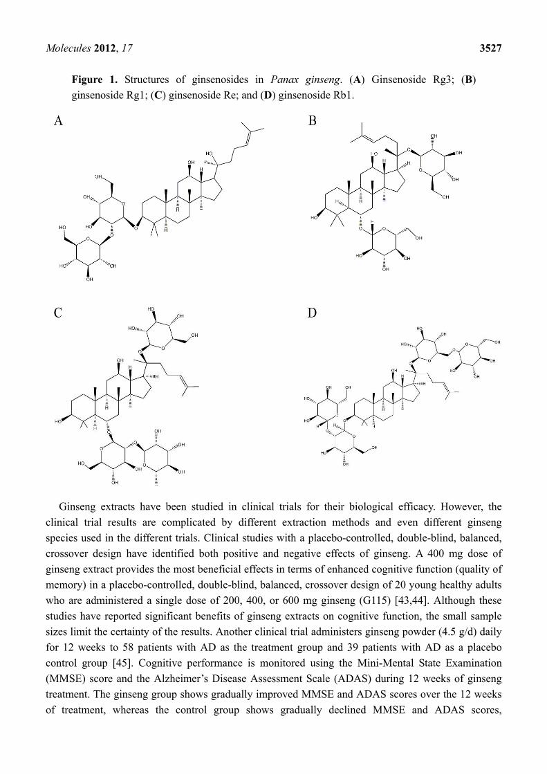
ginsenoside Rg3 (Figure 1A) significantly reduces the production of Aβ in CHO2B7 cells by 84% and
in Tg2576 transgenic mice by 31% [34]. Ginsenoside Rg3 reduces Aβ levels by promoting Aβ
degradation and by enhancing neprilysin gene expression, which is a rate-limiting enzyme in Aβ
degradation [35]. Furthermore, ginseng attenuates learning deficits in the damaged or aging brains of
rodents [34,36]. Additionally, ginsenoside Rg1 (Figure 1B) attenuates the amount of accumulated Aβ
and improves cognitive performance in a transgenic mouse model by activating the protein kinase
A/cAMP response element binding protein signaling pathway [37,38]. Ginsenoside Rg1 also reduces
Aβ production by modulating the APP process, which is accompanied by an improvement in cognitive
function [39,40].
Another ginsenoside in ginseng extract, ginsenoside Re (Figure 1C), protects PC12 cells against
Aβ-induced neurotoxicity [39]. In addition, ginsenoside Rb1 (Figure 1D) reverses Aβ-induced memory
loss in rats by attenuating neuroinflammation markers in the hippocampus [41]. Ginsenoside Rb1 also
exhibits beneficial effects on spatial learning by increasing synaptic density in the brain [42].
Molecules 2012, 17 3527
Figure 1. Structures of ginsenosides in Panax ginseng. (A) Ginsenoside Rg3; (B)
ginsenoside Rg1; (C) ginsenoside Re; and (D) ginsenoside Rb1.
Ginseng extracts have been studied in clinical trials for their biological efficacy. However, the
clinical trial results are complicated by different extraction methods and even different ginseng
species used in the different trials. Clinical studies with a placebo-controlled, double-blind, balanced,
crossover design have identified both positive and negative effects of ginseng. A 400 mg dose of
ginseng extract provides the most beneficial effects in terms of enhanced cognitive function (quality of
memory) in a placebo-controlled, double-blind, balanced, crossover design of 20 young healthy adults
who are administered a single dose of 200, 400, or 600 mg ginseng (G115) [43,44]. Although these
studies have reported significant benefits of ginseng extracts on cognitive function, the small sample
sizes limit the certainty of the results. Another clinical trial administers ginseng powder (4.5 g/d) daily
for 12 weeks to 58 patients with AD as the treatment group and 39 patients with AD as a placebo
control group [45]. Cognitive performance is monitored using the Mini-Mental State Examination
(MMSE) score and the Alzheimer’s Disease Assessment Scale (ADAS) during 12 weeks of ginseng
treatment. The ginseng group shows gradually improved MMSE and ADAS scores over the 12 weeks
of treatment, whereas the control group shows gradually declined MMSE and ADAS scores,

Molecules 2012, 17 3528
suggesting the beneficial effect of ginseng extracts on cognitive function and memory enhancement.
However, the beneficial effect of ginseng extract on memory declines gradually to the level of the
control group during a 12 week follow-up period without treatment.
2.2. Ginkgolides and Bilobalide from Gingko biloba L. (Ginkgoaceae)
Ginkgolides are a cyclic diterpenes of labdane type commonly isolated from G. biloba. EGb761,
extract of G. biloba leaves which contains 24% flavonoid glycosides, 6% terpenoids, and 5–10%
organic acids [46] has been extensively evaluated for its neuroprotective effects [47], and terpene
trilactones ginkgolides are the major pharmacologically active constituents in EGb761. For example,
pre-treatment of neuronal cells with ginkgolide A and B (Figure 2A,B) protects neuronal cells from
synaptic damage evaluated by the loss of synaptophysin, a presynaptic synaptic marker [48] and
increases neuronal survival against Aβ-induced toxicity [49]. Ginkgolide B rescues hippocampal neurons
from Aβ-induced apoptosis by increasing the production of brain-derived neurotrophic factor [50] and
reduces apoptotic death of neuronal cells in hemorrhagic rat brain [51]. In transgenic Caenorhabditis
elegans, ginkgolide A alleviates Aβ-induced adverse behavior including paralysis [52]. Ginkgolide B
reverses the Aβ-induced reduction of ACh release from hippocampal brain slices, suggesting potential
improvements in learning and memory deteriorated by Aβ [53]. Furthermore, Vitolo et al. reports that
ginkgolide J (Figure 2C) is the most potent inhibitor of Aβ-induced hippocampal neuronal cell death
among the ginkgolides in EGb761 [54]. Additionally, bilobalide (Figure 2D) reduces Aβ-induced
synaptic loss and subsequently enhances hippocampal neurogenesis and synaptogenesis [55].
Bilobalide also rescues chick embryonic neurons from apoptosis induced by serum deprivation or
staurosporine treatment [56,57].
Figure 2. Structures of ginkgolides and bilobalide in Gingko biloba. (A) Ginkgolide A;
(B) ginkgolide B; (C) ginkgolide J; and (D) bilobalide.

Molecules 2012, 17 3529
Despite these previous findings, regarding the neuroprotective effects of ginkgolides and
bilobalides in EGB761 and EGB761 itself, its clinical efficacy is inconsistent and remains
controversial. Therefore, clinical trials reflecting diverse races, dementia severity, and different doses
of EGb761 should be performed to evaluate the consistency of the effects of EGb761 against AD.
2.3. Cannabinoids from Cannabis sativa L. (Cannabaceae)
Cannabinoids are aromatic compounds containing a monoterpene moiety derived from isoprene
units, which are isolated from C. sativa in the Cannabaceae family. The diverse pharmacological
activities of cannabinoids are mediated by activating specific cannabinoid receptors including CB1 and
CB2 [58]. CB1 receptors, which mediate the psychoactive properties, are expressed in the nervous
system such as neurons and glial cells [59], whereas CB2 receptors are mainly located in immune
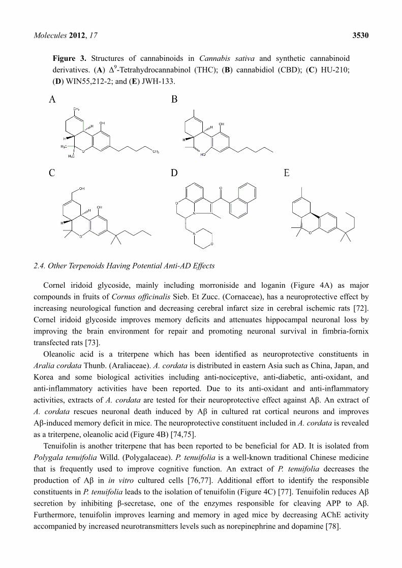
cells [60]. Δ9-Tetrahydrocannabinol (THC) (Figure 3A) mainly binds to CB1 receptors. This plant
contains about 60 cannabinoids, and one of the potential anti-AD agents is THC, the major
cannabinoid in C. sativa. THC is one of the widely-studied natural products and has anti-emetic,
anti-convulsive, anti-inflammatory, and analgesic effects [61]. A protective effect of THC against AD
has been reported. THC comparatively inhibits AChE and increases the availability of ACh. In addition,
THC reduces the inhibition of AChE-induced Aβ aggregation, and subsequently reduces Aβ-induced
toxicity [62] and is more efficient than commercially available AChE inhibitors such as tacrine and
donepezil. Furthermore, THC reduces behavioral and circadian disturbances in patients with severe
dementia [63].
Another major cannabinoid having neuroprotective effects against AD is cannabidiol (CBD)
(Figure 3B). CBD comprises about 40% of cannabis extracts and is the principle non-psychotrophic
constituent. The strong antioxidant effect of CBD provides neuroprotection by reducing oxidative
damage such as lipid peroxidation [64,65]. Furthermore, CBD alleviates Aβ-induced inflammatory
signals by reducing nitric oxide production by inhibiting p38 and nuclear factor-κB signaling
pathways [66]. Tau hyperphosphorylation, one of pathological hallmarks of AD, is also reduced
by CBD treatment, as it reduces glycogen synthase kinase-3β, an enzyme responsible for tau
hyperphosphorylation in patients with AD [67]. The neuroprotective effects of CBD have been
confirmed in an AD-mouse model induced with an intrahippocampal injection of Aβ (1–42) by a
reduction in glial activated pro-inflammatory mediators [68,69]. Because CBD lacks psychoactive
properties, it is one of the attractive potential anti-AD targets.
Additionally, some synthetic cannabinoids such as HU-210, WIN55, 212-2, and JWH-133
(Figure 3C–E), greatly reduce microglial activation and cytokine production in Aβ-administered rats.
Consequently, these synthetic compounds alleviate cognitive impairment by reducing the decrease in
neuronal marker levels [69–71]. These reports suggest that cannabinoids, particularly THC and CBD,
have potential to be developed as anti-AD therapeutics.
Molecules 2012, 17 3530
Figure 3. Structures of cannabinoids in Cannabis sativa and synthetic cannabinoid
derivatives. (A) Δ9-Tetrahydrocannabinol (THC); (B) cannabidiol (CBD); (C) HU-210;
(D) WIN55,212-2; and (E) JWH-133.
2.4. Other Terpenoids Having Potential Anti-AD Effects
Cornel iridoid glycoside, mainly including morroniside and loganin (Figure 4A) as major
compounds in fruits of Cornus officinalis Sieb. Et Zucc. (Cornaceae), has a neuroprotective effect by
increasing neurological function and decreasing cerebral infarct size in cerebral ischemic rats [72].
Cornel iridoid glycoside improves memory deficits and attenuates hippocampal neuronal loss by
improving the brain environment for repair and promoting neuronal survival in fimbria-fornix
transfected rats [73].
Oleanolic acid is a triterpene which has been identified as neuroprotective constituents in
Aralia cordata Thunb. (Araliaceae). A. cordata is distributed in eastern Asia such as China, Japan, and
Korea and some biological activities including anti-nociceptive, anti-diabetic, anti-oxidant, and
anti-inflammatory activities have been reported. Due to its anti-oxidant and anti-inflammatory
activities, extracts of A. cordata are tested for their neuroprotective effect against Aβ. An extract of
A. cordata rescues neuronal death induced by Aβ in cultured rat cortical neurons and improves
Aβ-induced memory deficit in mice. The neuroprotective constituent included in A. cordata is revealed
as a triterpene, oleanolic acid (Figure 4B) [74,75].
Tenuifolin is another triterpene that has been reported to be beneficial for AD. It is isolated from
Polygala tenuifolia Willd. (Polygalaceae). P. tenuifolia is a well-known traditional Chinese medicine
that is frequently used to improve cognitive function. An extract of P. tenuifolia decreases the
production of Aβ in in vitro cultured cells [76,77]. Additional effort to identify the responsible
constituents in P. tenuifolia leads to the isolation of tenuifolin (Figure 4C) [77]. Tenuifolin reduces Aβ
secretion by inhibiting β-secretase, one of the enzymes responsible for cleaving APP to Aβ.
Furthermore, tenuifolin improves learning and memory in aged mice by decreasing AChE activity
accompanied by increased neurotransmitters levels such as norepinephrine and dopamine [78].
Molecules 2012, 17 3531
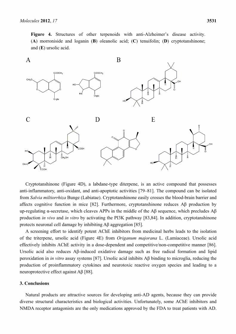
Figure 4. Structures of other terpenoids with anti-Alzheimer’s disease activity.
(A) morroniside and loganin (B) oleanolic acid; (C) tenuifolin; (D) cryptotanshinone;
and (E) ursolic acid.
Cryptotanshinone (Figure 4D), a labdane-type diterpene, is an active compound that possesses
anti-inflammatory, anti-oxidant, and anti-apoptotic activities [79–81]. The compound can be isolated
from Salvia miltiorrhiza Bunge (Labiatae). Cryptotanshinone easily crosses the blood-brain barrier and
affects cognitive function in mice [82]. Furthermore, cryptotanshinone reduces Aβ production by
up-regulating α-secretase, which cleaves APPs in the middle of the Aβ sequence, which precludes Aβ
production in vivo and in vitro by activating the PI3K pathway [83,84]. In addition, cryptotanshinone
protects neuronal cell damage by inhibiting Aβ aggregation [85].
A screening effort to identify potent AChE inhibitors from medicinal herbs leads to the isolation
of the triterpene, ursolic acid (Figure 4E) from Origanum majorana L. (Lamiaceae). Ursolic acid
effectively inhibits AChE activity in a dose-dependent and competitive/non-competitive manner [86].
Ursolic acid also reduces Aβ-induced oxidative damage such as free radical formation and lipid
peroxidation in in vitro assay systems [87]. Ursolic acid inhibits Aβ binding to microglia, reducing the
production of proinflammatory cytokines and neurotoxic reactive oxygen species and leading to a
neuroprotective effect against Aβ [88].
3. Conclusions
Natural products are attractive sources for developing anti-AD agents, because they can provide
diverse structural characteristics and biological activities. Unfortunately, some AChE inhibitors and
NMDA receptor antagonists are the only medications approved by the FDA to treat patients with AD.
Molecules 2012, 17 3532
Therefore, this review discussed some natural products and their molecular targets, particularly
terpenoids, which can be developed as potential anti-AD agents. The representative terpenoids with
anti-AD effects are ginsenosides from P. ginseng, ginkgolides and bilobalide from G. biloba, and
cannabinoids from C. sativa. The evaluation of biological activities by in vitro cell based assays and
in vivo animal studies indicate the beneficial effects of these compounds against AD. However, their
clinical efficacy is still controversial. Clinical trials should be designed to reflect diverse races,
dementia severity, and different doses of biologically active compounds.
Other compounds such as cornel iridoid glycoside, oleanolic acid, tenuifolin, cryptotanshinone and
ursolic acid have outstanding neuroprotective effects in in vitro assays. These compounds can exert
beneficial effects on central nervous system directly or indirectly by acting on peripheral targets.
Therefore, the methods to efficiently deliver the bioactive compounds to the brain should be
considered to develop terpenoids as anti-AD agents. In addition, the supply of large quantities of
biologically active compounds is essential to develop natural product-derived biologically active
compounds as therapeutic agents. To overcome this restriction, a mixture of two or three bioactive
compounds that act synergistically might be used as the alternative instead of a single compound.
Furthermore, a continuous search for bioactive compounds including terpenoids is expected to lead to
the discovery of therapeutic agents against AD from natural sources.
Conflict of Interest
The authors declare no conflict of interest.
Acknowledgments
This research was supported by the Korea Research Foundation Grant funded by the Korean
Government (KRF-2010-0003980) to S.-Y. Park.
References
1. Hebert, L.E.; Scherr, P.A.; Bienias, J.L.; Bennett, D.A.; Evans, D.A. Alzheimer disease in the US
population: Prevalence estimates using the 2000 census. Arch. Neurol. 2003, 60, 1119–1122.
2. Selkoe, D.J. The cell biology of beta-amyloid precursor protein and presenilin in Alzheimer’s
disease. Trends Cell. Biol. 1998, 8, 447–453.
3. Selkoe, D.J. Alzheimer’s disease: Genes, proteins, and therapy. Physiol. Rev. 2001, 81, 741–766.
4. Tanzi, R.E.; Bertram, L. Twenty years of the Alzheimer’s disease amyloid hypothesis: A genetic
perspective. Cell 2005, 120, 545–555.
5. Walsh, D.M.; Selkoe, D.J. A beta oligomers – a decade of discovery. J. Neurochem. 2007, 101,
1172–1184.
6. Kawahara, M.; Kuroda, Y. Molecular mechanism of neurodegeneration induced by Alzheimer’s
beta-amyloid protein: Channel formation and disruption of calcium homeostasis. Brain Res. Bull.
2000, 53, 389–397.

Molecules 2012, 17 3533
7. Lue, L.F.; Kuo, Y.M.; Roher, A.E.; Brachova, L.; Shen, Y.; Sue, L.; Beach, T.; Kurth, J.H.;
Rydel, R.E.; Rogers, J. Soluble amyloid beta peptide concentration as a predictor of synaptic
change in Alzheimer’s disease. Am. J. Pathol. 1999, 155, 853–862.
8. Roberson, E.D.; Mucke, L. 100 years and counting: Prospects for defeating Alzheimer’s disease.
Science 2006, 314, 781–784.
9. Heneka, M.T.; O’Banion, M.K. Inflammatory processes in Alzheimer’s disease. J. Neuroimmunol.
2007, 184, 69–91.
10. Yamamoto, N.; Matsubara, E.; Maeda, S.; Minagawa, H.; Takashima, A.; Maruyama, W.;
Michikawa, M.; Yanagisawa, K. A ganglioside-induced toxic soluble Abeta assembly. Its
enhanced formation from Abeta bearing the Arctic mutation. J. Biol. Chem. 2007, 282, 2646–2655.
11. Coulson, E.J. Does the p75 neurotrophin receptor mediate Abeta-induced toxicity in Alzheimer’s
disease? J. Neurochem. 2006, 98, 654–660.
12. de Felice, F.G.; Velasco, P.T.; Lambert, M.P.; Viola, K.; Fernandez, S.J.; Ferreira, S.T.;
Klein, W.L. Abeta oligomers induce neuronal oxidative stress through an N-methyl-D-aspartate
receptor-dependent mechanism that is blocked by the Alzheimer drug memantine. J. Biol. Chem.
2007, 282, 11590–11601.
13. Shankar, G.M.; Bloodgood, B.L.; Townsend, M.; Walsh, D.M.; Selkoe, D.J.; Sabatini, B.L.
Natural oligomers of the Alzheimer amyloid-beta protein induce reversible synapse loss by
modulating an NMDA-type glutamate receptor-dependent signaling pathway. J. Neurosci. 2007,
27, 2866–2875.
19. Magdesian, M.H.; Carvalho, M.M.; Mendes, F.A.; Saraiva, L.M.; Juliano, M.A.; Juliano, L.;
Garcia-Abreu, J.; Ferreira, S.T. Amyloid-beta binds to the extracellular cysteine-rich domain of
Frizzled and inhibits Wnt/beta-catenin signaling. J. Biol. Chem. 2008, 283, 9359–9368.
14. Nimmrich, V.; Grimm, C.; Draguhn, A.; Barghorn, S.; Lehmann, A.; Schoemaker, H.; Hillen, H.;
Gross, G.; Ebert, U.; Bruehl, C. Amyloid beta oligomers (A beta(1-42) globulomer) suppress
spontaneous synaptic activity by inhibition of P/Q-type calcium currents. J. Neurosci. 2008, 28,
788–797.
15. Mezler, M.; Barghorn, S.; Schoemaker, H.; Gross, G.; Nimmrich, V. Abeta oligomer directly
modulates P/Q-type calcium currents in Xenopus oocytes. Br. J. Pharmacol. 2012, 165, 1572–1583.
16. Kosik, K.S.; Joachim, C.L.; Selkoe, D.J. Microtubule-associated protein tau (tau) is a major
antigenic component of paired helical filaments in Alzheimer disease. Proc. Natl. Acad. Sci. USA
1986, 83, 4044–4048.
17. Kondo, J.; Honda, T.; Mori, H.; Hamada, Y.; Miura, R.; Ogawara, M.; Ihara, Y. The carboxyl third
of tau is tightly bound to paired helical filaments. Neuron 1988, 1, 827–834.
18. Iqbal, K.; Liu, F.; Gong, C.X.; Alonso Adel, C.; Grundke-Iqbal, I. Mechanisms of tau-induced
neurodegeneration. Acta Neuropathol. 2009, 118, 53–69.
20. Wallin, A.K.; Blennow, K.; Andreasen, N.; Minthon, L. CSF biomarkers for Alzheimer’s Disease:
levels of beta-amyloid, tau, phosphorylated tau relate to clinical symptoms and survival. Dement.
Geriatr. Cogn. Disord. 2006, 21, 131–138.
21. Busciglio, J.; Lorenzo, A.; Yeh, J.; Yankner, B.A. beta-amyloid fibrils induce tau phosphorylation
and loss of microtubule binding. Neuron 1995, 14, 879–888.
Molecules 2012, 17 3534
22. Greenberg, S.M.; Kosik, K.S. Secreted beta-APP stimulates MAP kinase and phosphorylation of
tau in neurons. Neurobiol. Aging 1995, 16, 403–407; discussion 407–408.
23. Takashima, A.; Honda, T.; Yasutake, K.; Michel, G.; Murayama, O.; Murayama, M.; Ishiguro, K.;
Yamaguchi, H. Activation of tau protein kinase I/glycogen synthase kinase-3beta by amyloid beta
peptide (25–35) enhances phosphorylation of tau in hippocampal neurons. Neurosci. Res. 1998,
31, 317–323.
24. Zheng, W.H.; Bastianetto, S.; Mennicken, F.; Ma, W.; Kar, S. Amyloid beta peptide induces tau
phosphorylation and loss of cholinergic neurons in rat primary septal cultures. Neuroscience 2002,
115, 201–121.
25. Leschik, J.; Welzel, A.; Weissmann, C.; Eckert, A.; Brandt, R. Inverse and distinct modulation of
tau-dependent neurodegeneration by presenilin 1 and amyloid-beta in cultured cortical neurons:
Evidence that tau phosphorylation is the limiting factor in amyloid-beta-induced cell death.
J. Neurochem. 2007, 101, 1303–1315.
26. Francis, P.T.; Palmer, A.M.; Sims, N.R.; Bowen, D.M.; Davison, A.N.; Esiri, M.M.; Neary, D.;
Snowden, J.S.; Wilcock, G.K. Neurochemical studies of early-onset Alzheimer’s disease. Possible
influence on treatment. N. Engl. J. Med. 1985, 313, 7–11.
27. Lipton, S.A. Pathologically-activated therapeutics for neuroprotection: Mechanism of NMDA
receptor block by memantine and S-nitrosylation. Curr. Drug Targets 2007, 8, 621–632.
28. Ferris, S.; Ihl, R.; Robert, P.; Winblad, B.; Gatz, G.; Tennigkeit, F.; Gauthier, S. Treatment effects
of Memantine on language in moderate to severe Alzheimer’s disease patients. Alzheimers
Dement. 2009, 5, 369–374.
29. Mecocci, P.; Bladstrom, A.; Stender, K. Effects of memantine on cognition in patients with
moderate to severe Alzheimer’s disease: Post-hoc analyses of ADAS-cog and SIB total and
single-item scores from six randomized, double-blind, placebo-controlled studies. Int. J. Geriatr.
Psychiatry 2009, 24, 532–538.
30. Newman, D.J.; Cragg, G.M. Natural products as sources of new drugs over the last 25 years.
J. Nat. Prod. 2007, 70, 461–477.
31. Wang, Y.; Huang, L.Q.; Tang, X.C.; Zhang, H.Y. Retrospect and prospect of active principles from
Chinese herbs in the treatment of dementia. Acta Pharmacol. Sin. 2010, 31, 649–664.
32. Liu, Z.Q.; Luo, X.Y.; Liu, G.Z.; Chen, Y.P.; Wang, Z.C.; Sun, Y.X. In vitro study of the
relationship between the structure of ginsenoside and its antioxidative or prooxidative activity in
free radical induced hemolysis of human erythrocytes. J. Agric. Food Chem. 2003, 51, 2555–2558.
33. Yun, T.K.; Lee, Y.S.; Lee, Y.H.; Kim, S.I.; Yun, H.Y. Anticarcinogenic effect of Panax ginseng
C.A. Meyer and identification of active compounds. J. Korean Med. Sci. 2001, 16, S6–S18.
34. Chen, F.; Eckman, E.A.; Eckman, C.B. Reductions in levels of the Alzheimer’s amyloid beta
peptide after oral administration of ginsenosides. FASEB J. 2006, 20, 1269–1271.
35. Yang, L.; Hao, J.; Zhang, J.; Xia, W.; Dong, X.; Hu, X.; Kong, F.; Cui, X. Ginsenoside Rg3
promotes beta-amyloid peptide degradation by enhancing gene expression of neprilysin. J. Pharm.
Pharmacol. 2009, 61, 375–380.
36. Zhao, R.; McDaniel, W.F. Ginseng improves strategic learning by normal and brain-damaged rats.
Neuroreport 1998, 9, 1619–1624.

Molecules 2012, 17 3535
37. Fang, F.; Chen, X.; Huang, T.; Luddy, J.S.; Yan, S.S. Multi-faced neuroprotective effects of
Ginsenoside Rg1 in an Alzheimer mouse model. Biochim. Biophys. Acta 2012, 1822, 286–292.
38. Shi, Y.Q.; Huang, T.W.; Chen, L.M.; Pan, X.D.; Zhang, J.; Zhu, Y.G.; Chen, X.C. Ginsenoside
Rg1 attenuates amyloid-beta content, regulates PKA/CREB activity, and improves cognitive
performance in SAMP8 mice. J. Alzheimers Dis. 2010, 19, 977–989.
39. Liang, W.; Ge, S.; Yang, L.; Yang, M.; Ye, Z.; Yan, M.; Du, J.; Luo, Z. Ginsenosides Rb1 and Rg1
promote proliferation and expression of neurotrophic factors in primary Schwann cell cultures.
Brain Res. 2010, 1357, 19–25.
40. Chen, L.M.; Lin, Z.Y.; Zhu, Y.G.; Lin, N.; Zhang, J.; Pan, X.D.; Chen, X.C. Ginsenoside Rg1
attenuates beta-amyloid generation via suppressing PPARgamma-regulated BACE1 activity in
N2a-APP695 cells. Eur. J. Pharmacol. 2012, 675, 15–21.
41. Wang, Y.; Liu, J.; Zhang, Z.; Bi, P.; Qi, Z.; Zhang, C. Anti-neuroinflammation effect of
ginsenoside Rbl in a rat model of Alzheimer disease. Neurosci. Lett. 2011, 487, 70–72.
42. Mook-Jung, I.; Hong, H.S.; Boo, J.H.; Lee, K.H.; Yun, S.H.; Cheong, M.Y.; Joo, I.; Huh, K.;
Jung, M.W. Ginsenoside Rb1 and Rg1 improve spatial learning and increase hippocampal
synaptophysin level in mice. J. Neurosci. Res. 2001, 63, 509–515.
43. Kennedy, D.O.; Scholey, A.B.; Wesnes, K.A. Dose dependent changes in cognitive performance
and mood following acute administration of Ginseng to healthy young volunteers. Nutr. Neurosci.
2001, 4, 295–310.
44. Reay, J.L.; Scholey, A.B.; Kennedy, D.O. Panax ginseng (G115) improves aspects of working
memory performance and subjective ratings of calmness in healthy young adults. Hum.
Psychopharmacol. 2010, 25, 462–471.
45. Lee, S.T.; Chu, K.; Sim, J.Y.; Heo, J.H.; Kim, M. Panax ginseng enhances cognitive performance
in Alzheimer disease. Alzheimer Dis. Assoc. Disord. 2008, 22, 222–226.
46. Le Bars, P.L. Magnitude of effect and special approach to Ginkgo biloba extract EGb 761® in
cognitive disorders. Pharmacopsychiatry 2003, 36, 44–49.
47. Shi, C.; Zhao, L.; Zhu, B.; Li, Q.; Yew, D.T.; Yao, Z.; Xu, J. Protective effects of Ginkgo biloba
extract (EGb761) and its constituents quercetin and ginkgolide B against beta-amyloid
peptide-induced toxicity in SH-SY5Y cells. Chem. Biol. Interact. 2009, 181, 115–123.
48. Bate, C.; Tayebi, M.; Williams, A. Ginkgolides protect against amyloid-beta1-42-mediated
synapse damage in vitro. Mol. Neurodegener. 2008, 3, 1.
49. Bate, C.; Salmona, M.; Williams, A. Ginkgolide B inhibits the neurotoxicity of prions or
amyloid-beta1-42. J. Neuroinflammation 2004, 1, 4.
50. Xiao, Q.; Wang, C.; Li, J.; Hou, Q.; Ma, J.; Wang, W.; Wang, Z. Ginkgolide B protects
hippocampal neurons from apoptosis induced by beta-amyloid 25-35 partly via up-regulation of
brain-derived neurotrophic factor. Eur. J. Pharmacol. 2010, 647, 48–54.
51. Hu, Y.Y.; Huang, M.; Dong, X.Q.; Xu, Q.P.; Yu, W.H.; Zhang, Z.Y. Ginkgolide B reduces
neuronal cell apoptosis in the hemorrhagic rat brain: Possible involvement of Toll-like receptor
4/nuclear factor-kappa B pathway. J. Ethnopharmacol. 2011, 137, 1462–1468.
52. Wu, Y.; Wu, Z.; Butko, P.; Christen, Y.; Lambert, M.P.; Klein, W.L.; Link, C.D.; Luo, Y.
Amyloid-beta-induced pathological behaviors are suppressed by Ginkgo biloba extract EGb 761
and ginkgolides in transgenic Caenorhabditis elegans. J. Neurosci. 2006, 26, 13102–13113.

Molecules 2012, 17 3536
53. Lee, T.F.; Chen, C.F.; Wang, L.C. Effect of ginkgolides on beta-amyloid-suppressed
acetylocholine release from rat hippocampal slices. Phytother. Res. 2004, 18, 556–560.
54. Vitolo, O.; Gong, B.; Cao, Z.; Ishii, H.; Jaracz, S.; Nakanishi, K.; Arancio, O.; Dzyuba, S.V.;
Lefort, R.; Shelanski, M. Protection against beta-amyloid induced abnormal synaptic function and
cell death by Ginkgolide J. Neurobiol. Aging 2009, 30, 257–265.
55. Tchantchou, F.; Lacor, P.N.; Cao, Z.; Lao, L.; Hou, Y.; Cui, C.; Klein, W.L.; Luo, Y. Stimulation of
neurogenesis and synaptogenesis by bilobalide and quercetin via common final pathway in
hippocampal neurons. J. Alzheimers Dis. 2009, 18, 787–798.
56. Ahlemeyer, B.; Mowes, A.; Krieglstein, J. Inhibition of serum deprivation- and staurosporine-induced
neuronal apoptosis by Ginkgo biloba extract and some of its constituents. Eur. J. Pharmacol. 1999,
367, 423–430.
57. Defeudis, F.V. Bilobalide and neuroprotection. Pharmacol. Res. 2002, 46, 565–568.
58. Howlett, A.C.; Barth, F.; Bonner, T.I.; Cabral, G.; Casellas, P.; Devane, W.A.; Felder, C.C.;
Herkenham, M.; Mackie, K.; Martin, B.R.; et al. International Union of Pharmacology. XXVII.
Classification of cannabinoid receptors. Pharmacol. Rev. 2002, 54, 161–202.
59. Herkenham, M.; Lynn, A.B.; Little, M.D.; Johnson, M.R.; Melvin, L.S.; de Costa, B.R.; Rice, K.C.
Cannabinoid receptor localization in brain. Proc. Natl. Acad. Sci. USA 1990, 87, 1932–1936.
60. Pertwee, R.G. Cannabinoid pharmacology: The first 66 years. Br. J. Pharmacol. 2006, 147,
S163–S171.
61. Carlini, E.A. The good and the bad effects of (−) trans-delta-9-tetrahydrocannabinol (Delta
9-THC) on humans. Toxicon 2004, 44, 461–467.
62. Eubanks, L.M.; Rogers, C.J.; Beuscher, A.E., IV; Koob, G.F.; Olson, A.J.; Dickerson, T.J.;
Janda, K.D. A molecular link between the active component of marijuana and Alzheimer’s disease
pathology. Mol. Pharm. 2006, 3, 773–777.
63. Walther, S.; Mahlberg, R.; Eichmann, U.; Kunz, D. Delta-9-tetrahydrocannabinol for nighttime
agitation in severe dementia. Psychopharmacology (Berl) 2006, 185, 524–528.
64. Hampson, A.J.; Grimaldi, M.; Axelrod, J.; Wink, D. Cannabidiol and (-)Delta9-tetrahydrocannabinol
are neuroprotective antioxidants. Proc. Natl. Acad. Sci. USA 1998, 95, 8268–8273.
65. Iuvone, T.; Esposito, G.; Esposito, R.; Santamaria, R.; di Rosa, M.; Izzo, A.A. Neuroprotective
effect of cannabidiol, a non-psychoactive component from Cannabis sativa, on beta-amyloid-induced
toxicity in PC12 cells. J. Neurochem. 2004, 89, 134–141.
66. Esposito, G.; de Filippis, D.; Maiuri, M.C.; de Stefano, D.; Carnuccio, R.; Iuvone, T. Cannabidiol
inhibits inducible nitric oxide synthase protein expression and nitric oxide production in
beta-amyloid stimulated PC12 neurons through p38 MAP kinase and NF-kappaB involvement.
Neurosci. Lett. 2006, 399, 91–95.
67. Esposito, G.; de Filippis, D.; Carnuccio, R.; Izzo, A.A.; Iuvone, T. The marijuana
component cannabidiol inhibits beta-amyloid-induced tau protein hyperphosphorylation through
Wnt/beta-catenin pathway rescue in PC12 cells. J. Mol. Med. (Berl) 2006, 84, 253–258.
68. Esposito, G.; Scuderi, C.; Savani, C.; Steardo, L., Jr.; de Filippis, D.; Cottone, P.; Iuvone, T.;
Cuomo, V.; Steardo, L. Cannabidiol in vivo blunts beta-amyloid induced neuroinflammation by
suppressing IL-1beta and iNOS expression. Br. J. Pharmacol. 2007, 151, 1272–1279.

Molecules 2012, 17 3537
69. Martin-Moreno, A.M.; Reigada, D.; Ramirez, B.G.; Mechoulam, R.; Innamorato, N.; Cuadrado, A.;
de Ceballos, M.L. Cannabidiol and other cannabinoids reduce microglial activation in vitro and
in vivo: Relevance to Alzheimer’s disease. Mol. Pharmacol. 2011, 79, 964–973.
70. Ramirez, B.G.; Blazquez, C.; Gomez del Pulgar, T.; Guzman, M.; de Ceballos, M.L. Prevention of
Alzheimer’s disease pathology by cannabinoids: Neuroprotection mediated by blockade of
microglial activation. J. Neurosci. 2005, 25, 1904–1913.
71. Martin Moreno, A.M.; Brera, B.; Spuch, C.; Carro, E.; Garcia-Garcia, L.; Delgado, M.; Pozo, M.A.;
Innamorato, N.G.; Cuadrado, A.; de Ceballos, M.L. Prolonged oral cannabinoid administration
prevents neuroinflammation, lowers beta-amyloid levels and improves cognitive performance in
Tg APP 2576 mice. J. Neuroinflamm. 2012, 9, 8.
72. Li, C.Y.; Li, L.; Li, Y.H.; Ai, H.X.; Zhang, L. Effects of extract from Cornus officinalis on nitric
oxide and NF-kappaB in cortex of cerebral infarction rat model. Zhongguo Zhong Yao Za Zhi
2005, 30, 1667–1670.
73. Zhao, L.H.; Ding, Y.X.; Zhang, L.; Li, L. Cornel iridoid glycoside improves memory ability and
promotes neuronal survival in fimbria-fornix transected rats. Eur. J. Pharmacol. 2010, 647, 68–74.
74. Cho, S.O.; Ban, J.Y.; Kim, J.Y.; Jeong, H.Y.; Lee, I.S.; Song, K.S.; Bae, K.; Seong, Y.H. Aralia
cordata protects against amyloid beta protein (25-35)-induced neurotoxicity in cultured neurons
and has antidementia activities in mice. J. Pharmacol. Sci. 2009, 111, 22–32.
75. Cho, S.O.; Ban, J.Y.; Kim, J.Y.; Ju, H.S.; Lee, I.S.; Song, K.S.; Bae, K.; Seong, Y.H.
Anti-ischemic activities of aralia cordata and its active component, oleanolic acid. Arch. Pharm.
Res. 2009, 32, 923–932.
76. Jia, H.; Jiang, Y.; Ruan, Y.; Zhang, Y.; Ma, X.; Zhang, J.; Beyreuther, K.; Tu, P.; Zhang, D.
Tenuigenin treatment decreases secretion of the Alzheimer’s disease amyloid beta-protein in
cultured cells. Neurosci. Lett. 2004, 367, 123–128.
77. Lv, J.; Jia, H.; Jiang, Y.; Ruan, Y.; Liu, Z.; Yue, W.; Beyreuther, K.; Tu, P.; Zhang, D. Tenuifolin,
an extract derived from tenuigenin, inhibits amyloid-beta secretion in vitro. Acta Physiol. (Oxf)
2009, 196, 419–425.
78. Zhang, H.; Han, T.; Zhang, L.; Yu, C.H.; Wan, D.G.; Rahman, K.; Qin, L.P.; Peng, C. Effects of
tenuifolin extracted from radix polygalae on learning and memory: A behavioral and biochemical
study on aged and amnesic mice. Phytomedicine 2008, 15, 587–594.
79. Kim, S.Y.; Moon, T.C.; Chang, H.W.; Son, K.H.; Kang, S.S.; Kim, H.P. Effects of tanshinone I
isolated from Salvia miltiorrhiza bunge on arachidonic acid metabolism and in vivo inflammatory
responses. Phytother. Res. 2002, 16, 616–620.
80. Ng, T.B.; Liu, F.; Wang, Z.T. Antioxidative activity of natural products from plants. Life Sci. 2000,
66, 709–723.
81. Park, E.J.; Zhao, Y.Z.; Kim, Y.C.; Sohn, D.H. PF2401-SF, standardized fraction of Salvia
miltiorrhiza and its constituents, tanshinone I, tanshinone IIA, and cryptotanshinone, protect
primary cultured rat hepatocytes from bile acid-induced apoptosis by inhibiting JNK
phosphorylation. Food Chem. Toxicol. 2007, 45, 1891–1898.
82. Kim, D.H.; Jeon, S.J.; Jung, J.W.; Lee, S.; Yoon, B.H.; Shin, B.Y.; Son, K.H.; Cheong, J.H.;
Kim, Y.S.; Kang, S.S.; et al. Tanshinone congeners improve memory impairments induced by
scopolamine on passive avoidance tasks in mice. Eur. J. Pharmacol. 2007, 574, 140–147.
Molecules 2012, 17 3538
83. Mei, Z.; Zhang, F.; Tao, L.; Zheng, W.; Cao, Y.; Wang, Z.; Tang, S.; Le, K.; Chen, S.; Pi, R.; et al.
Cryptotanshinone, a compound from Salvia miltiorrhiza modulates amyloid precursor protein
metabolism and attenuates beta-amyloid deposition through upregulating alpha-secretase in vivo
and in vitro. Neurosci. Lett. 2009, 452, 90–95.
84. Mei, Z.; Situ, B.; Tan, X.; Zheng, S.; Zhang, F.; Yan, P.; Liu, P. Cryptotanshinione upregulates
alpha-secretase by activation PI3K pathway in cortical neurons. Brain Res. 2010, 1348, 165–173.
85. Mei, Z.; Yan, P.; Situ, B.; Mou, Y.; Liu, P. Cryptotanshinione inhibits beta-amyloid aggregation
and protects damage from beta-amyloid in SH-SY5Y cells. Neurochem. Res. 2012, 37, 622–628.
86. Chung, Y.K.; Heo, H.J.; Kim, E.K.; Kim, H.K.; Huh, T.L.; Lim, Y.; Kim, S.K.; Shin, D.H.
Inhibitory effect of ursolic acid purified from Origanum majorana L on the acetylcholinesterase.
Mol. Cells 2001, 11, 137–143.
87. Heo, H.J.; Cho, H.Y.; Hong, B.; Kim, H.K.; Heo, T.R.; Kim, E.K.; Kim, S.K.; Kim, C.J.;
Shin, D.H. Ursolic acid of Origanum majorana L. reduces Abeta-induced oxidative injury.
Mol. Cells 2002, 13, 5–11.
88. Wilkinson, K.; Boyd, J.D.; Glicksman, M.; Moore, K.J.; El Khoury, J. A high content drug screen
identifies ursolic acid as an inhibitor of amyloid beta protein interactions with its receptor CD36.
J. Biol. Chem. 2011, 286, 34914–34922.
© 2012 by the authors; licensee MDPI, Basel, Switzerland. This article is an open access article
distributed under the terms and conditions of the Creative Commons Attribution license
(http://creativecommons.org/licenses/by/3.0/).