Tergal and Sternal Glands in Ants
47
PSYCHE Vol. 85 December, 1978 No. 4 TERGAL AND STERNAL GLANDS IN ANTS* BY BERT HOLLDOBLER AND HILTRUD ENGEL Department of Biology M CZ Laboratories, Harvard University Cambridge, Massachusetts INTRODUCTION Chemical signals, or pheromones as they are generally called, play a central role in the complex communication system of ant societies. During the last 20 years a number of exocrine glands have been identified as the anatomical sources for a diversity of pheromones which mediate sexual and social behavior in ants (for reviews see Wilson 1971, Blum 1977, H611dobler 1978). In recent years, however, several hitherto unknown exocrine glandular structures have been discovered in ants and the behavioral functions of some of them have already been determined. In this paper we will review these findings and will report the new results of our comparative morphological study of tergal and sternal glands in ants. MATERIAL AND METHODS For histological investigations live specimens were fixed in alco- holic Bouin (Dubosqu Brasil) or Carnoy (Romeis 1948), embedded in methyl methacrylate, and sectioned 8/ thick with a Jung Tetrander I microtome (Rathmayer 1962). The staining was Azao (Heidenhain). The SEM pictures were taken with an AMR 1000 A Scanning Electron Microscope. For some of the species which could only be identified to the generic level, the respective number is given of the voucher specimens, which are deposited in the ant collection of the MCZ (Harvard University). *Manuscript received by the editor May 3, 1979. 285
Transcript of Tergal and Sternal Glands in Ants
PSYCHEVol. 85 December, 1978 No. 4
TERGAL AND STERNAL GLANDS IN ANTS*
BY BERT HOLLDOBLER AND HILTRUD ENGEL
Department of BiologyMCZ Laboratories, Harvard University
Cambridge, Massachusetts
INTRODUCTION
Chemical signals, or pheromones as they are generally called, playa central role in the complex communication system of ant societies.During the last 20 years a number of exocrine glands have beenidentified as the anatomical sources for a diversity of pheromoneswhich mediate sexual and social behavior in ants (for reviews seeWilson 1971, Blum 1977, H611dobler 1978).
In recent years, however, several hitherto unknown exocrineglandular structures have been discovered in ants and the behavioralfunctions of some of them have already been determined. In thispaper we will review these findings and will report the new results ofour comparative morphological study of tergal and sternal glands inants.
MATERIAL AND METHODS
For histological investigations live specimens were fixed in alco-holic Bouin (Dubosqu Brasil) or Carnoy (Romeis 1948), embeddedin methyl methacrylate, and sectioned 8/ thick with a JungTetrander I microtome (Rathmayer 1962). The staining was Azao(Heidenhain). The SEM pictures were taken with an AMR 1000 AScanning Electron Microscope. For some of the species which couldonly be identified to the generic level, the respective number is givenof the voucher specimens, which are deposited in the ant collectionof the MCZ (Harvard University).
*Manuscript received by the editor May 3, 1979.
285
286 Psyche [December
RESULTS
Tergal glandsa. Pygidial gland
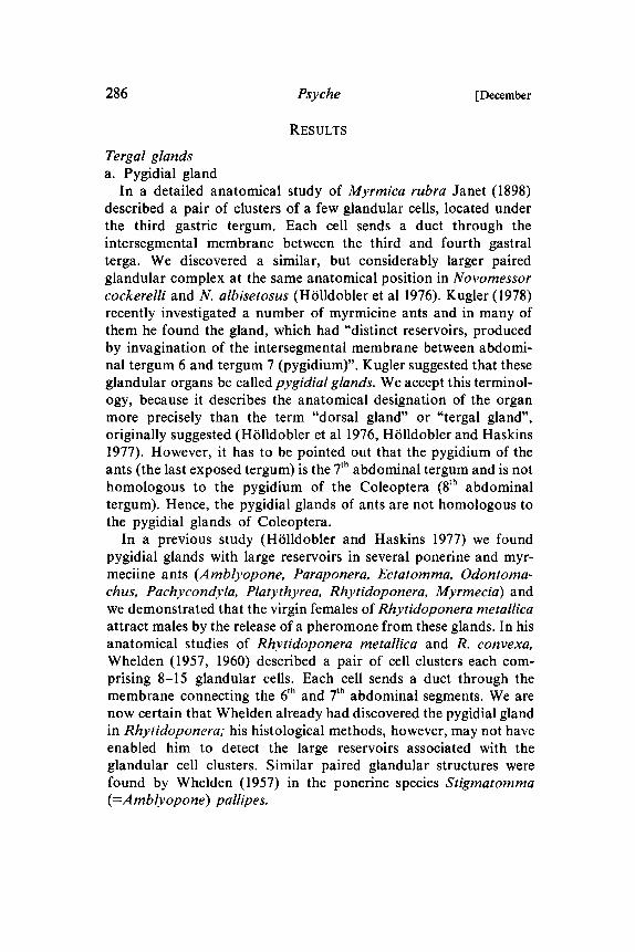
In a detailed anatomical study of Myrmica rubra Janet (1898)described a pair of clusters of a few glandular cells, located underthe third gastric tergum. Each cell sends a duct through theintersegmental membrane between the third and fourth gastralterga. We discovered a similar, but considerably larger pairedglandular complex at the same anatomical position in Novomessoreockerelli and N. albisetosus (H611dobler et al 1976). Kugler (1978)recently investigated a number of myrmicine ants and in many ofthem he found the gland, which had "distinct reservoirs, producedby invagination of the intersegmental membrane between abdomi-nal tergum 6 and tergum 7 (pygidium)". Kugler suggested that theseglandular organs be called pygidial glands. We accept this terminol-ogy, because it describes the anatomical designation of the organmore precisely than the term "dorsal gland" or "tergal gland",originally suggested (H611dobler et al 1976, H611dobler and Haskins1977). However, it has to be pointed out that the pygidium of theants (the last exposed tergum) is the 7th abdominal tergum and is nothomologous to the pygidium of the Coleoptera (8th abdominaltergum). Hence, the pygidial glands of ants are not homologous tothe pygidial glands of Coleoptera.
In a previous study (H611dobler and Haskins 1977) we foundpygidial glands with large reservoirs in several ponerine and myr-meciine ants (Amblyopone, Paraponera, Ectatomma, Odontoma-chus, Pachycondyla, Platythyrea, Rhytidoponera, Myrmecia) andwe demonstrated that the virgin females of Rhytidoponera metallicaattract males by the release of a pheromone from these glands. In hisanatomical studies of Rhytidoponera metallica and R. convexa,Whelden (1957, 1960) described a pair of cell clusters each com-prising 8-15 glandular cells. Each cell sends a duct through themembrane connecting the 6th and 7th abdominal segments. We arenow certain that Whelden already had discovered the pygidial glandin Rhytidoponera; his histological methods, however, may not haveenabled him to detect the large reservoirs associated with theglandular cell clusters. Similar paired glandular structures werefound by Whelden (1957) in the ponerine species Stigmatomma(=Amblyopone) pallipes.

1978] HOlldobler & Engel Glands in Ants 287
Finally, independently of our investigations, Maschwitz (pers.communication) found a pygidial gland in Leptogenys chinensis,which he called the "dorsal gland" (Maschwitz and SchiSnegge,1977).The new anatomical investigations presented in this paper reveal
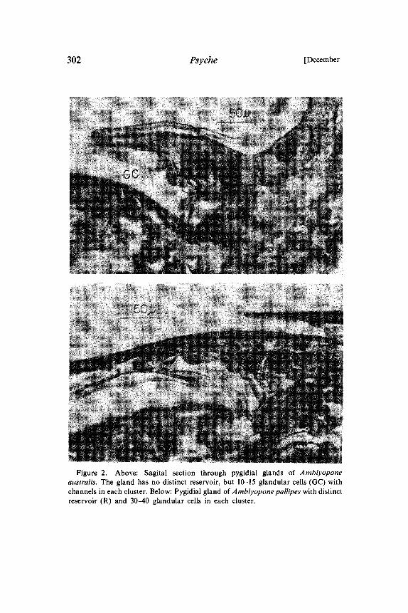
that the pygidial glands are much more common in ants thanpreviously assumed. Usually the organ consists of a pair of lateralclusters of glandular cells, each cell sending a duct through theintersegmental membrane between the 6th and 7th abdominal terga.Depending on the species, the intersegmental membrane can beinvaginated to different degrees, so that it can form a more or lessvoluminous reservoir (Fig. 1, 2, 3, 4). if no reservoir is present, theglandular structures can easily be missed during the dissection andhistological sectionings are therefore required to determine whetheror not the pygidial gland is present. As we have already indicated forNovomessor and as confirmed by Kugler (1978) for several othermyrmicine species, the pygidial gland can be associated with aspecial cuticular structure on the pygidium (7th tergum), (Fig. 5, 6,7). Our histological studies demonstrated, however, that the absenceof such structures does not necessarily indicate the absence of
Figure 1. Schematic illustration of glandular cells that send ducts through theintersegmental membrane. When the membrane is increasingly more invaginated, itforms an increasingly larger reservoir (a to c).
288 Psyche [December
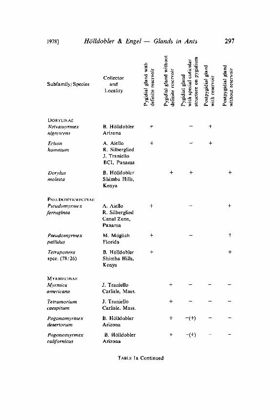
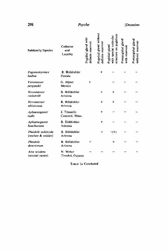
pygidial glands. Thus, in several myrmicine species, in which we(H611dobler et al 1976) and Kugler (1978) previously assumed thepygidial gland to be absent, we find that we now detect this organ byhistological methods. Tables a and b list the species of the majorsubfamilies that we investigated histologically and indicate the typeof tergal glands found.
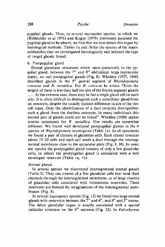
b. Postpygidial glandDorsal glandular, structures which open posteriorly to the py-
gidial gland, between the 7th and 8th abdominal terga (spiracularplate), we call postpygidial glands (Fig. 8). Whelden (1957, 1960)described glands in the 5th gastral segment of Rhytidoponeraconvexa and R. metallica. For R. convexa he writes: "Even thelargest of these is less than half the size of the fourth-segment glands
In the extreme case, there may be but a single gland cell on eachside. It is often difficult to distinguish such a unicellular gland froman oenocyte, despite the usually distinct difference in size of the twocell types. Only the identification of a duct certainly distinguishessuch a gland from the ductless oenocyte. In many individuals thissecond pair of glands could not be found". Whelden (1960) makessimilar statements for R. metallica. Our results are somewhatdifferent. We found well developed postpygidial glands in the 4species of Rhytidoponera investigated (Table a). In all specimenswe found a pair of clusters of glandular cells. Each cluster containsabout 15-20 cells and each cell sends a duct through the interseg-mental membrane close to the spiracular plate (Fig. 9, 10). In someant species the postpygidial gland consists of only a few glandularcells, in others the postpygidial gland is associated with a welldeveloped reservoir (Table la, lb).
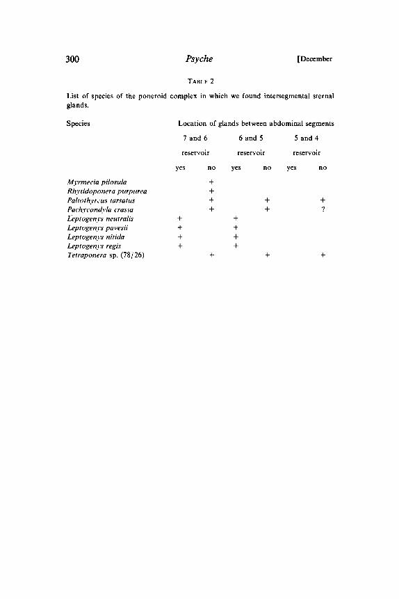
Sternal glandsIn several species we discovered intersegmental sternal glands
(Table 2). They can consist of a few glandular cells that send theirchannels through the intersegmental membrane, or of large clustersof glandular cells associated with voluminous reservoirs. Thesereservoirs are formed by invaginations of the intersegmental mem-branes (Fig. 8).
In several Leptogenys species (Fig. 12) we found two large sternalglands with reservoirs between the 7th and 6th, and 6th and 5th sterna.The latter glandular organ is usually associated with a specialcuticular structure on the 6th sternum (Fig. 12). In Paltothyreus
1978] HOlldobler & Engel- Glands in Ants 289
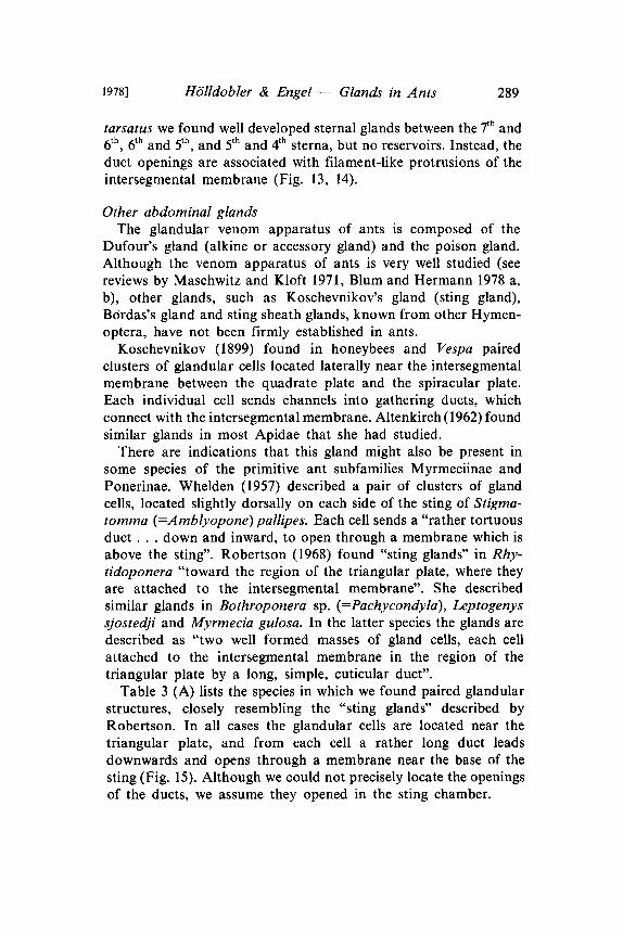
tarsatus we found well developed sternal glands between the 7th and6th, 6t and 5t, and 5t and 4t sterna, but no reservoirs. Instead, theduct openings are associated with filament-like protrusions of theintersegmental membrane (Fig. 13, 14).
Other abdominal glandsThe glandular venom apparatus of ants is composed of the
Dufour’s gland (alkine or accessory gland) and the poison gland.Although the venom apparatus of ants is very well studied (seereviews by Maschwitz and Kloft 1971, Blum and Hermann 1978 a,b), other glands, such as Koschevnikov’s gland (sting gland),Bordas’s gland and sting sheath glands, known from other Hymen-optera, have not been firmly established in ants.
Koschevnikov (1899) found in honeybees and Vespa pairedclusters of glandular cells located laterally near the intersegmentalmembrane between the quadrate plate and the spiracular plate.Each individual cell sends channels into gathering ducts, whichconnect with the intersegmental membrane. Altenkirch (1962) foundsimilar glands in most Apidae that she had studied.There are indications that this gland might also be present in
some species of the primitive ant subfamilies Myrmeciinae andPonerinae. Whelden (1957) described a pair of clusters of glandcells, located slightly dorsally on each side of the sting of Stigma-tomma (=Amblyopone)pallipes. Each cell sends a "rather tortuousduct.., down and inward, to open through a membrane which isabove the sting". Robertson (1968) found "sting glands" in Rhy-tidoponera "toward the region of the triangular plate, where theyare attached to the intersegmental membrane". She describedsimilar glands in Bothroponera sp. (=Pachycondyla), Leptogenyssjostedji and Myrmecia gulosa. In the latter species the glands aredescribed as "two well formed masses of gland cells, each cellattached to the intersegmental membrane in the region of thetriangular plate by a long, simple, cuticular duct".
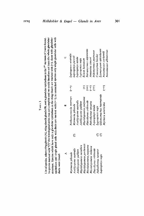
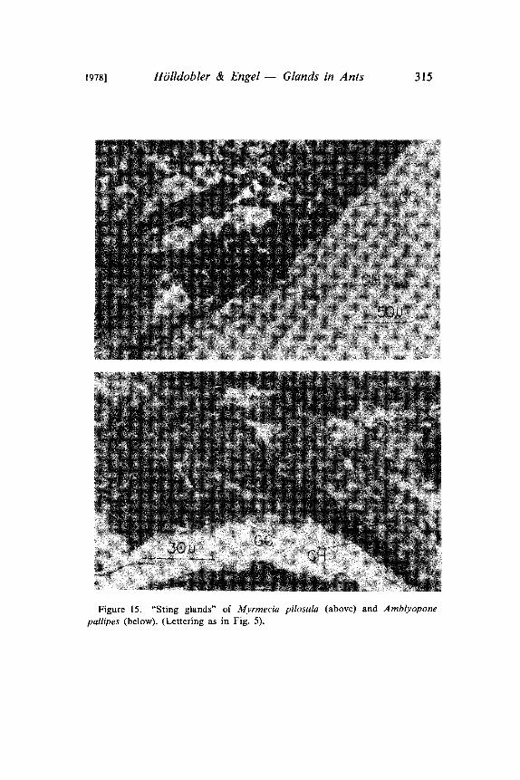
Table 3 (A) lists the species in which we found paired glandularstructures, closely resembling the "sting glands" described byRobertson. In all cases the glandular cells are located near thetriangular plate, and from each cell a rather long duct leadsdownwards and opens through a membrane near the base of thesting (Fig. 15). Although we could not precisely locate the openingsof the ducts, we assume they opened in the sting chamber.
290 Psyche [December
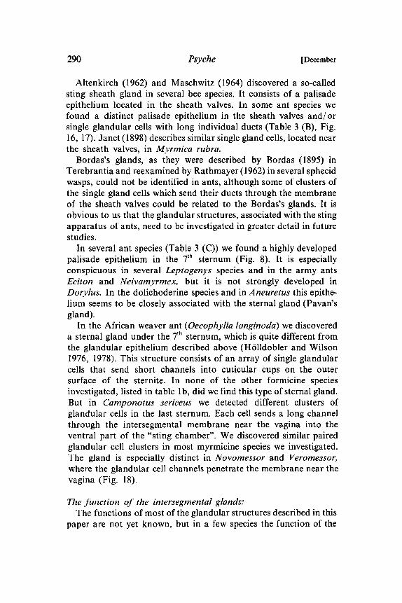
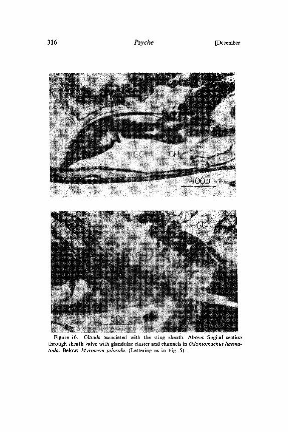
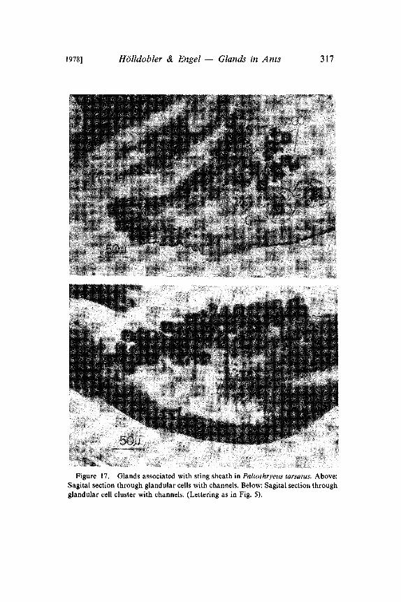
Altenkirch (1962) and Maschwitz (1964) discovered a so-calledsting sheath gland in several bee species. It consists of a palisadeepithelium located in the sheath valves. In some ant species wefound a distinct palisade epithelium in the sheath valves and/orsingle glandular cells with long individual ducts (Table 3 (B), Fig.16, 17). Janet (1898) describes similar single gland cells, located nearthe sheath valves, in Myrmica rubra.
Bordas’s glands, as they were described by Bordas (1895) inTerebrantia and reexamined by Rathmayer (1962) in several sphecidwasps, could not be identified in ants, although some of clusters ofthe single gland cells which send their ducts through the membraneof the sheath valves could be related to the Bordas’s glands. It isobvious to us that the glandular structures, associated with the stingapparatus of ants, need to be investigated in greater detail in futurestudies.
In several ant species (Table 3 (C)) we found a highly developedpalisade epithelium in the 7th sternum (Fig. 8). It is especiallyconspicuous in several Leptogenys species and in the army antsEciton and Neivamyrmex, but it is not strongly developed inDorylus. In the dolichoderine species and in Aneuretus this epithe-lium seems to be closely associated with the sternal gland (Pavan’sgland).
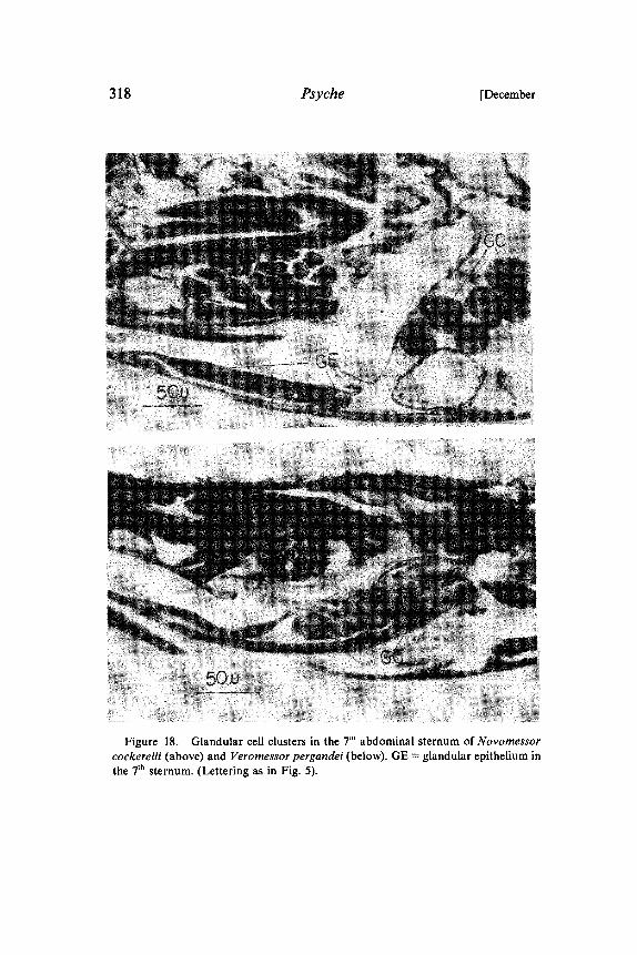
In the African weaver ant (Oecophylla longinoda) we discovereda sternal gland under the 7th sternum, which is quite different fromthe glandular epithelium described above (Htilldobler and Wilson1976, 1978). This structure consists of an array of single glandularcells that send short channels into cuticular cups on the outersurface of the sternite. In none of the other formicine speciesinvestigated, listed in table b, did we find this type of sternal gland.But in Camponotus sericeus we detected different clusters ofglandular cells in the last sternum. Each cell sends a long channelthrough the intersegmental membrane near the vagina into theventral part of the "sting chamber". We discovered similar pairedglandular cell clusters in most myrmicine species we investigated.The gland is especially distinct in Novomessor and Veromessor,where the glandular cell channels penetrate the membrane near thevagina (Fig. 18).
The function of the intersegmental glands:The functions of most of the glandular structures described in this
paper are not yet known, but in a few species the function of the
1978] HOlldobler & Engel Glands in Ants 291
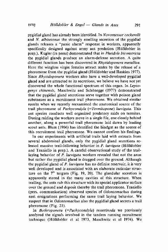
pygidial gland has already been identified. In Novomessor cockerelliand N. albisetosus the strongly smelling secretion of the pygidialglands releases a "panic alarm" response in workers, apparentlyspecifically designed against army ant predation (HiSlldobler inprep.). Kugler (in press) demonstrated that in Pheidole biconstrictathe pygidial glands produce an alarm-defense secretion. A quitedifferent function has been discovered in Rhytidoponera metallica.Here the wingless virgin females attract males by the release of apheromone from the pygidial gland (Htilldobler and Haskins 1977).Since Rhytidoponera workers also have a well-developed pygidialgland and are attracted to its secretions, we believe we have not yetdiscovered the whole functional spectrum of this organ. In Lepto-genys chinensis, Maschwitz and Sch6negge (1977) demonstratedthat the pygidial gland secretions serve together with poison glandsubstances as a recruitment trail pheromone. We obtained similarresults when we recently reexamined the anatomical source of thetrail pheromone of Pachycondyla (=Termitopone) laevigata. Thisant species conducts well organized predatory raids on termites.During raiding the workers move in a single file, one closely behindanother, along a powerful trail pheromone laid down by leadingscout ants. Blum (1966) has identified the hindgut as the source ofthis recruitment trail pheromone. We cannot confirm his findings.
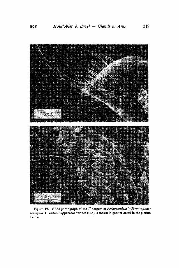
In our experiments with artificial trails laid with extracts fromseveral abdominal glands, only the pygidial gland secretions re-leased massive trail-following behavior in P. laevigata (Htilldoblerand Traniello in prep.). A careful observational study of the trail-laying behavior of P. laevigata workers revealed that not the anusbut rather the pygidial gland is dragged over the ground. Althoughthe pygidial gland of P. laevigata has no definite reservoir, it is verywell developed and is associated with an elaborate cuticular struc-ture on the 7th tergum (Fig. 19, 20). The glandular secretion isapparently stored in the many cavities of this structure. Whentrailing, the ants rub this structure with its special applicator surfaceover the ground and deposit thereby the trail pheromone. Traniello(pers. communication) observed species of Odontomachus duringnest emigrations performing the same trail laying behavior. Wesuspect that in Odontomachus also the pygidial gland secrets a trailpheromone (Fig. 21).
In Bothroponera (=Paehycondyla) tesserinoda we previouslyanalyzed the signals involved in the tandem running recruitmenttechnique (Htilldobler et al 1973, Maschwitz et al 1974). We
292 Psyche [December
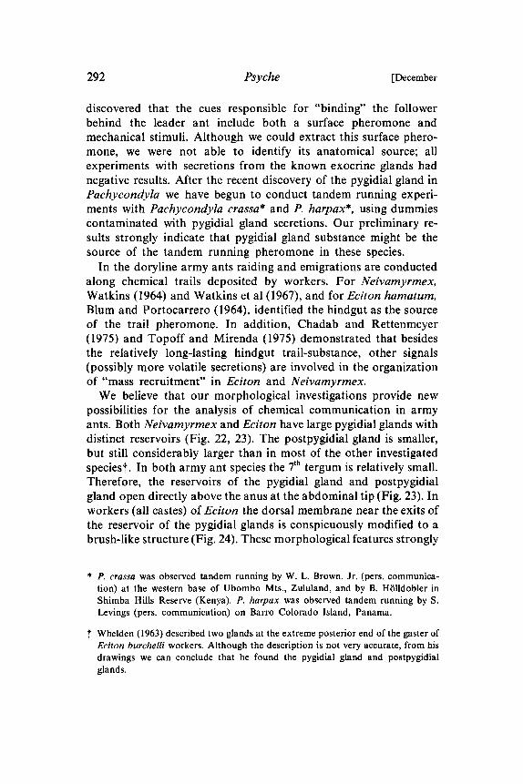
discovered that the cues responsible for "binding" the followerbehind the leader ant include both a surface pheromone andmechanical stimuli. Although we could extract this surface phero-mone, we were not able to identify its anatomical source; allexperiments with secretions from the known exocrine glands hadnegative results. After the recent discovery of the pygidial gland inPachycondyla we have begun to conduct tandem running experi-ments with Pachycondyla crassa* and P. harpax*, using dummiescontaminated with pygidial gland secretions. Our preliminary re-suits strongly indicate that pygidial gland substance might be thesource of the tandem running pheromone in these species.
In the doryline army ants raiding and emigrations are conductedalong chemical trails deposited by workers. For Neivamyrmex,Watkins (1964) and Watkins et al (1967), and for Eciton hamatum,Blum and Portocarrero (1964), identified the hindgut as the sourceof the trail pheromone. In addition, Chadab and Rettenmeyer(1975) and Topoff and Mirenda (1975) demonstrated that besidesthe relatively long-lasting hindgut trail-substance, other signals(possibly more volatile secretions) are involved in the organizationof "mass recruitment" in Eciton and Neivamyrmex.We believe that our morphological investigations provide new
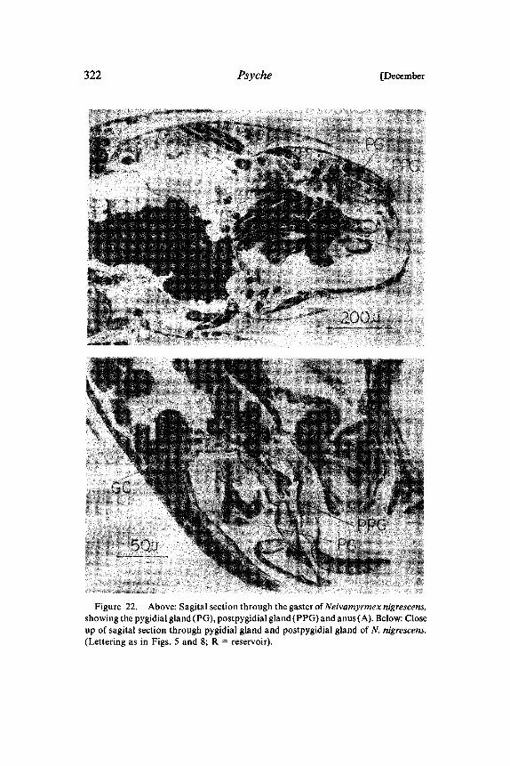
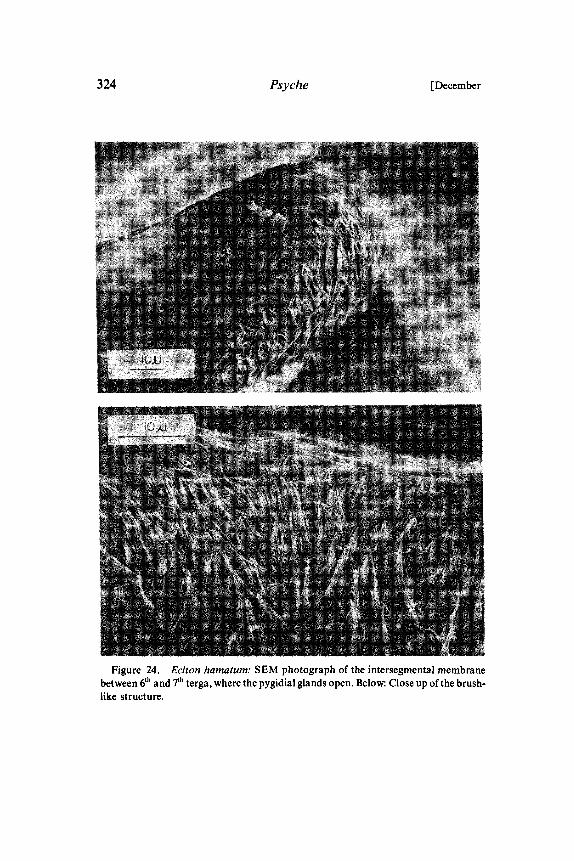
possibilities for the analysis of chemical communication in armyants. Both Neivamyrmex and Eciton have large pygidial glands withdistinct reservoirs (Fig. 22, 23). The postpygidial gland is smaller,but still considerably larger than in most of the other investigatedspecies’. In both army ant species the 7th tergum is relatively small.Therefore, the reservoirs of the pygidial gland and postpygidialgland open directly above the anus at the abdominal tip (Fig. 23). Inworkers (all castes) of Eciton the dorsal membrane near the exits ofthe reservoir of the pygidial glands is conspicuously modified to abrush-like structure (Fig. 24). These morphological features strongly
* P. crassa was observed tandem running by W. L. Brown, Jr. (pers. communica-tion) at the western base of Ubombo Mts., Zululand, and by B. H/511dotiler inShimba Hills Reserve (Kenya). P. harpax was observed tandem running by S.Levings (pers. communication) on Barro Colorado Island, Panama.
Whelden (1963) described two glands at the extreme posterior end of the gaster ofEciton burchelli workers. Although the description is not very accurate, from hisdrawings we can conclude that he found the pygidial gland and postpygidialglands.
1978] HOlldobler & Engel Glands in Ants 293
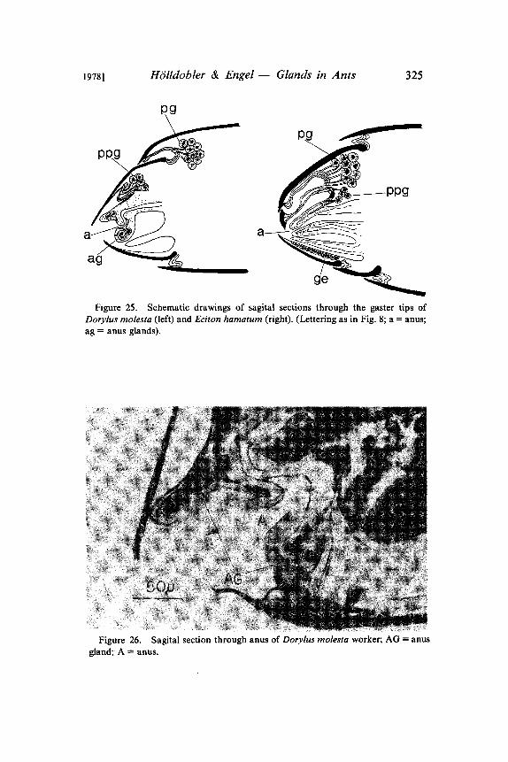
suggest that the tergal glands might be involved in the chemical trailcommunication of army ants. We have begun to test this hypothesiswith Eciton hamatum. The pygidial gland secretion of E. hamatumhas a strong, characteristic smell. The secretion is probably skatole(Traniello pers. communication), the substance that gives army antstheir typical "fecal odor". Recently Brown et al (1978) demonstratedthat skatole is an effective growth inhibitor for bacteria and fungiand repels insectivorous snakes (Watkins et al 1969). Our first,preliminary tests demonstrated that Eciton workers follow artificialtrails drawn with crushed pygidial glands. When we simultaneouslyoffered trails drawn with hindgut contents and pygidial glandsecretions, the latter were significantly preferred during the firstminute. When we used trails drawn with secretions of the poisongland or Dufour’s gland as controls, the ants always followed thepygidial gland trails. We have to stress, however, that theseexperiments must be considered pilot tests. The preliminary results,however, are striking enough to warrant a more detailed investiga-tion in the future. It is interesting to note that the anatomy of thepygidial gland in the African army ant, Dorylus molesta, is quitedifferent from that of Eciton and Neivamyrmex (Fig. 25). In thisspecies the 7th tergum is considerably larger than in species of thelatter genera, and the reservoirs of the pygidial glands do not openat the abdominal tip. In Dorylus, however, we found singleglandular cells with channels opening directly at the anus, a featurewe have not detected in other ant species (Fig. 25, 26).
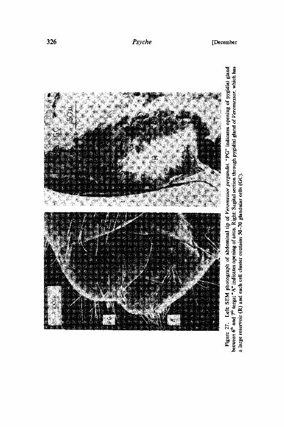
Finally, our morphological study of the pygidial gland of Ve-romessor pergandei has led to results that are suggestive of thefunction of this organ. In this species the 7th tergum is relativelysmall, and as a result the large reservoirs of the pygidial glands openat the tip of the gaster (Fig. 27). Veromessor forages in well-organized columns (Went et al 1972; Wheeler and Rissing 1975;Bernstein 1975). Several observations suggest that these foragingcolumns are organized by a trail pheromone, though no trailpheromone gland has yet been identified. Clearly, the large pygidialgland has to be considered as a possible source for the trailpheromone.The function of most of the newly discovered sternal glands is
unknown. Only in Paltothyreus tarsatus could we demonstrateexperimentally that foragers lay a recruitment trail with sternalgland secretions (HiSlldobler in prep.).

294 Psyche [December
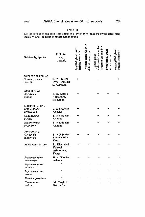
CONCLUSIONSSince we first found the pygidial gland widespread in the sub-
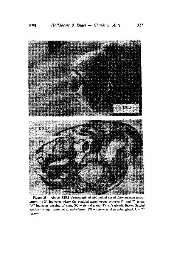
families Myrmeciinae and Ponerinae, we speculated that this glandmight be a primitive monophylogenetic trait in ants generally(Htilldobler and Haskins 1977). The results reported in the presentpaper fully confirm this assumption. A well-developed pygidialgland was found in the most primitive ant, Nothomyrmecia ma-crops, and in representatives of all major subfamilies except in theFormicinae (Table b). We agree with Kugler (1978) that the "analglands" cf the Dolichoderinae and Aneuretinae are homologous tothe pygidial glands of other ant subfamilies. Considering thevariation in the morphology of the pygidial glands, even within asingle subfamily, we think that the morphological variation of the"anal glands" of dolichoderine and aneuretine species does notwarrant a separate terminology. In fact the term "anal gland" ismisleading, because the glands do not exit from the anal opening ofthe gaster, as is sometimes inferred, but between the 6th and 7th
abdominal terga (Fig. 28). This was clearly demonstrated by Pavanand Ronchetti (1955). It is our view and also Kugler’s (pers.communication) that the "anal glands" should be called pygidialglands.Kugler (1978) concluded from his comparative studies of myrmi-
cine species that usually those species that have reduced or modifiedstings also have well-developed pygidial glands. He assumes that thepygidial gland replaces the sting apparatus as a chemical defensedevice. Our finding that well-developed pygidial glands occur inPogonomyrmex, a genus with a very effective sting apparatus, andin many stinging ponerine species, does not support Kugler’sconclusions.
ACKNOWLEDGEMENTSThis paper would not have been possible without the help of
many people. We would like to thank all the collectors mentioned inTable 1, including Donald W. Windsor, who helped finding theacacias in the Canal Zone. Special thanks to Robert W. Taylor, whosent us the precious Nothomyrmecia. Barry Bolton, William L.Brown, Jr., William H. Gotwald, Jr. and Roy Snelling identifiedmany species for us. We are grateful to Ed Seling for his superbassistance during the SEM work. Frank M. Carpenter’s manysuggestions improved the manuscript greatly. This work was sup-ported by NSF grant BNS 77-03884.

1978] H611dobler & Engel Glands in Ants 295
TABLE la
List of species of the poneroid complex (Taylor 1978) that were investigated histo-logically, and the types of tergal glands found. When the histological series wasincomplete and we could not make a definite statement, the column is marked with"?". When the cuticular structure on the pygidium was only slightly sculptured, wemarked the column with "-(+)".
Collectorand
LocalitySubfamily/Species
MYRMECIINAEMyrmeciapilosula
PONERINAEAmblyoponeaustralis
Amblyoponepallipes
Platythyreacribinoda
Rhytidoponerametallica
Rhytidoponeraperthensis
Rhytidoponerapurpurea
R. J. BartellR. W. TaylorBrindabellaRanges,Australia
C. P. HaskinsManjimup,W. Australia
J. TranielloCarlisle, Mass.
K. HortonR. SilbergliedShimba Hills,Kenya
C. P. HaskinsBlackwellRange,Queensland,Australia
C. P. HaskinsBoddington,W. Australia
C. P. HaskinsBlack Mountain,Kuranja,Queensland
+ +
+ +
+
+ +

296
Subfamily/Species
Rhytidoponeraviolacea
Paltothyreus
tarsatus
Pachycondylacrassa
Pachycondylalaevigata
Termitopone
Plectroctenastrigosa
Leptogenysneutralis
Leptogenyspavesii
Leptogenys
nitidia
Leptogenys
regis
Odontomachushaematoda
Psyche
Collectorand
Locality.x2_ " .x2_ "
C. P. HaskinsKings Park,W. Australia
B. H6ildoblerShimba Hills,
Kenya
B. H611doblerShimba Hills,Kenya
N. FranksJ. TranielloBCI, Panama
B. HtilldoblerShimba Hills,Kenya
C. P. HaskinsManjimup,W. Australia
[December
B. H611doblerShimba Hills,Kenya
B. HiSlldobler
Shimba Hills,Kenya
B. HiSlldobler
Shimba Hills,Kenya
C. P. HaskinsBCI, Panama
+ + ?
+ +
+ +
+ + +
+ + +
+ + +
+ + +
TABLE la Continued
1978] HOlldobler & Engel Glands in Ants
Subfamily/SpeciesCollector
andLocality
297
DORYLINAENeivamyrmexnigrescens
Ecitonhamatum
Dorylusmolesta
PSEUDOMYRMECINAEPseudomyrmexferruginea
Pseudomyrmexpallidus
Tetraponeraspec. (78 26)
MYRMECINAEMyrmicaamericana
Tetramoriumcaespitum
Pogonomyrmexdesertorum
Pogonomyrmexcalifornicus
B. H611doblerArizona
A. AielloR. SilbergliedJ. TranielloBCI, Panama
B. H611doblerShimba Hills,Kenya
A. Aiello +R. SilbergliedCanal Zone,Panama
M. MiSglich +Florida
B. H611dobler +Shimba Hills,Kenya
J. TranielloCarlisle, Mass.
J. TranielloCarlisle, Mass.
B. H/511dobler
Arizona
B. H611doblerArizona
+ +
+ +
/ +
+ -(+)
+ -(+)
TABLE la Continued
298
Subfamily/SpeciesCollector
andLocality
Psyche [December
Pogonomyrmexbadius
Veromessorpergandei
Novomessorcockerelli
Novomessoralbisetosus
Aphaenogasterrudis
Aphaenogasterhuachucana
Pheidole militicida(worker & soldier)
Pheidoledesertorum
A tta sexdens(several castes)
B. H611doblerFlorida
G. AlpertMexico
B. HiSlldoblerArizona
B. HiSlldoblerArizona
J. TranielloConcord, Mass.
B. H611doblerArizona
B. H611doblerArizona
B. H611doblerArizona
N. WeberTimehri, Guyana
-(+)
TABLE la Concluded
1978] HOlldobler & Engel Glands in Ants 299
TABLE b
List of species of the formicoid complex (Taylor 1978) that we investigated histo-logically, and the types of tergal glands found.
Subfamily/SpeciesCollector
andLocality
NOTHOMYRMECIINAENothomyrmeciamacrops
R. W. TaylorEyre PeninsulaS. Australia
+ +
ANEURETINAEAneurettsimoni
E. O. WilsonRatnapura,Sri Lanka
DOL1CHODERINAELiometopumapiculatum
Conomyrmabicolor
Iridomyrmexpruinosus
B. H6lldoblerArizona
B. HBlldoblerArizona
B. HBlldoblerArizona
FORM1CINAEOecopyllalonginoda
Pachycondyla spec.
Myrmecocystusmexicanus
Myrmecocystusmimicus
Myrmecocystusmendax
Formica perpilosa
Camponotussericeus
B. HiSlldoblerShimba Hills,KenyaR. SilbergliedNairobiArboretum,KenyaB. H611doblerArizona
M. M6glichSri Lanka
300 Psyche [December
TABLE 2
List of species of the poneroid complex in which we found intersegmental sternalglands.
Species Location of glands between abdominal segments
7and6 6and5 5and4
reservoir reservoir reservoir
Myrmecia pilosulaRhytidoponera purpureaPaitothyrcus tarsatus
Pach.vcondyla crassaLeptogenys neutralisLeptogenys pavesiiLeptogen.vs nitidaLeptogenys regisTetraponera sp. (78 26)
yes no yes no yes no
+++ + ++ + 9
+ ++ ++ ++ +
+ + +
1978] HOlldobler & Engel Glands in Ants 301
302 Psyche [December
Figure 2. Above: Sagital section through pygidial glands of Amblyoponeaustralis. The gland has no distinct reservoir, but 10-15 glandular cells (GC) withchannels in each cluster. Below: Pygidial gland of Amblyopone pallipes with distinctreservoir (R) and 30-40 glandular cells in each cluster.
1978] HOlldobler & Engel Glands in Ants 303
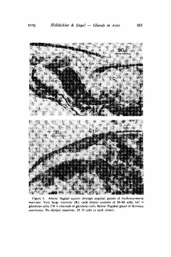
Figure 3. Above: Sagital section through pygidial glands of Nothomyrmeciamacrops: Very large reservoir (R); each cluster consists of 50-80 cells; GCglandular cells; CH channels of glandular cells. Below: Pygidial gland of Myrmicaamericana: No distinct reservoir, 10-15 cells in each cluster.
304 Psyche [December
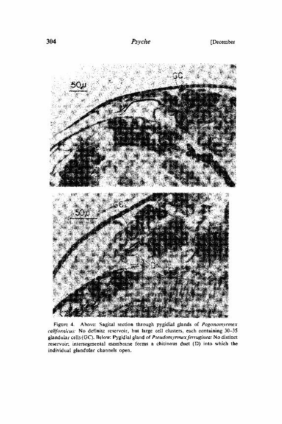
Figure 4. Above: Sagital section through pygidial glands of Pogonomyrmexcalifornicus: No definite reservoir, but large cell clusters, each containing 30-35glandular cells (GC). Below: Pygidial gland of Pseudomyrmexferruginea: No distinctreservoir; intersegmental membrane forms a chitinous duct (D) into which theindividual glandular channels open.
1978] H6lldobler & Engel Glands in Ants 305
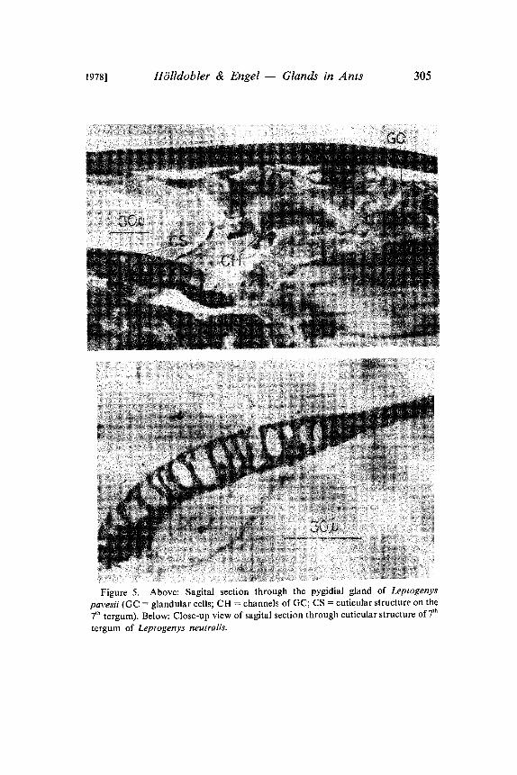
Figure 5. Above: Sagital section through the pygidial gland of Leptogenyspavesii (GC glandular cells; CH channels of GC; CS cuticular structure on the
7th tergum). Below: Close-up view of sagital section through cuticular structure of 7th
tergum of Leptogenys neutralis.
3O6 Psyche [December
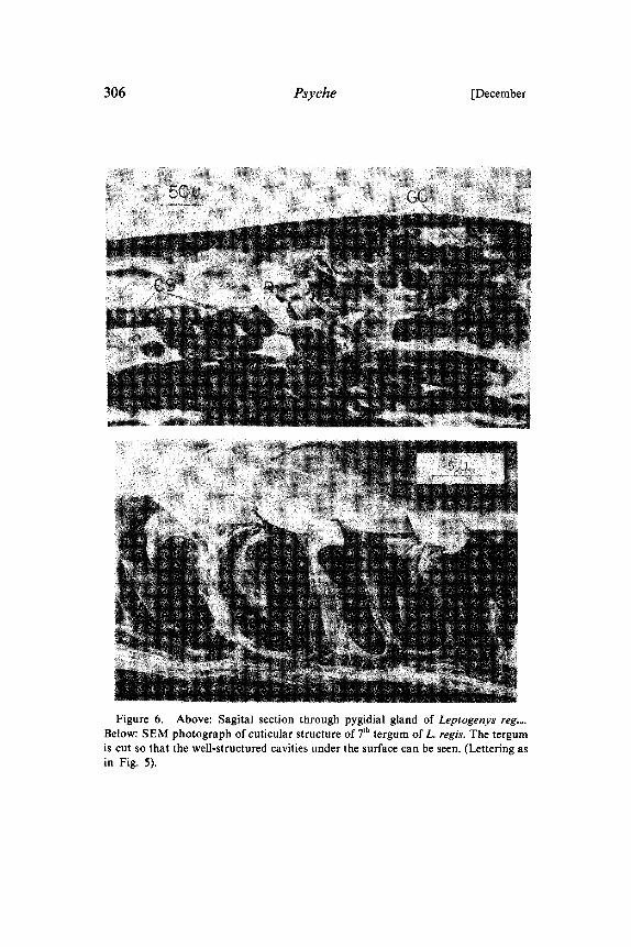
Figure 6. Above: Sagital section through pygidial gland of Leptogenys reg,o.Below: SEM photograph of cuticular structure of 7th tergum of L. regis. The tergumis cut so that the well-structured cavities under the surface can be seen. (Lettering asin Fig. 5).
1978] HOlldobler & Engel Glands in Ants 307
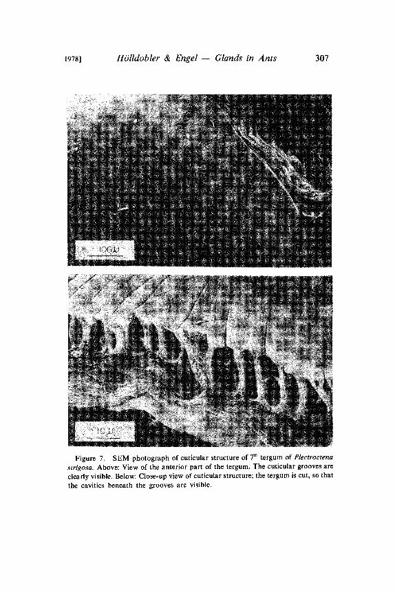
Figure 7. SEM photograph of cuticular structure of 7 tergum of Plectroctenastrigosa. Above: View of the anterior part of the tergum. The cuticular grooves are
clearly visible. Below: Close-up view of cuticular structure; the tergum is cut, so that
the cavities beneath the grooves are visible.
308 Psyche [December
PPg
ge
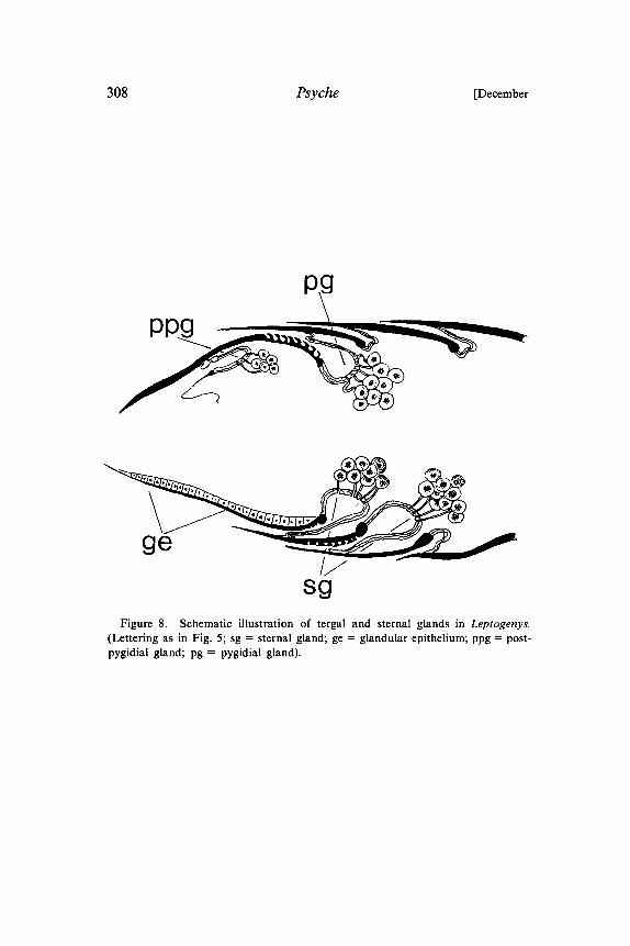
sFigure 8. Schematic illustration of tergal and sternal glands in Leptogenys.
(Lettering as in Fig. 5; sg sternal gland; ge glandular epithelium; ppg post-pygidial gland; pg pygidial gland).
1978] HOlldobler & Engel Glands in Ants 309
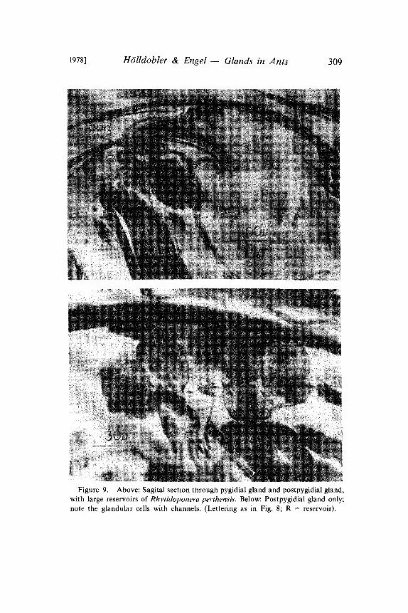
Figure 9. Above: Sagital section tlarougla pygidial gland and postpygidial gland,with large reservoirs of Rhytidoponera perthensis. Below: Postpygidial gland only;note the glandular cells with channels. (Lettering as in Fig. 8; R reservoir).
310 Psyche [December
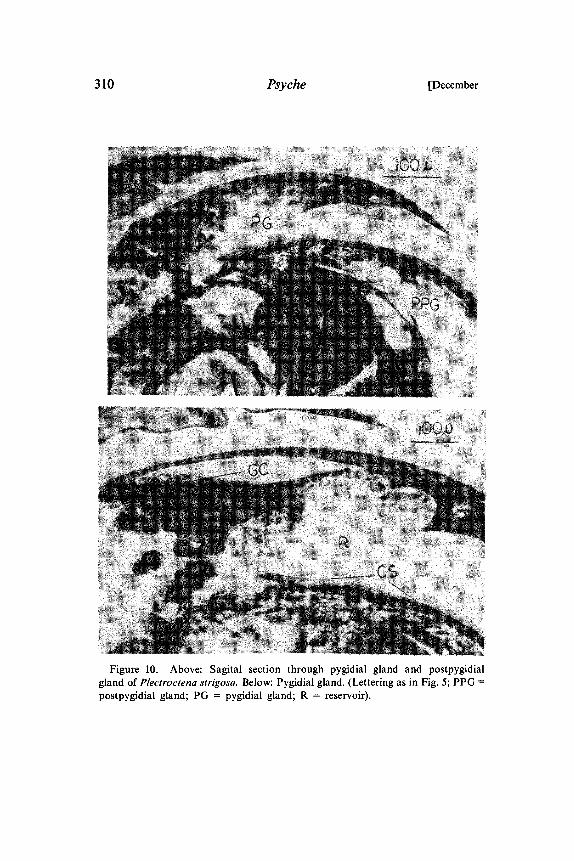
Figure 10. Above: Sagital section through pygidial gland and postpygidialgland of Plectroctena strigosa. Below: Pygidial gland. (Lettering as in Fig. 5; PPGpostpygidial gland; PG pygidial gland; R reservoir).
1978] H6lldobter & Engel Glands in Ants 311
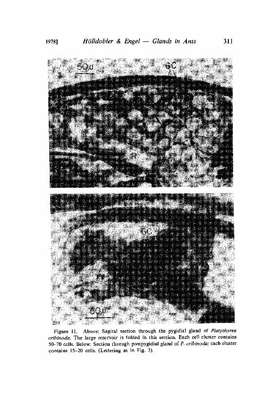
Figure 11. Above: Sagital section through the pygidial gland of Platythyrea
cribinoda. The large reservoir is folded in this section. Each cell cluster contains50-70 cells. Below: Section through postpygidial gland of P. cribinoda; each cluster
contains 15-20 cells. (Lettering as in Fig. 3).
312 Psyche [December
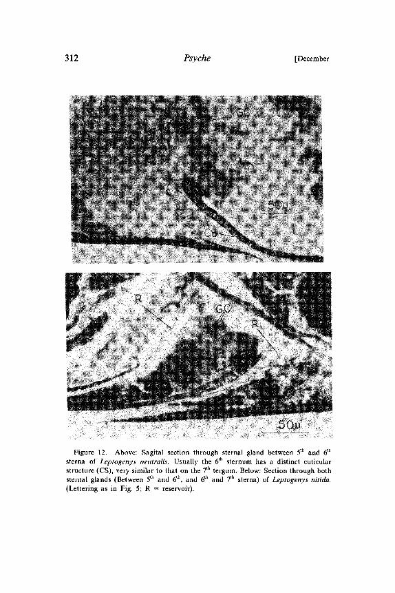
Figure 12. Above: Sagital section through sternal gland between 5th and 6th
sterna of Leptogenys neutralis. Usually the 6th sternum has a distinct cuticularstructure (CS), very similar to that on the 7th tergum. Below: Section through bothsternal glands (Between 5th and 6th, and 6th and 7th sterna) of Leptogenys nitida.(Lettering as in Fig. 5; R reservoir).
1978] H6lldobler & Engel Glands in Ants 313
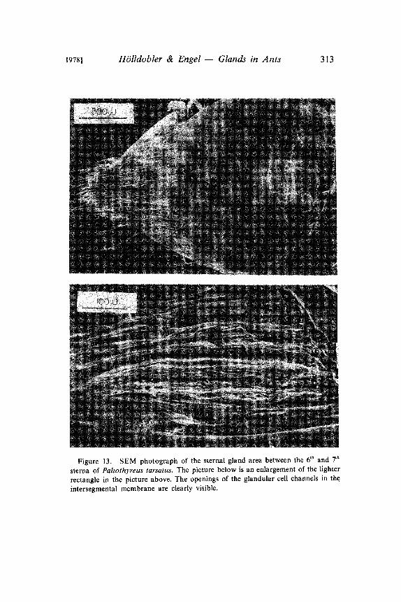
Figure 13. SEM photograph of the sternal gland area between the 6 and 7
sterna of Paltothyreus tarsatus. The picture below is an enlargement of the lighter
rectangle in the picture above. The openings of the glandular cell channels in theintersegmental membrane are clearly visible.
314 Psyche [December
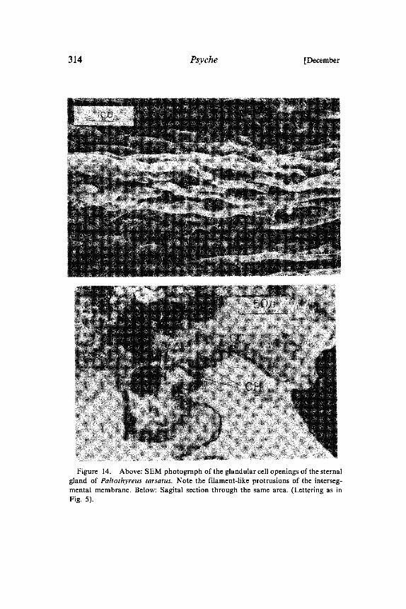
Figure 14. Above: SEM photograph of the glandular cell openings of the sternalgland of Paltothyreus tarsatus. Note the filament-like protrusions of the interseg-mental membrane. Below: Sagital section through the same area. (Lettering as inFig. 5).
1978] HOlldobler & Engel Glands in Ants 315
Figure 15. "Sting glands" of Myrmecia pilosula (above) and Amblyoponepallipes (below). (Lettering as in Fig. 5).
316 Psyche [December
Figure 16. Glands associated with the sting sheath. Above: Sagital sectionthrough sheath valve with glandular cluster and channels in Odontomachus haema-toda. Below: Myrmecia pilosula. (Lettering as in Fig. 5).
1978] HOlldobler & Engel Glands in Ants 317
Figure 17. Glands associated with sting sheath in Paltothryeus tarsatus. Above:Sagital section through glandular cells with channels. Below: Sagital section throughglandular cell cluster with channels. (Lettering as in Fig. 5).
318 Psyche [December
Figure 18. Glandular cell clusters in the 7 abdominal sternum of Novomessorcockerelli (above) and Veromessor pergandei (below). GE glandular epithelium inthe 7th sternum. (Lettering as in Fig. 5).
1978] HOlldobler & Engel Glands in Ants 319
Figure 19. SEM photograph of the 7th tergum of Pachycondyla (=Termitopone)laevigata. Glandular applicator surface (GA) is shown in greater detail in the picturebelow.
320 Psyche [December
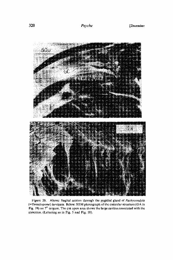
Figure 20. Above: Sagital section through the pygidial gland of Pachycondyla(=Termitopone) laevigata. Below: SEM photograph of the cuticular structure (GA inFig. 19) on 7th tergum. The cut open area shows the large cavities associated with thestructure. (Lettering as in Fig. 5 and Fig. 18).
1978] H6lldobler & Engel Glands in Ants 321
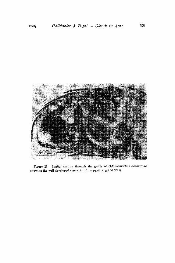
Figure 21. Sagital section through the gaster of Odontomachus haematoda,
showing the well developed reservoir of the pygidial gland (PG).
322 Psyche [December
Figure 22. Above: Sagital section through the gaster ofNeivamyrmexnigrescens,showing the pygidial gland (PG), postpygidial gland (PPG) and anus(A). Below: Closeup of sagital section through pygidial gland and postpygidial gland of N. nigrescens.(Lettering as in Figs. 5 and 8; R reservoir).

1978] HOlldobler & Engel Glands in Ants 323
Figure 23. Above: SEM photograph of tip of the gaster of Eciton hamatum.
Below: Sagital section through the same segments. (Lettering as in Fig. 8; S sting;GE glandular epithelium in 7th sternum).
324 Psyche [December
Figure 24. Eciton hamatum." SEM photograph of the intersegmental membranebetween 6th and 7th terga, where the pygidial glands open. Below: Close up of the brush-like structure.
19781 H6lldobler & Engel Glands in Ants 325
Figure 25. Schematic drawings of sagital sections through the gaster tips ofDorylus molesta (left) and Eciton hamatum (right). (Lettering as in Fig. 8" a anus;ag anus glands).
Figure 26. Sagital section through anus of Dorylus molesta worker; AG anus
gland; A anus.
326 Psyche [December
1978] HOlldobler & Engel Glands in Ants 327
Figure 28. Above: SEM photograph of abdominal tip of Liometopum apicu-latum. "PG" indicates where the pygidial gland opens between 6th and 7th terga."A" indicates opening of anus; SG sternal gland (Pavan’s gland). Below: Sagitalsection through gaster of L. apiculatum. PG reservoir of pygidial gland; 7. 7th
tergum.
328 Psyche [December
REFERENCES
ALTENKIRCH, G.1962. Untersuchungen fiber die Morphologie der abdominalen Hautdrtisen
einheimischer Apiden (Insecta, Hymenoptera) Zoologische Beitrige, 7,161-238.
BERNSTEIN, R. A.1974. Seasonal food abundance and foraging activity in some desert ants. The
American Naturalist, 108, 490-498.BLUM, M. S.
1966. The source and specificity of trail pheromones in Termitopone, Mono-morium and Huberia, and their relation to those of some other ants.Proceedings of the Royal Entomological Society of London, 41, 155-160.
1977. Behavioral responses of Hymenoptera to pheromones and allomones.Pp. 149-167 in Shorey, H. H. and J. J. McKelvey, Jr. (ed): Chemicalcontrol of insect behavior. John Wiley & Sons, New York 1977.
BLUM, M. S. AND H. R. HERMANN.1978a. Venoms and venom apparatuses of the Formicidae: Myrmeciinae,
Ponerinae, Dorylinae, Pseudomyrmecinae, Myrmicinae and Formici-nae. In: G. V. R. Born, O. Eichler, A. Farah, H. Heiken, A. D. Welch(ed). Handbook of Experimental Pharmacology, Springer-Verlag, Hei-delberg 1978, pp. 801-869.
BLUM, M. S. AND H. R. HERMANN.1978b. Venoms and venom apparatuses of the Formicidae: Dolichoderinae and
Aneuretinae. In: Handbook of Experimental Pharmacology, Springer-Verlag, pp. 871-894.
BLUM, M. S. AND C. m. PORTOCARRERO.1964. Chemical releasers of social behavior. IV. The hindgut as the source of
the odor trail pheromone in the neotropical army ant genus Eciton. Ann.Entomol. Soc. America 57, 793-794.
BORDAS, L.1895. Appareil glandulaire des Hymenopteres. Ann. Sci. Nat. Zool. 19, 289-
344.BROWN, C. A., J. F. WATKINS II AND D. W. ELDRIDGE.
1979. Depression of bacteria and fungi by the army ant secretion: Skatole.J. Kansas Entomol. Soc. 52, 119-122.
CHADAB, R. AND C. RETTENMEYER.1975. Mass recruitment by army ants. Science 188, 1124-1125.
HOLLDOBLER, B.1978. Ethological aspects of chemical communication in ants. Advances in the
Study of Behavior 8, 75-115.HOLLDOBLER, B., M. MOGLICH AND U. MASCHWITZ.
1973. Bothroponera tesserinoda (Formicidae): Tandemlauf beim Nestumzug.Encyclopaedia Cinematographica E 2040, pp. 3-14.
HOLLDOBLER, B., R. STANTON AND H. ENGEL.1976. A new exocrine gland in Novomessor (Hymenoptera: Formicidae) and
its possible significance as a taxonomic character. Psyche, 83, 32-41.
1978] HOlldobler & Engel Glands in Ants 329
HOLLDOBLER, B. AND C. P. HASKINS.1977. Sexual calling behavior in primitive ants. Science, 195, 793-794.
HOLLDOBLER, B. AND E. O. WILSON.1977. Weaver ants: Social establishment and maintenance of territory. Science,
195, 900-902.HOLLDOBLER, B. AND E. O. WILSON.
1978. The multiple recruitment systems of the African weaver ant Oecophyllalonginoda (Latreille) (Hymenoptera: Formicidae). Behav. Ecol. Socio-biol. 3, 19-60.
JANET, CH.1898. Etudes sur les Fourmis, les GuSpes et les Abeilles Note 17: SystSme
glandulaire tgumentaire de la Myrmica rubra. Observations diversessur les Fourmis. Paris, Georges Carr8 et C. Nand, Editeurs.
KOSCHEVNIKOV, G. A.1899. Zur Kenntnis der Hautdrtisen der Apidae und Vespidae. Anat. Anz. 15,
519-528.KUGLER, CH.
1978. Pygidial glands in the myrmicine ants (Hymenoptera, Formicidae).Insectes sociaux, 25, 267-274.
MASCHWITZ, U.1964. Gefahrenalarmstoffe und Gefahrenalarmierung bei sozialen Hymenop-
teren. Z. vergl. Physiol. 47, 596-655.MASCHWITZ, U., B. Ht3LLDOBLER AND M. MOGLICH.
1974. Tandemlaufen als Rekrutierungsverhalten bei Bothroponera tesserinodaForel (Formicidae: Ponerinae). Z. Tierpsychol. 35, 113-123.
MASCHWITZ, U. AND W. KLOFT.1971. Morphology and function of the venom apparatus of insects bees,
wasps, ants and caterpillars. In: Venomous animals and their venoms 3,1-60.
MASCHWlTZ, U. AND P. SCHONEGGE.1977. Recruitment gland of Leptogenys chinensis. Naturwissenschaften, 64,
589-590.PAVAN, M. AND G. RONCHETTI.
1955. Studi sulla morfologia esterna e anatomia interna dell’operaia di Iri-domyrmex humilis Mayr e ricerche chimiche e biologiche sulla iri-domirmecina. Atti della Societh Italiana di Scienze Naturali, Milano,94, 379-477.
RATHMAYER, W.1962. Methylmetacrylat als Einbettungsmedium fiir Insekten Experientia
(Basel) 18, 47-48.1962. Das Paralysierungsproblem beim Bienenwolf Philanthus triangulum
F. (Hym. Sphec.) Z. vergl. Physiol 45, 413-462.ROBERTSON, P. L.
1968. A morphological and functional study of the venom apparatus in repre-sentatives of some major groups of Hymenoptera. Aust. J. Zool. 16,133-166.
ROMEIS, B.1948. Mikroskopische Technik. Miinchen 1948.
330 Psyche [December
TAYLOR, R. W.1978. Nothomyrmecia macrops: A living-fossil ant rediscovered. Science 21}1,
979-985.TOPOFF, H. AND J. MIRENDA.
1975. Trail-following by the army ant Neivamyrmex nigrescens: Responses byworkers to volatile odors Ann. Entomol. Soc. Amer. 68, 1044-1046.
WATKINS, J. F.1964. Laboratory experiments on the trail following of army ants of the genus
Neivamyrmex (Formicidae: Dorylinae). J. Kansas Entomol. Soc. 37,22-28.
WATKINS, J. F., T. W. COLE AND R. S. BALDRIDGE.1967. Laboratory studies on interspecific trail following and trail preference of
army ants (Dorylinae). J. Kansas Entomol. Soc. 411, 146-151.WATKINS, J. F., F. R. GEHLBACH AND J. C. KROLL.
1969. Attractant-repellent secretions in blind snakes (Leptotyphlops dulcis)and army ants (Neivamyrmex nigrescens). Ecology 50, 1098-1102.
WENT, F. W., J. WHEELER AND G. C. WHEELER.1972. Feeding and digestion in some ants (Veromessor and Manica). Bio-
science 22, 82-88.WHEELER, J. AND S. W. RISSING.
1975. Natural history of Veromessor pergandei If. Behavior. The Pan-Pacific Entomologist 51., 303-314.
WHELDEN, R. M.1957. Notes on the anatomy of Rhytidoponera convexa Mayr ("violacea"
Forel) (Hymenoptera: Formicidae). Ann. Entomol. Soc. Amer. 51},271-282.
1957. Notes on the anatomy of the Formicidae I. Stigmatomma pallipes(Haldeman). J. New York Entomol. Soc. 65, 1-21.
1960. The anatomy of Rhytidoponera metallica F. Smith (Hymenoptera:Formicidae). Ann. Entmol. Soc. Amer. 53, 793-808.
1963. The anatomy of adult queen and workers of the army ants Ecitonburchelli Westwood and Eciton hamatum Fabricius. New YorkEntomol. Soc. 71, 90-115.
WILSON, E. O.1971. The insect societies. The Belknap Press of Harvard University Press,
Cambridge, Mass.

Submit your manuscripts athttp://www.hindawi.com
Hindawi Publishing Corporationhttp://www.hindawi.com Volume 2014
Anatomy Research International
PeptidesInternational Journal of
Hindawi Publishing Corporationhttp://www.hindawi.com Volume 2014
Hindawi Publishing Corporation http://www.hindawi.com
International Journal of
Volume 2014
Zoology
Hindawi Publishing Corporationhttp://www.hindawi.com Volume 2014
Molecular Biology International
GenomicsInternational Journal of
Hindawi Publishing Corporationhttp://www.hindawi.com Volume 2014
The Scientific World JournalHindawi Publishing Corporation http://www.hindawi.com Volume 2014
Hindawi Publishing Corporationhttp://www.hindawi.com Volume 2014
BioinformaticsAdvances in
Marine BiologyJournal of
Hindawi Publishing Corporationhttp://www.hindawi.com Volume 2014
Hindawi Publishing Corporationhttp://www.hindawi.com Volume 2014
Signal TransductionJournal of
Hindawi Publishing Corporationhttp://www.hindawi.com Volume 2014
BioMed Research International
Evolutionary BiologyInternational Journal of
Hindawi Publishing Corporationhttp://www.hindawi.com Volume 2014
Hindawi Publishing Corporationhttp://www.hindawi.com Volume 2014
Biochemistry Research International
ArchaeaHindawi Publishing Corporationhttp://www.hindawi.com Volume 2014
Hindawi Publishing Corporationhttp://www.hindawi.com Volume 2014
Genetics Research International
Hindawi Publishing Corporationhttp://www.hindawi.com Volume 2014
Advances in
Virolog y
Hindawi Publishing Corporationhttp://www.hindawi.com
Nucleic AcidsJournal of
Volume 2014
Stem CellsInternational
Hindawi Publishing Corporationhttp://www.hindawi.com Volume 2014
Hindawi Publishing Corporationhttp://www.hindawi.com Volume 2014
Enzyme Research
Hindawi Publishing Corporationhttp://www.hindawi.com Volume 2014
International Journal of
Microbiology