A Buffer Overflow Example João Paulo Magalhães ([email protected])April 2009.

TERESA MORUJO DE MAGALHÃES
NUTRITIONAL SUPPLEMENTATION WITH
MEDIUM CHAIN FATTY ACIDS IN DOGS WITH
COGNITIVE DYSFUNCTION SYNDROME: THE
TUTORS’ PERSPECTIVE
Supervisor: Professor Gonçalo da Graça Pereira
LUSÓFONA UNIVERSITY OF HUMANITIES AND
TECHNOLOGIES
FACULTY OF VETERINARY MEDICINE
LISBON
2015

1
DISSERTATION PRESENTED IN FULLFILMENT OF THE
REQUIREMENTS FOR THE DEGREE OF MASTERS IN
VETERINARY MEDICINE IN THE COURSE OF
INTEGRATED MASTER OF VETERINARY MEDICINE
GIVEN BY LUSÓFONA UNIVERSITY OF HUMANITIES
AND TECHNOLOGIES
WITH THE FOLLOWING ATTENDING JURY:
PRESIDENT: Professor Sofia Varela Van Haarten substituting
Professor Laurentina Pedroso
EXAMINER: Professor Joana Oliveira
SUPERVISOR: Professor Gonçalo da Graça Pereira
TERESA MORUJO DE MAGALHÃES
NUTRITIONAL SUPPLEMENTATION WITH
MEDIUM CHAIN FATTY ACIDS IN DOGS WITH
COGNITIVE DYSFUNCTION SYNDROME: THE
TUTORS’ PERSPECTIVE
LUSÓFONA UNIVERSITY OF HUMANITIES AND TECHNOLOGIES
FACULTY OF VETERINARY MEDICINE
LISBON
2015

2
To Polly,

3
Acknowledgements
To you, my dearest and best friend I ever had, Polly. A dog I had the pleasure to
know, love and grow up with. To say that you thought me the true meaning of selfless love
and respect of other beings is the least. I truly believe I would not have made this journey if
I had not met you. Thank you. Sincerely. “Saudade” is the only word I have to describe how
I feel not having you around every day. And sorrow is the one that describes how it feels
not to have you to celebrate this day with.
To my parents, the ones that truly made this dream possible and the ones who
would give it all up if it meant the pathway to my happiness. To my grandparents, being
parents two times is twice the work. To all that you thought me.
To my family, thank you for the support, laughter and tears we all shares
throughout the years. To Sandra, that we can both grow from the all the fights and, of
course from all the nights out and all the laughter we shared.
To my emotional family, Fátima, Inês and Zé. You are more than I could ask for.
To all the animals I have the pleasure to call family to, Chico, Eddie, ESA and
Oliver. And to the ones who were family even if only for a period of time Podenga, Camila,
Clotilde, Tofu, Dragão and the litters. You are the reason I chose to study Veterinary
Medicine.
To my, from ever and forever friend Margarida who always supported me, even
when I did not have the nicest things to say. And that we keep exploring the world together.
To my school friends Catarina, Cátia e Sara. What happens in our friendship stays
in our friendship. Thank you. To the beast teacher I ever had, Leonor.
To my “charruas” Pitacas e Ricardo, Verónica e Filipe, Nana e Pedro, Girabolhos,
Duarte, Tita, Cindy, Cláudia, Carla-da-Ota, Zé and Simões. You have seen me in my
wildest and stuck with me throughout the years.
To my college friends, Filipa without whom my life would be a lot more bland,
you are a big part of me, Catarina a woman I would trust my life to, Andreia, Patrícia and
Nuno. You made horrible times seem more pleasant. And awesome times perfect.
4
To Dr Gonçalo da Graça Pereira, you were not only a coordinator. You are a
mentor and a supporter that not only aims for my best but tries to bring confidence along
with it. And patience, a lot of patience. Thank you.
To my teachers who always thought me aiming for me to become a better
veterinarian. A special thanks to Ana Lúcia, Carla Sousa, Débora Araújo for the support. To
Castanha, a very special dog who endured a lot.
To all the LPDA team for letting me in your journey so early. You are a true
inspiration. Carla for being so crazy and so loud around cats, Célia for being so peaceful,
confident and wise. Marta for being such a huggable person – but makes up for it in
intelligence and sense of humor, Mónica who always as a word to say, even to yoghurt,
Teresa for me to have hope for myself since I see a lot of me in you, Patrícia for always
supporting the interns. To Pedro, for all the talks and funny moments. Started internship as
a colleague ended as a friend.
To all VetOeiras team who chose to teach me and show me top notch medicine. To
Dr Luís and Dr Rui who not only made the experience possible but also supported me
throughout the time. To Carina a vet who became a friend, a special thanks for always
believing in my capabilities and encouraging me to pursue my dreams. To Mariana longing
for the day I know half of what you do. To Elise for all the support and talks. To Gonçalo
for all the laughter we shared. To Mihaela who thought patience comes along with laughter.
To Ana, Ana Lúcia, Cláudia, Diogo, Jeni, Joana, Leonor, Lurdinhas, Paula e Sílvia. To my
colleagues Sónia and Mariana, my dream team. Thank you for listening to me even when I
was moody in the rounds or running away from surgery. To Tejo, a dog who stuck close to
my heart.
To all the animals whose lives are not respected. I cannot help you all, but I will try
to change the lives of the ones who cross my path.
To Dra Inês Viegas and Nestlé Purina®
for making this possible.

5
Resumo
A disfunção cognitiva canina é uma síndrome neurocomportamental na qual os animais
experienciam déficits contínuos na aprendizagem, memória e orientação espacial resultando
num declínio cognitivo gradual com crescente patologia cerebral. Apesar da prevalência
estimada desta síndrome variar entre 22,5% e 73,5%, a maioria destes casos não é tratada
desde cedo já que os tutores tendem a relacionar as alterações comportamentais dos seus
animais com o processo fisiológico do envelhecimento.
O estudo apresentado de seguida, tem como objectivo, averiguar se as alterações
comportamentais subsequentes de uma suplementação oral com ácidos gordos de cadeia
média será perceptível pelos tutores de uma população de cães urbanos.
Os efeitos de melhoramento cognitivo dos ácidos gordos de cadeia média estão bem
documentados e reportados na literatura. Contudo, quando estudando uma população de
cães geriátricos com alterações comportamentais subtis, não se verificam, na literatura,
estudos sobre a avaliação dos tutores do comportamento dos seus animais.
Quando se submeteram as variáveis do perído de estudo a testes de χ2, uma Taxa de
resposta global ao tratamento foi criada. Os resultados indicam melhoria de sinais clínicos
em 8,04% da população e agravamento dos mesmos em 5,36% dos casos. A maioria da
população de estudo, 86,61%, apresentou manutenção dos sinais clínicos de início de estudo
não tendo sofrido agravamento nem melhoria dos mesmos.
Palavras Chave: Disfunção cognitiva canina (DCC), comportamental, ácidos gordos de
cadeia média (AGCM), suplemento, tutor
6
Abstract
Canine cognitive dysfunction syndrome is a neurobehavioural syndrome in which
the animals experience ongoing deficits in learning, memory and spatial awareness resulting
in a gradual cognitive decline with increasing brain pathology. Although the estimated
prevalence of this syndrome in a population ranges between 22.5% and 73.5%, the majority
of those cases is not early addressed since the tutors tend to relate the behavioural changes
with the normal process of ageing.
The following study aimed to see if the behavioural changes subsequent of an oral
supplementation with medium chain fatty acids would be noticeable by the tutors of a
population of urban dogs.
The effects of medium chain fatty acids on cognition are well described in the
literature but not when addressing a population of geriatric dogs with an early onset of
behavioural changes in the tutors perspective.
When subjecting the variables throughout the trial period to χ2 tests a global
response to therapy rate was created. The results indicated an improvement in 8,04% of the
cases while a worsening in 5,36%. The majority of the study group, 86,61% considered that
the animals had not improved nor worsened their behavioural changes.
Key words: Canine cognitive dysfunction syndrome (CDS), behavioural, medium
chain fatty acids (MCTs), supplementation, tutors

7
Resumo das secções em português
Introdução
A disfunção cognitiva canina – DCC - é descrita como alguma alteração comportamental
em diferentes categorias como interacção social, comportamento de eliminação, ciclo do
sono e orientação (Overall, 2013). É uma síndrome neurocomportamental na qual o animal
experiencia deficits na aprendizagem, memória e orientação espacial contínuas e crescentes,
com exclusão de outras causas médicas para os mesmos sintomas (Landsberg, Hunthausen
& Ackerman, 2013 ; Overall, 2013).
O estudo desta síndrome, nos animais, começou para dar resposta a questões sobre
o Alzheimer onde, os animais estudados, eram usados como modelo para a doença de
medicina humana sendo todos estudos laboratoriais. Os primeiros artigos que focavam os
estudo da doença nos animais datam dos anos noventa (Cummings & Cotman, 1995). Desde
então, vários estudos foram elaborados com a finalidade de estudar esta doença como um
problema nos animais e, de maneira a, melhorar a sua qualidade de vida (Salvin, McGreevy,
Sachdev & Valenzuela, 2010).
Nem sempre esta síndrome é tratada desde o início da sua manifestação pois os tutores
tendem a relacionar as alterações comportamentais manifestadas como parte natural do
processo de envelhecimento (Neilson, Hart, Cliff & Ruehl, 2001).
O envelhecimento fisiológico cerebral do cérebro inclui determinadas alterações
tais como uma redução na massa cortical acompanhada com atrofia dos gânglios basais,
aumento do tamanho ventricular, calcificação meningeal, diminuição do número de
neurónios funcionais e aumento da sua degeneração (Borràs, Ferrer & Pumarola, 1999). Já
está comprovada uma correlação positiva entre atrofia cortical e disfunção cognitiva
(Rofina, Ederen, Toussaint, Secrève, van der Spek, van der Meer, et al., 2006). Existem
outras alterações que podem justificar a apresentação clínica da Síndrome de Disfunção
Cognitiva que incluem a insuficiência vascular cerebral por fibrose das paredes vasculares,
proliferação endotelial, mineralização, microhemorragias e angiopatia amilóide das
mesmas. Isto leva a uma diminuição da perfusão cerebral diminuindo o uso da glucose
como fonte primária de energia (Uchida, Okuda, Yamaguchi, Tateyama, Nakayama &
Goto, 1993 ; Borràs et al., 1999 ; Landsberg & Araujo, 2005).

8
Em relação ao que se pode apelidar de envelhecimento patológico existem três
processos que acontecem: Produção de radicais livres – o que leva a alterações oxidativas,
Formação de lesões cerebrais – nomeadamente a deposição de placas de β-amilóide e
alterações na disponibilidade de oxigénio pelo cérebro (Overall, 2013). Estas alterações têm
diversas consequências fisiológicas. Estudos indicam uma séria de alterações de
neurotransmissores em animais de laboratório e humanos. Estas alterações variam desde
diminuição dos níveis de dopamina e norepinefrina a alterações de metabolismo da glucose
diminuindo o uso pelo cérebro. Isto leva a formação de espécies reactivas de oxigénio
culminando, novamente, em danos oxidativos (Badino, Odore, Bergamasco, Barbero,
Osella, D’Angelo et al., 2013 ; London, Ohata, Takei, French & Rapoport, 1983 ; Beckman
& Ames, 1998). Sabe-se também que associado ao envelhecimento, para além da deplecção
de catecolaminas, existe um aumento de Monoaminoxidase B – MAOB - (Milgram, Ivy,
Head, Murphy, Wu, Ruehl et al., 1993).
Todavia, o seu envolvimento directo na síndrome a abordar, foi apenas provada
mais tarde quando se descreveu que um declínio em dopamina, e em todas as
catecolaminas, diminuição de acetilcolina e receptores muscarínicos têm, de facto, um papel
patogénico (Araujo, Studzinski, & Milgram, 2005). A sua minoração está associada a
declínio tanto motor como cognitivo e alterações da fase REM do sonho – fase do
Movimento Rápido do Olho (Araujo, Chan, Winka, Seymour & Milgram, 2004). Estão
também descritas diminuições em determinados factores neurotróficos cerebais - Fator
Neurotrófico Derivado do Cérebro e Factor de Crescimento Neural em cães geriátricos.
Apesar disso, ficou por provar a sua relação com a presença de DCC nestes animais (Head,
McClearly, Hahn, Milgram & Cotman, 2000).
O envelhecimento patológico patológico aparece secundário a um conjunto de
alterações: Aumento da produção de radicais livres por aumento da sua produção
mitocondrial e diminuição dos mecanismos para a sua eliminação, diminuição da energia
utilizável pelo cérebro e aumento de MAOB. É o aumento desres radicais livres que levará
a dano celular irreversível que varia entre disfunção a morte celular (Landsberg &
Denenberg, 2009).
Já foi provado, contudo, que existe uma correlação positiva entre a deposição de
placas de β-amilóide no córtex cerebral e a presença de DCC nesses mesmos animais.
Inclusivamente verificou-se que esta deposição leva a vários deficits em tarefas
9
discriminatórias, de aprendizagem reversa e aprendizagem espacial (Cummings et al.,
1996). O tipo de depósitos que existe no cérebro canino é diferente daquele que se encontra
no cérebro humano – nos primeiros apenas se encontram depósitos em forma de placas
(Papaioannou, Tooten, van Ederen, Bohl, Rofina, Tsangaris et al., 2001 ; Head, Rofina &
Zicker, 2008). O papel patológico destas deposições ainda não está completamente
explicado mas sabe-se que esta proteína é neurotóxica e a sua deposição leva a diminuição
da função neuronal o que resulta em perda de células, sinapses não funcionais e diminuição
de neurotransmissores (Cummings et al., 1996 ; Head, Callahan, Muggenburg, Cotman &
Milgram, 1998 ; Hauw, Crespeau, Uchihara, Akiyama, Checler et al., 2000 ; Gunn-Moore,
Moffat, Christie & Head, 2007).
Outra variável que influencia a apresentação clínica da síndrome descrita, para
além da quantidade de depósitos de β-amilóide, é a extensão e localização destes mesmos
depósitos. A área mais afectada pela deposição é variável segundo diferentes autores sendo,
no entanto, globalmente aceite que a localização de deposiação tende a alterar-se com a
idade: Começam por ser encontrados depósitos no lobo frontal, passando para o parietal,
sendo o lobo occipital o último a ser afectado (Colle et al., 2000 ; Head et al., 2000).
Para estudar o papel da genética na DCC há apenas um estudo onde se verificou
que a extensão e localização dos depósitos desta proteína podem ser influenciados pelo
genoma. Chegou-se a esta conclusão pois foi demonstrado que determinadas raças tendem a
ter deposição de β-amilóide mais cedo que outras raças e que entre ninhadas de cães há de
facto similaridades na extensão de depósitos (Bobik, Thompson & Russel, 1994).
Para avaliar a prevalência desta síndrome, foram elaborados diferentes estudos que
permitiram concluir que segundo diferentes autores e métodos de colheita de dados obtêm-
se resultados bastante diferentes. Estes resultados, dão-nos variâncias de prevalência entre
22.5% e 73.5% (Neilson et al., 2001 ; Osella, Re, Odore, Girardi, Badino, Barbero, et al.,
2007 ; Azkona, García-Bellenguer, Chacón, Rosado, Léon & Palacio, 2009). Deve ser
referido que alguns estudos podem sobrestimar a prevalência pois nem todas as causas
médicas de doença que possam ter a mesma apresentação clínica foram excluídas enquanto
outros a podem subestimar pois podem existir doenças concomitantes sem que isso exclua a
presença de DCC (Landsberg & Denenberg, 2009). O ponto de coerência é o
subdiagnóstico da síndrome pelos clínicos. Há um estudo que indica que numa população
de cães onde 14.2% tinha DCC apenas 1.9% havia sido diagnosticado (Salvin et al., 2010).

10
Todos estes estudos confirmam que as alterações cognitivas que se manifestam
como alterações comportamentais são facilmente ignoradas pelos tutores pois são
entendidas como parte normal do envelhecimento do seu animal de companhia (Landsberg
& Araujo, 2005 ; Osella et al., 2007).
Não há, até ao presente e que a autora tenha conhecimento, factores de risco
associados ao desenvolvimento de DCC com coerência entre autores (Fast, Schutt, Tpft,
Moller & Berendt, 2013 ; Fast et al., 2013 ; Azkona et al., 2009 ; Head et al., 2001).
Falando da apresentação clínica da síndrome é fácil entender que esta é bastante
variável e sem sinais patognomnónicos. Os sinais clínicos mais comuns são: Vocalização
nocturna e alterações no ciclo de sono-vigília; Alterações no comportamento social; Perda
de comportamentos ou tarefas previamente aprendidos pelo animal e demonstração de
ansiedade em situações nas quais previamente o animal se demonstrava confortável
(Overall., 2013).
Vários testes foram elaborados com o objectivo de avaliar certas alterações
comportamentais associadas com o envelhecimento. Sabe-se hoje que certas tarefas, como
aprendizagem reversa ou associadas a memória são sensíveis ao avançar da idade (Tapp,
Siwak, Estrada, Head, Muggenburg, Cotman et al., 2003). Os sinais mais frequentemente
encontrados em animais já diagnosticados com DCC são Diminuição das interacções
sociais, alterações no comportamento de eliminação seguidos de alterações no ciclo de
sono-vigília (Azkona et al., 2009).
Sendo que há outras doenças que podem partilhar a mesma apresentação clínica da
DCC, é de referir a extrema importância da exclusão das mesmas para que se chegue ao
diagnóstico da síndrome estudada (Landsberg & Denenberg, 2009). Os diagnósticos
diferenciais do clínico enquanto aborda um cão geriátrico com alterações comportamentais
devem incluir, para além da SDCC, outros problemas comportamentais primários do cão
geriátrico para além de outras causas orgânicas de doença. Deve também ser mencionado
que a exploração minuciosa e proactiva da história médica e comportamental do animal
deve ser considerada prioritária. Com isto, supõem-se verificar que não existem medicações
que façam parte do plano de tratamento de doenças concomitantes do animal que
justifiquem as alterações comportamentais encontradas para além de doenças que, só por si,
tambéma s justifiquem (Overall, 2013).

11
Não deve ser esquecido que para cada comportamento há um mecanismo
aprendido que o justifica. Quer isto dizer que, por exemplo, quando um animal demonstra
agressividade secundária a dor, percebe que com essa demonstração o que para ele é
considerado um perigo desaparece. Desta forma, é possível que o volte a demonstrar
noutras situações pois aprendeu que, para este exemplo, a agressividade lhe resolveu o
problema. Nestes casos são necessárias combinações de fármacos e planos de modificação
comportamental para eliminar os comportamentos indesejados (Landsberg & Denenberg,
2009).
Com o objectivo de chegar a um diagnóstico de SDCC o mais precocemente
possível, há que excluir outras causas que levam aos mesmos sinais clínicos através de uma
anamnese cuidada e extensa aquando da consulta. Para além disto, sugere-se a realização de
um exame físico, colheita de sangue para hematologia e bioquímica e endocrinologia
sanguínea e preenchimento de questionários já elaborados para rastreio de alterações
comportamentais em cães geriátricos. Estes questionários devem ter em conta uma
avaliação de dor, perda de capacidades sensitivas, avaliação neurológica de maneira a
facilitar a diferenciação entre causas médicas e comportamentais para as alterações
encontradas (Landsberg & Denenberg, 2009). Sugere-se uma avaliação a cada 6 meses
(Azkona et al., 2009).
Os passos descritos anteriormente dirigem o clínico a um diagnóstico e início
terapêutico precoce. Desta forma, consegue atrasar-se a progressão da doença com menor
risco de complicações resultando numa melhoria geral da qualidade de vida do animal e
aumento da sua esperança média de vida (Landsberg & Denenberg, 2009).
Para minorar o erro e o tempo despendido pelo clínico no diagnóstico desta
síndrome foram criadas várias escalas de auxílio de diagnóstico por diferentes autores.
Desta forma, três escalas serão enumeradas de seguida:
A escala “Age Age-Related Cognitive and Affective Disorders” será a primeira a ser
abordada. É uma escala numérica que, como o nome indica, avalia alterações
comportamentais de animais geriátricos diferenciando as mesmas em alterações cognitivas
tais como a tarefas aprendias e alterações emocionais onde se incluem o apetite ou o sono.
Foi elaborada com o objectivo de separar as alterações encontradas nestes dois grupos para
que seja mais fácil para o clínico fazer uma escolha correcta da terapêutica farmacológica a
implementar no animal. Esta escala está validada sendo que resultados altos nesta escala

12
estão relacionados com a presença de depósitos de β-amilóide no córtex cerebral,
nomeadamente no córtex temporal e hipocampo (Landsberg, Hunthausen & Ackerman,
2013 ; Colle et al., 2000).
A segunda escala a ser descrita será a Canine Cognitive Dysfunction Rating scale.
Esta escala, tal como a anteriormente descrita, é uma escala numérica que foi criada pela
necessidade da existência de uma escala clara e concisa para avaliação de alterações
comportamentais em cães geriátricos. Foi elaborada através de um conjunto de vinte e sete
comportamentos já identificados em cães com demência. Destes vinte e sete
comportamentos foram escolhidos treze que quando presentes adicionam pontos ao
resultado do animal. Se o resultado final do animal for superior a cinquenta, obtém-se um
diagnóstico de SDCC desde que seja acompanhado de uma avaliação médico-veterinária
para que se distinga entre SDCC e outros problemas comportamentais de animais
geriátricos. É uma escala também validada com uma precisão diagnóstica estimada de
99.3% (Salvin et al., 2010).
A terceira e última escala a ser enumerada na presente dissertação é a escala
“Disorientation, Social Interactions, Sleep-wake cycle, Housesoiling and Activity”. Esta, é
uma escala que agrupa as alterações comportamentais em diferentes categorias. Ao
contrário das anteriores, a presente escala não é numérica e o diagnóstico de SDCC é
baseado no número de sinais clínicos do animal mas pela presença de sinais clínicos de
diferentes categorias o que poderá ser indicativo de doença em vez de envelhecimento
fisiológico. Foi elaborada precisamente nesse sentido, auxiliando o clínico a direccionar o
seu diagnóstico clínico para SDCC em vez de outros problemas comportamentais e existem
diferentes versões por diversos autores (Overall, 2013 ; Landsberg, Hunthausen &
Ackerman, 2013).
Para elaborar um plano de tratamento para qualquer doença comportamental e,
inclusivamente, para SDCC, o clínico deverá começar por fazer um correcto diagnóstico
baseado na história médica e comportamental do animal a abordar. Posteriormente, diversos
exames complementares deverão ser elaborados para que se tenha um diagnóstico correcto
e, consequentemente, seja elaborado um plano de tratamento escrupuloso (Landsberg,
Hunthausen & Ackerman, 2013).
O primeiro passo que deve ser tomado será sempre tratar doenças concomitantes
ou que possam também elas levar a alterações comportamentais. Isto é particularmente

13
importante se for considerado que deixando este passo para trás e ao implementado um
plano de modificação comportamental este é provável que falhe. Especialmente se se tiver
em conta o mecanismo aprendido do comportamento (Landsberg, Hunthausen &
Ackerman, 2013).
Para que o plano de tratamento elaborado seja eficaz, deve ser focado em diversos
pontos tais como a modificação comportamental, o maneio ambiental, e o reaprender de
tarefas que em tempos o animal já foi capaz de elaborar. Foi aliás descrito, em diversos
estudos, que este treino é essencial para a manutenção da qualidade de vida do animal a
tratar pois desenvolve um papel de estimulante cognitivo (Milgram, Head, Zicker, Ikeda-
Douglas, Murphey, Muggenburg et al., 2004). Em associação devem ser adicionados planos
farmacológicos e nutricionais de tratamento da síndrome (Landsberg, Hunthausen &
Ackerman, 2013).
O enriquecimento ambiental pode ser obtido através de brincadeira interactiva,
treino e brinquedos do tipo puzzle com tarefas específicas. Sabe-se que existem várias
melhorias cognitivas associadas exclusivamente ao enriquecimento ambiental (van Praag,
Christie, Sejnowski & Gage, 1999 ; Milgram, Head, Zicker, Ikeda-Douglas, Murphey,
Muggenburg et al., 2005) até porque é através deste, que se consegue estimular o animal
resultando isto numa manutenção das funções cognitivas do mesmo e consequente atraso do
no declínio cognitivo (Milgram et al., 2004). Contudo, já foi provado que os melhores
resultados são obtidos quando se combinam estas técnicas com suplementos de
antioxidantes pois sabe-se que quando juntas têm um efeito sinérgico (Milgram et al.,
2005).
Outras formas de intervenção de igual importância são o uso único de treino
através de reforço positivo, manutenção das rotinas do animal ou, quando estritamente
necessário, fazer alterações progressivas e programadas para que se consiga manter uma
qualidade de vida razoável ao animal a tratar (McMillan, 2003 ; Landsberg, Hunthausen &
Ackerman, 2013).
O passo seguinte a tomar será sempre adaptar o plano de tratamento aos sinais
clínicos demonstrados pelo animal. Devem ser ponderadas alternativas a cada ponto
referido anteriormente de acordo com as capacidades não só cognitivas mas também físicas
demonstradas pelo animal afectado. Para além disso, o tratamento farmacológico a escolher
terá sempre por base os sinais mais graves presentes considerando sempre interacções

14
medicamentosas que possam existir com os planos de tratamento já elaborados para tratar
doenças concomitantes (Landsberg, Hunthausen & Ackerman, 2013).
Começando por falar das opções existentes para a terapêutica farmacológica vão
ser enumerados diversos princípios activos que podem auxiliar o tratamento da síndrome
abordada.
A selegilina é um inibidor da Monoaminoxidase B, uma enzima que catabolisa a
desaminação oxidativa de diversas aminas biogénicas, incluindo nas mesmas a dopamina.
Quer isto dizer, que ao inibirmos esta enzima o resultado será um aumento da dopamina
(Ruehl, Bruyette, DePaoli, Cotman, Head, Milgram et al., 1995). Outro benefício do uso
deste fármaco é o aumento de produção da enzima superóxido dismutase levando a uma
diminuição da quantidade de radicais livres cerebrais (Carillo, Ivy, Milgram, Head, Wu &
Kitani, 1994). Por último, leva também a um aumento da feniletilamina, um percursor da
dopamina com funções neuromoduladoras que facilita a trnamissão do impulso neuronal
(Milgram et al., 2005).
A propentofilina é um derivado das xantinas que actua como um inibidor selectivo
do transporte da adenosina e da fosdiesterase. Já tinha sido usado em cães com o objectivo
de melhorar a hemodinâmica dos animais pelos seus efeitos vasodilatadores. Isto pode ser
favorável em animais com SDCC pois pode aumentar a tolerância ao exercício (Siwak,
Grute, Woehrlé, Muggenburg, Murphey & Milgram, 2000). Para além disto, diminui a
letargia, o comportamento depressivo e influência a libertação de neurotransmissores e o
impulso neuronal. Já foi referido que pode reverter deficits na aprendizagem e memória em
ratos de laboratório (Fuji, Hiramatsu, Kameyama & Nabeshima 1993 ; Haragushi &
Kuribara, 1991).
Tendo como base estudos de medicina humana, percebeu-se que o uso de
suplementos nutricionais orais de ácidos gordos, minerais, vitaminas, antioxidantes e
nutritentes levam a uma melhoria dos sinais clínicos de pessoas com doença de Alzheimer
(Donini, Fellice & Canella, 2007). Então os nutracêuticos – qualquer substância
considerada comida ou parte de que melhora a saúde de quem o ingere (Boothe, 2004) –
surgem como solução para a neuroprotecção no cão geriátrico (Osella et al., 2008).
Alguns ácidos gordos como os ácidos docosahexaenóico e eicosapentaenóico ou
mesmo a fosfatidilserina que é um fosfolípido de membrana são considerados componentes
de reforço cerebral, essenciais para a optimização funcional do cérebro e tecido neuronal

15
(Heath et al., 2007). Para além destes benefícios a fosfatidilserina é ainda considerava chave
na transmissão de sinais eléctricos nas células nervosas (Osella et al., 2008). Promove
neuroprotecção de diversas formas e melhora a recuperação da memória e comportamento
exploratório (Vannucchi, Casamenti & Pepeu, 1990).
Outros ácidos gordos implicados na melhoria da cognição em humanos com
doença de Alzheimer são os ácidos gordos de cadeia média, onde se incluem os ácidos
docosahexaenóico e eicosapentaenóico. Estes, são uma fonte de energia alternativa para um
cérebro que usa ineficientemente a glucose (Reger, Henderson, Hale, Cholerton, Baker,
Watson et al., 2004). Conseguem-no pois o seu consumo aumenta a concentração de ácidos
gordos Ω-3 polinsaturados no cérebro por mobilização dos mesmos para o cérebro (Taha,
Henderson & Burnham., 2009). Isto pode melhorar a cognição porque estes ácidos estão
envolvidos na neurogénese e transmissão de sinal. Sabe-se que uma dieta com ácidos
gordos Ω-3 polinsaturados normaliza os níveis de factor neurotrófico derivado do cérebro e
reduz dano oxidativo levando a aumento da capacidade de aprendizagem em ratos com
dano cerebral induzido (Beltz, Tlusty, Benton & Sandeman., 2007 ; Wu, Ying & Gomex-
Pinilla., 2004).
O extracto de Gingko Biloba é apresentado como adjuvante da fosfatidilsierina já
que estimula alguns sistemas em animais idosos, nomeadamente o colinérgico e o
serotoninérgico (Lee, Chen & Wang, 2004). Outros benefícios comprovados deste extracto
são Aumento de dopamina no cérebro por inibir reversivelmente a MAOA e MAOB (White, Scates
& Cooper, 1996) e a protecção neuronal contra a apoptose por deposição de β – amilóide
(Yao, Drieu & Papadopoulos, 2001). Pensa-se que estes benefícios sejam justificados pelas
suas capacidades antioxidantes na prevenção de alterações oxidativas mitocondriais já
descritas em ratos de laboratótio (Sastre, Millán, Assunción, Plá, Juan, Pallardo et al.,
1998).
O ácido α – Lipóico é uma coenzima que está envolvido na utilização de hidratos
de carbono necessários para a produção de adenosina trifosfato na mitocôndria (Stoll,
Hartmann, Cohen & Muller, 1993). Está associada a melhorias na memória de longo prazo
em ratos e a sua junção com a acetil- L- carnitina está descrita como sinérgica (Liu, Head,
Charib, Yuan, Ingersoll, Hagen et al., 2002).
A piridoxina, também descrita como Vitamina B6, é de extrema importância pois
existem várias enzimas dependentes do fosfato piridoxal incluindo vários
16
neurotransmissores tais como a dopamina, norepinefrina, serotonina, ácido y-aminobutírico
e a taurina. Sabe-se quando há um declínio das concentrações desta vitamina existe,
consequentemente, uma deplecção nos níveis de serotonina apesar do mesmo não se
verificar em relação à dopamina. Apesar disto, o desequilíbrio entre os níveis destes dois
pode levar a alterações severas no sistema neuroendócrino (Dakshinamurti, Paulose,
Viswanathan, Siow & Sharma, 1990).
Já os suplementos nutricionais, são compostos que combinam vários dos
componentes atrás referidos e que surgiram como resposta comercial a uma necessidade de
existirem maneiras mais prácticas de administração destes nutracêuticos. Têm diferentes
apresentações, desde cápsulas a comprimidos para administração enteral até rações
comerciais suplementadas com diversos componentes. Cada um foi testado e os resultados
comprovados.
Não há terapia específica para a SDCC e o objectivo do tratamento é atrasar a
progressão da doença com manutenção da qualidade de vida do indivíduo. Para que estes
objectivos sejam atingidos, há uma variedade de opções que devem ser usadas em
conjugação e não como planos de tratamento individuais. As opções foram descritas
anteriormente e variam entre maneio ambiental, planos de modificação comportamental,
fármacos e suplementos nutricionais. A selecção de uma combinação adequada é a chave
para o sucesso de tratamento e há que ter em conta que o tempo de resposta ao tratamento
pode variar entre várias semanas a meses (Landsberg, Hunthausen & Ackerman, 2013).
O prognóstico é variável e o caso deve ser visto como um todo. O sucesso do plano
de tratamento dependerá do cumprimento do plano por parte do tutor, da existência de
doenças concomitantes, do período de diganóstico da doença e dos sinais clínicos exibidos
pelo animal. Apesar da SDCC não ser por si normalmente a causa de morte de um animal, a
resposta ao tratamento ao longo do tempo conjugado com a saúde geral do animal têm um
papel decisivo quando se tomam decisões relativamente ao uso da eutanásia (Landsberg,
Hunthausen & Ackerman, 2013).

17
Materiais e métodos
No presente estudo os objectivos foram responder à seguinte questão de
investigação: Avaliar se, na perspectiva do tutor, eram perceptíveis alterações
comportamentais nos seus animais de companhia aquando da alimentação com uma ração
enriquecida com ácidos gordos de cadeia média.
Para além da questão principal surgiram dois objectivos secundários: Avaliar o papel
dos ácidos gordos de cadeia média numa população de cães urbanos com alterações
comportamentais e fazer uma análise descritiva de uma população de cães urbanos
geriátricos.
Os critérios de selecção de indivíduos basearam-se na idade, que deveria ser igual ou
superior a oito anos, e deveriam apresentar um ou dois sinais clínicos compatíveis com
disfunção cognitiva. O grupo de estudo composto por dez animais que cumpriam os
critérios de inclusão foi escolhido de uma amostra de sessenta animais cujos tutores
responderam ao questionário para inclusão no estudo (que se encontra no anexo V) em
centros de atendimento médico-veterinário do distrito de Lisboa.
Os critérios de exclusão do estudo eram animais com idade inferior a oito anos de
idade, que demonstrassem três ou mais sintomas associados a SDCC ou que tivessem
resultados na hematologia ou bioquímica sanguínea compatíveis com doença sistémica.
Para além destes, estavam ainda excluídos animais que vivessem com o seu tutor há menos
de doze meses ou que estivessem em tratamento para SDCC ou outro problema
comportamental. Ficou também definido que aquando do início do estudo, todos os cães
que passassem por alterações extremas no seu ambiente físico ou emocional,
demonstrassem intolerância ao alimento ou fossem diagnosticados com alguma doença
médica seriam excluídos do grupo de estudo.
A avaliação cognitiva dos animais que foram incluídos no grupo de estudo foi feita
através do preenchimento de um questionário pelos tutores, denominado de “Assessment
Tool” que pode ser encontrado nos anexos do presente trabalho.
A colheita de dados foi feita através da colaboração com cinco centros de
atendimento médico-veterinário do distrito de Lisboa que disponibilizaram o questionário
de inclusão no estudo durante um período de doze meses, desde Maio de 2014 até Maio de
2015. Assim se formou a amostra de sessenta cães de onde foram seleccionados dez animais

18
que demonstravam apenas critérios de inclusão. Assim foi criado o grupo de estudo que
eram inicialmente dez animais. Dois animais foram excluídos durante o período de estudo
por diagnóstico de doença médica concomitante.
O desenho de estudo foi adaptado de maneira a maximizar os resultados estatísticos
que poderiam ser obtidos do grupo de estudo. Todo o grupo começou o estudo, no dia zero
com uma avaliação física e colheita de sangue para hematologia e bioquímica sanguínea.
Começaram também a comer uma dieta placebo – Dieta A – até ao dia trinta do estudo. A
sua composição pode ser encontrada no anexo VII.
Do dia trinta ao dia noventa os animais foram alimentados com uma dieta verum –
Dieta B – enriquecida com ácidos gordos de cadeia média cuja composição pode ser
encontrada no anexo VIII.
Durante todo o período de estudo com intervalos de trinta dias – dia 0, dia 30, dia 60
e dia 90 – foi pedido aos tutores que preenchessem o mesmo questionário para avaliação
comportamental dos seu animais de companhia, já enumerado anteriormente como
“Assessment Tool”. Este questionário está disponível para consulta no anexo IX.
A análise de dados foi feita através do uso de software próprio para o efeito: SPSS®
22 e Microsoft Excel® 2013. A análise estatística inicial incluiu uma análise descritiva da
amostra total e do grupo de estudo como dois grupos separados. Posteriormente foram
analisadas correlações entre as variáveis de cada grupo recorrendo a testes de χ2 já que
todas as variáveis eram categóricas.
19
Resultados
A análise descritiva da amostra total populacional (n=60) foi a primeira abordagem e
demonstrou que a população continha ligeiramente mais fêmeas que machos, 51.7% para
48.3%, e que 48.3% dos animais tinham entre oito e dez anos de idade, 36.7% tinham entre
onze e treze anos de idade restando 15% que tinham idade superior a treze anos aquando do
início do estudo. Relativamente a alterações no apetite as queixas mais reportadas foram a
alteração na conclusão da refeição seguido de alterações na capacidade física de se
alimentar. Quando a função de eliminação foi avaliada 73.3% dos tutores afirmaram não
existirem alterações relativamente à mesma sendo que a alteração mais referida foi o
aumento da frequência de necessidade de eliminação mesmo que sem eliminarem em locais
inapropriados. Relativamente às interacções com humanos também 73.3% dos tutores
afirmaram não existirem alterações sendo que 20% descrevem um ligeiro decréscimo de
interacção sem notarem que os animais deixem de reconhecer os humanos com quem
interagem. Já quando a interacção avaliada foi com outros animais de companhia apenas
66.7% dos tutores admitem não haver alterações. Já 15% admite existir um ligeiro
decréscimo de interacção seguido de 13.3% de tutores que afirmam existir um decréscimo
marcado de interacção com outros animais de companhia apesar de referirem sempre que o
animal reconhece os outros animais com quem interage. Por último, ao ser avaliado o ciclo
de sono-vigília 53.3% dos tutores referem não existirem quaisquer alterações sendo que
31.7% referem um aumento do número das horas de sono diurnas.
O passo seguinte foi a análise descritiva do grupo de estudo (n=8). Neste grupo a
análise do apetite revelou que apenas 12.5% da população teria alterações no mesmo sendo
que estas estariam relacionadas com a velocidade a que o animal se alimenta. Relativamente
ao comportamento de eliminação não existiram quaisquer alterações reportadas pelos
tutores. Passando às interacções com humanos apenas 37.5% dos tutores referem um
decréscimo de interacção mantendo o animal a capacidade de reconhecer os humanos com
quem interage. De seguida, foram avaliadas as interacções com outros animais. Nesta
categoria, 12.5% dos tutores afirmam que existe um ligeiro decréscimo de interacção,
outros 12.5% de tutores referem um decréscimo marcado de interacção com outros animais
de companhia apesar de referirem sempre que o animal reconhece os outros animais com
quem interage. Na mesma percentagem encontram-se animais que recusam qualquer
interacção com outros animais mas mantendo a capacidade de reconhecer os animais com

20
quem partilham o espaço. Sobre as alterações no ciclo de sono-vigilía apenas 37.5% dos
tutores referem alterações sendo estas o aumento do número de horas de sono diurnas.
Posteriormente, foram elaborados testes χ2 para avaliar correlações entre as
variáveis. Foram verificadas correlações positivas entre as variáveis “idade” e “Alteração na
frequência de eliminação e acidentes” (χ2 (6, N= 60) = 18,313, p =0,050), verificando um
agravamento da segunda variável com um aumento da idade. Foram também verificadas
correlações positivas entre as variáveis “Alteração nas interacções com os seres humanos” e
“idade” (χ2 (6, N= 60) = 12,959 p =0,011) e nas variáveis “Alteração nas interacções com
outros animais” e “idade” (χ2 (6, N= 60) = 15,598 p =0,016). Ambas demonstraram, tal
como na anterior, um agravamento das condições da variável com um aumento da variável
idade. Em último lugar foi também verificada uma correlação entre as variáveis “Alterações
no ciclo de sono-vigília” e “idade” (χ2 (6, N= 60) = 17,714 p =0,023) demonstrando
novamente um agravamento das condições da primeira variável aquando do aumento da
idade.
Passou-se então à análise de frequências do estudo de grupo (n=8) examinando o
questionário “Assessment Tool” nos diferentes momentos de resposta. Foram elaborados
vários testes χ2 mas não foram encontradas quaisquer correlações entre as variáveis. Desta
análise foi criada a taxa global de resposta ao tratamento. Esta taxa compara os resultados
globais de um indivíduo em dois momentos de avaliação – dia 0 e 90. Obtiveram-se
resultados de manutenção para a maioria do grupo: 86.61%. Já 8.04% do grupo respondeu
positivamente à taxa, demonstrando uma melhoria geral de sintomas sendo que 5.36%
demonstrou aumento do número de sintomas tendo sido considerado como agravamento de
doença.

21
Discussão
Os efeitos dos ácidos gordos na cognição já foram previamente estudados e sabe-se
que a mesma terá tendência a diminuir com o avançar da idade. Daí se ressalva a
importância para o clínico de saber como atrasar a progressão deste declínio cognitivo de
uma maneira eficiente (Heath et al., 2007 ; Reger et al., 2004 ; Taha et al., 2009).
Em relação ao estudo apresentado anteriormente, a primeira parte da análise foi uma
análise descritiva da população de todos os cães que foram avaliados no estudo. A
população não apresenta diferenças estatísticas quanto a grupos de idade ou género. Apenas
23,3% da população (n = 14) não tinha alterações comportamentais. Embora existam 76,7%
de animais que demonstraram alterações comportamentais, estas não têm obrigatoriamente
de estar relacionadas com SDCC. Portanto, a prevalência da SDCC na nossa população não
vai ser estimada. Apesar disso, nenhum desses animais tinha sido referenciado para um
Médico Veterinário especialista em Medicina Comportamental nem estavam as suas
alterações comportamentais a ser abordadas. Um estudo anterior demonstra que um
diagnóstico de SDCC apenas é concretizado em cerca de 13,3% de uma população (Salvin
et al., 2010).
Mostrou-se que 50% dos tutores da nossa população afirmam que os seus animais
têm alterações no apetite sendo a mais reportada a alteração da conclusão da ingestão de
uma refeição (18,3%). A segunda alteração mais reportada é a deterioração na capacidade
do animal para fisicamente manipular alimentos (11,7%). Ao avaliar o ciclo de sono-vigília,
foram relatadas alterações em quase metade da população (46,7%). Estes resultados
indicam que as alterações no apetite são os mais prevalentes seguidos pelas mudanças no
ciclo de sono-vigília. Isso não significa necessariamente que estes sinais são os mais
frequentes, nem o primeiro a aparecer em um cão que sofre de CDS mas pode indicar quais
são os sinais mais facilmente ou frequentemente detectados pelo tutor dos animais, ou que
são os sinais que mais facilmente têm impacto na vida dos tutores sendo, por isso mesmo,
mais facilmente notados.
Ao avaliar o grupo de estudo (n = 8) os resultados são coerentes com a população,
uma vez que a maior mudança relatada pelos tutores é a alteração no apetite dos animais
(87,5%). A correlação positiva entre as variáveis " Alteração na frequência de eliminação e
acidentes" sobre os comportamentos de eliminação, "Alteração nas interacções com os seres
humanos" e também "Alteração nas interacções com outros animais" sobre interacções

22
sociais e a variável "idade" facilmente demonstram que há um agravamento dos sintomas
com o aumento da idade. Estas correlações são significativas no sentido de validar os dados
de estudo, uma vez que de acordo com a literatura está o um aumento da prevalência da
disfunção cognitiva com o aumento da idade (Azkona et al., 2009).
Considerando uma população de sessenta cães, o facto de apenas oito animais terem
sido aceites no estudo foi uma consequência dos critérios de inclusão. O objectivo foi
estudar animais com comprometimento cognitivo leve e excluir os animais com
comprometimento cognitivo grave. Embora esta escolha tenha sido baseada no número de
sinais clínicos apresentados no momento do primeiro questionário não foi tido em conta
quais os sinais apresentados e a categoria de sinais de acordo com a escala DISHA,
apresentada pode também indicar a gravidade da doença (Osella et al., 2007).
Outro facto que deve ser enumerado é que, embora os animais tenham sido
submetidos a testes de hematologia e bioquímica, juntamente com um exame físico, não
podem ser excluídas todas as causas de doença concomitante já que não foram também
elaborados todos os exames complementares de diagnóstico que permitam fazer tal
afirmação. As probabilidades são baixas uma vez que apenas os animais com os resultados
satisfatórios nos testes foram incluídos. No entanto, sendo a SDCC uma doença com de
diagnóstico de exclusão, deve salientar-se que nenhum dos animais foi submetido a
qualquer teste de imagiologia cerebral levando a que outras patologias cerebrais não possam
ser excluídas.
Ao analisar a evolução dos sinais clínicos dos animais durante todo o período de
estudo, a taxa global de resposta à terapêutica não foi o que seria expectável considerando
que a maioria do grupo de estudo (n = 8) não apresentou melhorias nem piorias na sua
cognição na perspectiva dos seus tutores - 86,61%. A quantidade de animais que
apresentaram uma resposta global positiva à terapia foi de 8,04%. Ao comparar este valor
com outros estudos com suplementações com ácidos gordos de cadeia média foram obtidos
resultados mais baixos (Dodd et al, 2002;.. Wu et al, 2004).
Algumas razões que podem explicar o resultado anterior serão enumeradas de
seguida. O facto de o estudo ter sido realizado em um cenário de "vida real", em vez de em
um laboratório pode justificar grandemente os resultados. As principais diferenças estão
presentes nos pontos em que os animais não estão tão estreitamente monitorados e o facto
de que a avaliação comportamental foi da exclusiva responsabilidade do tutor, em vez de

23
um clínico, comportamentalista ou qualquer pessoal veterinário técnico. Istto pode ter
levado a menor grau de eficácia no alerta para alterações comportamentais subtis.
Combinando isso com o fato de que os animais escolhidos apresentavam sinais clínicos
leves e do período de estudo foi curto, é compreensível que as mudanças de comportamento
e, consequentemente, a resposta à terapia podem ter sido subestimadas.
Também deve ser mencionado que pode haver um viés na taxa global de resposta à
terapia. Deve ser tido em conta que a manutenção dos sinais clínicos pode ser também uma
resposta positiva à terapia. Esta hipótese pode ser considerada porque, tal como referido na
literatura, o objectivo do tratamento da síndrome é muitas vezes para atrasar a progressão
dos sinais clínicos e não eliminar sinais já instalados (Landsberg & Denenberg, 2009). Ao
considerar a avaliação da desorientação pode perceber-se que não houve mudança nessa
categoria porque nenhum dos animais apresentou quaisquer sinais de desorientação no
início do estudo. O facto desse sinal clínico não ter aparecido durante o período de estudo
pode ter sido um sinal benéfico do enriquecimento com ácidos gordos de cadeia média.
Estes podem ter ajudado os cães manter a sua memória de longo prazo sobre seus ambientes
familiares durante o estudo. Isso também pode ser considerado uma resposta positiva pois a
suplementação pode ter estabilizado estes animais. Outra possibilidade é a de que
simplesmente a região do cérebro afectada nestes animais não é a responsável por esse
comportamento.
Isto também deve ser tido em conta para os outros comportamentos avaliados. Por
exemplo, para avaliação das interacções sociais, a dieta suplementada estabilizou sete cães e
melhorou um cão considerando interacções com seres humanos. Para as interacções com
outros animais a dieta reduziu o número de cães com diminuição de interacções em
cinquenta porcento. Também estabilizou os níveis de interacções em sete cães. A dieta
suplementada também estabilizou sintomas noutras categorias, incluindo ciclos do ciclo
sono-vigília, comportamento de eliminação e níveis de actividade.
Uma vez que o estudo teve como avaliadores os tutores, seres humanos que são
emocionalmente ligados aos seus animais, o efeito placebo também devem ser considerado.
Especialmente se considerado que os croquetes da ração era diferente entre as dietas. Desta
forma, o tutor poderia facilmente entender que tinha havido uma mudança na dieta. Apesar
de não saber qual seria a dieta verum e a dieta placebo, a mudança pode ter influenciado as
respostas dos tutores aos questionários. Uma maneira possível de evitar estas possíveis
24
alterações seria, para além de ter croquetes iguais entre rações, ter também ter um médico
ou comportamentalista a preencher a avaliação comportamental do animal, a par do tutor
para o investigador para ser capaz de comparar os dois e estimar um percentual efeito
placebo mais com precisão.
Para entender completamente como um alimento seco suplementado vai mudar os
comportamentos e possivelmente atrasar a progressão da SDCC em cães são necessários
mais estudos. Esses estudos deverão ter populações maiores e períodos experimentais mais
longos. A separação da população em grupos placebo e verum como dois grupos de estudos
individuais pode surgir como solução. Evita, em primeiro lugar, a mudança de dieta durante
o estudo e possibilita a avaliação de flutuações comportamentais em populações de cães
geriátricos com declínio cognitivo. Outra alteração sugerida, seria agrupar os cães de acordo
com a categoria de sinais clínicos que mostram comprometimento em acordo com a escala
DISHA para que se verifique como cada categoria responde à suplementação.

25
Conclusão
O estudo anterior descreve o papel de uma dieta suplementada com ácidos gordos de
cadeia média numa população de cães com ligeiro declínio cognitivo quando abordado
pelos tutores.
Realizando uma série de questionários, as mudanças comportamentais do grupo de
estudo foram descritas ao longo de um período de alimentação placebo e, posteriormente,
de alimentação verum. Ao fazer isto, pode não só avaliar-se como cada tutor entende a
mudança de comportamento do seu animal de estimação, mas também notar que os
sintomas são mais sensíveis à suplementação.
Durante a realização deste estudo, verificou-se que nenhum destes cães estavam a
ser medicados para as suas mudanças de comportamento nem estavam a ser seguidos por
um comportamentalista. Foi, desta forma, demonstrado que o diagnóstico da SDCC é
facilmente subestimado, mesmo quando um animal visita regularmente o seu Médico
Veterinário, tal como descrito na literatura. Isto leva a uma revisão específica da formação
de veterinários de maneira a incluírem habilidades que permitam ao clínico realizar uma
anamnese precisa.
Nenhuma relação estatística foi encontrada na população entre a presença de
sintomas com género. Houve no entanto uma confirmação de que, quando não tratada, os
sinais clínicos piorar com a idade como se poderia suspeitar. Infelizmente, embora os dados
tenham sido validados, não foram encontradas correlações estatísticas entre as variáveis ou
ao longo do período do estudo. Apesar disto, ao avaliar os questionários individualmente
dieta suplementada ou estabilizou ou melhorou a maioria dos animais durante este estudo de
curto prazo. Isto é um resultado encorajador. Para estudos futuros e, a fim de avaliar mais
precisamente o cérebro da população de estudo, exames de imagiologia cerebral devem ser
incluídos.
As declarações anteriores indicam que mais estudos são necessários, de maneira a
analisar estatisticamente uma população de cães geriátricos com mais precisão. Desta forma
conseguir-se-á também perceber como os tutores percebem as mudanças de comportamento
em seus animais de companhia.
26
Abbreviations and symbols
CDS – Cognitive Dysfunction Syndrome
MRI – Magnetic Resonance Imaging
BDNF – Brain Derived Neurotrophic Factor
NGF – Nerve Growth Factor
MAOB – Monoamine oxidase B
DNA – Deoxyribonucleic acid
ARCAD – Age-Related Cognitive and Affective Disorders scale
CCDR – Canine Cognitive Dysfunction Rating scale
DISHA – Disorientation, Social Interactions, Sleep-wake cycle, Housesoiling and Activity
scale
EE – Environmental Enrichment
PEA - 2-phenylethyalamine
SSRI – Selective Serotonin Reuptake Inhibitors
TCA – Tricyclic Antidepressant
DHA – Docosahexaenoic Acid
EPA – Eicosapentaenoic Acid
PS – Phosphatidylserine
MCT – Medium Chain Triglycerides
PUFA – Ω-3 polyunsaturated fatty acid
EGb – Gingko Biloba Extract

27
General Index
1. Canine dysfunction syndrome 33
2. Biological basis for cognitive dysfunction 34
3. Prevalence and Risk factors 38
4. Clinical Signs and Diagnostic approach 39
4.1 The ARCAD scale 41
4.2 The CCDR scale 41
4.3 The DISHA scale 42
5. Treatment plans 43
5.1 Nutritional therapy 46
6. Materials and Methods 51
6.1 Aim of the study 51
6.2 Selection of subjects and criteria 51
6.3 Data collection 52
6.4 Experimental design 52
6.5 Data analysis 53
7. Results 54
7.1 Descriptive analysis of the population – Inclusion questionnaire 54
7.2 Descriptive analysis of the study group 61
7.3 Frequency analysis of trial period 68
7.4 Global Response to therapy rate 73
8. Discussion 74
9. Conclusion 78
10. Annexes 79

28
I. VetPlus Aktivait® Composition 79
II. Ceva Senilife® Composition 81
III. Hill's b/d® Canine Healthy Aging & Alertness Composition 82
IV. Test food ingredients and formulation 83
V. Symptoms associated with CDS based on the DISHA scale 84
VI. Questionnaire for inclusion in the study 85
VII. Purina ProPlan Sensitive Derma Composition 88
VIII. Purina ProPlan Senior Original Composition 90
IX. Questionnaire – Assessment tool 92
11. References 96

29
Figure Index
Figure 1: MRI - images taken from dogs showing the thalamus and hippocampus 34
Figure 2: Photomicrographs of β-amyloid immunoreactivity 36
Figure 3: Study design schematic 52

30
Graphic index
Graphic 1: Frequency analysis of changes in frequency of elimination of the IQ 56
Graphic 2: Frequency analysis of Bladder Control of the IQ 57
Graphic 3: Frequency analysis of interactions with other pets of the IQ 59
Graphic 4: Frequency analysis of Changes in the Sleep-Wake cycle of the IQ 60
Graphic 5: Frequency analysis of Changes in interactions with other pets of the SG 66

31
Table Index
Table 1: Stated roles of phosphatidylserine in neuroprotection (Osella et al., 2008). 27
Table 2: Frequency analysis of alterations in appetite of the IQ. 33
Table 3: Frequency analysis of ability to physically handle food of the IQ. 33
Table 4: Frequency analysis of ability to retain food of the IQ. 33
Table 5: Frequency analysis of ability to find food of the IQ. 33
Table 6: Frequency analysis of interest in food of the IQ. 33
Table 7: Frequency analysis of rate of eating of the IQ. 34
Table 8: Frequency analysis of completion of eating of the IQ. 34
Table 9: Frequency analysis of timing of eating of the IQ. 34
Table 10: Frequency analysis of preferred textures of the IQ. 34
Table 11: Frequency analysis of changes in frequency of elimination of the IQ. 35
Table 12: Frequency analysis of Bladder Control of the IQ. 36
Table 13: Frequency analysis of Bowel Control of the IQ. 37
Table 14: Frequency analysis of interactions with humans of the IQ. 37
Table 15: Frequency analysis of interactions with other pets of the IQ. 38
Table 16: Frequency analysis of changes in the Sleep-Wake cycle of the IQ. 39
Table 17: Frequency analysis of gender of the IQ. 40
Table 18: Frequency analysis of age groups of the IQ. 40
Table 19: Frequency analysis of appetite assessment of the SG. 40
Table 20: Frequency analysis of ability to handle food of the SG. 40
Table 21: Frequency analysis of ability to retain food of the SG. 40
Table 22: Frequency analysis of ability to find food of the SG. 41
Table 23: Frequency analysis of interest in food of the SG. 41

32
Table 24: Frequency analysis of rate of eating of the SG. 41
Table 25: Frequency analysis of rate of eating of the SG. 41
Table 26: Frequency analysis of timing of eating of the SG. 41
Table 27: Frequency analysis of preferred textures of the SG. 42
Table 28: Frequency analysis of changes in frequency of elimination of the SG. 42
Table 29: Frequency analysis of changes in frequency of bladder control of the SG. 43
Table 30: Frequency analysis of changes in bowel control of the SG 44
Table 31: Frequency analysis of changes in interactions with humans of the SG. 44
Table 32: Frequency analysis of changes in interactions with other pets of the SG. 45
Table 33: Frequency analysis of changes in the sleep-wake cycle of the SG. 46
Table 34: Frequency analysis of assessment of disorientation 1 of the SG. 47
Table 35: Frequency analysis of assessment of disorientation 2 of the SG. 47
Table 36: Frequency analysis of assessment of disorientation 3 of the SG. 47
Table 37: Frequency analysis of assessment of social interactions 1 of the SG. 48
Table 38: Frequency analysis of assessment of social interactions 2 of the SG. 48
Table 39: Frequency analysis of assessment of social interactions 3 of the SG. 48
Table 40: Frequency analysis of assessment of sleep-wake cycles 1 of the SG. 49
Table 41: Frequency analysis of assessment of sleep-wake cycles 2 of the SG. 49
Table 42: Frequency analysis of assessment of sleep-wake cycles 3 of the SG. 49
Table 43: Frequency analysis of assessment of housesoiling 1 of the SG. 50
Table 44: Frequency analysis of assessment of housesoiling 2 of the SG. 50
Table 45: Frequency analysis of assessment of activity levels 1 of the SG. 51
Table 46: Frequency analysis of assessment of activity levels 2 of the SG. 51
Table 47: Frequency analysis of assessment of activity levels 3 of the SG. 51

33
1. Canine Cognitive Dysfunction
A cognitive dysfunction is termed by any changes in certain behaviours as social
interaction, elimination, sleeping cycle and navigational (Overall, 2013). Besides these
changes the canine cognitive dysfunction syndrome can be described as a neurobehavioural
syndrome in which the animals experience deficits in learning, memory and spatial
awareness (Landsberg, Hunthausen & Ackerman, 2013). The described changes are
ongoing and all other medical causes for these must be ruled out (Overall, 2013). This is
shown as a gradual cognitive decline with increasing brain pathology (Landsberg &
Denenberg, 2009).
The study of cognitive dysfunction in animals started as a result of animals being
used as models to human Alzheimer’s disease. The first researches on canine cognitive
dysfunction are all laboratory studies. They took place in beginning of the nineties’ decade
(Price, Martin, Sisodia, Wagster, Koo, Walker et al., 1991 ; Cummings, Su, Cotman, White
& Russel, 1993 ; Cummings & Cotman,1995). Around the middle of the decade the first
publications that were focused in studying the canine cognitive dysfunction centered on the
animal itself and the veterinary medicine context can be found (Head et al., 1996).
From there and until nowadays several studies have been conducted to assess the
cognitive dysfunction syndrome – CDS - and to improve animals’ welfare. Once the
improvements in veterinary medicine expanded companion animals life expectancy it led to
a specific need to improve the treatment and management options for age-related diseases
(Salvin, McGreevy, Sachdev & Valenzuela, 2010).
Although it might be said that CDS is a common syndrome in older animals not the
majority of those is actually treated early on onset since the tutors tend to relate the
behavioural changes with the normal process of ageing (Neilson, Hart, Cliff & Ruehl,
2001).

34
2. Biological Basis for Canine Cognitive Dysfunction
The forebrain holds most of the memory and behaviour pathways. In this, the
hippocampus and the limbic system are included. Physiological ageing leads to several
changes in the brain. Those changes include a reduction in brain mass with cerebral and
basal ganglia atrophy with an increase in ventricular size, meningeal calcification, a
lessening in neurons and increase in degenerative neurons and axons with demyelination
and an elevation in lipofuscin apoptic bodies (Borràs, Ferrer & Pumarola, 1999).
The most common findings both in vivo imaging and in necropsies include cortical
atrophy and ventricular widening of the brain (Su, Head, Brooks, Wang, Muggenburg,
Adam et al., 1998). There is a confirmed correlation between cortical atrophy and cognitive
dysfunction (Rofina, Ederen, Toussaint, Secrève, van der Spek, van der Meer, et al., 2006).
Another suggestion to justify the clinical signs of CDS is the vascular insufficiency, fibrosis
of the vascular walls, endothelial proliferation, mineralization, micro hemorrhage and
amyloid angiopathy in cerebral veins that consequently decrease perfusion in the aged brain
of dogs and diminishes the use of glucose as the primary source of energy (Uchida, Okuda,
Yamaguchi, Tateyama, Nakayama & Goto, 1993 ; Borràs et al., 1999 ; Landsberg &
Araujo, 2005).
Figure 1 – Magnetic Resonance – MRI - images taken from dogs showing the
thalamus and hippocampus. There is an enlargement of the ventricles and cortical atrophy
with the increase of age. (Su et al., 1998).
35
In order for animals to go through pathological aging three main things have to
happen in the brain: 1) Production of free radicals (oxidative changes); 2) Formation of
lesions in the brain such as β-amyloid plaques deposition; and 3) Changes in the oxygen
availability to be used by the brain (Overall, 2013).
These changes secondary to aging lead to several physiological alterations. It has
been reported a series of neurotransmitter abnormalities in a variety of studies in several
laboratory animals and in humans. In one study the plasma levels of several
neurotransmitters were evaluated only to realize that in all aged dogs there is a severe
decrease of dopamine and norepinephrine levels (Badino, Odore, Bergamasco, Barbero,
Osella, D’Angelo et al., 2013). Other studies show that glucose and oxygen brain
metabolism changes with age in dogs leading to an impaired used of glucose as an energy
source (London, Ohata, Takei, French & Rapoport, 1983). It also results in the formation of
reactive oxygen species since they are products of cellular metabolism leading to oxidative
damage (Beckman & Ames, 1998).
Nevertheless, their direct involvement in cognitive dysfunction was only stated
later when it was realized that a decline in dopamine, and all other catecholamines, a
decrease in acetylcholine and a muscarinic receptors had an important pathogenic role
(Araujo, Studzinski, & Milgram, 2005). The impairment of the cholinergic system has also
been associated with both cognitive and motor decline and changes in the Rapid-Eye-
Movement sleep (Araujo, Chan, Winka, Seymour & Milgram, 2004).
There are also reports that demonstrate that some neurotrophic factors such as
Brain Derived Neurotrophic Factor – BDNF – and Nerve Growth Factor – NGF - are also
decreased in aged dogs. Although this findings, this decline was not proven to be directly
related to the existence of CDS in the dogs (Head, McClearly, Hahn, Milgram & Cotman,
2000).
Aging is also associated with a depletion of catecholamine neurotransmitters and
an increase in monoamine oxidase B – MAOB (Milgram, Ivy, Head, Murphy, Wu, Ruehl et
al., 1993). There is also a decrease in dopamine and dopaminergic receptors but that has not
been correlated with pathological aging nor cognitive dysfunction (Rehman & Masson,
2001 ; Landsberg & Denenberg, 2009).
In the ageing process the production of free radicals increases due to an increase of
mitochondrial production and a decrease in energy concurrent with an increase of MAOB

36
activity. The strategy to eliminate free radicals consists of enzymes - e.g. superoxidase
dismutase, glutathione peroxidase - and free radical scavengers - e.g. Vitamins A, C and E.
However, ageing is also responsible for a decrease in these processes. Consequently what
occurs is an increase in Reactive Oxygen Species and therefore cell damage that ranges
from dysfunction to mutation to cell death by reacting with deoxyribonucleic acid - DNA
lipids and proteins (Landsberg & Denenberg, 2009).
Figure 2: Photomicrographs of β-amyloid immunoreactivity in dogs at different life
stages. A – Young canine; B – Old canine with mild cognitive impairment; C – Old canine
with CDS; D – Severe case of Alzheimer’s Disease in a human patient (Cummings, Head,
Afagh, Milgram & Cotman, 1996).
In the canine ageing brain it the abnormal deposition of β-amyloid in the cerebral
cortex is correlated with Canine Cognitive Dysfunction. As a matter of fact it was observed
that this deposition is strongly associated with deficits in several tasks including
discrimination, reversal and spatial learning (Cummings et al., 1996). In the dog’s brain
there are no evidences of formation of neurofibrillary tangles – contrarily to the human
brain – instead they form β-amyloid plaques (Papaioannou, Tooten, van Ederen, Bohl,
Rofina, Tsangaris et al. 2001 ; Head, Rofina & Zicker, 2008). The exact pathological role of
β-amyloid deposition in the CDS brain is yet to be explained. What is already known is that

37
β-amyloid is neurotoxic and leads to an impairment in neuronal function which results in
degenerate synapses, cell loss and decreased neurotransmitters as stated before (Cummings
et al., 1996 ; Head, Callahan, Muggenburg, Cotman & Milgram, 1998 ; Colle, Hauw,
Crespeau, Uchihara, Akiyama, Checler et al., 2000 ; Gunn-Moore, Moffat, Christie & Head,
2007).
Besides the amount of deposition, the extent and location of these plaque
depositions is critical for the severity of deficits and clinical signs of this syndrome. The
main location of the deposits varies according to different authors. Some say they tend to
appear in the cerebral cortex, hippocampus and meningeal vessels (Head et al., 2000).
Others claim that besides the prefrontal cortex the temporal lobes are also affected but
exclude the brain stem or the cerebellum (Hou, White, Bobik, Marks & Russel, 1997).
Age is another variable, as it is known that the deposition sites vary according with
age. The first area of the brain to accumulate β-amyloid is the frontal lobe (Colle et al.,
2000 ; Head et al., 2000) which explains the early impairment in memory tasks shown by
CDS dogs. It seems that the β-amyloid deposition starts in the prefrontal cortex moving into
the entorhinal and parietal cortices being detected lastly in the occipital cortex (Head et al.,
2000).
Nonlipidic arteriosclerosis was also identified as a result of blood vessel fibrosis,
endothelial proliferation and mineralization as well as β-amyloid deposition (Colle et al.,
2000).
In terms of breed specifications and the genetic role in CDS, there is one study in
which it is stated that the extend of β-amyloid deposition and its distribution can be
influenced by genetics. This is listed as it was established that some breeds develop this
deposition at an earlier age. It was also stated that within litters there is a similar extent of β-
amyloid deposition (Bobik, Thompson & Russel, 1994).
To sum up, it seems that the appearance of the signs of CDS are concordantly with
the progression of the neuropathology and reflect the areas of the brain that are being
affected (Head et al., 2000).

38
3. Prevalence and Risk Factors
Various studies determine the prevalence of cognitive impairment in dogs through
interviewing the dogs’ tutors. The results give us an estimated prevalence that ranges
between 22.5% and 73.5% (Neilson et al., 2001 ; Osella, Re, Odore, Girardi, Badino,
Barbero et al., 2007 ; Azkona, García-Bellenguer, Chacón, Rosado, León & Palacio, 2009).
It must be taken into account that in some studies the prevalence of CDS may be
overestimated since not all medical causes were ruled out. On the other hand, in some
studies the real prevalence may be underestimated as some medical causes are found even
though CDS might be present concurrently (Landsberg & Denenberg, 2009). There is one
study that correlates the percentage of dogs with canine cognitive dysfunction and the ones
that had a previous diagnose of CDS. In this epidemiological approach an estimated
prevalence of 14.2% was described in their sample even though only 1.9% of the dogs had
been previously diagnosed (Salvin et al., 2010). Besides this, aggression and anxiety
disorders are the most common cause for referral to a behavioural practice in North
America (Landsberg, 1991).
All these results confirm that neurocognitive changes that conduct to behavioural
manifestations are commonly ignored or perceived by the dogs’ tutors as a part of the
normal ageing process (Landsberg & Araujo, 2005 ; Osella et al., 2007). They must be
aware that any behavioural change in a senior pet, even if it is one that is very subtle, may
not be a part of the normal ageing and it should be reported to their veterinarian as it may be
a sign not only of CDS but also of any medical condition (Landsberg & Denenberg, 2009).
If considering a diagnosis solely based in behavioural changes, the onset of this
syndrome in dogs is eleven years or older. In contrast, if laboratory studies are added, those
reports will show impairment in some memory tasks as early as six to eight years old
(Studzinski, Christie, Araujo, Burnham, Head, Cotman et al., 2006).
Until the present day there is still no risk factors associated with the development
of CDS in the dog (Fast, Schutt, Toft, Moller & Berendt, 2013). There are however some
studies that say this syndrome has no breed or gender preference (Fast et al., 2013) and
others who suggest that castrated dogs, from both genders, are significantly more affected
by CDS than fertile dogs (Azkona et al., 2009 ; Hart, 2001).
39
4. Clinical Signs and Diagnostic Approach
In CDS clinical signs are very variable and mostly non-specific to this disease. The
most common are: a)Nocturnal vocalization and changes in the sleep cycle,
b)Disorientation or other changes in spatial awareness, c)Alterations in the social behavior
(usually first as an extreme neediness changing into a very disengaged dog), d)Loss in
previous learned behaviours commonly showed by housesoiling and also e)Anxiety in
situations in which the dog was previously comfortable (Overall, 2013).
Several tests for the assessment of certain behaviour changes with ageing have
been done and validated. These show that visual discrimination is not usually affected by
age but memory tasks and reversal learning are sensitive to age (Tapp, Siwak, Estrada,
Head, Muggenburg, Cotman et al., 2003). There is one study that showed that 37.7% of the
animals previously diagnosed with CDS exhibited a decrease in social interaction, and in
the same amount housesoiling, whilst sleeping cycle changes were only displayed by 20.2%
of the population leaving disorientation to a 16.4% (Azkona et al., 2009).
Being CDS a neurobehavioural syndrome we have to rule out other medical causes
to the same clinical signs. Especially if we take into account the fact that in senior pets
behavioural manifestations might be the first or even the only signs of many medical
conditions (Landsberg & Denenberg, 2009). For instance pain in an arthritic dog may lead
to fewer interactions either with other pets or even humans, it can make them want to move
less and in consequence housesoiling happens or their ability to get food decreases and they
choose to eat less. Other normal ageing changes may lead to similar clinical signs as CDS
such as the impairment in their visual or olfactory systems and this may be manifested by
loss of interest in food, lower social interactions or even higher levels of reactivity (Overall,
2013).
The clinicians’ differential diagnoses list should also include other primary
behavioural problems of the dog. It is important to mention this, because there are several
behavioural changes with no organic cause but that are not CDS related as well. Those can
also worsen with the process of ageing or as a consequence of an environmental change.
It is also very important to truly explore the animals’ medical history because
many old pets tend to take medication for other conditions and this, with the medication
secondary effects may lead to some behavioural changes - becoming ataxic, changing their

40
appetite or the number of hours they sleep. If so this should not be mistaken with CDS
(Overall, 2013). Besides this, even when one or several medical conditions are diagnosed
and treated there may be a learnt mechanism of the undesired behavior. For instance, if the
dog addresses a specific behaviour such as pain related aggression, it might lead the animal
to use it in future occasions even when the threat is nor present simply because at first that
was the behaviour that solved its problem. Therefore, in order to resolve cases like the
example given before, it is needed a combination of therapeutics using not only a medical
treatment but also a behavioural modification technique (Landsberg & Denenberg, 2009).
With the purpose of ensuring the earliest detection of CDS and other medical
conditions common in senior companion animals such as arthritis, periodontitis or
neoplasia, these animals should be screened twice a year. The suggestion of an assessment
every six months is based on studies that show that CDS is only likely to progress in terms
of frequency or intensity of signs in blocks of six to twelve months (Azkona et al., 2009).
First of all a thorough medical history should be taken. In addition, this screening
should include physical examination, several laboratory tests and a comprehensive
questionnaire. This last tool should be left in the waiting room so that the tutor can complete
it in each visit and kept in the animal’s medical record for the clinician to be able to perform
comparisons and assess any alteration present in the animals’ behaviour (Landsberg,
Hunthausen & Ackerman, 2013). It should also be focused on pain assessment sensory loss
and neurological evaluation in order to ease the differentiation between medical and
behavioural causes (Landsberg & Denenberg, 2009).
Being memory impairment one of the first signs of CDS, this particular sign is very
difficult to be detected by tutors alone – with no laboratory testing. Only with a thorough
questionnaire will the clinician be able to consider a diagnose of CDS in the early stages of
disease development since the diagnosis requires a total rely on tutors’ history. (Landsberg,
Hunthausen & Ackerman, 2013).
The approach discussed above leads to an early diagnosis and intervention which
provides the best opportunity to delay the pathology with lower complication risk and
decrease the progress of several diseases. All this primes to the animals’ quality of life
improvement and possibly the longevity as well (Landsberg & Denenberg, 2009).

41
Since the clinical signs of CDS are non-specific and very variable, several scales
from different authors were created in order to assess the syndrome. From those three will
be enumerated.
4.1 The Age-Related Cognitive and Affective Disorders scale – ARCAD
The ARCAD designates a scoring system to evaluate Age-Related Cognitive and
Affective Disorders. This scoring addresses not only cognitive parameters, such as learned
tasks, but also emotional ones such as appetite or sleep.
It was designed not only to assess problems common in the elderly dog but also to
discriminate emotional and cognitive disorders as described above. That would help the
clinician in the pharmacological choice for the patient. (Landsberg, Hunthausen &
Ackerman, 2013)
There is actually a correlation between high ARCAD scores and the presence of β-
amyloid deposits in the brain particularly in the temporal cortex and hypothalamus. This
was specially seen in dogs with high emotional scores (Colle et al., 2000).
4.2 The Canine Cognitive Dysfunction Rating scale - CCDR
The first step on the development of this scale was the identification of twenty-
seven different behaviours that had previously been identified in dogs with dementia. It was
created from the need to have a scale with clear scoring and concise assessment of
behavioural changes.
In order to do so the authors isolated thirteen behaviours that when altered add
points to the animal’s score leading to a positive diagnose of CDS if the score is over fifty.
If the final score is lower than fifty the behavioural changes are considered normal ageing
or associated with other pathology, either medical our primarily behavioural. This rating
scale focuses on changes in orientation, memory, activity, olfactory function and
locomotion.
The definitive diagnose proposed included a score over fifty and a veterinary
assessment since this allows the professional to distinguish the neurobehavioural changes
related to CDS from the ones that occur in normal ageing.
42
The CCDR scale has a high diagnostic accuracy - 99.3% - with favorable
psychometric properties and a high reliability since the retest after two months also shows a
high diagnostic accuracy (Salvin et al., 2010).
4.3 The Disorientation, Social Interactions, Sleep-wake cycle, Housesoiling
and Activity scale - DISHA
The DISHA acronym is a scale that designates several signs that are present in the
CDS. This is not a numerical scale like the ones presented before but indicates a diagnosis
in the presence of signs of each category. When using this scale, the clinician does not get a
diagnosis based on a number but instead, by grouping the most common symptoms, the
clinician might better differentiate between physiological and pathological ageing.
It was designed with the aim of leading the clinician to a diagnosis of CDS rather
than other behavioural conditions of the senior companion animal.
There are several questionnaires that will help a diagnostic approach on the DISHA
basis by several authors (Overall, 2013 ; Landsberg, Hunthausen & Ackerman, 2013).

43
5. Treatment Plans
When approaching behaviour problems in general, the work towards the diagnosis
of those should be based on history and physical examination followed by any tests thought
necessary to rule out any medical causes for that problem. From this the clinician should
focus on getting a detailed behavioural history. Then, when the diagnosis is made
appropriate treatment must be followed. (Landsberg, Hunthausen & Ackerman, 2013)
Once focusing on treating this syndrome there are several steps in which the
clinician can focus. The first one is to treat any medical pathology concurrent with CDS or
that could be leading to the behavioural signs. This must definitely be addressed otherwise
the behavioural modification is likely to fail (Landsberg & Denenberg, 2009). Although,
even if the medical problems can be successfully addressed, if the dog has already learned
that a certain behaviour gives him a good outcome like aggression when trying to avoid
contact, it will be even harder to stop that behaviour. (Landsberg, Hunthausen & Ackerman,
2013)
In order to do so, the rest of the treatment should be focused on a combination of
behaviour modification, environmental management and retraining previously learnt tasks
that the dog cannot perform nowadays. It has been proven that in order to maintain the
dogs’ quality of life mental stimulation is an essential part of the treatment (Milgram, Head,
Zicker, Ikeda-Douglas, Murphey, Muggenburg et al., 2004). This must be done in
combination with pharmacological and nutritional therapy (Landsberg & Denenberg, 2009 ;
Landsberg, Hunthausen & Ackerman, 2013).
Environmental enrichment - EE - could be obtained through interactive playing,
exercising (always considering the animals’ mental and physical abilities), training and
presenting novelty and puzzle type toys with specific tasks. Studies in mice reveal that
running increases the dentate gyrus of the hippocampus leading to hippocampal
neurogenesis and as a result memory improvement (van Praag, Christie, Sejnowski & Gage,
1999). One task that is proven to improve a lot over time from EE when comparing to a
group of dogs with no enrichment, is discrimination learning task (Milgram, Head, Zicker,
Ikeda-Douglas, Murphey, Muggenburg et al., 2005). These activities will be a way to
stimulate the dog and help maintaining cognitive function and as well to postpone cognitive
decline (Milgram et al., 2004). Combining EE with an antioxidant supplemented diet
44
enhances the results in the animals’ cognition since when used concurrently they are more
effective than any of them when used alone (Milgram et al., 2005).
A very important rule that benefits a dog with CDS is the sole use of positive
reinforcement when desirable behaviours occur and ignore undesirable behaviours. The
positive punishment should never be used by the tutors in any occasion. If it is used,
especially in cases of aggression, it is likely to increase fear, anxiety and defensive
aggression (Landsberg, Hunthausen & Ackerman, 2013). A predictable but stimulating
routine should be created for the reason that older animals are less able to respond to stress
and maintain homeostasis. Because of that, sudden changes are more likely to cause stress
and anxiety. Besides that, the routines help the animal to improve cognition and temporal
orientation summing up to a general improvement in the dogs’ quality of life (McMillan,
2003). Any change that needs to be done must be done gradually and monitored. If the
animal has not only a marked cognitive decline but also a severe decrease in its sensory
acuity and processing, the different rooms in the animal’s environment can be individually
marked by an odor, a sound cue or even a tactile one to help maintain some degree of
environment awareness and welfare for the dog (Landsberg & Denenberg, 2009 ;
Landsberg, Hunthausen & Ackerman, 2013).
The next step is obviously adapting the treatment to the animals’ signs and
problems. The tutors should find alternatives considering the animals’ capabilities either
physical or mental. In some cases where the mental capabilities are very detrimental, or to
prevent it, the clinician should consider the most adequate drugs taking into account which
is the problem that causes most discomfort to the animal such as anxiety or restlessness.
The drug selection also depends on how marked the signs of CDS are. It can vary from
cerebral vasodilators to other drugs that will interact on metabolism, catabolism or
recaptation of specific neurotransmitters. Other item that must be taken into account is the
concurrent medical problems that may exist and the medication or natural supplements that
are being used to treat them (Landsberg, Hunthausen & Ackerman, 2013).
Selegiline or l-Deprenyl is an inhibitor of the monoamine oxidase-B – MAOB -
enzyme in the dog resulting in an increase of dopamine levels (Ruehl, Bruyette, DePaoli,
Cotman, Head, Milgram et al., 1995). It also increases the production of superoxide
dismutase in certain brain areas of rats (Carillo, Ivy, Milgram, Head, Wu & Kitani, 1994)
decreasing the amount of free radicals in the brain. Another substance that is increased is 2-
45
phenylethyalamine – PEA – which by its neuromodulator function enhances dopamine and
other catecholamine’s functions and this enhancement may improve neuronal impulse
transmission and ultimately cognition (Milgram et al., 1995). Another way this drug can
improve cognition is by its metabolites: L-amphetamine and L-methamphetamine can
improve cognition. However this drug should not be used concurrently with other MAOB
inhibitors nor with drugs that can increase serotonin transmission such as
Selective Serotonin Reuptake Inhibitors – SSRI’s or tricyclic antidepressants – TCA’s
(Landsberg, Hunthausen & Ackerman, 2013). The time that takes before an animal
responds to therapy is very variable ranging from one month (5%) to three months (15%)
(Ruehl et al., 1995).
Propentofylline has previously been used in adult dogs with the aim of improving
cerebral and peripheral compartments hemodynamics. This drug is a xanthine derivate that
acts as a selective inhibitor of adenosine and phosphodiesterase transport. It is broadly used
in human medicine both in patients with Alzheimer’s disease and vascular dementia (Kapl
& Rudolphi, 1998). When targeting aged dogs it is important to state that its effect in
hemodynamics can be favorable to the animals since it can raise exercise tolerance. Besides
that, it decreases dullness, lethargy and depressed demeanor since it acts as a vasodilator
and may influence neurotransmitter release and neuronal firing (Siwak, Grute, Woehrlé,
Muggenburg, Murphey & Milgram, 2000). Studies in rats and mice have shown that this
drug can reverse deficits in learning and memory caused by basal forebrain damage and
enhance performance of an avoidance task in mice (Fuji, Hiramatsu, Kameyama &
Nabeshima 1993 ; Haragushi & Kuribara, 1991).
There are some other drugs that may also be used if the clinician thinks it is
necessary to do so. Adrafinil is a stimulant used in human medicine to increase attention,
vigilance, memory and mood problems when associated with age (Israel, Fondarai, Lubin,
Salin & Hugonot, 1989). There is one study that stated that this drug enhanced locomotion
remaining effective for as long as the duration of the treatment (Siwak et al., 2000). In the
same study, no results were achieved when considering nicergoline and propentofylline. In
mice, a therapy was tested using Memantine, a N-methyl-D-aspartate receptor antagonist,
concluding that its use slowed the clinical signs of impaired cognition (Martinez-Coria,
Green, Billings, Kitazawa, Albrecht, Rammes et al., 2010).
46
There is no specific treatment for CDS but the aim of the treatment is to delay the
ageing process or improve the animal’s quality of life. In order to achieve this, there is a
range of different approaches that should be used with each other and not only individually.
This range varies from medical therapy to environmental enrichment, behavioural
modification therapy, cognitive drugs and drugs for specific behaviour signs. The
behavioural modification is the most important part of this therapy and should be always
considered. All the rest are complementary to this, but the selection of adequate
combination can be the way to success. When there is a need to use drugs, the animal can
take weeks to months to achieve the maximal effect of the drug and present signs of
improvement (Landsberg & Denenberg, 2009).
It has to be taken into account that treating behavioural problems in senior
companion animals is more difficult that doing so in the younger animals. That is so
because it is harder for older companion animals to learn new tasks since they are less able
to adapt to change. In addition to this, they are more likely to develop concurrent medical
conditions that have an impact on their behaviour. Finally, since CDS is a progressive
disease the medication that starts as effective can be less and less effective, ending up with
no use and so the clinician has to be alert and ready to make changes throughout the
progression of the syndrome (Landsberg & Denenberg, 2009).
The prognosis is variable and the whole case should be considered. It depends on
the tutors’ compliance, the existing concurrent medical problems, the diagnostic period and
the clinical signs the animal exhibits. Although CDS is not usually a cause for death or
euthanasia, since this syndrome is a progressive disease, the response to treatment over time
and the animal general health play an important role when it comes to making a decision
(Landsberg & Denenberg, 2009).
5.1 Nutritional therapy
As stated before, ageing in the dog is associated with several changes that might be
physiological or become pathological (Overall, 2013). Also realizing that the brain is the
organ that uses more oxygen and has a low level of antioxidant enzymes makes it
particularly susceptible to oxidative degeneration (Kidd, 2005). From human studies it was
also indicated that oral nutritional supplementation with not only one, but instead a
combination of fatty acids, minerals, vitamins, antioxidants and trophic nutrients led to an

47
improvement in cognitive functions subsequently decreasing morbidity associated with
Alzheimer’s Disease (Donini, Felice & Cannella, 2007).
Let’s consider neuroprotection – Neuron cells protection against degenerative
processes. This concept has to be taken into account for animals that suffer from
pathological ageing. This might be the change that promotes physiological brain ageing
resulting in a better quality of life. This is one of the strategies one can use when trying to
treat CDS (Osella, Re, Badino, Bergamasco & Miolo, 2008).
A nutraceutical comes from the junction of the words “Nutrition” and
“Pharmaceutical” by Dr Stephen DeFelice and can be described as any substance that is
either considered food or a part of it that is associated with improvements in the animals’
health. They should be administered orally and must be produced in a specific way –
purified or extracted (Boothe, 2004).
Nutraceuticals can arise as a solution for neuroprotection in the geriatric dog
(Osella et al., 2008). It is particularly important since the study that is about to be presented
recalls nutritional therapy to improve CDS signs.
Dietary supplements are known to improve antioxidants and reduce the toxic
effects of free radicals (Landsberg, Hunthausen & Ackerman, 2013). However, as
previously stated, the best results when approaching a multifactorial syndrome such as CDS
is a combination between an enriched environment and an enriched diet (Milgram et al.,
2004).
Some fatty acids such as docosahexaenoic and eicosapentaenoic acids – DHA and
EPA or even phosphatidylserine – PS -, a natural phospholipid, are considered brain-
strengthening components because they are mandatory for the functional optimization of
brain and neural tissue (Heath et al., 2007). The last one, PS, is a cell membrane
phospholipid that takes a key role in the transmission of electrical signals in nerve cells and
also in the response (Osella et al., 2008). It works in very different was to promote
neuroprotection – as showed in table 1 and it has be proven to improve memory retrieval
and exploratory behaviour in aged rats (Vannucchi, Casamenti & Pepeu, 1990). In humans,
the deficiency in these fatty acids early in life is associated with impairment in learning and
exploratory behaviour (Lucas, Morley, Cole, Lister & Leeson-Payne, 1992).

48
Table 1: Stated roles of phosphatidylserine in neuroprotection (Osella et al., 2008).
Other fatty acids known to improve cognition in humans with Alzheimer’s disease
are the medium chain triglycerides – MCTs - as they provide an alternate source of energy –
ketone bodies from β-hydroxybutyrate – to an aged brain that uses glucose inadequately
(Reger, Henderson, Hale, Cholerton, Baker, Watson et al., 2004). An alternate way in which
MCTs improve cognition is because the aged brain has a depletion in Ω-3 polyunsaturated
fatty acids – PUFAs – and although MCTs are not PUFAs, their consumption may increase
brain PUFAs by mobilizing them from other tissues to the brain itself. In beagle dogs it has
been stated that feeding a diet enriched with MCT oils will elevate the concentration of
PUFAs in the parietal cortex (Taha, Henderson & Burnham., 2009). This will ultimately
result in an improvement in cognitive function since PUFAs are known to be involved in
neurogenesis and cell signaling (Beltz, Tlusty, Benton & Sandeman., 2007). In rats it has
been proven that dietary enrichment with PUFAs will even normalize BDNF levels and
reduce oxidative damage resulting in an improvement of their learning ability after brain
injury (Wu, Ying & Gomex-Pinilla., 2004).

49
A coadjutant of PS is the Gingko Biloba Extract – EGb – for the simple reason that
it also stimulates some systems in aged animals such as the cholinergic or serotoninergic
one (Lee, Chen & Wang, 2004). Other stated benefits of EGb include the increase of
dopamine in the brain of mice by reversible inhibiting Monoamine oxidase A and MAOB
(White, Scates & Cooper, 1996) and the protection of neurons against apoptosis when that
cell death is happening because of the β-amyloid protein (Yao, Drieu & Papadopoulos,
2001). It is thought that these properties are due to its antioxidant abilities in preventing
oxidative changes in mice age-related mitochondrial changes (Sastre, Millán, Assunción,
Plá, Juan, Pallardo et al., 1998).
The α-Lipoic acid is a coenzyme that is involved in carbohydrate utilization
necessary for the production of Adenosine triphosphate in mitochondria (Stoll, Hartmann,
Cohen & Muller, 1993). This coenzyme is associated to improvements in long-term
memory in female mice. The association with Acetyl-L-carnitine has also been studied and
proved fruitful in mice (Liu, Head, Charib, Yuan, Ingersoll, Hagen et al., 2002).
Vitamin B6 – Pyridoxine - is another studied element in the
neurological/behavioural context. This vitamin has a very important role because there are
several pyridoxal phosphate dependent enzymes including quite a few neurotransmitters
such as dopamine, norepinephrine, serotonin, y-aminobutyric acid and taurine. In fact, when
a decline in the levels of pyridoxine is induced in rats, there is a depletion in serotonin but
not in dopamine in the brain. The depletion of dopamine, But, because the imbalance
between these two is altered, severe alterations in the neuroendocrine system can be
achieved (Dakshinamurti, Paulose, Viswanathan, Siow & Sharma, 1990).
There are several reports on this subject, but not all of them study each component
individually in aged dogs. For instance, there is a successful study with a supplement with
several components listed on annex I. This supplement, Aktivait®, demonstrated not only
great effect in managing some CDS signs such as Social interactions and housesoiling but
also in the tutor’s perception of the dogs’ condition culmination, obviously, in the
relationship between these and their tutors (Head, 2007).
Other supplement that was studied is Senilife®, whose composition is described in
annex II. In the preliminary trial the dogs treated with this supplement showed great
improvement in their symptoms although they did no achieve total remission. One thing
that must be stated is that the dogs enrolled for the trial did not have a baseline status of

50
severe CDS. Even though many had several different symptoms, they were all categorized
as dogs with mild cognitive impairment (Osella et al., 2007). However, the previous study
focused on client- owned dogs so a need was felt to test the supplement in a laboratory
stings, testing a memory performance of Beagle Dogs. This study found improvement in
dogs treated with the supplement in their short-term memory. The effects lasted for over
seventy days after the discontinuation of treatment suggesting a beneficial long-term effect
of oral nutritional supplementation with Senilife® (Araujo, Landsberg, Milgram & Miolo,
2008).
Another strategy presented was a study with a commercially available pet food –
Hills b/d® - whose composition is enumerated in annex III. The study indicated a global
improvement in cognition and behaviour in dogs by their tutors. The enhancements were in
behaviours from all categories of the DISHA scale and the authors estimate a placebo effect
ranging from twenty to fifty percent. (Dodd, Zicker, Jewell, Lowry, Fritsch, & Toll, 2002).
Alternatively Nestlé Purina Petcare presented a study in which beagle dogs were
being fed a diet containing 5.5% of MCTs versus a placebo group with no MCTs. From this
it was observed that the dogs fed with the supplemented diet had better results when it came
to discrimination and reversal learning, allocentric spatial ability and visuospatial function.
To sum up it showed that the diet improved cognitive ability and serum ketone levels (Pan,
Larson, Araujo, Lau, Rivera, Santana et al., 2010).
Other supplement tested - whose formulation can be found in annex IV – with
antioxidants and mitochondrial cofactors revealed that the aged dogs eating a supplemented
diet had better results when tested for landmark discrimination learning and had better long-
term memory maintenance. It must be stated that this was the first study that combined
substances with the aim of enhancing mitochondrial function and suppression of free
radicals which was assumed to work synergistically (Milgram, Head, Muggenburg,
Holowachuk, Murphey, Estrada, et al., 2002). In a posterior study using the same diet it was
concluded that the results were improved if combining the diet with an enrichment program
that included not only cognitive enrichment but also physical activities and environmental
enrichment (Milgram et al., 2004 ; Milgram et al., 2005).

51
6. Materials and Methods
6.1 Aim of the study
The aim of the present study was to assess if some behavioural changes were noticed
by the tutors of a geriatric dog population when these were being fed a diet supplemented
with medium chain fatty acids.
Besides this, the study also aimed to assess the role of a supplementation with
medium chain fatty acids in a urban and geriatric dog population with mild cognitive
decline – mild behavioural changes. It was also planned doing descriptive analysis of the
population that was addressed.
6.2 Selection of subjects and exclusion criteria
The subjects selected to enroll the study were chosen from a pool of 60 dogs. This
pool was created by leaving questionnaires in five small animal practices in the Lisbon
district. After an initial approach, a group of 10 dogs was elected based on the inclusion
criteria. These dogs were eight years old or older and had one or two symptoms associated
with CDS. They had to be considered generally healthy. A table was elaborated to aid the
identification of these symptoms, which can be found in annex V.
The exclusion criteria were dogs younger than eight years, that showed three or
more symptoms associated with cognitive dysfunction, had symptoms of clinical disease at
the examination or had hematology or biochemistry exams compatible with systemic
concomitant disease. Furthermore, all dogs that lived with their tutors for less than 1 year or
were undergoing treatment for Cognitive Dysfunction or other behavioural problems were
also excluded.
Once the study began, it was defined that all dogs that went through severe
changes in social or physical environment, showed intolerance to the food or were
posteriorly diagnosed with any systemic disease would be withdrawn.
In order for the cognitive status to be evaluated in the tutors’ perspective a
questionnaire was filled by the tutors.
It has to be stated that from the initial study group of ten dogs, two were withdrawn
during the study period due to presence of concurrent disease that was diagnosed during the
trial period. The remaining animals (n=8) were considered the final study group.

52
6.3 Data collection
The questionnaire referenced above was available for tutors in five small animal
veterinary practices in the district of Lisbon. The questionnaire was based on the original
questionnaire present in the book “Manual of Clinical Behavioral Medicine for Dogs and
Cats”. - annex VI. The questionnaires were available from May 2014 to May 2015.
6.4 Experimental design
The study layout for the initial ten selected cases – that remained eight for the final
study group - was designed to maximize the number of analysis that could be obtained
from the sample. The whole group started the study eating a placebo dry food from day one
to day thirty – Study diet A whose composition can be found in annex VII. In the next two
months, from day thirty to day ninety, the animals were fed a dry enriched food – Study diet
B – composition in annex VIII.
The tutors were asked to fill a numeric questionnaire specifically designed for the
study – the Assessment Tool - in the day zero and at each thirty days – day 0, 30, 60 and 90.
The aim of having the tutors fill the same questionnaire in four different occasions is to
more accurately assess behaviour changes throughout the period trial and, consequently,
throughout both diets. This questionnaire can be found in annex IX.
Figure 3: Study design schematic
To assess the general health of the enrolled subjects in the day 0 of the trial period
a physical examination was performed and blood was collected to have hematology and
biochemistry tests performed. To ensure the subjects remained healthy throughout the trial
period a physical examination was performed once a month- day 0, 30, 60 and 90. If clinical
signs of concurrent disease were found the animals were immediately withdrawn from the
study group.

53
6.5 Data analysis
The collected data was treated using SPSS® 22 and Microsoft Excel
® 2013.
The initial statistical analysis included a descriptive analysis of all the variables
from the inclusion questionnaire (n=60). Such variables were also tested using a χ2 test,
given that they were all categorical.
The smaller sample included in the study and the questionnaires’ data collected at
the four different moments were also subjected to a descriptive statistical analysis and the
variables were tested using χ2 tests.

54
7. Results
7.1 Descriptive analysis – Inclusion questionnaire, IQ (n=60)
1. Appetite assessment (may tick more than one).
a. No alterations in appetite
Table 2: Frequency analysis of alterations in appetite of the IQ.
b. Change in ability to physically handle food
Table 3: Frequency analysis of ability to physically handle food of the IQ.
c. Change in ability to retain to food (vomits or regurgitates)
Table 4: Frequency analysis of ability to retain food of the IQ.
d. Change in ability to find food
Table 5: Frequency analysis of ability to find food of the IQ.
e. Change in interest in food (may be olfactory, having to do with the ability to smell)
Table 6: Frequency analysis of interest in food of the IQ.
Frequency Percent
No 15 25,0
Yes 45 75,0
Total 60 100,0
Frequency Percent
No 53 88,3
Yes 7 11,7
Total 60 100,0
Frequency Percent
No 59 98,3
Yes 1 1,7
Total 60 100,0
Frequency Percent
No 60 100,0
Yes 0 0,0
Total 60 100,0
Frequency Percent
No 58 96,7
Yes 2 3,3
Total 60 100,0
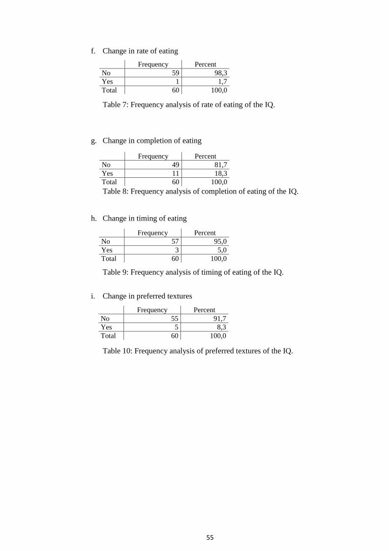
55
f. Change in rate of eating
Table 7: Frequency analysis of rate of eating of the IQ.
g. Change in completion of eating
Table 8: Frequency analysis of completion of eating of the IQ.
h. Change in timing of eating
Table 9: Frequency analysis of timing of eating of the IQ.
i. Change in preferred textures
Table 10: Frequency analysis of preferred textures of the IQ.
Frequency Percent
No 59 98,3
Yes 1 1,7
Total 60 100,0
Frequency Percent
No 49 81,7
Yes 11 18,3
Total 60 100,0
Frequency Percent
No 57 95,0
Yes 3 5,0
Total 60 100,0
Frequency Percent
No 55 91,7
Yes 5 8,3
Total 60 100,0

56
2. Assessment of elimination function
a. Changes in frequencies and “accidents”
1. No change in frequency and no “accidents”
2. Increased frequency, no “accidents”
3. Decreased frequency, no “accidents”
4. Increased frequency with “accidents”
5. Decreased frequency with “accidents”
6. No change in frequency, but “accidents”
Table 11: Frequency analysis of changes in frequency of elimination
behaviour of the IQ.
Frequency Percent
1 44 73,3
2 9 15,0
3 1 1,7
4 1 1,7
5 2 3,3
6 3 5,0
Total 60 100,0
0,010,020,030,040,050,060,070,080,090,0
100,0
44 9 1 1 2 3 60
1 2 3 4 5 6 Total
Valid
Changes in frequency and "accidents"
Graphic 1: Frequency analysis of changes in frequency of elimination
behaviour of the IQ.

57
b. Bladder Control
1. Leaks urine when asleep, only
2. Leaks urine when awake, only
3. Leaks urine when awake or asleep
4. Full-stream, uncontrolled urination when asleep, only
5. Full-stream, uncontrolled urination when awake, only
6. Full-stream, uncontrolled urination when awake or asleep
7. No leakage or uncontrolled urination, all urination controlled, but in
inappropriate or undesirable location
8. No change in urination control or behavior
Table 12: Frequency analysis of Bladder Control of the IQ.
Frequency Percent
1 1 1,7
2 1 1,7
3 3 5,0
4 0 0,0
5 0 0,0
6 1 1,7
7 5 8,3
8 49 81,7
Total 60 100,0
0,010,020,030,040,050,060,070,080,090,0
100,0
1 1 3 0 0 1 5 49 60
1 2 3 4 5 6 7 8 Total
Valid
Bladder Control
Graphic 2: Frequency analysis of Bladder Control of the IQ.

58
c. Bowel control
1. Defecates when asleep
2. Defecates without apparent awareness
3. Defecates when awake and aware of action, but in inappropriate or
undesirable locations
4. No changes in bowel control
Table 13: Frequency analysis of Bowel Control of the IQ.
3. Interactions with humans – which situation best describes that interaction?
a. No change in interaction with people
b. Recognizes people but slightly decreased frequency of interaction
c. Recognizes people but greatly decreased frequency of interaction
d. Withdrawal but recognizes other pets
e. Does not recognize other pets
Table 14: Frequency analysis of interactions with humans of the IQ.
Frequency Percent
1 0 0,0
2 0 0,0
3 4 6,7
4 56 93,3
Total 60 100,0
Frequency Percent
1 44 73,3
2 12 20,0
3 4 6,7
4 0 0,0
5 0 0,0
Total 60 100,0

59
4. Interactions with other pets – which situation best describes that interaction?
a. No change in interaction with other pets
b. Recognizes other pets but slightly decreased frequency of interaction
c. Recognizes other pets but greatly decreased frequency of interaction
d. Withdrawal but recognizes other pets
e. Does not recognize other pets
Table 15: Frequency analysis of interactions with other pets of the IQ.
Frequency Percent
1 40 66,7
2 9 15,0
3 8 13,3
4 3 5,0
5 0 0,0
Total 60 100,0
0
10
20
30
40
50
60
70
80
90
100
1 2 3 4 5 Total
66,7
15,0 13,3
5,0 0,0
100,0
Interactions with other pets
Graphic 3: Frequency analysis of interactions with other pets of the IQ.

60
5. Changes in sleep/wake cycle
a. No changes in sleep patterns
b. Sleeps more in day, only
c. Some change – awakens at night sleeps more in day
d. Much change – profoundly erratic nocturnal pattern and irregular daytime
pattern
e. Sleeps virtually all day, awake occasionally at night
f. Sleeps almost around the clock
Table 16: Frequency analysis of changes in the Sleep-Wake cycle of the IQ.
0
10
20
30
40
50
60
70
80
90
100
1 2 3 4 5 6 7 Total
53,3
31,7
0,0 0,0
8,3 5,0
1,7
100,0
Changes in the sleep-wake cycle
Frequency Percent
1 32 53,3
2 19 31,7
3 0 0,0
4 5 8,3
5 3 5,0
6 1 1,7
Total 60 100,0
Graphic 4: Frequency analysis of Changes in the Sleep-Wake cycle of the IQ.

61
Gender
Table 17: Frequency analysis of gender of the IQ.
Age groups
Table 18: Frequency analysis of age groups of the IQ.
7.2 Descriptive analysis – Study Group, SG (n=8)
1. Appetite assessment (may tick more than one).
a. No alterations in appetite
Table 19: Frequency analysis of appetite assessment of the SG.
b. Change in ability to physically handle food
Table 20: Frequency analysis of ability to handle food of the SG.
c. Change in ability to retain to food (vomits or regurgitates)
Table 21: Frequency analysis of ability to retain food of the SG.
Frequency Percent
Male 29 48,3
Female 31 51,7
Total 60 100,0
Frequency Percent
08 to 10 years old 29 48,3
11 to 13 years old 22 36,7
14 to 16 years old 9 15,0
Total 60 100,0
Frequency Percent
No 1 12,5
Yes 7 87,5
Total 60 100,0
Frequency Percent
No 8 100,0
Yes 0 0,0
Total 8 100,0
Frequency Percent
No 8 100,0
Yes 0 0,0
Total 8 100,0

62
d. Change in ability to find food
Table 22: Frequency analysis of ability to find food of the SG.
e. Change in interest in food (may be olfactory, having to do with the ability to smell)
Table 23: Frequency analysis of interest in food of the SG.
f. Change in rate of eating
Table 24: Frequency analysis of rate of eating of the SG.
g. Change in completion of eating
Table 25: Frequency analysis of rate of eating of the SG.
h. Change in timing of eating
Table 26: Frequency analysis of timing of eating of the SG.
Frequency Percent
No 8 100,0
Yes 0 0,0
Total 8 100,0
Frequency Percent
No 8 100,0
Yes 0 0,0
Total 8 100,0
Frequency Percent
No 8 100,0
Yes 0 0,0
Total 8 100,0
Frequency Percent
No 7 87,5
Yes 1 12,5
Total 8 100,0
Frequency Percent
No 8 100,0
Yes 0 0,0
Total 8 100,0

63
i. Change in preferred textures
Table 27: Frequency analysis of preferred textures of the SG.
2. 2. Assessment of elimination function
a. Changes in frequencies and “accidents”
i. No change in frequency and no “accidents”
ii. Increased frequency, no “accidents”
iii. Decreased frequency, no “accidents”
iv. Increased frequency with “accidents”
v. Decreased frequency with “accidents”
vi. No change in frequency but “accidents”
Table 28: Frequency analysis of changes in frequency of elimination behaviour of
the SG.
Frequency Percent
No 8 100,0
Yes 0 0,0
Total 8 100,0
Frequency Percent
1 8 100,0
2 0 0,0
3 0 0,0
4 0 0,0
5 0 0,0
6 0 0,0
Total 8 100,0

64
b. Bladder Control
i. Leaks urine when asleep, only
ii. Leaks urine when awake, only
iii. Leaks urine when awake or asleep
iv. Full-stream, uncontrolled urination when asleep, only
v. Full-stream, uncontrolled urination when awake, only
vi. Full-stream, uncontrolled urination when awake or asleep
vii. No leakage or uncontrolled urination, all urination controlled, but in
inappropriate or undesirable location
viii. No change in urination control or behavior
Table 29: Frequency analysis of changes in frequency of bladder control of the
SG.
Frequency Percent
1 0 0,0
2 0 0,0
3 0 0,0
4 0 0,0
5 0 0,0
6 0 0,0
7 0 0,0
8 8 100,0
Total 8 100,0

65
c. Bowel control
i. Defecates when asleep
ii. Defecates without apparent awarness
iii. Defecates when awake and aware of action, but in an inapprorpiate or
undesirable location
iv. No changes in bowel control
Table 30: Frequency analysis of changes in bowel control of the SG.
3. Interactions with humans – which situation best describes that interaction?
a. No change in interaction with people
b. Recognizes people but slightly decreased frequency of interaction
c. Recognizes people but greatly decreased frequency of interaction
d. Withdrawal but recognizes people
e. Does not recognize people
Table 31: Frequency analysis of changes in interactions with humans of the
SG.
Frequency Percent
1 0 0,0
2 0 0,0
3 0 0,0
4 8 100,0
Total 60 100,0
Frequency Percent
1 5 62,5
2 3 37,5
3 0 0,0
4 0 0,0
5 0 0,0
Total 60 100,0

66
4. Interactions with other pets – which situation best describes that interaction?
a. No change in interaction with other pets
b. Recognizes other pets but slightly decreased frequency of interaction
c. Recognizes other pets but greatly decreased frequency of interaction
d. Withdrawal but recognizes other pets
e. Does not recognize other pets
Table 32: Frequency analysis of changes in interactions with other pets of the SG.
Frequency Percent
1 5 62,5
2 1 12,5
3 1 12,5
4 1 12,5
5 0 0,0
Total 60 100,0
0
10
20
30
40
50
60
70
80
90
100
1 2 3 4 5 Total
62,5
12,5 12,5 12,5
0,0
100,0
Interactions with other pets
Graphic 5: Frequency analysis of Changes in interactions with other pets of the
SG.
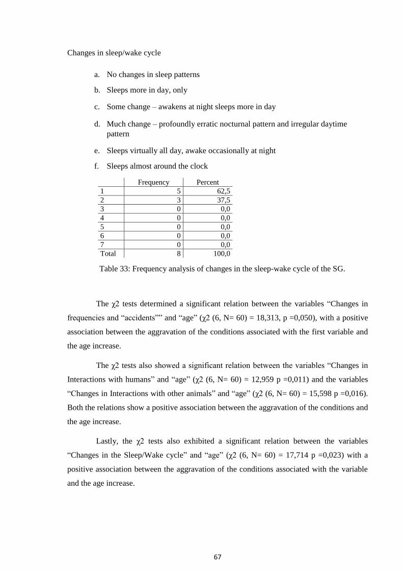
67
Changes in sleep/wake cycle
a. No changes in sleep patterns
b. Sleeps more in day, only
c. Some change – awakens at night sleeps more in day
d. Much change – profoundly erratic nocturnal pattern and irregular daytime
pattern
e. Sleeps virtually all day, awake occasionally at night
f. Sleeps almost around the clock
Table 33: Frequency analysis of changes in the sleep-wake cycle of the SG.
The χ2 tests determined a significant relation between the variables “Changes in
frequencies and “accidents”” and “age” (χ2 (6, N= 60) = 18,313, p =0,050), with a positive
association between the aggravation of the conditions associated with the first variable and
the age increase.
The χ2 tests also showed a significant relation between the variables “Changes in
Interactions with humans” and “age” (χ2 (6, N= 60) = 12,959 p =0,011) and the variables
“Changes in Interactions with other animals” and “age” (χ2 (6, N= 60) = 15,598 p =0,016).
Both the relations show a positive association between the aggravation of the conditions and
the age increase.
Lastly, the χ2 tests also exhibited a significant relation between the variables
“Changes in the Sleep/Wake cycle” and “age” (χ2 (6, N= 60) = 17,714 p =0,023) with a
positive association between the aggravation of the conditions associated with the variable
and the age increase.
Frequency Percent
1 5 62,5
2 3 37,5
3 0 0,0
4 0 0,0
5 0 0,0
6 0 0,0
7 0 0,0
Total 8 100,0

68
7.3 Frequency analysis of trial period, days 0, 30, 60 and 90
Assessing Disorientation
How frequently does your dog get lost in familiar locations?
(e. g. At home they may not find their way to their bed).
Day 0 Day 30 Day 60 Day 90
1 8 8 8 8
2 0 0 0 0
3 0 0 0 0
4 0 0 0 0
5 0 0 0 0
Table 34: Frequency analysis of assessment of disorientation 1 of the SG.
How frequently does your dog go to the wrong side of the door when leaving a room?
Day 0 Day 30 Day 60 Day 90
1 8 8 8 8
2 0 0 0 0
3 0 0 0 0
4 0 0 0 0
5 0 0 0 0
Table 35: Frequency analysis of assessment of disorientation 2 of the SG.
How frequently does your dog wander in the house with no apparent purpose?
Day 0 Day 30 Day 60 Day 90
1 8 3 4 4
2 0 4 2 2
3 0 1 2 2
4 0 0 0 0
5 0 0 0 0
Table 36: Frequency analysis of assessment of disorientation 3 of the SG.

69
Assessing Social Interaction
Interacting with humans. Remember the typical behaviour of your dog in adult life.
Nowadays the number of times your dog shows interest in interacting with humans
has…
(e. g. playing, petting, greeting)
Day 0 Day 30 Day 60 Day 90
1 0 0 0 0
2 0 0 0 0
3 8 8 7 7
4 0 0 1 1
5 0 0 0 0
Table 37: Frequency analysis of assessment of social interactions 1 of the SG.
Interacting with other pets Remember the typical behaviour of your dog in adult life.
Nowadays the number of times your dog shows interest in interacting with other pets
has…(e. g. playing, barking, smelling)
Day 0 Day 30 Day 60 Day 90
1 0 0 0 0
2 3 2 2 1
3 5 6 6 7
4 0 0 0 0
5 0 0 0 0
Table 38: Frequency analysis of assessment of social interactions 2 of the SG.
Remember some tasks you taught your dog. (e.g. sitting, laying down, fetching).
Nowadays the number of times your dog is capable of performing them has…
Day 0 Day 30 Day 60 Day 90
1 0 0 0 0
2 0 0 0 0
3 8 8 8 8
4 0 0 0 0
5 0 0 0 0
Table 39: Frequency analysis of assessment of social interactions 3 of the SG.

70
Assessing Sleep – Wake Cycles
Barking during the night. Remember the typical behaviour of your dog in adult life.
Nowadays the number of times your dog barks during the night for no apparent reason
has…
Day 0 Day 30 Day 60 Day 90
1 3 3 2 3
2 2 2 2 2
3 3 3 4 3
4 0 0 0 0
5 0 0 0 0
Table 40: Frequency analysis of assessment of sleep-wake cycles 1 of the SG.
Sleeping during the day Remember the typical behaviour of your dog in adult life.
Nowadays the number of times your dog spends most of his/her day sleeping has…
Day 0 Day 30 Day 60 Day 90
1 0 0 0 0
2 0 0 0 0
3 5 6 6 5
4 1 0 0 2
5 2 2 2 1
Table 41: Frequency analysis of assessment of sleep-wake cycles 2 of the SG.
Staying awake during the night. Remember the typical behaviour of your dog in adult
life. Nowadays the number of times your dog spends most of his/her night awake has…
Day 0 Day 30 Day 60 Day 90
1 2 2 2 2
2 0 1 0 0
3 4 5 5 5
4 2 0 1 1
5 0 0 0 0
Table 42: Frequency analysis of assessment of sleep-wake cycles 3 of the SG.

71
Assessing Housesoiling
Urinating in a “wrong” place. Remember the typical behaviour of your dog in adult life.
Nowadays the number of times your dog urinates in a place he is not supposed to has…
Day 0 Day 30 Day 60 Day 90
1 3 3 3 3
2 0 0 0 0
3 4 5 4 4
4 1 0 1 1
5 0 0 0 0
Table 43: Frequency analysis of assessment of housesoiling 1 of the SG.
Defecating in a “wrong” place. Remember the typical behaviour of your dog in adult
life. Nowadays the number of times your dog defecates in a place he is not supposed to
has… Day 0 Day 30 Day 60 Day 90
1 3 3 3 3
2 0 0 0 0
3 5 5 5 5
4 0 0 0 0
5 0 0 0 0
Table 44: Frequency analysis of assessment of housesoiling 2 of the SG.

72
Assessing Activity Levels
Vocalization. Remember the typical behaviour of your dog in adult life. Nowadays the
number of times your dog vocalizes has… Day 0 Day 30 Day 60 Day 90
1 1 1 1 1
2 0 0 0 0
3 6 6 6 5
4 0 0 0 1
5 1 1 1 1
Table 45: Frequency analysis of assessment of activity levels 1 of the SG.
Appetite. Remember the typical behaviour of your dog in adult life. Nowadays your
dog’s appetite has… (e. g. eating more, asking your for food) Day 0 Day 30 Day 60 Day 90
1 0 0 0 0
2 0 1 3 4
3 4 4 2 2
4 4 3 3 2
5 0 0 0 0
Table 46: Frequency analysis of assessment of activity levels 2 of the SG.
Greeting behaviour. Remember the typical behaviour of your dog in adult life.
Nowadays the number of times your dog greets you / other elements of the family has… Day 0 Day 30 Day 60 Day 90
1 0 0 0 0
2 0 0 0 0
3 5 6 6 5
4 3 2 2 3
5 0 0 0 0
Table 47: Frequency analysis of assessment of activity levels 3 of the SG.
Several χ2 tests were elaborated between the variables and the day of the
questionnaire but no relations, neither positive nor negative, were found between these.

73
7.4 Global response to therapy rate
After testing the variables an additional analysis compared the global results
between the days zero and ninety. From these comparisons, a global response to therapy
rate was determined.
When comparing these two moments 8.04% of the study group (n =8) had a
general improvement in the lessening of their clinical signs. The majority of the study group
- 86.61%, was generally considered to have no improvement nor aggravation in their
clinical signs and finally 5.36% of the study group experienced a general worsening of the
CDS signs.

74
8. Discussion
The effects of fatty acids in the cognition have been previously studied (Heath et
al., 2007 ; Reger et al., 2004 ; Taha et al., 2009). Cognition may decrease with age and it is
important for the clinician to know how to delay its progress in an efficient way.
Regarding the study previously presented, the first part of the analysis was a
descriptive analysis of the population of all the dogs that were assessed in the study. The
population had no statistical differences regarding age groups nor gender. Only 23.3% of
the population (n=14) had no behavioural alterations. Although 76.7% had some described
alterations not all of them have to be related to CDS. Therefore, the prevalence of CDS in
our population will not be estimated. Despite of that, none of these animals had been
referred to behaviouralists nor were their clinical signs being addressed. A previous study
shows that a veterinary diagnosis of CDS is accomplished in 13,3% of a population (Salvin
et al., 2010).
It was shown that 50% of the tutors of our population claim that their animals have
alterations in appetite being the most reported one the “change in the completion of eating a
meal” (18,3%) followed by an impairment in the ability of the animal to physically handle
food (11,7%). When assessing sleep-wake cycles changes, these were reported in almost
half of the population (46,7%). These results state that the alterations in the appetite are the
most prevalent followed by the changes in the sleep-wake cycle. This does not necessarily
mean that these signs are neither the most frequent nor the first to appear in a dog suffering
from CDS but rather might indicate which are the signs more easily or frequently detected
by the animals’ tutor, or the ones that impact the most the tutors’ life.
When assessing the study group (n=8) the results are coherent with the population,
since the major change reported by the tutors is the alteration in the animals’ appetite
(87,5%). The positive association between the variables “Changes in the frequencies and
accidents” regarding housesoiling, “Changes in interactions with humans” and also
“Changes in interactions with other animals” regarding social interactions and the variable
“age” show that there is an aggravation of the symptoms with the age increase. These
significant relations validate the data since according to literature the prevalence of
cognitive impairment is expected to increase with the increase of age (Azkona et al., 2009).

75
Considering a population of sixty dogs, the fact that only eight animals were
enrolled in the study was a consequence of the inclusion criteria. The aim was to study
animals with mild cognitive impairment and exclude the animals with severe cognitive
impairment. Although this choice was based upon the number of clinical signs presented at
the moment of the first questionnaire it was not taken into account which signs were
presented. For instance none of the animals demonstrated disorientation as a clinical sign.
The category of signs presented, according to the DISHA scale, might also indicate the
severity of the disease (Osella et al., 2007). Other reasons include refusal from the tutor,
biochemistry tests / hematology coherent with systemic disease, lack or excess of
behavioural changes.
Another fact that should be nominated is that although the animals underwent
hematology and biochemistry tests along with physical examination, it cannot be confirmed
that the animals did not have a concomitant systemic disease. The odds are low since only
animals with test results satisfying were included. But, because several diagnostic tests were
left undone – such as urinalysis our fecal cytology - it cannot be stated that the animals had
no concurrent disease. Since CDS is an exclusion diagnostic disease it must be stated that
none of the animals had an MRI or Computerized Axial Tomography – CAT scan done so
other brain pathologies cannot be excluded.
When analyzing the animals’ evolution throughout the 90 days trial-period, the
global response to therapy was not what was expected considering that the majority of the
study group (n=8) had no improvement nor impairment in their overall cognition when
addressed by the tutors – 86,61%. The amount of animals that showed a positive global
response to therapy was 8,04%. When comparing this value with other studies with MCT’s
the previous study achieved a lower score (Dodd et al., 2002 ; Wu et al., 2004). Lastly, a
smaller percentage also worsened throughout the study – 5,36%.
Some reasons that can explain the previous result are the fact that the study was
conducted in a “real life” scenario rather than in a laboratory. The major differences are the
fact that the animals are not so closely monitored and the fact that the behavioural
evaluation was solely the tutor responsibility rather than a clinician, behaviourist or any
veterinary staff. This may lead to a lower degree of efficacy on noticing subtle behavioural
changes. Combining that with the fact that the animals chosen had mild clinical signs and

76
the short study period it is understandable that the behavioural changes and, consequently,
the response to therapy may have been underestimated.
It must also be stated that there may be a bias in our global response to therapy rate
if it is considered that the maintenance of the clinical signs might also be a positive
response to therapy. This can be considered because, as stated in the literature, the goal of
the treatment of this syndrome is often to slow down the progression of signs rather than
eliminating them (Landsberg & Denenberg, 2009). When considering the disorientation
assessment one can notice that there was no change in that category because none of the
animals presented any disorientation signs to begin with. Even there was no further
improvement due to the fact that they were not disoriented at the beginning, the MCTs diet
may have helped the dogs maintain their long term memory about their familiar
environments during the study. This might also be considered a positive response since the
MCT’s in the diet might have stabilized these animals or it might just be that the brain
region affected in these animals is not responsible for that behaviour.
This is also true for the other behaviours assessed. For instance for social
interaction assessment, MCT’s diet stabilized 7 dogs and improved 1 dog in interacting with
humans and reduced the number of dogs that had decreased interaction with other pets from
2 to 1, and stabilizing the interactions in other 7 dogs. The MCT diet either stabilized the
symptoms or improved the symptoms in other assessments including sleep-wake cycle,
housesoiling and activity levels.
Since the study dealt with humans who are emotionally attached to the animal they
were evaluating, the placebo effect also must be taken into account. Specially if considered
that Study diet A and Study diet B had different shaped kibbles, the tutor could easily
understand that there had been a change in the diet. Although not knowing which was the
verum and which was the placebum this might have influenced the tutors when answering
the questionnaires. A possible way this fact could be avoided, is not only having equally
shaped kibbles but also having a clinician or behaviourist completing the behaviour
assessment of the animal along with the tutor for the investigator to be able to compare both
and estimate a placebum effect percentage more accurately.
To completely understand how a supplemented dry food will change the
behaviours and slow the progression of CDS in dogs further studies are needed with bigger
populations and extended trial periods. Other vicissitudes that may be addressed is

77
separating the placebo and the verum populations as two individual group studies in order
to firstly avoid the change of diet during the study and to more easily compare the
fluctuations of behaviour changes in populations of geriatric dogs with cognitive
impairment. Other suggested alteration would be to group the dogs accordingly to the
category of clinical signs they show impairment in according to the DISHA scale to identify
how they answer the supplementation.
Other possible
78
9. Conclusion
The previous study describes the role of a supplemented diet in a population of
dogs with cognitive impairment when addressed by the tutors.
Conducting a series of questionnaires the behavioural changes of the study group
was described throughout a period of non-supplemented food and a period of supplemented
food. By doing this one can assess not only how does a tutor perceive the change of
behaviour in his pet but also notice which symptoms are more sensitive to the
supplementation.
When realizing that none of these dogs were being medicated for their behavioural
changes nor were being followed by a behaviourist, it was shown that the diagnostic of
CDS is easily underestimated even when an animal is regularly visiting the clinician as
described in the literature. This leads to a specific revision of veterinary training and include
skills that allow the clinician to perform an accurate anamnesis.
No statistical relation was found in our population between the presence of
symptoms with gender. There was however a confirmation that, when untreated, the clinical
signs worsen with age as one could suspect. Unfortunately, although our data was validated
as a group no statistical relations were found between the variables or along the period of
the study. Besides this, when assessing the questionnaires individually the MCT’s diet
either stabilized or improved most of the measurements alone during this short-term study
which is an encouraging result. For further studies and in order to more precisely evaluate
the brain MRI’s or CAT scans should be included.
The previous statements indicate that further studies are needed in order to more
accurately statistically analyze a population of geriatric dogs and to realize how the tutors
perceive the changes of behaviour in their companion animals.

79
ANNEXES
Annex I
VetPlus Aktivait® Composition
Composition per capsule: Small breeds (< 10 Kg)
DHA/EPA 35 mg
L-carnitine 20 mg
Vitamin C 20 mg
N-acetylcysteine 13.5mg
Alpha-lipoic acid 10 mg
Vitamin E 10 mg
Acetyl – carnitine 5 mg
Coenzyme Q10 1 mg
Phosphatidylserine 1 mg
Selenium (as selenometionina) 25mcg

80
VetPlus Aktivait®
Composition per capsule: Medium / Large breeds (> 10 Kg)
DHA/EPA 70 mg
L-carnitine 40 mg
Vitamin C 40 mg
N-acetylcysteine 27 mg
Alpha-lipoic acid 20 mg
Vitamin E 20 mg
Acetyl - carnitine 10 mg
Coenzyme Q10 2 mg
Phosphatidylserine 2 mg
Selenium (as selenometionina) 50mcg

81
Annex II
Ceva Senilife® Composition
Composition per capsule: Small breeds (< 10 Kg)
Phosphatidylserine 25 mg
Gingko Biloba extract 10 mg
Vitamin B6 20.5 mg
Vitamin E 33.5mg
Resveratrol (Grape extract) 5 mg
Other ingredients: cod liver oil, seed oil products and byproducts, gelatin, glycerin,
glyceryl, monostearate, titanium dioxide, FD&C Red 40
Guaranteed Analysis: Proteins 22.5%, Oils and Fats 46%, Cellulose <1%,
Moisture/Humidity 6%, Ash 6%, Vitamin E (33.5 mg), Vitamin B6 (20.5 mg)
Ceva Senilife XL®
Composition per capsule: Small breeds (< 10 Kg)
Phosphatidylserine 62.5 mg
Gingko Biloba extract 25 mg
Vitamin B6 51.25 mg
Vitamin E 83.75 mg
Resveratrol (Grape extract) 12.5 mg
Other ingredients: cod liver oil, seed oil products and byproducts, gelatin, glycerin,
glyceryl, monostearate, titanium dioxide, FD&C Red 40
Guaranteed Analysis: Proteins 18.5%, Oils and Fats 52%, Cellulose <1%,
Moisture/Humidity 5%, Ash 7%, Vitamin E (83.75 mg), Vitamin B6 (51.25 mg)

82
Annex III
Hill's Prescription Diet®
b/d® Canine Healthy Aging & Alertness
Ingredients
Whole Grain Corn, Chicken By-Product Meal, Pork Fat, Brewers Rice, Soybean Mill Run,
Flaxseed, Soybean Meal, Fish Meal, Lactic Acid, Chicken Liver Flavor, Dried Egg Product,
Soybean Oil, Dried Carrots, Dried Spinach, Dried Grape Pomace, Dried Tomato Pomace,
Dried Citrus Pulp, Potassium Chloride, Oat Fiber, Choline Chloride, vitamins (Vitamin E
Supplement, L-Ascorbyl-2-Polyphosphate (source of Vitamin C), Niacin Supplement,
Thiamine Mononitrate, Vitamin A Supplement, Calcium Pantothenate, Vitamin B12
Supplement, Pyridoxine Hydrochloride, Riboflavin Supplement, Vitamin D3 Supplement,
Folic Acid, Biotin), L-Lysine, Iodized Salt, Calcium Carbonate, L-Tryptophan, Taurine,
minerals (Ferrous Sulfate, Zinc Oxide, Copper Sulfate, Manganous Oxide, Calcium Iodate,
Sodium Selenite), L-Carnitine, Mixed Tocopherols for freshness, Lipoic Acid, Phosphoric
Acid, Beta-Carotene, Natural Flavors.

83
Annex IV
Test food ingredients and formulation
The basis of the food was formulated to meet the American Association of Feed Control
Official recommendations on adult dog food.
The supplement included – on the dry matter basis - L-Carnitine (330ppm), DL-α-Lipoic
acid (150ppm), DL-α tocopherol acetate (1150ppm), taurine (1095 ppm), ascorbic acid (100
ppm), and the following were included at the percentage of 1%: spinach flakes, tomato
pomace, grape pomace, carrot granules and citrus pulp.
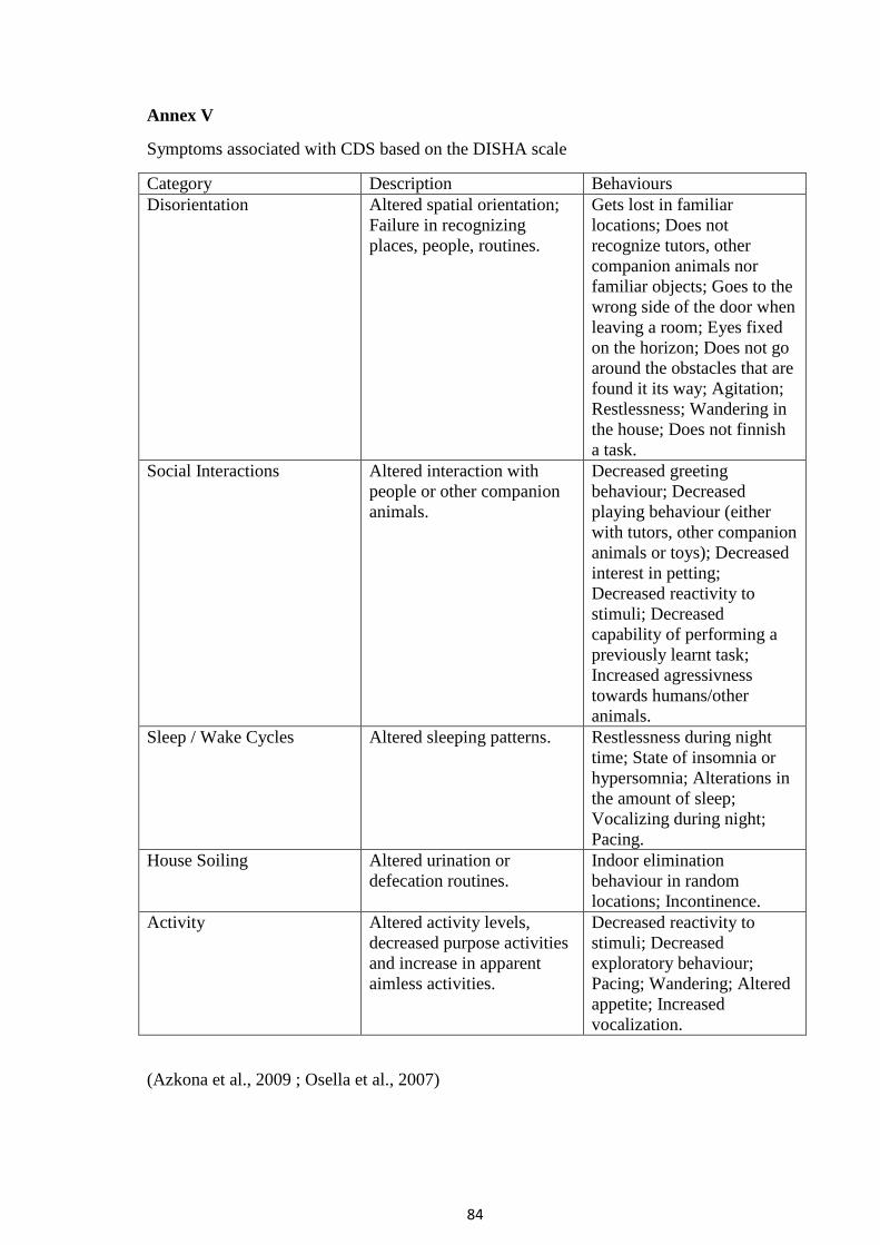
84
Annex V
Symptoms associated with CDS based on the DISHA scale
Category Description Behaviours
Disorientation Altered spatial orientation;
Failure in recognizing
places, people, routines.
Gets lost in familiar
locations; Does not
recognize tutors, other
companion animals nor
familiar objects; Goes to the
wrong side of the door when
leaving a room; Eyes fixed
on the horizon; Does not go
around the obstacles that are
found it its way; Agitation;
Restlessness; Wandering in
the house; Does not finnish
a task.
Social Interactions Altered interaction with
people or other companion
animals.
Decreased greeting
behaviour; Decreased
playing behaviour (either
with tutors, other companion
animals or toys); Decreased
interest in petting;
Decreased reactivity to
stimuli; Decreased
capability of performing a
previously learnt task;
Increased agressivness
towards humans/other
animals.
Sleep / Wake Cycles Altered sleeping patterns. Restlessness during night
time; State of insomnia or
hypersomnia; Alterations in
the amount of sleep;
Vocalizing during night;
Pacing.
House Soiling Altered urination or
defecation routines.
Indoor elimination
behaviour in random
locations; Incontinence.
Activity Altered activity levels,
decreased purpose activities
and increase in apparent
aimless activities.
Decreased reactivity to
stimuli; Decreased
exploratory behaviour;
Pacing; Wandering; Altered
appetite; Increased
vocalization.
(Azkona et al., 2009 ; Osella et al., 2007)

85
Annex VI
Questionnaire for inclusion in the study.
Please answer these questions comparing the behaviours the dog exhibits now with behaviours the dog exhibited in adult age (consider five years old).
1. Appetite assessment (may tick more than one).
a. No alterations in appetite
b. Change in ability to physically handle food
c. Change in ability to retain to food (vomits or regurgitates)
d. Change in ability to find food
e. Change in interest in food (may be olfactory, having to do with the ability
to smell)
f. Change in rate of eating
g. Change in completion of eating
h. Change in timing of eating
i. Change in preferred textures
3. 2. Assessment of elimination function (tick only one in each section)
a. Changes in frequencies and “accidents”
i. No change in frequency and no “accidents”
ii. Increased frequency, no “accidents”
iii. Decreased frequency, no “accidents”
iv. Increased frequency with “accidents”
v. Decreased frequency with “accidents”
vi. No change in frequency but “accidents”
b. Bladder Control
i. Leaks urine when asleep, only
ii. Leaks urine when awake, only
iii. Leaks urine when awake or asleep
iv. Full-stream, uncontrolled urination when asleep, only

86
v. Full-stream, uncontrolled urination when awake, only
vi. Full-stream, uncontrolled urination when awake or asleep
vii. No leakage or uncontrolled urination, all urination controlled, but in
inappropriate or undesirable location
viii. No change in urination control or behavior
c. Bowel control
i. Defecates when asleep
ii. Defecates without apparent awarness
iii. Defecates when awake and aware of action, but in an inappropriate or
undesirable location
iv. No changes in bowel control
3. Interactions with humans – which situation best describes that interaction? (tick only
one)
a. No change in interaction with people
b. Recognizes people but slightly decreased frequency of interaction
c. Recognizes people but greatly decreased frequency of interaction
d. Withdrawal but recognizes other pets
e. Does not recognize other pets
4. Interactions with other pets – which situation best describes that interaction? (Tick
only one)
a. No change in interaction with other pets
b. Recognizes other pets but slightly decreased frequency of interaction
c. Recognizes other pets but greatly decreased frequency of interaction
d. Withdrawal but recognize other pets
5. Changes in sleep/wake cycle (tick only one)
a. No changes in sleep patterns
b. Sleeps more in day, only

87
c. Some change – awakens at night sleeps more in day
d. Much change – profoundly erratic nocturnal pattern and irregular daytime
pattern
e. Sleeps virtually all day, awake occasionally at night
f. Sleeps almost around the clock
6. Is there anything else you think we should know?
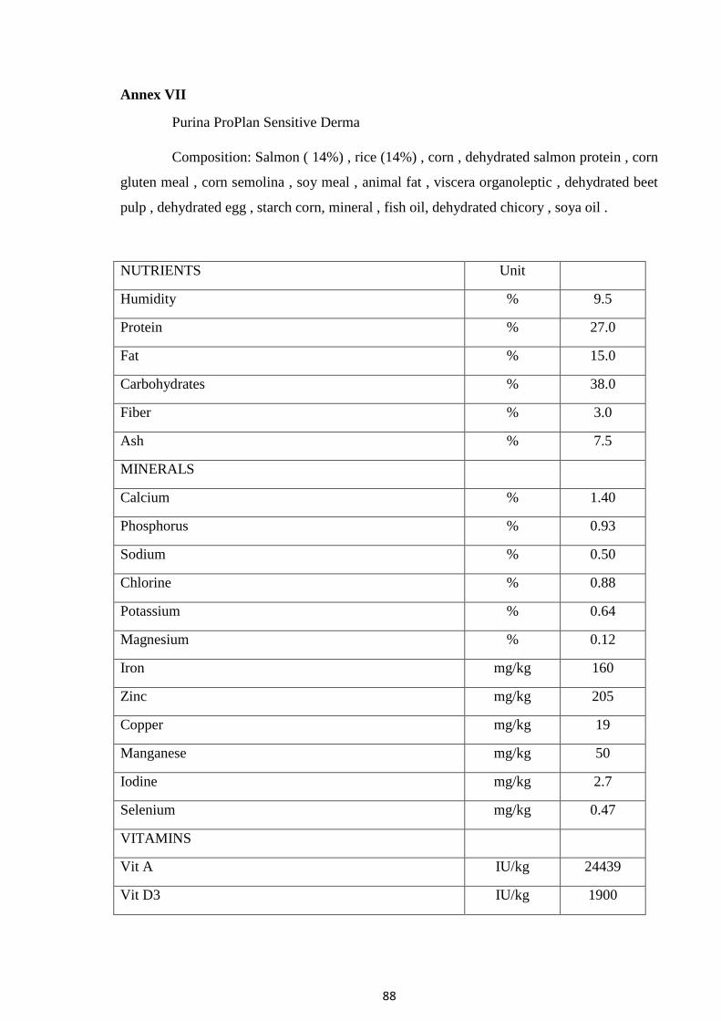
88
Annex VII
Purina ProPlan Sensitive Derma
Composition: Salmon ( 14%) , rice (14%) , corn , dehydrated salmon protein , corn
gluten meal , corn semolina , soy meal , animal fat , viscera organoleptic , dehydrated beet
pulp , dehydrated egg , starch corn, mineral , fish oil, dehydrated chicory , soya oil .
NUTRIENTS Unit
Humidity % 9.5
Protein % 27.0
Fat % 15.0
Carbohydrates % 38.0
Fiber % 3.0
Ash % 7.5
MINERALS
Calcium % 1.40
Phosphorus % 0.93
Sodium % 0.50
Chlorine % 0.88
Potassium % 0.64
Magnesium % 0.12
Iron mg/kg 160
Zinc mg/kg 205
Copper mg/kg 19
Manganese mg/kg 50
Iodine mg/kg 2.7
Selenium mg/kg 0.47
VITAMINS
Vit A IU/kg 24439
Vit D3 IU/kg 1900

89
Vit E mg/kg 479
Vit K mg/kg 0.17
Vit C mg/kg 70
Vit B1 mg/kg 30
Vit B2 mg/kg 13
Vit B3 mg/kg 159
Vit B5 mg/kg 47
Vit B6 mg/kg 16
Vit B8 mg/kg 0.15
Vit B9 mg/kg 4.60
Vit B12 mg/kg 0.25
Colina mg/kg 3540
Taurina mg/kg 750
ESSENTIAL FATTY ACIDS
Ómega- 6 % 2.0
Linoleic acid % 1.7
Omega- 3 % 1.1
DHA % 0.50
EPA + DHA % 0.92
ENERGY
Calculated metabolizable energy Kcal/g 3.550

90
Annex VIII
Purina ProPlan Senior Original
Composition: dehydrated poultry protein , Wheat, Corn , Chicken (14%) , gluten,
soy flour, fractions of vegetable oil, rice (4%) , dried beet pulp , minerals , corn semolina ,
animal fat , viscera organoleptic , Fish oil.
NUTRIENTS Unit
Humidity % 9.5
Protein % 29.0
Fat % 15.0
Carbohydrates % 37.0
Fiber % 2.0
Ash % 7.5
MINERALS
Calcium % 1.3
Phosphorus % 0.86
Sodium % 0.52
Chlorine % 1.02
Potassium % 0.86
Magnesium % 0.12
Iron mg/kg 201
Zinc mg/kg 219
Copper mg/kg 20
Manganese mg/kg 57
Iodine mg/kg 2.8
Selenium mg/kg 0.4
VITAMINS
Vit A IU/kg 16545
Vit D3 IU/kg 713
Vit E mg/kg 559

91
Vit K mg/kg 0.10
Vit C mg/kg 82
Vit B1 mg/kg 19
Vit B2 mg/kg 10
Vit B3 mg/kg 122
Vit B5 mg/kg 33
Vit B6 mg/kg 12
Vit B8 mg/kg 0.18
Vit B9 mg/kg 3.30
Vit B12 mg/kg 0.19
Colina mg/kg 2025
Taurina mg/kg 872
ESSENTIAL FATTY ACIDS
Ómega- 6 % 2.0
Linoleic acid % 1.9
Omega- 3 % 0.3
DHA % 0.07
EPA + DHA % 0.23
ENERGY
Calculated metabolizable energy Kcal/g 3 585

92
Annex IX
Questionnaire – Assessment tool
Assessing Disorientation
(1 – Never; 2 – Seldomly; 3 – Sometimes; 4 – Frequently; 5 - Always)
0 1 2 3 4 5
1. How frequently does your dog get
lost in familiar locations? (e. g. At
home they may not find their way to
their bed)
2. How frequently does your dog go to
the wrong side of the door when
leaving a room?
3. How frequently does your dog
wanders in the house with no apparent
purpose?

93
Assessing Social Interaction
(1 – Almost never shows interest; 2 –Decreased; 3 – The same; 4 – More often; 5 - Always)
0 1 2 3 4 5
1. Interacting with humans.
Remember the typical behaviour of
your dog in adult life.
Nowadays the number of times your
dog shows interest in interacting with
humans has…
(e. g. playing, petting, greeting)
2.Interacting with other pets
Remember the typical behaviour of
your dog in adult life.
Nowadays the number of times your
dog shows interest in interacting with
other pets has…(e. g. playing, barking,
smelling)
3. Remember some tasks you taught
your dog. (e.g. sitting, laying down,
fetching)
Nowadays the number of times your
dog is capable of performing them
has…

94
Assessing Sleep – Wake Cycles
(1 – Almost never; 2 –Decreased; 3 – The same; 4 – More often; 5 - Always)
0 1 2 3 4 5
1. Barking during the night.
Remember the typical behaviour of
your dog in adult life.
Nowadays the number of times your
dog barks during the night for no
apparent reason has…
2. Sleeping during the day
Remember the typical behaviour of
your dog in adult life.
Nowadays the number of times your
dog spends most of his/her day
sleeping has…
3. Staying awake during the night.
Remember the typical behaviour of
your dog in adult life.
Nowadays the number of times your
dog spends most of his/her night
awake has…
Assessing House Soiling
(1 – Almost never; 2 –Decreased; 3 – The same; 4 – More often; 5 - Always)
0 1 2 3 4 5
1. Urinating in a “wrong” place.
Remember the typical behaviour of
your dog in adult life.
Nowadays the number of times your
dog urinates in a place he is not
supposed to has…
2. Defecating in a “wrong” place.
Remember the typical behaviour of
your dog in adult life.
Nowadays the number of times your
dog defecates in a place he is not
supposed to has…

95
Assessing Activity Levels
(1 – Almost never; 2 –Decreased; 3 – The same; 4 – More often; 5 - Always)
0 1 2 3 4 5
1. Vocalization.
Remember the typical behaviour of
your dog in adult life.
Nowadays the number of times your
dog vocalizes has…
2. Appetite.
Remember the typical behaviour of
your dog in adult life.
Nowadays your dog’s appetite has…
(e. g. eating more, asking your for
food)
3. Greeting behaviour.
Remember the typical behaviour of
your dog in adult life.
Nowadays the number of times your
dog greets you / other elements of the
family has…
(Azkona et al., 2009 ; Osella et al., 2007)

96
10. References
Araujo, J.A., Chan, A. D. F., Winka, L. L., Seymour, P. A. & Milgram N. W. (2004). Dose-
specific effects of scopolamine on canine cognition: Impairment of visuospatial memory, but
no visuospatial discrimination. Psychopharmacology, 175, 92 – 98.
Araujo, J.A., Studzinski, C. M. & & Milgram N. W. (2005). Further evidence for the
cholinergic hypothesis of aging and dementia from the canine model of aging, Progress in
Neuro-Psychopharmacology & Biological Psychiatry, 29, 411 -422.
Araujo, J.A., Landsberg G. M., Milgram N. W. & Miolo, A. (2008). Improvement of short-
term memory performance in aged beagles by a nutraceutical supplement containing
phosphatidylserine, Gingko biloba, vitamin E and pyridoxine, Canadian Veterinary Journal,
49, 379 – 385.
Azkona, G., García-Bellenguer, S., Chacón, G., Rosado, B., León, M. & Palacio J. (2009).
Prevalence and risk factors of behavioral changes associated with age-related cognitive
impairment in geriatric dogs, Journal of Small Animal Practice, 50, 87 – 91.
Badino, P., Odore, R., Bergamasco, L., Barbero, R., Osella, M., D’Angelo, A., Re, G. &
Girardi, G. (2013). Concentrations of platelet α2-adrenoreceptors, lymphocyte muscarinic
receptors, and blood monoamines in dogs (Canis familiaris) affected by canine cognitive
dysfunction syndrome, Journal of Veterinary Behavior, 8, 146 – 153.
Beckman. K. B. & Ames, B. N. (1998). The Free Radical Theory of Aging Matures,
Physiological Reviews, 78 (2), 547–581.
Beltz, B. S., Tlusty, M. F., Benton, J. L. & Sandeman, D. C. (2007). Omega-3 fatty acids
upregulate adult neurogenesis, Neuroscience Letters, 415, 154 – 158.
Bobik, M., Thompson, T., Russel, M. J. (1994). Amyloid deposition in various breeds of
dogs, Society for Neuroscience abstracts, 20, 172.
Boothe, D. M. (2004). Balancing fact and fiction of novel ingredients: definitions, regulations
and evaluation, The Veterinary Clinics Small Animal Practice, 34, 7 – 38.

97
Carillo, M. C., Ivy, G. O., Milgram, N. W., Head, E., Wu, P., & Kitani, K. (1994). Deprenyl
increases activity of superoxide dismutase, Life Sciences, 54(20), 1483 - 9.
Colle, M-A., Hauw, J. J., Crespeau, F., Uchihara, T., Akiyama, H., Checler, F., Pageat, P. &
Duykaerts, C. (2000). Vascular and parenchymal Aβ deposition in the aging dog: correlation
with behavior, Neurobiology of Aging, 21, 695 – 704.
Cummings, B. J., Su, J., Cotman, C., White, R. & Russel, M. (1993). β-amyloid accumulation
in aged canine brain: A model of early plaque formation in Alzheimer’s disease.
Neurobiology of Aging, 14, 547 – 560.
Cummings, B. J., Head, E., Afagh, A. F., Milgram, N. W. & Cotman C. W. (1996). β-amyloid
accumulation correlates with cognitive dysfunction in the aged canine. Neurobiology of
Learning and Memory, 66, 11 – 23.
Cummings B. J. & Cotman, C. (1995). Image analysis of β-amyloid “load” in Alzheimer’s
disease and relation to dementia severity. The Lancet, 346, 1524 – 1528.
Dakshinamurti, K., Paulose, C. S., Viswanathan, M., Siow Y. L., & Sharma S. K. (1990).
Neurobiology of Pyridoxine, Annals of the New York Academy of Sciences, 585(1), 128-144.
Dodd, C., Zicker, SC., Jewell, D. E., Lowry, S. R., Fritsch, D., Toll, P. W. (2002). Effects of
an investigational food on age related behavioral changes in dogs, Paper presented at: Hill’s
European Symposium on Canine Brain Ageing; Barcelona.
Donini, L.M., Felice, M. R. & Cannella, C. (2007). Nutritional status determinants and
cognition in the elderly, Archives of Gerontology and Geriatrics Journal, 44, 143 – 153.
Fast, R., Schutt, T., Toft, N., Moller, A. & Berendt M. (2013). An Observational Study with
Long-Term Follow-Up of Canine Cognitive Dysfunction: Clinical Characteristics, Survival
and Risk Factors, Journal of Veterinary Internal Medicine, 27 (4), 822 – 829.
Fuji, K., Hiramatsu, M., Kameyama, T., Nabeshima, T. (1993). Effects of repeated
administration of propentofylline on memory impairment produced by basal forebrain lesions
in rats, European Journal of Pharmacology, 236, 411 – 417.

98
Gunn-Moore, D., Moffat, K., Christie, L. A. & Head, E. (2007). Cognitive dysfunction and
the neurobiology of ageing in cats, Journal of Small Animal Practice, 48, 546 – 553.
Haraguchi, H. & Kuribara, H. (1991). Behavioral effects of Adenosine Agonists: Evaluation
by punishment, Discrete Shuttle Avoidance and Activity Tests in Mice, The Japanese Journal
of Pharmacology, 55, 303 – 310.
Hart, B. L. (2001). Effect of gonadectomy on subsequent development of age-related
cognitive impairment in dogs, Journal of the American Veterinary Medical Association, 219
(1), 51 – 56.
Head, E., Hartley, J., Kameka, A., Mehta, R., Ivy, G., Ruehl, W. & Milgram, N.W. (1996).
The effects of L-Deprenyl on spatial short term memory in young and aged dogs. Prog
Neuro-Psychopharmacology & Biology Psychiatric, 20, 515 – 530.
Head, E., Callahan, H., Muggenburg, B. A., Cotman C. W. & Milgram, N.W. (1998). Visual-
discrimination learning ability and β-amyloid accumulation in the dog, Neurobiology of
aging, 19 (5), 415–425.
Head, E., McCleary. R., Hahn, F. F., Milgram, N.W. & Cotman, C. W. (2000). Region-
specific age at onset of β-amyloid in dogs, Neurobiology of aging, 21, 89 – 96.
Head, E. (2007). Combining an Antioxidant-Fortified diet with behavioral enrichment leads to
cognitive improvement and reduced brain pathology in aging canines, Annals of the New York
Academy of Sciences, 1114, 398 – 406.
Head, E., Rofina, J., Zicker S. (2008). Oxidative Stress, Aging and CNS disease in the Canine
Model of Human Brain Aging, Veterinary Clinics of North American Small Animal Practice,
38 (1), 167 – vi.
Hou, Y., White, R. G., Bobik, M., Marks, J. S. & Russel, M. J. (1997). Distribution of β-
amyloid in the canine brain, Neuroreport, 8, 1009 – 1012.
Israel, L., Fondarai, J., Lubin, S., Salin, B., & Hugonot, R. (1989). Olmifon et patients âgés
ambulatoires: efficacité, versus placebo, de l'adrafinil sur l'éveil dans les activités de la vie
quotidienne, Psychologie médicale, 21(8), 1235-1255.

99
Kapl, D., & Rudolphi, K. A. (1998). New pharmacologic aspects in the neurologic profile of
propentofylline (Karsivan ad us. vet.), Tierarztliche Praxis. Ausgabe K,
Kleintiere/Heimtiere, 26(5), 317-321.
Kidd, P. M. (2005). Neurodegeneration from mitochondrial insufficiency: Nutrients, Stem
Cells, Growth Factors, and Prospects for Brain Rebuilding Using Integrative Management,
Alternative Medicine Review, 10 (4), 268 – 293.
Landsberg G. M. (1991). The distribution of canine behavior cases at three behavior referral
practices, Journal of Veterinay Medicine, 86 (10), 1011 – 1018.
Landsberg G. M., & Araujo, J. A., (2005). Behavior Problems in Geriatric Pets, Veterinary
Clinics of Small Animal Practice, 35, 675 – 698.
Landsberg G. M. & Denenberg, S. (2009). Behaviour problems in the senior pet. In Horwitz,
D. F. & Mills, D. S., BSAVA Manual of Canine and Feline Behavioural Medicine (2nd
ed., pp
127 – 135). Gloucester: British Small Animal Veterinary Association.
Landsberg, G. M., Hunthausen, W. & Ackerman, L. (2013).The effects of aging on behavior
in senior pets. In Landsberg, G. M., Hunthausen, W., Ackerman, L., Behavior Problems of the
Dog and Cat (3rd
ed., pp. 211 – 235). Edinburgh: Saunders.
Lee, T-f., Chen, C-f. & Wang, C. H. (2004). Effect of Ginkgolides on β-amyloid-suppressed
Acetylocholine Release from Rat Hippocampal Slices, Phytotherapy Research, 18, 556 – 560.
Liu, J., Head, E., Gharib, A. M., Yuan, W., Ingersoll, R. T., Hagen, T. M., Cotman, C. W. &
Ames, B. N. (2002). Memory loss in old rats is associated with brain mitochondrial decay and
RNA/DNA oxidation: Partial reversal by feeding acetyl-L-carnitine and/or R-α-lipoic acid,
Proceedings of the National Academy of Sciences USA, 99 (4), 2356 – 2361.
London, E., Ohata. M., Takei, H., French, A. W. & Rapoport S.I. (1983). Regional Cerebral
Metabolic Rate for Glucose in Beagle Dogs of Different Ages, Neurobiology of Aging, 4, 121
– 126.
Lucas, A., Morley, R., Cole, T. J., Lister, G. & Leeson-Payne, C. (1992). Breast milk and
subsequent intelligence quotient in children born preterm, The Lancet, 339, 261 – 264.

100
Martinez-Coria, H., Green, K. N., Billings, L. M., Kitazawa, M., Albrecht, M., Rammes, G.,
Parsons, C. G., Sandeep, G., Banerjee, P. & LaFerla, F. M. (2010). Memantime improves
cognition and reduces Alzheimer’s like Neuropathology in Transgenic Mice, The American
Journal of Pathology, 176 (2), 870 – 880.
McMillan, F. D., (2003). Maximizing quality of life in ill animals, Journal of the American
Animal Hospital Association, 39, 227 – 235.
Milgram, N.W., Ivy, G. O., Head, E., Murphy, M.P., Wu, P.H., Ruehl, W.W., Yu, P.H.,
Durden, D.A., Davis, B.A., Paterson, I.A. & Boulton A.A. (1993). The effect of L-Deprenyl
on Behavior, Cognitive Function and Biogenic Amines in the Dog, Neurochemical Research,
18 (12), 1211 – 1219.
Milgram, N.W., Head, E., Muggenburg, B. A., Holowachuk, D., Murphey, H., Estrada, J.,
Ikeda-Douglas, C. J., Zicker, S. C. & Cotman C. W. (2002). Landmark discrimination
learning in the dog: effects of age, an antioxidant fortified food, and cognitive strategy,
Neuroscience and Biobehavioral Reviews, 26, 679 – 695.
Milgram, N. W., Head, E., Zicker, S. C., Ikeda-Douglas, C. J., Murphey, H., Muggenburg, B.
A., Siwak, C. T., Tapp, P. D., Lowry, S. R. & Cotman C. W. (2004). Long-term treatment
with antioxidants and a program of behavioral enrichment reduces age-dependent impairment
in discrimination and reversal learning in beagle dogs, Experimental Gerontology, 39, 753 –
765.
Milgram, N. W., Head, E., Zicker, S. C., Ikeda-Douglas, C., Murphey, H., Muggenburg, B.
A., Siwak, C. T., Tapp, D. & Cotman C. W. (2005). Learning ability in aged Beagle dogs is
preserved by behavioral enrichment and dietary fortification: a two-year longitudinal study,
Neurobiology of Aging, 26, 77 – 90.
Neilson, J., Hart, B., Cliff, K. & Ruehl, W. (2001). Prevalence of behavioral changes
associated with age-related cognitive impairment in dogs, Journal of the American Veterinary
Medical Association, 218 (11), 1787 – 1791.
Osella, M. C., Re, G., Odore, R., Girardi, C., Badino, P., Barbero, R. & Bergamasco, L.
(2007). Canine cognitive dysfunction syndrome: Prevalence, clinical signs and treatment with
a neuroprotective nutraceutical, Applied Animal Behavior Science, 105, 297 – 310.

101
Osella, M. C., Re, Badino, P., Bergamasco, L. & Miolo, A. (2008). Phosphatidylserine (PS)
as a potential nutraceutical for canine brain aging: A review, Journal of Veterinary Behavior,
3, 41 – 51.
Overall, K. L. (2013). Abnormal Canine Behaviors and Behavioral Pathologies not Primarily
Involving Pathological Aggression. In Overall, K. L., Manual of Clinical Behavioral
Medicine (1st ed., pp 231 - 309). St Louis: Elsevier.
Pan, Y., Larson, B., Araujo, J. A., Lau, W., Rivera, C., Santana, R., Gore, A. & Milgram N.
W. (2010). Dietary suplementation with medium chain TAG has long-lasting cognition-
enhancing effects in aged dogs, British Journal of Nutrition, 103, 1746 – 1754.
Papaioannou, N., Tooten, P.C.J., van Ederen, A.M., Bohl, J.R.E., Rofina, J., Tsangaris, T. &
Gruys, E. (2001). Immunohistochemical investigation of the brain of aged dogs. I. Detection
of neurofibrillary tangles and 4-hydroxynonenal protein, an oxidative damage product, in
senile plaque, The Journal of Protein Folding Disorders, 8, 11-21.
Price, D. L., Martin, L.J., Sisodia, S. S., Wagster. M. V., Koo, E. H., Walker, L. C., Koliatsos,
V. E. & Cork L. C. (1991). Aged nun-human primates: An animal model of age-associated
neurodegenerative disease, Brain Pathology, 1, 287 – 296.
Reger, M. A., Henderson, S. T., Hale, C., Cholerton, B., Baker, L. D., Watson, G. S., Hyde,
K., Chapman D. & Craft S. (2004). Effects of β-hydroxybutyrate on cognition in memory
impaired adults, Neurobiology of aging, 25, 311 – 314.
Rehman, H. U. & Masson E. A. (2001). Neuroendocrinology of ageing, British Geriatrics
Society, 30, 279-287.
Rofina, J.E., van Ederen, A.M., Touissaint, M.J.M., Secrève, M., van der Spek, A., van der
Meer, I., van Eerdenburg, F.J.C.M. & Gruys, E. (2006). Cognitive disturbances in old dogs
suffering from the canine counterpart of Alzheimer’s disease, Journal of Brain Research,
1069 (1), 216 – 226.
Ruehl, W.W., Bruyette, D. S., DePaoli, A., Cotman, C. W., Head, E., Milgram, N. W.
& Cummings, B. J. (1995). Canine cognitive dysfunction as a model for human age-related
cognitive decline, dementia and Alzheimer's disease: clinical presentation, cognitive testing,
pathology and response to 1-deprenyl therapy, Progress in Brain Research, 1 0, 217-25.

102
Salvin, H. E., McGreevy, P. D., Sachdev, P. S. & Valenzuela, M. J. (2010). Under diagnosis
of canine cognitive dysfunction: A cross sectional survey of older companion dogs, The
Veterinary Journal, 184, 277 – 281.
Sastre, J., Millán, A., Assunción, J. G., Plá, R., Juan, G., Pallardo, F. V., O’Connor, E.,
Martin, J. A., Droy-Lefaix, MT. & Vina, J. (1998). A Gingko biloba extract (EGb 761)
prevents mitochondrial aging by protecting against oxidative stress, Free Radical Biology &
Medicine, 24 (2), 298 – 304.
Siwak, C. T., Gruet, P., Woehrlé, F., Muggenburg, B. A., Murphey, H., Milgram N. W.
(2000). Comparison of the effects of adrafinil, propentofylline, and nicergoline on behavior in
aged dogs, American Journal of Veterinary Research, 61 (11), 1410 – 1414.
Stoll, S., Hartmann, H., Cohen S. A. & Muller, E. (1993). The potent free radical scavenger α-
Lipoic Acid improves memory in aged mice: Putative Relationship to NMDA Receptor
Deficits, Pharmacology Biochemistry and Behavior, 46, 799 – 805.
Studzinski, C. M., Christie L-A., Araujo, J. A., Burnham W. M., Head, E., Cotman, C. W. &
Milgram N. W. (2006). Visuospatial function in the beagle dog: An early marker of cognitive
decline in a model of human aging and dementia, Neurobiology of Learning and Memory, 86,
197 – 204.
Su, M-Y., Head, E., Brooks, W. M., Wang, Z., Muggenburg, B. A., Adam G. E., Sutherlan,
R., Cotman, C. W. & Nalcioglu, O. (1998). Magnetic Resonance Imaging of Anatomic and
Vascular Characteristics in a Canine Model of Human Aging, Neurobiology of Aging, 19 (5),
479 – 485.
Taha, A. Y., Henderson, S. T. & Burnham, W. M. (2009). Dietary Enrichment with Medium
Chain Triglycerides (AC-1203) Elevates Polyunsaturated Fatty Acids in the Parietal Cortex of
Aged Dogs: Implications for Treating Age-Related Cognitive Decline, Neurochemical
Research, 34, 1619 – 1625.
Tapp, P. D., Siwak, C. T., Estrada, J., Head, E., Muggenburg, B. A., Cotman, C. W. &
Milgram N. W. (2003). Size and reversal learning in the Beagle dog as a measure of executive
function and inhibitory control in aging, Learning and Memory, 10, 64 – 73.

103
Uchida, K., Okuda, R., Yamaguchi, R., Tateyama, S., Nakayama, H. & Goto, N. (1993).
Double-labeling immunohistochemical studies on canine senile plaques and cerebral amyloid
angiopathy, Journal of Veterinary Medical Sciences, 55, 637 – 642.
Van Praag, H., Christie, B. R., Sejnowski, T. J. & Gage F. H. (1999). Running enhances
neurogenesis, learning and long-term potentiation in mice, Proceedings of the National
Academy of Sciences USA, 96 (23), 13427 – 13431.
Vannuchi, M. G., Casamenti, F. & Pepeu G. (1990). Decrease of acetylcoline release from
cortical slices in aged rats: Investigations into its reversal by phosphatidylserine, Journal of
Neurochemistry, 55 (3), 819 – 825.
White, H. L., Scates P. W. & Cooper, B. R. (1996). Extracts of Gingko Biloba leaves inhibit
monoamine oxidase, Life Sciences, 58 (16), 1315 – 1321.
Wu, A., Ying, Z. & Gomez-Pinilla, F. (2004). Dietary Omega-3 Fatty Acids Normalize
BDNF Levels, Reduce Oxidative Damage, and Counteract Learning Disability after traumatic
Brain Injury in Rats, Journal of Neurotrauma, 21 (10), 1457 – 1467.
Yao, Z-x., Drieu, K. & Papadopoulos, V. (2001). The Gingko biloba extract EGb 761 rescues
the PC12 neuronal cells from β-amyloid-derived diffusible neurotoxic ligands, Brain
Research, 889, 181 – 190.