Teratogenicity of Zinc Deficiency in the Rat: Study of the Fetal Skeleton
21
Teratogenicity of Zinc Deficiency in the Rat: Study of the Fetal Skeleton R.M.C. Da Cunha Ferreira, I. Monreal Marquiegui, and I. Villa Elizaga Departments of Pediatrics (R.M.C.C.F., I.V.E.) and Biochemistry (I.M.M.) University Clinic of Navarra, University of Navarra, Pamplona, Spain ABSTRACT Zinc deficiency (ZD) is teratogenic in rats, and fetal skeletal defects are prominent. This study identifies fetal skeletal malformations that affect calcified and non-calcified bone tissue as a result of gestational zinc deficiency in rats, and it assesses the effect of maternal ZD in fetal bone calcification. Pregnant Sprague-Dawley rats (180-250 g) were fed 1) a control diet (76.4 μg Zn/g diet) ad libitum (group C), 2) a zinc-deficient diet (0 μg/g) ad libitum (group ZD), or 3) the control diet pair-fed to the ZD rats (group PF). On day 21 of gestation, laparotomies were performed. Fetuses were weighed, examined for external malformations, and stained in toto with a double-staining technique for the study of skeletal malformations. Maternal and fetal tissues were used for Zn, Mg, Ca, and P determinations. Gross external malformations were present in 97% of the ZD fetuses. No external malformations were found in fetuses from groups C and PF. Ninety-one percent of cleared ZD fetuses had multiple skeletal malformations, whereas only 3% of the fetuses of group PF had skeletal defects; no skeletal malformations were found in fetuses from group C. Some of the skeletal malformations described in the ZD fetuses, mainly affecting non-calcified bone, were not mentioned in previous reports, thus stressing the importance of using double-staining techniques. Examination of stained fetuses and counting of ossification centers revealed important calcification defects in ZD fetuses. These effects were confirmed by lower Ca and P concentrations in fetal bone with alteration of the Ca:P ratio. INTRODUCTION Severe maternal zinc deficiency (ZD) is known to be teratogenic in rats. Fetal malformations due to maternal zinc deficiency affect almost every tissue and skeletal mal-formations have been reported (Hurley and Swenerton, '66; Hurley and Mutch, '73; Vojnik and Hurley, '78; Hickory et al., '79; Masters et al., '83; Rogers et al., '85). Address reprint requests to Dr. Roque M.C. da Cunha Ferreira, Department of Pediatrics, Hospital de Santa Maria, Universidade de Lisboa, Au. Egas. Moni, 1699 LISBOA Codex, Portugal.
Transcript of Teratogenicity of Zinc Deficiency in the Rat: Study of the Fetal Skeleton

Teratogenicity of Zinc Deficiency in the
Rat: Study of the Fetal Skeleton
R.M.C. Da Cunha Ferreira, I. Monreal Marquiegui, and I. Villa Elizaga
Departments of Pediatrics (R.M.C.C.F., I.V.E.) and Biochemistry (I.M.M.)
University Clinic of Navarra, University of Navarra, Pamplona, Spain
ABSTRACT
Zinc deficiency (ZD) is teratogenic in rats, and fetal skeletal defects are prominent. This
study identifies fetal skeletal malformations that affect calcified and non-calcified bone
tissue as a result of gestational zinc deficiency in rats, and it assesses the effect of
maternal ZD in fetal bone calcification. Pregnant Sprague-Dawley rats (180-250 g) were
fed 1) a control diet (76.4 µg Zn/g diet) ad libitum (group C), 2) a zinc-deficient diet (0
µg/g) ad libitum (group ZD), or 3) the control diet pair-fed to the ZD rats (group PF).
On day 21 of gestation, laparotomies were performed. Fetuses were weighed, examined
for external malformations, and stained in toto with a double-staining technique for the
study of skeletal malformations. Maternal and fetal tissues were used for Zn, Mg, Ca,
and P determinations. Gross external malformations were present in 97% of the ZD
fetuses. No external malformations were found in fetuses from groups C and PF.
Ninety-one percent of cleared ZD fetuses had multiple skeletal malformations, whereas
only 3% of the fetuses of group PF had skeletal defects; no skeletal malformations were
found in fetuses from group C. Some of the skeletal malformations described in the ZD
fetuses, mainly affecting non-calcified bone, were not mentioned in previous reports,
thus stressing the importance of using double-staining techniques. Examination of
stained fetuses and counting of ossification centers revealed important calcification
defects in ZD fetuses. These effects were confirmed by lower Ca and P concentrations
in fetal bone with alteration of the Ca:P ratio.
INTRODUCTION
Severe maternal zinc deficiency (ZD) is known to be teratogenic in rats. Fetal
malformations due to maternal zinc deficiency affect almost every tissue and skeletal
mal-formations have been reported (Hurley and Swenerton, '66; Hurley and Mutch, '73;
Vojnik and Hurley, '78; Hickory et al., '79; Masters et al., '83; Rogers et al., '85).
Address reprint requests to Dr. Roque M.C. da Cunha Ferreira,
Department of Pediatrics, Hospital de Santa Maria,
Universidade de Lisboa, Au. Egas. Moni, 1699 LISBOA
Codex, Portugal.
Previous studies using stained fetuses (Hurley and Swenerton, '66; Hickory et al., '79;
Rogers et al., '85) searched only for abnormalities of calcified bone and thus were not
able to provide information about cartilage defects. In the present study we used a
double-staining technique for bone and cartilage to identify skeletal malformations in
both calcified and non-calcified bone tissue from fetuses of ZD dams. This has enabled
us to detect previously unrecognized malformations that involved mainly non-calcified
bone. Counting of ossification centers of sternum, hands and feet, and fetal bone
determinations of Ca and P concentrations were performed to assess skeletal
calcification, which was found to be impaired in ZD fetuses. Maternal bone
concentrations of Ca and P also were determined. The effect of maternal zinc deficiency
on tissue concentration of Zn in the dams and fetuses was examined to confirm the
deficiency state.
MATERIALS AND METHODS
Virgin female Sprague-Dawley rats (180 - 250 g) were purchased from a commercial
source (Panlab, S.A., Barcelona, Spain). They were individually housed in suspended
stainless steel cages in a room controlled for temperature (20-25°C) and light (13/11 hr
light/dark cycle). All animals were acclimated for a minimum of 7 days and were fed a
complete purified diet (Table 1). Females were mated overnight with stock-fed males of
the same strain. Mating was confirmed by the presence of vaginal plugs and/or sperm-
positive smears. On day 0 of gestation rats were divided into three groups and fed 1) the
control diet (Table 1) ad libitum (group C, n = 8); 2) a severe zinc-deficient diet ad
libitum (group ZD, n = 8); and 3) the control diet in restricted amounts on a one-to-one
basis to the. ZD animals (group PF, n = 8). The composition of the zinc-deficient diet
was similar to that of the control diet, except that it did not contain ZnCO3. All rats
received distilled deionized water ad libitum.
The diets were analyzed at the beginning and at regular intervals during the experiment.
Zinc concentration (mean) of the control diet was 76.4 µg/g; that of the deficient diet
was below the sensitivity of the method used (0.05 µg/g) and is thus expressed as 0
µg/g. Both the control and the deficient diet were found to contain (mean): Fe 103.5
µg/g, Cu 7.1 µg/g, Ca 9.2 mg/g, and Mg 1.0 mg/g. Extreme care was taken on the
manufacture and manipulation of the diets to avoid contamination: all ingredients were
provided in plastic or glass containers; diets were always prepared and mixed by Dr.
Roque M.C. da Cunha Ferreira in previously EDTA-washed mixer and subsequently
stored in double-plastic bags; diets were only manipulated by Dr. Cunha Ferreira and
Dr. I. Monreal Marquiegui with disposable plastic gloves.
Daily food intake as well as weight of the dams on days 0, 5, 10, 15, and 21 were
measured. On day 21 of gestation dams were anesthetized with ether and laparotomies
were performed.
Blood was collected by cardiac puncture into disposable plastic syringes (Sardstead,
Numbrecht, Federal Republic of Germany) with Zinc-free heparin (Sigma Chemical
Co., St. Louis, MO). Blood was centrifuged for 10 min at 3,000 rpm, and the plasma
was removed with plastic transfer pipettes (Sardstead) and stored in disposable
polystyrene tubes until analyzed.

The number of live fetuses and resorptions (detected by persistence of metrial nodes)
was counted. The fetuses were removed, weighed, asid examined for external
malformations. All fetuses, except two in each uterine horn (used for bone analysis and
histology), were immediately sacrificed, eviscerated, stained in toto with alizarin red
and alcian blue, and cleared according to the method of Kimmel and Trammel ('81).
The fetuses were then examined blind for skeletal malformations and for assessment of
skeletal calcification. The number of ossification centers in the sternum, hands
(metacarpals and anterior proximal phalanges), and feet (metatarsals) were used as
indicators of skeletal calcification (Aliverti et al., '79).
Maternal bone and fetal liver and bone were removed, washed in distilled deionized
water, and stored in plastic film at 4°C for subsequent analysis. The livers from all
fetuses of each dam were pooled, and fetal bone determinations were done on tibiae
extracted from one fetus in each litter.
Zinc, copper, calcium, and magnesium concentrations were determined by flame atomic
absorption spectrophotometry (Perkin-Elmer, model 305-B, Perkin-Elmer Corp.,
Norwalk, CN). Concentrations of iron and phosphorus were determined by a
colorimetric method (Merck, Darmstadt, Federal Republic of Germany) with a
spectrophotometer "Zeiss-PM2" (Carl Zeiss, Oberkochen, Federal Republic of
Germany). Solid samples were previously ashed in concentrated nitric acid (16 M)
according to Clegg et al. ('81).
One-way analysis of variance, chi-square, and Duncan's multiple range test were used to
evaluate statistical significance. Correlation coefficient was assessed by the Pearson
product moment. A probability of less than 0.05 was considered significant. The litter
was used as the statistical unit for calculation of fetal weight and ossification nuclei, and
so these values represent means of litter means within each group.
RESULTS
Maternal food intake and weight gain during pregnancy
Maternal food intake and weight gain during gestation were significantly affected by
dietary zinc deficiency (Table 2). Dams fed the zinc-deficient diet (group ZD) ate
significantly less than those of group C. Food intake of ZD dams decreased significantly
from day 5 onward, following a 4-day cyclic pattern, and it was minimal in the last 4
days of gestation (Fig. 1). Accordingly, at the end of gestation, ZD dams had gained
significantly less weight than did C or PF dams. Significant differences between the
weight of the dams of groups ZD and C were already present by day 10 (Table 3). By
day 15 there were also significant differences between the weight of the dams of groups
ZD and PF.
Reproductive parameters / external malformations
Dietary zinc deficiency during gestation had severe effects on reproductive parameters
as summarized in Table 4. Fetal weight was significantly less in ZD fetuses compared
with C and PF fetuses. All litters from ZD dams had malformed fetuses, and gross

external malformations were present in 97% of all ZD fetuses (Table 5). Defects of the
tail and extremities occurred in 88% and 86% of all ZD fetuses, respectively, and were
present in all litters. There was also a high frequency of malformations of the head
(47%) and trunk (39%) in fetuses of group ZD (Fig. 2). On external observation, no
malformations were found in fetuses from groups C and PF.
Skeletal malformations
The study of cleared specimens revealed that 91% of the 44 fetuses examined in group
ZD had skeletal malformations, whereas only 3% of 74 fetuses examined from group
PF and none of 63 fetuses examined from group C were affected. Skeletal
malformations found in fetuses of group PF were fused vertebrae (one fetus) and double
hemi-vertebrae (one fetus). Bilobulated vertebrae and different configurations of the
xiphoid process were found in all groups. In the ZD group, malformations of the tail
(100% of specimens examined-35/35) (Fig. 3a), ribs (84%-36/43) (Fig. 3b-e), and
extremities (68%-30/44) (Figs. 4 and 5) were found in all eight litters. There were
malformations of the spine (74%-32/43) (Fig. 3bc) in seven litters; malformations of the
sternum (50%-15/30) were present in five litters and micrognathia (25%-11/44) in three.
Fetal skeletal malformations found in ZD fetuses are presented in Table 6. In relation to
malformations of the appendicular skeleton, forelimbs were malformed in 66% of the
specimens examined (29/44) and the hindlimbs in 39% (16/41), affecting eight and
seven litters, respectively. Malformations of proximal segments were generally bilateral,
while malformations of the hands and feet (except oligodactylies) were mostly
unilateral. In a total of nine fetuses from two litters the forearm had a single curved
bone with radial convexity (Fig. 4ab); its proximal end was similar to the olecranon,
while its distal end was similar to the distal epiphysis of the radius. In eight of these
nine fetuses the bone of the forearm continued the humerus by a synarthrosis. One fetus
from a different litter exhibited shortening of the forearm with the ulna curved over the
radius. In each of these ten cases there was a lateral deviation of the hand. Whenever
present, clinodactyly was also laterally orientated. Oligodactyly was mostly bilateral
and, with two exceptions, there were always four digits present; missing digits were
from the central group and occasionally it was the fifth digit. Oligodactyly resulted from
total transverse fusion of phalanges in three cases. With respect to the hindlimbs, 80%
of tibial defects were a curved bone over an hypoplastic fibula (Fig. 5a-d).
Oligodactylies were always due to lack of digits of the central group, except in one case
when total transversal fusion of phalanges was present.
Calcification defects
Malformations in the fetal skeleton of ZD fetuses were accompanied by major defects
of skeletal calcification. In many cases the long bones of ZD fetuses showed smaller
areas of calcification than the controls or no calcification at all. There were no signs of
calcification of the fibula in 27% of the specimens examined (11/41), affecting fetuses
from five litters (Fig. 5a-d). The number of ossification nuclei in the sternum, hands,
and feet was significantly lower in the fetuses of the ZD group (Table 7).

Tissue trace element concentrations
Dietary zinc deficiency during gestation had a significant effect on zinc
concentrations of maternal tissues (Table 8). Plasma and bone zinc concentrations were
lower in the ZD dams than in either PF or C dams. Plasma Zn concentrations in PF
dams was also lower than in C dams. There were no significant differences in maternal
bone zinc among groups PF and C. Zinc concentrations in fetal liver were not
influenced by maternal zinc deficiency, while those of fetal bone were significantly
lower in the ZD group. There was no significant effect of maternal dietary zinc
deficiency on calcium and phosphorus concentrations in maternal plasma and bone.
Concentrations of Ca and P in fetal bone were significantly lower in group ZD
compared with groups PF and C. Ca:P ratio was also significantly lower in group ZD.
Considering all three groups, there was a correlation (P < .001) between fetal bone zinc
concentrations and fetal bone Ca (r = 0.80), P (r = 0.75), and Ca:P ratio (r = 0.77).
Maternal dietary zinc deficiency had no effect on magnesium concentrations in maternal
and fetal tissues.
DISCUSSION
Maternal food intake and weight gain
The decrease in food intake and the cyclic pattern of intake found in ZD dams are
common findings in animals fed zinc-deficient diets and have been reported in non-
pregnant and pregnant rats (Chesters and Quarterman, '70; Williams and Mills, '70;
Chesters and Will, '73; Masters et al., '83; Rogers et al., '85). There is still controversy
about the mechanisms involved in causing both phenomena (Chesters and Quarter-man,
'70; Chesters and Will, '73; Wallwork et al., '81; Masters et al., '83; Rogers et al., '85).
Apparently the cyclic pattern of food intake is due to alternate periods of protein
catabolism and anabolism secondary to decreased food intake and tissue zinc liberation,
respectively (Chesters and Will, '73; Masters et al., '83). From day 17 of gestation until
day 21 the ZD dams ate minimal amounts of the diet, and this has been correlated with
the higher requirements of the maternal-fetal unit at this stage (Masters et al., '83;
Rogers et al., '85): the rapid fetal accumulation of zinc in the last days of gestation
would not allow the maternal reutilization of the zinc liberated from tissue catabolism,
thus diminishing further food intake (Feaster et al., '55; Hurley and Swenerton, '71;
Williams et al., '77; Masters et al., '83).
Total weight gain and net weight gain were significantly less in ZD dams, due to both
lower food intake and lower food efficiency. These findings agree with those of
previous studies (Hurley and Swenerton, '66; Vojnik and Hurley, '78; Masters et al., '83;
Reinstein et al., '84; Rogers et al., '85). There is lack of agreement as to whether
differences in total weight gain merely reflect the differences in the weight of the uterus
plus the fetuses, placentae, and adhering membranes (Rogers et al., '85) or correspond to
weight changes in the dams. In the present study we found a significantly lower net
weight of the dams. Also, the fact that at day 15 of gestation, that is, prior to the period
of rapid fetal growth, there were already significant differences between the weight of
the dams of groups ZD and PF indicates that zinc deficiency affects the growth of the
pregnant rat.

Prenatal development
In the present study maternal zinc deficiency severely affected embryonic and fetal
development. A higher percentage of resorptions (Vojnik and Hurley, '78; Masters et
al., '83; Rogers et al., '85), a lower fetal weight (Hurley and Swenerton, '66; Vojnik and
Hurley, '78; Masters et al., '83; Reinstein et al., '84; Rogers et al., '85), and a high
incidence of malformations (Hurley and Swenerton, '66; Hurley and Mutch, '73; Re-
instein et al., '84; Rogers et al., '85) as a consequence of maternal zinc deficiency have
been extensively reported in the literature. There is a wide variation in the percentage of
malformed fetuses reported (45-98%); this is mainly due to different methods and
criteria of evaluation. Some authors consider external malformations exclusively
(Hurley and Mutch, '73; Reinstein et al., '84), while others include visceral and/or
skeletal malformations (Hurley and Swenerton, '66). Hurley and Cosens ('74) described
a relationship between the concentration of zinc in the diet of pregnant rats and the
frequency of fetal malformations. They reported that feeding diets containing 0.4-1.25
µg Zn/g diet to Sprague-Dawley rats caused almost all fetuses to be malformed. When
the dietary zinc content was increased to 2.5 µg/g, the frequency of malformed fetuses
decreased to 67%. The percentage of malformed fetuses (99%) found in our study
(considering external and skeletal malformations) is in accordance with the data of
Hurley and Cosens ('74) and is also similar to the 98% described earlier by Hurley and
Swenerton ('66).
External malformations
External malformations found in ZD fetuses corresponded with those reported by other
authors (Hurley and Swenerton, '66; Hurley and Mutch, '73; Hickory et al., '79; Masters
et al., '83; Reinstein et al., '84; Rogers et al., '85). We did not find some malformations
mentioned in other studies, such as anencephaly (Hurley and Mutch, '73),
hydroanencephaly (Hurley and Swenerton, '66), and cleft palate (Hurley and Swenerton,
'66; Hurley and Mutch, '73; Masters et al., '83; Reinstein et al., '84; Rogers et al., '85), as
the study of these malformations requires sectioning for internal examination (Wilson,
'65). Abnormal curvatures of the spine have not been considered because of the pliable
nature of the fetuses. The incidence of external malformations varies in the different
studies; comparing our findings with those of other authors using a different protein
source (Cox et al., '69), different dietary zinc concentration (Masters et al., '83;
Reinstein et al., '84), or other animal strain (Rogers et al., '85), we assume that when
contamination is ruled out, the main source of variation is dietary zinc concentration.
Indeed, the concentration of zinc in the diets used by Masters et al. ('83) and Reinstein
et al. ('84) was just slightly higher (0.7 µg/g and 1.2 µg/g) than that in our diet, and the
incidence of external malformations was much lower. Nevertheless, the decreased
incidence of malformations when small amounts of zinc are present was documented by
Fosmire et al. ('77); this group found that the addition of 1µg Zn/g to a zinc-deficient
diet prevents teratogenesis. Also, different methods of observation may account, to
some extent, for the differences found.
The mechanisms involved in the genesis of malformations due to maternal zinc
deficiency are as yet unknown. They can probably be explained by the fundamental role
of zinc in the synthesis of proteins and nucleic acids (Hurley, '81).

Skeletal malformations
The main interest in using in toto staining techniques is that the observation of cleared
fetuses reveals skeletal malformations not discernible by external observation or
sectioning. The high number of skeletal malformations found in ZD fetuses and their
type and distribution agrees with the findings of other authors (Hurley and Swenerton,
'66; Hickory et al., '79; Rogers et al., '85). Many of these malformations correspond
with the external malformations. As for the other groups, a binucleated vertebral body,
as that found in a fetus from group PF, can be considered a variant of normal, as it
affected only one vertebra (Kimmel and Wilson, '73); bilobulated vertebral centrae and
different configurations of the xiphoid process that were found in fetuses from all
groups are also variants of normal (Kimmel and Wilson, '73). It is difficult to compare
different studies in relation to the type and frequency of skeletal malformations.
Although previous reports (Hurley and Swenerton, '66; Hickory et al., '79; Rogers et al.,
'85) described the more frequent skeletal defects, the type of malformations described
are scarce, and observations lack many details. Several skeletal malformations described
in the present study such as vertebral fusion, absence of the sternum, dysplasia of long
bones, synarthrosis, synchondrosis, fusion of metacarpals and metatarsals, clinodactyly,
and duplications have not been acknowledged.
Methods used by other authors (Hurley and Swenerton, '66; Hickory et al., '79; Rogers
et al., '85) could only reveal calcified bone, thus giving limited information on the fetal
skeleton, especially if the stage of calcification of the term fetus is considered. We
believe that some bones reported to be missing in earlier works, namely, the mandible
(Hurley and Swenerton, '66), vertebrae (Hurley and Swenerton, '66), and tibia (Hickory
et al., '79; Rogers et al., '85) were possibly present and would have been identified if
cartilage had been stained.
Double-staining techniques reveal both calcified and non-calcified bone tissue and thus
enable a much more detailed and precise study of the fetal skeleton. A correct and
comprehensive description and classification of skeletal fetal malformations can only be
done when such methods are used. The study of double-stained specimens provides
additional information on the teratogenic effects of maternal zinc deficiency. We found
defects resulting from insults at different stages of embryonic and fetal development,
thus confirming the rapid effect of nutritional zinc deficiency and a constant need of a
maternal supply of this metal throughout gestation. Also, the wide distribution of
skeletal malformations reflects a generalized defect, suggesting a deviation in essential
metabolic pathways.
Although skeletal malformations due to maternal zinc deficiency could well be
attributed to defects in the synthesis of proteins and nucleic acids, it is likely that more
specific mechanisms are also involved. Zinc is known to play a role in bone metabolism
at various levels. Zinc influences regulatory mechanisms of bone growth, namely,
calcium regulatory hormones such as vitamin D3 (Yamaguchi and Sakashita, '86) and
growth factors such as somatomedin C (Cossack, '84; Oner et al., '84). On a local level,
it has been demonstrated that zinc is involved in the metabolism of the bone and
cartilage matrix, in cellular proliferation and function, and in the process of
calcification. It is needed for collagen synthesis (Bergman et al., '70; Fernandez-Madrid
et al., '71, '73; Philip and Kurup, '78; Starcher et al., '80; Suwarnasarn et al., '82) and
metabolism (Starcher et al., '80; Suwarnasarn et al., '82) and for the synthesis of

glycosaminoglycans (Lema and Sandstead, '70; Philip and Kurup, '78; Suwarnasarn et
al., '82). Morphological and functional derangements of chondrocytes in endochondral
growth sites (Diamond and Hurley, '70; Bergman et al., '70, '72; Suwarnasarn et al., '82)
and of osteoblasts (Follis et,al., '41; Bergman et al., '70) have been described in ZD rats.
A role for zinc in the division and maturation of chondrocytes and in the differentiation
and functional ability of osteoblasts and osteoblasts has been confirmed in skeletal
tissue organ culture (Rest, '76). The role of zinc in the process of calcification is
discussed below.
Skeletal calcification
Fetuses from ZD dams showed major defects of skeletal calcification and retarded
skeletal development, as evidenced by significantly fewer calcification nuclei in the
sternum, hands, and feet. These ossification defects found in the ZD fetal skeleton
concur with the biochemical data. Ossification defects in the skull, vertebrae, ribs, and
long bones, and missing ossification centers in the sternum, hands, and feet of ZD
fetuses have been mentioned in previous works (Hurley and Swenerton, '66; Hickory et
al., '79; Rogers et al., '85), but evaluation was mainly based on subjective interpretation
of fetal ossification. Counting of ossification nuclei in 21-day fetuses provides objective
data in determining the stage of skeletal ossification, which, in turn, is a reliable index
for evaluating fetal development (Ali-verti et al., '79). Defective bone mineralization
and a delay in skeletal maturation in association with gestational zinc deficiency have
been shown in other animals (Hurley, '81), including primates (Leek et al., '84). Similar
effects have also been described as a result of post-natal zinc deficiency in animals
(Calhoun et al., '74; Leek et al., '88), and in man (Halsted et al., '72); these effects are
reversed by zinc supplementation (Calhoun et al., '74; Ronaghy et al., '74). Zinc is
necessary in the process of calcification; possible mechanisms involved are mentioned
below.
Tissue zinc, calcium and phosphorus
Maternal zinc deficiency had a significant effect on zinc concentrations in maternal
plasma and bone and in fetal bone. Plasma zinc is considered to be a good indicator of
zinc status in controlled experimental conditions (Walker and Kelleher, '78); the
significantly lower values in group ZD confirmed the maternal zinc deficiency in this
group. Bone (tibia) zinc concentration was significantly decreased in ZD dams,
confirming the data of previous studies (Hurley and Swenerton, '71). No differences
were found in bone concentrations of calcium and phosphorus, supporting the findings
of Murray and Messner ('81) that the lower bone zinc concentration found in ZD
animals reflect a decreased incorporation of the metal in bone tissue, rather than its
liberation from this tissue. In the fetuses from ZD dams bone zinc concentrations were
significantly lower, but liver zinc concentration was similar to that found groups PF and
C.
Other authors noted a significant decrease in zinc concentration in fetal liver (Hurley
and Swenerton, '71; Halas et al., '82; Hurley et al., '83; Reinstein et al., '84; Rogers et
al., '85) and bone (Halas et al., '82). It is possible that the normal fetal liver zinc found
in this study represents the initial phase of the metabolic cycle of zinc liberated from

maternal tissues, mainly in the last days of gestation (Masters et al., '83) which is
rapidly transferred to the fetus (Feaster et al., '55). We cannot agree 'with Halas et al.
('82) who have considered fetal liver and bone zinc concentrations to reflect fetal zinc
metabolic status. Our results indicate that fetal liver zinc is not useful as an indicator of
zinc status in the term fetus. We did not find any effect of gestational zinc deficiency in
the magnesium status of the dams and the fetuses.
Maternal zinc deficiency did not affect calcium and phosphorus concentrations in
maternal plasma. Previous work in pregnant rats (Hurley and Tao, '72) also showed no
effect of maternal zinc deficiency in serum calcium. Tibiae from ZD fetuses had
significantly lower calcium and phosphorus concentrations when compared with those
of other groups. Ca:P ratio also was affected, indicating an impairment of bone
calcification and confirming the observations in stained fetuses. Kinetic studies with 45
Ca in ZD young rats revealed a specific alteration of calcium metabolism, namely, a
decrease in calcium incorporation and bone anabolism (Hurley et al., '69). Other authors
(Brown et al., '78; Murray and Messner, '81) also demonstrated less accumulation of
calcium and phosphorus in bone of zinc-deficient young rats, with no alteration of Ca:P
ratio.
We could not find any previous data on calcium and phosphorus concentrations in ZD
fetal bone. Biochemical determinations of Ca and P in fetal bone confirm the
observations of severe defects in calcification detected in stained specimens. According
to Brown et al. ('78), Zn:Ca and Zn:P ratios are not important in the mechanisms of
calcification. Apparently, the role of zinc in the process of calcification is non-
structural, acting as a co-factor or catalyst in that process (Brown et al., '78). This is
probably related to the activation of alkaline phosphatase, a zinc-metalloenzyme that
plays a fundamental role in bone calcification (Bourne, '72; Matsuzawa and Anderson,
'71; Salomon, '74). Supporting this hypothesis is the fact that an important part of
skeletal zinc is found in areas of active calcification (Haumont, '61; Haumont and
Vincent, '61; Haumont and MacLean, '66; Bergman, '70; Vincent, '63) in association
with alkaline phosphatase (Aitken, '76). It has been shown in rats that zinc deficiency
reduces the activity of bone alkaline phosphatase (Prasad et al., '67; Prasad and
Oberleas, '71; Huber and Gershoff, '73; Roth and Kirchgessner, '74; Adeniyi and
Heaton, '80), an effect that is rapidly reversed with zinc repletion (Prasad et al., '67;
Roth and Kirchgessner, '74). On the other hand, oral administration of zinc sulfate to
weanling rats produces an increase in bone alkaline phosphatase (Yamaguchi and
Yamaguchi, '86). Interestingly enough, when all groups were considered, Zn
concentrations of fetal bone correlated significantly with Ca and P concentrations. This
suggests a close dependency of Ca and P accumulation in bone on the amount of Zn
present, thus stressing the importance of this trace metal in the mechanisms of
calcification.
In summary, the present study has provided further evidence of the high incidence of
skeletal malformations in fetuses from severe zinc-deficient dams, with information on
defects of non-calcified bone. It has also shown a significant impairment of calcification
in ZD fetal skeleton, confirming a defective fetal development.

ACKNOWLEDGMENTS
This work was supported by a grant from Fundación Echebano (Pamplona, Spain).
EDDA-Uniasa (Granada, Spain) kindly provided the vitamin mix and the salt mix for
the diets. The authors thank Dr. Michael J.G. Farthing, M.D., M.R.C.P, for reviewing
the manuscript, and Dr. João M.B. Cabral and Mr. Duarte Trigueiros for their statistical
advice. Dr. Roque da Cunha Ferreira was supported in part by a research grant from
Fundação Calouste Gulbenkian (Lisbon, Portugal).
LITERATURE CITED
1. Adeniyi, F.A., and F.W. Heaton (1980) The effect of zinc deficiency on alkaline
phosphatase (EC 3.1.3.1) and its isoenzymes. Br. J. Nutr., 43:561-569.
2. Aitken, J.M. (1976) Factors affecting the distribution of zinc in the human. Calcif.
Tissue Res., 20:23-30.
3. Aliverti, V., L. Bonanomi, E. Giavini, V.G. Leone, and L. Marione (1979) The
extent of fetal ossification as an index of delayed development in teratogenic studies
on the rat. Teratology, 20:237-242.
4. Bergman, B. (1970) The distribution of 65
Zn in the endochondral growth sites of the
mandibular condyle and proximal end of the tibia in young rats: An
autoradiographic and gamma scintilation study. Odontol. Revy., 21:1-4.
5. Bergman, B., U. Friberg, S. Lohmander, and T. Öberg (1970) Morphologic and
autoradiographic observations on the effect of zinc deficiency on endochondral
growth sites in the white rat. Odontol. Revy., 21 :379-399.
6. Bergman, B., U. Friberg, S. Lohmander, and T. Öberg (1972) The importance of
zinc to cell proliferation in endochondral growth sites in the white rat. Scand. J.
Dent. Res., 80:486-92.
7. Bourne, G.H. (1972) Phosphatase and calcification. In: The Biochemistry and
Physiology of Bone. G.H. Bourne, ed. Academic Press, New York, Vol. 2, pp. 79-
120.
8. Brown, E.D., W. Chan, and J.C. Smith (1978) Bone mineralization during a
developing zinc deficiency. Proc. Soc. Exp. Biol. Med., 157:211-214.
9. Calhoun, N.R., J.C. Smith, and K.L. Becker (1974) The role of zinc in bone
metabolism. Clin. Orthop., 103:212-234.
10. Chesters, J.K., and J. Quarterman (1970) Effects of zinc deficiency on food intake
and feeding patterns of rats. Br. J. Nutr., 24:1061-1069.
11. Chesters, J.K., and M. Will (1973) Some factors controlling food intake by zinc-
deficient rats. Br. J. Nutr., 30:555-566.
12. Clegg, M.S., C.L. Keen, B. Lönnerdal, and L.S. Hurley (1981) Influence of ashing
techniques on the analysis of trace elements in animal tissue. I. Wet ashing. Biol.
Trace Element Res., 3:107-115.
13. Cossack, Z.T. (1984) Somatomedin-C and zinc deficiency. Experientia, 40:498.
14. Cox, D.H., R.C. Chu, and S.A. Schlicker (1969) Zinc deficiency in the maternal rat
during gestation and zinc, iron, copper and calcium content and enzyme activity in
maternal and fetal tissues. J. Nutr., 98:449-458.
15. Diamond, I., and L.S. Hurley (1970) Histopathology of zinc-deficient fetal rats. J.
Nutr., 100:325-329.
16. Feaster, J.P., S.L. Hansard, J.T. McCall, and G.K. Davis (1955) Absorption,
deposition and placental transfer of zinc-65 in the rat. Am. J. Physiol., 181:287-290.
17. Fernandez-Madrid, F., A.S. Prasad, and D. Oberleas (1971) Effect of zinc deficiency
on collagen metabolism. J. Lab. Clin. Med., 78:853.
18. Fernandez-Madrid, F., A.S. Prasad, and D. Oberleas (1973) Effect of zinc deficiency
on nucleic acids, collagen and noncollagenous protein of connective tissue. J. Lab.
Clin. Med., 82:951-961.
19. Follis Jr, R.H., H.G. Day, and E.V. McCollum (1941) Histological studies of the
tissues of rats fed a diet extremely low in zinc. J. Nutr., 22:223-237.
20. Fosmire, G.J., S. Greeley, and H.H. Sandstead (1977) Maternal and fetal response to
various sub-optimal levels of zinc intake during gestation in the rat. J. Nutr.,
107:1543-1550.
21. Halas, E.S., J.C. Wallwork, and H.H. Sandstead (1982) Mild zinc deficiency and
undernutrition during the prenatal and postnatal periods in rats: Effects on weight,
food consumption and brain catecholamine concentration. J. Nutr., 112:542-551.
22. Halsted, J.A., H.A. Ronaghy, P. Abadi, M. Haghshenass, G.H. Amirhakemi, R.M.
Barakat, and J.G. Reinhold (1972) Zinc deficiency in man: The Shiraz experiment.
Am. J. Med., 53:277-284.
23. Haumont, S. (1961) Distribution of zinc in bone tissue. J. Histochem. Cytochem.,
9:141-145.
24. Haumont, S., and F.E. McLean (1966) Zinc and the physiology of bone. In: Zinc
Metabolism. A.S. Prasad, ed. Charles C Thomas, Springfield, IL, pp. 169-186.
25. Haumont, S., and J. Vincent (1961) Zn et calcification du squelette. Experientia,
17:296-297.
26. Hickory, W., R. Nanda, and F.A. Catalanotto (1979) Fetal skeletal malformations
associated with moderate zinc-deficiency during pregnancy. J. Nutr., 109: 883-891.
27. Huber, A.M., and S.N. Gershoff (1973) Effects of dietary zinc on zinc enzymes in
the rat. J. Nutr., 103:1175-1181.
28. Hurley, L.S. (1981) Teratogenic aspects of manganese, zinc, and copper nutrition.
Physiol. Rev., 61:249-295.
29. Hurley, L.S., and G. Cosens (1974) Reproduction and prenatal development in
relation to dietary zinc level. In: Trace Element Metabolism in Animals-2. W.G.
Hoekstra, J.W. Suttle, H.E. Ganther, and W. Mertz, eds. University Park Press,
Baltimore, pp. 516-518.
30. Hurley, L.S., J. Gowan, and G. Milhaud (1969) Calcium metabolism in manganese-
deficient and zinc-deficient rats. Proc. Soc. Exp. Biol. Med., 130:856-860.
31. Hurley, L.S., C.L. Keen, and B. Lönnerdal (1983) Aspects of trace element
interactions during development. Fed. Proc. 42:1735-1739.
32. Hurley, L.S., and P.B. Mutch (1973) Prenatal and post-natal development after
transitory gestational zinc deficiency in rats. J. Nutr., 103:649-656.
33. Hurley, L.S., and H. Swenerton (1966) Congenital mal-formations resulting from
zinc deficiency in rats. Proc. Soc. Exp. Biol. Med., 123:692-696.
34. Hurley, L.S., and H. Swenerton (1971) Lack of mobilization of bone and liver zinc
under teratogenic conditions of zinc deficiency in rats. J. Nutr., 101:597-603.
35. Hurley, L.S., and S.H. Tao (1972) Alleviation of teratogenic effects of zinc
deficiency by simultaneous lack of calcium. Am. J. Physiol., 222:322-325.
36. Kimmel, C.A., and C. Trammell (1981) A rapid procedure for routine double
staining of cartilage and bone in fetal and adult animals. Stain Technol., 56:271-273.
37. Kimmel, C.A., and J.G. Wilson (1973) Skeletal deviations in rats: Malformations or
variations. Teratology, 8:309-316.

38. Leek, J.C., C.L. Keen, J.B. Vogler, M.S. Golub, L.S. Hurley, A.G. Hendrickx, and
M.E. Gershwin (1988) Long-term marginal zinc deprivation in rhesus monkeys. IV.
Effects on skeletal growth and mineralization. Am. J. Clin. Nutr., 47:889-895.
39. Leek, J.C. J.B. Vogler, M.E. Gershwin, M.S. Golub, L.S. Hurley, and A.G.
Hendrickx (1984) Studies of marginal zinc deprivation in rhesus monkeys. V. Fetal
and infant skeletal defects. Am. J. Clin. Nutr., 40:1203-1212.
40. Lema, O., and H.H. Sandstead (1970) Zinc deficiency: Effect on epiphyseal growth.
Clin. Res., 18:458.
41. Masters, D.G., C.L. Keen, B. Lönnerdal, and L.S. Hurley (1983) Zinc deficiency
teratogenicity: The protective role of maternal tissue catabolism. J. Nutr., 113:905-
912.
42. Matsuzawa, T., and H.C. Anderson (1971) Phosphatases of epiphyseal cartilage
studied by electron microscopic cytochemical methods. J. Histochem. Cytochem.,
19: 801-808.
43. Murray, E.J., and H.H. Messner (1981) Turnover of bone zinc during normal and
accelerated bone loss in rats. J. Nutr., 111:1941-1947.
44. Oner, G., B. Bhaumick, and R.M. Bala (1984) Effect of zinc deficiency on serum
somatomedin levels and skeletal growth in young rats. Endocrinology, 114: 1860-
1863.
45. Philip, B., and P.A. Kurup (1978) Dietary zinc and levels of collagen, elastin and
carbohydrate components of glycoproteins of aorta, skin and cartilage in rats. Indian
J. Exp. Biol., 16:370-372.
46. Prasad, A.S., and D. Oberleas (1971) Changes in activities of zinc-dependent
enzymes in zinc-deficient tissues in rats. J. Appl. Physiol., 31:842-846.
47. Prasad, A.S., D. Oberleas, P. Wolf, J.P. Horwitz, R. Collins, and J.M. Vazquez
(1967) Studies on zinc deficiency: Changes in trace elements and enzyme activities
in tissues of zinc-deficient rats. J. Clin. Invest., 46:549-557.
48. Reinstein, N.H., B. Lönnerdal, C.L. Keen, and L.S. Hurley (1984) Zinc-copper
interactions in rat: Fetal outcome and maternal and fetal zinc, copper, and iron. J.
Nutr., 114:1266-1279.
49. Rest, J.R. (1976) The histological effects of copper and zinc on chick embryo
skeletal tissues in organ culture. Br. J. Nutr., 36:243-254.
50. Rogers, J.M., C.L. Keen, and L.S. Hurley (1985) Zinc deficiency in pregnant Long-
Evans hooded rats: Teratogenicity and tissue trace elements. Teratology, 31: 89-
100.
51. Ronaghy, H.A., J.G. Reinhold, M. Mahloudji, P. Ghavami, M.R. Spivey Fox, and
J.A. Halsted (1974) Zinc supplementation of malnourished school-boys in Iran:
Increased growth and other effects. Am. J. Clin. Nutr., 27:112-121.
52. Roth, H.P., and M. Kirchgessner (1974) Zum Einfluss unterschiedlicher
Diätzinkgehalte auf die Activität der alkalischen Phosphatase im Knochem. Z.
Tierphysiol. Tierernähr. Futtermittelkunde, 33:57-61.
53. Salomon, C.D. (1974) A fine structural study on the extracellular activity of alkaline
phosphatase and its role in calcification. Calcif. Tissue Res., 15:201-212.
54. Starcher, B.C., C.H. Hill, and J.C. Madaras (1980) Effect of zinc deficiency on bone
collagenase and collagen turnover. J. Nutr., 110:2095-2102.
55. Suwarnasarn A., J.C. Wallwork, G.I. Lykken, F.N. Low, and H.H. Sandstead (1982)
Epiphyseal plate development in the zinc-deficient rat. J. Nutr., 112: 1320-1328.
56. Vincent, J. (1963) Microscopic aspects of mineral metabolism in bone tissue with
special reference to calcium, lead and zinc. Clin. Orthop., 26:161-175.
57. Vojnik, C., and L.S. Hurley (1978) Abnormal prenatal lung development resulting
from maternal zinc deficiency in rats. J. Nutr., 107:862-872.
58. Wallwork, J.C., C.J. Fosmire, and H.H. Sandstead (1981) Effect of zinc deficiency
on appetite and plasma amino-acid concentrations in the rat. Br. J. Nutr., 45:127-
136.
59. Walker, B.E., and J. Kelleher (1978) Plasma, whole blood and urine zinc in the
assessment of zinc deficiency in the rat. J. Nutr., 108:1702-1707.
60. Williams, R.B., N.T. Davis, and I. McDonald (1977) The effects of pregnancy and
lactation on copper and zinc retention in the rat. Br. J. Nutr., 38:407-416.
61. Williams, R.B., and C.F. Mills (1970) The experimental production of zinc-
deficiency in the rat. Br. J. Nutr., 24:989:1003.
62. Wilson, J.G. (1965) Embriologic considerations in teratology. In: Teratology;
Principles and Techniques. J.G. Wilson and J. Warkany, eds. University of Chicago
Press, Chicago, pp. 251-277.
63. Yamaguchi, M., and T. Sakashita (1986) Enhancement of vitamin D3 effect on bone
metabolism in weanling rats orally administered zinc sulphate. Acta Endocrinol.,
111 :285-288.
64. Yamaguchi, M., and R. Yamaguchi (1986) Action of zinc on bone metabolism in
rats: Increases in alkaline phosphatase activity and DNA content. Biochem.
Pharmacol., 35:773-777.
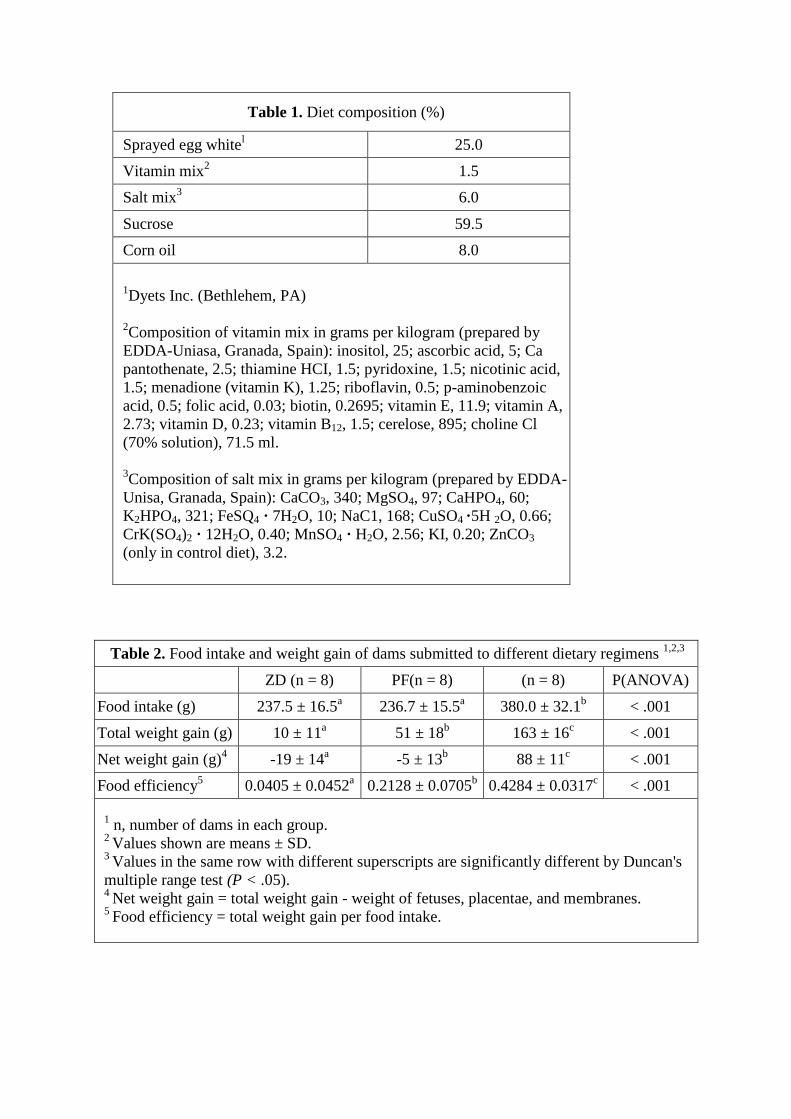
Table 1. Diet composition (%)
Sprayed egg whitel 25.0
Vitamin mix2 1.5
Salt mix3 6.0
Sucrose 59.5
Corn oil 8.0
1Dyets Inc. (Bethlehem, PA)
2Composition of vitamin mix in grams per kilogram (prepared by
EDDA-Uniasa, Granada, Spain): inositol, 25; ascorbic acid, 5; Ca
pantothenate, 2.5; thiamine HCI, 1.5; pyridoxine, 1.5; nicotinic acid,
1.5; menadione (vitamin K), 1.25; riboflavin, 0.5; p-aminobenzoic
acid, 0.5; folic acid, 0.03; biotin, 0.2695; vitamin E, 11.9; vitamin A,
2.73; vitamin D, 0.23; vitamin B12, 1.5; cerelose, 895; choline Cl
(70% solution), 71.5 ml.
3Composition of salt mix in grams per kilogram (prepared by EDDA-
Unisa, Granada, Spain): CaCO3, 340; MgSO4, 97; CaHPO4, 60;
K2HPO4, 321; FeSQ4 · 7H2O, 10; NaC1, 168; CuSO4 ·5H 2O, 0.66;
CrK(SO4)2 · 12H2O, 0.40; MnSO4 · H2O, 2.56; KI, 0.20; ZnCO3
(only in control diet), 3.2.
Table 2. Food intake and weight gain of dams submitted to different dietary regimens 1,2,3
ZD (n = 8) PF(n = 8) (n = 8) P(ANOVA)
Food intake (g) 237.5 ± 16.5a 236.7 ± 15.5
a 380.0 ± 32.1
b < .001
Total weight gain (g) 10 ± 11a 51 ± 18
b 163 ± 16
c < .001
Net weight gain (g)4 -19 ± 14
a -5 ± 13
b 88 ± 11
c < .001
Food efficiency5 0.0405 ± 0.0452
a 0.2128 ± 0.0705
b 0.4284 ± 0.0317
c < .001
1 n, number of dams in each group.
2 Values shown are means ± SD.
3 Values in the same row with different superscripts are significantly different by Duncan's
multiple range test (P < .05). 4
Net weight gain = total weight gain - weight of fetuses, placentae, and membranes. 5
Food efficiency = total weight gain per food intake.

Table 3. Weight of dams submitted to different dietary regimens on days 0, 5,
10, 15 and 21 of gestation 1,2,3
Day ZD
PF C P(ANOVA)
0 201 ± 7 204 ± 10 196 ± 10 NS
5 ―4 219 ± 9 222 ± 8 NX
10 219 ± 14a 225 ± 14
a 245 ± 8
b < .002
15 223 ± 15a 237 ± 10
b 276 ± 8
b < .001
21 211 ± 12a 255 ± 17
b 358 ± 16
c < .001
1 n, number of dams in each group.
2 Values shown are means ± SD.
3 Values in the same row with different superscripts are significantly different by
Duncan's multiple range test (P < .05). 4
Not measured.
Table 4. Reproductive parameters in dams submitted to different dietary regimens 1,2
Reproductive parameters ZD (n = 8) PF (n = 8) C (n = 8) P3
Implantation sites per litter4 12.9 ± 1.8 13.9 ± 2.4 12.6 ± 2.2 NS†
Live fetuses per litter4 9.6 ± 2.9
a 13.2 ± 2.1
b 11.9 ± 2.0
a,b < .02†
Fetal weight5 2.43 ± 0.50
a 3.54 ± 0.61
b 5.46 ± 0.19
c < .001†
Resorptions
Percent of implantation sites 25.2* 4.5** 5.9** < .001‡
Litters with resorptions 6/8 3/8 6/8
Malformations6
Percent of live fetuses 98.7* 1.9** 0** < .001‡
Litters with malformations 8/8 1/8 0
Sites affected7
Percent of implantation sites 99.0* 5.4** 5.9** < .001‡
Litters affected 8/8 4/8 6/8 1n, number of dams in each group.
2Values in the same row with different superscripts are significantly different (P < .05) by Duncan's
multiple range test (alphabetical superscripts) and chi-squared test (asterisks). 3ANOVA P value (†) or chi-square P value (‡)
4Values shown are means ± SD.
5Values shown are means of litter means SD.
6Malformations detected by external observation or by examination of fetal skeleton in stained
specimens. 7Resorptions and malformations.

Table 5. Malformations detected by external observation of the fetuses from zinc-
deficient dams1
Malformation Affected fetuses (%)2 Litters affected
Head
Exencephaly 1 1
Meningoencephalocele 8 3
Meningocele 19 4
Hydrocephaly 4 1
Microphthalmia 17 2
Micrognathia 35 5
Trunk
Umbilical hernia 12 4
Gastroschisis 27 5
Forelimbs2
Syndactyly 44 (65) 7
Shortening 21 (100) 3
Dysplasia3 17 (85) 3
Oligodactyly 40 (84) 7
Hindlimbs2
Syndactyly 12 (33) 5
Shortening 17 (100) 3
Dysplasia 53 (80) 6
Oligodactyly 4 (0) 2
Extra elements 3 (100) 1
Tail
Short4 3 2
Angulated 3 2
Wavy 26 4
Curly 57 6
1All fetuses (77) from all litters (8) examined.
2Percentage of bilateral defects in brackets.
3Predominantly lateral.
4Only fetuses that did not present any other defect of the tail.

Table 6. Skeletal malformations detected in fetuses from zinc-deficient dams by
examination of cleared specimens1
Malformation2
Fetuses
affected
(%)
Litters
affected3
Head (44) Micrognathia 25.0 3
Spine (43) Fused vertebrae 41.9 6 Hemivertebrae 74.4 7
Tail (35) Short 71.4 6 Fused vertebrae 95.2 8 Malformed 97.1 8
Ribs (43) Absent 69.8
4 7
Short 30.2 5 Fused 7.0 2 Dysplastic 25.6 5
Sternum (30) Absent 46.7 5 Fused ossification nuclei 3.3 1
Forelimbs: arm and forearm (44) Curved humerus 2.3 1 Synarthrosis (elbow)
5 18.2 1
Dysplasia and/or hypoplasia of ulna and radius 25.0 4 Forelimbs: hands (42)
Fused metacarpals 11.9 4 Transversally fused phalanges
6 11.9 5
Longitudinal synchondrosis (carpus and phalanges) 2.4 1 Clinodactyly 19.0 3 Digital hypoplasia 4.8 2 Oligodactyly
7 57.1 7
Duplication 2.4 1 Hindlimbs: Thigh and leg (41)
Hypoplastic femur
19.5 3 Hypoplastic tibia 14.6 2 Dysplastic tibia 24.4 5 Absent fibula 4.9 1 Hypoplastic fibula 24.4 5
Hindlimbs: feet (38) Fused metatarsals 15.8 2 Transversally fused phalanges
6 2.6 1
Clinodactyly 2.6 1 Digital hypoplasia 5.3 1 Oligodactyly
7 23.7 4
Duplication 2.6 1 1Does not include calcification defects.
2Number of specimens examined in brackets.
3Eight litters examined.
4Total absence in 25.6% and partial absence in 44.2%.
5Synchondrosis, except for one fetus that presented unilateral synostosis.
6Whether or not accompanied by fusion of metacarpals or metatarsals.
7Includes total and partial ectrodactylies.

Table 7. Ossification nuclei of fetuses from dams submitted to different
dietary regimens1,2,3
Location ZD PF C P (ANOVA)
Sternum (16) 1.95 ± 1.42 a (74) 4.56 ± 0.88
b (57) 5.45 ± 0.69
b < .001
Hands4 (80) 1.96 ± 0.95
a (146) 5.50 ± 1.60
b (116) 6.61 ± 0.59
b < .001
Feet5 (69) 2.57 ± 1.20
a (147) 3.96 ± 0.07
b (124) 4.22 ± 0.22
b < .001
1Number of specimens examined in brackets.
2Values shown are means of litter means ± SD.
3Values in the same row with different superscripts are significantly different by Duncan's
multiple range test (P < .05). 4Metacarpals and anterior proximal phalanges.
5Metatarsals.
Table 8. Effect of gestational zinc deficiency on concentrations of zinc, calcium, and
phosphorus in maternal and fetal tissues 1,2,3
ZD (n = 8) PF (n = 8) C (n = 8)4 P (ANOVA)
Maternal plasma
Zinc5 26.00 ± 7.09
a 50.37 ± 9.65
b 94.50 ± 12.25
c < .001
Magnesium6 1.96 ± 0.27
1.77 ± 0.28 1.91 ± 0.16 NS
Calcium6 8.53 ± 1.22
a 7.22 ± 0.36
b 8.82 ± 0.39
a < .001
Phosphorus6 7.42 ± 1,27
a 5.92 ± 0.76
b 6.47 ± 1.28
a b < .05
Maternal Tibia
Zinc7 183.72 ± 16.36
a 222.21 ± 17.33
b 227.04 ± 16.79
b < .01
Magnesium8 11.40 ± 0.78 11.35 ± 1.31 12.44 ± 2.85 NS
Calcium8 183.74 ± 7.26 183.99 ± 17.59 197.45 ± 37.45 NS
Phosphorus8 97.48 ± 3.88 96.64 ± 5.24 98.13 ± 4.39 NS
Fetal liver
Zinc7 50.29 ± 13.30 46.20 ± 11.48 53.31 ± 8.00 NS
Fetal tibia
Zinc7 88.25 ± 37.60
a 142.62 ± 18.33
b 164.14 ± 15.07
b < .001
Magnesium8 3.58 ± 1.74 3.46 ± 1.60 2.14 ± 0.67 NS
Calcium8 46.82 ± 10.08
a 85.91 ± 10.46
b 79.59 ± 8.72
b < .001
Phosphorus8 38.05 ± 1.74
a 56.07 ± 4.32
b 50.10 ± 5.80
c < .001
Ca:P ratio 1.22 ± 0.13 a 1.53 ± 0.10
b 1.59 ± 0.03
b < .001
1n, number of dams and litters in each group.
2Values shown are mean ± SD.
3Values in the same row with different superscripts are significantly different by Duncan's
multiple range test (P < .05). 4For Zn, Ca, and P in fetal tibia n = 7.
5Concentrations are expressed in micrograms per 100 ml.
6Concentrations are expressed in milligrams per 100 ml.
7Concentrations are expressed in micrograms per gram.
8Concentrations are expressed in milligrams per gram.
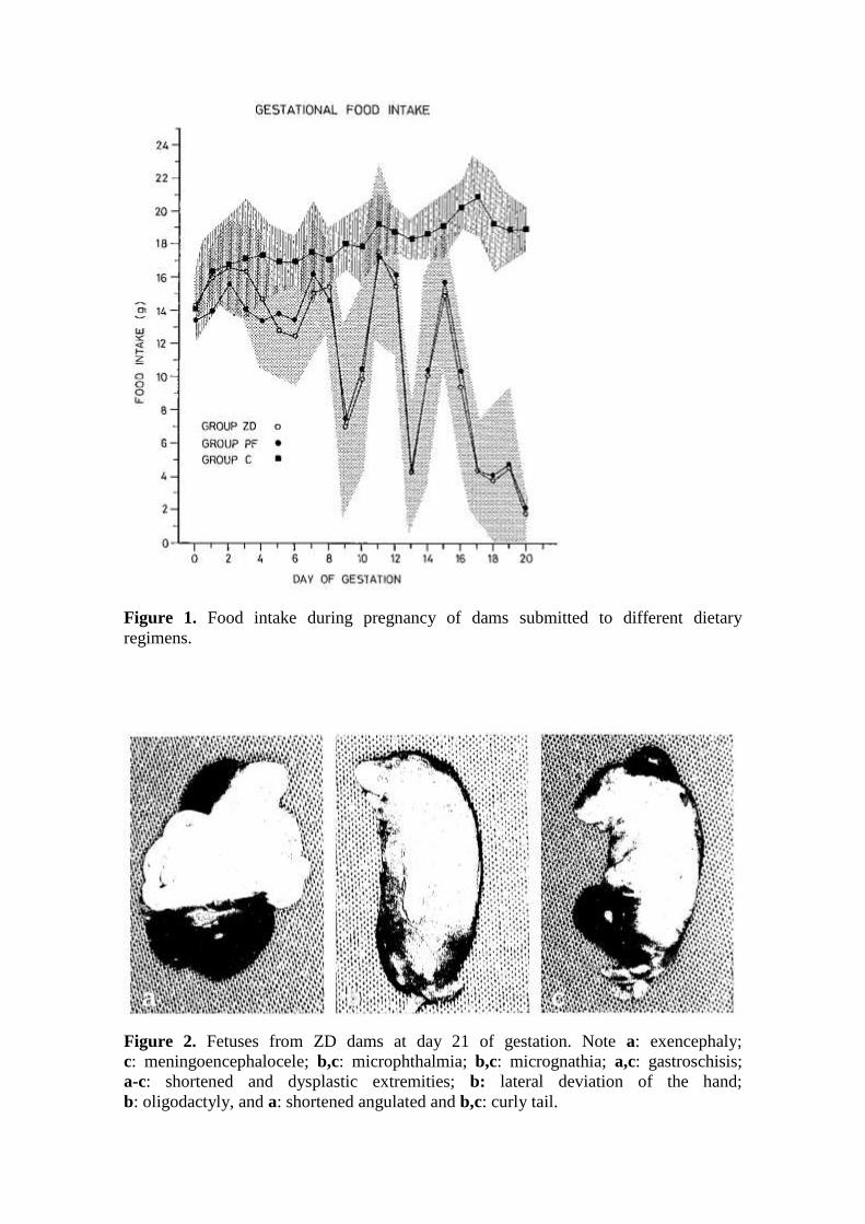
Figure 1. Food intake during pregnancy of dams submitted to different dietary
regimens.
Figure 2. Fetuses from ZD dams at day 21 of gestation. Note a: exencephaly;
c: meningoencephalocele; b,c: microphthalmia; b,c: micrognathia; a,c: gastroschisis;
a-c: shortened and dysplastic extremities; b: lateral deviation of the hand;
b: oligodactyly, and a: shortened angulated and b,c: curly tail.

Figure 3. Skeletal defects in stained and cleared fetuses from ZD dams at day 21 of
gestation. a: Short tail with fused vertebrae and spina bifida. b,c: Spine with fused
vertebrae and hemivertebrae. b: Total and c: partial absence of ribs, and c-e: short,
fused, and malformed ribs.
Figure 4. Skeletal malformations of forelimbs in stained fetuses from ZD dams at day
21 of gestation. a,b: Synarthrosis of the elbow and dysplastic bones of the forearm;
a,b,d: fused carpal bones; b,d: fused metacarpals; b,d: transversally fused phalanges,
and a-c: oligodactyly.

Figure 5. Skeletal malformations of hind limbs in stained fetuses from ZD dams at day
21 of gestation. a-c: Hypoplastic femur; a-d: hypoplastic and dysplastic tibia and fibula;
d: fused metatarsals and transverselly fused phalanges; e: clinodactyly; and d: digital
hypoplasia.