TENDINOPATHIES ABOUT THE KNEE -...
68
Tendinopathies About The Knee Diagnosis, Conservative / Surgical Treatment Chih-Hwa Chen, MD Department of Orthopaedic Surgery Taipei Medical University Hospital Taipei Medical University Taipei, Taiwan
Transcript of TENDINOPATHIES ABOUT THE KNEE -...
Tendinopathies About The KneeDiagnosis, Conservative / Surgical Treatment
Chih-Hwa Chen, MD
Department of Orthopaedic SurgeryTaipei Medical University Hospital
Taipei Medical UniversityTaipei, Taiwan

Taipei, Taiwan
Tallinn, Estonia

• Tendon unit:• Tendon
• Myotendinous junction
• Enthesis: tendon-bone insertion
• Tendon: • Endotenon
• Peritendon: epitenon / paratennon
• Tendon sheath
• Bursa
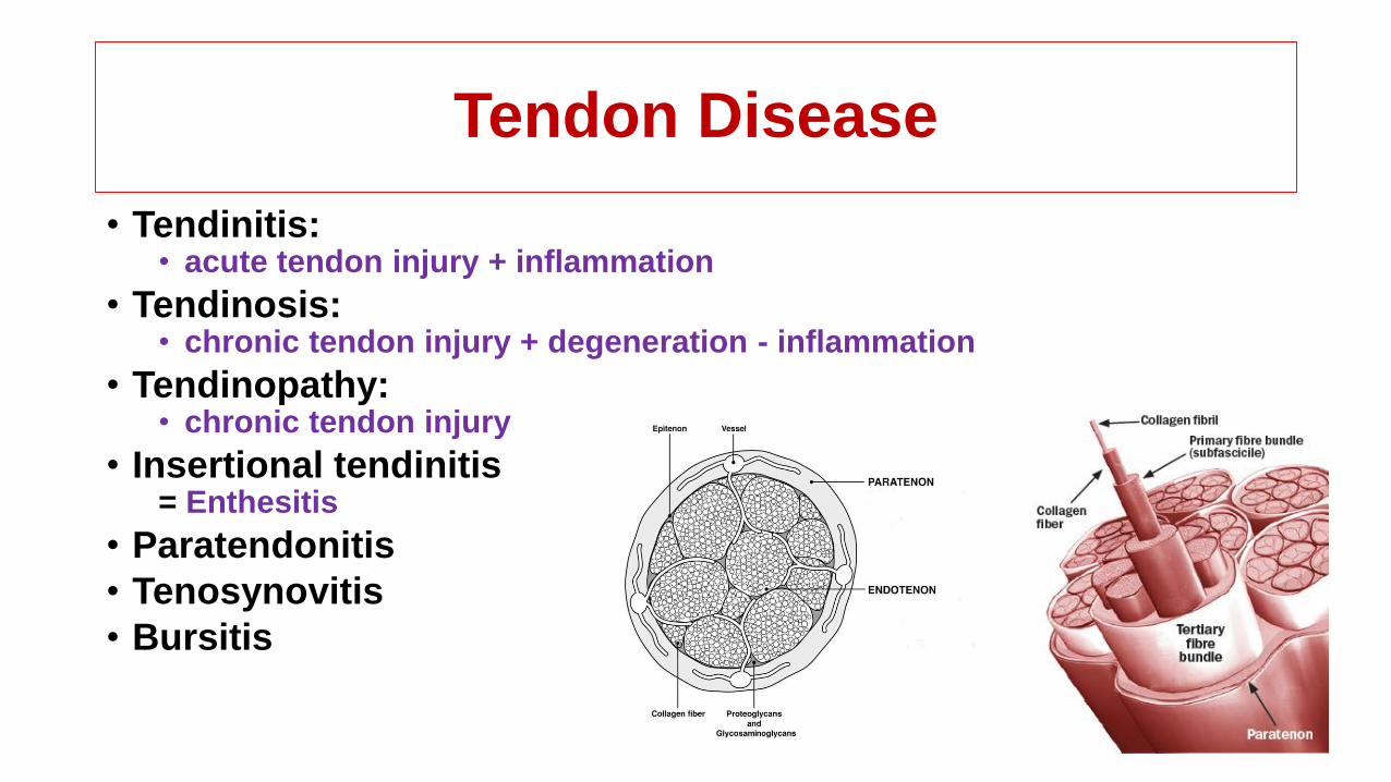
Tendon Disease
• Tendinitis: • acute tendon injury + inflammation
• Tendinosis: • chronic tendon injury + degeneration - inflammation
• Tendinopathy: • chronic tendon injury
• Insertional tendinitis = Enthesitis
• Paratendonitis
• Tenosynovitis
• Bursitis
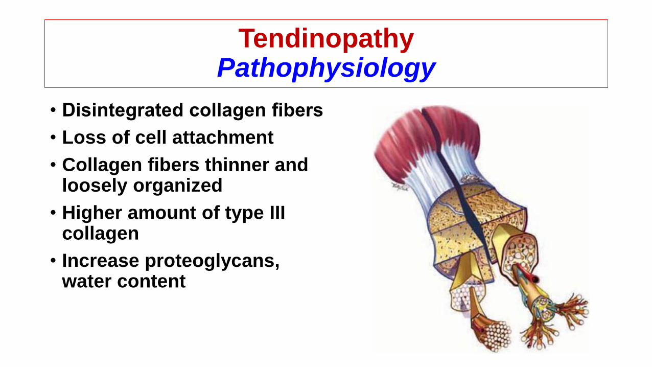
TendinopathyPathophysiology
• Disintegrated collagen fibers
• Loss of cell attachment
• Collagen fibers thinner and loosely organized
• Higher amount of type III collagen
• Increase proteoglycans, water content
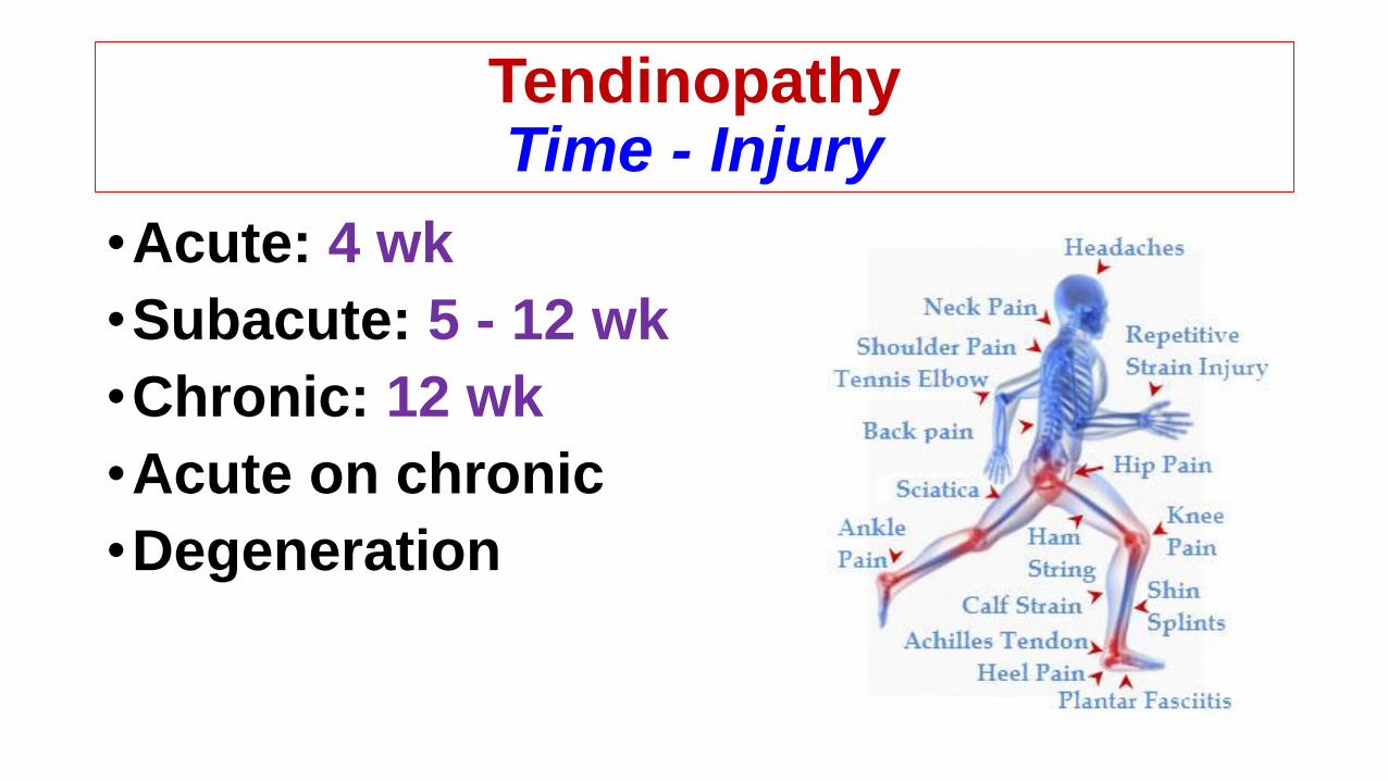
TendinopathyTime - Injury
•Acute: 4 wk
•Subacute: 5 - 12 wk
•Chronic: 12 wk
•Acute on chronic
•Degeneration

TendinopathyMechanism
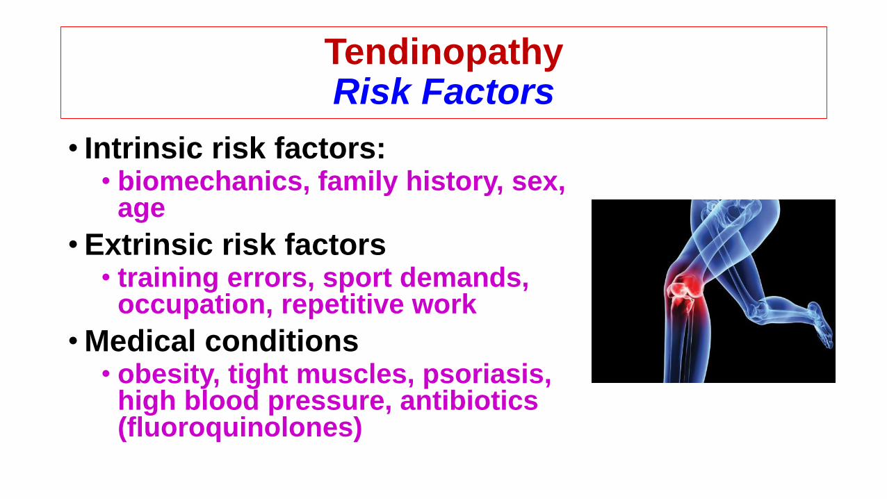
Tendinopathy Risk Factors
• Intrinsic risk factors:• biomechanics, family history, sex,
age
•Extrinsic risk factors• training errors, sport demands,
occupation, repetitive work
•Medical conditions• obesity, tight muscles, psoriasis,
high blood pressure, antibiotics (fluoroquinolones)

Tendinopathy Additional Features
• Calcification• Primary / Dystrophic
• Bony change• Overlaying spur, Insertional spur, Traction spur
• Joint pathology• OA, ligament injury, chondral tear, meniscus tear
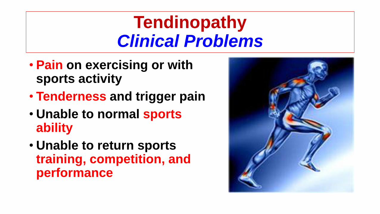
TendinopathyClinical Problems
• Pain on exercising or with sports activity
• Tenderness and trigger pain
• Unable to normal sports ability
• Unable to return sports training, competition, and performance
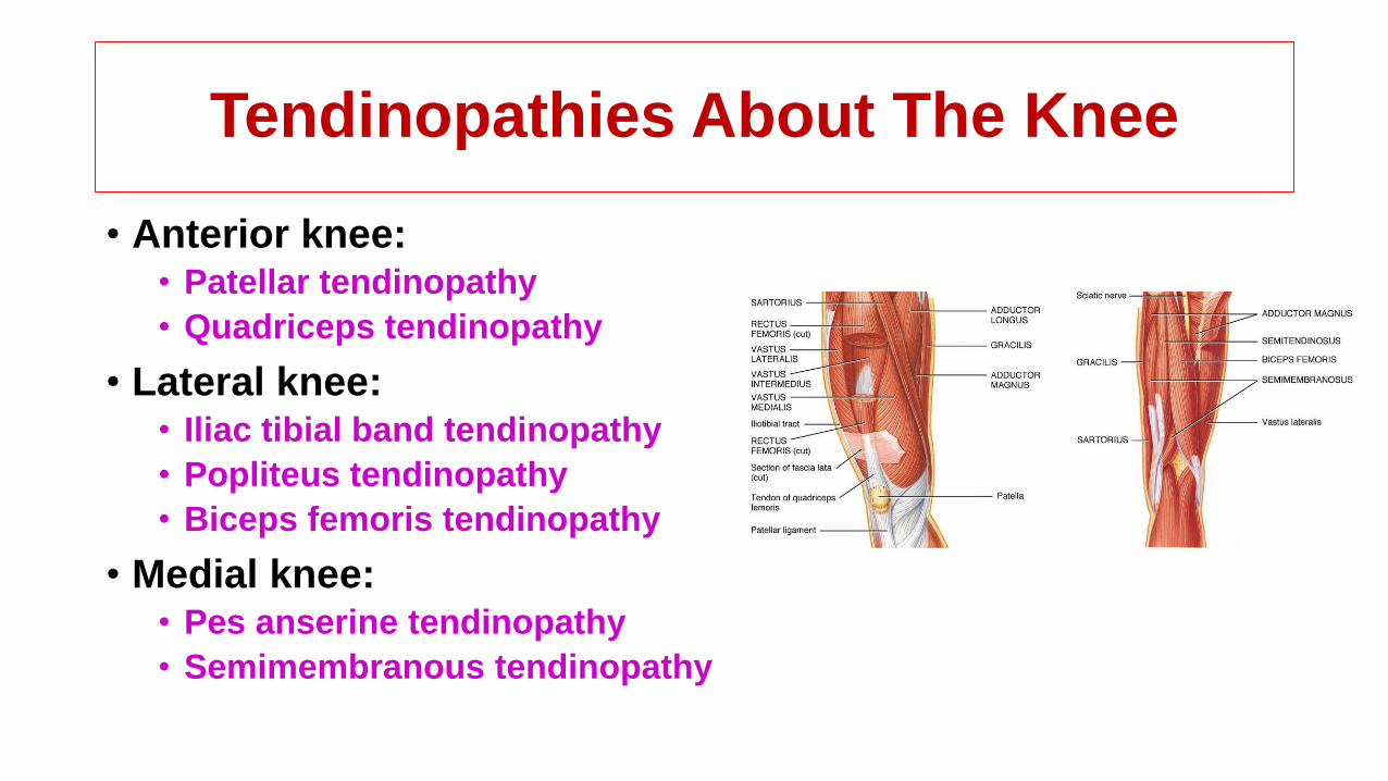
Tendinopathies About The Knee
• Anterior knee:• Patellar tendinopathy
• Quadriceps tendinopathy
• Lateral knee:• Iliac tibial band tendinopathy
• Popliteus tendinopathy
• Biceps femoris tendinopathy
• Medial knee:• Pes anserine tendinopathy
• Semimembranous tendinopathy

Patellar Tendinopathy
Jumper’s KneeAnteriorKnee

Patellar TendinopathyStructure
•Epidemiology• incidence
• Up to 20% of jumping athletes
• Pathophysiology• mechanism
• repetitive, forceful, eccentric contraction of the extensor mechanism
• histology• degenerative, rather than inflammatory
• Micro-tears of the tendinous tissue are commonly seen
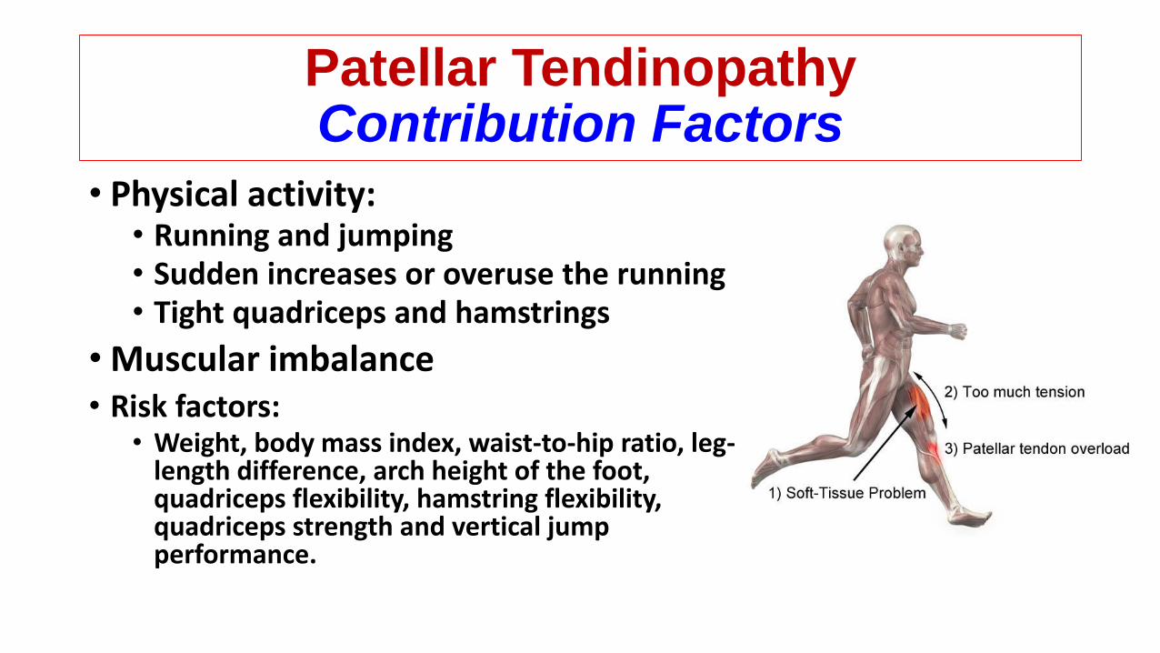
Patellar TendinopathyContribution Factors
• Physical activity:• Running and jumping • Sudden increases or overuse the running• Tight quadriceps and hamstrings
• Muscular imbalance• Risk factors:
• Weight, body mass index, waist-to-hip ratio, leg-length difference, arch height of the foot, quadriceps flexibility, hamstring flexibility, quadriceps strength and vertical jump performance.

Study Factor
Risk factor /
associated
factor
Patellar
tendinopathy /
tendon pathology
Comment
Visnes
CookGender Both Both Men at higher risk
Malliaras Waist circumference Associated PathologyIncreased waist circumference associated with
increased pathology
Cook Imaging abnormality Risk Tendinopathy Adolescents only
Cook Hamstring length Associated Pathology Less extensible hamstrings associated with pathology
Witvrouw Hamstring length Risk TendinopathyLess extensible hamstrings increase risk of patellar
tendinopathy
Witvrouw Quadriceps length Risk TendinopathyStiffer quadriceps increase risk of patellar
tendinopathy
Malliaras Dorsiflexion Associated PathologyReduced dorsiflexion associated with increased
pathology
EdwardsAltered landing
strategiesAssociated Pathology
Less knee bend at landing, altered hip strategies
associated with pathology
Lian Jumping ability Both TendinopathyBetter jumping ability associated with patellar
tendinopathy
Culvenor Fat pad size Associated TendinopathyIncreased fat pad size associated with patellar
tendinopathy
Gaida
JannsenLoading Associated Tendinopathy Excess loading associated with patellar tendinopathy
Patellar TendinopathyDiagnosis
• Classification:• Blazina classification system
• phase I• pain after activity only
• phase II• pain at the beginning of activity, disappearing after warm-up, and
reappearing after completion of an activity
• phase III• persistent pain with or without activities• deterioration of performance• unable to participate in sports.
• phase IV• complete rupture of the patellar tendon
Patellar Tendinopathy Diagnosis
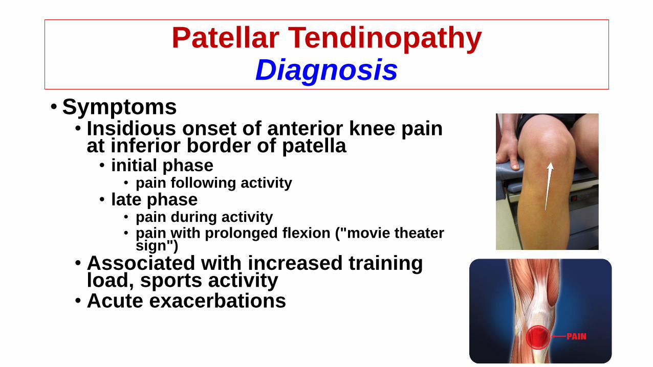
•Symptoms • Insidious onset of anterior knee pain
at inferior border of patella• initial phase
• pain following activity
• late phase• pain during activity• pain with prolonged flexion ("movie theater
sign")
• Associated with increased training load, sports activity
• Acute exacerbations
Patellar Tendinopathy Diagnosis
• Physical exam• inspection
• may have swelling over tendon and lower pole of patella
• palpation• tenderness at inferior border of patella
• provocative tests• Basset's sign
• tenderness to palpation at distal pole of patella in full extension
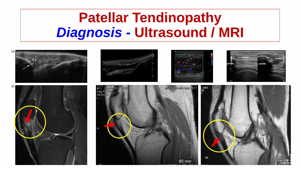
Patellar Tendinopathy Diagnosis - Ultrasound / MRI
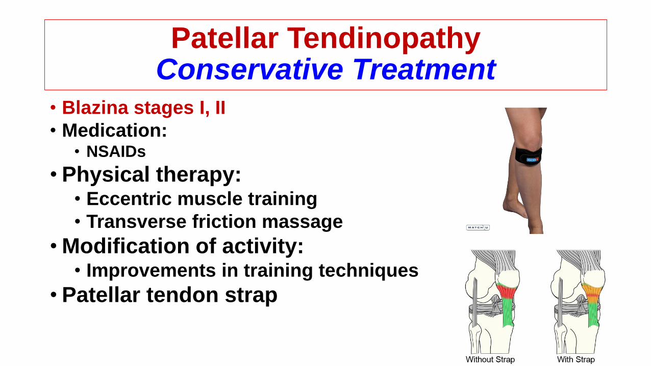
Patellar TendinopathyConservative Treatment
• Blazina stages I, II
• Medication:• NSAIDs
• Physical therapy:• Eccentric muscle training
• Transverse friction massage
• Modification of activity:• Improvements in training techniques
• Patellar tendon strap
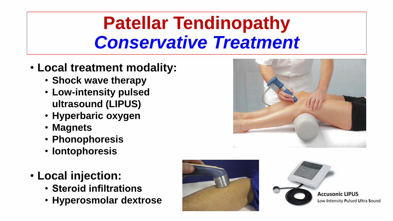
Patellar TendinopathyConservative Treatment
• Local treatment modality:• Shock wave therapy
• Low-intensity pulsed
ultrasound (LIPUS)
• Hyperbaric oxygen
• Magnets
• Phonophoresis
• Iontophoresis
• Local injection:• Steroid infiltrations
• Hyperosmolar dextrose
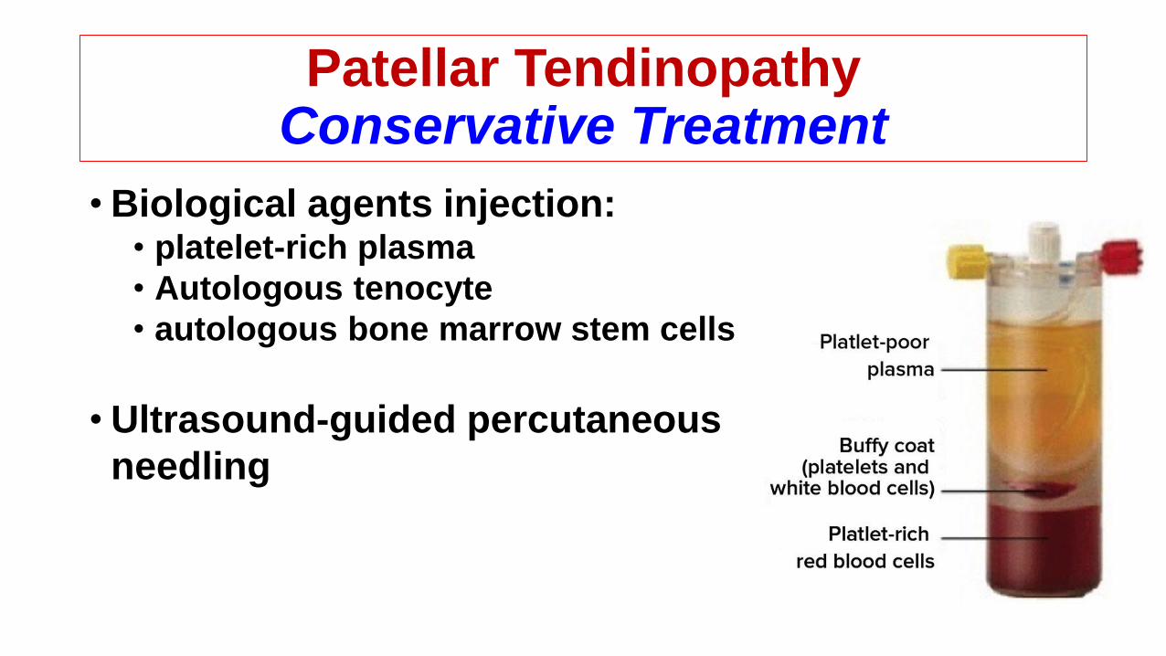
Patellar TendinopathyConservative Treatment
• Biological agents injection:• platelet-rich plasma
• Autologous tenocyte
• autologous bone marrow stem cells
• Ultrasound-guided percutaneous
needling

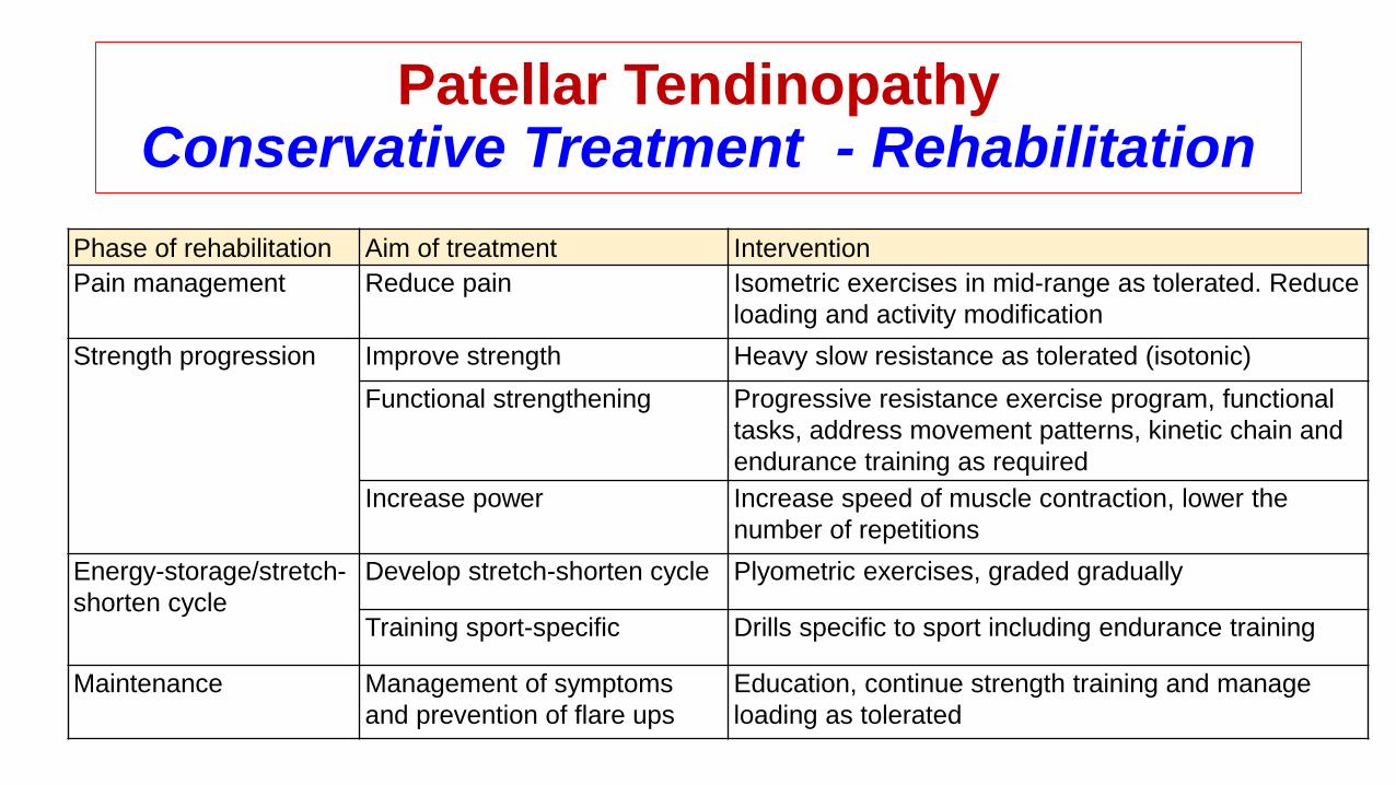
Patellar TendinopathyConservative Treatment - Rehabilitation
• Activity modification:
• Cryotherapy:
• Joint motion and kinematics assessment:
• Stretching:
• Strengthening:
• Sport-specific proprioceptive training and plyometric
• Ultrasonography or phonophoresis
• patellofemoral brace
• McConnell taping
Patellar TendinopathyConservative Treatment - Rehabilitation
Phase of rehabilitation Aim of treatment Intervention
Pain management Reduce pain Isometric exercises in mid-range as tolerated. Reduce
loading and activity modification
Strength progression Improve strength Heavy slow resistance as tolerated (isotonic)
Functional strengthening Progressive resistance exercise program, functional
tasks, address movement patterns, kinetic chain and
endurance training as required
Increase power Increase speed of muscle contraction, lower the
number of repetitions
Energy-storage/stretch-
shorten cycle
Develop stretch-shorten cycle Plyometric exercises, graded gradually
Training sport-specific Drills specific to sport including endurance training
Maintenance Management of symptoms
and prevention of flare ups
Education, continue strength training and manage
loading as tolerated
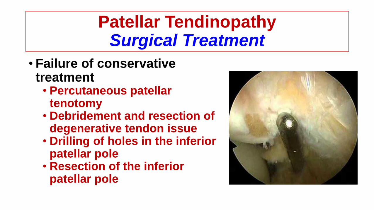
Patellar Tendinopathy Surgical Treatment
• Failure of conservative treatment• Percutaneous patellar
tenotomy • Debridement and resection of
degenerative tendon issue• Drilling of holes in the inferior
patellar pole • Resection of the inferior
patellar pole
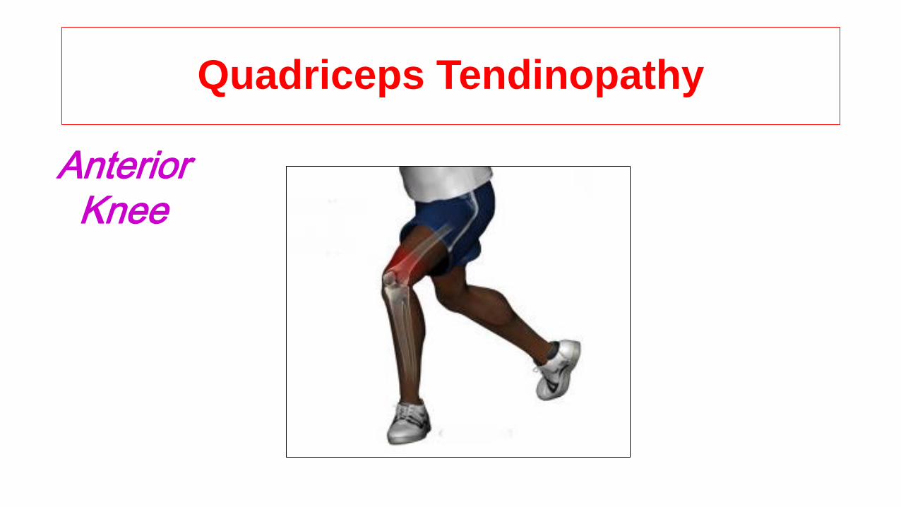
Quadriceps Tendinopathy
AnteriorKnee
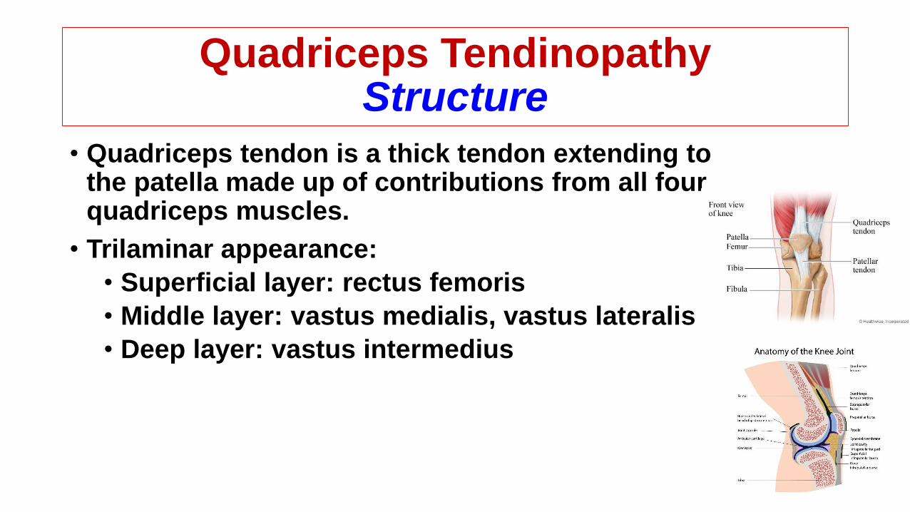
Quadriceps TendinopathyStructure
• Quadriceps tendon is a thick tendon extending to the patella made up of contributions from all four quadriceps muscles.
• Trilaminar appearance:
• Superficial layer: rectus femoris
• Middle layer: vastus medialis, vastus lateralis
• Deep layer: vastus intermedius
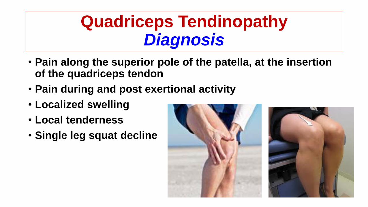
Quadriceps TendinopathyDiagnosis
• Pain along the superior pole of the patella, at the insertion of the quadriceps tendon
• Pain during and post exertional activity
• Localized swelling
• Local tenderness
• Single leg squat decline
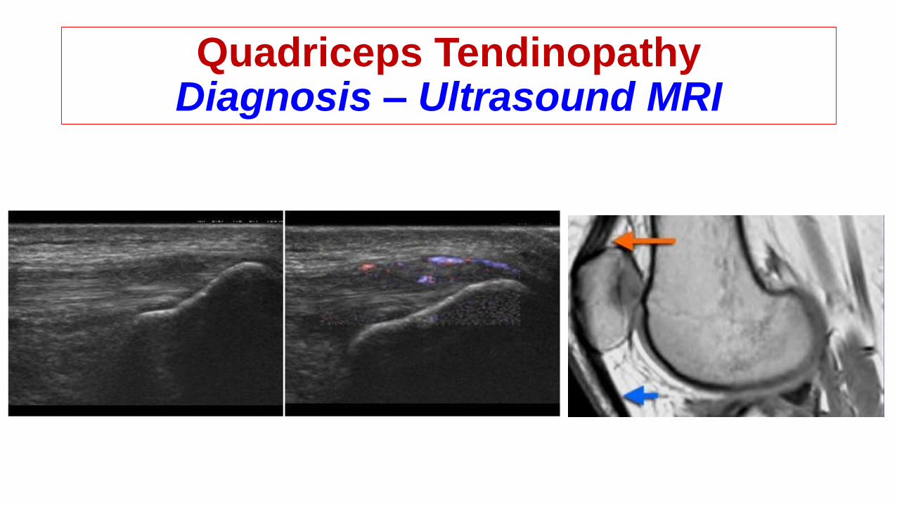
Quadriceps TendinopathyDiagnosis – Ultrasound MRI

Quadriceps TendinopathyContribution Factors
• Joint stiffness (particularly the hip, knee, ankle or lower back)
• Muscle tightness (particularly the quadriceps, hamstrings or calfs)
• Inappropriate or excessive training
• Inadequate warm up
• Muscle weakness (especially the quadriceps and / or gluteals)
• Poor pelvic or core stability
• Inadequate rehabilitation following a previous quadriceps injury
• Poor foot posture or other biomechanical issues
• Inappropriate footwear
• Medical disease:• Hyperparathyroidism • calcium pyrophosphate deposition • diabetes mellitus •
steroid induced tendinopathy • fluroquinolone induced tendinopathy • osteomalacia • chronic renal insufficiency • gout • uraemia

Quadriceps TendinopathyConservative Treatment
•Eccentric exercises
•Stretching
•PRP
•Shock wave therapy
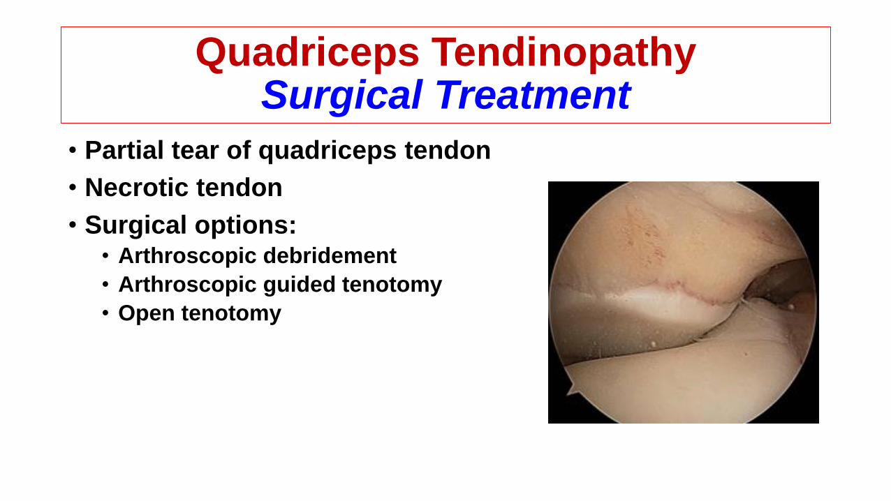
Quadriceps TendinopathySurgical Treatment
• Partial tear of quadriceps tendon
• Necrotic tendon
• Surgical options: • Arthroscopic debridement
• Arthroscopic guided tenotomy
• Open tenotomy

Iliotibial Band Tendinopathy
Runner’s Knee
Cyclist’s KneeLateralKnee
Iliotibial Band TendinopathyStructure
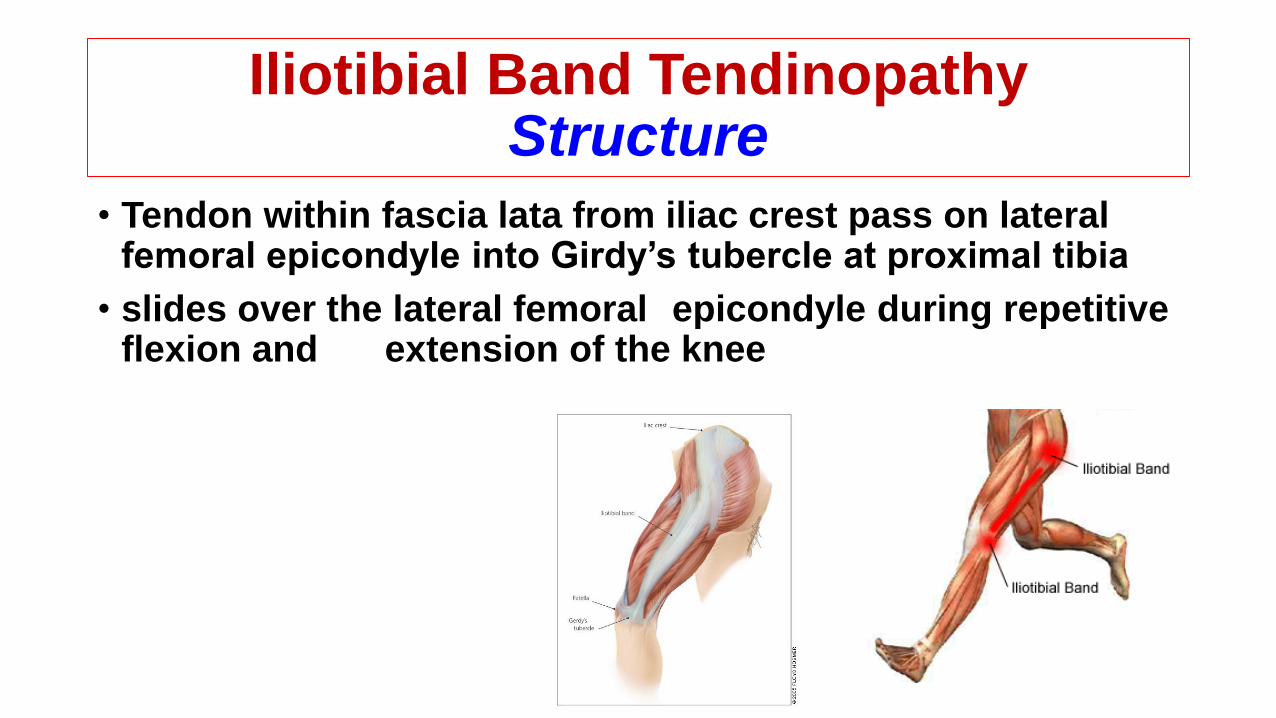
• Tendon within fascia lata from iliac crest pass on lateral femoral epicondyle into Girdy’s tubercle at proximal tibia
• slides over the lateral femoral epicondyle during repetitive flexion and extension of the knee
Iliotibial Band TendinopathyDiagnosis
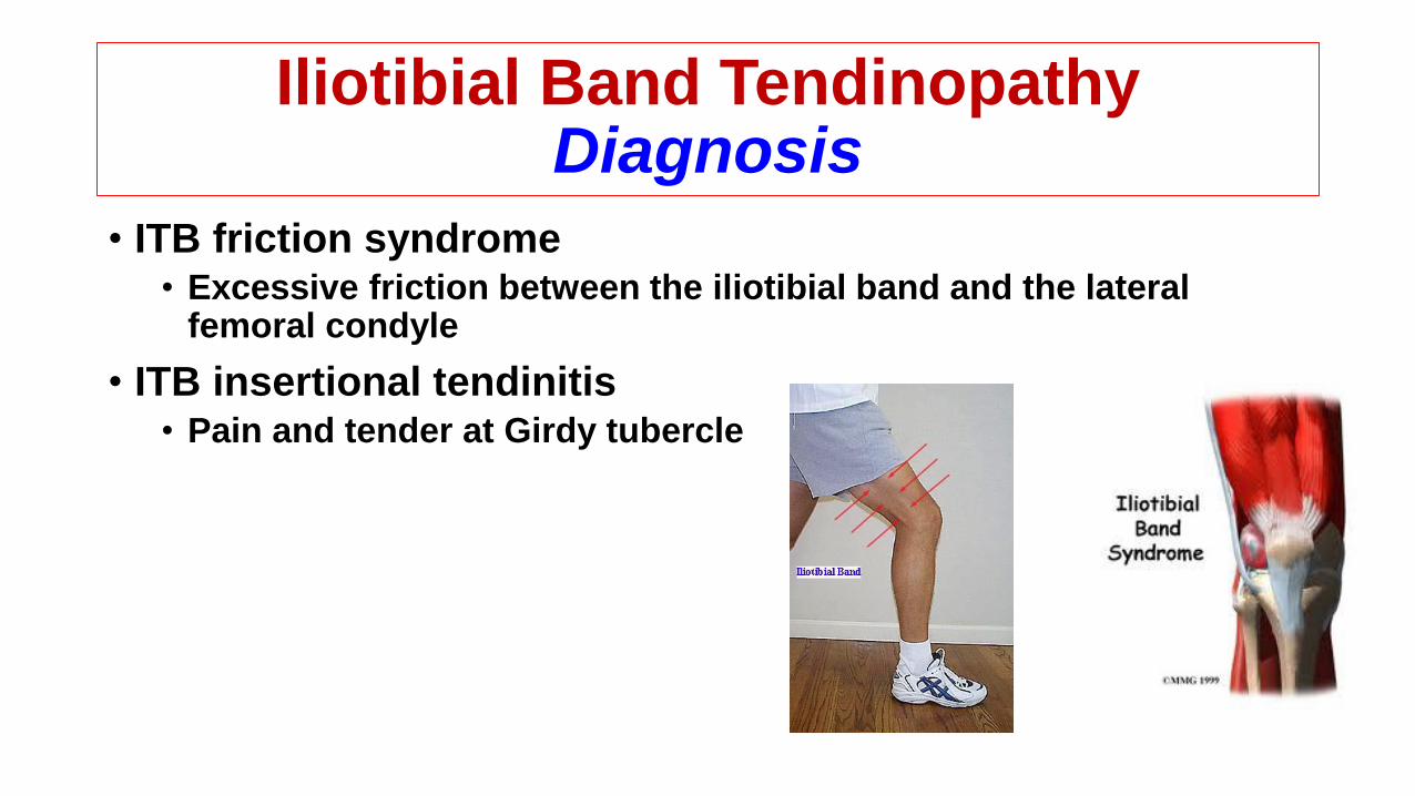
• ITB friction syndrome• Excessive friction between the iliotibial band and the lateral
femoral condyle
• ITB insertional tendinitis• Pain and tender at Girdy tubercle
Iliotibial Band TendinopathyDiagnosis
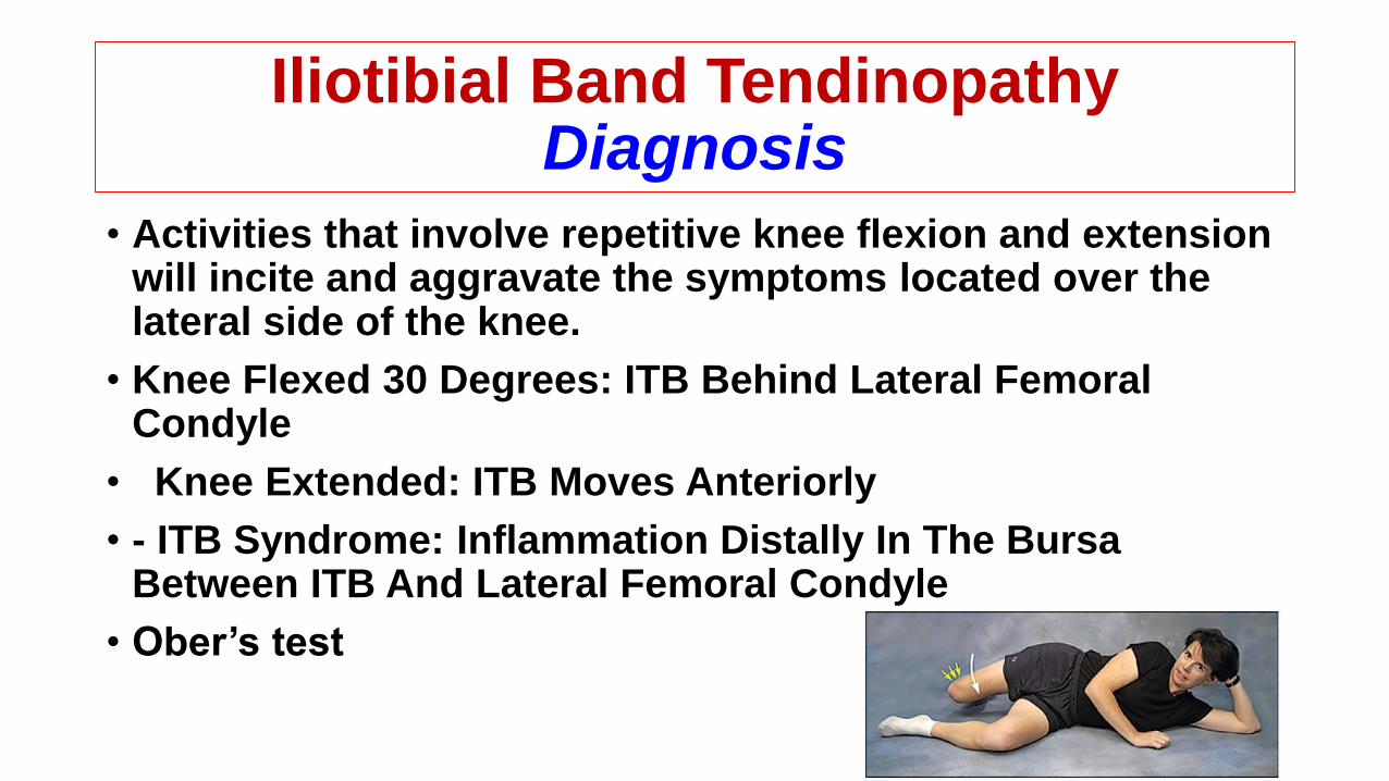
• Activities that involve repetitive knee flexion and extension will incite and aggravate the symptoms located over the lateral side of the knee.
• Knee Flexed 30 Degrees: ITB Behind Lateral Femoral Condyle
• Knee Extended: ITB Moves Anteriorly
• - ITB Syndrome: Inflammation Distally In The Bursa Between ITB And Lateral Femoral Condyle
• Ober’s test
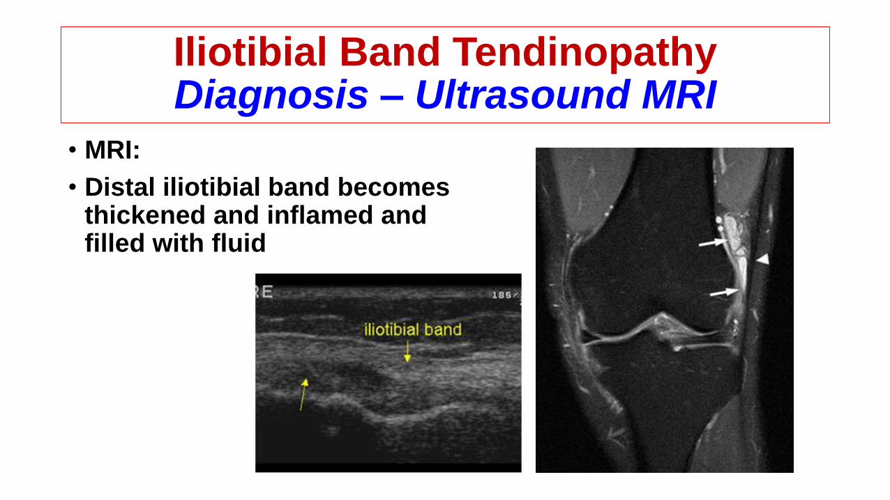
Iliotibial Band TendinopathyDiagnosis – Ultrasound MRI
• MRI:
• Distal iliotibial band becomes thickened and inflamed and filled with fluid
Iliotibial Band TendinopathyContribution Factors
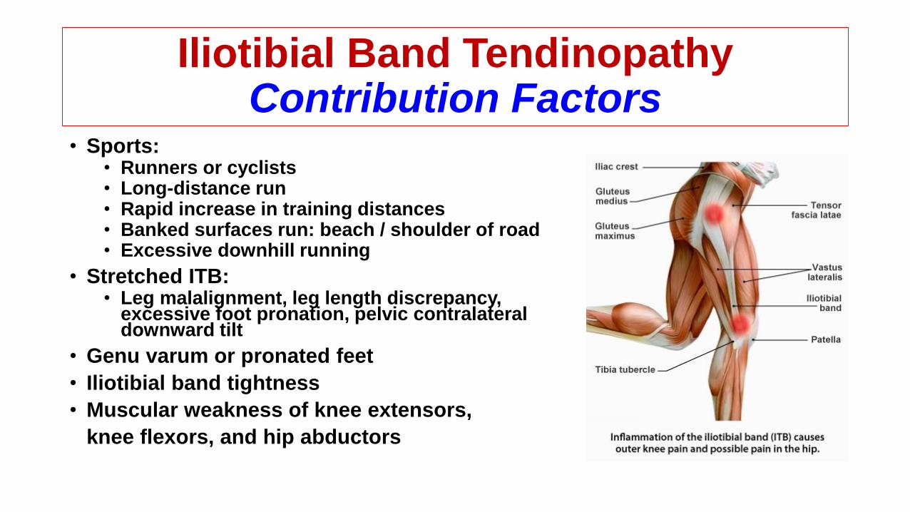
• Sports:• Runners or cyclists• Long-distance run• Rapid increase in training distances• Banked surfaces run: beach / shoulder of road• Excessive downhill running
• Stretched ITB:• Leg malalignment, leg length discrepancy,
excessive foot pronation, pelvic contralateral downward tilt
• Genu varum or pronated feet
• Iliotibial band tightness
• Muscular weakness of knee extensors,
knee flexors, and hip abductors
Iliotibial Band TendinopathyConservative Treatment
• Reduction of training distance
• NSAIDS
• Stretching ITB
• Strengthen ipsilateral hip abductors• Correction of mal-alignments
• Utilize proper warm-up and stretching techniques
• Avoidance of aggravating activities
• Orthotics
• Local infiltration of corticosteroid
Iliotibial Band TendinopathySurgical Treatment
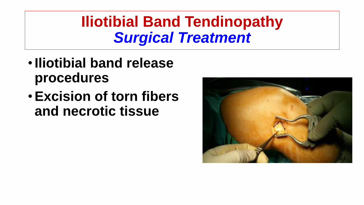
• Iliotibial band release procedures
•Excision of torn fibers and necrotic tissue
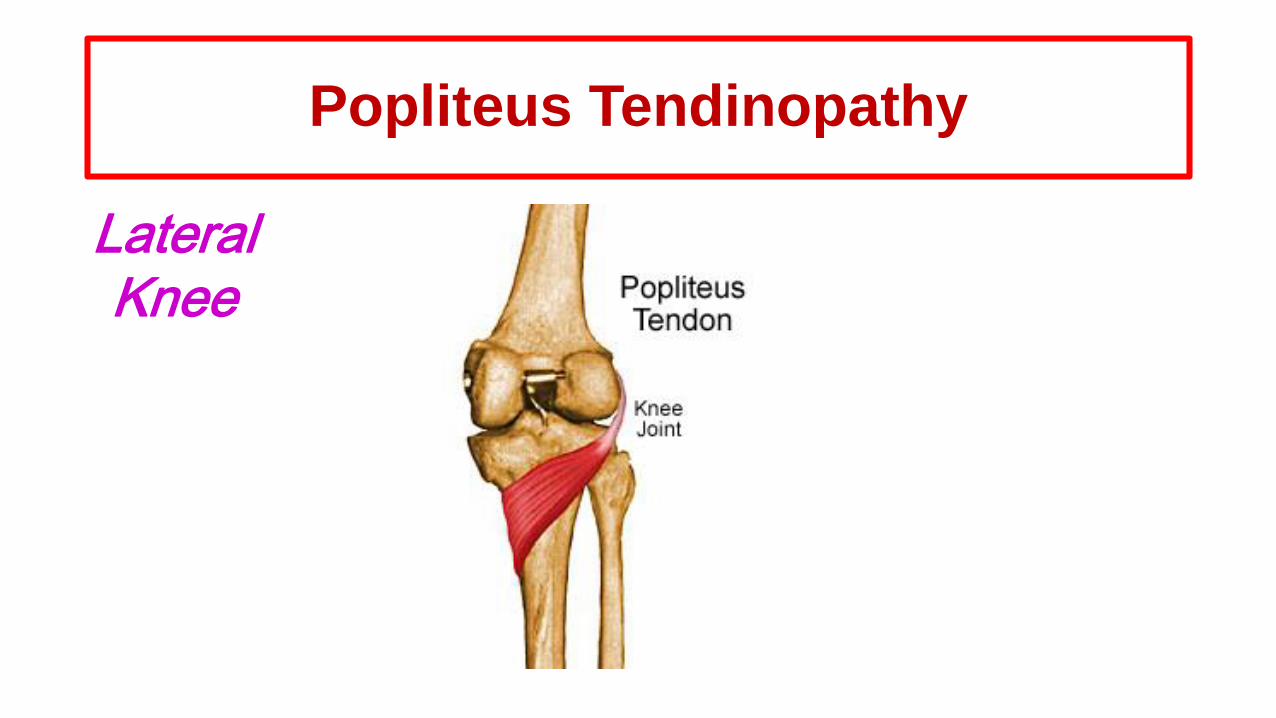
Popliteus Tendinopathy
LateralKnee
Popliteal TendinopathyStructure
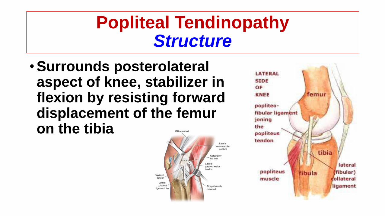
•Surrounds posterolateral aspect of knee, stabilizer in flexion by resisting forward displacement of the femur on the tibia
Popliteal TendinopathyDiagnosis
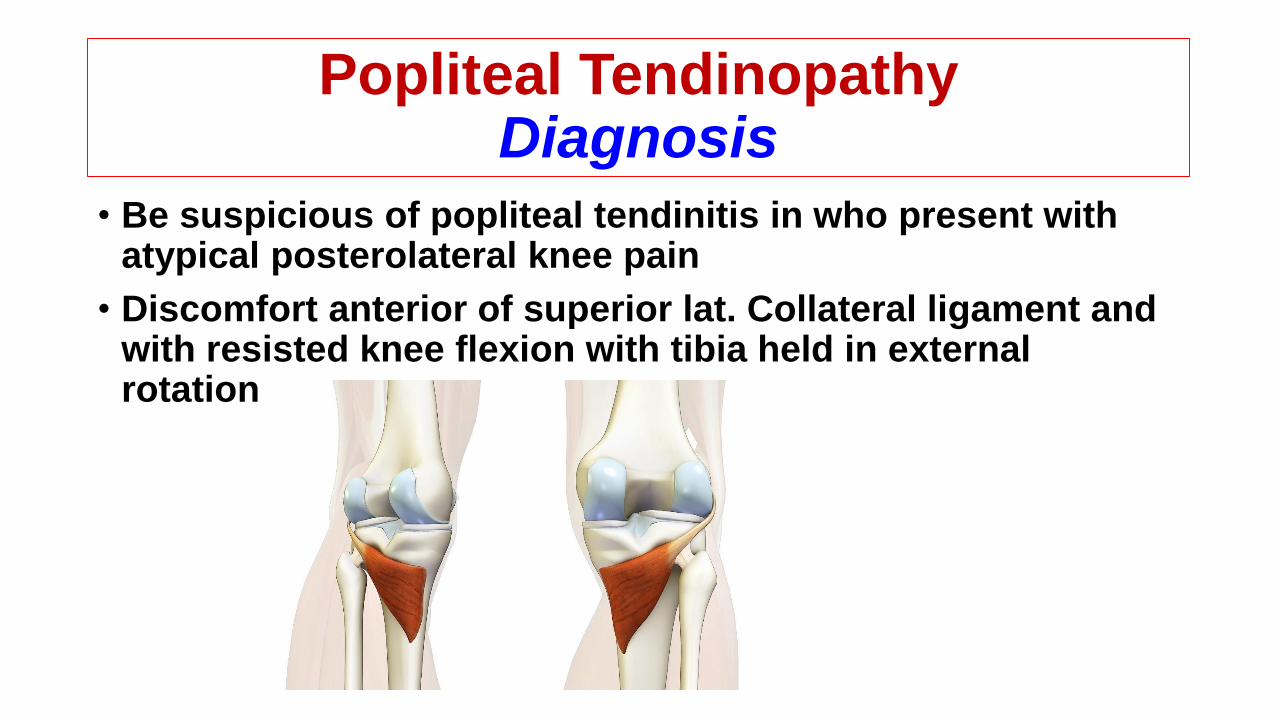
• Be suspicious of popliteal tendinitis in who present with atypical posterolateral knee pain
• Discomfort anterior of superior lat. Collateral ligament and with resisted knee flexion with tibia held in external rotation

Popliteal TendinopathyContribution Factors
•Cross-country running
•Extensive downhill walking or running
•Long-distance runners and walkers
Popliteal TendinopathyConservative Treatment
•Reduction training distance
•NSAIDS
•Stretching knee flexors
•Electrotherapy
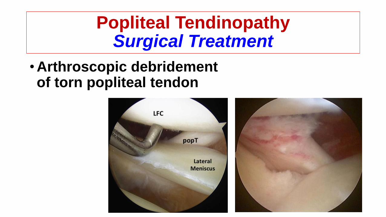
Popliteal TendinopathySurgical Treatment
•Arthroscopic debridement of torn popliteal tendon
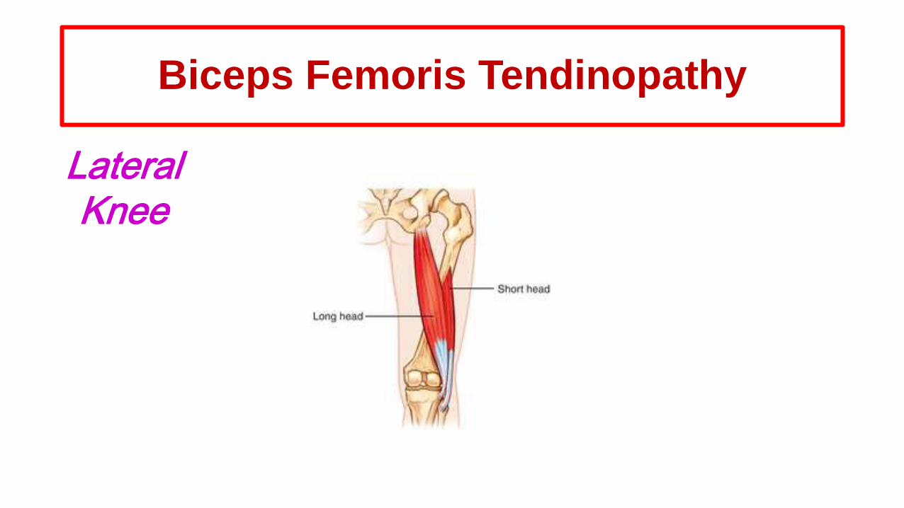
Biceps Femoris Tendinopathy
LateralKnee
Biceps Femoris TendinopathyStructure
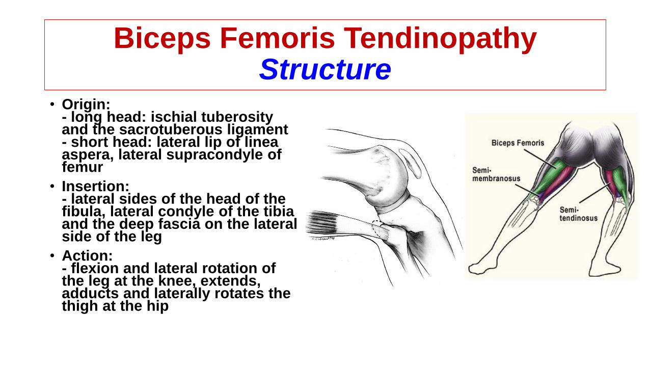
• Origin:- long head: ischial tuberosity and the sacrotuberous ligament- short head: lateral lip of linea aspera, lateral supracondyle of femur
• Insertion:- lateral sides of the head of the fibula, lateral condyle of the tibia and the deep fascia on the lateral side of the leg
• Action:- flexion and lateral rotation of the leg at the knee, extends, adducts and laterally rotates the thigh at the hip
Biceps Femoris TendinopathyDiagnosis
• Tenderness at the site where the tendon enters the bone
• Swelling at the site where the tendon enters the bone
• Pain with resisted flexion of the knee
• Stiffness of the knee after physical activity or exercise
• Tightness of the hamstring muscles resulting in limitation of hip flexion

Biceps Femoris TendinopathyContribution Factors
• Lower extremity muscle imbalances
• Decreased lower body flexibility
• Obese or overweight
• Advanced age
• Malalignment abnormalities of the leg
• Excessive running

Biceps Femoris TendinopathyConservative Treatment
• Rest
• Ice
• Massage therapy
• Eccentric exercise
• NSAID
• Ultrasound therapy
• Electrotherapy
• Taping
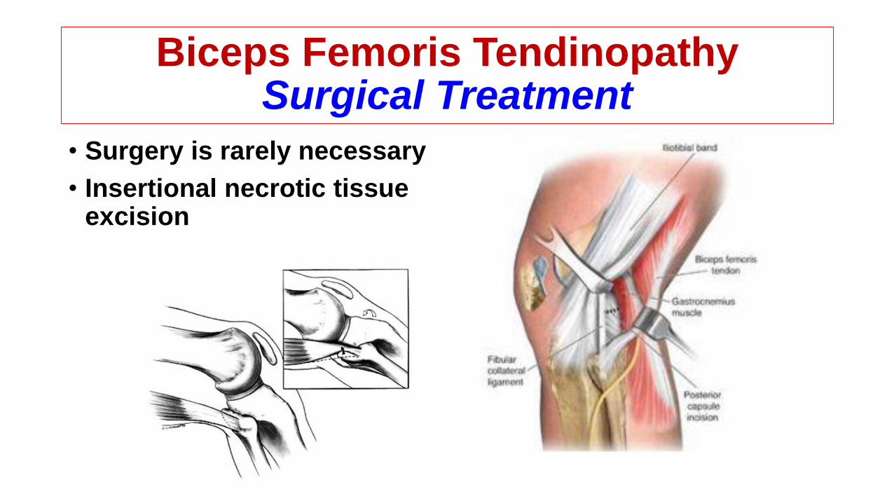
Biceps Femoris TendinopathySurgical Treatment
• Surgery is rarely necessary
• Insertional necrotic tissue excision

Pes Anserine Tendinopathy
MedialKnee

Pes Anserine TendinopathyStructure
• The tendinous aponeurosis of the sartorius, gracilis, and semitendinosus
• Per anserinus bursa: located directly beneath this aponeurosis and lies on top of the underlying superficial medial collateral ligament
Pes Anserine TendinopathyDiagnosis
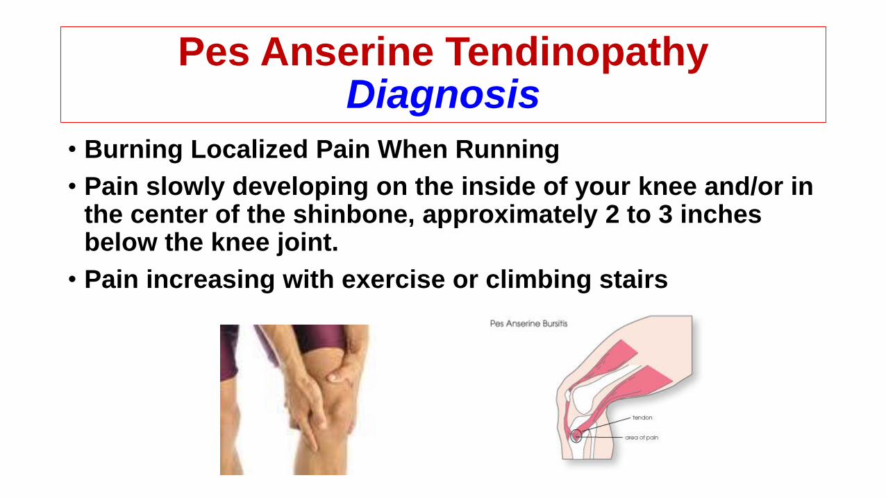
• Burning Localized Pain When Running
• Pain slowly developing on the inside of your knee and/or in the center of the shinbone, approximately 2 to 3 inches below the knee joint.
• Pain increasing with exercise or climbing stairs
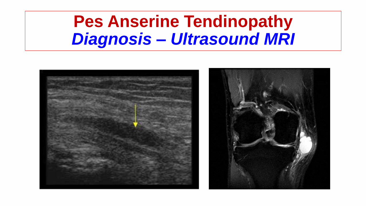
Pes Anserine TendinopathyDiagnosis – Ultrasound MRI
Pes Anserine TendinopathyContribution Factors
• Tight hamstrings, inadequate stretching, previous hamstring injury, hamstring orientation training programme
• Excessive genu valgum and weak vastus medialis
• Running with one leg higher than the other
• Running on a slope or crowned road
Pes Anserine TendinopathyConservative Treatment
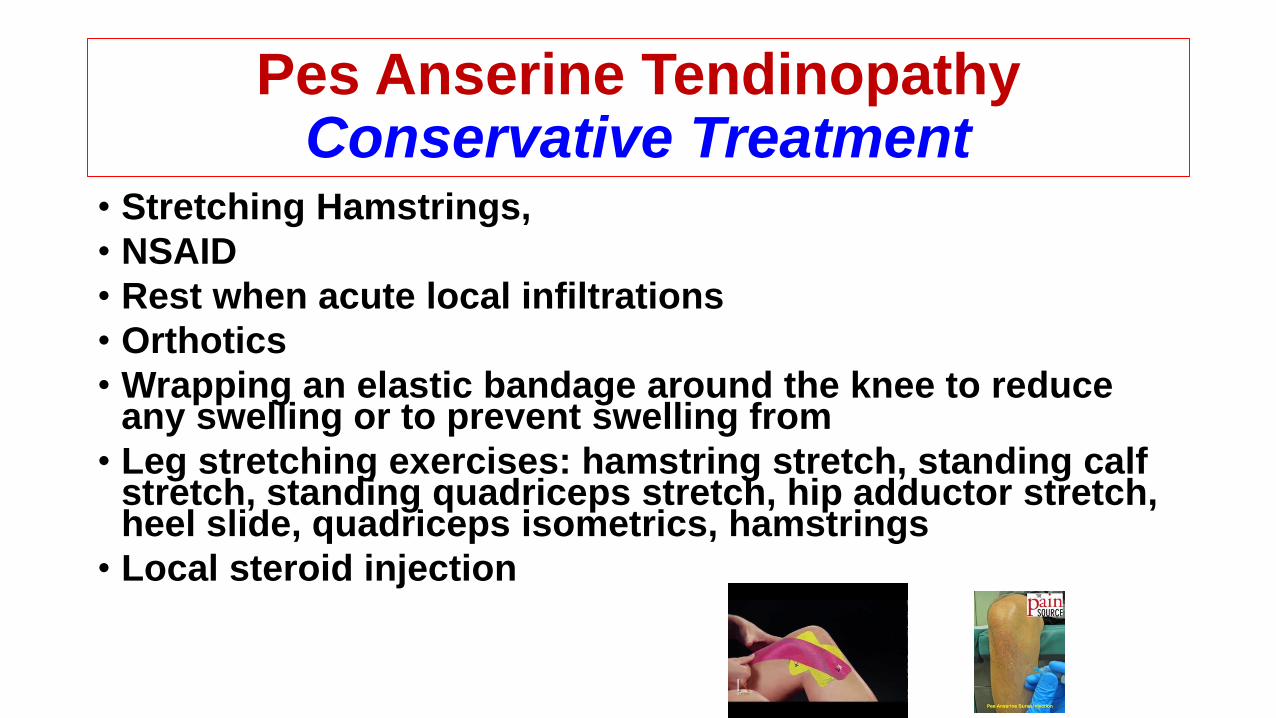
• Stretching Hamstrings,
• NSAID
• Rest when acute local infiltrations
• Orthotics
• Wrapping an elastic bandage around the knee to reduce any swelling or to prevent swelling from
• Leg stretching exercises: hamstring stretch, standing calf stretch, standing quadriceps stretch, hip adductor stretch, heel slide, quadriceps isometrics, hamstrings
• Local steroid injection
Pes Anserine TendinopathySurgical Treatment
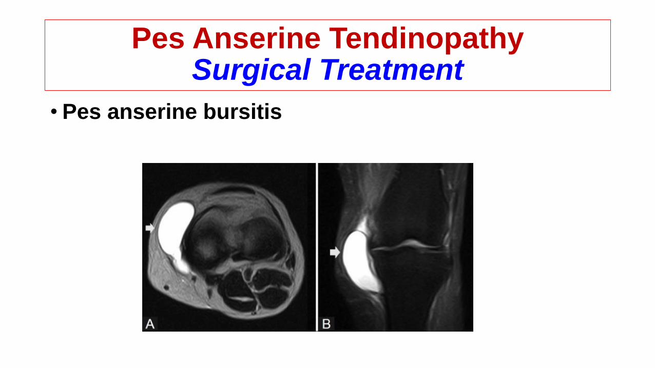
• Pes anserine bursitis

Semimembranosus Tendinopathy
MedialKnee
Semimembranosus TendinopathyStructure
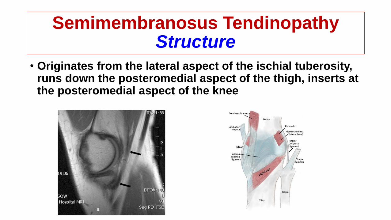
• Originates from the lateral aspect of the ischial tuberosity, runs down the posteromedial aspect of the thigh, inserts at the posteromedial aspect of the knee
Semimembranosus Tendinopathy Diagnosis
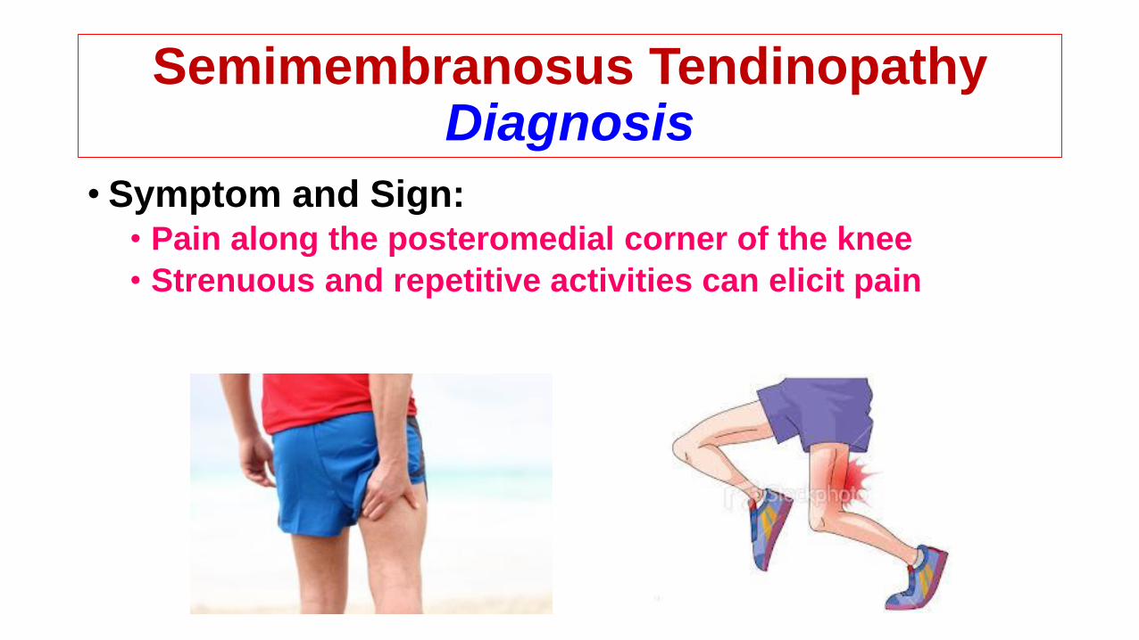
• Symptom and Sign:• Pain along the posteromedial corner of the knee
• Strenuous and repetitive activities can elicit pain
Semimembranosus Tendinopathy Diagnosis
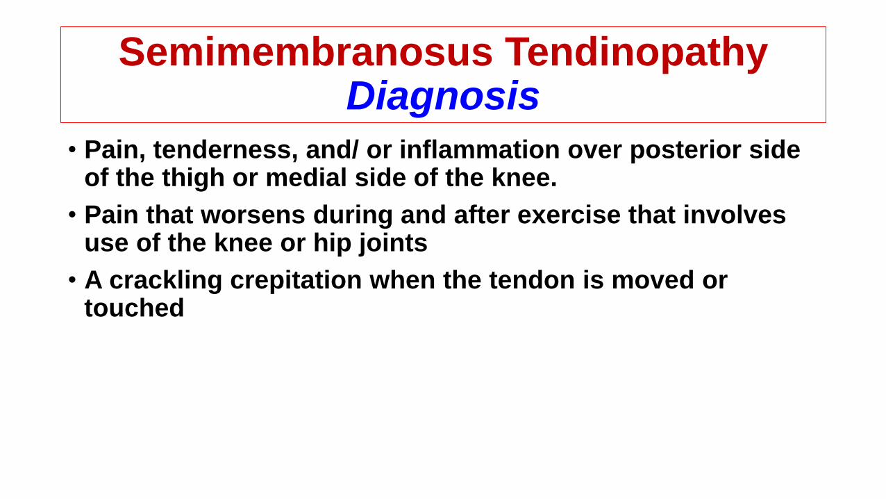
• Pain, tenderness, and/ or inflammation over posterior side of the thigh or medial side of the knee.
• Pain that worsens during and after exercise that involves use of the knee or hip joints
• A crackling crepitation when the tendon is moved or touched
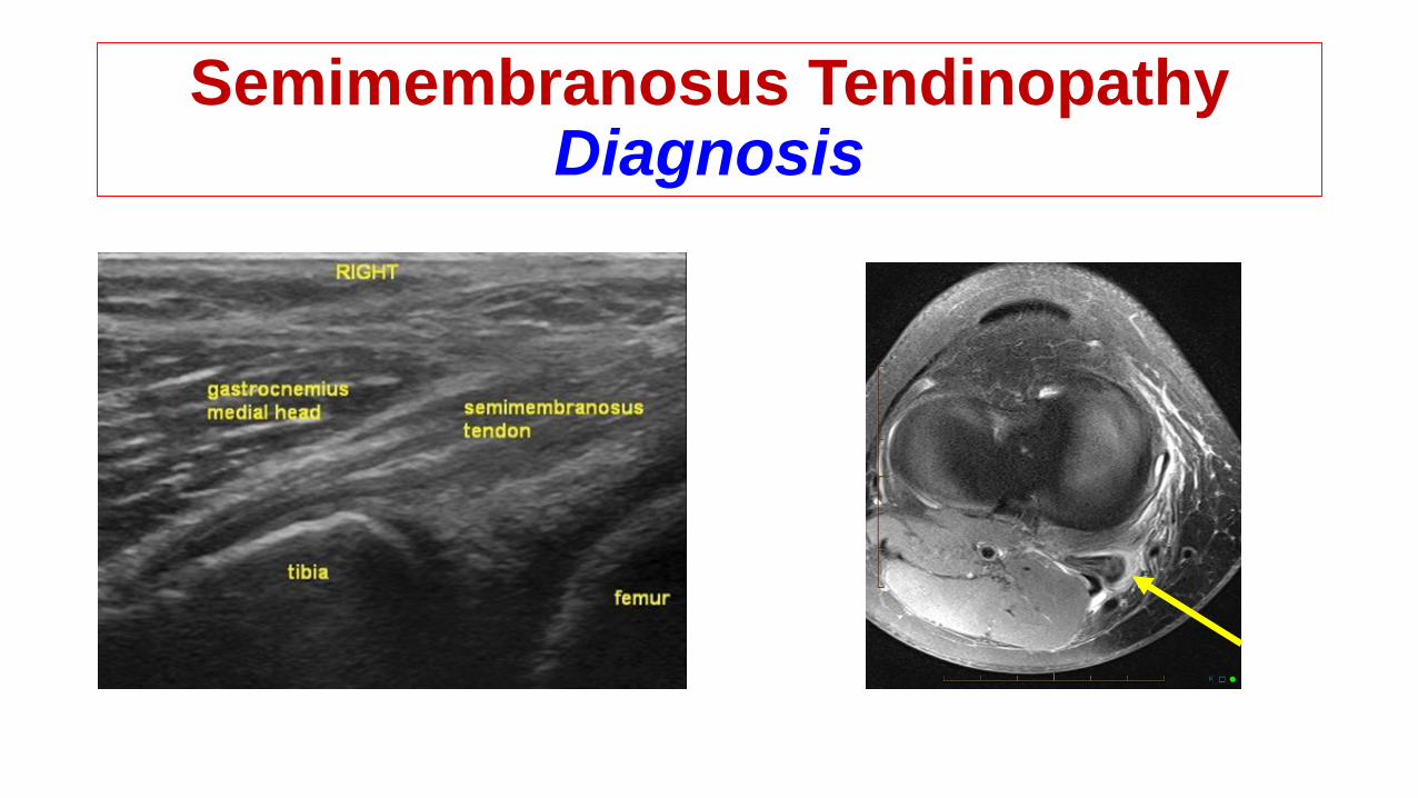
Semimembranosus TendinopathyDiagnosis
Semimembranosus TendinopathyContribution Factors
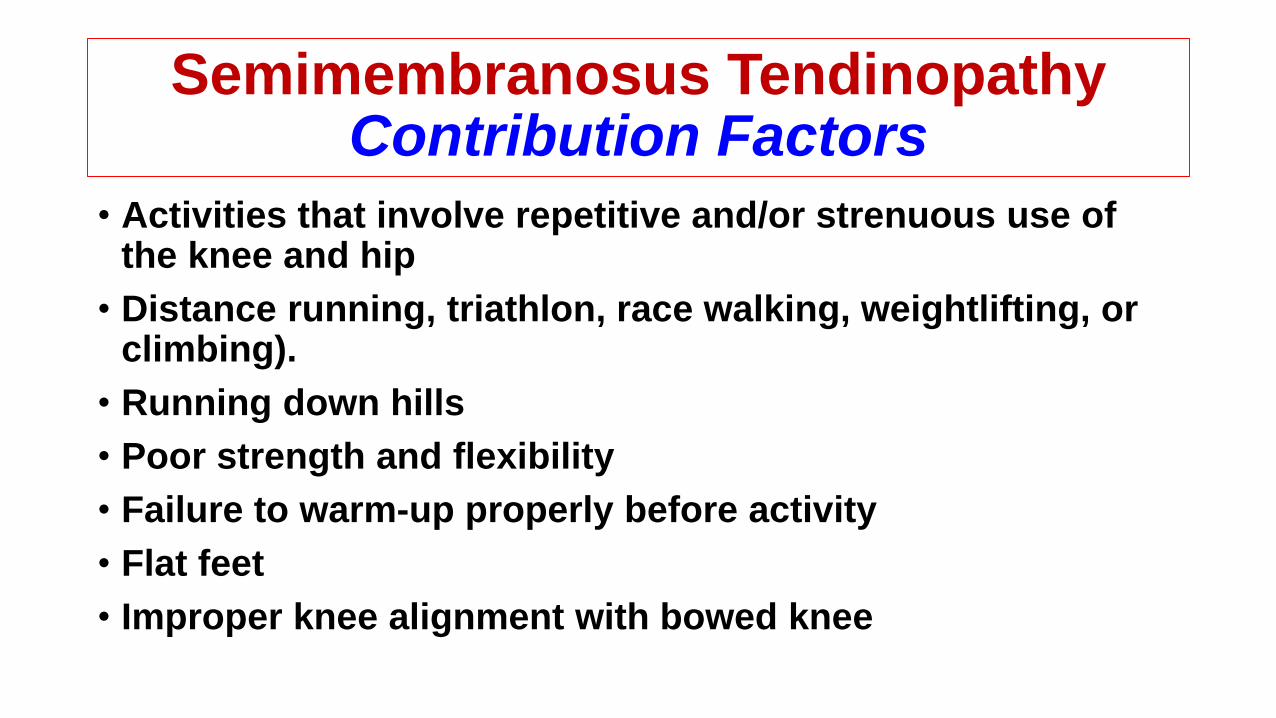
• Activities that involve repetitive and/or strenuous use of the knee and hip
• Distance running, triathlon, race walking, weightlifting, or climbing).
• Running down hills
• Poor strength and flexibility
• Failure to warm-up properly before activity
• Flat feet
• Improper knee alignment with bowed knee
Semimembranosus TendinopathyConservative Treatment
• Relative rest from painful activities
• Pain-relieving modalities
• NSAID
• Physical therapy with hamstring strengthening and stretching
• Proper shoe fit to prevent over pronation

Semimembranosus Tendinopathy Surgical Treatment
• Recalcitrant cases of SMT after failure of conservative treatment
• SM-rerouting procedure:• Places the SM tendon adjacent to the posterior border of the MCL
• Relieve the chronic irritation of the SM tendon at the posterior medial corner