Ten-year longitudinal study of thyroid function in ...
29
18 September 2017 intestazione repositorydell’ateneo Ten-Year Longitudinal Study of Thyroid Function in Children with Down's Syndrome / Iughetti L; Predieri B; Bruzzi P; Predieri F; Vellani G; Madeo SF; Garavelli L; Biagioni O; Bedogni G; Bozzola M.. - In: HORMONE RESEARCH IN PAEDIATRICS. - ISSN 1663-2818. - STAMPA. - 82:2(2014), pp. 113-121. Original Ten-Year Longitudinal Study of Thyroid Function in Children with Down's Syndrome Publisher: Published DOI:10.1159/000362450 Terms of use: openAccess Publisher copyright (Article begins on next page) Testo definito dall’ateneo relativo alle clausole di concessione d’uso Availability: This version is available at: 11380/1026715 since: 2017-03-16T09:42:24Z This is the peer reviewd version of the followng article: brought to you by CORE View metadata, citation and similar papers at core.ac.uk provided by Archivio istituzionale della ricerca - Università di Modena e Reggio Emilia
Transcript of Ten-year longitudinal study of thyroid function in ...

18 September 2017
intestazione repositorydell’ateneo
Ten-Year Longitudinal Study of Thyroid Function in Children with Down's Syndrome / Iughetti L; Predieri B; Bruzzi P;Predieri F; Vellani G; Madeo SF; Garavelli L; Biagioni O; Bedogni G; Bozzola M.. - In: HORMONE RESEARCH INPAEDIATRICS. - ISSN 1663-2818. - STAMPA. - 82:2(2014), pp. 113-121.
Original
Ten-Year Longitudinal Study of Thyroid Function in Children with Down's Syndrome
Publisher:
PublishedDOI:10.1159/000362450
Terms of use:openAccess
Publisher copyright
(Article begins on next page)
Testo definito dall’ateneo relativo alle clausole di concessione d’uso
Availability:This version is available at: 11380/1026715 since: 2017-03-16T09:42:24Z
This is the peer reviewd version of the followng article:
brought to you by COREView metadata, citation and similar papers at core.ac.uk
provided by Archivio istituzionale della ricerca - Università di Modena e Reggio Emilia

1
Ten-year longitudinal study of thyroid function in children with Down’s syndrome
Lorenzo Iughettia, Barbara Predieri
a, Patrizia Bruzzi
a, Flavia Predieri
a, Giulia Vellani
a,
Simona Filomena Madeoa, Livia Garavelli
b, Ornella Biagioni
c, Giorgio Bedogni
d, Mauro
Bozzolae
a Department of Medical and Surgical Sciences of the Mother ,Children and Adults,
University of Modena and Reggio Emilia, Modena, Italy; b
Clinical Genetics Unit, Obstetric
and Pediatric Department, Istituto di Ricovero e Cura a Carattere Scientifico, Arcispedale
Santa Maria Nuova, Reggio Emilia, Italy; c Department of Mental Health, AUSL, Modena,
Italy; d
Liver Research Center, Basovizza, Trieste, Italy and International Center for the
Assessment of Nutritional Status (ICANS), University of Milan, Milan, Italy; e Internal
Medicine and Therapeutics Department, University of Pavia, Italy.
ESPE membership: Lorenzo Iughetti, Mauro Bozzola
Short title: Thyroid dysfunction in Down's syndrome children
Type of manuscript: Original paper
Word count for the abstract: 200 Word count for the manuscript: 3127
Number of References: 46
Number of Tables: 2 Number of Figures: 5
Keywords: Thyroid function; Thyroid autoantibodies; Natural history; Down's syndrome;
Children
Financial disclosure: nothing to declare
Address correspondence to:
Lorenzo IUGHETTI, M.D., Ph.D.
Department of Medical and Surgical Sciences of the Mother, Children and Adults –
University of Modena and Reggio Emilia
Via del Pozzo, 71 – 41124 Modena, Italy
Phone: +39 059 422 2182; Fax: +39 059 422 4583
E-mail: [email protected]
2
Abstract
Background/Aims The natural history of thyroid function in children with Down
syndrome (DS) is relatively unknown We hypothesized that in these patients the occurrence
of thyroid dysfunction rises during development.
Methods - Thyroid function was assessed yearly in 145 children with DS, all followed from
birth up to 10 years of age. Heteroskedastic binary and ordinary logistic regression for
repeated measures were used to evaluate the relationship of thyroid function with continuous
time.
Results - Congenital hypothyroidism was detected in 7% of cases. The probability of
acquired thyroid dysfunction increased from 30% at birth to 49% at 10 years (p<0.001). The
subclinical hypothyroidism (S-HT) was nearly stable during the follow-up. The probability of
hypothyroidism increased from 7% to 24% at 10 years (p<0.001). Positive anti-thyroglobulin
antibodies were associated with a higher odds of more severe hypothyroidism [odds ratio
(OR)=3.6]. Positive anti-thyroid peroxidase antibodies (TPOab) were a better predictor of
more severe hypothyroidism, (OR=6.1). Diffuse hypoechogenicity on thyroid ultrasound was
found in 34 out of 145 children.
Conclusion - The probability of thyroid dysfunction increases during development is higher
than previously reported. Such children should be carefully monitored annually to early
identify thyroid dysfunction.
3
Introduction
Down's syndrome (DS), the most common chromosomal disorder, is associated with
several concomitant diseases, including thyroid dysfunction [1-3]. Thyroid disorders are more
common in patients with DS than in the general population and they have been estimated to
range between 4% and 19.5% [4-8] with an increase in frequency, up to 54%, as the children
age [5, 9-10]. These abnormalities include congenital hypothyroidism (1–3.6%) [7, 11-14],
primary hypothyroidism (0.3-3.2%) [7, 14-15], autoimmune thyroiditis (0.3–1.4%) [16], and
subclinical hypothyroidism (12.5–32.9%) [7, 10, 14-15, 17]. In addition, hyperthyroidism (0–
2%) occurs in children with DS as well [7, 10, 18].
Pathogenesis, natural course, prognostic factors, and therapy of thyroid dysfunction in
children with DS remain the objects of debate in the literature because epidemiological and
long-term cohort studies are scarce. Pediatric endocrinologists frequently face the decision of
what to do regarding these subjects.
In the general population the natural history of Hashimoto’s thyroiditis in children and
adolescents is not yet fully known. Thyroid-stimulating hormone concentrations showed large
fluctuations over time, and a trend toward progressively deteriorating thyroid function was
demonstrated [19-20]. A recent review on subclinical hypothyroidism in children showed that
it is a remitting process with a rate of evolution toward overt hypothyroidism ranging between
0 and 28.8% [21].
We hypothesized that the occurrence of thyroid diseases in children with DS rises in
the course of development. Therefore, we investigated the probability of each thyroid disease
in a cohort of 145 children with DS from birth to 10 years of age.

4
Subjects and methods
Clinical and biochemical assessment.
We conducted a cohort study from 1995 to 2010 including 205 Caucasian neonates
with DS (95.6% with primary trisomy 21, 3.4% with translocation, and 1% with mosaicism)
assessed at the Department of Pediatrics of the University of Modena and Reggio Emilia,
Italy. Twenty neonates were not recruited because the parents refused the study, while during
the follow-up children were excluded because of the development of severe heart disease in
18 cases, leukemia in 4, seizure in 3, and coeliac disease in 10. We include this description of
these subjects to avoid the lost to follow-up because of sudden death [22] and the diagnostic
bias due to other exposure status. Specifically, patients with known autoimmune diseases [23]
or using drugs that may interfere with thyroid function [24-25] are prone to develop
autoimmune thyroiditis or subclinical hypothyroidism. Furthermore, 2 subjects refused to
continue the study and 3 were lost to follow-up. Written informed consent was obtained from
all parents at the moment of recruitment in the study and before the first data collection. The
study was approved by the Ethics Committee of the University of Modena and Reggio Emilia.
Thyroid function status was tested annually from birth over a 10-year period. Blood
samples were analyzed for thyroid-stimulating hormone (TSH), free thyroxine (F-T4),
thyroglobulin antibody (TGab), and thyroid peroxidase antibody (TPOab) within the same
laboratory. Plasma TSH and F-T4 were measured by fluorometric assay (AutoDELFIA
automatic immunoassay system). TGab and TPOab were quantified by an immunometric
assay (IMMULITE 2000): values >40 IU/ml and >35 IU/ml were defined as positive,
respectively. Ultrasonography of thyroid gland was performed by an experienced pediatric
radiologist who was blinded to the thyroid status of the subjects. In subjects with congenital
hypothyroidism (C-HT) it was immediately performed while in the other ones it was

5
firstly done at 2 years of age and repeated at time of thyroid dysfunction and/or thyroid
autoantibodies development. In these cases a 2-year thyroid ultrasound follow-up was
performed.
After exclusion of children with confirmed C-HT (newborn screening based on the
measurement of TSH ≥ 20 μIU/ml on eluates of dry blood collected on filter paper after 24 h
of life as the primary screening test and confirmed by plasma thyroid function test performed
after 8 days of life), all DS children were annually grouped according to both F-T4 and TSH
levels as:
- euthyroidism (EuT): normal F-T4 and TSH ≤ 5 μIU/ml
- hypothyroidism (HT): low F-T4 and TSH ≥ 10 μIU/ml
- subclinical hypothyroidism (S-HT): normal F-T4 and TSH > 5 μIU/ml
- hyperthyroidism (HyperT): high F-T4 and low TSH
Children with C-HT were excluded from the present prospective analysis, but not at
first year evaluation of thyroid dysfunction probability.
Subjects with HT were treated with L-thyroxine and were considered in HT group
during the whole follow up period. Replacement therapy was also started in children with S-
HT but only if TSH levels were persistently ≥ 10 μIU/ml [21] and these patients have always
been included in the S-HT group.
Statistical analysis
Heteroskedastic binary and ordinal logistic regression for repeated measures were used
to evaluate the changes of the binary and ordinal outcomes of interest with time [26-27].
These analyses were implemented using a heteroskedastic ordinal generalized linear model
6
(OGLM) with cluster confidence intervals to take into account repeated measures. The
OGLM reduces to a logistic regression model when the outcome is binary and estimates the
odds of more severe vs. less severe disease when the outcome is ordinal. The binary outcomes
of interest were: 1) thyroid dysfunction (0 = no; 1 = yes), 2) TGab positivity (0 = no; 1 = yes),
TPOab positivity (0 = no; 1 = yes). The ordinal outcome of interest was hypothyroidism (0 =
EuT, 1 = S-HT and, 2 = overt HT). Time was modeled as continuous (years, 1 to 10 by steps
of 1) and the possibility to control for heteroskedasticy was used to model non-linear time-
outcome relationships. To test whether TGab and TPOab are associated with hypothyroidism
we added them, first separately and then together, with time as predictors of the logistic
ordinal model (multivariable analysis). The odds ratio (OR) associated with TGab and TPOab
is a time-averaged measure of their association with hypothyroidism. Statistical significance
was set to a value of p<0.05. Statistical analysis was performed using STATA MP Version
13.0.

7
Results
From 1995-2010, 145 of 205 patients with DS were longitudinally evaluated for the
purpose of the study. Overall, 59% of the subjects were boys.
During the first year of evaluation, C-HT was confirmed in 10 of 145 cases (6.9%; 6
out 10 had thyroid agenesis, 2 had thyroid ectopia and, 2 had thyroid hypoplasia), 4 children
had HT (2.7%; with normal thyroid ultrasound characteristics), 27 had S-HT (18.6%; 2 out of
27 had thyroid hypoplasia and 1-year after developed thyroid autoantibodies), while the other
104 had normal thyroid function.
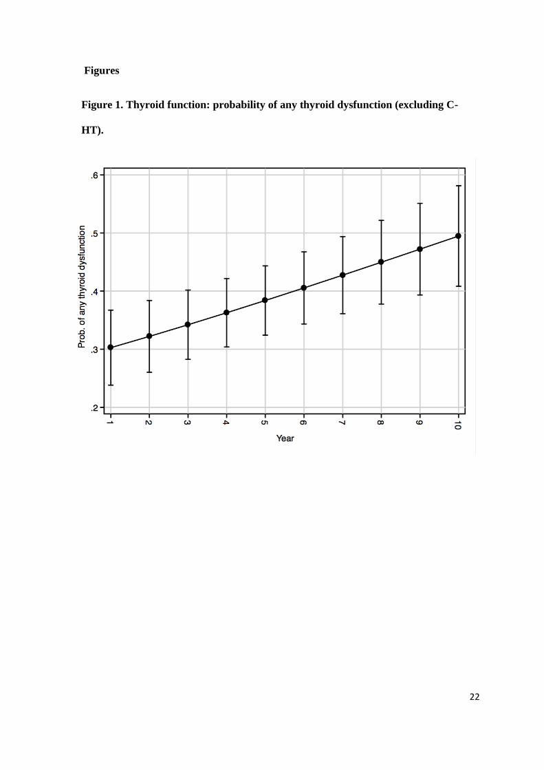
Figure 1 depicts the probability of any thyroid dysfunction during the study. Such
probability increased from 30% (95%CI 24 to 37%) at 1st year to 49% (41% to 58%) at 10
th
year (p<0.001, heteroskedastic binary logistic regression with cluster confidence intervals).
Thyroid dysfunction was classified as HyperT in 1 case and as S-HT or overt HT in the
remaining cases. Further analyses were performed on the subjects who did not develop
HyperT.
Figure 2 depicts the probability of EuT, S-HT and overt HT during the follow-up. The
probability of EuT decreased from 71% (65% to 78%) at 1st year to 52% (44% to 60%) at 10
th
year. S-HT had a fluctuating course between normal, compensated and overt HT so its
probability was nearly stable, being 22% (17% to 28%) at 1st year and 24% (17% to 31%) at
10th
year. Lastly, the probability of overt HT increased from 7% (3% to 10%) at 1st year to
24% (17% to 32%) at 10th
year (p<0.001, heteroskedastic ordinal logistic regression with
cluster confidence intervals).
Considering the EuT group we found that, after 10 years, 64 patients still had normal
function, 17 developed HT, and 22 developed S-HT. In the S-HT group one third developed

8
EuT at the end of the study without replacement therapy, while one third developed HT. The
thyroid dysfunctions were almost equally distributed between the genders (data not show).
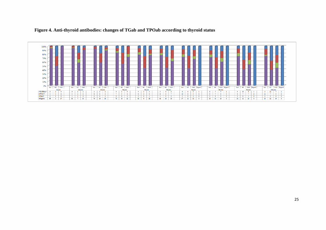
Figures 3 and 4 depict the probability of positive thyroid autoantibodies (ATA) during
the study. The probability of positive TGab increased from 3% (5‰ to 5%) at 1st year to 25%
(17% to 33%) at 10th
year with corresponding figures of 5% (2% to 7%) and 37% (30% to
45%) for TPOab (p<0.001, heteroskedastic binary logistic regression with cluster confidence
intervals) (Figure 3). The significant increase in the probability of ATA was mainly found in
subjects with EuT (32 out of 99 initially negative, 32.3%) and S-HT (14 out of 27 initially
negative, 51.8%) (Figure 4).
Combining the data according to thyroid function and the development of ATA, over a
10-year period, we found two different natural course patterns for both EuT and S-HT, mainly
depending on the presence or absence of ATA. In particular, 60% of ATA-positive EuT
subjects developed a thyroid dysfunction, whereas 73% of ATA-negative EuT cases remained
in EuT group (p=0.002). Among children with S-HT at first visit, 50% of ATA-positive cases
had not changed at the last visit, whereas 28.6% developed HT. This prevalence was lower
than in ATA-negative cases (38.5%); only 21.4% of ATA-positive subjects developed EuT, in
contrast to the 46.1% we found among the ATA-negative cases (Figure 5).
At multivariable ordinal logistic regression, TGab positivity was associated with an
higher odds of more severe vs. less severe hypothyroidism but the estimate was imprecise
(OR=3.6, 95%CI 1.2 to 11.0, p=0.02). On the contrary, TPOab positivity was strongly
associated with more severe vs. less severe hypothyroidism (OR=6.1, 2.3 to 16.1, p<0.001).
Adding TGab to TPOab was associated with no improvement in the prediction of the severity
of hypothyroidism as compared to TPOab alone (OGLM not shown).

9
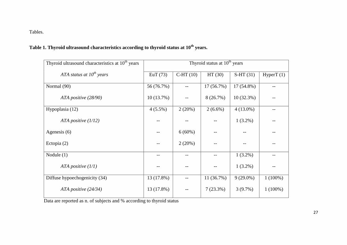
The characteristics of thyroid ultrasounds at the 10-year follow-up are reported in
Table 1. Overall, 62% of subjects had normal echogenicity on ultrasound and 23.5% had
diffuse hypoechogenicity (p<0.001). The prevalence of thyroid dysfunctions in children with
hypoechogenicity was significantly higher than in individuals with normal thyroid
echogenicity (61.8% vs. 37.8%; p=0.028). Considering ATA status we found that subjects
with hypoechogenicity had a significantly higher percentage of ATA positivity than those
with normal echogenicity (70.6 % vs. 31.0%; p<0.001). Interestingly, all subjects with diffuse
hypoechogenicity on thyroid ultrasound but no circulating ATA (29.4%) had thyroid
dysfunctions (6 S-HT and 4 HT) (Table 2).
After exclusion of patients with C-HT, at the end of the follow-up 55 (41%) of our
children were treated for thyroid disorders. Specifically, L-thyroxine replacement therapy was
started in all children who developed HT and in 24 out of 31 (77.5%) patients with S-HT.
Almost all patients with ATA-positive S-HT (88%) required treatment in contrast to the 64%
of ATA-negative cases. Methimazole was used for the only child with HyperT.
To assess the possibility that the level of initial TSH were predictive of a future
thyroid dysfunction requiring L-thyroxine replacement therapy, we correlated the 1st
year TSH values with the last ones before beginning of the treatment. Excluding data
from the C-HT patients and the 4 subjects already having HT at 1-year of life, the TSH
levels at 1st year (4.92±1.83 IU/ml) and the ones just before therapy (12.3±13.5 IU/ml)
were not significantly correlated (Spearman R=0.234, p=0.098). The same results were
observed analyzing data according to 1st year thyroid function: EuT (Spearman
R=0.060, p=0.7322) and S-HT (Spearman R=0.317, p=0.230). Moreover, considering
data from DS patients with S-HT at the end of the study, we found that both TSH

10
(Spearman R=-0.063, p=0.737) and FT4 (Spearman R=0.069, p=0.710) levels were not
significantly correlated with BMI z-score.
11
Discussion
Several studies have investigated the prevalence of thyroid dysfunction in children and
adults with DS [4, 10-12, 14-15, 28-29] but, to our best knowledge, this is the first
prospective study to examine annually, over a 10-year period, the natural history of thyroid
disorders in very young children with DS. Our data confirm the high probability of thyroid
dysfunctions in children with DS, increasing year by year, when compared to reference ranges
for the general population.
In our cohort, C-HT probability was higher than reported in the literature, ranging
from 1% to 2.7% [11-13]. Early thyroid screening increases the prevalence of newborns
demonstrating a modest elevation of blood spot TSH concentrations due to the physiological
neonatal surge, increasing the number of false-positive results. Newborns with DS generally
have a mild hypothyroid state because of decreased thyroxine concentration that left-shifted
normal distribution and mildly elevated TSH levels [30]. To avoid false-positive C-HT due to
early measurement, we used a TSH screening cut-off of ≥ 20 μIU/ml and this allowed us to
exclude additional cases of C-HT that were mainly due to functional disorders [31].
Moreover, to confirm the positive screening C-HT cases we adopted a diagnostic algorithm
that included the subsequent confirmation by low plasma FT4, high TSH levels and thyroid
ultrasound characteristics (6 agenesis, 2 ectopia, and 2 hypoplasia).
Our children with DS suffered from two other different thyroid diseases such as S-HT
mainly developed during the first 3 years of life and HT with a later onset; however, these
thyroid dysfunctions were both present as early as the 1st year of life. These cases of S-HT
and HT, considering the thyroid ultrasound characteristics and the development of ATA,
cannot be misdiagnosed with C-HT. Beyond the neonatal period, the incidence of elevated

12
TSH values in DS increases, being reported as high as 85% of infants under the age of 12
months [32].
Despite the limitation of the low number and the high heterogeneity of patients
included in the published studies, in the general population S-HT is a benign and remitting
process with a risk of progression to overt HT of approximately 0-12.5% [19, 21, 33-35]. In a
retrospective multi-centre study [34], including a large pediatric cohort, it was demonstrated
that TSH values tended to normalize proportionally to the degree of the TSH elevation at the
initiation of the study. In subjects with TSH between 5.5 and 10 IU/ml, 73.6% normalized
their TSH, about 25% maintained that range, and about 2% had their TSH increased over 10
μIU/ml but with normal FT4. In subjects with initially highly elevated TSH, 40% normalized
their TSH, 33.1% reduced their TSH to a value between 5.5 and 10 μIU/ml, 24.9%
maintained their TSH higher than 10 μIU/ml. Overall, 70.1% of initially abnormal TSH levels
normalize in the second evaluation while HT requiring replacement therapy was found in
0.4% of the cohort.
In our cohort we found that an increase in TSH levels such to support the definition of
S-HT is not correlated with BMI z-score and may be considered a common transient
condition in DS and not necessarily imply the occurrence of HT. Considering our data, we
recommend a careful approach and follow up for children with DS, thyroid hypoplasia and S-
HT, irrespective of the presence or absence of ATA. A recent study demonstrated that the
distribution plot for TSH had a significant shift of the curve to higher values in subjects
with DS respect to healthy controls, suggesting that hyperthyrotropinaemia is an innate
attribute of chromosome 21 trisomy [36]. Moreover, some authors [5, 37-38] have
proposed that S-HT is probably related to inappropriate secretion of TSH or thyroid
insensitivity to TSH, rather than to autoimmune thyroiditis.

13
As well as an increase in the probability of thyroid dysfunction we also found an
increased probability of ATA during the 10-year period. It is important to note that in our EuT
children, the presence of ATA, mainly TPOab, significantly raised the risk of developing
thyroid dysfunctions during the long-term follow-up. These data suggest that ATA might
represent a marker of deteriorating thyroid function unlike 1st year TSH levels that are not
predicting for future thyroid dysfunction requiring therapy, both in initially EuT and S-
HT subjects with DS.
The prevalence of ATA in DS children was reported to range from 16% [38] to 39%
[39]. Moreover, the increased TSH levels were demonstrated to be positively correlated with
TPOab which were already indicated as a key factor in the follow-up of DS subjects [40]. So,
our results have confirmed the previously reported findings on the important role of both
elevated TSH and ATA in the development of HT [10, 14, 16, 17, 39, 41], suggesting that
children with DS should be tested more frequently than others.
In the general population it was demonstrated that in children and adolescents with
both S-HT and autoimmune thyroiditis ATA increased over time [19, 35]. However, the
progressive increase in TPOab raised the risk of developing HT by 3.5-fold [20], lower than
data we found in our DS population.
Up to 10% of patients with autoimmune thyroiditis are ATA negative [42], so thyroid
ultrasound findings may be important in order to differentiate children with a mild S-HT and
autoimmune thyroiditis from those with other causes of thyroid dysfunction. Our data support
the hypothesis that some children with DS may have a persistent mild HT presumably of
thyroidal origin related to the trisomic state of chromosome 21 [43].

14
Thyroid hormones influence almost all aspects of normal development during
childhood. They play a crucial role as regulators of neurodevelopment, growth and skeletal
development, and metabolism [44]. The early diagnosis of thyroid dysfunctions is important
and pediatricians can often recognize them in their early stages, by maintaining an appropriate
index of suspicion. Recognition of HT can be very difficult because it has a subtle
presentation and symptoms overlap with features of DS, including impaired intellectual
development in young children, decreased linear growth, dry skin and fine hair, excess
weight, dentition abnormalities, and decreased physical activity [2]. In our DS children with
thyroid dysfunctions the incidence of replacement therapy was 41%, over the 10-year period.
The medical treatment of S-HT is a topic of debate [5, 11, 14]; however, almost all patients
with ATA required treatment [45]. Currently we do not know at which TSH level clinical
signs and potential adverse effects on lipid profile, cardiac function, and neuropsychiatric
performance will appear, or whether treatment can prevent them. However, it was
demonstrated that S-HT, even of many years’ duration, and HT, if appropriately treated
within a few months, do not impair growth [19]. In the general pediatric population, there is
no clear evidence of the beneficial effects of L-thyroxine on growth and thyroid volume [21],
but van Trotsenburg et al. [46] have reported that in children with DS replacement therapy for
the first 24 months of life probably results in improvements of psychomotor development and
growth. However, this study was designed to determine the rate of annual thyroid
dysfunctions in this population, not the appropriateness of treatment.
In summary, in our study thyroid dysfunctions were relatively common in children
with DS and, because symptoms might be mistaken for symptoms related to the natural
course of DS, they may represent a significant health risk if not identified. We suggest that
DS patients, especially those with EuT or S-TH associated with positive ATA, should be

15
annually monitored to precociously identify thyroid diseases and to begin an adequate
replacement therapy when proper.
16
References
1. Radetti G, Drei F, Betterle C, Mengarda G: Down's syndrome, hypothyroidism and
insulin-dependent diabetes mellitus. Helv Paediatr Acta 1986;41:377-380.
2. Prasher VP: Down syndrome and thyroid disorders: a review. Downs Syndr Res Pract
1999;6:25-42.
3. Weijerman ME, de Winter JP: Clinical practice. The care of children with Down
syndrome. Eur J Pediatr 2010;169:1445-1452.
4. Pueschel SM, Jackson IM, Giesswein P, Dean MK, Pezzullo JC: Thyroid function in
Down syndrome. Res Dev Disabil 1991;12:287-296.
5. Gibson PA, Newton RW, Selby K, Price DA, Leyland K, Addison GM: Longitudinal
study of thyroid function in Down's syndrome in the first two decades. Arch Dis Child
2005;90:574-578.
6. Murphy J, Philip M, Macken S, Meehan J, Roche E, Mayne PD, O'Regan M, Hoey HM:
Thyroid dysfunction in Down's syndrome and screening for hypothyroidism in children
and adolescents using capillary TSH measurement. J Pediat Endocrinol Metab
2008;21:155-163.
7. Regueras L, Prieto P, Muñoz-Calvo MT, Pozo J, Arguinzoniz L, Argente J:
Endocrinological abnormalities in 1.105 children and adolescents with Down syndrome.
Med Clin (Barc) 2011;136:376-381.
8. McGowan S, Jones J, Brown A, Reynolds L, Leyland K, Charleton P, Rahim M, Mansor
M, Ritha S, Donaldson M, Scottish Down Syndrome Thyroid Screening Group: Capillary
TSH screening programme for Down's syndrome in Scotland, 1997-2009. Arch Dis Child
2011;96:1113-1117.

17
9. Kishnani PS, Crissman BG: Special issue: current perspectives on Down syndrome:
selected medical and social issues. Am J Med Genet C Semin Med Genet 2006;142C:127-
205.
10. Unachak K, Tanpaiboon P, Pongprot Y, Sittivangkul R, Silvilairat S, Dejkhamron P,
Sudasna J: Thyroid functions in children with Down's syndrome. J Med Assoc Thai
2008;91:56-61.
11. Fort P, Lifshitz F, Bellisario R, Davis J, Lanes R, Pugliese M, Richman R, Post EM,
David R: Abnormalities of thyroid function in infants with Down syndrome. J Pediatr
1984;104:545-549.
12. Cutler AT, Benezra-Obeiter R, Brink SJ: Thyroid function in young children with Down
syndrome. Am J Dis Child 1986;140:479-483.
13. Jaruratanasirikul S, Patarakijvanich N, Patanapisarnsak C: The association of congenital
hypothyroidism and congenital gastrointestinal anomalies in Down's syndrome infants. J
Pediatr Endocrinol Metab 1998;11:241-246.
14. Tüysüz B, Beker DB: Thyroid dysfunction in children with Down's syndrome. Acta
Paediatr 2001;90:1389-1393.
15. Shaw CK, Thapalial A, Nanda S, Shaw P: Thyroid dysfunction in Down syndrome.
Kathmandu Univ Med J (KUMJ) 2006;4:182-186.
16. Karlsson B, Gustafsson J, Hedov G, Ivarsson SA, Annerén G: Thyroid dysfunction in
Down's syndrome: relation to age and thyroid autoimmunity. Arch Dis Child
1998;79:242-245.
17. Rubello D, Pozzan GB, Casara D, Girelli ME, Boccato S, Rigon F, Baccichetti C, Piccolo
M, Betterle C, Busnardo B: Natural course of subclinical hypothyroidism in Down's
syndrome: prospective study results and therapeutic considerations. J Endocrinol Invest
1995;18:35-40.

18
18. Goday-Arno A, Cerda-Esteva M, Flores-Le-Roux JA, Chillaron-Jordan JJ, Corretger JM,
Cano-Pérez JF: Hyperthyroidism in a population with Down syndrome (DS). Clin
Endocrinol (Oxf) 2009;71:110-114.
19. Radetti G, Gottardi E, Bona G, Corrias A, Salardi S, Loche S, Study Group for Thyroid
Diseases of the Italian Society for Pediatric Endocrinology and Diabetes (SIEDP/ISPED):
The natural history of euthyroid Hashimoto's thyroiditis in children. J Pediatr
2006;149:827-832.
20. Radetti G, Maselli M, Buzi F, Corrias A, Mussa A, Cambiaso P, Salerno M, Cappa M,
Baiocchi M, Gastaldi R, Minerba L, Loche S: The natural history of the normal/mild
elevated TSH serum levels in children and adolescents with Hashimoto's thyroiditis and
isolated hyperthyrotropinaemia: a 3-year follow-up. Clin Endocrinol (Oxf) 2012;76:394-
398.
21. Monzani A, Prodam F, Rapa A, Moia S, Agarla V, Bellone S, Bona G: Natural history of
subclinical hypothyroidism in children and adolescents and potential effects of
replacement therapy: a review. Eur J Endocrinol 2012;168:R1-R11.
22. Myrelid A, Jonsson B, Guthenberg C, von Döbeln U, Annerén G, Gustafsson J: Increased
neonatal thyrotropin in Down syndrome. Acta Paediatr 2009;98:1010-1013.
23. Elfström P, Montgomery SM, Kämpe O, Ekbom A, Ludvigsson JF: Risk of thyroid
disease in individuals with celiac disease. J Clin Endocrinol Metab 2008;93:3915-3921.
24. Kim SH, Chung HR, Kim SH, Kim H, Lim BC, Chae JH, Kim KJ, Hwang YS, Hwang H:
Subclinical hypothyroidism during valproic acid therapy in children and adolescents with
epilepsy. Neuropediatrics 2012;43:135-139.
25. van Santen HM, Thonissen NM, de Kraker J, Vulsma T: Changes in thyroid hormone
state in children receiving chemotherapy. Clin Endocrinol (Oxf) 2005;62:250-257.

19
26. Hilbe JM: Generalized linear models and extensions. College Station, Tex.: Stata Press;
2012.
27. Williams R: Fitting heterogeneous choice models with oglm. Stata Journal 2010;10:540-
567.
28. Loudon MM, Day RE, Duke EM: Thyroid dysfunction in Down's syndrome. Arch Dis
Child 1985;60:1149-1151.
29. Prasher V, Ninan S, Haque S: Fifteen-year follow-up of thyroid status in adults with
Down syndrome. J Intellect Disabil Res 2011;55:392-396.
30. van Trotsenburg ASP, Vulsma T, van Santen HM, Cheung W, de Vijlder JJM: Lower
neonatal screening thyroxine concentrations in Down Syndrome newborns. J Clin
Endocrinol Metab 2003;88:1512-1515.
31. Deladoëy J, Ruel J, Giguère Y, Van Vliet G: Is the incidence of congenital
hypothyroidism really increasing? A 20-year retrospective population-based study in
Québec. J Clin Endocrinol Metab 2011;96:2422-2429.
32. Sharav T, Collins R, Baab P: Growth studies in infants and children with Down's
syndrome and elevated levels of thyrotropin. Am J Dis Child 1988;142:1302-1306.
33. Surks MI, Ortiz E, Daniels GH, Sawin CT, Col NF, Cobin RH, Franklyn JA, Hershman
JM, Burman KD, Denke MA, Gorman C, Cooper RS, Weissman NJ: Subclinical thyroid
disease: scientific review and guidelines for diagnosis and management. JAMA
2004;291:228-238.
34. Lazar L, Frumkin RB, Battat E, Lebenthal Y, Phillip M, Meyerovitch J: Natural history of
thyroid function tests over 5 years in a large pediatric cohort. Endocrinol Metab
2009;94:1678-1682.

20
35. Zois C, Stavrou I, Svarna E, Seferiadis K, Tsatsoulis A: Natural course of autoimmune
thyroiditis after elimination of iodine deficiency in northwestern Greece. Thyroid
2006;16:289-293.
36. Meyerovitch J, Antebi F, Greenberg-Dotan S, Bar-Tal O, Hochberg Z:
Hyperthyrotropinaemia in untreated subjects with Down's syndrome aged 6 months
to 64 years: a comparative analysis. Arch Dis Child 2012;97:595-598.
37. Selikowitz M: A five-year longitudinal study of thyroid function in children with Down
syndrome. Dev Med Child Neurol 1993;35:396-401.
38. Faria CD, Ribeiro S, Kochi C, Silva AP, Ribeiro BN, Marçal LT, Santos FH, Eduardo CP,
Monte O, Longui CA: TSH neurosecretory dysfunction (TSH-nd) in Down syndrome
(DS): low risk of progression to Hashimoto's thyroiditis. Arq Bras Endocrinol Metabol
2011;55:628-631.
39. Ivarsson SA, Ericsson UB, Gustafsson J, Forslund M, Vegfors P, Annerén G: The impact
of thyroid autoimmunity in children and adolescents with Down syndrome. Acta Paediatr
1997;86:1065-1607.
40. Dias VM, Nunes JC, Araújo SS, Goulart EM: Etiological assessment of
hyperthyrotropinemia in children with Down's syndrome. J Pediatr (Rio J) 2005;81:79-84.
41. Freeman SB, Torfs CA, Romitti MH, Royle MH, Druschel C, Hobbs CA, Sherman SL:
Congenital gastrointestinal defects in Down syndrome: a report from the Atlanta and
National Down Syndrome Projects. Clin Genet 2009;75:180-184.
42. Cappa M, Bizzarri C, Crea F: Autoimmune Thyroid Diseases in Children. J Thyroid Res
2011;2011:675-703.
43. van Trotsenburg AS, Kempers MJ, Endert E, Tijssen JG, de Vijlder JJ, Vulsma T:
Trisomy 21 causes persistent congenital hypothyroidism presumably of thyroidal origin.
Thyroid 2006;16:671-680.

21
44. Roberts CGP, Ladenson PW: Hypothyroidism. Lancet 2004;363:793-803.
45. Popova G, Paterson WF, Brown A, Donaldson MD: Hashimoto's thyroiditis in Down's
syndrome: clinical presentation and evolution. Horm Res 2008;70:278-284.
46. van Trotsenburg ASP, Vulsma T, van Rozenburg-Marres SL, van Baar AL, Ridder JC,
Heymans HS, Tijssen JG, de Vijlder JJ: The effect of thyroxine treatment started in the
neonatal period on development and growth of two-year-old Down syndrome children: a
randomized clinical trial. J Clin Endocrinol Metab 2005;90:3304-3311.
22
Figures
Figure 1. Thyroid function: probability of any thyroid dysfunction (excluding C-
HT).

23
Figure 2. Thyroid function: probability of EuT, S-HT, and HT.

24
Figure 3. Anti-thyroid antibodies: probability of positive TGab and TPOab.
25
Figure 4. Anti-thyroid antibodies: changes of TGab and TPOab according to thyroid status

26
Figure 5. Changes of thyroid function according to development of antibodies during follow-up: 1st vs. 10
th year.
27
Tables.
Table 1. Thyroid ultrasound characteristics according to thyroid status at 10th
years.
Thyroid ultrasound characteristics at 10th
years
ATA status at 10th
years
Thyroid status at 10th
years
EuT (73) C-HT (10) HT (30) S-HT (31) HyperT (1)
Normal (90)
ATA positive (28/90)
56 (76.7%)
10 (13.7%)
--
--
17 (56.7%)
8 (26.7%)
17 (54.8%)
10 (32.3%)
--
--
Hypoplasia (12)
ATA positive (1/12)
Agenesis (6)
Ectopia (2)
4 (5.5%)
--
--
--
2 (20%)
--
6 (60%)
2 (20%)
2 (6.6%)
--
--
--
4 (13.0%)
1 (3.2%)
--
--
--
--
--
--
Nodule (1)
ATA positive (1/1)
--
--
--
--
--
--
1 (3.2%)
1 (3.2%)
--
--
Diffuse hypoechogenicity (34)
ATA positive (24/34)
13 (17.8%)
13 (17.8%)
--
--
11 (36.7%)
7 (23.3%)
9 (29.0%)
3 (9.7%)
1 (100%)
1 (100%)
Data are reported as n. of subjects and % according to thyroid status

28
Table 2. Thyroid autoantibodies (ATA) status according to thyroid ultrasound characteristics during the 10-years follow-up.
Thyroid ultrasound characteristics Thyroid status ATA status at 10th
years
10th
years 10th
years TG/TPOab + (24) TPOab + (24) TGab + (6) Negative (81)
Normal (90)
EuT
HT
S-HT
4 (4.4%)
2 (2.2%)
1 (1.1%)
1 (1.1%)
20 (22.2%)
8 (8.9%)
7 (7.8%)
5 (5.5%)
4 (4.4%)
--
--
4 (4.4%)
62 (69.0%)
46 (51.2%)
9 (10%)
7 (7.8%)
Hypoplasia (10)
EuT
HT
S-HT
--
--
--
--
1 (10%)
--
--
1 (10%)
--
--
--
--
9 (90%)
4 (40%)
2 (20%)
3 (30%)
Nodule (1)
S-HT
1 (100%)
1 (100%)
--
--
--
--
--
--
Diffuse hypoechogenicity (34)
EuT
HT
S-HT
HyperT
19 (55.9%)
8 (23.6%)
7 (20.6%)
3 (8.8%)
1 (2.9%)
3 (8.8%)
3 (8.8%)
--
--
--
2 (5.9%)
2 (5.9%)
--
--
--
10 (29.4%)
--
4 (11.8%)
6 (17.6%)
--
Data are reported as n. of subjects and % according to thyroid ultrasound characteristics (patients with C-HT were all negative for ATA so their
data were excluded from the table)


![Papillary thyroid carcinoma coexists with undifferentiated ... · Papillary thyroid carcinoma (PTC) is the commonest thyroid carcinoma worldwide [1], while undifferentiated thyroid](https://static.fdocuments.us/doc/165x107/605714f9a806da25134f71a8/papillary-thyroid-carcinoma-coexists-with-undifferentiated-papillary-thyroid.jpg)