Ten Berge, D. - Stanford University
13
LETTERS Embryonic stem cells require Wnt proteins to prevent differentiation to epiblast stem cells Derk ten Berge 1,2,4 , Dorota Kurek 1 , Tim Blauwkamp 2 , Wouter Koole 2 , Alex Maas 3 , Elif Eroglu 2 , Ronald K. Siu 2 and Roel Nusse 2 Pluripotent stem cells exist in naive and primed states, epitomized by mouse embryonic stem cells (ESCs) and the developmentally more advanced epiblast stem cells (EpiSCs; ref. 1). In the naive state of ESCs, the genome has an unusual open conformation and possesses a minimum of repressive epigenetic marks 2 . In contrast, EpiSCs have activated the epigenetic machinery that supports differentiation towards the embryonic cell types 3–6 . The transition from naive to primed pluripotency therefore represents a pivotal event in cellular differentiation. But the signals that control this fundamental differentiation step remain unclear. We show here that paracrine and autocrine Wnt signals are essential self-renewal factors for ESCs, and are required to inhibit their differentiation into EpiSCs. Moreover, we find that Wnt proteins in combination with the cytokine LIF are sufficient to support ESC self-renewal in the absence of any undefined factors, and support the derivation of new ESC lines, including ones from non-permissive mouse strains. Our results not only demonstrate that Wnt signals regulate the naive-to-primed pluripotency transition, but also identify Wnt as an essential and limiting ESC self-renewal factor. We visualized activation of the Wnt pathway in ESCs using R1 cells carrying the Wnt reporter 7xTcf–eGFP (enhanced green fluorescent protein; ref. 7), cultured on mouse embryo fibroblast (MEF) feeder layers. ESC colonies with sharp boundaries and hard-to-distinguish individual cells—characteristics of undifferentiated colonies—showed higher levels of reporter activity than flattened colonies with distinct individual cells (Fig. 1a,b). We verified the Wnt responsiveness of the reporter by its induction by purified Wnt3a protein (Fig. 1c,d), and by its extinction by the Wnt antagonist Fz8CRD, a soluble domain of the Wnt receptor that binds and sequesters Wnt proteins (Fig. 1e,f). These data demonstrate that R1 ESCs grown on MEFs experience paracrine or autocrine stimulation by Wnt ligands. Indeed, Wnts are expressed by ESCs themselves (Supplementary Fig. S1a) and by MEFs (ref. 8). 1 Erasmus MC Stem Cell Institute, Department of Cell Biology, Erasmus Medical Center, P.O. Box 2040, 3000 CA Rotterdam, The Netherlands. 2 Howard Hughes Medical Institute, and Department of Developmental Biology, Stanford University School of Medicine, Stanford, California 94305, USA. 3 Department of Cell Biology, Erasmus Medical Center, P.O. Box 2040, 3000 CA Rotterdam, The Netherlands. 4 Correspondence should be addressed to D.t.B. (e-mail: [email protected]) Received 19 April 2011; accepted 5 July 2011; published online 14 August 2011; DOI: 10.1038/ncb2314 To determine whether these endogenous Wnt ligands aid in self-renewal, we FACS-sorted the 7xTcf–eGFP cells into four populations, on the basis of eGFP level. Cells with less eGFP were less likely to establish colonies positive for the ESC marker alkaline phosphatase (Fig. 1g). Moreover, a higher percentage of cells formed colonies when plated in the presence of Wnt3a protein (Fig. 1g), demonstrating that endogenous Wnt ligands support ESC self-renewal. To quantify to what extent ESC self-renewal depends on Wnt signals, we measured the expansion of cells able to establish alkaline phosphatase-positive colonies in the presence of Fz8CRD over three passages at clonal density. The Wnt antagonist reduced, and at high concentration completely suppressed, self-renewal (Fig. 1h and Supplementary Fig. S1b). This effect was countered by addition of Wnt3a protein (Fig. 1h and Supplementary Fig. S1b), demonstrating that it relied on the Wnt-binding ability of Fz8CRD. Furthermore, ESC self-renewal was also suppressed by the small-molecule inhibitor IWP2, which interferes with the ability of cells to produce active Wnt proteins by blocking Porcupine, an enzyme essential for acylating Wnt proteins 9 (Fig. 1i). Importantly, this effect was rescued by addition of Wnt3a protein (Fig. 1i), demonstrating that the inhibition is specific for Wnt signal production. Thus, endogenous Wnt ligands are essential for self-renewal of R1 ESCs. ESC lines such as E14 and CGR8 self-renew in the absence of MEFs, indicating that they produce sufficient Wnt proteins themselves. Using E14 ESCs carrying a LacZ reporter targeted into the Wnt target gene Axin2 (ref. 10; also known as Conductin), we indeed detected widespread reporter activation in the absence of MEFs (Fig. 1j). This was due to endogenous Wnt proteins as the reporter was extinguished by IWP2 and by Fz8CRD, which was counteracted by the addition of Wnt3a protein (Fig. 1j–o). E14 ESCs therefore produce active Wnt proteins. In addition, we found that CGR8 ESCs also express Wnt-encoding genes in the absence of MEFs (Supplementary Fig. S1a). Inhibition of the endogenous Wnt signals by Fz8CRD or IWP2 resulted in differentiation of both E14tg2a and CGR8 cells, as indicated by the loss of alkaline phosphatase expression (Fig. 1p,r,t NATURE CELL BIOLOGY ADVANCE ONLINE PUBLICATION 1 © 2011 Macmill an Publishers Li mi ted. A ll rights reserved.
Transcript of Ten Berge, D. - Stanford University

L E T T ERS
Embryonic stem cells require Wnt proteins to preventdifferentiation to epiblast stem cellsDerk ten Berge1,2,4, Dorota Kurek1, Tim Blauwkamp2, Wouter Koole2, Alex Maas3, Elif Eroglu2, Ronald K. Siu2
and Roel Nusse2
Pluripotent stem cells exist in naive and primed states,epitomized by mouse embryonic stem cells (ESCs) and thedevelopmentally more advanced epiblast stem cells (EpiSCs;ref. 1). In the naive state of ESCs, the genome has an unusualopen conformation and possesses a minimum of repressiveepigenetic marks2. In contrast, EpiSCs have activated theepigenetic machinery that supports differentiation towards theembryonic cell types3–6. The transition from naive to primedpluripotency therefore represents a pivotal event in cellulardifferentiation. But the signals that control this fundamentaldifferentiation step remain unclear. We show here thatparacrine and autocrine Wnt signals are essential self-renewalfactors for ESCs, and are required to inhibit theirdifferentiation into EpiSCs. Moreover, we find that Wnt proteinsin combination with the cytokine LIF are sufficient to supportESC self-renewal in the absence of any undefined factors, andsupport the derivation of new ESC lines, including ones fromnon-permissive mouse strains. Our results not only demonstratethat Wnt signals regulate the naive-to-primed pluripotencytransition, but also identify Wnt as an essential and limitingESC self-renewal factor.
We visualized activation of the Wnt pathway in ESCs using R1 cellscarrying the Wnt reporter 7xTcf–eGFP (enhanced green fluorescentprotein; ref. 7), cultured on mouse embryo fibroblast (MEF) feederlayers. ESC colonies with sharp boundaries and hard-to-distinguishindividual cells—characteristics of undifferentiated colonies—showedhigher levels of reporter activity than flattened colonies with distinctindividual cells (Fig. 1a,b). We verified the Wnt responsiveness of thereporter by its induction by purified Wnt3a protein (Fig. 1c,d), and byits extinction by the Wnt antagonist Fz8CRD, a soluble domain of theWnt receptor that binds and sequesters Wnt proteins (Fig. 1e,f). Thesedata demonstrate that R1 ESCs grown onMEFs experience paracrine orautocrine stimulation by Wnt ligands. Indeed, Wnts are expressed byESCs themselves (Supplementary Fig. S1a) and byMEFs (ref. 8).
1Erasmus MC Stem Cell Institute, Department of Cell Biology, Erasmus Medical Center, P.O. Box 2040, 3000 CA Rotterdam, The Netherlands. 2Howard HughesMedical Institute, and Department of Developmental Biology, Stanford University School of Medicine, Stanford, California 94305, USA. 3Department of Cell Biology,Erasmus Medical Center, P.O. Box 2040, 3000 CA Rotterdam, The Netherlands.4Correspondence should be addressed to D.t.B. (e-mail: [email protected])
Received 19 April 2011; accepted 5 July 2011; published online 14 August 2011; DOI: 10.1038/ncb2314
To determine whether these endogenous Wnt ligands aid inself-renewal, we FACS-sorted the 7xTcf–eGFP cells into fourpopulations, on the basis of eGFP level. Cells with less eGFP wereless likely to establish colonies positive for the ESC marker alkalinephosphatase (Fig. 1g). Moreover, a higher percentage of cells formedcolonies when plated in the presence of Wnt3a protein (Fig. 1g),demonstrating that endogenousWnt ligands support ESC self-renewal.To quantify to what extent ESC self-renewal depends on Wnt
signals, we measured the expansion of cells able to establish alkalinephosphatase-positive colonies in the presence of Fz8CRD over threepassages at clonal density. The Wnt antagonist reduced, and athigh concentration completely suppressed, self-renewal (Fig. 1h andSupplementary Fig. S1b). This effect was countered by addition ofWnt3a protein (Fig. 1h and Supplementary Fig. S1b), demonstratingthat it relied on the Wnt-binding ability of Fz8CRD. Furthermore,ESC self-renewal was also suppressed by the small-molecule inhibitorIWP2, which interferes with the ability of cells to produce active Wntproteins by blocking Porcupine, an enzyme essential for acylating Wntproteins9 (Fig. 1i). Importantly, this effect was rescued by addition ofWnt3a protein (Fig. 1i), demonstrating that the inhibition is specificforWnt signal production. Thus, endogenousWnt ligands are essentialfor self-renewal of R1 ESCs.ESC lines such as E14 and CGR8 self-renew in the absence of
MEFs, indicating that they produce sufficient Wnt proteins themselves.Using E14 ESCs carrying a LacZ reporter targeted into the Wnt targetgene Axin2 (ref. 10; also known as Conductin), we indeed detectedwidespread reporter activation in the absence of MEFs (Fig. 1j). Thiswas due to endogenous Wnt proteins as the reporter was extinguishedby IWP2 and by Fz8CRD, which was counteracted by the additionof Wnt3a protein (Fig. 1j–o). E14 ESCs therefore produce activeWnt proteins. In addition, we found that CGR8 ESCs also expressWnt-encoding genes in the absence ofMEFs (Supplementary Fig. S1a).Inhibition of the endogenous Wnt signals by Fz8CRD or IWP2
resulted in differentiation of both E14tg2a and CGR8 cells, asindicated by the loss of alkaline phosphatase expression (Fig. 1p,r,t
NATURE CELL BIOLOGY ADVANCE ONLINE PUBLICATION 1
© 2011 M acmillan Publishers Limited. A ll rights reserved.

L E T T ERS
+Wnt3a
+IW
P2
+Fz8
CR
D
+Wnt3a
+IW
P2
+Fz8
CR
D0
Dimmes
tDim
Bright
Bright
est
+Vehicle +Wnt3a
Per
cent
age
of A
P-p
ositi
ve c
olon
ies
102030405060708090
1 2 3
+Wnt3a 600 ng ml–1
AP
+ co
loni
es, c
umul
ativ
e
100
101
102
103
104
105+Vehicle
Passage number
1 2 3
Passage number
Control
+Fz8CRD0.5 µg ml–1
+Fz8CRD2 µg ml–1
+Fz8CRD8 µg ml–1
1 2 3
AP
+ co
loni
es, c
umul
ativ
e
100
101
102
103
104
105
Passage number
+Vehicle+Wnt3a+IWP2+Wnt3a +IWP2
+Vehicle
1 2 3
+Wnt3a
+IWP2
+Fz8CRD
AP
+ co
loni
es, c
umul
ativ
e
100
101
102
103
104
+IWP2+Wnt3a
+Vehicle
Passage number
a b
c d
e
i j k
l m
n o
p q v
r s
t u
f
g h
Figure 1 ESC self-renewal requires Wnt signals. (a–f) The 7xTcf–eGFPreporter is active in a subset (arrow) of ESCs cultured for 2 dayson MEFs (a,b); Wnt3a protein activates the reporter in all cells (c,d),whereas Fz8CRD extinguishes it (e,f). (a,c,e) Phase-contrast microscopy;(b,d,f) eGFP. (g) The ability of 7xTcf–eGFP cells to form alkalinephosphatase-positive (AP+) colonies in the absence of MEFs correlatedwith the level of eGFP, and was enhanced by the presence of Wnt3aprotein (mean± s.e.m., n = 3). (h) The expansion of R1 ESCs able toform alkaline phosphatase-positive colonies on MEFs was progressivelyrepressed by increasing concentrations of the Wnt antagonist Fz8CRD.This effect was counteracted by simultaneous addition of Wnt3a protein(mean+ s.e.m., n = 3). (i) The expansion of R1 ESCs able to establish
alkaline phosphatase-positive colonies on MEFs was repressed by IWP2.This repression was relieved by simultaneous addition of Wnt3a protein(240ngml�1) (mean+s.e.m., n =3). (j–o) Axin2LacZ ESCs cultured in theabsence of MEFs, untreated (j) or treated for 3 days with IWP2 (l,m),2 µgml�1 Fz8CRD (n,o) and/or 200ngml�1 Wnt3a (k,m,o) and stainedwith X-gal and Nuclear Red. (p–u) CGR8 ESCs cultured in the absence ofMEFs, untreated (p) or treated for three passages with IWP2 (r,s), 2 µgml�1
Fz8CRD (t,u) and/or 200ngml�1 Wnt3a (q,s,u) and stained for alkalinephosphatase. (v) The expansion of CGR8 ESCs able to form alkalinephosphatase-positive colonies in the absence of MEFs was repressedby IWP2 or 500ngml�1 Fz8CRD, and promoted by 200ngml�1 Wnt3aprotein. Scale bars, 100 µm (a–f, j–o), 500 µm (p–u).
and Supplementary Fig. S2a). Again, addition of Wnt3a proteinrescued these effects (Fig. 1p–u and Supplementary Fig. S2a). Wenext quantified the effect of Wnt inhibition on the self-renewalon CGR8 cells and the E14 sublines E14tg2a and IB10. At clonaldensity, self-renewal of these ESC lines was very limited, indicatingthe importance of paracrine signalling (Fig. 1v and SupplementaryFig. S2b). The remaining self-renewal ability was eliminated by Fz8CRDor IWP2, indicating that it depended on endogenous Wnt signals(Fig. 1v and Supplementary Fig. S2b). Furthermore, addition of Wnt3aprotein strongly promoted self-renewal at clonal density (Fig. 1v andSupplementary Fig. S2b), further confirming that Wnt proteins are theparacrine factors necessary for ESC self-renewal.Interestingly, when R1 ESCs were cultured on MEFs in the presence
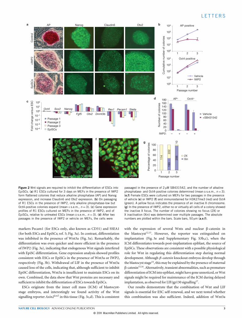
of IWP2, they started to resemble EpiSCs, losing alkaline phosphataseexpression and acquiring a flattened morphology (Fig. 2a). Thecolonies remained positive for the ESC and EpiSC markers Oct4and Nanog, and induced the EpiSC markers Claudin6 and Otx2(ref. 4; Fig. 2a and Supplementary Fig. S3a). To confirm that the ESCsdifferentiated towards EpiSCs, we passaged the cells as small clumpsusing collagenase IV, as is required to preserve EpiSCs (refs 3,4),and evaluated their marker profile, growth-factor dependence andepigenetic state. In the presence of IWP2, only Oct4-positive colonies
negative for alkaline phosphatase expanded (Fig. 2b and SupplementaryFig. S3a), and the cells acquired amarker-gene expression profile similarto that of EpiSCs (Fig. 2c). Furthermore, similarly to EpiSCs, IWP2-treated ESCs became dependent on Activin/Nodal signalling throughthe ALK receptors (ActivinA receptors, type 1), as colony formationwas severely repressed by the ALK inhibitor SB431542 (Fig. 2d). Finally,EpiSCs differ from ESCs in their epigenetic state, most markedly shownby the presence of an inactive X chromosome in female EpiSCs. Indeed,IWP2 treatment of female ESCs induced the formation of trimethylhistoneH3 Lys 27 (H3K27me3) foci, which is diagnostic for the inactiveX chromosome (Fig. 2e–g). In addition, the treated cells activated theDNA methylation machinery as shown by the induction of the denovo DNA methylase gene Dnmt3b (Fig. 2c). Combined, these datademonstrate that Wnt signals are required to inhibit the differentiationof ESCs into EpiSCs. This is in agreement with a report that ESCsmutant for theWnt signal transducer �-catenin (also known asCtnnb1)show amorphology andmarker pattern similar to that of EpiSCs11.ESCs readily differentiate into EpiSCs on transfer to EpiSCmedium6,
and we tested whether Wnt3a protein would be sufficient to block thistransition. R1 ESCs plated in serum-free N2B27medium supplementedwith basic fibroblast growth factor (bFGF), ActivinA and LIF rapidlyacquired EpiSC character, as indicated by FACS analysis for the
2 NATURE CELL BIOLOGY ADVANCE ONLINE PUBLICATION
© 2011 M acmillan Publishers Limited. A ll rights reserved.
L E T T ERS
Control +IWP2
0
20
40
60
80
100P
erce
ntag
e of
col
onie
s
1 2 3Passage number
2XXin11
4
1020
6
193
Con
trol
+IW
P2
EpiS
Cs
AP Nanog Claudin6 Otx2
1 2 3
AP positive
Cum
ulat
ive
num
ber o
f col
onie
s
1 2 3
Oct4 positive
Passage number
VehicleIWP2
100
101
102
103
104
100
101
102
103
104
Fold
cha
nge
vers
us E
SC
100
10–1
10–2
10–3
10–4
101
102
Oct4 Sox2 Nanog
Fgf5 Otx2 Dnmt3b
Rex1 Pecam1 Stella
Passage 1Passage 2Passage 3EpiSCs 0
20406080
100120140160180
IWP2: – + – +
AP+ Oct4+
Num
ber o
f col
onie
s
Vehicle+SB431542
a
c
e fg
d
b
222
Figure 2 Wnt signals are required to inhibit the differentiation of ESCs intoEpiSCs. (a) R1 ESCs cultured for 3 days on MEFs in the presence of IWP2form flattened colonies that reduce alkaline phosphatase (AP) and Nanogexpression, and increase Claudin6 and Otx2 expression. (b) On passagingof R1 ESCs in the presence of IWP2, only alkaline phosphatase-low butOct4-positive colonies expand (mean+s.e.m., n =3). (c) Gene expressionprofiles of R1 ESCs cultured on MEFs in the presence of IWP2, and ofEpiSCs, relative to untreated ESCs (mean±s.e.m., n = 3). (d) After twopassages in the presence of IWP2 or vehicle on MEFs, the cells were
passaged in the presence of 2 µM SB431542, and the number of alkalinephosphatase- and Oct4-positive colonies determined (mean±s.e.m., n=3).(e,f) Female ESCs were cultured on MEFs for two passages in the presenceof vehicle (e) or IWP2 (f) and immunostained for H3K27me3 (red) and Oct4(green). A yellow focus indicates the presence of an inactive X chromosome.(g) In the presence of IWP2, either no or virtually all cells of a colony showedthe inactive X focus. The number of colonies showing no focus (2X) orX inactivation (Xin) was determined over multiple passages. The absolutenumbers are plotted within the bars. Scale bars, 50 µm (a,e,f).
markers Pecam1 (for ESCs only, also known as CD31) and SSEA1(for both ESCs and EpiSCs; ref. 5; Fig. 3a). In contrast, differentiationwas inhibited in the presence of Wnt3a (Fig. 3a). Remarkably, thedifferentiation was even quicker and more efficient in the presenceof IWP2 (Fig. 3a), indicating that endogenous Wnt signals interferedwith EpiSC differentiation. Gene expression analysis showed profilesconsistent with ESCs or EpiSCs in the presence of Wnt3a or IWP2,respectively (Fig. 3b). Withdrawal of LIF in the presence of Wnt3acaused loss of the cells, indicating that, although sufficient to inhibitEpiSC differentiation, Wnt3a is insufficient to maintain ESCs on itsown. Combined, the data show that Wnt proteins are necessary andsufficient to inhibit the differentiation of ESCs towards EpiSCs.ESCs originate from the inner cell mass (ICM) of blastocyst-
stage embryos, and interestingly we found activity of the Wntsignalling reporter Axin2LacZ in this tissue (Fig. 3c,d). This is consistent
with the expression of several Wnts and nuclear �-catenin inthe blastocyst12,13. However, the reporter was extinguished onimplantation (Fig. 3e and Supplementary Fig. S3b,c), when theICM differentiates towards post-implantation epiblast, the source ofEpiSCs. These observations are consistent with a possible physiologicalrole for Wnt in regulating this differentiation step during normaldevelopment. Although �-catenin knockout embryos develop throughthe blastocyst stage14, this may be explained by the presence of maternal�-catenin15,16. Alternatively, transient abnormalities, such as prematuredifferentiation of ICM into epiblast,might have gone unnoticed, orWntsignals might be required for maintenance of the ICM during delayedimplantation, as observed for LIF/gp130 signalling17.Our results demonstrate that the combination of Wnt and LIF
signals is essential for ESC self-renewal, and we next tested whetherthis combination was also sufficient. Indeed, addition of Wnt3a
NATURE CELL BIOLOGY ADVANCE ONLINE PUBLICATION 3
© 2011 M acmillan Publishers Limited. A ll rights reserved.

L E T T ERS
1.6% 93.4% 70.4% 0.3%
0.6% 96.4% 1.4% 93.9% 7.6% 87%
58.3% 20.2% 50.1% 5.9%
1.9%89.8%5.4%91.1%54.7% 39.3%
38.9% 50.7%+W
nt3a
No
Wnt
3a+I
WP
2Passage 1 Passage 2 Passage 3
ESCs EpiSCs
Pecam1
SS
EA-1
LFAW p. 1LFAW p. 2LFAW p. 3
LFAI p. 1LFAI p. 2LFAI p. 3
EpiSCs
Oct4 Sox2 Nanog Rex1 Stella Pecam1
Fgf5Dnmt3b
102
101
100
10–1
10–2
10–3
10–4
Fold
cha
nge
vers
us E
SC
a
b
c d e
Figure 3 Wnt3a protein is sufficient to inhibit the differentiation of ESCsinto EpiSCs. (a) FACS plots of EpiSCs and of R1 cells passaged every 3 daysin N2B27 supplemented with LIF, bFGF and ActivinA, in the presenceof Wnt3a protein (240ngml�1) or IWP2 (2 µM) as indicated, and stainedwith anti-SSEA1-PE (phycoerythrin) and anti-Pecam1-FITC (fluoresceinisothiocyanate) antibodies (10,000 cells/plot). (b) Gene expression profilesof EpiSCs and R1 ESCs cultured in N2B27 supplemented with LIF, bFGF,ActivinA and either Wnt3a (LFAW) or IWP2 (LFAI), relative to untreatedESCs (mean±s.e.m., n =3). (c–e) The Axin2LacZ/+ reporter indicates Wntactivity in the E3.5 (c) and E4.5 (d) ICM, but is inactive in E5.5 implantedembryos (e). Scale bars, 25 µm.
protein supported the expansion of R1 ESCs at clonal density in theabsence of MEFs (Fig. 4a), and even in serum-free N2B27 mediumsupplemented with LIF (Fig. 4b and Supplementary Fig. S4a,b). In
N2B27+ LIF+Wnt3a, the cells adhered poorly to gelatin-coatedsurfaces but plating efficiency was restored by further fibronectincoating, and the expansion was then similar to that on MEFs (compareFig. 4a,f). No adaptation period was necessary for culture in N2B27+LIF+Wnt3a. The cells formed colonies with a distinct, dome-shapedmorphology that stained intensely for alkaline phosphatase (compareFig. 4c,d), and real-time PCR analysis demonstrated normal expressionlevels of ESCmarkers (Fig. 4e). 5-bromodeoxyuridine analysis revealedno role of Wnt3a in proliferation, indicating that inhibition ofdifferentiation is its main function in self-renewal (SupplementaryFig. S4c). Using single-cell deposition in N2B27+LIF+Wnt3a, R1cells formed colonies in 55 out of 95 wells. Several clones were furtherexpanded over the course of eight passages (24 days) and functionallytested by blastocyst injections, resulting in chimaeras with highcoat-colour chimaerism, a strong sex distortion in favour ofmales and ahigh proportion of germline transmission (Supplementary Table S1 andFig. S4d). These results demonstrate thatWnt3a protein in combinationwith LIF is sufficient to support self-renewal of germline-competentESCs at similar efficiencies to serumandMEFs, even at clonal density.An alternative way to activate the Wnt pathway is by inhibiting the
glycogen synthase kinase 3 (GSK3) kinases, which stabilizes �-catenin18
(Supplementary Fig. S4e). However, the GSK3 inhibitor CHIR99021was less effective than Wnt3a in supporting clonal expansion of ESCs(Fig. 4f), indicating that the other effects of GSK3 inhibition19 aredisadvantageous. Further concerns about GSK3 inhibition arise fromits interference with chromosomal alignment at mitosis and from itsinduction of chromosome instability20,21.Recently, the combined inhibition of the GSK3 and MAP-kinase
kinase (MEK) kinases was reported to support ESC self-renewal in theabsence of LIF (ref. 22). We found that, in combination with the MEKinhibitor PD0325901, Wnt3a protein was as effective as CHIR99021in supporting limited self-renewal (Fig. 4f). The self-renewal effectof GSK3 inhibition can therefore be ascribed to its ability to activatethe Wnt pathway, consistent with a recent report23. Interestingly, inthe presence of LIF, MEK inhibitor lowered the requirement for Wntprotein in ESC self-renewal (compare Fig. 4g and b), but providedno benefit when sufficient Wnt3a protein was provided (Fig. 4f andSupplementary Fig. S4f). MEK inhibition did not activate the Wntpathway, as neither the 7xTcf–eGFP reporter nor theAxin2LacZ reporterresponded to PD0325901 in ESCs (not shown), nor did Wnt3a inhibitthe MEK pathway (Fig. 4h), suggesting that the interaction betweenthese pathways is further downstream.We next investigated the effect of Wnt on ESC derivation, using
N2B27 medium in the absence of MEFs. ESC derivation from wholeblastocysts of the mouse strain 129Sv was 96% efficient when Wnt3aand LIF were combined with the MEK inhibitor PD0325901, whichaids ESC derivation by preventing the differentiation of ICM intoprimitive endoderm in whole-blastocyst culture24 (SupplementaryTable S2). Following derivation, Wnt3a and LIF were sufficient tosupport expansion of the new lines. The newly derived cells stainedpositive for alkaline phosphatase, Oct4 and Nanog (SupplementaryFig. S5a), and several lines contributed to germline chimaeras followingblastocyst injections (Supplementary Table S3 and Fig. S5b).Most mouse strains are non-permissive for ESC derivation; that
is, no ESCs develop from embryos cultured on MEFs in thepresence of LIF (ref. 25). However, we readily derived ESCs from
4 NATURE CELL BIOLOGY ADVANCE ONLINE PUBLICATION
© 2011 M acmillan Publishers Limited. A ll rights reserved.

L E T T ERS
0
1
2
Oct4
Sox2NanogSte
lla
Pecam
1
Feeders + serum + LIFSerum + LIF + Wnt3aLIF + Wnt3a
Expr
essi
on le
vel
p-ERK
ERKPD –9 hWnt3a
1 2 3
AP
+ co
loni
es, c
umul
ativ
e
100
101
102
103
104
105
Passage number
MEFs + Wnt3a25 ng ml–1
MEFs
Wnt3a100 ng ml–1
Wnt3a25 ng ml–1
No MEFs,no Wnt3a
IWP21 2 3
100
101
102
103
104
AP
+ co
loni
es, c
umul
ativ
e
Passage number
Wnt3a100 ng ml–1
Wnt3a50 ng ml–1
Wnt3a25 ng ml–1
Wnt3a13 ng ml–1
No Wnt3a
1 2 30
100
101
102
103
104
105
AP
+ co
loni
es, c
umul
ativ
e
Passage number
LIF + PD + Wnt3aLIF + Wnt3aLIF + PD + CHIRLIF + CHIRLIF + PDPD + CHIRPD + Wnt3aLIF + PD + IWP2CHIRWnt3a 200 ng ml–1
1 2 3100
101
102
103
104
AP
+ co
loni
es, c
umul
ativ
e
Passage number
Wnt3a100 ng ml–1
Wnt3a50 ng ml–1
Wnt3a25 ng ml–1
Wnt3a13 ng ml–1
No Wnt3a
IWP2
a
f
g h
b c
e
d
Figure 4 LIF and Wnt3a are sufficient to support ESC self-renewal.(a,b) Expansion over multiple passages of alkaline phosphatase-positive(AP+) R1 ESC colonies in medium containing serum and LIF (a) orin N2B27 containing LIF (b) (mean+ s.e.m., n = 3). (c,d) Alkalinephosphatase-stained R1 ESCs cultured on MEFs (c) or maintained forsix passages in N2B27 medium supplemented with LIF and Wnt3a (d). (e)Gene expression profile of R1 ESCs following four passages in the indicatedconditions. (f) Expansion of alkaline phosphatase-positive R1 ESC colonies
in N2B27 medium. Where indicated, Wnt3a was added at 200ngml�1.(g) Expansion of alkaline phosphatase-positive R1 ESC colonies in N2B27medium containing LIF and PD0325901. (h) MEK activity (indicated bythe presence of phospho-ERK (p-ERK; extracellular signal-regulated kinase)on western blot) in R1 ESCs cultured in N2B27 with LIF, was repressedby PD0325901 (PD) but not by Wnt3a, or by withdrawal of Wnt3a for 9 h(�9h). Uncropped images of blots are shown in Supplementary Fig. S4g.Scale bar, 200 µm (c,d).
non-permissive FVB/N embryos on addition of Wnt3a protein tothese conditions, and also in the absence of MEFs, using N2B27supplemented with LIF, Wnt3a and PD0325901 (SupplementaryTable S2). The new lines stained positive for alkaline phosphatase,Oct4 and Nanog (Fig. 5a–c and Supplementary Fig. S5a), several linescontributed to high-percentage chimaeras (Supplementary Table S4)and germline transmission was observed from line FN3 (Fig. 5d).Following derivation, Wnt3a and LIF were sufficient to supportexpansion of FVB/N ESCs (Fig. 5e). We found that FVB/N ESCsrequired Wnt3a supplementation for expansion on MEFs (Fig. 5f),indicating that MEFs provide insufficient Wnt ligands to supportthem. Non-permissive ESCs therefore differ from their permissivecounterparts by an increased requirement forWnt ligands.This work shows that ESC self-renewal depends on extrinsic
Wnt signals, and identifies the Wnt pathway as a regulatorysignal between ESCs and EpiSCs. Moreover, we show thatthe combination of Wnt3a protein with LIF is sufficientto support the expansion of germline-competent ESCs, atclonal efficiencies similar to those obtained with feeders. Wntsignals may inhibit differentiation to EpiSCs through thetranscription factor TCF3, which regulates the expression ofmultiple-pluripotency genes26–28.
A function of Wnt signals in ESCs has been suspected since itwas reported that Apc modulates the differentiation of ESCs throughthe �-catenin pathway29. Although subsequent reports demonstratedthat Wnt ligands can improve ESC self-renewal30–32, no requirementor specific function for Wnt signals in ESC self-renewal had beenestablished. In fact, for some time it has been unclear whether theself-renewing state of ESCs requires extrinsic signals or is autonomous.Although the cytokine LIF had been identified as an essential ESCself-renewal factor33,34, a recent report shows that combined inhibitionof the MEK signal transducers and the GSK3 kinases (2i condition) issufficient to support ESC self-renewal22, suggesting that extracellularfactors are not required. However, GSK3 inhibition has many effects19,among which is activation of the Wnt signalling pathway18. Ourdemonstration that Wnt3a can substitute for the GSK3 inhibitor in the2i condition indicates that the effect of GSK3 inhibition can be ascribedto its ability to activate theWnt pathway.The hitherto unappreciated requirement forWnt signals for ESC self-
renewal may have implications for the establishment of naive pluripo-tent cells from other species, including humans. This is underscoredby our demonstration that Wnt3a protein supports the derivation ofgermline-competent ESCs from non-permissive FVB mice. The Wntrequirement also explains findings of an instable naive state in repro-
NATURE CELL BIOLOGY ADVANCE ONLINE PUBLICATION 5
© 2011 M acmillan Publishers Limited. A ll rights reserved.

L E T T ERS
01 2 3A
P+
colo
nies
, cum
ulat
ive
101
102
103
104
Passage number
Wnt3a200 ng ml–1
PD + Wnt3a200 ng ml–1
PD, no Wnt
No WntMEFs
101
100
102
103
104
1 2 3Passage number
MEFs+ Wnt3a120 ng ml–1
c
AP
+ co
loni
es, c
umul
ativ
ea b d e f
Figure 5 Wnt3a supports derivation of non-permissive ESCs. (a–c) Alkalinephosphatase, Oct4 and Nanog stainings, respectively, of the newly derivedFVB/N ESC line FN3. Scale bar, 200 µm. (d) Chimaera (black–white spotted)obtained from injection of C57Bl/6 blastocysts with passage 7 ESC lineFN3 together with FVB mate and pups showing germline transmission (white
pups). (e) Expansion of alkaline phosphatase-positive (AP+) colonies fromnewly derived FVB/N ESCs in N2B27 medium with LIF (mean+ s.e.m.,n = 3). PD, PD0325901. (f) Expansion of alkaline phosphatase-positivecolonies from FVB/N ESCs on MEFs in medium containing serum and LIF(mean+s.e.m., n=3).
grammed non-permissive NOD cells35 as a consequence of insufficientparacrine support by Wnt proteins. Finally, the dependence of ESCself-renewal on (heterogeneous) paracrine Wnt signals may explain theheterogeneity that is observed in ESC cultures, with significant subpop-ulations showing a partial differentiation towards EpiSCs (ref. 5). ⇤
METHODSMethods and any associated references are available in the onlineversion of the paper at http://www.nature.com/naturecellbiology
Note: Supplementary Information is available on the Nature Cell Biology website
ACKNOWLEDGEMENTSThese studies were supported by theHowardHughesMedical Institute, the ErasmusMC Stem Cell Institute and grants from the California Institute of RegenerativeMedicine (RC1-00133-1), the National Institutes of Health (DK67834-01) and theEuropean Union (FP7-PEOPLE-2009-RG-256560). We thank H. Zeng for technicaladvice, J. Kong-A-San for assistance with blastocyst injections and R. van der Lindenfor assistance with FACS.We are grateful for the use of the Cellavista imager and theassistance of V. Vincent and Roche Diagnostics.
AUTHOR CONTRIBUTIONSD.t.B., D.K. and T.B. designed and carried out experiments, analysed data and wrotethe paper. W.K., A.M., R.S. and E.E. designed and carried out experiments andanalysed data. R.N. designed experiments and wrote the paper.
COMPETING FINANCIAL INTERESTSThe authors declare no competing financial interests.
Published online at http://www.nature.com/naturecellbiologyReprints and permissions information is available online at http://www.nature.com/reprints
1. Nichols, J. & Smith, A. Naive and primed pluripotent states. Cell Stem Cell 4,487–492 (2009).
2. Niwa, H. Open conformation chromatin and pluripotency. Genes Dev. 21,2671–2676 (2007).
3. Brons, I. G. et al. Derivation of pluripotent epiblast stem cells from mammalianembryos. Nature 448, 191–195 (2007).
4. Tesar, P. J. et al. New cell lines from mouse epiblast share defining features withhuman embryonic stem cells. Nature 448, 196–199 (2007).
5. Hayashi, K., Lopes, S. M., Tang, F. & Surani, M. A. Dynamic equilibrium andheterogeneity of mouse pluripotent stem cells with distinct functional and epigeneticstates. Cell Stem Cell 3, 391–401 (2008).
6. Guo, G. et al. Klf4 reverts developmentally programmed restriction of ground statepluripotency. Development 136, 1063–1069 (2009).
7. ten Berge, D. et al. Wnt signaling mediates self-organization and axis formation inembryoid bodies. Cell Stem Cell 3, 508–518 (2008).
8. Sato, N., Meijer, L., Skaltsounis, L., Greengard, P. & Brivanlou, A. H. Maintenanceof pluripotency in human and mouse embryonic stem cells through activationof Wnt signaling by a pharmacological GSK-3-specific inhibitor. Nat. Med. 10,55–63 (2004).
9. Chen, B. et al. Small molecule-mediated disruption of Wnt-dependent signaling intissue regeneration and cancer. Nat. Chem. Biol. 5, 100–107 (2009).
10. Lustig, B. et al. Negative feedback loop of Wnt signaling through upregulation of con-ductin/axin2 in colorectal and liver tumors. Mol. Cell Biol. 22, 1184–1193 (2002).
11. Anton, R., Kestler, H. A. & Kuhl, M. �-catenin signaling contributes to stemnessand regulates early differentiation in murine embryonic stem cells. FEBS Lett. 581,5247–5254 (2007).
12. Kemp, C., Willems, E., Abdo, S., Lambiv, L. & Leyns, L. Expression of all Wntgenes and their secreted antagonists during mouse blastocyst and postimplantationdevelopment. Dev. Dyn. 233, 1064–1075 (2005).
13. Wang, Q. T. et al. A genome-wide study of gene activity reveals developmentalsignaling pathways in the preimplantation mouse embryo. Dev. Cell 6,133–144 (2004).
14. Haegel, H. et al. Lack of �-catenin affects mouse development at gastrulation.Development 121, 3529–3537 (1995).
15. Ohsugi, M. et al. Expression and cell membrane localization of catenins duringmouse preimplantation development. Dev. Dyn. 206, 391–402 (1996).
16. De Vries, W. N. et al. Maternal �-catenin and E-cadherin in mouse development.Development 131, 4435–4445 (2004).
17. Nichols, J., Chambers, I., Taga, T. & Smith, A. Physiological rationale forresponsiveness of mouse embryonic stem cells to gp130 cytokines. Development128, 2333–2339 (2001).
18. Stambolic, V., Ruel, L. & Woodgett, J. R. Lithium inhibits glycogen synthasekinase-3 activity and mimics wingless signalling in intact cells. Curr. Biol. 6,1664–1668 (1996).
19. Doble, B. W. & Woodgett, J. R. GSK-3: tricks of the trade for a multi-tasking kinase.J. Cell Sci. 116, 1175–1186 (2003).
20. Tighe, A., Ray-Sinha, A., Staples, O. D. & Taylor, S. S. GSK-3 inhibitors inducechromosome instability. BMC Cell Biol. 8, 34 (2007).
21. Acevedo, N., Wang, X., Dunn, R. L. & Smith, G. D. Glycogen synthase kinase-3regulation of chromatin segregation and cytokinesis in mouse preimplantationembryos. Mol. Reprod. Dev. 74, 178–188 (2007).
22. Ying, Q. L. et al. The ground state of embryonic stem cell self-renewal. Nature 453,519–523 (2008).
23. Kelly, K.F. et al. �-catenin enhances Oct-4 activity and reinforces pluripotencythrough a TCF-independent mechanism. Cell Stem Cell 8, 214–227 (2011).
24. Nichols, J., Silva, J., Roode, M. & Smith, A. Suppression of Erk signallingpromotes ground state pluripotency in the mouse embryo. Development 136,3215–3222 (2009).
25. Gardner, R. L. & Brook, F. A. Reflections on the biology of embryonic stem (ES) cells.Int. J. Dev. Biol. 41, 235–243 (1997).
26. Cole, M. F., Johnstone, S. E., Newman, J. J., Kagey, M. H. & Young, R. A. Tcf3is an integral component of the core regulatory circuitry of embryonic stem cells.Genes Dev. 22, 746–755 (2008).
27. Tam, W. L. et al. T-cell factor 3 regulates embryonic stem cell pluripotency andself-renewal by the transcriptional control of multiple lineage pathways. Stem Cells26, 2019–2031 (2008).
28. Yi, F., Pereira, L. & Merrill, B. J. Tcf3 functions as a steady-state limiter oftranscriptional programs of mouse embryonic stem cell self-renewal. Stem Cells 26,1951–1960 (2008).
29. Kielman, M. F. et al. Apc modulates embryonic stem-cell differentiation bycontrolling the dosage of �-catenin signaling. Nat. Genet. 32, 594–605 (2002).
30. Hao, J., Li, T. G., Qi, X., Zhao, D. F. & Zhao, G. Q. WNT/�-catenin pathwayup-regulates Stat3 and converges on LIF to prevent differentiation of mouseembryonic stem cells. Dev. Biol. 290, 81–91 (2006).
31. Ogawa, K., Nishinakamura, R., Iwamatsu, Y., Shimosato, D. & Niwa, H. Synergisticaction of Wnt and LIF in maintaining pluripotency of mouse ES cells.Biochem. Biophys. Res. Commun. 343, 159–166 (2006).
32. Singla, D. K., Schneider, D. J., LeWinter, M. M. & Sobel, B. E. wnt3a but not wnt11supports self-renewal of embryonic stem cells. Biochem. Biophys. Res. Commun.345, 789–795 (2006).
33. Williams, R. L. et al. Myeloid leukaemia inhibitory factor maintains thedevelopmental potential of embryonic stem cells. Nature 336, 684–687 (1988).
34. Smith, A. G. et al. Inhibition of pluripotential embryonic stem cell differentiation bypurified polypeptides. Nature 336, 688–690 (1988).
35. Hanna, J. et al. Metastable pluripotent states in NOD-mouse-derived ESCs.Cell Stem Cell 4, 513–524 (2009).
6 NATURE CELL BIOLOGY ADVANCE ONLINE PUBLICATION
© 2011 M acmillan Publishers Limited. A ll rights reserved.

DOI: 10.1038/ncb2314 METHODS
METHODSRecombinant proteins and small molecules. Recombinant ActivinA andhuman bFGF proteins were purchased from Peprotech. Fz8CRD–Fc fusionprotein (Supplementary Fig. S5c) and control Fc were produced as described36.Recombinant mouse Wnt3a protein was produced in Drosophila S2 cells grownin suspension culture, and purified by Blue Sepharose affinity and gel filtrationchromatography as described37 (Supplementary Fig. S5c). Wnt3a activity wasdetermined in a luciferase reporter assay using L cells stably transfected withthe SuperTOPFlash reporter as described38 (Supplementary Fig. S4e). Someexperiments were reproduced using recombinant humanWnt3a protein generouslydonated by R&D Systems.
IWP2, CHIR99021, SB431542 (all purchased from Stemgent) and PD0325901(Merck) were diluted from 2mM (IWP2) or 10mM (all others) stocks indimethylsulphoxide, and used at 2, 3, 6 and 0.9 µM, respectively. Media,recombinant proteins and small molecules were changed daily in all experimentsexcept when indicated otherwise.
Cell and embryo culture. R1 and CGR8 ESCs were obtained from the StanfordTransgenic Facility. E14tg2a ESCs were purchased from ATCC. Axin2LacZ ESCs10were donated by W. Birchmeier (Berlin). Female C57Bl6/Mus musculus castaneus
E8 ESCs were donated by J. Gribnau (Rotterdam).Routine culture of R1 and E8 ESCs was carried out on a feeder layer of
irradiated primary MEFs (GlobalStem) in mESC medium (DMEM plus 15%fetal bovine serum (Hyclone), 1mM sodium pyruvate, MEM non-essential aminoacids, 50 µM�-mercaptoethanol, 100Uml�1 penicillin, 100 µgml�1 streptomycin(all from Invitrogen) and 1,000Uml�1 LIF (Chemicon)) on gelatin-coated plates.
N2B27 medium39 consisted of one volume DMEM/F12 combined with onevolume Neurobasal medium, supplemented with 0.5% N2 Supplement, 1%B27 Supplement, 0.033% BSA 7.5% solution, 50 µM�-mercaptoethanol, 2mMGlutamax, 100 U/ml penicillin and 100 µgml�1 streptomycin (all from Invitrogen).
CGR8, Axin2LacZ and E14tg2a ESCs were maintained on gelatin-coated plates inmESC medium.
Cells were passaged as a single-cell suspension using 0.25% trypsin–EDTA. Forserum-free culture, trypsin was quenched using soybean trypsin inhibitor (Sigma).
129S2 C1a EpiSCs were donated by L.Vallier, and maintained on plates coatedwith gelatin and FCS in N2B27 medium supplemented with 12 ngml�1 bFGFand 20 ngml�1 ActivinA. The cells were passaged every 3–4 days as clumps using0.5mgml�1 collagenase IV (Sigma). For culture on MEFs, EpiSCs were maintainedin mEpiSC medium (DMEM/F12 plus 15% knockout serum replacement (KOSR),MEM non-essential amino acids, 100 µM�-mercaptoethanol (all Invitrogen) and10 ngml�1 bFGF).
E5.5 Axin2LacZ/+ embryos10 were obtained from matings of homozygousAxin2LacZ/LacZ males with wild-type females. Embryos were placed in mESCmediumwithout LIF, supplementedwith 3 µMCHIR99021 or vehicle and cultured overnightat 37 �C in 5% CO2 atmosphere. Cultured embryos were fixed for 2min in 1%paraformaldehyde, washed in PBS and stained with 5-bromo-4-chloro-3-indolyl-�-d-galactoside (X-gal). Axin2LacZ EpiSCswere derived fromE5.5Axin2LacZ/+ embryosas described4.
Correlation between Wnt reporter and ability to form ESC colonies. For7xTcf–eGFP FACS, cells were plated on MEFs and cultured for 2 days, dissociatedwith 0.25% trypsin–EDTA and resuspended in PBS with 1% serum and 0.05%propidium iodide, and live cells sorted into four categories on the basis of eGFPintensity using a BD FACSAria II cell sorter. Gates were set such that the dimmestand brightest categories each contained 13% of the total population, whereas theintermediate categories each contained 33% (the remainder were lost in the gapsbetween the gates). Sorted cells were plated on gelatin-coated plates in mESCmedium, containing 300 ngml�1 Wnt3a as indicated, at a density of 250 cells/cm�2,and stained for alkaline phosphatase after 4 days. The stained plates were driedand imaged using a Cellavista automated imager equipped with a ⇥4 objectiveand bright-field illumination (Roche Diagnostics). The total number of coloniesin each well (alkaline phosphatase-positive and negative) was counted manually,whereas the number of alkaline phosphatase-positive colonies was determined byusing CellProfiler biological image analysis software40 to ensure consistency. Thepercentage of alkaline phosphatase-positive colonies was calculated and plotted±s.e.m. (n= 3).
Clonal self-renewal assays. Toquantify self-renewal overmultiple passages, singlecells were plated at a density of 100 cells cm�2 in gelatin-coated six-well plates and ingelatin-coated 24-well plates in triplicate, containing an MEF layer when indicated.
For assays in N2B27, the plates were additionally coated with 15 µgml�1 humanplasma fibronectin (Sigma). Every 3 days, six-well plates were trypsinized to singlecells, and passaged to a new set of plates at a dilution that would lead to a density nothigher than but as close as possible to 100 cells cm�2. At the same time, the 24-wellplates were stained for alkaline phosphatase, or for Oct4 using an Oct4 antibody and3,30-diaminobenzidine (DAB) staining (see Marker staining). Stained plates wererinsed with water, dried and scanned using a CellCelector automated imager (Aviso)equipped with a ⇥4 objective and bright-field illumination. The number of positivecolonies was determined using CellProfiler biological image analysis software40. Thecumulative number of colonies was determined bymultiplying the colony counts bythe dilution factor used for passaging. Results are plotted as the mean of three wells±s.e.m.
ESC to EpiSC differentiation assays. A single-cell suspension of R1 ESCs wasplated onto MEFs at a density of 100 cells cm�2 in mESC medium in six-well plates,and in 24-well plates in triplicate. EpiSCs were plated as small clumps onto MEFs inmEpiSCmedium. One day after plating, IWP2 was added as indicated. After 3 moredays, the 24-well plates were processed for marker staining, and the six-well platespassaged 1:10 using 0.5mgml�1 collagenase IV every 3 days. At the second passage,further 24-well plates were prepared with 6 µM SB431542 included in the mediafor the ALK-inhibitor assay. Stained plates were manually counted. The cumulativenumber of colonies was determined bymultiplying the colony counts by the dilutionfactor used for passaging. Results are plotted as the mean± s.e.m. of three wells.
For serum-free conditions, a single-cell suspension of R1 ESCs was plated ontogelatin/serum-coated plates at a density of 10,000 cells cm�2 in N2B27 mediumsupplemented with LIF and 120 ngml�1 Wnt3a. The next day, the media werereplaced with N2B27/LIF supplemented with 12 ngml�1 bFGF, 20 ngml�1 ActivinAand 120 ngml�1 Wnt3a or 1 µM IWP2 as indicated. The cells were then passaged1:4–1:10 every 3 days as small clumps using 0.5mgml�1 collagenase IV. At everypassage, a portion of the cells was triturated to single-cell suspension, stainedwith anti-SSEA1–PE (eBioscience 12-8813) and anti-Pecam1–FITC (BDBiosciences553372) antibodies, and analysed using a Facscan flow cytometer.
ESC derivation. E3.5 blastocysts were obtained from natural matings of 129Svor FVB/N mice and plated intact in 96-well plates on MEFs or on plates coatedfirst with gelatin, followed by 15 µgml�1 human plasma fibronectin (Sigma) inPBS. An equal volume of fresh medium was added on days 2 and 3, followed bydaily medium changes. The medium used for derivation on MEFs consisted ofDMEMplus 15%KOSR (Invitrogen),MEMnon-essential amino acids (Invitrogen),50 µM�-mercaptoethanol and 1,000Uml�1 LIF (Chemicon). In the absence ofMEFs, derivation was carried out in N2B27 medium containing 1,000Uml�1 LIF.Wnt3a protein or the MEK inhibitor PD0325901 was added as indicated to aconcentration of 300 ngml�1 or 1 µM, respectively. When both Wnt3a and MEKinhibitor were added, the MEK inhibitor was removed after the outgrowths werepassaged. After 6 days, outgrowths were trypsinized and transferred to fresh 96-wellplates. Newly formed cell lines were expanded for aminimum of seven passages, andanalysed by alkaline phosphatase, Nanog andOct4 staining and blastocyst injectionsusing C57Bl/6 host blastocysts. The percentage of coat colour chimaerism of thechimaeras was estimated visually. To determine germline transmission, chimaerasfrom 129Sv-derived ESCs were mated with C57Bl/6 mice, and chimaeras fromFVB/N-derived ESCs were mated with FVB/N mice.
Marker staining and immunohistochemistry. Alkaline phosphatase activitywas detected using the SCR004 kit (Millipore). For immunostaining, cells werefixed with 4% paraformaldehyde for 15min, washed thrice with PBS/0.5% TritonX-100 (PBT), permeabilized for 20min with ice-cold methanol and blocked with10% normal donkey serum (NDS)/PBT for 30min. Samples were then incubatedwith primary antibody in NDS/PBT overnight at 4 �C, washed three times withPBT and either stained using a Vector ABC Elite kit and DAB (Vector), orprimary antibodies detected by DyLight 488-labelled secondary antibodies (JacksonImmunoResearch), followed by imaging. Antibodies and concentrations: anti-Oct4(Santa Cruz sc-8628, 0.8 µgml�1), anti-Nanog (Cosmo Bio REC-RCAB0002P-F,1:250), anti-Claudin6 (Santa Cruz sc-17669, 2 µgml�1), anti-Otx2 (Abcam ab21990,1 µgml�1), anti-H3K27me3 (Abcam ab6002, 1:100), anti-ERK and anti p-ERK (CellSignalling 9102 and 4376S, respectively, both 1:1,000).
Gene expression analysis. Total RNA was prepared using a QIAGEN RNeasymini kit with on-column DNase digestion, followed by reverse transcription usingSuperscript II (Invitrogen). When MEFs were present, cells were first plated for30min on gelatin-coated plates to enable MEFs to attach and ESCs or EpiSCs
NATURE CELL BIOLOGY
© 2011 M acmillan Publishers Limited. A ll rights reserved.

METHODS DOI: 10.1038/ncb2314
gently flushed off the plate. Quantitative PCR with reverse transcription was carriedout on a Roche Lightcycler 480 using Lightcycler 480 SYBR Green Master mix(Roche). Relative quantification was carried out using glyceraldehyde-3-phosphatedehydrogenase as a reference gene. All PCRs were carried out in triplicate, and themean crossing point was used for quantification. Primer sequences were designedusing Lightcycler Probe Design Software 2.0 (Roche) such that they spanned splicejunctions when possible.
For detection of Wnt gene expression in R1 and CGR8 cells, PCR reactions werecarried out in quadruplicate. Primers were tested on cDNA derived from E12.5whole-embryo RNA, and product length was verified on gels. Primer sequences areprovided in Supplementary Table S5.
36. Hsieh, J. C., Rattner, A., Smallwood, P. M. & Nathans, J. Biochemicalcharacterization of Wnt–frizzled interactions using a soluble, biologically activevertebrate Wnt protein. Proc. Natl Acad. Sci. USA 96, 3546–3551 (1999).
37. Willert, K. et al. Wnt proteins are lipid-modified and can act as stem cell growthfactors. Nature 423, 448–452 (2003).
38. Mikels, A. J. & Nusse, R. Purified Wnt5a protein activates or inhibits �-catenin–TCFsignaling depending on receptor context. PLoS Biol 4, e115 (2006).
39. Ying, Q. L., Stavridis, M., Griffiths, D., Li, M. & Smith, A. Conversion ofembryonic stem cells into neuroectodermal precursors in adherent monoculture. Nat.Biotechnol. 21, 183–186 (2003).
40. Lamprecht, M. R., Sabatini, D. M. & Carpenter, A. E. CellProfiler: free, versatilesoftware for automated biological image analysis. Biotechniques 42, 71–75 (2007).
NATURE CELL BIOLOGY
© 2011 M acmillan Publishers Limited. A ll rights reserved.

S U P P L E M E N TA RY I N F O R M AT I O N
WWW.NATURE.COM/NATURECELLBIOLOGY 1
DOI: 10.1038/ncb2314
Figure S1 Wnt signals are expressed by and required for the self-renewal of ESCs. (a) Expression level of Wnt genes in R1 and CGR8 ESCs relative to Gapdh (mean+/-s.e.m., n=4; ND, not detected). R1 cells were cultured on MEFs in medium with serum and LIF, CGR8 cells on gelatine-coated plates in medium with serum and LIF. (b) R1 ESCs were cultured at
clonal density on MEFs in medium with serum and LIF supplemented with Fz8CRD and/or 600 ng/ml Wnt3a protein as indicated, passaged every 3 days using trypsin, and stained at every passage for AP. Shown are representative fields of view of the experiment that is quantified in Fig. 1h. Scale bar: 500 μm.
Passage 1 Passage 3Passage 2
Control
Fz8CRD 0.5 ug/ml
Fz8CRD 2 ug/ml
Fz8CRD 8 ug/ml
Fz8CRD 2 ug/ml+ Wnt3a
Fz8CRD 8 ug/ml+ Wnt3a
Fz8CRD 0.5 ug/ml+ Wnt3a
+ Wnt3a
Expression relative to Gapdh
10E-5 10E-4 10E-3 10E-2 10E-1W nt1W nt2bW nt3W nt3aW nt4W nt5aW nt5bW nt7aW nt7bW nt8bW nt9aW nt10bW nt11W nt16
NDND
R1 ESCs
CGR8 ESCs
10E-4W nt1W nt2bW nt3W nt3aW nt4W nt5aW nt5bW nt7aW nt7bW nt8bW nt9aW nt10bW nt11W nt16
10E-110E-210E-310E-5
Expression relative to Gapdh
ND
a b
ten Berge et alFig. S1
© 2011 M acmillan Publishers Limited. A ll rights reserved.

S U P P L E M E N TA RY I N F O R M AT I O N
2 WWW.NATURE.COM/NATURECELLBIOLOGY
Figure S2 Wnt signals are required for the self-renewal of feeder-independent ESCs. (a) CGR8 and E14tg2a ESCs cultured in the absence of MEFs in medium with serum and LIF, supplemented with IWP2, 2 µg/ml Fz8CRD, and/or 200 ng/ml Wnt3a as indicated, passaged every 3 days, and
stained at every passage for AP. (b) Expansion over multiple passages of AP-positive E14tg2a and IB10 ESC colonies in medium containing serum and LIF, and supplemented with IWP2, Fz8CRD, and/or 200 ng/ml Wnt3a where indicated. Scale bar: 500 μm (a).
+Wnt3a
+ Vehicle
+ IWP2
+ Fz8CRD
+ Vehicle
+ IWP2
+ Fz8CRD
Passage 1
Passage 2
CGR8 E14tg2a+Wnt3a
a
b
1E+0
1E+1
1E+2
1E+3
1E+4
1E+5
1 2 3
Wnt3a 80 ng/mlWnt3a 20 ng/mlWnt3a 5 ng/mlno Wnt3aIWP2
IB10 ESCs in presence of serum + LIF
AP
+ co
loni
es, c
umul
ativ
e
1 2 3
E14tg2a ESCs in presence of serum + LIF
Passage number Passage number
1E+0
1E+1
1E+2
1E+3
1E+4Vehicle
IWP2
Fz8CRD 0.5ug/mlWnt3a
IWP2 + Wnt3a
Fz8CRD 0.5ug/ml + Wnt3a
ten Berge et alFig. S2
© 2011 M acmillan Publishers Limited. A ll rights reserved.

S U P P L E M E N TA RY I N F O R M AT I O N
WWW.NATURE.COM/NATURECELLBIOLOGY 3
Figure S3 Wnt signals are required to inhibit the differentiation of ESCs into EpiSCs. (a) R1 ESCs were cultured on MEFs in medium with serum and LIF supplemented with 2 µM IWP2 as indicated, passaged every 3 days as small clumps using Collagenase IV, and stained at every passage for AP and Oct4. Shown are representative fields of view of the experiment that is quantified in Fig. 2b. (b) To determine whether Axin2LacZ in the E5.5 epiblast responds to the Wnt/beta-catenin pathway, E5.5 Axin2LacZ/+ embryos were cultured for 24 hrs in the presence of the GSK3-inhibitor CHIR99021 or vehicle, and stained with X-gal. Red arrow points to the epiblast. Blue staining in the epiblast
indicates that the reporter is able to respond to activation of the Wnt/beta-catenin pathway. (c) To confirm that the Axin2LacZ reporter is able to respond to Wnt proteins, EpiSCs were derived from E5.5 Axin2LacZ/+ embryos, cultured in N2B27 containing bFGF and ActivinA, treated with the indicated factors for 2 days, and stained with X-gal and Nuclear Red. The cells display weak local spontaneous activation of the reporter which is repressed by IWP2 and Fz8CRD, indicating that it is the result of endogenous Wnt proteins. Treatment with Wnt3a protein (200 ng/ml) induced the reporter, confirming that it is indeed responsive to Wnt proteins. Scale bars: 500 μm (a,c), 25 μm (b).
Passage 1 Passage 2 Passage 3
VehicleAlkPhos
IWP2AlkPhos
EpiSCs - Oct4
VehicleOct4
IWP2Oct4
EpiSCs - AlkPhos
a + Vehicle
+ CHIR99021 (GSK3-inhibitor)
b
+ IWP22 uM
+ Wnt3a
+ Fz8CRD 2 ug/ml
Vehicle
c
ten Berge et alFig. S3
© 2011 M acmillan Publishers Limited. A ll rights reserved.

S U P P L E M E N TA RY I N F O R M AT I O N
4 WWW.NATURE.COM/NATURECELLBIOLOGY
Figure S4 LIF and Wnt3a are sufficient to support expansion of ESCs. (a,b) As a control, at the last passage of the quantitative self renewal assays a second set of plates was prepared and stained for Oct4, in addition to the AP staining. Virtual all colonies that were present stained positive for either marker. Shown are representative fields of view of passage 3 of the quantitative self renewal assay measuring the expansion of R1 ESC colonies in serumfree N2B27 medium containing LIF and 120 ng/ml Wnt3a, stained for AP (a) or Oct4 (b). (c) BrdU analyses indicating that Wnt3a protein does not promote ESC proliferation. R1 ESCs were plated in N2B27 medium containing LIF or Wnt3a as indicated, and cultured for the indicated amount of time. The cells were then labeled with BrdU for 4 hrs, counted, and the amount of incorporated BrdU determined using the Roche Cell Proliferation
ELISA. The results are plotted as BrdU incorporation normalized for cell count. Using one-way ANOVA, no significant difference was observed between the different media regardless of culture duration (p>0.05). Results shown are the mean±s.e.m, n=4. (d) Chimeric mice displaying 100% coat colour chimerism obtained by injection of C57Bl/6 blastocysts with clonally expanded R1 cells. (e) Supertopflash reporter assay demonstrates activity of purified Wnt3a protein. The GSK3 inhibitor CHIR099021 causes maximum stimulation of the Wnt pathway at 1.5-3 µM. (f) Representative fields of view of passage 3 of the experiment that is quantified in Fig. 4f. (g) Full length western blots showing MEK activity in R1 ESCs cultured in N2B27 with LIF, indicated by the presence of phospho-ERK on western blot. Molecular weight markers (kD) are indicated at the left of the image. Scale bars: 500 μm (a,b), 200 μm (f).
a b
d
f
LIF+Wnt3a LIF+Wnt3a+PD
LIF+IWP2+PD
0
100
200
300
400
500
600
10 100 1000 10000
Concentration [ng/ml Wnt3a; nM CHIR]
Fold
indu
ctio
n
Wnt3a CHIRe g
150
2025
375075
100
150
2025
375075
100
- ERK
- p-ERK
-
-9 hr
s
PD Wnt3
a
xx
c
012
3456
789
LIF+W
nt3a
LIF, n
o Wnt3a
Wnt3a, n
o LIF
no gr
owth
factors
Brd
U in
corp
orat
ion/
cell
[a.u
.]
01
234
567
89
LIF+W
nt3a
LIF, n
o Wnt3a
Wnt3a, n
o LIF
no gr
owth
factors
01
23
45
67
89
LIF+W
nt3a
LIF, n
o Wnt3a
Wnt3a, n
o LIF
no gr
owth
factors
48 hrs culture24 hrs culture 72 hrs culture
ten Berge et alFig. S4
© 2011 M acmillan Publishers Limited. A ll rights reserved.

S U P P L E M E N TA RY I N F O R M AT I O N
WWW.NATURE.COM/NATURECELLBIOLOGY 5
Figure S5 Pluripotency marker stainings and germline transmission of newly derived ESCs. (a) 129Sv and FVB/N ESC lines (SV and FN, respectively) derived in N2B27/LIF/Wnt3a/PD325901 were cultured in N2B27/LIF/Wnt3a for at least 3 passages, and colonies stained for the ESC markers alkaline phosphatase (red), Oct4 (green), and Nanog (green). (b) Chimera obtained
from injection of C57Bl/6 blastocysts with passage 8 ESC line SV3 (40% coat colour chimerism) together with pups showing germline transmission (agouti pups). Brown areas indicate contribution of the ESCs. (c) Silver-stained gel demonstrating purity of Wnt3a and Fz8CRD protein preps. Molecular weight markers (kD) are indicated at the left of the image. Scale bar: 200 μm (a).
AlkPhos Oct4 Nanog AlkPhos Oct4 Nanog
SV1
SV21
SV14
SV13
SV12
SV11
SV10
SV9
SV8
SV7
SV6
SV5
SV4
SV2
SV15
SV16
SV17
SV18
SV19
SV20
SV22
SV23
SV24
AlkPhos Oct4 Nanog
FN1
FN3
FN2
SV3
a
b
Wnt3a
BSAFz8CRD
37
50
75100150250
2520
Wnt3a Fz8CRDc
ten Berge et alFig. S5
© 2011 M acmillan Publishers Limited. A ll rights reserved.