Temporally and spatially coordinated roles for Rho, Rac ...1724 respond with similar cathodally...
13
1723 Research Article Introduction Nervous system function requires ‘hard-wired’ circuits resulting from the extension of neuronal processes (neurites) over long distances to specific targets. Growth cones, the tips of extending neurites, are guided in embryos and during regeneration by extracellular cues including gradients of chemoattractive and chemorepulsive molecules (Yamamoto, 2002). Steady, direct current (DC) electric fields (EFs) co-exist with these molecular gradients for prolonged periods (Hotary and Robinson, 1991; Shi and Borgens, 1995) (reviewed by McCaig et al., 2002; McCaig et al., 2005). The basis for the EF is the transepithelial potential (TEP) across the intact epithelium. In Xenopus embryos, a TEP of approximately +60 mV (inside positive) is generated by the inward movement of Na + ions across the skin. In regions where epithelial integrity is disturbed, for example by breakdown of tight junctions prior to cell migration, the TEP collapses locally but remains intact distally. This generates a voltage gradient (EF) within the tissues beneath (parallel to) the epithelium. The neural tube, the precursor of the brain and spinal cord, shares an ectodermal origin with skin and generates its own TEP such that the entire nervous system develops within a natural EF of hundreds of mV/mm (Hotary and Robinson, 1991; Shi and Borgens, 1994; Shi and Borgens, 1995). The EF varies developmentally and spatially, and is highest across developmentally key regions, such as the floor plate (McCaig et al., 2005). EFs are usually omitted from the list of recognised growth cone guidance cues despite evidence that disruption of naturally occurring EFs perturbs normal nervous system development (Hotary and Robinson, 1992; Hotary and Robinson, 1994; Metcalf and Borgens, 1994), successful completion of a Phase I clinical trial in which a DC EF was applied to neurologically complete human spinal cord injuries (Shapiro et al., 2005), and evidence that EFs direct neurite growth in vivo (Song et al., 2004) and in vitro (Hinkle et al., 1981; McCaig, 1987; Rajnicek et al., 1998). Neurites of dissociated, embryonic, Xenopus spinal neurons grow faster towards the negative pole of the EF (cathode), retract from the positive pole (anode) and branch more often towards the cathode, and growth cones turn to migrate towards the cathode. Cathodally directed growth occurs in an EF as small as 7 mV/mm (Hinkle et al., 1981), which is at least 50 times smaller than the EFs that developing neurons encounter naturally in vivo. Significantly, regenerating mammalian nerves in vivo Although it is known that neuronal growth cones migrate towards the cathode of an applied direct current (DC) electric field (EF), resembling the EF present in the developing nervous system, the underlying mechanism remains unclear. Here, we demonstrate temporally and spatially coordinated roles for the GTPases Rac, Cdc42 and Rho and their effectors. Growth cones of cultured Xenopus embryonic spinal neurons turned towards the cathode but collective inhibition of Rho, Rac and Cdc42 attenuated turning. Selective inhibition of Rho, Cdc42 or Rac signalling revealed temporally distinct roles in steering by an electrical gradient. Rho, Rac and Cdc42 are each essential for turning within the initial 2 hours (early phase). Later, Rho and Cdc42 signals remain important but Rac signalling dominates. The EF increased Rho immunofluorescence anodally. This correlated spatially with collapsed growth cone morphology and reduced anodal migration rates, which were restored by Rho inhibition. These data suggest that anodally increased Rho activity induces local cytoskeletal collapse, biasing growth cone advance cathodally. Collapse might be mediated by the Rho effectors p160 Rho kinase and myosin light chain kinase since their inhibition attenuated early turning. Inhibitors of phosphoinositide 3-kinase, MEK1/2 or p38 mitogen-activated protein kinase (MAPK) did not affect turning behaviour, eliminating them mechanistically. We propose a mechanism whereby Rac and Cdc42 activities dominate cathodally and Rho activity dominates anodally to steer growth cones towards the cathode. The interaction between Rho GTPases, the cytoskeleton and growth cone dynamics is explored in the companion paper published in this issue. Our results complement studies of growth cone guidance by diffusible chemical gradients and suggest that growth cones might interpret these co-existing guidance cues selectively. Key words: Axon guidance, Electric field, Rho GTPases Summary Temporally and spatially coordinated roles for Rho, Rac, Cdc42 and their effectors in growth cone guidance by a physiological electric field Ann M. Rajnicek*, Louise E. Foubister and Colin D. McCaig School of Medical Sciences, Institute of Medical Sciences, University of Aberdeen, Aberdeen, Scotland, AB25 2ZD, UK *Author for correspondence (e-mail: [email protected]) Accepted 19 January 2006 Journal of Cell Science 119, 1723-1735 Published by The Company of Biologists 2006 doi:10.1242/jcs.02896 Journal of Cell Science
Transcript of Temporally and spatially coordinated roles for Rho, Rac ...1724 respond with similar cathodally...

1723Research Article
IntroductionNervous system function requires ‘hard-wired’ circuitsresulting from the extension of neuronal processes (neurites)over long distances to specific targets. Growth cones, the tipsof extending neurites, are guided in embryos and duringregeneration by extracellular cues including gradients ofchemoattractive and chemorepulsive molecules (Yamamoto,2002). Steady, direct current (DC) electric fields (EFs) co-existwith these molecular gradients for prolonged periods (Hotaryand Robinson, 1991; Shi and Borgens, 1995) (reviewed byMcCaig et al., 2002; McCaig et al., 2005). The basis for theEF is the transepithelial potential (TEP) across the intactepithelium. In Xenopus embryos, a TEP of approximately +60mV (inside positive) is generated by the inward movement ofNa+ ions across the skin. In regions where epithelial integrityis disturbed, for example by breakdown of tight junctions priorto cell migration, the TEP collapses locally but remains intactdistally. This generates a voltage gradient (EF) within thetissues beneath (parallel to) the epithelium. The neural tube,the precursor of the brain and spinal cord, shares an ectodermalorigin with skin and generates its own TEP such that the entirenervous system develops within a natural EF of hundreds of
mV/mm (Hotary and Robinson, 1991; Shi and Borgens, 1994;Shi and Borgens, 1995). The EF varies developmentally andspatially, and is highest across developmentally key regions,such as the floor plate (McCaig et al., 2005).
EFs are usually omitted from the list of recognised growthcone guidance cues despite evidence that disruption ofnaturally occurring EFs perturbs normal nervous systemdevelopment (Hotary and Robinson, 1992; Hotary andRobinson, 1994; Metcalf and Borgens, 1994), successfulcompletion of a Phase I clinical trial in which a DC EF wasapplied to neurologically complete human spinal cord injuries(Shapiro et al., 2005), and evidence that EFs direct neuritegrowth in vivo (Song et al., 2004) and in vitro (Hinkle et al.,1981; McCaig, 1987; Rajnicek et al., 1998). Neurites ofdissociated, embryonic, Xenopus spinal neurons grow fastertowards the negative pole of the EF (cathode), retract from thepositive pole (anode) and branch more often towards thecathode, and growth cones turn to migrate towards the cathode.Cathodally directed growth occurs in an EF as small as 7mV/mm (Hinkle et al., 1981), which is at least 50 times smallerthan the EFs that developing neurons encounter naturally invivo. Significantly, regenerating mammalian nerves in vivo
Although it is known that neuronal growth cones migratetowards the cathode of an applied direct current (DC)electric field (EF), resembling the EF present in thedeveloping nervous system, the underlying mechanismremains unclear. Here, we demonstrate temporally andspatially coordinated roles for the GTPases Rac, Cdc42 andRho and their effectors. Growth cones of cultured Xenopusembryonic spinal neurons turned towards the cathode butcollective inhibition of Rho, Rac and Cdc42 attenuatedturning. Selective inhibition of Rho, Cdc42 or Racsignalling revealed temporally distinct roles in steering byan electrical gradient. Rho, Rac and Cdc42 are eachessential for turning within the initial 2 hours (early phase).Later, Rho and Cdc42 signals remain important but Racsignalling dominates. The EF increased Rhoimmunofluorescence anodally. This correlated spatiallywith collapsed growth cone morphology and reducedanodal migration rates, which were restored by Rhoinhibition. These data suggest that anodally increased Rho
activity induces local cytoskeletal collapse, biasing growthcone advance cathodally. Collapse might be mediated bythe Rho effectors p160 Rho kinase and myosin light chainkinase since their inhibition attenuated early turning.Inhibitors of phosphoinositide 3-kinase, MEK1/2 or p38mitogen-activated protein kinase (MAPK) did not affectturning behaviour, eliminating them mechanistically. Wepropose a mechanism whereby Rac and Cdc42 activitiesdominate cathodally and Rho activity dominates anodallyto steer growth cones towards the cathode. The interactionbetween Rho GTPases, the cytoskeleton and growth conedynamics is explored in the companion paper published inthis issue. Our results complement studies of growth coneguidance by diffusible chemical gradients and suggest thatgrowth cones might interpret these co-existing guidancecues selectively.
Key words: Axon guidance, Electric field, Rho GTPases
Summary
Temporally and spatially coordinated roles for Rho,Rac, Cdc42 and their effectors in growth coneguidance by a physiological electric fieldAnn M. Rajnicek*, Louise E. Foubister and Colin D. McCaigSchool of Medical Sciences, Institute of Medical Sciences, University of Aberdeen, Aberdeen, Scotland, AB25 2ZD, UK*Author for correspondence (e-mail: [email protected])
Accepted 19 January 2006Journal of Cell Science 119, 1723-1735 Published by The Company of Biologists 2006doi:10.1242/jcs.02896
Jour
nal o
f Cel
l Sci
ence

1724
respond with similar cathodally directed growth, turning andbranching in a naturally occurring EF of about 40 mV/mm(Song et al., 2004; Sta Iglesia and Vanable, Jr, 1998).
Assuming a threshold EF strength of 10 mV/mm, the voltagegradient across a 10 �m wide growth cone is 0.1 mV. Themechanism by which a growth cone senses this small gradientand translates it into a directional response remains unclear butseveral parallels exist with growth cone chemotropism, inwhich growth cones detect and amplify (through spatiallyregulated signal transduction) shallow chemical gradients(McCaig et al., 2005). Mechanistically, both require signallingthrough cytoplasmic Ca2+, phospholipase C (PLC), andactivation of receptors for neurotrophin-3 (NT-3), brain-derived growth factor (BDNF), acetylcholine (ACh) andfibroblast growth factor (FGF). EF guidance is presumed toinvolve EF-induced asymmetric localisation or activation ofchannels or receptors in the plasma membrane (reviewed byMcCaig et al., 2002; McCaig et al., 2005), particularly voltage-gated Ca2+ channels (Stewart et al., 1995) and receptors forACh, NT-3 and BDNF (Fig. 7A). How asymmetric activationof these molecules steers growth cones towards the cathode hasnot been established but any proposal must link thesemolecules to spatial regulation of the cytoskeleton.
A probable link between the candidate molecules and thecytoskeleton is the Rho family of small GTPases, whichincludes Rac, Rho and Cdc42. These molecules aredownstream of activation of the candidate membrane receptors(and Ca2+ signals), upstream of effectors of cytoskeletaldynamics, and are proposed to mediate directional migrationof growth cones (Jin et al., 2005; Mueller, 1999) and other cellsin chemical gradients (Fukata et al., 2003; Kaibuchi et al.,1999). Genetic disruption of GTPase activity induces growthcone path-finding defects in embryos (Kaufmann et al., 1998;Kim et al., 2002), suggesting that precise spatiotemporalregulation of Rho GTPases is crucial for normal growth coneguidance.
According to the predominant model, growth conechemoattraction results from activation of Cdc42 and Rac onthe side of the growth cone facing the attractant (andconsequent inhibition of Rho), whereas chemorepulsion ismediated by activation of Rho on the side facing the repellent(and inhibition of Rac and Cdc42) (Mueller, 1999; Song andPoo, 2001; Patel and Van Vactor, 2002). Activation of Cdc42and Rac stimulates growth cone filopodia and lamellipodia,respectively, whereas RhoA activation stimulates contractionof the actin cytoskeleton leading to growth cone collapse(Kozma et al., 1997; Kim et al., 2002; Yuan et al., 2003).
The aim of the present study was to explore the role of thesmall GTPases Rac, Cdc42 and Rho and their effectors in EF-induced growth cone steering. The mechanistic link betweensignalling by the small GTPases, the cytoskeleton and growthcone dynamics in an EF are explored in the companion paperin this issue (Rajnicek et al., 2006). Here, we show that Rac,Cdc42 and Rho are each involved in guidance of growth conesby EFs and that Rac signalling predominates during the latephase (>2 hours) of EF exposure. Turning during the earlyphase (<2 hours) requires activity of the Rho GTPase effectorsmyosin light chain kinase (MLCK), citron kinase (citron-K),rhotekin-rhophilin-protein kinase N (PKN) and p160 Rhokinase (p160 ROCK). By contrast, cathodal turning does notrequire phosphoinositide 3-kinase (PI 3-kinase), p38 mitogen-
Journal of Cell Science 119 (9)
activated protein kinase (MAPK) or extracellular signal relatedkinase (ERK1/2) activity, which have been implicatedpreviously in EF guidance of non-neuronal cells (Zhao et al.,2002) and chemoattraction of growth cones (Campbell andHolt, 2003). These data suggest mechanistic differencesbetween steering in chemical and electrical gradients. A modelfor the intracellular cascades involved in EF guidance ofgrowth cones is presented.
It is particularly important to elucidate the mechanisms thatunderpin EF influences on neuronal growth and guidance inlight of a recently completed clinical trial testing the ability ofa DC EF to stimulate human spinal cord regeneration (Shapiroet al., 2005). Since Rho is activated following spinal cordinjury (Madura et al., 2004) and Rho inhibition improves adultmammalian spinal cord repair (Fourneir et al., 2003; Sung etal., 2003), an understanding of the interaction between RhoGTPases and EF-induced growth cone guidance would suggestnovel combinations of electrical/pharmacological therapies.
ResultsNeuronal growth cones migrate directionally towards thecathodeConsistent with previous studies (e.g. Hinkle et al., 1981; Pateland Poo, 1982; Rajnicek et al., 1998), Xenopus spinal neuronswithout an EF exhibited randomly oriented neurite extensionwhereas those exposed to an EF of 150 mV/mm for 5 hoursreoriented strikingly towards the cathode (Fig. 1A-D). Withoutan EF, growth cones turned +3±6° and migrated at 19.6±2.1�m/hour; in an EF, growth cones migrated faster (31.4±2.3�m/hour) and turned –53±5° towards the cathode (expect 0° ifrandom; negative values indicate net cathodal turning, positivevalues indicate net anodal turning). The EF also increased thefrequency of cathodal turning; 76% (65/85) turned towards thecathode, and decreased the frequency of anodal turning to 8%(7/85) compared with no EF controls in which 27% (13/49) ofgrowth cones turned towards the cathode and 39% (19/49)turned towards the anode. The controls are indistinguishablefrom the theoretical frequency of 33% for random growth.
Control experiments (drug, no EF) were performed for allpharmacological treatments in Figs 1-6 (data not shown).Neurite extension was random (not different from the ‘withoutEF’ data above). The mean angle turned during 5 hours rangedfrom –6±8° in Rho 23-40 (n=49) to +8±6° in 10 ng/ml toxinB (TxB) (n=40).
Rho GTPases are involved in cathodal growth coneguidanceThe Rho family of small GTPases has been implicated inchemoattractive turning of Xenopus growth cones (Yuan et al.,2003; Mueller, 1999). We used the same pharmacologicalinhibitors of Rho GTPases and their effectors from thosestudies to explore the mechanism for cathodal turning. Thebroad-spectrum Rho GTPase inhibitor TxB derived fromClostridium difficile inhibits RhoA, Rac 1 and Cdc42collectively (Chavez-Olarte et al., 1997) and attenuated turningin 0.01 ng/ml or 10 ng/ml (Fig. 1A,B,D). The mean angleturned during 5 hours of EF exposure (Fig. 1A) and thefrequency of cathodal turning (Fig. 1B) were reducedcompared with EF-treated growth cones in the absence of anyinhibitor, but the frequency of anodal turning was not affectedby TxB (Fig. 1B). Similar cathodal attenuation was observed
Jour
nal o
f Cel
l Sci
ence
1725Nerve guidance by electrical gradients
in 0.1 ng/ml TxB (n=50, data not shown). Thediminished turning response in TxB was not a resultof failure of neurite extension since growth conemigration rates exceeded (in 0.01 ng/ml) or wereidentical to (in 0.1 ng/ml or 10 ng/ml) those withoutthe drug (Fig. 1C). These data implicate Rac, Cdc42and Rho collectively in cathodal orientation but do notallow discrimination of the selective requirements ofRac, Rho or Cdc42.
Cdc42 and Rac mediate cathodal turningWe dissected the involvement of individual familymembers in EF guidance using custom-synthesisedpeptides that perturb Rac or Cdc42 signallingselectively (Vastrik et al., 1999) and C3 transferase(from Clostridium botulinum, also known as C3exoenzyme) to inhibit RhoA (Aktories et al., 1989;Narumiya et al., 1990). The synthetic peptidescorrespond to amino acids 17-32 on the N-terminus ofRac 1 (Rac 17-32) or Cdc42 (Cdc42 17-32) and areeach tagged with a TAT internalisation sequence on theC-terminus. The peptides correspond to the CRIB(Cdc42/Rac-1 binding) domain on Rac or Cdc42.These peptides were selected because, from anoverlapping series of peptides, only Rac 17-32 but notCdc42 17-32 or Rho 17-32 inhibited collapse of dorsalroot ganglion growth cones (Vastrik et al., 1999),demonstrating peptide specificity on growth conebehaviour. In addition, Rac 17-32 or Cdc42 17-32 eachprevented retraction of axons upon contact with arepulsive cue (Thies and Davenport, 2002), suggestinga role in neurite guidance. The peptides compete withactivated Rac or Cdc42 for binding to effector proteinsand therefore prevent signalling downstream (Vastriket al., 1999). Peptides were added to the cultures atleast 1 hour prior to EF exposure and remained in themedium throughout the experiment.
In 10 �g/ml Rac 17-32, growth cones migratedrandomly during the entire 5 hours of EF exposure(Fig. 2). The mean angle turned (Fig. 2A) and thefrequencies of cathodal and anodal turning (Fig. 2B)were the same as for growth cones without an EF.Therefore, inhibition of Rac signalling abolishedcathodal orientation throughout the entire 5 hours.
The effect of Cdc42 17-32 on growth cone turningwas biphasic. In 10 �g/ml Cdc42 17-32, growth conesdid not turn at all during the first 2 hours of EFexposure (–0.3±2°) and, by 5 hours, had turned only–13±4 deg (n=177). Therefore, cathodal turning wasabolished by Cdc42 17-32 in the first 2 hours andcathodal turning at 5 hours was attenuatedsubstantially (P<0.0001 compared with EF alone) butnot abolished (P=0.0274 compared with no EF) (Fig. 2A,D).A similar trend was observed for the frequency of cathodalturning (Fig. 2B). The frequency turning at 2 hours (27%,48/177) was reduced (P<0.001) compared with 72% (61/85)without the peptide. By 5 hours, only 40% (71/177) had turnedcathodally, significantly less than without the peptide (76%,65/85, P<0.001) (Fig. 2B). Although it attenuated cathodalturning, Cdc42 17-32 did not abolish turning at 5 hours, as Rac17-32 had done (Fig. 2A). The peptides also differ in their
effect on neurite outgrowth. Growth cones migrated morerapidly in Rac 17-32 than in Cdc42 17-32 (Fig. 2C). Since thepeptides differ by only two amino acids and contain identicalTAT sequences, the differences in growth cone behavioursuggest different functional specificities and reducesignificantly the possibility that the TAT sequence itself causesthe inhibitory effects. Moreover, growth cone morphologydiffered in the two peptides (Rajnicek et al., 2006) in a mannerconsistent with selective inhibition of either Rac or Cdc42,
Fig. 1. Collective inhibition of Rac, Rho and Cdc42 with toxin B attenuatescathodal growth cone steering by an EF. (A) Mean angle turned by growthcones during 5 hours. In the absence of an EF, growth cones migraterandomly (mean angle turned would be 0° for random migration) but, in anEF of 150 mV/mm, growth cones turn towards the cathode. Negative valuesindicate net cathodal deflection. The number of growth cones measured is inparentheses. The mean angle turned at 5 hours was compared with ‘no drug +EF’ using a Student’s two-tailed t test; TxB, toxin B; ###P<0.0001.(B) Percentage of growth cones (see A for total) that turn towards the cathode(filled bars) or anode (open bars) in 5 hours. The dotted line is the expectedfrequency for random orientation (33%). Asterisks compare cathodal oranodal frequencies with no drug + EF; ***P<0.001. (C) Mean rate of growthcone advance during 5 hours of EF exposure compared with no drug + EF(black bar) using a two-tailed Student’s t test. ##P=0.002; ###P<0.0001. See Afor number of growth cones. (D) Composite drawings made from images ofindividual, dissociated neurons at the end of a 5-hour experiment. Somaswere superimposed at the coloured dot and the path of each neurite wastraced. The EF vector is horizontal, with cathode at left and anode at right.Bar, 100 �m for all drawings. ns, not significant.
Jour
nal o
f Cel
l Sci
ence

1726
again suggesting specificity of the compounds in the presenceof identical TAT sequences. Filopodia were retained butlamellipodia were reduced significantly in Rac 17-32.Conversely, growth cones in Cdc42 17-32 had few filopodiabut well developed lamellipodia.
Disrupting Rho signalling inhibits cathodal turningRhoA involvement in cathodal turning was tested using C3transferase, which ADP-ribosylates RhoA in the effectordomain, therefore preventing interaction with effectors(Aktories et al., 1989; Narumiya et al., 1990). Cathodal turningat 5 hours was attenuated by 1 �g/ml or 20 �g/ml (Fig. 2). Theturning response in 20 �g/ml C3 transferase was quantitativelyand temporally similar to that in Cdc42 17-32, with no turningwithin the first 2 hours, severely attenuated turning between 2and 5 hours (Fig. 2A) and fewer growth cones turningcathodally by 5 hours than without the drug (Fig. 2B). Cathodalturning was attenuated throughout the entire 5 hours in 1 �g/mlC3 transferase but less than in 20 �g/ml (Fig. 2A). The
frequency of cathodal turning was also decreased in 20 �g/ml(Fig. 2B). Attenuated turning in 20 �g/ml was not attributableto poor growth cone advance because, although the migrationrate was reduced compared with EF-treated cells without aninhibitor, it was the same as for growth cones without any drugor EF (Fig. 2C).
In non-neuronal cells, L-�-lysophosphatidic acid (LPA)induces Rho activation through the LPA receptor-PDZ-Rho-GEF complex (Yamada et al., 2005). In neurons, LPA activatesG-protein-coupled LPA receptors, elevates endogenous Rhowithin seconds, and induces growth cone collapse (Kozma etal., 1997; Kranenburg, 1999). In Xenopus neurons, LPA-induced growth cone collapse and repulsive steering in agradient of LPA are abolished by TxB and when neuronsexpress dominant-negative RhoA but not dominant-negativeCdc42 (Yuan et al., 2003), thus linking LPA and Rho-mediatedturning in Xenopus growth cones. Since Rho inhibitionattenuated cathodal growth cone turning (Fig. 2), we used LPAto determine if stimulating Rho activity enhanced cathodal
turning. Unexpectedly, 1 �M LPA had no effect(data not shown). At a lower concentration (100nM), LPA attenuated the angle and frequency ofturning (Fig. 2A,B,D) but increased the migrationrate (Fig. 2C).
Therefore, a high concentration of C3 transferase(20 �g/ml) attenuated turning, a high concentrationof LPA (1 �M) allowed turning and lowerconcentrations of C3 transferase (1 �g/ml) and LPA(100 nM) attenuated turning. These data suggestthat a critical balance of Rho activation andinactivation is essential for cathodal guidance. Thisidea is supported by the observation that the neuritepaths of 100 nM LPA (Fig. 2D) and 1 �g/ml C3-transferase-treated neurons (n=91, data not shown)are wavy, suggesting that episodes of cathodalturning are interspersed with episodes of adaptation,as has been proposed for Xenopus growth conechemotropism (Ming et al., 2002).
RhoA immunofluorescence is higher anodallyIf asymmetric Rho signalling mediates cathodalgrowth cone turning in a manner analogous tochemotropism (Jin et al., 2005) then Rho activitywould be enhanced anodally. We tested this notionby measuring relative Rho immunofluorescence inanode-facing and cathode-facing sides of growthcones (single plane confocal images). Rho levelswere expressed as the ratio of anode-to-cathodeimmunofluorescence. A symmetrically fluorescentgrowth cone would have an intensity ratio of 1 but,if Rho fluorescence were higher anodally, the ratiowould be >1 and, if it is higher cathodally, the ratiowould be <1. Rho fluorescence was uniform incontrol growth cones (no EF no drug;ratio=1.07±0.06, n=15) but was higher anodallythan cathodally in EF-treated growth cones(1.51±0.11, n=10; P=0.0013). Similarly, Rhofluorescence in 100 nM LPA was uniform withoutan EF (0.98±0.04; n=5) but was elevated anodallyin an EF (1.21±0.04, n=61; P=0.0017). Whenconsidered collectively (regardless of LPA), Rho
Journal of Cell Science 119 (9)
Fig. 2. Effect of selective inhibition of Rac, Rho or Cdc42 and elevation of Rhoon EF growth cone guidance. See Fig. 1 for format. No drug + EF data arerepeated from Fig. 1 for ease of comparison. (A) Mean angle turned since thestart. #P<0.05; ##P<0.005; ###P<0.0001. (B) Frequency of cathodal (filled bars)and anodal (open bars) turning compared with no drug + EF (black bar).*P<0.05; **P=0.002; ***P<0.001. (C) The rate of neurite extension in an EFfor 5 hours. #P<0.005; ##P=0.0035; ###P<0.0001. (D) Composite drawings ofneurite paths for EF-treated cells. Compare with no drug in Fig. 1D. Bar, 100�m for all drawings. ns, not significant.
Jour
nal o
f Cel
l Sci
ence

1727Nerve guidance by electrical gradients
fluorescence was uniform in growth cones without an EF butwas elevated anodally in EF-treated growth cones (Fig. 3A).
Collapse of the anode-facing sides of growth conescorrelates temporally with cathodal turning (Rajnicek et al.,2006). We therefore examined the spatial relationship betweenEF-induced Rho immunofluorescence and growth conemorphology (Fig. 3). Only growth cones oriented at a rightangle to the EF direction (90±45°, some with LPA) wereincluded. We counted the filopodia and measured thelamellipodial area (excluding filopodia) of the cathode- andanode-facing regions from which Rho immunofluorescencehad been determined. Each was expressed as a ratio of theanode-to-cathode values for individual growth cones (Fig. 3A).In 13 control (no EF) growth cones, the Rhoimmunofluorescence intensity, distribution of filopodia andlamellipodial area were symmetrical (ratio ~1.0) but, in 40 EF-treated growth cones, elevation of Rho on the anode-facingsides correlated spatially with anodal reduction of filopodia andlamellipodia.
Rho activation induces growth cone collapse and neuriteretraction (Kozma et al., 1997; Thies and Davenport, 2002;Kranenburg et al., 1999); therefore, anodal accumulation ofRho would be predicted to cause anode-facing growth cones tocollapse and either to retract or advance more slowly than
cathode-facing ones. Our data and a previous report (McCaig,1987) support these ideas. Growth cones rarely faced the anodeafter 5 hours of EF exposure, but Fig. 3 shows a neuron whosegrowth cones faced the cathode and anode directly. The Rhoimmunofluorescence gradient in the cathode-facing growthcone (* in Fig. 3F,G) was less steep (ratio 1.12) and it had amore complex morphology than the relatively collapsed anode-facing growth cone (*** in Fig. 3F,G), which had a steeper Rhofluorescence gradient (ratio 2.07). Rho immunofluorescence ishigher in the shorter (180 �m), anode-facing neurite than thelonger (245 �m), cathode-facing neurite (Fig. 3F), suggestinga link between Rho elevation and reduced anodal migrationrate. Our time-lapse data support this notion. Of 85 EF-treatedgrowth cones in the present study, only two faced the anode at5 hours and these migrated at 3.1±0.2 �m/hour compared with33.0±2.4 �m/hour for 71 growth cones facing cathodally.Therefore, compared with growth cones without an EF(19.6±2.1 �m/hour, n=49), cathodal rates increase and anode-facing growth cones migrate very slowly. If Rho activityunderlies reduced rates, then C3 transferase inhibition of Rhoshould ‘rescue’ anode-facing growth cone advance. Indeed, in1 �g/ml C3 transferase, anode-facing growth cones migratefaster (61.3±16.1 �m/hour, n=6) than cathode-facing growthcones (17.5±2.2 �m/hour, n=100, P=0.0440) and, in 20 �g/ml
Fig. 3. Anodal RhoA elevation correlates spatially withcollapsed morphology. (A) Mean anode-to-cathode ratios forRho immunofluorescence intensity, number of filopodia andlamellipodial area for 40 growth cones (350 total filopodia)oriented within 45° of the EF direction and 13 growth coneswith no EF (182 total filopodia). P values (p; two-tailedStudent’s t test) compare no EF and +EF ratios. A ratio of 1.0(dotted line) indicates a symmetric growth cone, whereasratios >1 and <1 indicate relative anodal and cathodal bias,respectively. (B,D) Confocal images of growth cones in100nM LPA labelled with Rhodamine-phalloidin (red) and anantibody to RhoA (green). Image planes have been merged.(C,E) Confocal image of RhoA immunofluorescence withfluorescence intensity pseudocoloured on the scale shown.Dotted outlines indicate the regions used to calculate ratios.(F) RhoA immunofluorescence intensity plot for the cell inG. The green line represents Rho fluorescence measuredalong a line extending from the tip of the cathode-facinggrowth cone, along the neurite contour to the tip of theanode-facing growth cone. The black line representsfluorescence intensity when the same line is shifted to abackground position near, but not overlapping, the cell.Asterisks indicate corresponding regions in image G. (G) Aneuron in an EF for 5 hours labelled as in B. Insets (H,I,K,L)show detail of cathode-facing and anode-facing growthcones. (J,M) Fluorescence intensity plots for the linesindicated on I and L. Mean intensities (±s.e.m.) for thecathode-facing (blue) and anode-facing (red) sides of eachplot are indicated by black bars. Ratios indicate mean anodeintensity compared with mean cathode intensity for eachgrowth cone. Cathode- versus anode-facing means werecompared with a two-tailed Student’s t test.Jour
nal o
f Cel
l Sci
ence

1728
C3 transferase, rates are identical in each direction (22.1±1.0�m/hour, n=14 anodally and 18.3±1.5 �m/hour, n=42cathodally). Therefore, Rho inhibition rescues anodalattenuation of growth cone advance.
Inhibition of RhoA effectorsHaving established a role for RhoA in cathodal turning, weexplored the effector molecules that Rho might use to directcytoskeletal dynamics. We used 10 �M Y27632 (Uehata et al.,1997) to inhibit p160 ROCK, which mediates growth conecollapse in response to Rho activation (Wahl et al., 2000) andtwo cell-permeable peptides, Rho 23-40 and Rho 75-92,corresponding to the binding sites for citron-K and rhophilin-rhotekin-PKN, respectively (Fujisawa et al., 1998), therebypreventing their downstream signalling. Y27632 (10 �M) hasbeen used previously to implicate ROCK in Rho-mediatedXenopus growth cone chemotropism (Yuan et al., 2003).Y27632 (10 �M) and Rho 23-40 (10 �g/ml) each abolishedcathodal turning during the first 2 hours, but not between 2 and5 hours (Fig. 4A,B,D), which is consistent with our observation
that Rho signalling is especially important in the initial 2 hoursof EF exposure (Fig. 2A). With Y27632 and Rho 23-40, theextent of growth cone turning in 2 hours was the same as thatwithout an EF (–4±3° and –2±2°, respectively). 26% (38/145)of growth cones turned cathodally in Y27632 and 30%(86/289) turned cathodally in Rho 23-40 (Fig. 4A). Turningresumed at 2 hours and, by 5 hours, the angle and frequencyof cathodal turning were similar in each inhibitor (Fig. 4A,B).Neurite growth rates were unchanged in 10 �M Y27632 butincreased in Rho 23-40 compared with EF-treated growthcones without inhibitor (Fig. 4C). 5 �M Y27632 had no effecton growth cone turning (–48±4° at 5 hours) but it increased themigration rate to 53.0±2.0 �m/hour (P<0.0001, n=231).
The Rho 75-92 peptide decreased, but did not abolish, theearly phase of cathodal turning (Fig. 4A). For example, growthcones turned only –12±3° in Rho 75-92 by 2 hours comparedwith –29±4° without the peptide (P=0.0008). By 5 hours, theangle and frequency of cathodal turning were not different toEF-treated cells without the peptide (Fig. 4A,B). Collectively,these data implicate Rho activity (especially the citron-K-binding domain) in cathodal turning and suggest that the earlyphase requires p160 ROCK activity linked to Rho activation.
The ability of Rho 23-40 to abolish the early phase of turningyet Rho 75-92 permitted limited cathodal turning, togetherwith the distinct inhibitory responses of Rac 17-32 and Cdc 17-32 described above, further suggest that the presence of theTAT sequence is not responsible for the effects of the peptides.
MLCK is involved in early turningHaving revealed roles for Rac, Cdc42 and Rho signalling incathodal orientation (Figs 1, 2) we explored the potentialinvolvement of effector molecules downstream from theseGTPases. Wortmannin (500 nM) (Davies et al., 2000) andbutanedione monoxime (BDM) (2 mM), an inhibitor of myosinATPase (Herrmann et al., 1992), were used to inhibit myosinlight chain kinase (MLCK) activity. BDM has been usedpreviously in studies of Xenopus growth cones to implicateMLCK in motility (Ruchhoeft and Harris, 1997) and to revealthat Cdc42 and Rho signals converge at MLCK to underpinsteering in chemical gradients (Yuan et al., 2003). DMSO wasused as a solvent for wortmannin and other inhibitors used inthis and the companion paper in this issue (Rajnicek et al.,2006), so the influence of DMSO on cathodal growth conemigration was tested from 0.0004% to 0.3% DMSO to reflectthe range of solvent concentrations in the medium. Thecathodal responses did not vary within this range so data werepooled. Over the 5 hours of EF exposure in DMSO-containingmedium, the angle turned (Fig. 5A, Fig. 6A) and the frequencyof cathodal turning were the same as without DMSO (Fig.5A,B, Fig. 6A,B), but growth cones migrated faster in DMSOthan without it (Fig. 5C, Fig. 6C).
Wortmannin (0.1% DMSO final concentration) or BDM(dissolved directly in culture medium) attenuated, but did notabolish, cathodal turning during the first 3 hours (Fig. 5A).Attenuation was quantitatively and temporally similar for bothdrugs. At 2 hours, growth cones in 500 nM wortmannin turnedonly –11±4° with 35% (39/111) to the cathode, significantlyless (P=0.0034 and P<0.002, respectively) than in DMSO(–23±2°, 57% 249/440). Similarly, in BDM, they turned only–8±2° with 30% (44/147) compared with –29±4° and 72%(61/85) without the drug. However, by 5 hours, the mean angle
Journal of Cell Science 119 (9)
Fig. 4. Inhibition of Rho effectors attenuates EF-induced growthcone guidance. Format is as in Fig. 1. (A) Mean angle turned during5 hours. No drug + EF data are repeated from Fig. 1 for comparison;ns, not significant; #P<0.05; ##P=0.0002; ###P<0.0001.(B) Frequency of cathodal or anodal turning. *P<0.05; ***P<0.001.(C) Rate of growth cone advance in an EF. ##P=0.0002; ###P<0.0001.(D) Composite drawings of neurons in an EF (cathode to the left) for5 hours. Compare with no drug + EF in Fig. 1D. Bar, 100 �m for alldrawings.
Jour
nal o
f Cel
l Sci
ence

1729Nerve guidance by electrical gradients
and the frequency of turning were not different compared withthe relevant controls (Fig. 5A,B,D). A lower concentration ofwortmannin (5 nM, n=87) yielded similar results. Thefrequency of cathodal turning was reduced at 2 hours (44%,38/87) compared with DMSO controls (P<0.05) but not at 5hours (61%, 53/87 in 5 nM wortmannin; 68% 301/440 inDMSO). The angle turned in 5 nM wortmannin was notsignificantly different to the angle turned in 500 nMwortmannin. By 5 hours, growth cones had turned –30±6° in5 nM wortmannin compared with –37±5° in 500 nM. The rateof growth cone migration in BDM was identical to controlswith no drug but it was decreased by 500 nM (Fig. 5) or 5 nMwortmannin (27.5±1.3 �m/hour) compared with DMSO(45.0±1.1 �m/hour; P=0.0013). These data implicate MLCKactivity in cathodal turning during the early phase.
PI 3-kinase activity is not essential for cathodal turningPI 3-kinase is activated by Rac during chemotaxis (Servant et
al., 2004), and the specific PI 3-kinase inhibitor LY294002attenuates cathodal migration of epithelial cells (Zhao et al.,2002) and mediates adaptation of Xenopus growth cone turningin chemical gradients (Ming et al., 2002). We therefore usedLY294002 (10 �M) to inhibit PI 3-kinase (Vlahos et al., 1994;Davies et al., 2000) in EF-treated neurons. AlthoughLY294002 enhanced the rate of growth cone migration in anEF (Fig. 5C), it did not affect the angle (Fig. 5A,D) or thefrequency (Fig. 5B) of cathodal turning. These responses aredifferent than those in wortmannin, a less specific inhibitor ofPI 3-kinase (Davies et al., 2000), which reduced cathodalturning during the early phase and slowed growth cone advance(Fig. 5). Therefore, we conclude that PI 3-kinase signalling isnot required for either phase of cathodal growth cone steeringand the effect of wortmannin on cathodal orientation is not aresult of inhibition of PI 3-kinase.
Fig. 5. Effects of inhibition of myosin-based contraction (using BDMor wortmannin) or PI 3-kinase (using LY294002 or wortmannin) onEF-induced growth cone guidance. Formatted as in Fig. 1. No drug +EF data are repeated from Fig. 1; ns, not significant compared withrevelant no drug + EF or DMSO + EF control. (A) Mean angleturned. #P<0.05; ##P<0.0004; ###P<0.0001. (B) Frequency of turningduring 5 hours. *P<0.05; ***P<0.001. (C) Growth rates during5 hours of EF exposure. ##P=0.0002 compared with no drug + EF(black bar); P<0.0001 compared with DMSO + EF (grey bar).(D) Composite drawings of neurons in an EF (cathode to the left) for5 hours. Bar, 100 �m for all drawings. Compare BDM and DMSOdrawings to no drug + EF in Fig. 1D, and compare wortmannin andLY294002 to DMSO.
Fig. 6. Effect of inhibition of MAPK signalling on EF-inducedgrowth cone guidance. Format as is Fig. 1. Control data are repeatedfrom Fig. 1; ns, not significant. (A) Mean angle turned during 5hours is not affected by any inhibitor. (B) Percentage of growth conesthat turn cathodally or anodally during 5 hours. None of theinhibitors affected the frequency of cathodal turning and onlySB202190 slightly increased the anodal frequency compared with theDMSO control; *, P<0.05; ***P<0.0001. (C) Rate of growth coneadvance during 5 hours in an EF. P values compared with no drug +EF: DMSO, ###P<0.0001; SB203580, #P<0.05. P values comparedwith DMSO + EF: U0126 and SB202190, ###P<0.001.(D) Composite drawings of neurons in an EF (cathode to the left)for 5 hours. Bar, 100 �m for all drawings. Compare U0126 andSB202091 to DMSO at top of panel and compare SB203580 to nodrug + EF in Fig. 1D.
Jour
nal o
f Cel
l Sci
ence
1730
MEK1/2 and p38 MAPK signalling are not required forcathodal turningMEK1/2, ERK1/2 and p38 MAPK (p38) signalling have beenimplicated in cathodal migration of epithelial cells (Wang etal., 2003; Zhao et al., 2002) and chemoattraction of Xenopusgrowth cones (Campbell and Holt, 2003; Ming et al., 2002), sowe tested their involvement in EF growth cone guidance withthe same inhibitors used for studies of Xenopus growth conechemoattraction (Campbell and Holt, 2003; Ming et al., 2002).ERK1/2 activation by MEK1/2 was inhibited using 30 �MU0126 (0.3% DMSO) (Duncia et al., 1998), whereas p38 wasinhibited using 300 nM SB202190 (0.003% DMSO) or 10 �MSB203580 (dissolved in culture medium) (Davies et al., 2000).None of these inhibitors affected the mean angle turned or thefrequency of cathodal turning (Fig. 6A,B,D). Growth conesmigrated faster in SB203580 and SB202190 than without anydrug but they were slower in U0126 than in DMSO alone (Fig.6C). Antibody staining (not shown) confirmed ERK1 and p38MAPK presence in growth cones. Immunofluorescence of totalp38 MAPK (active and inactive, assessed as for total Rho) wasuniform (ratio=0.96±0.07, n=6) across EF-treated growthcones oriented at a right angle to the EF (90±45°), suggestingthat the EF does not induce a gradient of total p38 MAPK.Together with the pharmacological data, this indicates that p38MAPK and ERK1/2 signalling are not essential for directedcathodal growth cone migration.
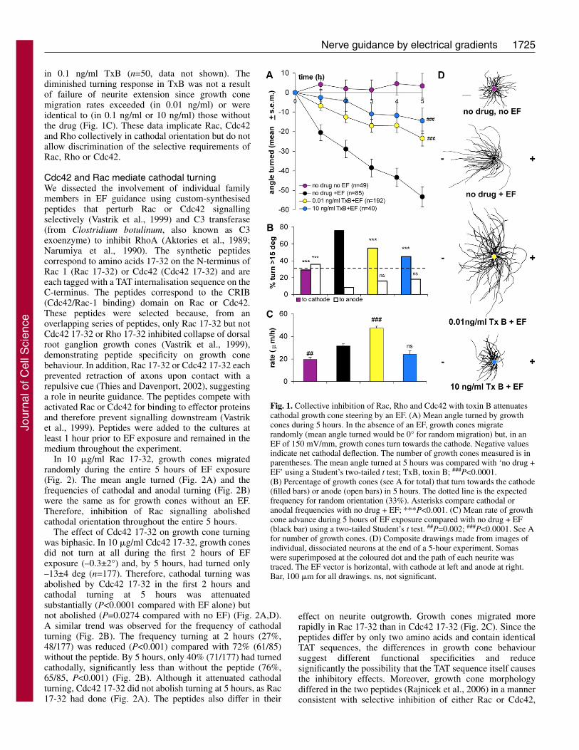
DiscussionWe demonstrate that the GTPases Cdc42, Rac and Rho mediategrowth cone steering in a physiological EF by spatiotemporalregulation of GTPase activity and their effectors. In thecompanion paper in this issue (Rajnicek et al., 2006), wedescribe the cytoskeletal requirements for EF steering, theeffects of inhibition of Rac and Cdc42 signalling onasymmetric growth cone morphology and the consequences forEF guidance. The present study expands understanding of the
cathodal guidance mechanism considerably by establishing arequirement for seven additional pathway components (Cdc42,Rac, Rho, p160 ROCK, citron-K, rhophilin-rhotekin-PKN andMLCK), eliminating three others (p38 MAPK, ERK1/2 and PI3-kinase) (Fig. 7A,B) and demonstrating an EF-inducedasymmetry of total RhoA, but not p38 MAPK, in EF-treatedgrowth cones. It also highlights similarities and differences inthe signal transduction mechanisms for growth cone guidanceby chemical and electrical gradients, suggesting that thesedirectional cues, which co-exist during development andregeneration, might be integrated or interpreted selectively.
Proposed mechanism for growth cone guidance by anEFDuring development and regeneration, growth cones integratemultiple, co-existing, extracellular cues to find appropriatesynaptic targets (reviewed by Yamamoto et al., 2003; Patel andVan Vactor, 2002; Mueller, 1999). The most frequently studiedguidance cues are diffusible or substratum-bound chemicalgradients, but the naturally occurring EF of 400-1000 mV/mmin the developing (Hotary and Robinson, 1991; Hotary andRobinson, 1992; Shi and Borgens, 1994) and regenerating(Song et al., 2004) nervous system is often neglected. Thisstems, in part, from misunderstanding or ignorance of the basicphysiological principles of bioelectricity (reviewed by McCaiget al., 2005). DC EFs merit serious consideration because anendogenous EF is present for sustained periods during normaldevelopment and its disruption perturbs normal nervous systemdevelopment (Hotary and Robinson, 1992; Hotary andRobinson, 1994); furthermore, EFs co-exist with the othertypes of guidance cues mentioned above. Although the abilityof an EF to enhance neuronal differentiation, determine thedirection of amphibian (Xenopus) growth cone migration andneurite branching is long established (e.g. Hinkle et al., 1981;Patel and Poo, 1982; Rajnicek et al., 1998), the mechanism bywhich growth cones detect and translate small voltage
gradients into cathodal migration remains largelyunknown.
Journal of Cell Science 119 (9)
Fig. 7. Working model for cathodal growth cone turningin an EF. See Discussion for detail. (A) The currentmodel for growth cone steering of Xenopus growthcones by an EF. Pathway components implicatedpreviously in cathodal turning are in boxes and greyovals indicate components explored in the presentstudy. Pharmacological inhibitors used in the presentstudy are shown in grey text at their points of action.(B) Proposed pathway linking Rho GTPase activity tocathodal turning. In this scheme, Cdc42 and Racsignalling dominate cathodally, with concurrentsuppression of Rho. Conversely, Rho signallingdominates anodally, with suppression of Cdc42/Racactivity through Rho activation (not shown). Thus, onthe anode-facing side of the growth cone, the signallingevents and consequences on cytoskeletal dynamicswould be opposite to those indicated. Pathwaycomponents studied in this study are indicated in greyovals and the inhibitors used (with the exception ofLPA, which activates Rho) are indicated in grey text attheir sites of action. Although the pathways are roughlylinear for ease of presentation, extensive cross-talkwould be expected.
Jour
nal o
f Cel
l Sci
ence

1731Nerve guidance by electrical gradients
Hypothetical model for cathodal steeringThe current model for EF-induced growth cone steering ispresented in Fig. 7A. Central to the scheme is the premise thatasymmetric activity of membrane proteins arises either byselective cathodal activation or by accumulation of specificproteins in the cathode-facing membrane. This idea is based onthe finding that ACh receptors (AChRs) move rapidly to thecathode-facing side of cells (Orida and Poo, 1978) and AChRactivity is required for EF growth cone guidance (see below).Candidate proteins include those whose activities are essentialfor cathodal guidance of Xenopus growth cones. In addition tothe AChR, they include TrkB (the BDNF receptor), TrkC (theNT-3 receptor) and voltage-gated Ca2+ channels (VGCCs).AChR activity and Ca2+ elevation are linked in this scheme.Growth cones release ACh spontaneously, which binds to theAChR, activating a signalling cascade that includes MAPK,PLC and PI 3-kinase, elevating cytoplasmic Ca2+, which iselevated further by the AChR itself, which is ‘leaky’ to Ca2+.Activation of TrkB and TrkC induce release of ACh from thegrowth cone, which activates the AChR, further elevating Ca2+.Elevated Ca2+ induces an additional Ca2+ rise through Ca2+-induced Ca2+ release from thapsigargin and ryanodine-sensitive intracellular stores. The absolute requirement for Ca2+
in EF-induced guidance of growth cones has been disputed byPalmer et al. (Palmer et al., 2000) but they assessed overallneurite orientation after 12 hours rather than dynamic growthcone turning. We revealed differences in the signals thatunderpin cathodal steering in the initial 2 hours and >2 hours.The time-lapse studies used to develop the prevailing model ofgrowth cone chemotropism (Song and Poo, 2001) are short;typically 30 to 60 minutes (e.g. Wen et al., 2004; Ming et al.,2002; Campbell and Holt, 2003), so it would be interesting todetermine whether chemotropic signalling requirementschange temporally in a manner similar to EF growth conesteering.
There are several unknown elements in the EF guidancescheme (Fig. 7A). For example, PLC activity is essential forcathodal turning (Erskine et al., 1995), but it is not knownwhether cathodal steering through TrkB and AChR activationinvolves MAPK and PI 3-kinase activity. How these collectivesignals regulate cytoskeletal dynamics to steer growth conescathodally is unknown. We explored these questions byinhibiting components of the signalling pathway presented inFig. 7B coupled with studies (Rajnicek et al., 2006) ofinhibition of cytoskeletal dynamics and the roles of Rac andCdc42 on regional growth cone morphology and cathodalsteering.
p38 MAPK, ERK1/2 and PI 3-kinase activities are notrequired for cathodal steeringWe tested the roles of p38 MAPK, ERK1/2 and PI 3-kinase incathodal growth cone steering (Fig. 7A) for several reasons:(1) MAPK and PI 3-kinase are activated upon activation ofTrkB and AChRs (Li et al., 2005), which mediate Xenopusgrowth cone steering in chemical and electrical gradients; (2)Rac activation stimulates PI 3-kinase during neutrophilchemotaxis (Servant et al., 2004); (3) PI 3-kinase and p38MAPK activities are required for Xenopus growth cone turningin chemical gradients (Campbell and Holt, 2003; Ming et al.,2002); and (4) cathodal migration of epithelial cells requiresERK1/2 and PI 3-kinase (Wang et al., 2003; Zhao et al., 2002).
Surprisingly, we failed to identify roles for these moleculesin cathodal growth cone turning (Figs 5, 6). This indicates thatthe molecular mechanisms underpinning cathodal migrationdiffer in epithelial cells and growth cones. Although theintracellular mechanisms that mediate Xenopus growth conesteering in chemical and electrical gradients share componentssuch as TrkB, TrkC, AChR and Rho GTPases, they differ inothers, including nerve growth factor receptors, PI 3-kinase,p38 MAPK and ERK1/2 (McCaig et al., 2005). This suggeststhe possibility that, in embryos, where chemical and electricalgradients co-exist, growth cones might interpret the gradientsseparately, or perhaps combine them through molecularcrosstalk, yielding net behaviours that enhance, integrate ornegate individual guidance responses.
The Rho GTPases Cdc42, Rac and Rho are involved ingrowth cone steeringOur observation that collective inhibition of Rac, Rho andCdc42 with TxB attenuates turning (Fig. 1) implicates them allin the EF response. Rho GTPase activation might be regulatedby spatial changes in cytoplasmic Ca2+ (Henley and Poo, 2004;Jin et al., 2005) and provide a mechanism by which localisedincreases can be coupled to regional cytoskeletal dynamics(Patel and Van Vactor, 2002). Furthermore, the GTPases areactivated in a cascade whereby GTP-Cdc42 activates Rac andGTP-Rac inactivates Rho (Raftopoulou and Hall, 2004). Rhoactivation is accompanied by de-activation of Cdc42, whichamplifies the intracellular signalling gradient induced by theextracellular electrical gradient, yielding changes incytoskeletal dynamics and membrane insertion that underpingrowth cone steering (Giniger, 2002). Consistent with studiesin which pharmacological manipulation of Rho activity withLPA mediated chemotropism of Xenopus growth cones (Yuanet al., 2003), and our observation that total RhoA is elevatedanodally (Fig. 3), we hypothesise that the electrical gradientactivates the Cdc42 and/or Rac pathway cathodally (withconsequent inactivation of Rho) and Rho is activated on theanode-facing side of the growth cone (with consequentinactivation of Cdc42/Rac). This hypothesis is appealingbecause, unlike chemical gradients, electrical gradientssimultaneously provide an attractive cue cathodally and arepulsive cue anodally to amplify the signalling gradient withinthe growth cone. Several lines of evidence support the notionthat a Cdc42/Rac versus Rho gradient underpins cathodalsteering: (1) a gradient of (but not global) AChR activationinduces growth cone filopodia and lamellipodia in aCdc42/Rac-dependent manner (Kozma et al., 1997); (2) Ca2+-dependent ACh release is mediated by Rac, not Rho (Doussauet al., 2000); (3) ACh release and AChR activation are requiredfor cathodal turning (Erskine and McCaig, 1995; McCaig etal., 2000); and (4) Rac-dependent filopodial asymmetry(increased cathodally) (Rajnicek et al., 2006) mediatescathodal growth cone steering.
How does asymmetric GTPase activity bias growth coneadvance cathodally?The mechanism for selective activation of Cdc42 and Raccathodally is not clear but in the context of proven roles foractivation of TrkB (the BDNF receptor), TrkC, AChR, VGCCsand Ca2+ release from stores (Stewart et al., 1995; McCaig etal., 2000), it is likely that local Ca2+ elevation triggers cAMP-
Jour
nal o
f Cel
l Sci
ence

1732
dependent protein kinase A (PKA) activation. PKA mediateschemoattraction of Xenopus growth cones in a BDNF gradient(Song et al., 1997) and PKA activates (indirectly) Cdc42 andRac but inactivates Rho (Howe, 2004); thus, enhancing PKAactivity cathodally could regulate attractive cathodal turning.Palmer et al. found that PKA inhibition or stimulationpromoted cathodal neurite orientation after 12 hours but theydid not measure dynamic growth cone turning so the role ofPKA remains equivocal (Palmer et al., 2000). Furthermore,overexpression of inactive Cdc42 but not Rho prevents steeringof Xenopus growth cones by a gradient of intracellular Ca2+
(Jin et al., 2005), providing a causal link between spatialregulation of Ca2+, Rho GTPase activity and growth conesteering. However, the role of PKA in Ca2+ regulation of RhoGTPases was not examined.
A simplified scheme that might underlie cathodal growthcone turning is presented in Fig. 7B. On the cathode-facingsides of growth cones, Cdc42 is activated as a consequence ofCa2+ elevation (and possibly PKA), which activates the N-WASP–ARP2/3 pathway, causing actin nucleation andfilopodial and lamellipodial extension. Our data (Fig. 2)implicate the CRIB domain of Cdc42 in this pathway. Cdc42activation stimulates Rac activation (also perhaps throughPKA) and GTP-Rac stimulates PAK-1. PAK-1 activates LIMkinase (LIMK), which inactivates the actin-severing proteincofilin, thereby increasing net actin polymerisation. In parallel,PAK-1 inhibits MLCK, which prevents myosin-II-basedcontraction of the actin network. Here, we provide evidence forroles for the CRIB domain of Rac (Fig. 2A) and MLCK (Fig.5A). Rac activation inhibits Rho activation (Raftopoulou andHall, 2002) in parallel with PKA inhibition of Rho, whichprevents collapse of the cathode-facing growth cone andfavours extension towards the cathode.
RhoA involvement in cathodal growth cone guidanceHow does the EF influence events on the anodal sides ofgrowth cones? We propose that RhoA is redistributed (Fig. 3)and/or activated locally by an unidentified mechanism. Thismight result from relatively low cytoplasmic Ca2+ affectingnucleotide exchange (Henley and Poo, 2004). The result is netactivation of Rho and stimulation of p160 ROCK and MLCK,with a tendency to growth cone collapse (Fig. 7B). Support forthis idea is provided by the present study because: (1) total Rhoimmunofluorescence was higher on the anode-facing sides ofgrowth cones (Fig. 3F); (2) elevated Rho correlated spatiallywith a collapsed growth cone morphology (Fig. 3E,G); (3) Rhoinhibition with C3 transferase (Fig. 2) attenuated cathodalturning and ‘rescued’ anodal attenuation of migration rates;and (4) inhibition of p160 ROCK (Fig. 4) or MLCK (Fig. 5)abolished or attenuated early turning.
Neurites in low concentrations of the Rho inhibitor (C3transferase) or the Rho activator (LPA) migrated in ‘zig-zag’patterns in an EF (Fig. 2D) and LPA attenuated cathodalturning (Fig. 2A), suggesting that tight regulation of Rho or itseffectors is essential for cathodal guidance. Although ourimmunofluorescence data (Fig. 3D) are not specific for activeRho, the increased concentration of total Rho anodallysuggests that, even if the EF elevated Rho uniformly, it isprobable that Rho-GTP would be higher anodally.
We addressed this issue using western blots from neuraltubes (20 in each case) that were either exposed to an EF or
not (data not shown). The EF elevated total Rho compared withno EF controls but less than when exposed to LPA (no EF),which activates Rho globally. This is consistent with, but doesnot prove (see below), the idea that Rho is elevated anodallyby an EF. We did not measure Rho activation in neuronsbiochemically because available assays use the rhotekin-binding site and in our study the peptide specific for therhotekin site (Rho 75-92) was the least effective at blockingcathodal guidance (Fig. 4A). Moreover, whole-cellbiochemical techniques would be insufficient to correlate Rholevels spatially with EF polarity.
Our pharmacological data (Figs 4 and 5) suggest a model(Fig. 7B) in which Rho activation of p160 ROCK, citron-K,MLCK and rhotekin-rhophilin-PKN are essential for EFsteering, especially during the early phase. The quantitativelyidentical responses in Y27632 and Rho 23-40, and theirsimilarity to the quantitatively identical responses in BDM andwortmannin, are consistent with the idea that these pathwaysconverge mechanistically at MLCK. Rho-GTP activates p160ROCK, leading to myosin-II-based contraction throughMLCK, retrograde actin flow and collapse of the anode-facingside of the growth cone. In parallel, Rho induces activation ofcitron-K, which activates MLCK, further contributing togrowth cone collapse. Rho activation of rhophilin-rhotekin-PKN signalling increases actin depolymerisation through �-actinin (Raftopoulou and Hall, 2004). In addition, PKNphosphorylates Tau, which destabilises microtubules(Taniguchi et al., 2001). Although citron-K regulates mousecortical dendrite outgrowth (Di Cunto et al., 2003), we are notaware of any previous evidence for roles for PKN or citron-Kin growth cone guidance.
Microtubules and GTPasesAlthough most emphasis has been on the effect of Rho GTPaseactivity on the actin cytoskeleton in growth cone guidance,microtubules are also important in path-finding (Lee et al., 2004;Gordon-Weeks, 2003; Raftopoulou and Hall, 2004). In thecompanion paper in this issue, we show that disruption of eithermicrofilament or microtubule dynamics inhibits cathodal turning(Rajnicek et al., 2006), so we have incorporated microtubulesinto our hypothetical model (Fig. 7B). In addition to effects onmicrofilaments, Rho GTPases influence microtubule functionand stability. For example, Rac and Cdc42 localise to thegrowing ends of microtubules, establishing polarisation of theleading edge during migration (Fukata et al., 2002), and promotemicrotubule elongation and dynamics cathodally.
In summary, we have revealed temporally and spatiallydistinct requirements for members of the Rho family of smallGTPases (Rac, Rho and Cdc42) in growth cone guidance byEFs. Our data further suggest involvement of the effectors p160ROCK and MLCK but not ERK1/2, PI 3-kinase or p38 MAPK.Rho is activated following brain injury in humans (Brabeck etal., 2004) or spinal cord injury in rats (Sung et al., 2003;Madura et al., 2004) and Rho inhibition in adult rats improvedrecovery from spinal cord injury (Brabeck et al., 2004;Fournier et al., 2003; Sung et al., 2003). These observations,in conjunction with the success of a recent clinical trial usingEF stimulation to treat neurologically complete human spinalcord injuries (Shapiro et al., 2005), highlight the need tounderstand the potential interaction of these cues in growthcone guidance.
Journal of Cell Science 119 (9)
Jour
nal o
f Cel
l Sci
ence

1733Nerve guidance by electrical gradients
Materials and MethodsCell cultureGeneration of embryos and primary cultures of Xenopus laevis spinal neurons weredescribed previously (Rajnicek et al., 1998). Embryos obtained by in vitrofertilisation were maintained in 5% DeBoer’s solution (5.5 mM NaCl, 0.07 mMKCl, 0.02 mM CaCl2, pH 7.2) at 12-23°C until stage 20-22 (Nieuwkoop and Faber,1956). The jelly coat and vitelline envelope were removed and the dorsal third ofthe embryo (which contains the neural tube) was transferred to a solution of 1 mg/mltype I collagenase (Sigma) in Steinberg’s solution [58 mM NaCl, 0.67 mM KCl,0.44 mM Ca(NO3)2, 1.3 mM MgSO4, 4.6 mM Trizma Base, pH 7.9]. Isolated neuraltubes were disaggregated in Ca2+-Mg2+-free (CMF) Steinberg’s (58 mM NaCl, 0.67mM KCl, 4.6 mM Trizma Base, 0.4 mM EDTA, pH 7.9) then CMF was replacedwith culture medium composed of 20% (v/v) modified Leibovitz L-15 mediumwithout L-glutamine (ICN Biomedical), 2% (v/v) penicillin (5000IU/ml)/streptomycin (5000 �g/ml), 1% (v/v) calf serum, 77% (v/v) Steinberg’ssolution (pH 7.9). Neural tubes were then triturated to yield a suspension of isolatedneurons. Cells were plated directly into the central trough of the chamber describedbelow and used for experiments when neurites were at least one cell diameter (~30�m) long. In most cases, drug-containing medium was perfused through thechamber immediately before the first (0 hour) images were collected but in somecases (see below) drugs were perfused earlier.
Electric field applicationA DC EF was applied to neurons using a chamber design that included agar-saltbridges to eliminate contamination of the culture medium by electrode products andto isolate cells from pH changes at the electrodes (McCaig et al., 2005). Experimentsusing a modification of this chamber proved that directional responses of growthcones are not caused by chemotropic gradients established within the culturechamber by the EF (Hinkle et al., 1981) since neurites still responded cathodallyunder perfusion, which prevented establishment of chemical gradients.
The chamber was a 100 mm tissue culture dish (Falcon) to which two strips ofno 1 coverslip (64 mm long � 12 mm wide) were secured parallel to each other, 1cm apart, with silicone adhesive (Dow Corning RTV 3140). The adhesive wasallowed to cure at least 24 hours before plating cells into the central trough definedby the coverslips. A third coverslip (64 mm � 24 mm) was secured over the top ofthe cells using a non-curing silicone compound (Dow Corning DC4). This createsa rectangular EF chamber 64 mm long, 10 mm wide and ~0.5 mm high. Electricalcontact to the cells was made through 15 cm long agar-salt bridges (glass tubingfilled with 1% w/v agar in Steinberg’s solution). One end of each bridge rested ina pool of culture medium continuous with that in the central trough of the chamberand the other end of each bridge rested in a beaker of Steinberg’s solution containinga Ag/AgCl electrode connected to a DC power supply in series with a variableresistor. The EF used for all experiments was 150 mV/mm. The field strength wasdetermined by measuring the voltage drop across the chamber at the start of theexperiment. Drops of fresh medium (with or without pharmacological inhibitors)were added to the chambers coincident with hourly measurement of the EF strength.This prevented evaporation and replenished inhibitor-containing mediumthroughout the 5-hour experiment.
Pharmacological reagentsThe following reagents were dissolved directly into culture medium: butanedionemonoxime (BDM) (Sigma), C3 transferase from Clostridium botulinum (BiomolResearch Labs), L-�-lysophosphatidic acid (LPA) (Sigma), SB230850.HCl(Tocris), toxin B (TxB) from Clostridium difficile (Sigma), Y27632 (Tocris). DMSOwas the solvent for LY294002 (Sigma), SB202190 (Tocris), U0126 (Promega) andwortmannin (Sigma). Since wortmannin is light sensitive, the dish was covered withfoil between hourly image acquisitions.
Four short peptide sequences corresponding to effector binding domains onRhoA, Rac 1 and Cdc42 were custom synthesised (University of AberdeenProteomics Facility, Aberdeen, UK). The synthesis, specificity and efficacy ofthese peptides in inhibiting their respective GTPase effector domains have beenreported previously (Fujisawa et al., 1998; Vastrik et al., 1999). The peptidescorrespond to amino acids 23-40 of RhoA (the class III binding domain), aminoacids 75-92 of RhoA (the class I binding domain), amino acids 17-32 of Rac 1,and amino acids 17-32 of Cdc42 (Rac and Cdc42 CRIB domains). Each peptidewas tagged with the TAT internalisation sequence (GRKKRRQRRRPPQC) at itsC-terminus to facilitate transport into the cytoplasm (Dunican and Doherty, 2001).The peptides were dissolved directly into culture medium. Neurons were pre-incubated in the peptides or C3 transferase for at least 1 hour prior to theexperiment to allow uptake by the cells. The compounds had no effect on growthcone behaviour in EFs when incubated for shorter durations, suggesting that thecytoplasmic concentration of drug was too low to exert an effect at pre-incubationtimes less than 1 hour.
Analysis of growth cone turningThe angle of growth cone orientation relative to the horizontal EF was determinedat hourly intervals for 5 hours. Digital images were captured using a Nikon Diaphotinverted phase contrast microscope equipped with a monochrome CCD TV camera
coupled to Leica Quantimet imaging software. Images of identified cells werecaptured hourly, and measurements of neurite length and angle were made fromprinted images. The camera was aligned relative to the EF chamber so that the EFvector was horizontal, with the cathode to the left of each image and the anode tothe right. Since growth cones tend to ‘wobble’ by about 10° during randomlydirected growth (no directional cue), a growth cone was considered to have ‘turned’if there was a net change in angle >15° since the start of the experiment. The meanangles turned were compared using a Student’s two-tailed t test. The frequency ofgrowth cones turning toward the cathode, anode, or failing to turn were comparedusing a D-test (Bailey, 1981). The expected frequency in each of the three categorieswould be 33% for a population of growth cones migrating randomly.
Confocal microscopyCells were fixed and permeabilised simultaneously for 20-30 minutes in 4% (v/v)formaldehyde in phosphate-buffered saline with 0.1% Tween-20 (PBST). Cells wererinsed three times in PBST for 5 minutes and then blocked for 1 hour with 10 mg/mlbovine serum albumin (BSA) in PBST. Cells were rinsed three times with PBSTand incubated overnight (4°C) in primary antibody (see below) diluted in PBSTfollowed by three more PBST rinses. Fluorescein (FITC)-conjugated AffiniPureGoat Anti-Rabbit IgG (Jackson ImmunoResearch) diluted 1:100 in PBST was addedto visualise the primary antibody in combination with (1:50 dilution in PBST)Rhodamine-phalloidin (Molecular Probes) to visualise filamentous actin. Cells wereincubated in labelling solution for at least 1 hour at 37°C or overnight at 4°Cfollowed by PBST rinses. Vectashield (Vector Labs) was added to preventphotobleaching. Primary antibodies: rabbit polyclonal anti-RhoA IgG (Santa CruzBiotechnology) diluted 1:100. Controls in which either the primary or secondaryantibody was omitted revealed autofluorescence of yolk platelets in the cell bodybut no autofluorescence or non-specific antibody binding in growth cones. Cellswere observed with a BioRad Microradiance laser-scanning confocal microscopeusing a 60�/1.3 NA water immersion objective on an Olympus microscope.
Fluorescence intensity plotsThe relative intensity of anode/cathode RhoA immunofluorescence was quantified(Metamorph software, Universal Imaging) from greyscale images of growth conesdouble labelled with Rhodamine-phalloidin and an antibody to RhoA. The EFdirection was horizontal in all images and intensity measurements were made fromcathode- and anode-facing sides of growth cones within a single confocal imageplane (at the level of the lamellipodium). This avoids regional intensity artefactsintroduced by summing a z-series of confocal images in which one region of thegrowth cone spans more z-planes than another (for example, upon asymmetriccollapse and rounding). The outline of the growth cone (including lamellipodia, butexcluding filopodia) was traced onto the Rhodamine-phalloidin image plane, whichreveals the entire growth cone. The outline was transferred to the Rho fluorescenceimage plane and was divided into anode- and cathode-facing regions by a line thatdescribes the angle of the growth cone relative to the EF (e.g. Fig. 3C). The meanintensity of greyscale pixel values (from 0=black to 255=white) was calculated foreach region (none contained areas of pixel saturation). Background subtraction wasnot performed since relative comparisons were made within single images andbackground values were uniform (Fig. 3F). Fluorescence symmetry was quantifiedas the ratio (average anode intensity/average cathode intensity) for each growthcone. A symmetrically fluorescent growth cone ratio=1 but if fluorescence wasbrighter anodally than cathodally the value would be >1, and if it was brightercathodally than anodally the value would be <1. All growth cones were initiallyanalysed (regardless of orientation relative to the EF) but subsequent analysiseliminated those facing the anode or cathode directly, leaving only those orthogonalto the EF direction (±45°). Mean ratios were compared using a two-tailed Student’st test.
Supported by The Wellcome Trust.
ReferencesAktories, K., Braun, U., Rosener, S., Just, I., and Hall, A. (1989). The rho gene product
expressed in E. coli is a substrate of botulinum ADP-ribosyltransferase C3. Biochem.Biophys. Res. Commun. 158, 209-213.
Bailey, N. T. J. (1981). Statistical Methods in Biology (2nd edn.), pp. 38-39. London:Hodder and Stoughton.
Brabeck, C., Beschorner, R., Conrad, S., Mittelbronn, M., Bekure, K., Meyermann,R., Schluesener, H. J., and Schwab, J. M. (2004). Lesional expression of RhoA andRhoB following traumatic brain injury in humans. J. Neurotrauma 21, 697-706.
Campbell, D. S. and Holt, C. E. (2003). Apoptotic pathway and MAPKs differentiallyregulate chemotropic responses of retinal growth cones. Neuron 376, 939-952.
Chaves-Olarte, E., Weidmann, M., Eichel-Streiber, C. and Thelestam, M. (1997).Toxins A and B from Clostridium difficile differ with respect to enzymatic potencies,cellular substrate specificities, and surface binding to cultured cells. J. Clin. Invest. 100,1734-1741.
Davies, S. P., Reddy, H., Caivano, M. and Cohen, P. (2000). Specificity and mechanismof action of some commonly used protein kinase inhibitors. Biochem. J. 351, 95-105.
Di Cunto, F., Ferrara, L., Curtetti, R., Imaisio, S., Guazzone, S., Broccoli, V., Bulfone,
Jour
nal o
f Cel
l Sci
ence

1734
A., Altruda, F., Vercelli, A. and Sikengo, L. (2003). Role of citron kinase in dendriticmorphogenesis of cortical neurons. Brain Res. Bull. 60, 319-327.
Doussau, F., Gasman, S., Humeau, Y., Vitiello, F., Popoff, M., Boquet, P., Bader, M.-F. and Poulain, B. (2000). A rho-related GTPase is involved in Ca2+-dependentneurotransmitter exocytosis. J. Biol. Chem. 275, 7764-7770.
Duncia, J. V., Santella, J. B., III, Higley, C. A., Pitts, W. J., Wityak, J., Frietze, W.E., Rankin, F. W., Sun, J.-H., Earl, R. A., Tabaka, A. C. et al. (1998). MEKinhibitors: the chemistry and biological activity of U0126, its analogs, and cyclizationproducts. Bioorg. Med. Chem. Lett. 8, 2839-2844.
Dunican, D. J. and Doherty, P. (2001). Designing cell-permeant phosphopeptides tomodulate intracellular signalling pathways. Biopolymers 60, 45-60.
Erskine, L. and McCaig, C. D. (1995). Growth cone neurotransmitter activationmodulates electric field guided nerve growth. Dev. Biol. 171, 330-339.
Erskine, L., Stewart, R. and McCaig, C. D. (1995). Electric field-directed growth andbranching of cultured frog nerves: effects of aminoglycosides and polycations. J.Neurobiol. 26, 523-536.
Fournier, A. E., Takizawa, B. T. and Strittmatter, S. M. (2003). Rho kinase inhibitionenhances axonal regeneration in the injured CNS. J. Neurosci. 23, 1416-1423.
Fujisawa, K., Madaule, P., Ishizaki, T., Watanabe, G., Bito, H., Saito, Y., Hall, A. andNarumia, S. (1998). Different regions of Rho determine Rho-selective binding ofdifferent classes of Rho target molecules. J. Biol. Chem. 273, 18943-18949.
Fukata, M., Watanabe, T., Noritake, J., Nakagawa, M., Yamaga, M., Iwamatsu, A.,Perez, F. and Kaibuchi, K. (2002). Rac and cdc42 capture microtubules throughIGGAP1 and CLIP-170. Cell 109, 873-885.
Fukata, M., Nakagawa, M. and Kaibuchi, K. (2003). Roles of Rho-family GTPases incell polarisation and directional migration. Curr. Opin. Cell Biol. 15, 590-597.
Giniger, E. (2002). How do Rho family GTPases direct axon growth and guidance? Aproposal relating signalling pathways to growth cone mechanics. Differentiation 70,385-396.
Gordon-Weeks, P. (2003). Microtubules and growth cone function. J. Neurobiol. 58, 70-83.
Henley, J. and Poo, M.-M. (2004). Guiding neuronal growth cones using Ca2+ signals.Trends Cell Biol. 14, 320-330.
Herrmann, C., Wray, J., Travers, F. and Barman, T. (1992). Effect of 2,3-butanedionemonoxime on myosin and myofibrillar ATPases. An example of an uncompetitiveinhibitor. Biochemistry 31, 12227-12232.
Hinkle, L., McCaig, C. D. and Robinson, K. R. (1981). The direction of growth ofdifferentiating neurons and myoblasts from frog embryos in an applied electric field.J. Physiol. 314, 121-135.
Hotary, K. B. and Robinson, K. R. (1991). The neural tube of the Xenopus embryomaintains a potential difference across itself. Dev. Brain Res. 59, 65-73.
Hotary, K. B. and Robinson, K. R. (1992). Evidence for a role for endogenous electricalfields in chick embryo development. Development 114, 985-996.
Hotary, K. B. and Robinson, K. R. (1994). Endogenous electrical currents and voltagegradients in Xenopus embryos and the consequences of their disruption. Dev. Biol. 166,789-800.
Howe, A. K. (2004). Regulation of actin-based cell migration by cAMP/PKA. Biochim.Biophys. Acta 1692, 159-174.
Jin, M., Guan, C.-B., Jiang, Y.-A., Chen, G., Zhao, C.-T., Cui, K., Song, Y.-Y, Wu,C.-P., Poo, M.-M. and Yuan, X.-B. (2005). Ca2+-dependent regulation of RhoGTPases triggers turning of nerve growth cones. J. Neurosci. 25, 2338-2347.
Kaibuchi, K., Kuroda, S. and Amano, M. (1999). Regulation of the cytoskeleton andcell adhesion by the rho family GTPases in mammalian cells. Annu. Rev. Biochem. 68,459-486.
Kaufmann, N., Wills, Z. P. and Van Vactor, D. (1998). Drosophila Rac1 controls motoraxon guidance. Development 125, 453-461.
Kim, M. D., Kolodziej, P. and Chiba, A. (2002). Growth cone pathfinding andfilopodial dynamics are mediated separately by cdc42 activation. J. Neurosci. 22,1794-1806.
Kozma, R., Sarner, S., Ahmed, S. and Lim, L. (1997). Rho family GTPases andneuronal growth cone remodelling: relationship between increased complexity inducedby Cdc42Hs, Rac1, and acetylcholine and collapse induced by RhoA andlysophosphatidic acid. Mol. Cell. Biol. 17, 1201-1211.
Kranenburg, O., Poland, M., van Horck, F. P. G., Drechsel, D., Hall, A. andMoolenaar, W. H. (1999). Activation of rhoA by lysophosphatidic acid and G�12/13
subunits in neuronal cells: induction of neurite retraction. Mol. Biol. Cell 10, 1851-1857.
Lee, H., Engel, U., Rusch, J., Scherrer, S., Sheard, K. and Van Vactor, D. (2004). Themicrotubule plus end tracking protein Orbit/MAST/CLASP acts downstream of thetyrosine kinase Abl in mediating axon guidance. Neuron 42, 913-926.
Li, Y., Jia, Y.-C., Cui, K., Li, N., Zheng, Z.-Y., Wang, Y.-Z. and Yuan, X.-B. (2005).Essential role of TRP channels in the guidance of nerve growth cones by brain derivedneurotrophic factor. Nature 434, 894-898.
Madura, T., Yamashita, T., Kubo, T., Fujitani, M., Hosokawa, K. and Tohyama M.(2004). Activation of Rho in the injured axons following spinal cord injury. EMBORep. 5, 412-417.
McCaig, C. D. (1987). Spinal neurite reabsorption and regrowth in vitro depend on thepolarity of an applied electric field. Development 100, 31-41.
McCaig, C. D., Sangster, L. and Stewart, R. (2000). Neurotrophins enhance electricfield-directed growth cone guidance and directed nerve branching. Dev. Dyn. 217, 299-308.
McCaig, C. D., Rajnicek, A. M., Song, B. and Zhao, M. (2002). Has electrical growthcone guidance found its potential? Trends Neurosci. 25, 354-358.
McCaig, C. D., Rajnicek, A. M., Song, B. and Zhao, M. (2005). Controlling cellbehavior electrically: current views and future potential. Physiol. Rev. 85, 943-978.
Metcalf, M. E. M. and Borgens, R. B. (1994). Weak applied voltages interfere withamphibian morphogenesis and pattern. J. Exp. Zool. 268, 322-338.
Ming, G. L., Wong, S. T., Henley, J., Yuan, X. B., Song, H. J., Spitzer, N. C. and Poo,M. M. (2002). Adaptation in the chemotactic guidance of nerve growth cones. Nature417, 411-418.
Mueller, B. K. (1999). Growth cone guidance: first steps to a deeper understanding. Annu.Rev. Neurosci. 22, 351-388.
Narumiya, S., Morii, N., Sekine, A. and Kozaki, S. (1990). ADP-ribosylation of therho/rac gene products by botulinum ADP-ribosyltransferase: identity of the enzymeand effects on protein and cell functions. J. Physiol. (Paris) 84, 267-272.
Nieuwkoop, P. D. and Faber, J. (1956). Normal Table of Xenopus laevis (Daudin).Amsterdam: North Holland.
Orida, N. and Poo, M. M. (1978). Electrophoretic movement and localisation ofacetylcholine receptors in the embryonic muscle cell membrane. Nature 275, 31-35.
Palmer, A. M., Messerli, M. A. and Robinson, K. R. (2000). Neuronal galvanotropism isindependent of external Ca2+ entry or internal Ca2+ gradients. J. Neurobiol. 45, 30-38.
Patel, B. N. and Van Vactor, D. L (2002). Axon guidance: they cytoplasmic tail. Curr.Opin. Cell Biol. 14, 221-229.
Patel, N. and Poo, M. M. (1982). Orientation of neurite growth by extracellular electricfields. J. Neurosci. 2, 483-496.
Raftopoulou, M. and Hall, A. (2004). Cell migration: rho GTPases lead the way. Dev.Biol. 265, 23-32.
Rajnicek, A. M., Robinson, K. R. and McCaig, C. D. (1998). The direction of neuritegrowth in a weak DC electric field depends on the substratum: contributions ofadhesivity and net surface charge. Dev. Biol. 203, 412-423.
Rajnicek, A. M., Foubister, L. E. and McCaig, C. D. (2006). Growth cone steering bya physiological electric field requires dynamic microtubules, microfilaments and Rac-mediated filopodial asymmetry. J. Cell Sci. 119, 1736-1745.
Ruchhoeft, M. L. and Harris, W. A. (1997). Myosin functions in Xenopus retinalganglion cell growth cone motility in vivo. J. Neurobiol. 32, 567-578.
Servant, G., Weiner, O. D., Herzmark, P., Balla, T., Sedat, J. W. and Bourne, H. R.(2004). Polarization of chemoattractant receptor signalling during neutrophilchemotaxis. Science 287, 1037-1040.
Shapiro, S., Borgens, R., Pascuzzi, R., Roos, K., Groff, M., Purvines, S., Rodgers, R.B., Hagy, S. and Nelson, P. (2005). Oscillating field stimulation for complete spinalcord injury in humans: a phase 1 trial. J. Neurosurg. Spine 2, 3-10.
Shi, R. and Borgens, R. B. (1994). Embryonic neuroepithelium sodium transport, theresulting physiological potential and cranial development. Dev. Biol. 165, 105-116.
Shi, R. and Borgens, R. B. (1995). Three dimensional gradients of voltage duringdevelopment of the nervous system as invisible coordinates for the establishment ofthe embryonic pattern. Dev. Dyn. 202, 101-114.
Song, B., Zhao, M., Forrester, J. and McCaig, C. (2004). Nerve regeneration and woundhealing are stimulated and directed by an endogenous electrical field in vivo. J. CellSci. 117, 4681-4690.
Song, H. and Poo, M.-M. (2001). The cell biology of neuronal navigation. Nat. Cell Biol.3, E81-E88.
Song, H.-J., Ming, G.-l. and Poo, M.-M. (1997). cAMP-induced switching in turningdirection of nerve growth cones. Nature 388, 275-279.
Sta Iglesia, D. D. and Vanable, J. W., Jr (1998). Endogenous lateral electric fieldsaround bovine corneal lesions are necessary for and can enhance normal rates of woundhealing. Wound Repair Regen. 6, 531-542.
Stewart, R., Erskine, L. and McCaig, C. D. (1995). Calcium channel subtypes andintracellular calcium stores modulate electric field-stimulated and -oriented nervegrowth. Dev. Biol. 171, 340-351.
Sung, J.-K., Miao, L., Calvert, J. W., Huang, L., Harkey, H. L. and Zhang, J. H.(2003). A possible role of RhoA/Rho-kinase in experimental spinal cord injury in rat.Brain Res. 959, 29-38.
Taniguchi, T., Kawamata, T., Mukai, H., Hasegawa, H., Isagawa, T., Yasuda, M.,Hashimoto, T., Terashima, A., Nakai, M., Mori, H. et al. (2001). Phosphorlylationof tau is regulated by PKN. J. Biol. Chem. 276, 10025-10031.
Thies, E. and Davenport, R. W. (2002). Independent roles of rho-GTPases in growthcone and axonal behavior. J. Neurobiol. 54, 358-369.
Uehata, M., Ishizaki, T., Satoh, H., Ono, T., Kawahara, T., Morishita, T., Tamakawa,H., Yamagami, K., Inui, J., Maekawa, M. et al. (1997). Calcium sensitization ofsmooth muscle mediated by a Rho-associated protein kinase in hypertension. Nature389, 990-994.
Vastrik, I., Eickholt, B. J., Walsh, F. S., Ridley, A. and Doherty, P. (1999). Sema-3induced growth cone collapse is mediated by Rac 1 amino acids 17-32. Curr. Biol. 9,991-998.
Vlahos, C. J., Matter, W. F., Hui, K. Y. and Brown, R. F. (1994). A specific inhibitorof phosphatidylinositol 3-kinase, 2-(4-morpholinyl)-8-phenyl-4H-1-benzopyran-4-one(LY294002). J. Biol. Chem. 269, 5241-5248.
Wahl, S., Barth, H., Ciossek, T., Aktories, K. and Mueller, B. K. (2000). Ephrin-A5induces collapse of growth cones by activating Rho and Rho kinase. J. Cell Biol. 149,263-270.
Wang, E., Zhao, M., Forrester, J. V. and McCaig, C. D. (2003). Electric fields and MAPkinase signaling can regulate early wound healing in lens epithelium. Invest.Ophthalmol. Vis. Sci. 44, 244-249.
Wen, Z., Guirland, C., Min, G.-l. and Zheng, J. Q. (2004). A CaMKII/calcineurinswitch controls the direction of Ca2+-dependent growth cone guidance. Neuron 43, 835-846.
Journal of Cell Science 119 (9)
Jour
nal o
f Cel
l Sci
ence

1735Nerve guidance by electrical gradients
Yamada, T., Ohoka, Y., Kogo, M. and Inagaki, S. (2005). Physical and functionalinteractions of the lysophosphatidic acid receptors with PDZ domain-containingrho guanine nucleotide exchange factors (RhoGEFs). J. Biol. Chem. 280, 19358-19363.
Yamamoto, N., Tamada, A. and Murakami, F. (2003). Wiring of the brain by a rangeof guidance cues. Prog. Neurobiol. 68, 393-407.
Yuan, X. B., Jin, M., Xu, X., Song, Y. Q., Wu, C. P., Poo, M. M. and Duan, S. (2003).Signalling and crosstalk of Rho GTPases in mediating axon guidance. Nat. Cell Biol.5, 38-45.
Zhao, M., Pu, J., Forrester, J. V. and McCaig, C. D. (2002). Membrane lipids, EGFreceptors, and intracellular signals colocalize and are polarized in epithelial cellsmoving directionally in a physiological electric field. FASEB J. 16, 857-859.
Jour
nal o
f Cel
l Sci
ence