Temporal variation in the symbiosis and growth of the … · well-lit areas due to low grazing...
12
MARINE ECOLOGY PROGRESS SERIES Mar Ecol Prog Ser Vol. 348: 161–172, 2007 doi: 10.3354/meps07050 Published October 25 INTRODUCTION Endosymbiotic mutualisms with dinoflagellates of the genus Symbiodinium, algal cells more commonly known as zooxanthellae, are widespread among scleractinian corals. For reef-building tropical corals, this association provides the bulk of the coral’s nutrition via translocation of zooxanthella photo- synthetic products (Muscatine 1990). Scleractinians at high latitudes do not build reefs and are not as diverse or abundant as they are in the tropics, but many of them do maintain relationships with zooxan- thellae (Shuhmacher & Zibrowius 1985). However, unlike tropical coral–zooxanthella relationships, tem- perate associations tend to be facultative rather than obligate, meaning that they can persist both with and without notable symbiont populations. Several factors contribute to the reduced prevalence of this symbio- sis at high latitudes relative to the tropics, including reduced irradiance for photosynthesis (Weston 1980, Miller 1995), limited input from zooxanthellae to coral calcification and energetic demands under lower temperature and irradiance (Jacques et al. 1983), and an environment that inherently selects for competitively superior macroalgae over corals in well-lit areas due to low grazing pressure and high nutrient concentrations (Miller & Hay 1996, Grace 2004). In spite of this, some temperate scleractinians © Inter-Research 2007 · www.int-res.com *Email: [email protected] Temporal variation in the symbiosis and growth of the temperate scleractinian coral Astrangia poculata J. Dimond 1, 2, *, E. Carrington 1, 3 1 Department of Biological Sciences, University of Rhode Island, 100 Flagg Rd., Kingston, Rhode Island 02881, USA 2 Present address: Marine Biological Laboratory, 7 MBL St., Woods Hole, Massachusetts 02543, USA 3 Present address: Department of Biology and Friday Harbor Laboratories, University of Washington, 620 University Rd., Friday Harbor, Washington 98250, USA ABSTRACT: Seasonal variation in the algal symbiosis and growth of Astrangia poculata, a faculta- tively symbiotic temperate scleractinian, was explored in Rhode Island, USA. Coral pigmentation and growth were measured simultaneously and repeatedly in both zooxanthellate (corals with symbionts) and azooxanthellate (symbiont free) colonies at 2 sites (~10 km apart) over a 15 mo period using non- destructive digital image analysis methods. A chlorophyll density proxy based on coral pigmentation was derived from multivariate analysis of color data from coral images, and polyps were enumerated to measure colony growth. Among zooxanthellate corals, predicted chlorophyll density exhibited significant seasonal fluctuations that were positively related to temperature, with maxima occurring during late summer and early autumn. Pigmentation dynamics in azooxanthellate corals were more variable, although many of these corals displayed temporal fluctuations in pigmentation. Growth also exhibited seasonal fluctuations positively related to temperature, and ceased during the coldest 3 to 4 mo of the year. Corals lost live polyps during the winter as a result of tissue thinning and dormancy, which rendered the colony unable to fend off settling organisms. Although zooxanthellate colonies were able to grow faster than azooxanthellate colonies, coral pigmentation explained only 23% of the variation in growth rate, emphasizing the importance of heterotrophy as the primary source of nutrition for A. poculata at this northern margin of its range. KEY WORDS: Temperate coral · Astrangia · Coral–Algal symbiosis · Seasonal variation · Scleractinian · Coral growth · Zooxanthellae Resale or republication not permitted without written consent of the publisher
Transcript of Temporal variation in the symbiosis and growth of the … · well-lit areas due to low grazing...

MARINE ECOLOGY PROGRESS SERIESMar Ecol Prog Ser
Vol. 348: 161–172, 2007doi: 10.3354/meps07050
Published October 25
INTRODUCTION
Endosymbiotic mutualisms with dinoflagellates ofthe genus Symbiodinium, algal cells more commonlyknown as zooxanthellae, are widespread amongscleractinian corals. For reef-building tropical corals,this association provides the bulk of the coral’snutrition via translocation of zooxanthella photo-synthetic products (Muscatine 1990). Scleractinians athigh latitudes do not build reefs and are not asdiverse or abundant as they are in the tropics, butmany of them do maintain relationships with zooxan-thellae (Shuhmacher & Zibrowius 1985). However,unlike tropical coral–zooxanthella relationships, tem-
perate associations tend to be facultative rather thanobligate, meaning that they can persist both with andwithout notable symbiont populations. Several factorscontribute to the reduced prevalence of this symbio-sis at high latitudes relative to the tropics, includingreduced irradiance for photosynthesis (Weston 1980,Miller 1995), limited input from zooxanthellae tocoral calcification and energetic demands underlower temperature and irradiance (Jacques et al.1983), and an environment that inherently selects forcompetitively superior macroalgae over corals inwell-lit areas due to low grazing pressure and highnutrient concentrations (Miller & Hay 1996, Grace2004). In spite of this, some temperate scleractinians
© Inter-Research 2007 · www.int-res.com*Email: [email protected]
Temporal variation in the symbiosis and growth ofthe temperate scleractinian coral Astrangia poculata
J. Dimond1, 2,*, E. Carrington1, 3
1Department of Biological Sciences, University of Rhode Island, 100 Flagg Rd., Kingston, Rhode Island 02881, USA
2Present address: Marine Biological Laboratory, 7 MBL St., Woods Hole, Massachusetts 02543, USA
3Present address: Department of Biology and Friday Harbor Laboratories, University of Washington, 620 University Rd., Friday Harbor, Washington 98250, USA
ABSTRACT: Seasonal variation in the algal symbiosis and growth of Astrangia poculata, a faculta-tively symbiotic temperate scleractinian, was explored in Rhode Island, USA. Coral pigmentation andgrowth were measured simultaneously and repeatedly in both zooxanthellate (corals with symbionts)and azooxanthellate (symbiont free) colonies at 2 sites (~10 km apart) over a 15 mo period using non-destructive digital image analysis methods. A chlorophyll density proxy based on coral pigmentationwas derived from multivariate analysis of color data from coral images, and polyps were enumeratedto measure colony growth. Among zooxanthellate corals, predicted chlorophyll density exhibitedsignificant seasonal fluctuations that were positively related to temperature, with maxima occurringduring late summer and early autumn. Pigmentation dynamics in azooxanthellate corals were morevariable, although many of these corals displayed temporal fluctuations in pigmentation. Growth alsoexhibited seasonal fluctuations positively related to temperature, and ceased during the coldest 3 to4 mo of the year. Corals lost live polyps during the winter as a result of tissue thinning and dormancy,which rendered the colony unable to fend off settling organisms. Although zooxanthellate colonieswere able to grow faster than azooxanthellate colonies, coral pigmentation explained only 23% of thevariation in growth rate, emphasizing the importance of heterotrophy as the primary source ofnutrition for A. poculata at this northern margin of its range.
KEY WORDS: Temperate coral · Astrangia · Coral–Algal symbiosis · Seasonal variation ·Scleractinian · Coral growth · Zooxanthellae
Resale or republication not permitted without written consent of the publisher

Mar Ecol Prog Ser 348: 161–172, 2007
commonly maintain Symbiodinium populations asdense as tropical corals (Jacques et al. 1983, Schiller1993).
Coral–Symbiodinium symbioses are dynamic.Recent studies on tropical corals have shown thatsymbiotic zooxanthella populations undergo regularfluctuations that are responses to environmentalfactors, including seasonal temperature and lightcycles and variations in dissolved inorganic nutrientconcentrations (Stimson 1997, Brown et al. 1999,Fagoonee et al. 1999, Fitt et al. 2000, Costa et al. 2005).These fluctuations in algal density correlate withphotosynthetic capacity (Warner et al. 2002), whichmay ultimately drive tissue biomass cycles (Fitt et al.2000). Thus, while recognizing that corals can activelyregulate the population density of their symbionts(Baghdasarian & Muscatine 2000), the extrinsicenvironment is a fundamental determinant of zoo-xanthella dynamics and, ultimately, coral physiologyand growth. Little is known, however, about howcoral–zooxanthella symbioses vary seasonally intemperate seas that experience reduced irradiance,colder temperatures, and greater seasonal variation inenvironmental conditions than do tropical regions. Ithas been suggested that zooxanthella symbioses in seaanemones are seasonally more stable at high latitudesthan in the tropics (Muller-Parker & Davy 2001), butthis hypothesis has not been evaluated with anemonesor corals. Latitudinal comparisons of cnidarian–zoo-xanthella symbioses in general are therefore incom-plete without an understanding of the long-term fieldecology of temperate associations (Muller-Parker &Davy 2001). Ultimately, such comparisons help placethe ecology of both temperate and tropical associationswithin a broader context, considering the full range ofcnidarian and Symbiodinium adaptations.
Astrangia poculata (= A. danae, Peters et al. 1988) isa small facultatively symbiotic coral found both azoo-xanthellate and zooxanthellate to varying degrees inshallow coastal waters along the northwest Atlantic,reaching its northern limit around Cape Cod. Zooxan-thellae are obtained post-settlement rather than frompreceding generations (Szmant-Froelich et al. 1980),suggesting that all corals have equal ability to hostsymbionts. Although there is considerable overlap inthe distribution of zooxanthellate and azooxanthellatecolonies, irradiance plays a key role in determining theextent of symbiosis in this coral, limiting it to depthsreceiving 80 to 90% of the surface irradiance (Weston1980). The facultative nature of symbiosis in A. pocu-lata has been useful for several studies investigatingthe influence of symbionts on coral nutrition (Szmant-Froelich & Pilson 1980, 1984), calcification and meta-bolism (Jacques & Pilson 1980, Jacques et al. 1983,Cummings 1983), recovery from sedimentation (Peters
& Pilson 1985), and paleotemperature estimates(Cohen et al. 2002), but there is limited information ontemporal, spatial, or within-individual dynamics ofsymbiosis and growth in natural settings (but seeCummings 1983).
The present study investigated the symbiosis andgrowth of zooxanthellate and azooxanthellate Astran-gia poculata colonies over a 1 yr period in RhodeIsland, USA. Seasonal dynamics of growth andsymbiosis were compared at 2 sites with differentphysical regimes in order to evaluate the relativeimportance of spatial versus temporal effects andpotential abiotic influences, including temperatureand irradiance. In order to make repeated measure-ments and to measure symbiosis and growthsimultaneously in this small coral, it was necessary touse non-destructive photographic methods to estimatechlorophyll density (Edmunds et al. 2003).
MATERIALS AND METHODS
Experimental design. A field experiment was con-ducted for 15 mo (14 May 2004 to 23 July 2005) at2 sites in Rhode Island, USA: an open bay site in Ft.Wetherill State Park in Jamestown (FW; 41° 28’ 40’’ N,71° 21’ 34’’ W), and a tidal river site approximately10 km to the southwest, in the lower PettaquamscuttRiver (PR; 41° 26’ 55’’ N, 71° 26’ 58’’ W) in Narragansett.Ft. Wetherill is a rocky point with moderate exposureto wave-action at the extreme southern end of Narra-gansett Bay, facing Rhode Island Sound. The Petta-quamscutt River is 10 km in length and averages 2 mdeep, draining a 3634 ha watershed into westernRhode Island Sound. At both sites, 7 plots of approxi-mately 25 cm2 were cleared of algae and other organ-isms on southwest-facing vertical rock surfaces at 3 mdepth below mean low low water. Plots were spacedapproximately 50 cm apart and spanned a horizontallength of about 5 m.
A reciprocal transplant design was employed to con-trol for the possibility that corals from these 2 siteswere inherently different from each other with respectto their growth or ability to host symbionts. FourAstrangia poculata colonies were placed in each plot:zooxanthellate and azooxanthellate individuals nativeto the site and zooxanthellate and azooxanthellateindividuals collected from the opposite site. Samplesize was thus 14 corals of each symbiotic state at eachsite (56 corals total). Initial symbiotic state was visuallyestimated during collection; unpigmented corals wereconsidered azooxanthellate, and corals with completebrown tissue pigmentation were considered zooxan-thellate. Corals were collected with hammer and chiselfrom 3 m depth near the cleared plots, then haphaz-
162

Dimond & Carrington: Temperate coral symbiosis and growth
ardly reattached to the vertical substrate using under-water epoxy (Kop-Koat A-788) and marked withuniquely numbered aluminum tags glued beneath.Eight corals at PR were lost after the start of the studyin May 2004; since this was early in the study, thesewere subsequently replaced in June 2004 with newindividuals. Six other corals (3 at each site) were lost invarious stages during the study, but were not re-placed. Plots were periodically maintained by remov-ing macroalgal recruits to ensure that unobstructedphotographs could be taken and corals were notexposed to algal competition. Relieving corals fromalgal interference also reduced variability in the lightenvironment.
Digital photography and image analysis. Approxi-mately monthly (until January 2005, after whichsampling occurred approximately every other month),all corals were individually photographed with a 4-megapixel Olympus C-4000 digital camera set manu-ally in macro-setting with full internal flash, apertureof 2.8, and shutter speed of 1/60. All corals werephotographed with their polyps withdrawn so thatindividual polyps could be seen and to avoid anyeffects of polyp expansion on image analysis; this wasaccomplished by lightly touching each coral before theimage was taken. A metal framer attached to thecamera housing ensured that images were capturedfrom a fixed distance of 14 cm and that the coral wasalways positioned for optimal lighting. Two sets of red,green, and blue (RGB) electrical tape bands wrappedaround 1.5 cm diameter PVC tubing were attachedto the end of the framer such that the coral beingphotographed was surrounded by these bands. Thebands were used to standardize the RGB brightnessof all images in subsequent image analyses (Edmundset al. 2003), as described below.
SigmaScan Pro 5 (SPSS) image analysis software wasused to measure coral pigmentation. For every coralimage, each individual polyp was outlined using thetrace tool, which automatically recorded the red,green, and blue brightness into a spreadsheet. Tracingeach polyp rather than the whole coral was necessaryto avoid capturing pigmentation between polyps fromthe bare skeleton or the often abundant red or greenendolithic algae. Polyp RGB values were then aver-aged. To reduce variation attributed to differences inimage exposure and brightness, the average polypRGB was standardized relative to all images in thedataset (every image from all sites and sampling dates)by subtracting the pooled average RGB values of theRGB tape bands on the framer present in all images.Principal component analysis (PCA) on a correlationmatrix (complete analysis performed by SYSTAT v.11,SYSTAT) of the standardized polyp RGB values wasused to generate a single variable that explained a
large proportion of the variance (Edmunds et al. 2003).This unit-less principle component was dubbed ‘polypcolor’ and multiplied by –1 to make the directionalityof the values more intuitive (i.e. the more symbiontpigments, the more positive the polyp color value).Polyp color values were then adjusted relative to thelowest value in the dataset by adding the positive inte-ger of the lowest value, such that the lowest valuebecame zero. For example, the lowest polyp colorvalue was –4.121, so 4.121 was added to all values.
Establishing a chlorophyll density proxy. SinceAstrangia poculata has no host pigments and istranslucent white when azooxanthellate, the intensityof brown pigmentation in a coral colony is directlyrelated to the density of Symbiodinium pigments.Chlorophyll was chosen over zooxanthella density as asymbiosis parameter because zooxanthellae undergoseasonal fluctuations in pigment concentration relatedto photoadaptation (Cummings 1983, Fitt et al. 2000),and therefore any correlation between zooxanthelladensity and coral color would not be consistent overtime. To quantify the relationship between coral colorand algal pigments, 20 corals of varying symbioticstatus were photographed in situ in September 2004using the methods described above, then immediatelybrought back to the laboratory for chlorophyll ana-lysis. Colony tissue was removed using a Waterpik(Johannes & Wiebe 1970), then homogenized in ablender. Aliquots (60 to 70 ml) were centrifuged, andthe resulting pellet was resuspended in 4 ml of 100%acetone for 24 h of extraction at 4°C. After centri-fugation, the extract was analyzed for chlorophylls aand c2 using a Spectronic Genesys spectrophotometerand equations from Jeffrey & Humphrey (1975). Totalchlorophyll concentrations were expressed in terms ofcoral surface area by using the aluminum foil methodof Marsh (1970). RGB color data from these 20 coralsthat were processed for their chlorophyll content werestandardized, along with the field study dataset, andwere included in the overall PCA so that their polypcolor values would be computed relative to all samplesin the field study. This allowed conversion of polypcolor values to predicted chlorophyll density usingthe regression curve.
Measuring coral growth. Although the Astrangiapoculata colonies chosen for the present study wereencrusting in morphology, polyp number was thechosen measure of growth over colony area because itwas assumed to better capture the often 3-dimensionalgrowth of a colony, and it proved to be a more usefulmeasure of live colony surface during the cold seasonwhen corals were dormant and other organismsencroached on or near their skeletons. Only livingpolyps with obvious tissue presence were counted.Daily growth rates between sampling points were
163

Mar Ecol Prog Ser 348: 161–172, 2007
calculated by dividing the number of polyps accu-mulated from one sampling date to the next by thenumber of days in that period.
Environmental data. Light and temperature wererecorded with data loggers (Onset Computers) mooredat 3 m depth adjacent to experimental plots at eachsite. Hobo HLI light intensity loggers (spectralresponse = 300 to 1100 nm) encased in cylindricaltransparent polycarbonate housings recorded lumi-nous flux (lumens m–2) at 30 min intervals during mostof the summer of 2004, providing a measure of relativelight differences between the 2 sites. Due to excessivefouling of light logger housings by sediment andorganisms after approximately 10 d of immersion, onlythe first 9 d of light data were utilized. Since lightloggers were exchanged and downloaded approxi-mately monthly over the course of the summer, three9 d periods were used for analysis of a total of 27 d.Maximum and cumulative daily light intensity werecomputed in order to compare the light environment atthe 2 sites, and differences were evaluated with apaired t-test.
Temperature was logged year-round every 10 minwith Stowaway TidBit devices (±0.5°C accuracy).Temperature loggers at FW were lost during most ofthe summer of 2004 (18 June 2005 to 8 September2005); the gap in the time series was filled withdata from nearby NOAA CO-OPS Weather Station8452660 in Newport, RI (www.tidesandcurrents.noaa.gov; 41° 30’ 20’’ N, 71° 19’ 35’’ W). The Newport stationdata were adjusted to predicted FW values with alinear equation obtained through regression analysisof warm season periods for which both Newport andFW data were available (September 2004 and June/July 2005; y = 1.1x – 2.6; R2 = 0.87). Monthly meanswere computed for the 15 mo of temperature records,and a paired t-test was used to examine differences inthe mean temperature between sites.
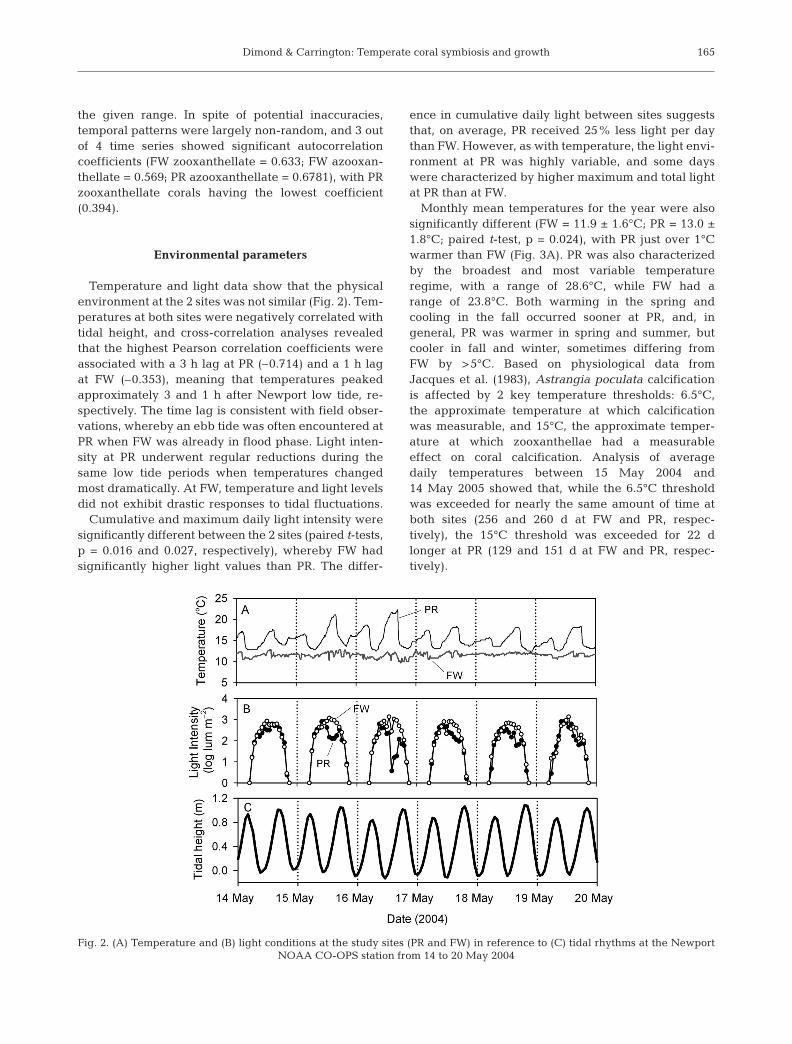
Local water-level data for a 6 d period between14 and 20 May 2004 were obtained from the NOAAweather station in Newport. These hourly data wereused to examine the potential influence of tidalrhythms on light and temperature conditions at thestudy sites. Cross-correlation analyses were employedto analyze the relationship between tide and tempera-ture.
Statistical analyses. Multivariate analysis of variance(MANOVA) was used to perform repeated-measuresanalyses of both predicted chlorophyll and growthdata, with month as the repeated factor. SYSTAT v.11(SYSTAT Inc.) was used for these analyses. Parametricassumptions were met with a log(x + 1) transformationfor predicted chlorophyll data and a log(x + 2) transfor-mation for growth data. One-way MANOVAs testingcollection site as the factor found no significant differ-
ences in predicted chlorophyll (df = 1, F = 0.239, p =0.627) or growth (df = 1, F = 1.148, p = 0.289) based oncollection site. Subsequently, both datasets were ana-lyzed with 2-way MANOVAs, with site and symbioticstate as factors. To avoid unbalanced analyses due tocorals lost during the study, a total of 47 missing values(7% of predicted chlorophyll data and 7.6% of growthdata) was replaced with treatment means. The degreesof freedom and mean squares for the error terms ofthe within-subjects components of these tests wereadjusted accordingly (Underwood 1997).
RESULTS
Chlorophyll density proxy
PCA of the RGB color values yielded a single princi-pal component (polyp color) that explained 93.5% ofthe variance. Component loadings for red, green, andblue variables were 0.951, 0.978, and 0.971, respec-tively, indicating a strong and largely equal contribu-tion of each variable to the principal component. Therelationship between polyp color and total chlorophylldensity was non-linear, best described by a polynomialfunction (y = 0.044x2 + 0.0335x; R2 = 0.89; Fig. 1). Thecurvilinear shape of the function suggests that esti-mates of chlorophyll are least reliable at low polypcolor values, but improve in accuracy with increasingpigmentation, as shown by 95% prediction limits(Fig. 1). Prediction limits indicate the 95% probabilityof data from a subsequent experiment falling within
164
Fig. 1. Astrangia poculata. Relationship between polyp colorand areal chlorophyll (n = 20; y = 0.044x2 + 0.0335x; R2 = 0.89)
Dimond & Carrington: Temperate coral symbiosis and growth
the given range. In spite of potential inaccuracies,temporal patterns were largely non-random, and 3 outof 4 time series showed significant autocorrelationcoefficients (FW zooxanthellate = 0.633; FW azooxan-thellate = 0.569; PR azooxanthellate = 0.6781), with PRzooxanthellate corals having the lowest coefficient(0.394).
Environmental parameters
Temperature and light data show that the physicalenvironment at the 2 sites was not similar (Fig. 2). Tem-peratures at both sites were negatively correlated withtidal height, and cross-correlation analyses revealedthat the highest Pearson correlation coefficients wereassociated with a 3 h lag at PR (–0.714) and a 1 h lagat FW (–0.353), meaning that temperatures peakedapproximately 3 and 1 h after Newport low tide, re-spectively. The time lag is consistent with field obser-vations, whereby an ebb tide was often encountered atPR when FW was already in flood phase. Light inten-sity at PR underwent regular reductions during thesame low tide periods when temperatures changedmost dramatically. At FW, temperature and light levelsdid not exhibit drastic responses to tidal fluctuations.
Cumulative and maximum daily light intensity weresignificantly different between the 2 sites (paired t-tests,p = 0.016 and 0.027, respectively), whereby FW hadsignificantly higher light values than PR. The differ-
ence in cumulative daily light between sites suggeststhat, on average, PR received 25% less light per daythan FW. However, as with temperature, the light envi-ronment at PR was highly variable, and some dayswere characterized by higher maximum and total lightat PR than at FW.
Monthly mean temperatures for the year were alsosignificantly different (FW = 11.9 ± 1.6°C; PR = 13.0 ±1.8°C; paired t-test, p = 0.024), with PR just over 1°Cwarmer than FW (Fig. 3A). PR was also characterizedby the broadest and most variable temperatureregime, with a range of 28.6°C, while FW had arange of 23.8°C. Both warming in the spring andcooling in the fall occurred sooner at PR, and, ingeneral, PR was warmer in spring and summer, butcooler in fall and winter, sometimes differing fromFW by >5°C. Based on physiological data fromJacques et al. (1983), Astrangia poculata calcificationis affected by 2 key temperature thresholds: 6.5°C,the approximate temperature at which calcificationwas measurable, and 15°C, the approximate temper-ature at which zooxanthellae had a measurableeffect on coral calcification. Analysis of averagedaily temperatures between 15 May 2004 and14 May 2005 showed that, while the 6.5°C thresholdwas exceeded for nearly the same amount of time atboth sites (256 and 260 d at FW and PR, respec-tively), the 15°C threshold was exceeded for 22 dlonger at PR (129 and 151 d at FW and PR, respec-tively).
165
Fig. 2. (A) Temperature and (B) light conditions at the study sites (PR and FW) in reference to (C) tidal rhythms at the Newport NOAA CO-OPS station from 14 to 20 May 2004

Mar Ecol Prog Ser 348: 161–172, 2007
Seasonal patterns of symbiosis and growth
Although the seasonal trends of predicted chloro-phyll density were significantly different betweenzooxanthellate and azooxanthellate corals (Table 1,Fig. 3B), pigmentation in both types of corals wasdynamic. Pigmentation in zooxanthellate corals in-creased from starting levels in May (but note initialdrop at FW) before reaching a peak in late summer/
early autumn, then decreased and stabilized some-what during the winter. The temporal response ofinitially azooxanthellate corals ranged from nearlyzero accumulation of pigmentation to completechanges whereby all polyps in a colony became fullysymbiotic at some point during the year-long study. Infact, only 3 out of 27 azooxanthellate corals did notgain noticeable pigmentation. A significant differencebetween sites (Table 1, Fig. 3B) is particularly evident
166
Fig. 3. Astrangia poculata. (A) Temperatures (3 m depth) at Pettaquamscutt River (PR, light gray) and Ft. Wetherill (FW, darkgray) during the study period. Solid horizontal line indicates the period for which adjusted NOAA CO-OPS station data weresubstituted for lost data loggers at FW. Dotted horizontal lines indicate the physiologically important temperature thresholdsdelineated by Jacques et al. (1983): 6.5°C, the temperature above which calcification was measurable, and 15°C, the temperatureabove which symbiosis had a measurable effect on calcification. (B) Mean (±SE) predicted chlorophyll density of corals over time.
(C) Mean (±SE) growth rate of corals over time. Z: zooxanthellate corals; AZ: azooxanthellate corals

Dimond & Carrington: Temperate coral symbiosis and growth
among azooxanthellate corals. Although azooxanthel-late corals at both sites exhibited an increase inpigmentation, PR coral pigmentation increased sub-stantially more than at FW. Among zooxanthellatecorals, there was a 1 mo difference in the timing ofmaximum coral pigmentation between sites, with PRcorals reaching their peak before FW corals.
Coral growth also underwent significant seasonalvariation, but did not differ significantly between sites(Table 2, Figs. 3C & 4). However, growth differedsignificantly depending on initial symbiotic state, withzooxanthellate corals growing significantly more thanazooxanthellate corals. The majority of polyp growthoccurred in the 8 mo between May and December. Inthe winter and early spring, many corals substantiallydeclined in live polyp number due to tissue loss. Thisloss, unlike the growth that occurred under warmerconditions, affected both types of corals similarly.Apparently, corals cease feeding and contract theirpolyps during this period (Jacques et al. 1983, J.D.
pers. obs.), which renders them more susceptible tosedimentation stress (Peters & Pilson 1985) and algalovergrowth. This is compounded by tissue thinningthat reduces the coenosarc to nearly nothing, essen-tially removing connectivity between polyps and open-ing up bare skeleton for the recruitment of algae, bar-nacles, and other organisms (J.D. pers. obs.). Despitethis loss, tissue and skeletal growth resumed betweenthe final 2 sampling periods in May and July 2005.
Based on linear regression analysis of polyp color anddaily coral growth rates averaged for the major period ofcoral growth (May to December), predicted chlorophylldensity explained 23% of the variation in coral growth(n = 56, y = 0.0586x + 0.0888, R2 = 0.23, p < 0.001; Fig. 5).Growth was highly temperature dependent (ANCOVA,p < 0.001), and zooxanthellate coral growth was morestrongly influenced by temperature than growth inazooxanthellate corals (ANCOVA, temperature × symbi-otic state, p < 0.003), particularly at the higher range oftemperatures (Fig. 6). Plotted on similar linear scales, the
167
Table 1. MANOVA results for predicted chlorophyll density. Sym: the initial symbiotic state of corals
Source df MS F p
Between subjectsSite 1 1.45 5.448 <0.023Sym 1 28.43 106.8 <0.001Site × Sym 1 1.028 3.862 <0.055Error 52 0.266
Within subjectsMonth 11 0.632 31.99 <0.001Month × Site 11 0.107 5.426 <0.001Month × Sym 11 0.420 21.29 <0.001Month × Site × Sym 11 0.097 4.923 <0.001Error 5250 0.021
Table 2. MANOVA results for daily growth rate. Sym: the initial symbiotic state of corals
Source df MS F p
Between subjectsSite 1 0.010 2.074 <0.156Sym 1 0.038 8.252 <0.006Site × Sym 1 0.007 1.543 <0.220Error 52 0.005
Within subjectsMonth 10 0.224 65.02 <0.001Month × Site 10 0.013 3.770 <0.001Month × Sym 10 0.013 3.787 <0.001Month × Site × Sym 10 0.001 0.235 <0.993Error 5200 0.004
Fig. 4. Astrangia poculata. Mean (±SE) number of polyps per colony over time. PR: Pettaquamscutt River; FW: Ft. Wetherill;Z: zooxanthellate corals; AZ: azooxanthellate corals

Mar Ecol Prog Ser 348: 161–172, 2007
temperature dependence of calcium carbonate de-position rates reported by Jacques et al. (1983) closelymatch the polyp budding rates measured in the presentstudy, and the differences between zooxanthellate andazooxanthellate rates are similar.
DISCUSSION
Chlorophyll density estimation
Coral pigmentation measured from digital imagesreflected a strong non-linear relationship with chloro-phyll density in Astrangia poculata. The curvilinearshape of the function suggests that estimates of chloro-phyll density at low polyp color values may be lessreliable, and this could be partially responsible for thehigher standard errors among azooxanthellate corals.However, significant autocorrelation of both azoo-xanthellate time series suggests that these data arewell represented; this autocorrelation may have bene-fited from the use of a repeated-measures designas opposed to random collections. Thieberger et al.(1995), who first developed the photographic tech-nique linking coral color to chlorophyll density, ob-tained similar color–chlorophyll relationships for 3tropical coral species in the Red Sea. Maguire et al.(2002) and Edmunds et al. (2003) adapted the tech-nique for digital images and software that could mea-sure color in the RGB spectrum.
The use of chlorophyll density over zooxanthelladensity as a symbiosis parameter was chosen for thisstudy due to seasonal fluctuations in chlorophyllcontent per zooxanthella cell (i.e. photoadaptation)that have been observed in tropical corals (Brown et al.1999, Fitt et al. 2000, Warner et al. 2002) as well as inAstrangia poculata (Cummings 1983). Such fluctua-tions preclude the use of a single relationship betweencolor and zooxanthella density, obtained at any onetime of year, to make estimates of zooxanthella densityover time. Despite the advantages of a color–chloro-phyll curve, the limitation is that it cannot distinguishthe contribution of changes in zooxanthella densityversus changes in zooxanthella pigment content topolyp pigmentation. Additionally, these non-destruc-tive methods cannot account for variations in tissuemass that add a third dimension to any measure ofsymbiosis on an areal basis.
Temporal variation in coral pigmentation
Nearly all corals, regardless of their initial symbioticstate, underwent some degree of temporal change inpigmentation. However, the trend among zooxanthel-late corals, with high pigmentation occurring duringlate summer/early autumn and subsequent lows dur-ing winter and spring, are most indicative of a seasonalcycle. In spite of coarse temporal resolution, 5 randomcoral collections from PR by Cummings (1983) over a1 yr period suggested similar zooxanthella density andtissue mass patterns. Chlorophyll a per cell, con-versely, was highest in winter and spring and lowest insummer and fall (Cummings 1983). Taken together,the 2 patterns appear to concur with the pigmentationpatterns for zooxanthellate corals in the present study.For example, the stable to very slow reduction inpigmentation observed during the winter months maybe the result of an increase in cell-specific chlorophyllthat could compensate for the potential loss in pig-mentation from a reduction in zooxanthella densities.Additionally, the somewhat dramatic reduction inpigmentation among both zooxanthellate and azoo-xanthellate corals (except for PR zooxanthellate corals)in July 2004 may reflect a drop in cell-specificchlorophyll associated with the longest and mostintense days of sunlight of the year. Alternatively, thistemporary loss of color could be interpreted as anincrease in tissue mass, perhaps accompanied by a lagin zooxanthella growth that essentially dilutes coralpigmentation until the new coral cells becomeoccupied by symbionts in subsequent months.
Studies of tropical scleractinians indicate thattemperature is perhaps the most important driver ofseasonal cycles in pigmentation and symbiont density
168
Fig. 5. Astrangia poculata. Predicted chlorophyll densityversus growth rate for all corals, based on means for the 8 moof net growth (May to December) (n = 56; y = 0.0586x +
0.0888; R2 = 0.23; p < 0.001)

Dimond & Carrington: Temperate coral symbiosis and growth
(Brown et al. 1999, Fitt et al. 2000), and Astrangiapoculata appears to be no exception. The positivelinear response of zooxanthella photosynthesis totemperature (Jacques et al. 1983) and the resultingeffect on the algal population growth rate is probablylargely responsible for seasonal pigmentation trends.Although zooxanthella photosynthesis may be negligi-ble below 5°C (Jacques et al. 1983), zooxanthellaeappear to build up chlorophyll for the low light ofwinter (Cummings 1983) and even divide in spite oflow temperatures (J.D. unpubl. data). Laboratory incu-bations suggest that although zooxanthella mitoticrates in hospite are nearly 3 times higher atsummertime temperatures (20°C in September), divi-sion nonetheless occurs at 3°C in March (J.D. unpubl.data). During warmer months, increased coral tissuebiomass (Cummings 1983, J.D. pers. obs.) is likely tosupport more zooxanthellae and thus further enhancepigmentation (see Fitt et al. 2000). Variations in tissuemass from month to month were clear though non-quantifiable from coral images, with a trend very simi-lar to coral pigmentation. Since A. poculata is capableof metabolic regulation at the higher range of temper-atures (11 to 23°C; Jacques et al. 1983), energy alloca-tion to tissue growth should increase with temperature.
In contrast to the temporal pigmentation trendsreported here for zooxanthellate Astrangia poculata,zooxanthella and chlorophyll density among tropicalscleractinians tend to be highest during the coolesttimes of year and lowest during the warmest periods(Stimson 1997, Brown et al. 1999, Fitt et al. 2000, but
see Costa et al. 2005). This may relate to the general-ization that corals and their symbionts in the tropics areliving closer to their upper rather than their lowerthermal tolerance threshold, while the opposite is trueof A. poculata and probably other temperate sclerac-tinians. Seasonal depressions of zooxanthellae andtheir pigments in the tropics can be driven by 2 differ-ent factors: (1) an increase in both host and symbiontrespiratory metabolism with higher temperatures thatreduces both host tissue mass and algal density, and(2) excessive thermal and/or irradiance stress, whichcauses photoinhibition and photodamage in zooxan-thellae, a phenomenon more typically associated withsevere coral bleaching (Fitt et al. 2001). Seasonalreductions in A. poculata pigmentation may not be asextreme as in tropical corals due to cold-suppression ofmetabolism in both host and symbiont (Jacques et al.1983), but it is plausible that photoinhibition also playsa role in reducing zooxanthella densities at lowtemperatures, since low and high temperature photo-inhibition share similarities (Gombos et al. 1994,Warner et al. 1999).
Seasonal irradiance cycles ultimately drive air andwater temperatures, but they also influence zooxan-thella photosynthetic processes (Warner et al. 2002)and therefore act as additional drivers of coral pigmen-tation cycles (Stimson 1997, Brown et al. 1999, Warneret al. 2002). It is expected that corals received photo-synthetically active radiation (PAR) levels within therange predicted by Narragansett Bay light extinctioncoefficients (Oviatt et al. 2002), which fall within the
169
Fig. 6. Astrangia poculata. Temperature versus coral growth: polyp budding rates from the present study compared withlaboratory calcium carbonate deposition rates from Jacques et al. (1983). For polyp budding data, each point represents the meangrowth and temperature for 1 mo sampling intervals. Values <5°C and –0.1 polyps d–1 were excluded from the graph.
PR: Pettaquamscutt River; FW: Ft. Wetherill; Z: zooxanthellate corals; AZ: azooxanthellate corals

Mar Ecol Prog Ser 348: 161–172, 2007
same range of saturating irradiance for photosynthesisin Astrangia poculata symbionts (120 µmol quanta m–2
s–1 at 15°C up to 400 µmol quanta m–2 s–1 at 23°C;Jacques et al. 1983). Typical surface irradiance inRhode Island peaks in June and reaches its minimumin December (Pilson 1985), meaning that pigmentationin A. poculata is associated with the moderateirradiance levels of the late summer and early fall. Itis therefore hypothesized that irradiance influencesthe seasonal patterns of pigmentation in A. poculataprimarily through photoadaptation effects on algal cellchlorophyll content (Cummings 1983) rather thanthrough the photodamage effects associated with thehigh irradiance levels of the tropical spring andsummer (Gorbunov et al. 2001, Warner et al. 2002).However, seasonal variations in water-column lightattenuation may add an important caveat; for example,water clarity in the region is typically highest duringthe colder months, when surface irradiance is rela-tively low, and vice versa (J.D. pers. obs.).
Pigmentation trends in azooxanthellate Astrangiapoculata colonies were not analogous to zooxanthel-late corals. Most notable is the lack of an early autumnpeak in color and an overall increase in pigmentationthat was subtle at FW but marked at PR. Despiteinitially low pigmentation among all azooxanthellatecorals at the beginning of the study, all but 3 out of27 azooxanthellate corals gained at least some pig-mentation at some point over the course of a year.Some of this variability may be attributed to manipula-tion effects and removal of the algal canopy, whichmay have increased corals’ original light and/ortemperature (in the case of transplantation from FW toPR) regime. Since many azooxanthellate corals are infact inhabited by small zooxanthella populations thatare below visual detection densities and even standardhemocytometer count detection densities (J.D. unpubl.data), presumably, changes in light or temperaturecould readily trigger increases in symbiont density.While some azooxanthellate corals may have re-sponded to a higher light environment, the consider-able increase in pigmentation among PR azooxanthel-late corals compared to a more subtle increase amongFW corals suggests that other factors, such as tempera-ture, may have been more influential.
Asynchrony in the pigmentation and growth trendsof both zooxanthellate and azooxanthellate coralsbetween the study sites is likely a reflection of thedissimilar environmental regimes. In the Pettaquams-cutt River, an ebb tide apparently released turbidwater that caused increased light attenuation. Thiswater, having been entrained in a relatively shallowestuary averaging 2 m in depth, was either warmed orcooled relative to Rhode Island Sound water depend-ing on the season. Zooxanthellate corals at PR reached
their maximum pigmentation at least 1 mo before thoseat FW, perhaps a result of higher summertime temper-atures in the river. In addition, as mentioned above, PRazooxanthellate corals underwent a substantial in-crease in pigmentation during the study period thatwas not observed at FW. In spite of a chronicallyreduced irradiance regime, PR may be an especiallyfavorable environment for symbiosis due to prolongedhigher temperatures. Randomized coral collections byCummings (1983) from the Pettaquamscutt River sug-gested a higher prevalence of symbiosis compared tosites in Narragansett Bay and Rhode Island Sound, andpersonal observations (by J.D.) support these data.Temperatures at PR exceeded the 15°C threshold for22 d longer than they did at FW and averaged >4°Cwarmer at their maximum, so it is possible that at PR,the high prevalence of zooxanthellate corals, the in-crease in pigmentation among azooxanthellate corals,and the earlier peak in growth and pigmentation arereflective of these differences.
Temporal variation in coral growth and relation topigmentation
Similarly to coral pigmentation trends, growth ofAstrangia poculata showed considerable seasonal vari-ation. The results of this field study corroborate withthe laboratory measurements of Jacques et al. (1983)and show that skeletal growth at this northern marginof the coral’s range is limited to the approximately 8 mobetween April and December. The present study, how-ever, documents an important ecological observation:the inactivity of corals during the coldest periods of theyear results in their inability to avoid sedimentationand settlement of any bare skeleton by other organ-isms, which leads to tissue loss particularly around theedges of the colony. Tissue re-growth in the spring canoccur, but only in calices that remain mostly bare,while areas of the skeleton settled by other organismsare permanently lost. These processes may help toexplain the northern range limit of this coral. Cape Codacts as a well-defined barrier between waters fed bythe Labrador Current to the north and the Gulf Streamto the south. Temperatures north of Cape Cod reachsimilar seasonal lows as southern New England, but donot reach 20°C during the warm season, and it is there-fore hypothesized that corals settling north of the capewould struggle to grow enough in the warm season toovercome partial colony loss during the winter. How-ever, this hypothesis allows that A. poculata would notfirst be overcome by reproductive or developmentallimitations.
Zooxanthella photosynthesis significantly enhancescalcium carbonate deposition in symbiotic scleractini-
170
Dimond & Carrington: Temperate coral symbiosis and growth
ans (Jacques et al. 1983, Gattuso et al. 1999). Thissymbiosis effect is apparent in the higher polypbudding rates of initially zooxanthellate Astrangiapoculata, as well as in the significant relationshipbetween pigmentation and growth rate over thegrowing season. Given that enhanced calcification inzooxanthellate corals has been questioned by recentstudies (Marshall 1996, Marshall & Clode 2004), thepresent study offers unequivocal field documentationof symbiosis enhancement within a single scleractinianspecies. However, because the relationship describedonly 23% of the variation in growth rate, it is expectedthat other factors such as food intake (Szmant-Froelich& Pilson 1984, Miller 1995), microhabitat differences,and individual genotypic variation also contributed tooverall variability in growth. Since A. poculata mayobtain benthic prey as a substantial proportion of itsfood (Grace 1996), prey capture for an individualcolony could be more spatially and temporally variablethan it would be if prey were predominantly plank-tonic. Additionally, if enhancement of calcification byzooxanthella photosynthesis in A. poculata occurs onlyat temperatures of approximately 15°C and above(Jacques et al. 1983), symbiosis would have an effecton growth for only 4 (FW) to 5 (PR) of the approxi-mately 8 mo of growth. At deeper depths, the rapidattenuation of PAR severely limits the benefits andextent of symbiosis (Weston 1980, Jacques et al. 1983)and the role of heterotrophy becomes more critical.
Acknowledgements. J.D. is indebted to J. Barber for fieldassistance. In addition, the authors thank the following peoplefor advice and/or assistance: M. Boller, P. Edmunds, S. Grace,M. Gustafson, B. Longval, G. Moeser, J. Ritchie, R. Rotjan, K.Sebens, J. Sylvester, and L. Trueblood. Valuable feedback onearlier versions of the manuscript was provided by C. Oviatt,M. Pilson, B. Siebel, and 3 anonymous reviewers. This studywas supported by an American Academy of UnderwaterSciences student scholarship to J.D., as well as by the URIDepartment of Biological Sciences, and National ScienceFoundation grants to E.C.
LITERATURE CITED
Baghdasarian G, Muscatine L (2000) Preferential expulsion ofdividing algal cells as a mechanism for regulatingalgal–cnidarian symbioses. Biol Bull (Woods Hole) 199:278–286
Brown BE, Dunne RP, Ambarsari I, Le Tissier MDA, Sat-apoomin U (1999) Seasonal fluctuations in environmentalfactors and variation in symbiotic algae and chlorophyllpigments in four Indo-Pacific coral species. Mar Ecol ProgSer 191:53–69
Cohen AL, Owens KE, Lane GD, Shimizu N (2002) The effectof algal symbionts on the accuracy of Sr/Ca paleotemper-atures from coral. Science 296:331–333
Costa CF, Sassi R, Amaral FD (2005) Annual cycle of symbi-otic dinoflagellates from three species of scleractiniancorals from coastal reefs of northeastern Brazil. CoralReefs 24:191–193
Cummings C (1983) The biology of Astrangia danae. PhD dis-sertation, University of Rhode Island, Kingston
Edmunds PJ, Gates RD, Gleason DF (2003) The tissue compo-sition of Montastrea franksi during a natural bleachingevent in the Florida Keys. Coral Reefs 22:54–62
Fagoonee I, Wilson HB, Hassell MP, Turner JR (1999) Thedynamics of zooxanthellae populations: a long-term studyin the field. Science 283:843–845
Fitt WK, McFarland FK, Warner ME, Chilcoat GC (2000) Sea-sonal patterns of tissue biomass and densities of symbioticdinoflagellates in reef corals and relation to coral bleach-ing. Limnol Oceanogr 45:677–685
Fitt WK, Brown BE, Warner ME, Dunne RP (2001) Coralbleaching: interpretation of thermal tolerance limits andthermal thresholds in tropical corals. Coral Reefs 20:51–65
Gattuso JP, Allemand D, Frankignoulle M (1999) Photo-synthesis and calcification at cellular, organismal, andcommunity levels: a review on interactions and control bycarbonate chemistry. Am Zool 39:160–183
Gombos Z, Wada H, Murata N (1994) The recovery of photo-synthesis from low-temperature photoinhibition is accel-erated by the unsaturation of membrane lipids: a mecha-nism of chilling tolerance. Proc Natl Acad Sci USA 91:8787–8791
Gorbunov MY, Kolber ZS, Lesser MP, Falkowski PG (2001)Photosynthesis and photoprotection in symbiotic corals.Limnol Oceanogr 46:75–85
Grace SP (1996) The effects of water flow on Astrangia pocu-lata. MS thesis, University of Rhode Island, Kingston
Grace SP (2004) Ecomorphology of the temperate sclerac-tinian Astrangia poculata: coral–macroalgal interactionsin Narragansett Bay (Rhode Island). PhD dissertation,University of Rhode Island, Kingston
Jacques TG, Pilson MEQ (1980) Experimental ecology of thetemperate scleractinian Astrangia danae. I. Partition ofrespiration, photosynthesis, and calcification between hostand symbionts. Mar Biol 60:167–178
Jacques TG, Marshall N, Pilson MEQ (1983) Experimentalecology of the temperate scleractinian Astrangia danae. II.Effect of temperature, light intensity and symbiosis withzooxanthellae on metabolic rate and calcification. MarBiol 76:135–148
Jeffrey SW, Humphrey GF (1975) New spectrophotometricequations for determining chlorophylls a, b, c1, and c2 inhigher plants, algae and natural phytoplankton. BiochemPhysiol Pflanz 167:191–194
Johannes RE, Wiebe WJ (1970) A method for determination ofcoral tissue biomass and composition. Limnol Oceanogr15:822–824
Maguire B, Gleason DF, Mojica R, Mojica EC (2002) Changesin photographic R, G, and B numerical values quantita-tively reflect pigment and zooxanthella decrease duringPorites astreoides bleaching. In: Kasim Moosa MK, Soe-modihardjo S, Nontji A, Soegiarto A, Romimohtarto K,Sukarano, and Suharsono (eds) Proc 9th Int Coral ReefSymp. International Coral Reef Society, Bali, p 1129–1138
Marsh JA (1970) Primary productivity of reef-building cal-careous red algae. Ecology 51:255–263
Marshall AT (1996) Calcification in hermatypic and aherma-typic corals. Science 271:637–639
Marshall AT, Clode P (2004) Calcification rate and the effectof temperature in a zooxanthellate and an azooxanthellatereef coral. Coral Reefs 23:218–224
Miller MW (1995) Growth of a temperate coral: effects of tem-perature, light, depth, and heterotrophy. Mar Ecol ProgSer 122:217–225
Miller MW, Hay ME (1996) Coral–seaweed–grazer–nutrient
171

Mar Ecol Prog Ser 348: 161–172, 2007
interactions on temperate reefs. Ecol Monogr 66:323–344Muller-Parker G, Davy SK (2001) Temperate and tropical sea-
anemone symbioses. Invertebr Biol 120:104–123Muscatine L (1990) The role of symbiotic algae in carbon and
energy flux in reef corals. In: Dubinsky Z (ed) Ecosystemsof the world, Vol 25. Coral reefs. Elsevier, Amsterdam
Oviatt C, Keller A, Reed L (2002) Annual primary productionin Narragansett Bay with no bay–wide winter–springphytoplankton bloom. Estuar Coast Shelf Sci 54:1013–1026
Peters EC, Pilson MEQ (1985) A comparative study of theeffects of sediment on symbiotic and asymbiotic coloniesof the coral Astrangia danae Milne Edwards and Haime,1849. J Exp Mar Biol Ecol 92:215–230
Peters EC, Cairns SD, Pilson MEQ, Wells JW, Jaap WC, LangJC, Vasleski CE, St. Pierre Gollahon L (1988) Nomencla-ture and biology of Astrangia poculata (= A. danae, = A.astreiformis) (Cnidaria: Anthozoa). Proc Biol Soc Wash101:234–250
Pilson MEQ (1985) Annual cycles of nutrients and chlorophyllin Narragansett Bay, Rhode Island. J Mar Res 43:849–873
Schiller C (1993) Ecology of the symbiotic coral Cladocoracaespitosa (L.) (Faviidae, Scleractinia) in the Bay of Piran(Adriatic Sea). II. Energy budget. PSZN I: Mar Ecol 14:221–238
Schuhmacher H, Zibrowius H (1985) What is hermatypic? Aredefinition of ecological groups in corals and other organ-isms. Coral Reefs 4:1–9
Stimson J (1997) The annual cycle of density of zooxanthel-lae in the tissues of field and laboratory-held Pocillopora
damicornis (Linnaeus). J Exp Mar Biol Ecol 214:35–48Szmant-Froelich A, Pilson MEQ (1980) The effects of feeding
frequency and symbiosis with zooxanthellae on the bio-chemical composition of Astrangia danae Milne Edwardsand Haime, 1849. J Exp Mar Biol Ecol 48:85–97
Szmant-Froelich A, Pilson MEQ (1984) Effects of feedingfrequency and symbiosis with zooxanthellae on nitrogenmetabolism and respiration of the coral Astrangia pocu-lata. Mar Biol 81:153–162
Szmant-Froelich A, Yevich P, Pilson MEQ (1980) Gameto-genesis and early development of the temperate coralAstrangia danae (Anthozoa: Scleractinia). Biol Bull (WoodsHole) 158:257–269
Thieberger Y, Kizner Z, Achituv Y, Dubinsky Z (1995) Anovel, nondestructive bioassay for assessing areal chloro-phyll a in hermatypic cnidarians. Limnol Oceanogr 40:1166–1173
Underwood AJ (1997) Experiments in ecology: their logicaldesign and interpretation using analysis of variance. Cam-bridge University Press
Warner ME, Fitt WK, Schmidt GW (1999) Damage to photo-system II in symbiotic dinoflagellates: a determinant ofcoral bleaching. Proc Natl Acad Sci USA 96:8007–8012
Warner ME, Chilcoat GC, McFarland FK, Fitt WK (2002) Sea-sonal fluctuations in the photosynthetic capacity of photo-system II in symbiotic dinoflagellates in the Caribbeanreef-building coral Montastrea. Mar Biol 141:31–38
Weston MP (1980) Distribution of the dinoflagellate symbiosisin Astrangia danae as affected by light attenuation. MSthesis, Old Dominion University, Norfolk
172
Editorial responsibility: Charles Birkeland (ContributingEditor), Honolulu, Hawaii, USA
Submitted: December 27, 2006; Accepted: May 8, 2007Proofs received from author(s): September 24, 2007