Tejido Conectivo Parte B
14
Tejido Conectivo Parte B Informe #3 Laboratorio Biología # 240 Profesor: Javier Cabello
Transcript of Tejido Conectivo Parte B

Tejido Conectivo Parte B
Informe #3
Laboratorio Biología # 240
Profesor: Javier Cabello
Figure 4-8 The Cells and Fibers of Connective Tissue Proper
Elastic fibers
Collagen fibers
Fibroblast
Free macrophage
Connective tissue proper LM 502
Areolar

Figure 4-10a Adipose and Reticular Tissues
Adipose Tissue
LOCATIONS: Deep to the skin, especially at sides, buttocks, breasts; padding around eyes and kidneys
FUNCTIONS: Provides padding and cushions shocks; insulates (reduces heat loss); stores energy
Adipose tissue
Adipocytes (white adipose
cells)
LM 300
Adiposo

Figure 4-10b Adipose and Reticular Tissues
Reticular Tissue
FUNCTIONS: Provides supporting framework
LOCATIONS: Liver, kidney, spleen, lymph nodes, and bone marrow
Reticular tissue from liver
Reticular Tissue
Reticular fibers
LM 375
Reticular

Figure 4-11a Dense Connective Tissues
Dense Regular Connective Tissue
Collagen fibers
Fibroblast nuclei
Tendon LM 440
LOCATIONS: Between skeletal muscles and skeleton (tendons and aponeuroses); between bones or stabilizing positions of internal organs (ligaments); covering skeletal muscles; deep fasciae
FUNCTIONS: Provides firm attachment; conducts pull of muscles; reduces friction between muscles; stabilizes relative positions of bones
Denso Regular

Figure 4-11b Dense Connective Tissues
Collagen fiber
bundles
Deep dermis LM 111
Dense Irregular Connective Tissue
LOCATIONS: Capsules of visceral organs; periostea and perichondria; nerve and muscle sheaths; dermis FUNCTIONS: Provides strength to resist forces applied from many directions; helps prevent overexpansion of organs such as the urinary bladder
Denso Irregular
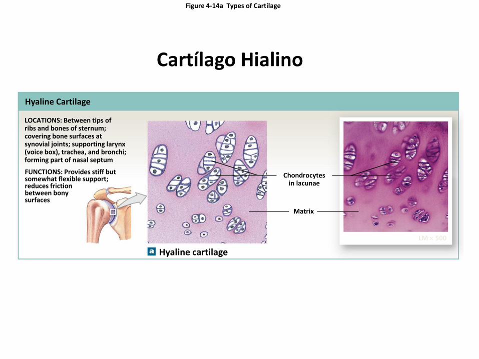
Figure 4-14a Types of Cartilage
Hyaline Cartilage
LOCATIONS: Between tips of ribs and bones of sternum; covering bone surfaces at synovial joints; supporting larynx (voice box), trachea, and bronchi; forming part of nasal septum FUNCTIONS: Provides stiff but somewhat flexible support; reduces friction between bony surfaces
Hyaline cartilage
LM 500
Matrix
Chondrocytes in lacunae
Cartílago Hialino

Figure 4-14b Types of Cartilage
Elastic Cartilage
LOCATIONS: Auricle of external ear; epiglottis; auditory canal; cuneiform cartilages of larynx
FUNCTIONS: Provides support, but tolerates distortion without damage and returns to original shape
Elastic cartilage
Elastic fibers in matrix
Chondrocyte in lacuna
LM 358
Cartílago Elástico

Figure 4-14c Types of Cartilage
Fibrocartilage
LOCATIONS: Pads within knee joint; between pubic bones of pelvis; intervertebral discs
FUNCTIONS: Resists compression; prevents bone- to-bone contact; limits movement
Fibrocartilage
Fibrous matrix
Chondrocytes in lacunae
LM 400
Cartílago Fibroso
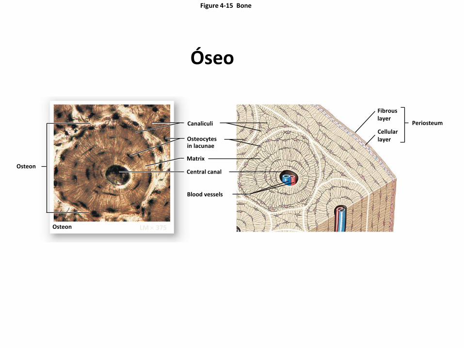
Figure 4-15 Bone
Canaliculi
Osteocytes in lacunae
Matrix
Central canal
Blood vessels
LM 375
Osteon
Osteon
Fibrous layer
Cellular layer
Periosteum
Óseo

Figure 4.22
Sangre humana (Blood Smear)

Figure 4-12 Formed Elements of the Blood
Red blood cells
Red blood cells account for roughly half the volume of whole blood and give blood its color.
Red blood cells, or erythrocytes (e-RITH-ro-sıts), are responsible for the transport of oxygen (and, to a lesser degree, of carbon dioxide) in the blood.
¯ ¯

Figure 4-12 Formed Elements of the Blood
White blood cells
Eosinophil
Neutrophil
Basophil
White blood cells, or leukocytes (LOO-ko-sıts; leuko-, white), help defend the body from infection and disease.
¯ ¯
Eosinophils and neutro- phils are phagocytes. Ba- sophils promote inflamma- tion much like mast cells in other connective tissues.
Lymphocytes are un- common in the blood but they are the domi- nant cell type in lymph, the second type of fluid connective tissue.
Monocytes are phagocytes similar to the free macro- phages in other tissues.

Figure 4-12 Formed Elements of the Blood
Platelets
Platelets are membrane-enclosed packets of cytoplasm that function in blood clotting.
These cell fragments are involved in the clotting response that seals leaks in damaged or broken blood vessels.