Technologic Applications in Nuclear Molecular Imaging Arif Sheikh, MD Assistant Professor Director...
45
Technologic Applications in Technologic Applications in Nuclear Molecular Imaging Nuclear Molecular Imaging Arif Sheikh, MD Arif Sheikh, MD Assistant Professor Assistant Professor Director of Targeted Radionuclide Director of Targeted Radionuclide Therapy Therapy and Cardiovascular Nuclear and Cardiovascular Nuclear Medicine Medicine Divisions of Nuclear and General Divisions of Nuclear and General Medicine Medicine Departments of Radiology Departments of Radiology and Internal Medicine and Internal Medicine University of North Carolina University of North Carolina
-
Upload
abigail-worton -
Category
Documents
-
view
219 -
download
4
Transcript of Technologic Applications in Nuclear Molecular Imaging Arif Sheikh, MD Assistant Professor Director...

Technologic Applications in Technologic Applications in Nuclear Molecular ImagingNuclear Molecular Imaging
Arif Sheikh, MDArif Sheikh, MDAssistant ProfessorAssistant Professor
Director of Targeted Radionuclide TherapyDirector of Targeted Radionuclide Therapyand Cardiovascular Nuclear Medicineand Cardiovascular Nuclear Medicine
Divisions of Nuclear and General MedicineDivisions of Nuclear and General MedicineDepartments of RadiologyDepartments of Radiology
and Internal Medicineand Internal Medicine University of North Carolina University of North Carolina

What Information does an Image Convey?
• Importance of image information
• Unique information portrayed
• Clinical relevance of imaging
• Understanding of technology behind the image

Issues with Nuclear Imaging• Nuclear Imaging
» Low resolution scans» High physical sensitivity
studies» Rely on observation of
physiology» Creates pictures from
individual photon counts
» Low target to background ratio
» Poisson statistics
• Other Radiologic Imaging» Higher resolution» Anatomic pathology
readily visible» Sensitivity is lower than
Nuclear Imaging» Uses ‘energy spectra’
to create images» High target to
background ratio» Gaussian statistics

History of Radiologic Imaging• Historically, much of modern diagnostic imaging was
performed by Nuclear Medicine» Neuro (Stroke) Brain scan → CT, MRI
» Cardiac (Functional) MUGA → Echocardiography (ultrasound)
» Pulmonary (Embolic Disease) V/Q → CT Angiography
» Gastrointestinal• Gallbladder (HIDA) → Ultrasound, CT, MRI• Liver/spleen (Metastases) → CT, MRI
» Genitourinary (Torsion) Testicular Scan → Ultrasound
• Despite advancements, many of these procedures are still used today!

Important Milestones in Nuclear Imaging
• Imaging» Initially, monitored
uptake over body» Anger Camera (image
generation)» Dynamic imaging» Gating» Tomographic (SPECT)
imaging» PET imaging» Attenuation Correction» CT based correction
• Other technologies» Processing
• Filtered back projection• Iterative reconstruction• Scatter correction• Resolution recovery
» Crystals• Sodium Iodide (SPECT)• BGO, LSO, etc. (PET)
» Computer processing• Film uses• Digital displays

Applications in Nuclear Medicine
• Wide range of applications (most versatile as a single imaging system)
• Essentially, evaluates pathology based on functional imaging
• Use of physiologic principles to guide surgeries
• Identification of tissue type based on molecular and/or physiologic parameters

Wide Variations of Nuclear Images
• Image appearance depends upon radiopharmaceutical characteristics• Normal image depends upon the physiologic distribution of tracer• Identification of pathology depends upon deviation from base image

Planar vs. SPECT Anger Camera Images
• Planar Imaging» Better counts
» Improved resolution
» Improved physical sensitivity
» Ability to do dynamic images
» Performed much quicker than SPECT imaging
• SPECT Imaging» Better contrast
» Improved clinical sensitivity
» Tomographic (3-dimensional) views of target region

Bone Imaging

Planar vs. SPECT vs. PET

Frontal view of thyroid
NodulesR L
Field of view of image detector
Thyroid Gland
Pinhole Imaging

Copyright restrictions may apply.
Mandel, S. J. JAMA 2004;292:2632-2642.Mandel, S. J. JAMA 2004;292:2632-2642.
123I Scans of Hyperfunctioning Thyroid Nodules
With suppression of extranodular thyroid
Without suppression of Extranodular Thyroid

Renal Scanning

Resolution vs. Sensitivity

Gated Slice Display

Surgical Mapping Theory• Certain cancers (melanoma, breast, etc.) tumours
may sit in lymph nodes undetected at time of initial diagnosis/surgery
• Tumours spread/metastasize through lymphatic channels
• Detection of tumours in lymph nodes will alter therapeutic approach
• Need to detect specific lymph nodes in entire basins which likely have disease
• Need to be accurate enough to minimize chance of ‘missing’ the target (sentinel) lymph node

Sentinel Lymph Node Mapping• Sentinel Lymph Node (SLN) is the 1st node
to which the tumour will likely metastasize• Detection and examination of this lymph
node will decide whether disease has spread
• Use particles that will travel in lymphatic channels from the tumour to the local lymph nodes to the sentinel lymph node
• Sulfur colloid (~0.1-1 m) particle is taken up by lymph nodes

infraclavicularinfraclavicular
apicalapicallaterallateral
parasternalparasternal
subareolarsubareolarplexusplexuscross-cross-drainagedrainage
from thoracic wallfrom thoracic wall
pectoralpectoral
subscapularsubscapularcentralcentral
to abdom.to abdom.wallwallsubcutaneoussubcutaneous
plexusplexus

Malignant Melanoma of the Back

rightrightgroingroin
rightrightaxillaaxilla
leftleftaxillaaxilla
rightrightaxillaaxilla
leftleftaxillaaxilla

Dual Isotope Imaging• Prostascint® is a
tracer to detect prostate cancer
• 111In based, targets tumour sites
• Also large amount of blood pool activity
• 99mTc-RBC distribute in blood pool, helping distinguish vessels from lymph node uptake

18F +
-
p
PET Principle

PET vs. SPECT• PET
» Improved spatial resolution (clinically)
» Inherent attenuation correction
» Can ‘quantitate’ biologic phenomenon easily
» Isotopes more biologically relevant
• 11C, 13N, 15O• Newer molecular tracers
» Dynamic tomographic images possible
» Shorter imaging times
• SPECT» Better count sensitivity» Ability for simultaneous
multi (dual) isotope imaging
» Wider range of pharmaceutical chemistries
» Isotopes (currently) more readily available
» Less cost (currently)

Prostate Cancer
Transverse Coronal Saggital
DG: 57 years old with High DG: 57 years old with High Grade; local resection, now Grade; local resection, now mass post-RT. Recurrence? mass post-RT. Recurrence? Pelvic Exent. possible?Pelvic Exent. possible?

SPECT SestaMIBI (Rest-Stress)
StrStr
RstRst
StrStr
RstRst
StrStr
RstRst
StrStr
RstRst
Mount Sinai School of Medicine

82Rb-PET Study (Rest-Stress)
Stress
Rest
Stress
Rest
Stress
Rest
Stress
Rest
Mount Sinai School of Medicine

Thyroid Cancer
Metastasis localized to the posterior vertebral bodyMetastasis localized to the posterior vertebral body

Advances in Processing:FBP vs. OSEM (Iterative)
• A major reason for the rise in popularity of PET imaging
• Improved diagnostic abilities• Algorithms were once not
possible due to limitations in computing power
• Optimal algorithms need to account for many variables» The type of camera» Radioisotope» Attenuation Correction (CT
vs. rod sources)» Counts/pixel

Quantitative Imaging• Counts per pixel are a representation of a physiologic
process• Change in process over time allows quantitative analysis
to examine physiologic phenomenon• Quantitation may be imaging based, or simply on counts
(eg. blood samples, urine collections, etc.)• Collection based studies
» Urea breath test for Helicobacter pylori
» Schilling’s test for B12 deficiency
» Calculation of Glomerular Filtration Rate with 125I-iodothalamate

Camilleri M. N Engl J Med 2007;356:820-829
Patterns of Gastric Emptying in Healthy People and in Patients with Diabetic Gastroparesis

MUGA Scans

Image Based Dosimetry
dttAA voxvox ~
Time-activity curveTime-activity curve
Time (d)Time (d)
ÃÃvoxvox
AAvox vox ((tt))
rad
ioac
tivi
ty (
MB
q)
rad
ioac
tivi
ty (
MB
q)
0 1 2
Day1
Day2
Day0
Cumulated activity

124I-Iodine in Thyroid Cancer
61.89 Gy/GBq
54.86 Gy/GBq
33.78 Gy/GBq

Technical Developments
• Multipinhole SPECT• Procedure specific cameras• Advances in OSEM algorithms• Newer detector materials• Radiopharmaceuticals• Transition to ‘Molecular Imaging

The D-SPECT Difference
BROADVIEWTM Technology
• 9 solid state detectors
10X the sensitivity
Twice the resolution
• Each detector can look at 100’s to 1000’s of different angles
• Each detector is independently addressable

Patient 510- Stress/Rest
Conventional D-SPECT, Spectrum Dynamics

Biologic vs. Technologic Challenges
• Ultimately, Biology trumps technology• Newer focus is development of better tracers for
evaluation of biology• Technology is developing to better allow for newer
tracer imaging• Thus, the field of ‘Molecular Imaging’ is
developing» Disease specific tracers» Multiple different tracers to look at the same disease

Papillary Thyroid CaFDG-PET versus 131I scans

FDG vs 124I in Thyroid Cancer
PET-PET-124124IIPET-FDGPET-FDG

FDG Prognosis of Distantly Metastatic Disease (Thyroid Ca)
Wang W et al: Prognostic value of [18F]fluorodeoxyglucose positron emission tomographicWang W et al: Prognostic value of [18F]fluorodeoxyglucose positron emission tomographicscanning in patients with thyroid cancer. J Clin Endo Metab 2000 85:1107-13scanning in patients with thyroid cancer. J Clin Endo Metab 2000 85:1107-13

SPECT-CT (111In-Octreotide)vs PET-CT (FDG)
Patient with rising calcitonin, undetected source on anatomic imaging or octreoscan
SPECT-CTSPECT-CT PET-CTPET-CT

Wilex Renal Cell Carcinoma Wilex Renal Cell Carcinoma 124124I-cG250 Clinical Imaging TrialI-cG250 Clinical Imaging Trial013-004 (CG July 7 Clear cell Fuhrman 2)013-004 (CG July 7 Clear cell Fuhrman 2)
Benign

Gene Imaging
HSV1-tk
Mammalian-tk
124I-FIAU
X
124I-FIAU-PO4
124I-FIAU
FIAU (2'-fluoro-2'-deoxy-5'-iodo-1-FIAU (2'-fluoro-2'-deoxy-5'-iodo-1-ββ-d-arabinofuranosyluracil)/-d-arabinofuranosyluracil)/Thymidine Kinase Thymidine Kinase
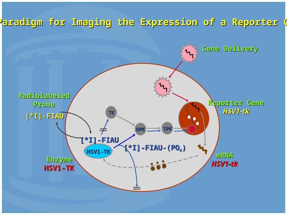
HSV1-TKHSV1-TK
TKTK
DPKDPK TPKTPK
[*I]-FIAU[*I]-FIAU
[*I]-FIAU[*I]-FIAU
[*I]-FIAU-(PO[*I]-FIAU-(PO44))
Reporter GeneReporter GeneHSV1-tkHSV1-tk
RadiolabeledRadiolabeledProbeProbe
Gene DeliveryGene Delivery
mRNAmRNAHSV1-tkHSV1-tk
Paradigm for Imaging the Expression of a Reporter GeneParadigm for Imaging the Expression of a Reporter Gene
EnzymeEnzymeHSV1-HSV1-
TKTK

Summary• Nuclear Imaging relies on a vast array of
technologies in the creation and evaluation of imaging
• Technological advancements are important, but still secondary to biological understanding of disease
• Important development of the field moving from physiologic processes to Molecular Imaging
• Molecular Imaging promises to play an important part of future Medical Imaging
• Paradigm shift into the development of personalized medicine