Techniken der Virologie.08 - Helmholtz Zentrum … der Virologie. 3 ... Indirect Immunofluorescence...
62
1 Virology
Transcript of Techniken der Virologie.08 - Helmholtz Zentrum … der Virologie. 3 ... Indirect Immunofluorescence...
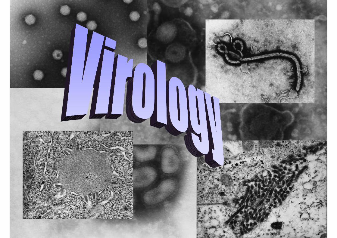
1Virology
Techniken der Virologie
3
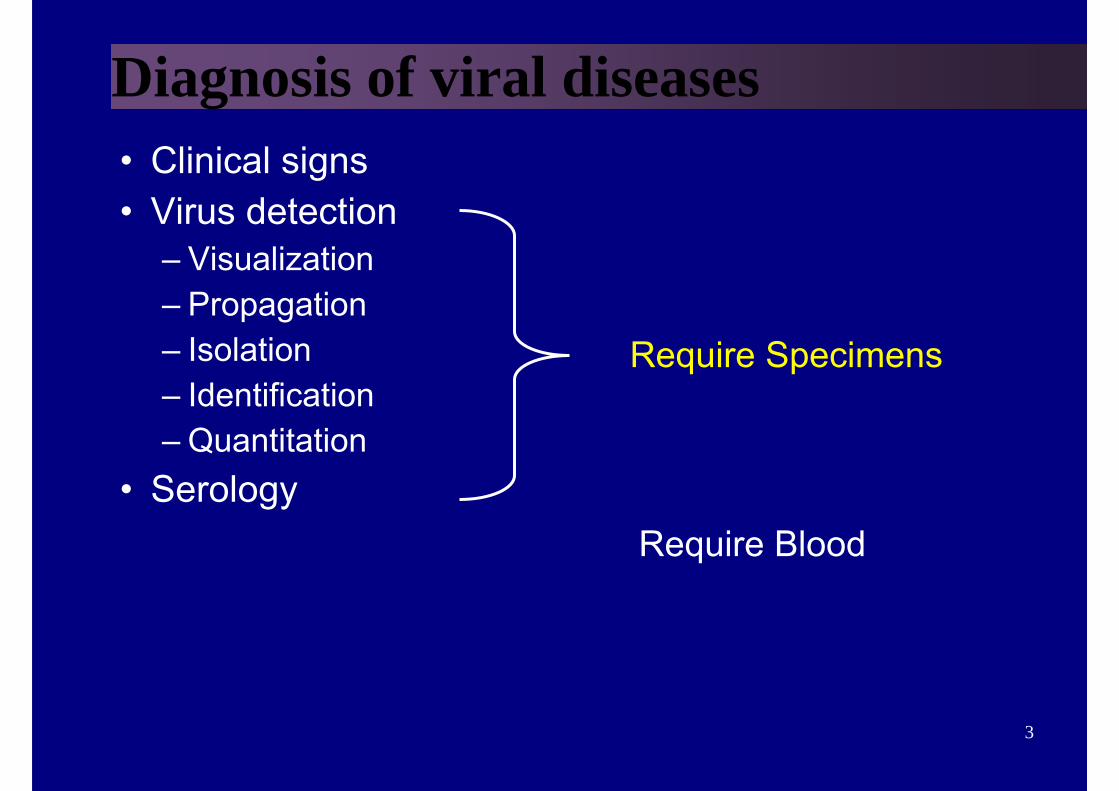
Diagnosis of viral diseases• Clinical signs• Virus detection
– Visualization– Propagation– Isolation – Identification– Quantitation
• Serology
Require Specimens
Require Blood
4
SPECIMEN COLLECTION● Specimens are to be collected from the freshest part of the
lesion, where the most active and current virus replication is presumed to happen.
● Attempt to take the sample as sterile or as clear as possible.
● This is very important, as cell culture is ideal medium for bacterial growth.
● A contaminated sample negates VI as a valid diagnostic test.
● If the sample is definitely contaminated, antibiotics can be added.
5
Diagnostic Methods in Virology
1. Direct Examination
2. Virus Propagation Isolation
3. Virus Detection Methods
4. Serology
6
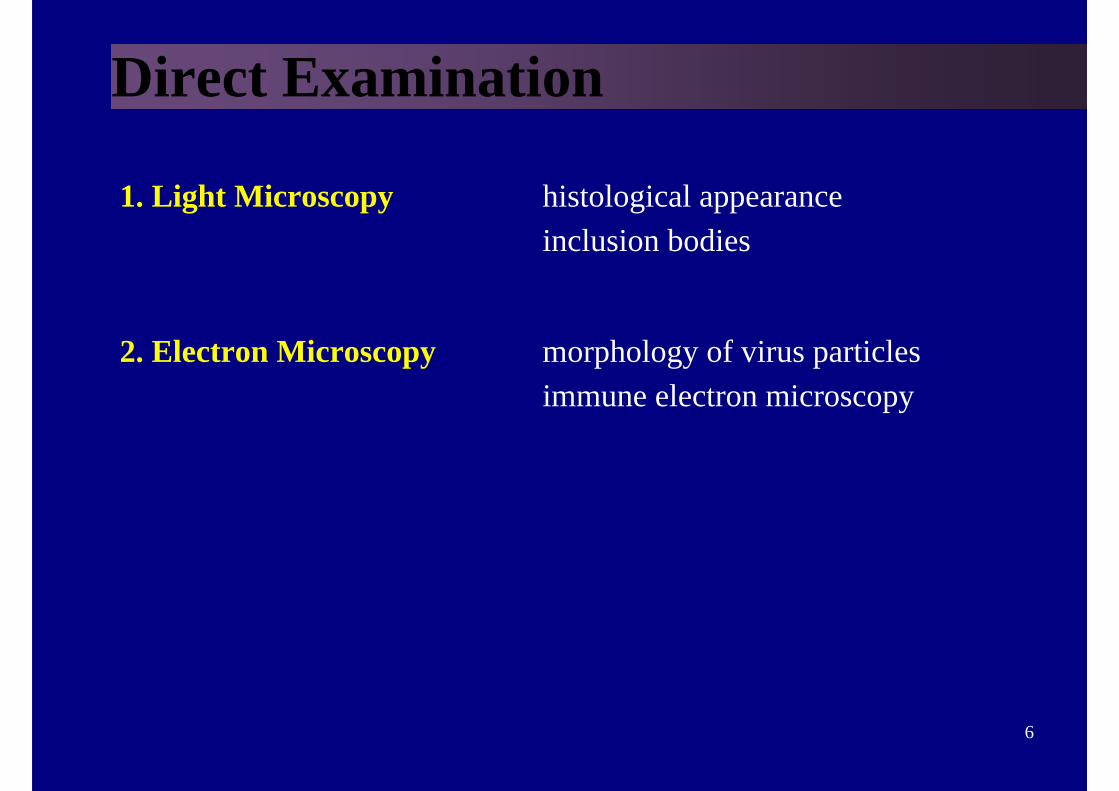
Direct Examination
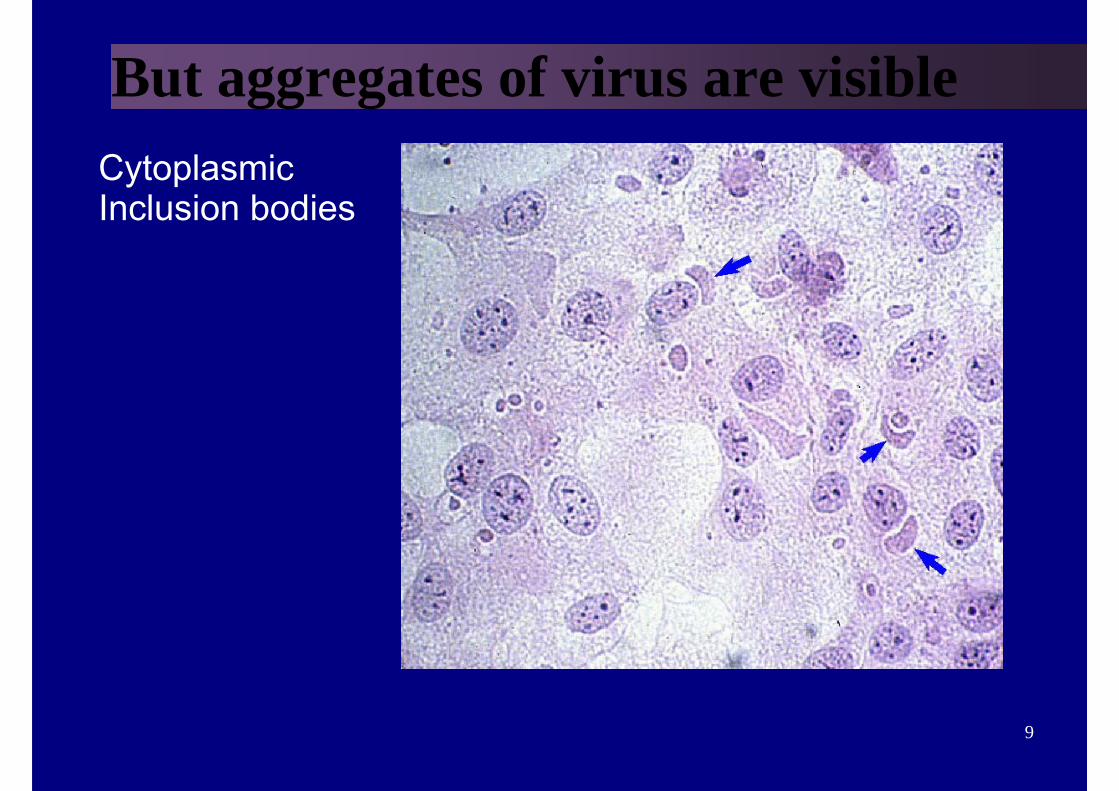
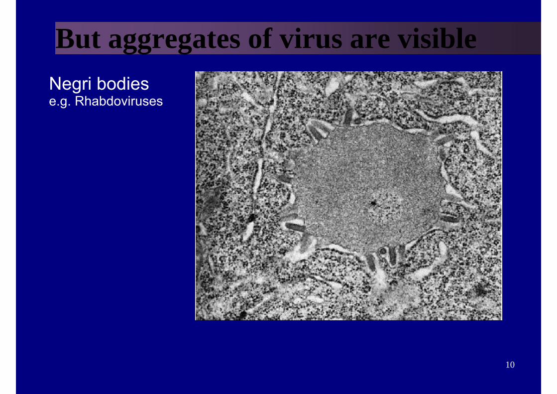
1. Light Microscopy histological appearanceinclusion bodies
2. Electron Microscopy morphology of virus particles immune electron microscopy
7
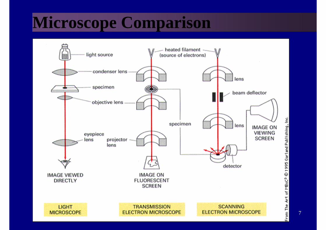
Microscope Comparison
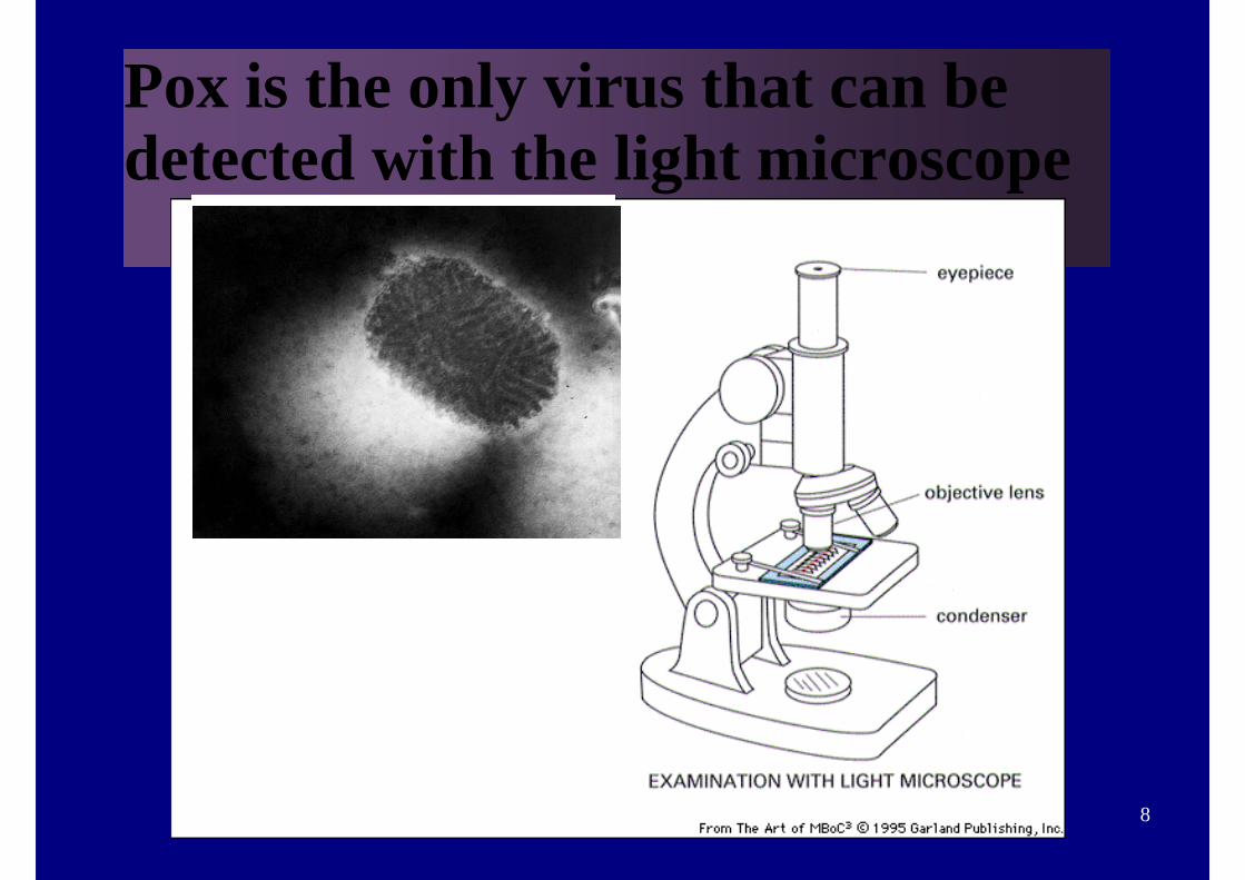
8
Pox is the only virus that can be detected with the light microscope
9
But aggregates of virus are visibleCytoplasmicInclusion bodies
10
But aggregates of virus are visibleNegri bodiese.g. Rhabdoviruses
11
Electron Microscopy106 virus particles per ml required for visualization, x 50,000 -60,000 magnification normally used. Viruses may be detected in the following specimens.
Faeces Rotavirus, AdenovirusNorovirusesAstrovirus, Calicivirus
Vesicle Fluid HSVVZV
Skin scrapings papillomavirus
12
Visualization of individual virus particles● Most biological materials show little contrast
with their surroundings unless they are stained.
● In the case of light microscopy, contrast can be enhanced by using coloured stains which selectively absorb certain wavelengths.
● The electrons in the electron microscope are absorbed very little by biological material and contrast is obtained mainly by electron scattering
13
Staining● To heighten the contrast between viruses
and the background, use is made of electron-dense "stains".
● These are usually compounds of heavy metals of high atomic number, that serve to scatter the electrons from regions covered with the stain.
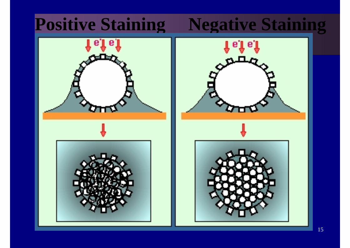
● If virus particles are coated with stain (positive staining), fine detail may be obscured.
14
Negative Staining• Negative staining overcomes this problem by
staining the background and leaving the virus relatively untouched.
● The negative stain is moulded round the virus particle, outlining its structure, and is also able to penetrate between small surface projections and to delineate them.
● If there are cavities within the virus particle that are accessible to the stain, these will be revealed and some of the internal structure of the virus may be disclosed
15
Positive Staining Negative Staining
16
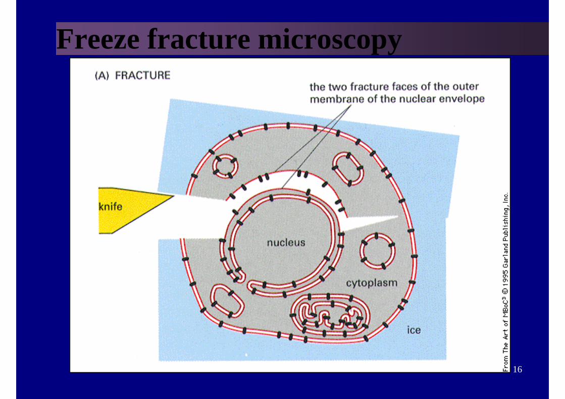
Freeze fracture microscopy
17
Problems with Electron Microscopy
• Expensive equipment
• Expensive maintenance
• Require experienced observer
• Sensitivity often low
18
Diagnostic Methods in Virology
1. Direct Examination
2. Virus Propagation and Isolation
3. Virus Detection Methods
4. Serology
19
Virus propagation and isolation● VIRUSES NEED HOST CELLS TO
REPLICATE
20
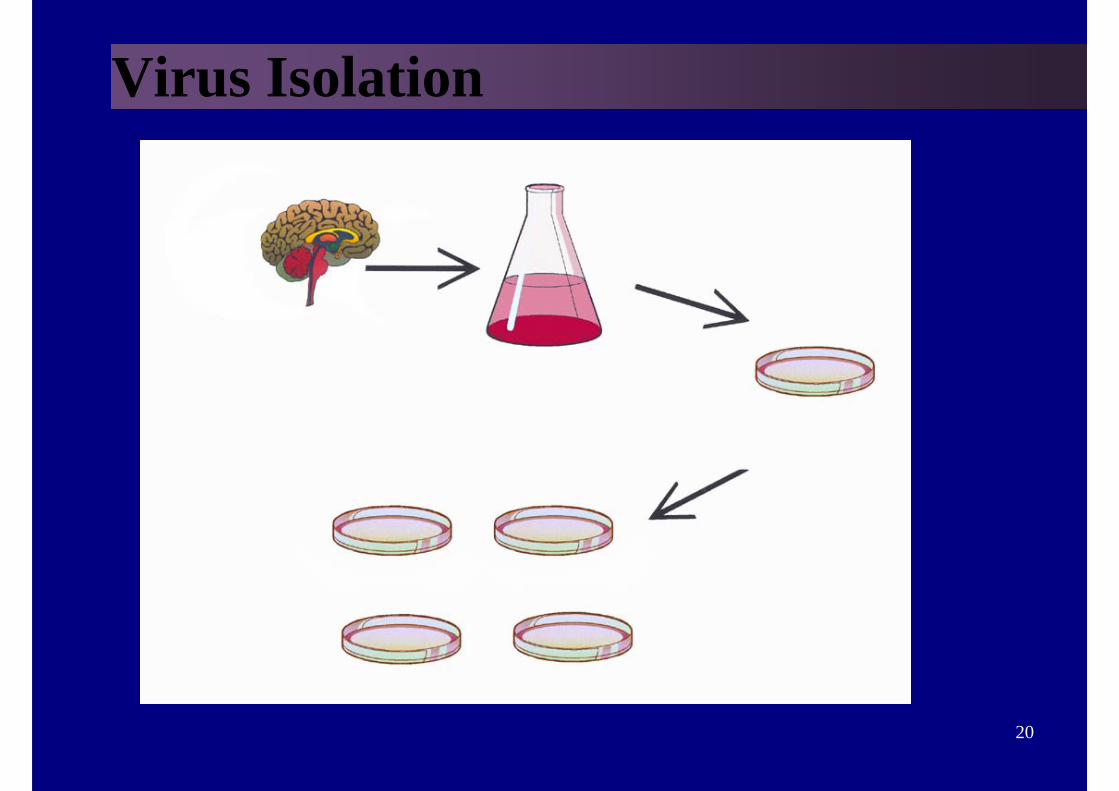
Virus Isolation
21
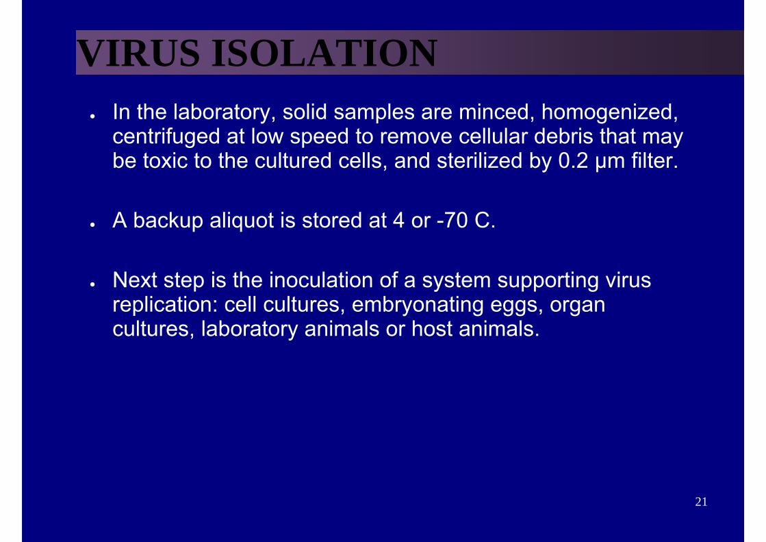
VIRUS ISOLATION● In the laboratory, solid samples are minced, homogenized,
centrifuged at low speed to remove cellular debris that may be toxic to the cultured cells, and sterilized by 0.2 µm filter.
● A backup aliquot is stored at 4 or -70 C.
● Next step is the inoculation of a system supporting virus replication: cell cultures, embryonating eggs, organ cultures, laboratory animals or host animals.
22
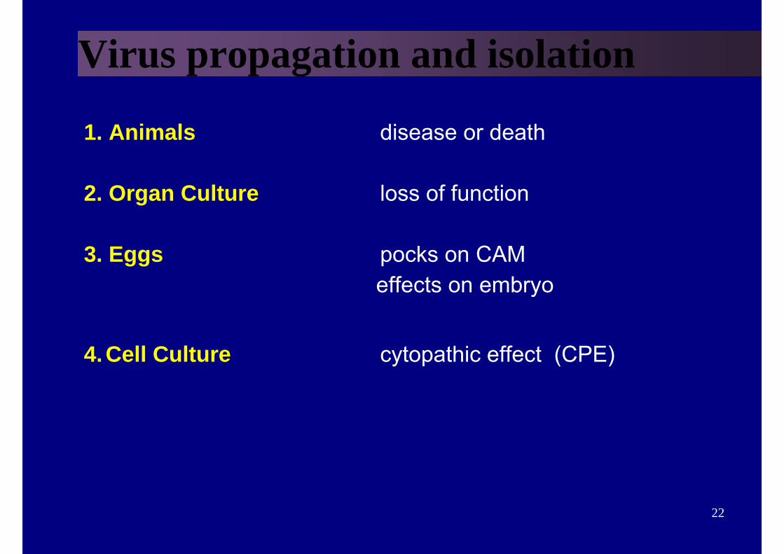
Virus propagation and isolation
1. Animals disease or death
2. Organ Culture loss of function
3. Eggs pocks on CAMeffects on embryo
4.Cell Culture cytopathic effect (CPE)
23
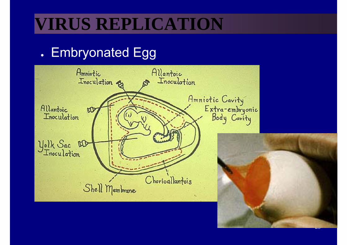
VIRUS REPLICATION● Embryonated Egg
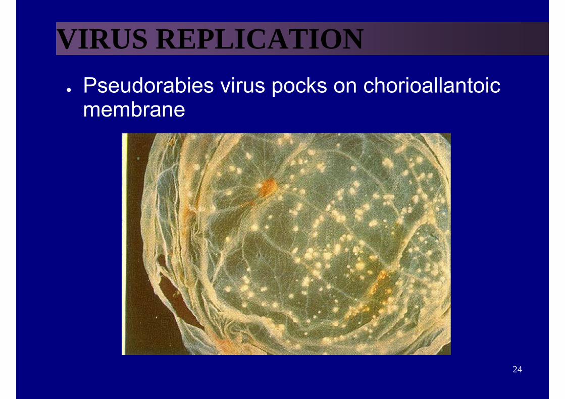
24
VIRUS REPLICATION● Pseudorabies virus pocks on chorioallantoic
membrane
25
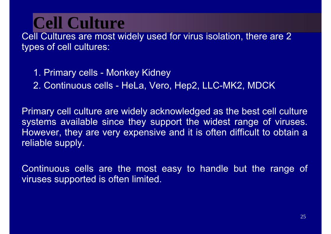
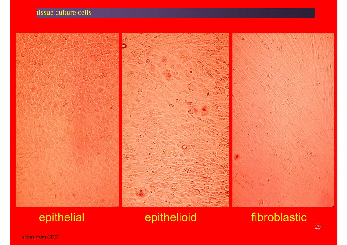
Cell CultureCell Cultures are most widely used for virus isolation, there are 2 types of cell cultures:
1. Primary cells - Monkey Kidney2. Continuous cells - HeLa, Vero, Hep2, LLC-MK2, MDCK
Primary cell culture are widely acknowledged as the best cell culture systems available since they support the widest range of viruses. However, they are very expensive and it is often difficult to obtain a reliable supply.
Continuous cells are the most easy to handle but the range of viruses supported is often limited.
26
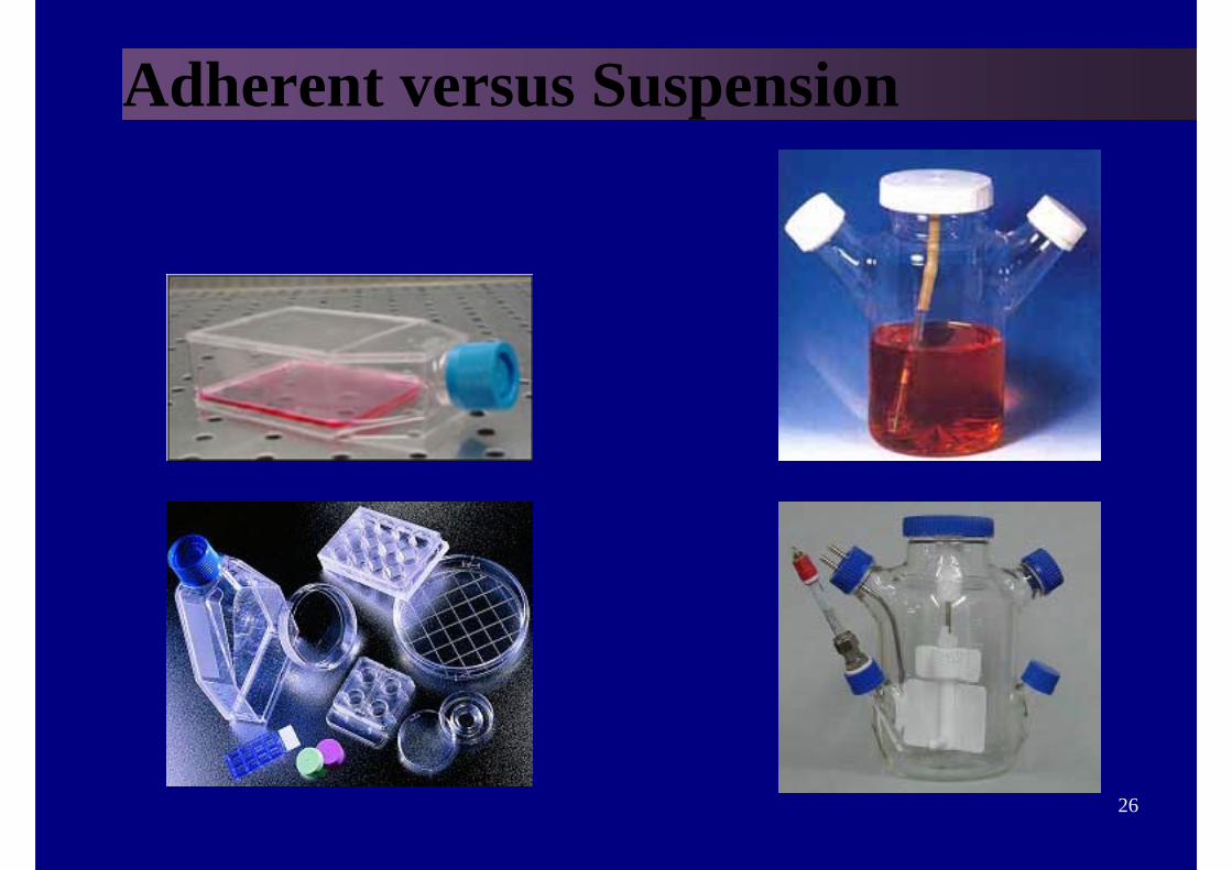
Adherent versus Suspension
27
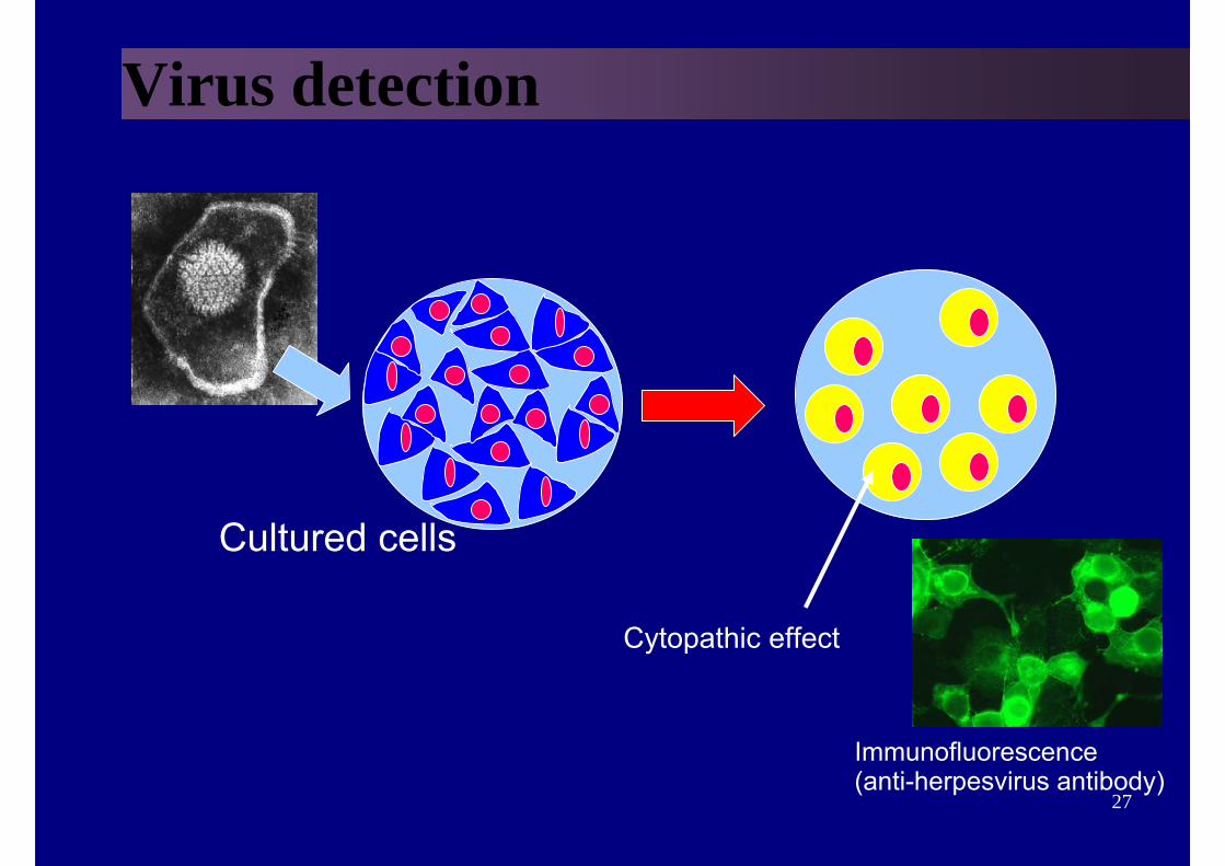
Virus detection
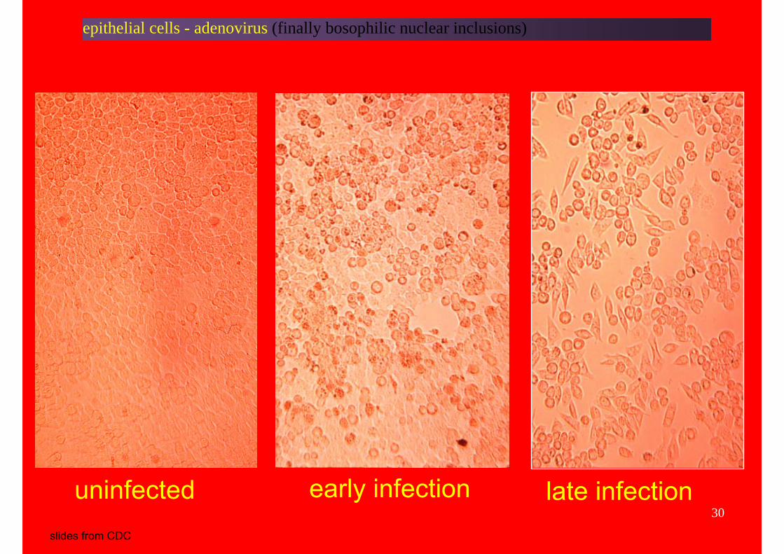
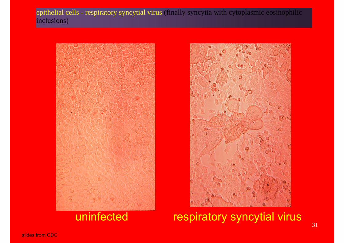
Cultured cells
Cytopathic effect
Immunofluorescence(anti-herpesvirus antibody)
28
Problems with cell culture
• Long period (up to 4 weeks) required for result.• Often very poor sensitivity, sensitivity depends on a
large extent on the condition of the specimen.• Susceptible to bacterial contamination.• Susceptible to toxic substances which may be present
in the specimen.• Many viruses will not grow in cell culture e.g. Hepatitis
B, Diarrhoeal viruses, parvovirus, papillomavirus.
29
tissue culture cells
epithelial epithelioid fibroblasticslides from CDC
30
epithelial cells - adenovirus (finally bosophilic nuclear inclusions)
uninfected early infection late infectionslides from CDC
31
epithelial cells - respiratory syncytial virus (finally syncytia with cytoplasmic eosinophilic inclusions)
uninfected respiratory syncytial virusslides from CDC
32
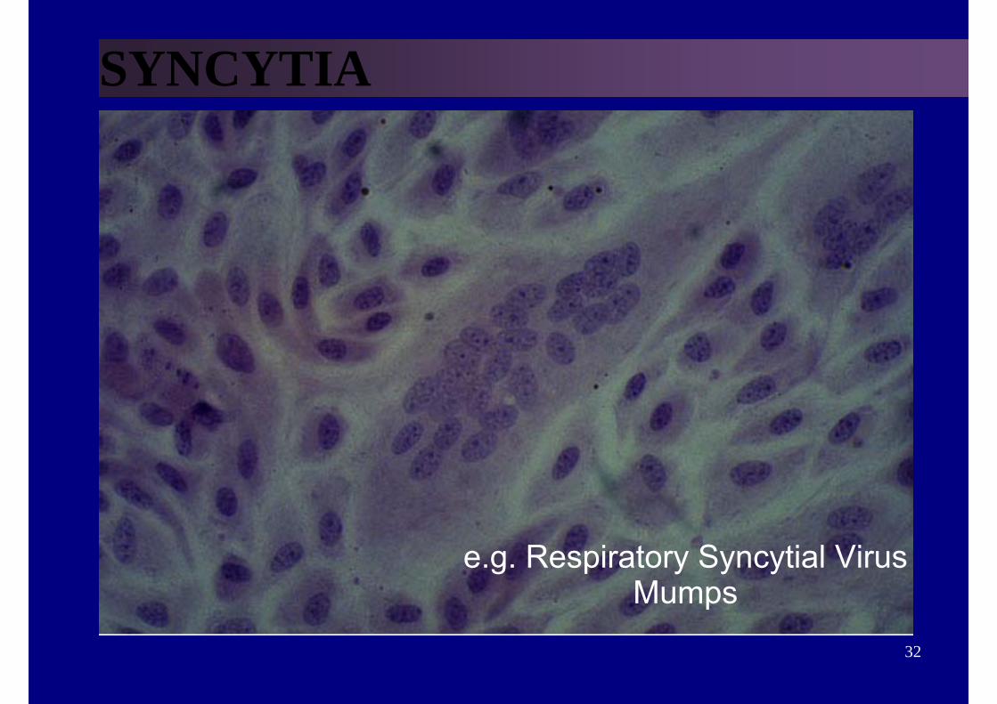
SYNCYTIA
e.g. Respiratory Syncytial VirusMumps
33
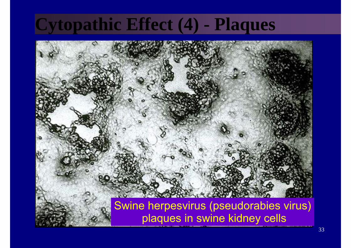
Cytopathic Effect (4) - Plaques
Swine herpesvirus (pseudorabies virus)plaques in swine kidney cells
34
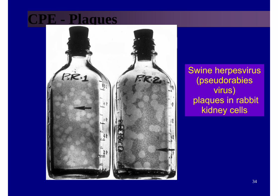
CPE - Plaques
Swine herpesvirus (pseudorabies
virus)plaques in rabbit
kidney cells
35
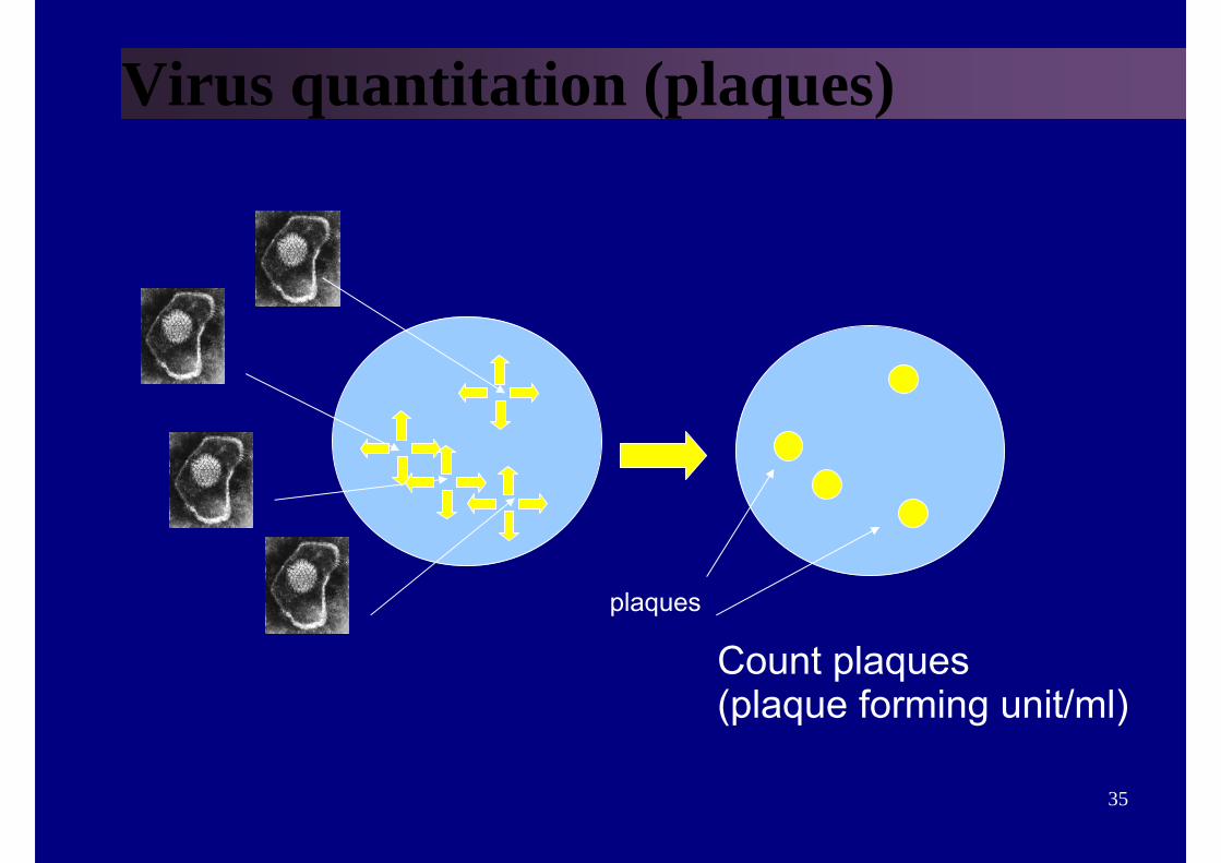
Virus quantitation (plaques)
plaques
Count plaques(plaque forming unit/ml)
36
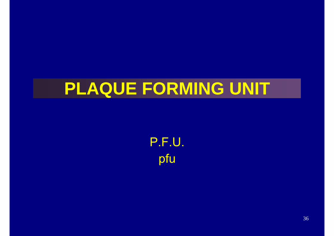
PLAQUE FORMING UNIT
P.F.U.pfu
37
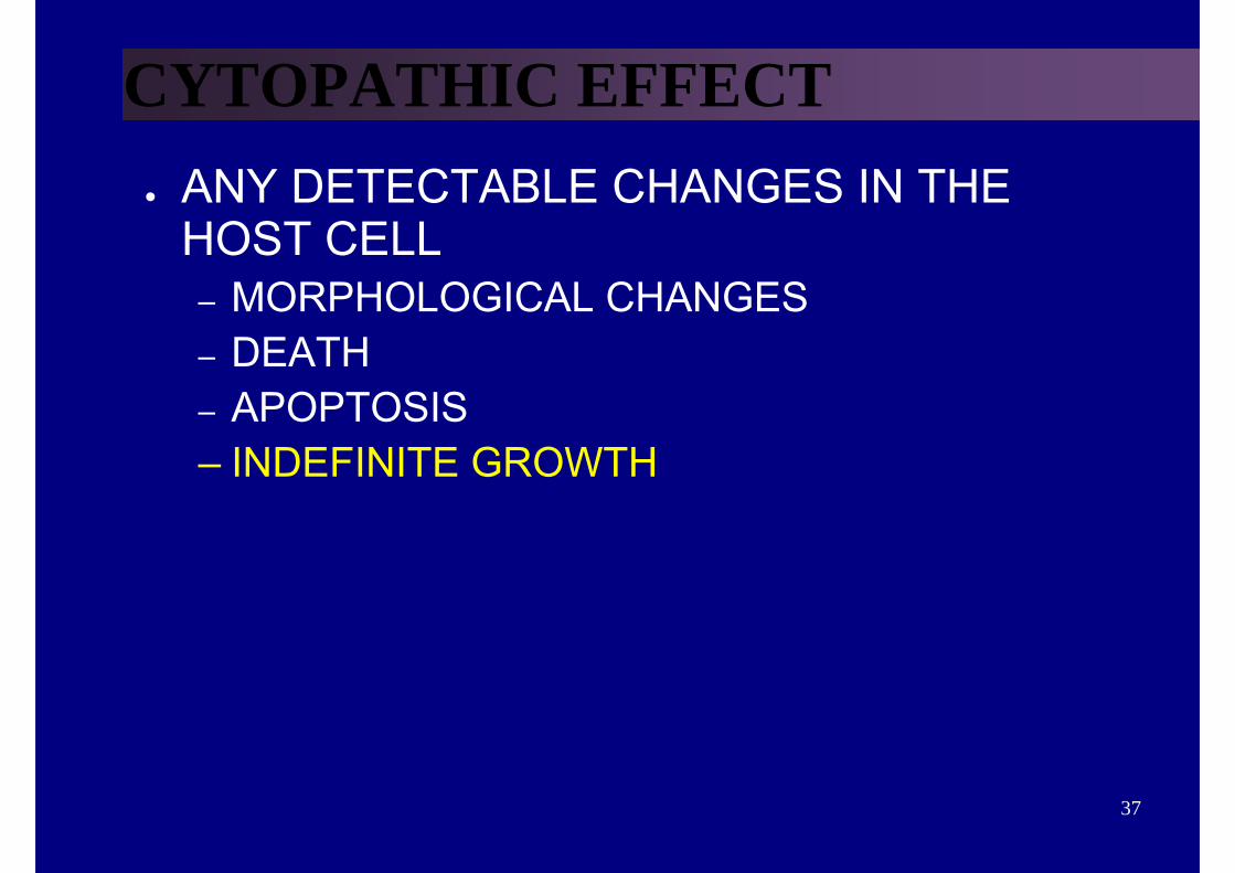
CYTOPATHIC EFFECT● ANY DETECTABLE CHANGES IN THE
HOST CELL– MORPHOLOGICAL CHANGES– DEATH– APOPTOSIS– INDEFINITE GROWTH
38
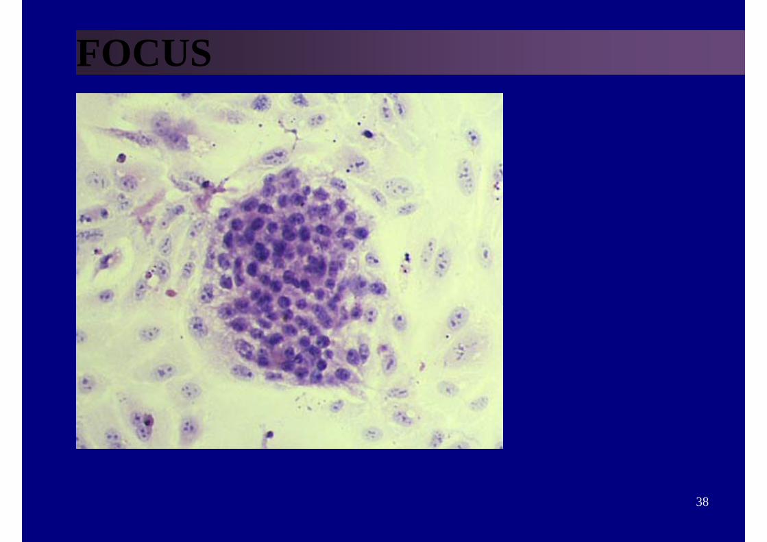
FOCUS
39
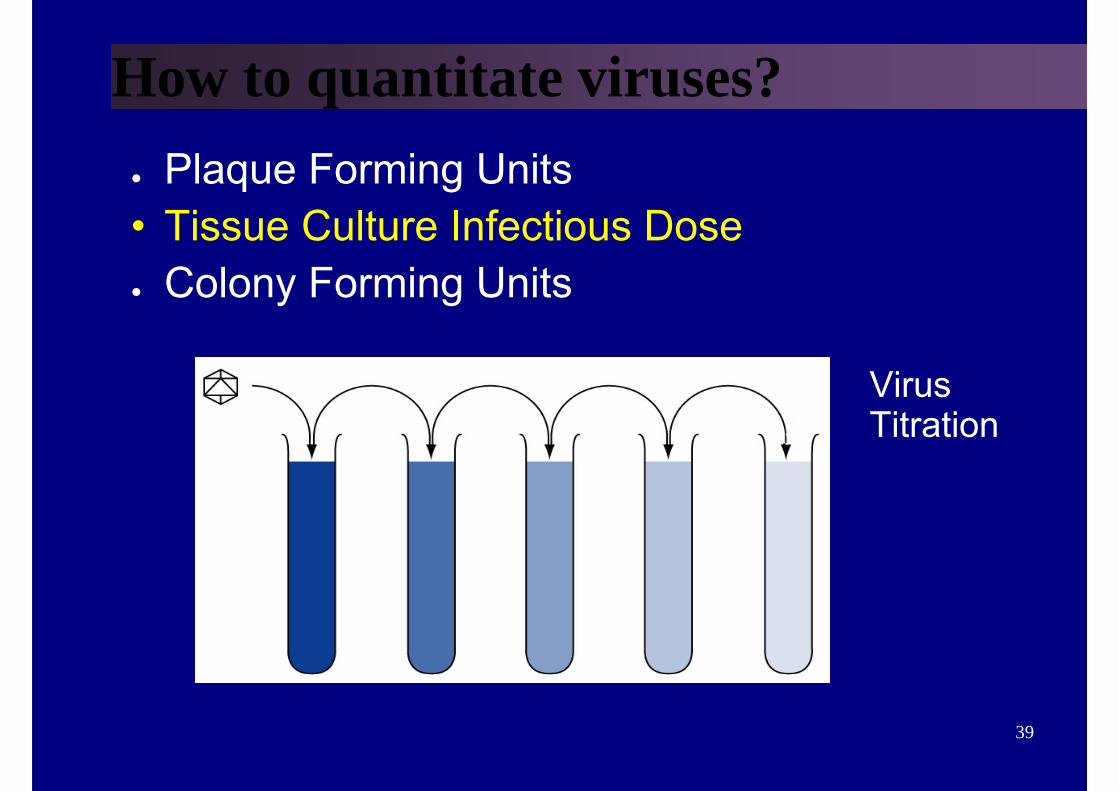
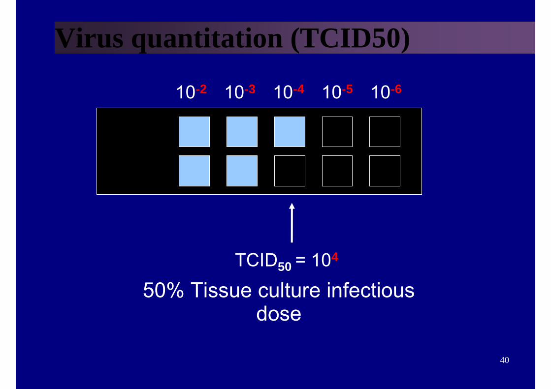
How to quantitate viruses?● Plaque Forming Units• Tissue Culture Infectious Dose● Colony Forming Units
VirusTitration
40
Virus quantitation (TCID50)
10-2 10-3 10-4 10-5 10-6
TCID50 = 104
50% Tissue culture infectiousdose
41
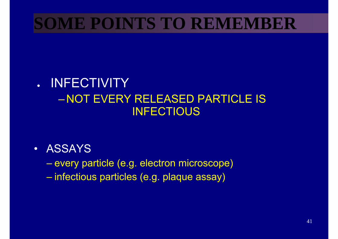
SOME POINTS TO REMEMBER
● INFECTIVITY– NOT EVERY RELEASED PARTICLE IS
INFECTIOUS
• ASSAYS– every particle (e.g. electron microscope)– infectious particles (e.g. plaque assay)
42
Tests to detect virus (viral antigen)
● Haemagglutination
43
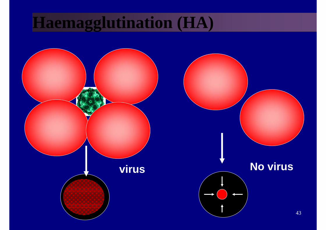
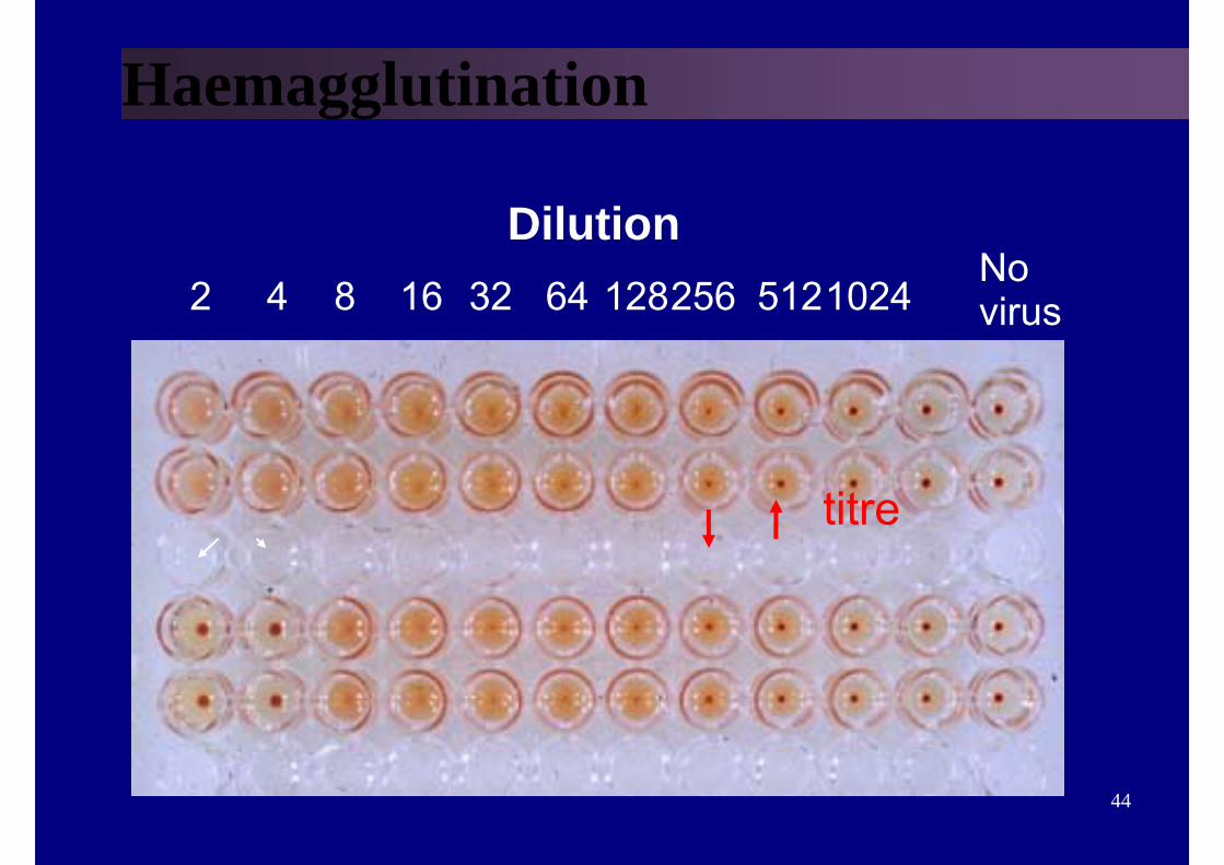
Haemagglutination (HA)
virus No virus
44
Haemagglutination
Dilution2 4 8 16 32 64 128256 512
titre
1024Novirus
45
Tests to detect virus (viral antigen)
● Haemagglutination• ELISA
46
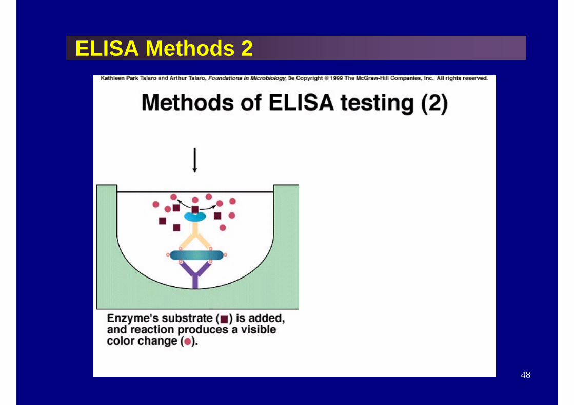
VIRUS ANTIGEN DETECTION● ELISA
● Antibody binds to antigen; ● enzyme-labeled anti-IgG binds to antibody; ● substrate changes color
47
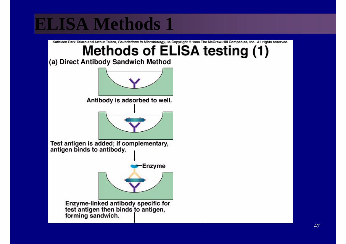
ELISA Methods 1
48
ELISA Methods 2
49
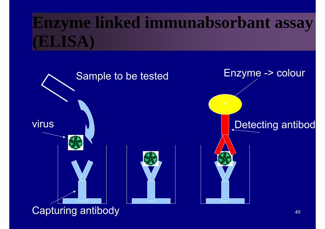
Enzyme linked immunabsorbant assay (ELISA)
Sample to be tested
virus
Capturing antibody
Detecting antibody
Enzyme -> colour
50
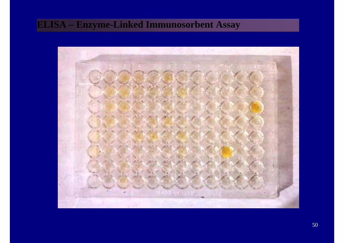
ELISA – Enzyme-Linked Immunosorbent Assay
51
ELISA Machine
52
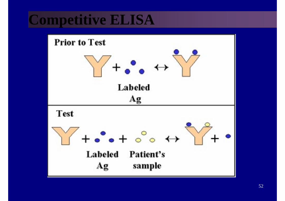
Competitive ELISA
53
Tests to detect virus (viral antigen)● Haemagglutination● ELISA• Immunofluorescence
54
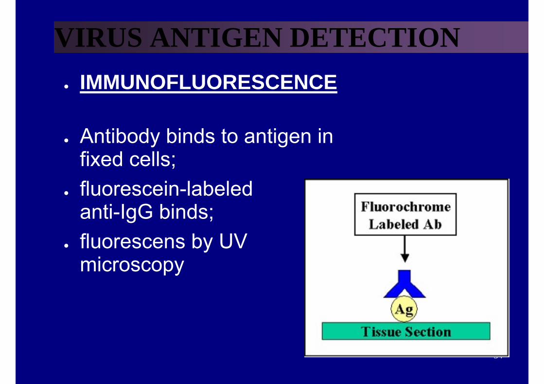
VIRUS ANTIGEN DETECTION● IMMUNOFLUORESCENCE
● Antibody binds to antigen in fixed cells;
● fluorescein-labeled anti-IgG binds;
● fluorescens by UV microscopy
55
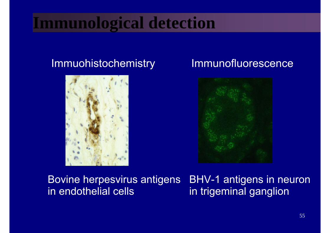
Immunological detection
Immuohistochemistry Immunofluorescence
Bovine herpesvirus antigensin endothelial cells
BHV-1 antigens in neuronin trigeminal ganglion
56
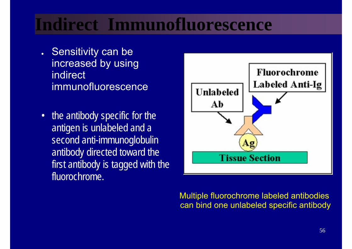
Indirect Immunofluorescence● Sensitivity can be
increased by using indirect immunofluorescence
• the antibody specific for the antigen is unlabeled and a second anti-immunoglobulin antibody directed toward the first antibody is tagged with the fluorochrome.
Multiple fluorochrome labeled antibodiescan bind one unlabeled specific antibody
57
Diagnosis of viral diseases• Detection of exposure (Serology)
– Virus neutralization– Haemagglutination inhibition– ELISA– Limitations
58
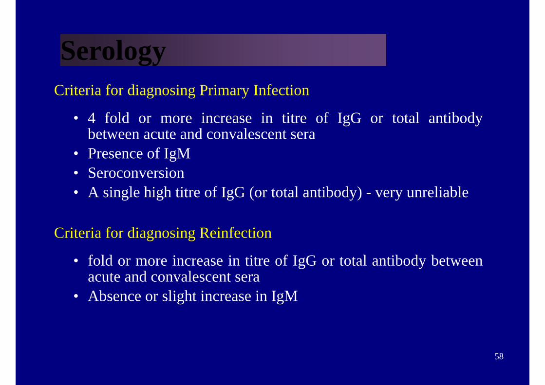
SerologyCriteria for diagnosing Primary Infection
• 4 fold or more increase in titre of IgG or total antibody between acute and convalescent sera
• Presence of IgM• Seroconversion• A single high titre of IgG (or total antibody) - very unreliable
Criteria for diagnosing Reinfection
• fold or more increase in titre of IgG or total antibody between acute and convalescent sera
• Absence or slight increase in IgM
59
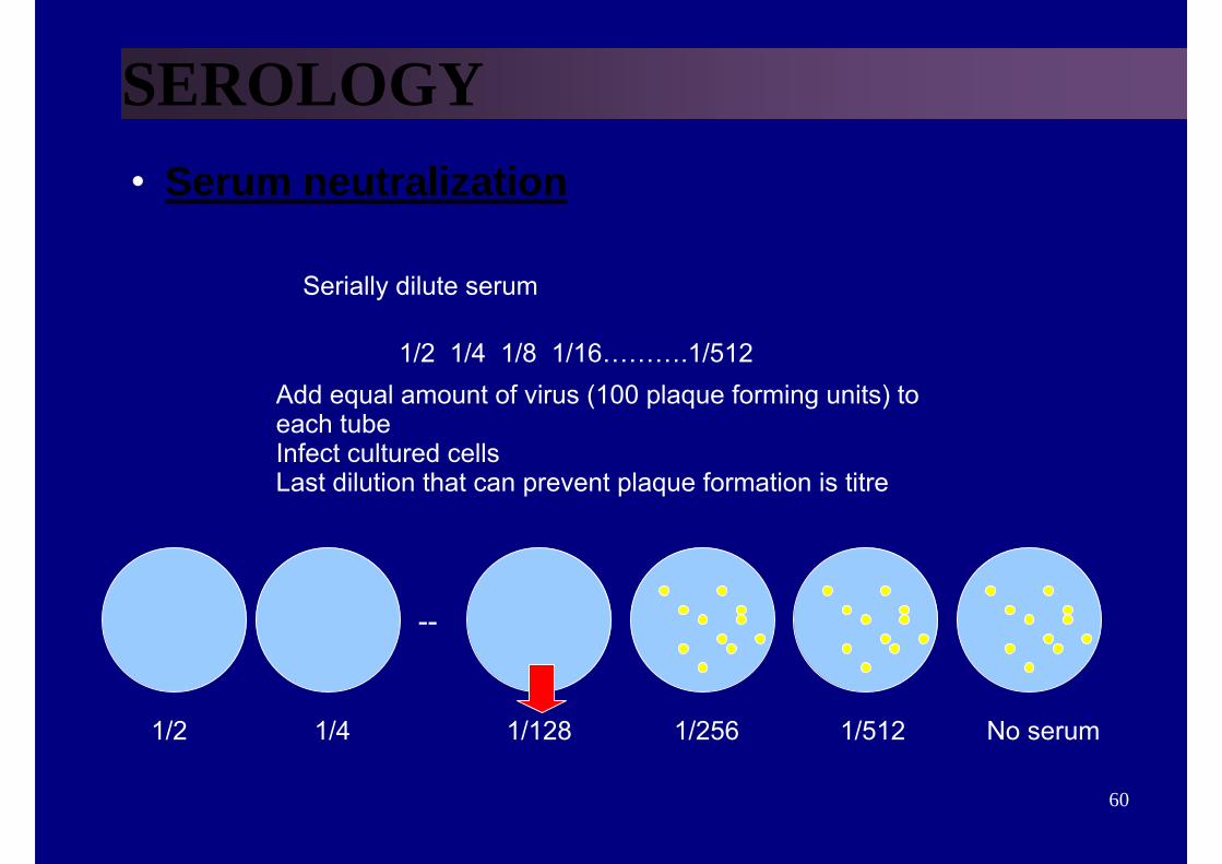
SEROLOGY● Serum Neutralization
● Antibody neutralizes infectivity of virion;inhibits cytopathology, reduces plaques, or protects animals
60
SEROLOGY• Serum neutralization
Serially dilute serum
1/2 1/4 1/8 1/16……….1/512Add equal amount of virus (100 plaque forming units) toeach tubeInfect cultured cellsLast dilution that can prevent plaque formation is titre
No serum1/2 1/4 1/256 1/5121/128
--
61
Usefulness of Serological Results
● How useful a serological result is depends on the individual virus. ● E.g. for viruses such as rubella and hepatitis A, onset of clinical
symptoms coincide with production of antibodies. Detection of IgM or rising titres of IgG in the patient’s serum indicates active disease.
● However, many viruses often produce clinical disease before the appearance of antibodies such as respiratory and diarrhoeal viruses. So in this case, any serological diagnosis would be retrospective and therefore will not be that useful.
● There are also viruses which produce clinical disease months or years after seroconversion e.g. HIV and rabies. For these viruses, the mere presence of antibody is sufficient to make a definitive diagnosis.
62
Problems with Serology• Long period of time required for diagnosis for paired acute and
convalescent sera.• Mild local infections such as HSV genitalis may not produce a
detectable humoral immune response.• Antigenic cross-reactivity between related viruses e.g. HSV and
VZV, Japanese B encephalitis and Dengue, may lead to false positive results.
• immunocompromised patients often give a reduced or absent humoral immune response.
• Patients with infectious mononucleosis SLE may react non-specifically giving a false positive result.
• Patients given blood or blood products may give a false positiveresult due to the transfer of antibody.