Technetium-99m BIDA Biliary Scintigraphy in the Evaluation of the
7
he differentiation ofjaundice due to the hepatocel lular disease (medical jaundice) from extrahepatic bili ary obstruction (surgical jaundice) is important in plan fling the appropriate diagnostic and therapeutic procedures. Currently, there are a number of radiologic tech niques available in the investigation of a jaundiced patient. These include: ultrasonography (US), corn puted tomography (CT), percutaneous transhepatic cholangiography (PTC), and endoscopic retrograde cholangio-pancreatography (ERCP) ( 1). Some of these (PTC and ERCP) are invasive and/or expensive. Fur thermore, they delineate anatomic lesions but do not provide a functional evaluation. On the other hand, radionuclide hepatobiliary scanning is a noninvasive study and provides the only means of functional evaluation along with imaging. In selecting a single screening test which can differ entiate medical from surgical causes of jaundice, one has to take into account ease of performance, cost effectiveness, complications associated with the proce dure, and degree of accuracy. With the introduction of newer radiopharrnaceuticals such as p-butyl acetanilid iminodiacetic acid (BIDA) labeled with technetium 99m (99mTc) interest in hepatobiliary scintiscanning has increased (2—4). There has not been uniform agreement concerning Received July 19, 1985; revision accepted Mar. 6, 1986. For reprints contact: M.D. Ram, MD, PhD, Chief, Surgical Service, V. A. Medical Center, Lexington, KY 405 11. the diagnostic value of scintiscanning in jaundiced pa tients (5—7).Klingensmith et al. stated that US and hepatobiliary scintigraphy have a complementary role in the evaluation ofbiliary obstruction (8). Their group also suggested that intrahepatic cholestasis can be di agnosed by hepatobiliary scintigraphy (9). This study reviews our experience with the use of radionuclide hepatobiliary imaging as a screening procedure in jaundiced patients. MATERIALS AND METHODS During the period March 1, 1979, through April 30, 1983, we studied 96 patients who were clinically jaun diced. All patients were from our institution and in cluded 79 males and 17 females. The age range was 1 mo to 92 yr (mean 52 yr). The levels of serum bilirubin ranged from 2.1—38.8 mg/dl (normal 0. 1— 1.1 mg/dl). Excluded from this review were patients who carried a diagnosis of either acute or chronic cholecystitis alone and also patients who have had previous biliary enteric bypass. These have been previously reviewed by one of us (2—4,10). In addition to scintiscanning, all patients underwent clinical, chemical, radiologic, and endoscopic evalua tions as required. The final diagnosis in each patient was based on the results of all studies noted above and additionally in most patients based on operative find ings or needle biopsy of the liver (details in results). 1407 Volume27 • Number9 • September1986 Technetium-99m BIDA Biliary Scintigraphy in the Evaluation of the Jaundiced Patient Anthony W. Lee, Madhira D. Ram, Wei-Jen Shih, and Karen Murphy Surgical and Nuclear Medicine Services, V. A. Medical Center and the Department of Surgery, University ofKentucky Medical Center, Lexington, Kentucky Biliary scintigraphy using @â€oeTc p-butyl acetanilidiminodiacetic acid (BIDA) was performed as partof thediagnosticevaluationon96 patientswithjaundice(serumbilirubin>2 mg/dl)to assessits valuein this groupof patients.Theresultsof scintigraphyrevealed(a)no obstruction to the flow of the scintigraphic agent into the duodenum in 54 patients, (b) delayed appearance of the agent (normal upper limit 60 mm) in the duodenum indicating partial obstruction in 22 patients, and (c) complete obstruction of the duct demonstrated by absenceof agentinthe duodenumin 20 patients.Thefindingswerecorrelatedwith thefinal diagnosisandthe overallresultsshowaccuracyof 92.7%,sensitivityof 97.3%,and specificityof 89.8%.Biliaryscintigraphywas thusfoundto be usefulin differentiating nonobstructive, partiallyobstructive,andcompletelyobstructivecausesofjaundice. J Nucl Med 27:1407—1412,1986 by on April 10, 2019. For personal use only. jnm.snmjournals.org Downloaded from
Transcript of Technetium-99m BIDA Biliary Scintigraphy in the Evaluation of the
he differentiation ofjaundice due to the hepatocellular disease (medical jaundice) from extrahepatic biliary obstruction (surgical jaundice) is important in planfling the appropriate diagnostic and therapeuticprocedures.
Currently, there are a number of radiologic techniques available in the investigation of a jaundicedpatient. These include: ultrasonography (US), cornputed tomography (CT), percutaneous transhepaticcholangiography (PTC), and endoscopic retrogradecholangio-pancreatography (ERCP) ( 1). Some of these(PTC and ERCP) are invasive and/or expensive. Furthermore, they delineate anatomic lesions but do notprovide a functional evaluation. On the other hand,radionuclide hepatobiliary scanning is a noninvasivestudy and provides the only means of functionalevaluation along with imaging.
In selecting a single screening test which can differentiate medical from surgical causes of jaundice, onehas to take into account ease of performance, costeffectiveness, complications associated with the procedure, and degree of accuracy. With the introduction ofnewer radiopharrnaceuticals such as p-butyl acetanilidiminodiacetic acid (BIDA) labeled with technetium99m (99mTc) interest in hepatobiliary scintiscanninghas increased (2—4).
There has not been uniform agreement concerning
Received July 19, 1985; revision accepted Mar. 6, 1986.For reprints contact: M.D. Ram, MD, PhD, Chief, Surgical
Service, V. A. Medical Center, Lexington, KY 405 11.
the diagnostic value of scintiscanning in jaundiced patients (5—7).Klingensmith et al. stated that US andhepatobiliary scintigraphy have a complementary rolein the evaluation ofbiliary obstruction (8). Their groupalso suggested that intrahepatic cholestasis can be diagnosed by hepatobiliary scintigraphy (9). This studyreviews our experience with the use of radionuclidehepatobiliary imaging as a screening procedure injaundiced patients.
MATERIALS AND METHODS
During the period March 1, 1979, through April 30,1983, we studied 96 patients who were clinically jaundiced. All patients were from our institution and included 79 males and 17 females. The age range was 1mo to 92 yr (mean 52 yr).
The levels of serum bilirubin ranged from 2.1—38.8mg/dl (normal 0. 1—1.1 mg/dl). Excluded from thisreview were patients who carried a diagnosis of eitheracute or chronic cholecystitis alone and also patientswho have had previous biliary enteric bypass. Thesehave been previously reviewed by one of us (2—4,10).In addition to scintiscanning, all patients underwentclinical, chemical, radiologic, and endoscopic evaluations as required. The final diagnosis in each patientwas based on the results of all studies noted above andadditionally in most patients based on operative findings or needle biopsy of the liver (details in results).
1407Volume27 •Number9 •September1986
Technetium-99m BIDA Biliary Scintigraphyin the Evaluation of the Jaundiced PatientAnthony W. Lee, Madhira D. Ram, Wei-Jen Shih, and Karen Murphy
Surgical and Nuclear Medicine Services, V. A. Medical Center and the Department of Surgery,University ofKentucky Medical Center, Lexington, Kentucky
Biliary scintigraphy using @“Tcp-butyl acetanilidiminodiacetic acid (BIDA) was performed aspartof the diagnosticevaluationon 96 patientswith jaundice(serumbilirubin>2 mg/dl)toassessits valuein this groupof patients.Theresultsof scintigraphyrevealed(a)noobstruction to the flow of the scintigraphic agent into the duodenum in 54 patients, (b)delayed appearance of the agent (normal upper limit 60 mm) in the duodenum indicatingpartial obstruction in 22 patients, and (c) complete obstruction of the duct demonstrated byabsenceof agentin the duodenumin 20 patients.Thefindingswerecorrelatedwith the finaldiagnosisandthe overallresultsshowaccuracyof 92.7%,sensitivityof 97.3%,andspecificityof 89.8%.Biliaryscintigraphywas thus foundto be usefulin differentiatingnonobstructive,partiallyobstructive,andcompletelyobstructivecausesof jaundice.
J Nucl Med 27:1407—1412,1986
by on April 10, 2019. For personal use only. jnm.snmjournals.org Downloaded from

I24kii.
Informed consent was obtained from all subjects.Technetium-99m BIDA was used as scanning agentand was prepared from a commercial kit and 5mCi of[99mTc]BIDA was injected intravenously. Using agamma camera,t images were obtained at 2, 5, 10, 15,30, 45, and 60 mm after the injection and then at 15-mm intervals for up to 90 to 120 mm. Further scanswere obtained at 4 to 6 hr as needed and up to 24 hr ifindicated. Each image accumulated 300—500kcounts.The details of this technique were previously described(2,11).
RESULTS
In a normal scintigram, the liver is visualized at 5 to10 mm after injection of the agent, the gallbladder at
,@â€1̃ 5—20 mm and complete images ofthe liver, galiblad
der, common bile duct, and proximal small bowel areobtained between 25 and 30 mm. The criteria used todefine extrahepatic biliary obstruction are based onwhether the gallbladder and the common bile duct werevisualized or not and whether there is radionuclideactivity in bowel or not and also the time of appearanceof this activity.
The results of scintigraphy in this group were classifled into three categories as follows: (a) nonobstructivegroup (no obstruction to the flow of the scintigraphicagent into the duodenum), 54 patients; (b) partiallyobstructive (partial obstruction on the basis of delayedappearance of the agent: 60 mm or more in the bowel),22 patients (Fig. 1); and (c) completely obstructive(absence of the scanning agent in the small bowel evenafter 24 hr), 20 patients (Fig. 2).
In eight patients who were categorized as partialobstruction, in addition to delayed radioactive tracerexcretion in bowel, there was a scintigraphic pattern ofintrahepatic bile pooling along the area and/or segmental defect in common bile duct (Fig. 3). This characteristic pattern has been previously documented (12).
There are two scintigraphic patterns of completeobstruction of the common bile duct. One is that of afairly rapid hepatic uptake of the tracer by the liver butno visualization ofthe hepatic ducts, the common duct,gallbladder, or the bowel even up to 24 hr after injection(13,14) (Fig. 2). The other pattern is a hyperacutecomplete common bile duct obstruction, scintigraphicfeatures ofwhich include rapid uptake and visualizationof the hepatic ducts, common bile duct, and the gallbladder, but no appearance of activity in the bowelthrough the 24-hr study period ( 15) (Fig. 4). Only onepatient in the study showed this pattern.
The results of scintigraphy were correlated with thefinal diagnosis (Tables 1, 2, and 3). The following definitions were used: (a) true negative—no obstruction ordelay of agent into the duodenum in the scan and noobstruction demonstrated, (b) true positive—obstruction or delay of agent into the duodenum by scan andobstruction confirmed, (c) false positive—obstructionor delay of agent into the duodenum by scan but noobstruction present, and (d) false negative—no obstruction or delay of agent into the duodenum by scan butobstruction present.
Based on the above, the sensitivity of the scan was97.3%, the specificity was 89.8%, and the overallaccuracy was 92.7%.
DISCUSSION
Our results suggest that scintigraphy is useful in theinitial evaluation of the jaundiced patient. The differentiation of obstructive from nonobstructive jaundicehas been a subject of several conflicting reports (17—20). The area of greatest difficulty centers around thefact that at very high levels of serum bilirubin, lack ofimages may be due to hepatocellular disease (poorhepatic uptake) or extrahepatic biliary obstruction(poor excretion) ( 16,21 ). In the former situation thetracer remains in the blood pool for a long time and
FIGURE 1Partial obstruction of common bileduct: Fairly rapid radiotracer hepaticuptake and no visualizationof bileducts, gallbladder,and bowel in 5-,10-, 15-, 30-, and 60-mm images;colonicactivityseenin 24-hr imagesbut substantial radiotracer remainingin liver at 24 hr, indicatingseverepartialobstruction
1408 Lee, Ram, Shih et al The Journal of Nuclear Medicine
by on April 10, 2019. For personal use only. jnm.snmjournals.org Downloaded from
2h
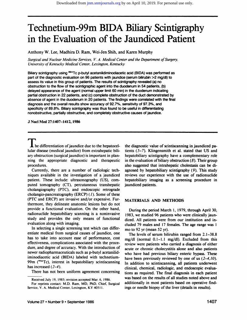
FIGURE 2Complete obstruction due to biliaryatresia in 2-mo-old girl: Radiotracerisrapidlytakenby liverbut bileducts,gallbladder,and bowel are persistently not visualized, even in 20-hrimage
the biliary tract will not be imaged. However, in patientswith very high levels of bilirubin, these results may beequivocal because of dilution of the tracer ( 17).
The scintigraphic patterns associated with biliaryobstruction and jaundice are briefly outlined below.
1. In complete obstruction ofthe common bile duct,there is a fairly rapid hepatic uptake of the tracer, butno visualization ofthe hepatic ducts, the common duct,gallbladder, or the bowel 24 hr after injection (13)(Fig. 1).
2. Hyperacute complete common bile duct obstruc
tion has scintigraphic features that include rapid hepaticuptake and visualization of the hepatic ducts, the common bile duct, and the gallbladder, but no appearanceof activity in the bowel through and up to the 24-hrstudy period ( 15) (Fig. 4). This pattern is very rarebecause such a patient is usually studied immediatelyfollowing the onset of the common duct obstructionwhile the gallbladder is still continuing to reabsorbwater. There is an initial imaging of the biliary systemwhile there is net bile flow until the bile is maximallyconcentrated. Eventually when the pressure in the sys
FIGURE 3Partial obstruction associated wfthintrahepaticbilepoolingandnarrowingin commonbileduct: Bilepoolingseen in 15-, 30-, 45-, and 60-mmimages;abruptlynarrowingcommonduct seen in 30-, 45-, and 60-mmimages
‘mom-@i@m‘@30m-@.@,@45@60m120m
1409Volume27 •Number9 •September1986
15m 30m
20h
by on April 10, 2019. For personal use only. jnm.snmjournals.org Downloaded from

AverageFinalclinical
diagnosisNo.of
patientsS.bilirubin
(mg/dl)Confirmationof
diagnosisAlcOhOliC
cirrhosis127.1LiverbiopsyAlcoholichepatitis77.7LiverbiopsyUver
metastasis85.1LiverbiopsyDruginducedcho 48.5LiverbiopsylestasisSepsis41
0.4Exploratory celiotomyHepatitis41
2.6Elevated hepatitisB, surface anti
genLiver
failure45.9HistoryNeonatalhepatitis29.1UverbiopsySarcoidosis13.6Liver
biopsyChronicpassive11 3.5LiverbiopsycongestionCirrhosis13.0Liver
biopsyHodgkin'sdisease16.8LiverbiopsyCytomegalovirus
in 11 4.3LiverbiopsyfectionInfected
aneurysm16.5Exploratory celiotomySubphrenic
abscess12.8Exploratory celiotomyHepatoma11
1.0Operation and biopsyCholedocholithiasis13.4Ultrasonography
andoperationTotal54.
False negative.
TABLE 1NonobstructiveGroupby Scan
2m
Ih
5m lOm
15m 30m 45m
•13.5 h
FIGURE 4Hyperacutecompletecommonbileductobstruction:Rapidhepatic tracer uptake, in 30-mm to 24-hr images andvisualizationof gallbladder; no bowel activity seen throughout studyup to 24 hr
tem is high, net bile flow will cease and the scintigraphicpattern of complete common duct obstruction willresult(14—16).
3. The pattern that is seen in patients with biliaryatresia includes a delayed hepatic uptake with no visualization of hepatic ducts, common bile duct andbowel throughout the 24-hr period. In those patientswho have been pretreated with phenobarbital, this pattern is highly reliable for a diagnosis of biliary atresiaand separates this group from those with neonatal hepatitis in whom gstrointestinal excretion is evident (21).
4. In intrahepatic cholestasis, there is a rapid hepaticuptake and delayed appearance of activity in the bowelprecluding a complete obstruction ofthe common duct.Since the hepatic function is intact, the scintigraphicpattern is similar to that due to partial obstruction ofthe excretory mechanism (21,22). In our study, therewere two patients with drug induced cholestasis whofall into the partially obstructive group and were misdiagnosed. One other patient with alcoholic hepatitisand a serum bilirubin of 20.5 mg/dl was also misdiagnosed as “partiallyobstructive,― probably on the basisof the impaired uptake by the hepatocytes.
5. Hepatic failure. In this group of patients, there isa severely decreased hepatic uptake and persistentlyhigh cardiac or blood-pool activity with poor or evennonvisualization of the hepatic ducts, common bileduct, and the bowel. It is virtually impossible to differentiate this condition from partially obstructed orcompletely obstructed groups (13).
Interest in scintigraphy was revived in the mid 1970swith the introduction of new agents which are 99mTc@
24 h
labeled derivatives of iminodiacetic acid such asdimethylacetanilidiminodiacetic acid (HIDA) andp-butylacetanilidiminodiacetic acid (BIDA). From pastexperience, we have noted that using [99mTc]HIDA,wecannot accurately evaluate patients with serum bilirubin levels above 6 mg/dl whereas [99mTc]BIDAcan beused even with serum bilirubin levels >30 mg/dl (2).
Technetium-99m BIDA was used as an investigational drug in this study and has the advantage thatmore than 95% of this compound is excreted throughthe bile (23). BIDA is protein bound and this highprotein binding and hepatic excretion is similar to themanner in which rose bengal is handled by the liver.The amount of renal excretion of BIDA is <5% and isnot affected by hepatic function. Other IDA derivativeshave a renal excretion in the range of 10—20%ofadministered dose (24). However, in patients with hepatic dysfunction, the proportion of renal excretionincreases and the more severe the hepatic dysfunction,the greater the degree ofrenal excretion (25). The higherrenal excretion further impairs hepatobiliary scanningand imaging because of the overlap of shadows andeffect ofthe blood pool.
Our study yielded a sensitivity of 97.3%. There was
1410 Lee,Ram,Shihet al TheJournalof NuclearMedicine
by on April 10, 2019. For personal use only. jnm.snmjournals.org Downloaded from

FinalclinicaldiagnosisNo.of
patientsAverage
S.bilirubin(mg/dl)Confirmation ofdiagnosisCholedocholithiasis96.7Operation
andcholangiogramPancreatitis33.1Laboratory(amylase)andul
trasonographyCommonbileduct stricture212.2Operation andcholangiogramPseudocyst—pancreas16.4Ultrasonography
andbariumstudiesCarcinoma—gallbladderI19.5Operation
andbiopsyLeiomyosarcoma—stomachdisplacing commonI5.6Operation andbiopsyductDrug
inducedcholestasi&28.1LiverbiopsySepsis•12.2History,exploratorycell
otomyGasgangreneabdominalwai112.4Physical findings,culturesAlcoholic
hepatiti&120.59UverbiopsyTotal22.
False positive.
FinalclinicaldiagnosisNo.
ofpatientsAverage
S.bilirubin(mg/dl)Confirmation
ofdiagnosisCarcinoma
of pancreas81 5.5Operation andbiopsyBiliary
atresia(extrahe 79.4Uver biopsyandpatic)operationCommon
bileduct stric16.7OperationtureCommon
bile duct ste12.4OperationnosisIntra-abdominal
mass13.8Operationobstructingcommonbile
ductHemOrrhagiCpancreati115.3OperationtisPrimary
biliarycirrhosis'113.0LiverbiopsyTotal20.
False positive.
TABLE2Partially Obstructive Group
only one false negative (2.7%). The patient had a mildlyelevated bilirubin with choledocholithiasis and mayvery well not have had enough extrahepatic obstructionto cause a delay in appearance of the agent into theduodenum. Our false positives consist of six patients(5.76%). These patients had serum bilirubin in therange of 8.0—20.6 mg/dl. The dilution of tracer byretained bile may have caused the delay in imaging ofthe agent into the duodenum ( 17).
By performing a BIDA scan as the initial procedurein the workup ofjaundiced patients, only 10% of the“nonobstructive―group (medical jaundice) of patients
TABLE3CompletelyObstructiveGroup
would be subjected to further diagnostic workup,whereas 90% would be treated medically after the scanresults. In the “obstructive―group (surgical jaundice),all but one patient would have had further appropriatediagnostic studies before their operation. Scintigraphy,therefore, is clearly of benefit as an initial screening
procedure. It has the advantages of being noninvasiveand it can provide a functional evaluation of the hepatobiliary system in patients with elevated serum bilirubin. It also has the advantages over ultrasonography inthese patients because dilated loops of bowel do notinterfere with the imaging.
As with other noninvasive studies it is not completelyinfallible. A precise anatomic diagnosis is not feasiblewith scintigraphy. In equivocal cases, clinical featuresand appropriate use of other studies, both invasive andnoninvasive, become important.
We feel that with a level ofaccuracy over 90%, biliaryscintigraphy warrants serious consideration as a screening test because it offers a safe, simple procedure freefrom complications, for primary evaluation ofjaundiced patients.
In conclusion, biliary scintigraphy is useful as aninitial screening tool in differentiating jaundiced patients into nonobstructive, partially obstructive andcompletely obstructive groups.
FOOTNOTES
. CIS-Us, Inc., Lake Success, NY.
t Siemens or General Electric.
REFERENCES
1. Toombs BD, Sandier CM: Medical versus surgicaljaundice: When and how the radiologist can help. TexMed77:52—58, 1981
Volume 27 •Number 9 •September 1986 1411
by on April 10, 2019. For personal use only. jnm.snmjournals.org Downloaded from

2. Ram MD, Hagihara PF, Kim EE, et al: Evaluation ofbiliary disease by scintigraphy. Am J Surg 141:77—83,1981
3. Ram MD, Mattingly SS, Kim EE, et al: Biliary scmtiscanning in acute cholecystitis. World J Surg 6: 110—114,1982
4. Ram MD: The value of scintigraphy in the diagnosisof biliary disease. Ann R Coil Surg Eng 63:333—336,1981
5. Matzen P, Malchow-Moller A, Brun B, et al: Ultrasonography, computed tomography, and cholescintigraphy in suspected obstructive jaundice—A prospective comparative study. Gastroenterology 84:1492—1497,1983
6. O'Connor KW, Snodgrass PJ, Swonder JE, et al: Ablinded prospective study comparing four currentnoninvasive approaches in the differential diagnosisof medical versus surgical jaundice. Gastroenierology84:1498—1504,1983
7. Zeman RK, Burrell MI, Gold JA, et al: The intrahepatic and extrahepatic bile ducts in surgical jaundice:Radiological evaluation and therapeutic implications.CRC Cr11Rev DiagImaging21:1—36,1984
8. Klingensmith M, Johnson MC, Kuni CC, et al: Complementary role of Tc-99m diethyl IDA and US inlarge and small duct biliary tract obstruction. Radio!-ogy138:177—184,1981
9. Kuni CC, Klingensmith WC, Fritzberg AR: Evaluation of intrahepatic cholestasis with radionuclide hepatobiliary imaging. Gastrointest Radio! 9:163—166,1984
10. Tidmore H, Ram MD: Hepato-biliary scintiscanningin the evaluation of biliary enteric anastomoses. AmSurg5l:158—l61,1985
11. Williams W, Krishnamurthy GT, Brar HS, et al: Scmtigraphic variations of normal biliary physiology. JNuclMed25:160—165,1984
12. Krishnamurthy GT, Lieberman OJ, Brar HD: Detection, localization and quantitation of degree of common bile duct obstruction by scintigraphy. JNuc!Med26:726—735,1985
13. Blue PW: Biliary scanning interpretation using Tc
99m DISIDA. Clin Nucl Med 10:742—745,198514. Klingensmith WC, Whitney WP, Spitzer VM, et al:
Effect of complete biliary tract obstruction on serialhepatobiliary imaging in an experimental model: Concisc communication. J NuclMed 22:866—868,1981
15. Blue PW: Hyperacute complete common bile ductobstruction demonstrated with Tc-99m IDA cholescintigraphy. Nuc! Med Commun 6:275—279,1985
16. Floyd JL, Collins TL: Discordance of sonography andcholescintigraphy in acute biliary obstruction. Radio!-ogy140:501—502,1983
17. Taavisainen M, Korhola 0, Riihimaki E, et al: Technetium-99m-diethyl-IDA cholescintigraphy in the differential diagnosis ofjaundice. Scand J Gastroentero!14:567—575,1979
18. Rosenthall L, Shaffer EA, Lisbona R, et al: Diagnosisofhepatobiliary disease by 99mTc-HIDA cholescintigraphy. Radiology 126:467—474,1978
19. Scott BB, Evans JA, Unsworth J: The initial investigation of jaundice in a district general hospital: Astudy of ultrasonography and hepatobiliary scintigraphy.BrJRadiol53:557—562,1980
20. Nadel M, Srenson TI, Jerichau I, et al: Hepatobiliaryscintigraphy with 99mTc-labelled diethyl acetanilideiminodiacetic acid in the differential diagnosis ofjaundice.Dan Med Buil27:278—280,1980
21. Majd M, Reba RC, Altman RP: Hepatobiliary scintigraphy with 99mTc-PIPIDA in the evaluation of neonataljaundice.Pediatrics67:140—145,1981
22. Kuni CC, Klingensmith WC: Atlas of RadionuclideHepatobi!iary Imaging, Boston, GK Hall MedicalPublishers, 1983
23. Nicholson RW, Herman KL, Shields RA, et al: Theplasma protein binding of HIDA. Eur J Nuc! Med5:311—312,1980
24. Wiston BW, Subramanian G, Van Heertuan RL, etal: An evaluation of Tc-99m labelled hepatobiliaryagents.JNuclMed 18:455—561,1977
25. Subramanian G, McAfee JG, Henderson RW, et al:The influence of structural changes on biodistributionof Tc-99m labelled N-substituted IDA derivatives. JNuc!Med 18:624,1977
1412 Lee,Ram,Shihet al TheJournalof NuclearMedicine
by on April 10, 2019. For personal use only. jnm.snmjournals.org Downloaded from
1986;27:1407-1412.J Nucl Med. Anthony W. Lee, Madhira D. Ram, Wei-Jen Shih and Karen Murphy Technetium-99m BIDA Biliary Scintigraphy in the Evaluation of the Jaundiced Patient
http://jnm.snmjournals.org/content/27/9/1407This article and updated information are available at:
http://jnm.snmjournals.org/site/subscriptions/online.xhtml
Information about subscriptions to JNM can be found at:
http://jnm.snmjournals.org/site/misc/permission.xhtmlInformation about reproducing figures, tables, or other portions of this article can be found online at:
(Print ISSN: 0161-5505, Online ISSN: 2159-662X)1850 Samuel Morse Drive, Reston, VA 20190.SNMMI | Society of Nuclear Medicine and Molecular Imaging
is published monthly.The Journal of Nuclear Medicine
© Copyright 1986 SNMMI; all rights reserved.
by on April 10, 2019. For personal use only. jnm.snmjournals.org Downloaded from


![Technetium-99m-TetrofosminasaNew …jnm.snmjournals.org/content/34/2/222.full.pdf · 2006-12-18 · Onemilliliteroftheliquid[@mTc(tetrofosmin)2O2]+prepara tioncontainingapproximately5mCi(185MBq)@mTcwas](https://static.fdocuments.us/doc/165x107/5e8de0441bc88d4af97c7fd0/technetium-99m-tetrofosminasanew-jnm-2006-12-18-onemilliliteroftheliquidmtctetrofosmin2o2prepara.jpg)