Targeting of NAD metabolism in pancreatic cancer cells: potential
38
1 Targeting of NAD metabolism in pancreatic cancer cells: potential novel therapy for pancreatic tumors Claudia C.S. Chini # , Anatilde M. Gonzalez Guerrico # , Veronica Nin, Juliana Camacho-Pereira, Carlos Escande, Maria Thereza Barbosa, and Eduardo N. Chini * *To whom correspondence should be address: Eduardo Nunes Chini. [email protected] . Signal Transduction Laboratory. Kogod Aging Center, Mayo Clinic Cancer Center, Department of Anesthesiology, Mayo Clinic College of Medicine, Rochester, Minnesota 55905 Authors have no conflict of interest. #Authors contributed equally The authors declare no conflict of interest related to the current study. Abbreviations: Cluster of Differentiation 38 (glycoprotein) (CD38); Nicotinamide phosphoribosyltransferase (Nampt); Nicotinamide adenine dinucleotide (NAD); (3-(4,5- Dimethylthiazol-2-yl)-2,5-diphenyltetrazolium bromide (MTT). Running title: NAD anabolism and catabolism interplay in cancer cells. Research. on March 28, 2019. © 2013 American Association for Cancer clincancerres.aacrjournals.org Downloaded from Author manuscripts have been peer reviewed and accepted for publication but have not yet been edited. Author Manuscript Published OnlineFirst on September 11, 2013; DOI: 10.1158/1078-0432.CCR-13-0150
Transcript of Targeting of NAD metabolism in pancreatic cancer cells: potential
1
Targeting of NAD metabolism in pancreatic cancer cells:
potential novel therapy for pancreatic tumors
Claudia C.S. Chini#, Anatilde M. Gonzalez Guerrico#, Veronica Nin, Juliana
Camacho-Pereira, Carlos Escande, Maria Thereza Barbosa, and Eduardo N. Chini*
*To whom correspondence should be address: Eduardo Nunes Chini.
[email protected]. Signal Transduction Laboratory. Kogod Aging Center,
Mayo Clinic Cancer Center, Department of Anesthesiology, Mayo Clinic College
of Medicine, Rochester, Minnesota 55905
Authors have no conflict of interest.
#Authors contributed equally
The authors declare no conflict of interest related to the current study.
Abbreviations: Cluster of Differentiation 38 (glycoprotein) (CD38); Nicotinamide
phosphoribosyltransferase (Nampt); Nicotinamide adenine dinucleotide (NAD); (3-(4,5-
Dimethylthiazol-2-yl)-2,5-diphenyltetrazolium bromide (MTT).
Running title: NAD anabolism and catabolism interplay in cancer cells.
Research. on March 28, 2019. © 2013 American Association for Cancerclincancerres.aacrjournals.org Downloaded from
Author manuscripts have been peer reviewed and accepted for publication but have not yet been edited. Author Manuscript Published OnlineFirst on September 11, 2013; DOI: 10.1158/1078-0432.CCR-13-0150
2
Statement of translational relevance: Pancreatic cancer is one of the top five causes of
cancer-related deaths around the globe. No effective therapies are available for pancreatic
cancer and new and effective therapies are urgently needed. The role of NAD metabolism
in pancreatic cancer has never been studied. NAD homeostasis is maintained by
equilibrium between synthesis and degradation. In this study, we demonstrated for the
first time that pancreatic cancer cells use the salvage pathway for NAD synthesis and that
Nampt inhibition is effective both in vitro and in vivo to decrease tumor cell growth.
Furthermore, we identified the NADase CD38 as a key factor in the responsiveness of
pancreatic cancer cells to NAD synthesis inhibition in vitro. Our data provides extremely
novel pre-clinical knowledge in pancreatic cancer NAD metabolism and opens a new
avenue for further clinical studies of manipulating NAD metabolism for the treatment of
this deadly disease.
Research. on March 28, 2019. © 2013 American Association for Cancerclincancerres.aacrjournals.org Downloaded from
Author manuscripts have been peer reviewed and accepted for publication but have not yet been edited. Author Manuscript Published OnlineFirst on September 11, 2013; DOI: 10.1158/1078-0432.CCR-13-0150

3
Abstract
Purpose: Here, we describe a novel interplay between NAD synthesis and degradation
involved in pancreatic tumor growth.
Experimental Design: We used human pancreatic cancer cells both in vitro (cell culture
experiments) and in vivo (xenograft experiments) to demonstrate the role of NAD
synthesis and degradation in tumor cell metabolism and growth.
Results: We demonstrated that pharmacological and genetic targeting of Nampt, the key
enzyme in the NAD salvage synthesis pathway, inhibits cell growth and survival of
pancreatic cancer cells. These changes were accompanied by a reduction of NAD levels,
glycolytic flux, lactate production, mitochondrial function, and levels of ATP. The
massive reduction in overall metabolic activity induced by Nampt inhibition was
accompanied by a dramatic decrease in pancreatic tumor growth. The results of the
mechanistic experiments showed that neither the NAD-dependent enzymes PARP-1, nor
SIRT1 play a significant role on the effect of Nampt inhibition on pancreatic cancer cells.
However, we identified a role for the NAD degradation pathway mediated by the
NADase CD38 on the sensitivity to Nampt inhibition. The responsiveness to Nampt
inhibition is modulated by the expression of CD38; low levels of this enzyme decrease
the sensitivity to Nampt inhibition. In contrast, its overexpression decreased cell growth
in vitro and in vivo and further increases the sensitivity to Nampt inhibition.
Conclusions: Our study demonstrates that NAD metabolism is essential for pancreatic
cancer cell survival and proliferation and that targeting NAD synthesis via the Nampt
pathway could lead to novel therapeutic treatments for pancreatic cancer.
Research. on March 28, 2019. © 2013 American Association for Cancerclincancerres.aacrjournals.org Downloaded from
Author manuscripts have been peer reviewed and accepted for publication but have not yet been edited. Author Manuscript Published OnlineFirst on September 11, 2013; DOI: 10.1158/1078-0432.CCR-13-0150
4
Introduction
In a series of seminal studies in the early 1900’s, Otto Warburg defined unique metabolic
features of cancer cells (1-4). These metabolic changes are critical fortumor cell survival,
proliferation, and metastatic potential (1-5). However, it was not until recently that cancer
cell metabolism became the focus of intense investigation (1-11).
Nicotinamide adenine dinucleotide (NAD) is a crucial co-factor in redox reactions in
metabolic pathways of nearly every cell (7, 12). It has been shown that NAD participates
in multiple physiological processes (7, 13-20). In addition, NAD metabolism appears to
have a crucial role in fate of tumor cells (21-24). Cellular NAD levels are maintained at
stable levels via equilibrium between NAD degradation and NAD synthesis. NAD
synthesis is mediated by two distinct mechanisms, the salvage and the de novo pathway
(7, 12). NAD degradation is mainly regulated by CD38 (13-18), with other enzymes
including sirtuins, Poly (ADP-ribose) polymerases (PARPs), and ADP-ribosyl-
transferases (ARTs) playing a complementary role.
In this study, we investigated a novel hypothesis that the interplay between (NAD)
synthetic and degrading pathways was involved in the regulation of pancreatic
tumorigenesis. We studied how inhibition of Nampt, the rate limiting enzyme of the
salvage pathway, affects NAD levels, metabolism, cellular energy production, and
tumorigenesis. We also studied the role of NAD degrading enzymes in modulating this
response.
Research. on March 28, 2019. © 2013 American Association for Cancerclincancerres.aacrjournals.org Downloaded from
Author manuscripts have been peer reviewed and accepted for publication but have not yet been edited. Author Manuscript Published OnlineFirst on September 11, 2013; DOI: 10.1158/1078-0432.CCR-13-0150
5
Material and methods
Cell lines
PaTu8988t, Panc-1, SU86.86, Panc04.03 and HPDE cells were provided by Dr. D.
Billadeau or from ATCC. Cultures used for experiments were reinitiated every 4-6
months from the cryopreserved stocks. The pancreatic cancer cells lines possess K-ras
and/or p53 mutations that were validated by DNA sequence analysis using published
primers flanking each mutated exon. PaTu8988t and Panc-1 cells were maintained in
high-glucose DMEM supplemented with 10% FBS and penicillin/streptomycin
(Invitrogen, Eugene, OR, USA). SU86.86 and Panc04.03 cells were grown in RPMI
medium supplemented with 10% FBS and penicillin/streptomycin. HPDE cells were
grown in SFM-keratinocyte medium supplemented with 5 ng/ml of EGF and 50 µg/ml of
bovine pituitary extract. For all the experiments, cells were maintained in media
containing 1% FBS for at least 48 hours unless specified.
Reagents and antibodies
Except when specified, all reagents and chemicals were purchased from Sigma Chemical.
Antibodies were from: CD38 (Epitomics), Nampt (Bethyl), NaprT1 (Proteintech), P21
(Santa Cruz Biotechnology). EX527 was from Cayman .PARP-1 inhibitor (4-amino-1,8-
naphthalimide) was from Enzo Life Sciences.
MTT assay and trypan blue dye exclusion assay
Cells were plated in 96 well plates (3-5x10³/well) and treated with the drugs for 48-72
hours at 37 0C. Cell viability was determined by the standard MTT assay or trypan blue
Research. on March 28, 2019. © 2013 American Association for Cancerclincancerres.aacrjournals.org Downloaded from
Author manuscripts have been peer reviewed and accepted for publication but have not yet been edited. Author Manuscript Published OnlineFirst on September 11, 2013; DOI: 10.1158/1078-0432.CCR-13-0150
6
assay. IC50 were calculated using CalcuSyn software (Biosoft, Cambridge, UK). The
values represent the mean ± SD from 3 independent experiments.
Short interfering RNA
Non-targeting siRNA (Dharmacon # D001210-03-20) was used as control. For CD38
siRNAs IDT (HSC.RNAI.N001775.12.2) and Dharmacon (J-004581-06) were used.
Nampt siRNAs were a pool of 3 target-specific siRNAs (sc-45843, Santa Cruz), and a
human on-target plus probe (J-009222-05, Dharmacon). Transfections were performed
with 50 nM of siRNA using Lipofectamine RNAiMAX (Invitrogen) according to the
manufacturer’s instruction.
Transfection and western-blots
Panc-1 cells were transfected with Flag or Flag-CD38 vector using lipofectamine 2000
(Invitrogen). For stable transfections Panc-1 cells were co-transfected with Flag-
CD38/puromycin vector or Flag- vector/puromycin vector and selected with 4 µg/ml of
puromycin. Western-blots were performed using standard laboratory techniques as
described before (14, 16).
β-Galactosidase staining
Cells were washed in PBS, fixed for 10 min with 3% formaldehyde, washed and
incubated for 24 hours at 37oC with β-Gal staining solution: X-Gal 1 mg/ml, 40 mM
citric acid, sodium phosphate (pH 6.0), 5 mM potassium ferrocyanide, 5 mM potassium
ferricyanide, 150 mM NaCl, 2 mM MgCl2.
Research. on March 28, 2019. © 2013 American Association for Cancerclincancerres.aacrjournals.org Downloaded from
Author manuscripts have been peer reviewed and accepted for publication but have not yet been edited. Author Manuscript Published OnlineFirst on September 11, 2013; DOI: 10.1158/1078-0432.CCR-13-0150

7
Soft agar colony formation assay
Cells were seeded at a density of 10,000/well in 6-well plates in 0.35% agar over 0.6%
bottom agar layer in growth media containing 5% FBS and increasing concentrations of
FK866. Cell colonies were grown in a humidified 5% CO2 incubator at 37 °C. Colonies
measuring ≥ 50 µm were counted after 7-10 days of culture using a cell colony counter
(Gelcount, Oxford Optronix). Experiments were repeated 3 times, each in triplicates.
NAD quantification and NADase activity.
NAD was measured by an enzymatic cycling assay as extensively described by us (14,
16, 18). NADase activity was determined by us using etheno-NAD as a substrate (14,
16-18).
Determination of glycolytic intermediates
Nuclear magnetic resonance (NMR) metabolomic analysis was performed with Glucose
C13 as a tracer. Briefly, culture media of non-treated and FK866-treated cells were
replaced by glucose-free DMEM supplemented with D-[U-13C] 5 mM glucose and cells
were incubated for 1 hour. Metabolic analysis was by one-dimensional 13C spectra of
media and cell extracts. Spectra of Panc-1 cells metabolites were acquired with a Brucker
DRX 400 MHz using a triple resonance probe (TXI). Spectra processing and analysis
were performed using Topspin 2.0 and metabolite assignment was done by chemical shift
comparison of known metabolites deposited in the Human Metabolome Database v 1.0.
Research. on March 28, 2019. © 2013 American Association for Cancerclincancerres.aacrjournals.org Downloaded from
Author manuscripts have been peer reviewed and accepted for publication but have not yet been edited. Author Manuscript Published OnlineFirst on September 11, 2013; DOI: 10.1158/1078-0432.CCR-13-0150
8
ATP measurements
ATP levels were measured in tumor tissues using the Aposensor ATP luminescence assay
from BioVision, and in cells using the ATPlite Luminescence assay system from
PerkinElmer.
Lactate production
Lactate assay was performed using hydrazine/glycine buffer (pH 9.2), 5mg/mL β-NAD+
and 15 units/mL lactate dehydrogenase. NADH formation was monitored at 340nm (25).
Oxygen Consumption of intact cells
O2 consumption rates were measured polarographically using high-resolution
respirometry (Oroboros-O2K, Insbruck, Austria). FK886 or vehicle treatment cells were
used to measure oxygen consumption at the same time at 37 0C (26). After recording
routine (basal) oxygen consumption in DMEM-serum free, oligomycin 2μg/mL was
added to inhibit ATP synthesis (leak respiration), followed by titration with the uncoupler
FCCP (carbonyl cyanide p-trifluoromethoxyphenylhydrazone) until it reached maximum
uncoupled respiration (0,2 – 2,5μM). 2μM Rotenone was added to inhibit complex I.
Oxygen consumption after rotenone (not-mitochondria related) was subtracted from all
other oxygen consumption measurements. Coupled respiration was calculated as the
difference between routine and leak respiration.
Tumor xenograft study
Female athymic nu/nu mice were obtained from the National Cancer Institute (NCI) . The
experimentswere performed under the supervision and approval of the Institutional
Animal Care and Use Committee at Mayo Clinic (protocol A39511).
Research. on March 28, 2019. © 2013 American Association for Cancerclincancerres.aacrjournals.org Downloaded from
Author manuscripts have been peer reviewed and accepted for publication but have not yet been edited. Author Manuscript Published OnlineFirst on September 11, 2013; DOI: 10.1158/1078-0432.CCR-13-0150
9
Subconfluent Panc-1 cells were harvested, and suspensions consisting of single cells with
90% viability were used for subcutaneous injections in both flanks of 5-6 week mice
(4x10⁶ cells in 100 μl of PBS:matrigel (1:1)/site).11 days after implantation, (tumor
volume ~60 mm3), mice were randomized in two groups: (i) untreated control (vehicle;
PBS containing 1% Hydroxypropyl)-β-cyclodextrine and 12% propylenglycol); (ii)
FK866 (15mg/kg, twice daily by i.p. injection). Tumor volumes were measured weekly
with a caliper and calculated using the formula V=4/3π(l x w x d), were l is the length, w
is the width and d is the depth.
Quantification of mRNA
mRNAs from tissue samples were prepared from biospecimens obtained from the Mayo
Clinic Tissue Registry under an approved Institutional review board protocol. RNA was
isolated from a set of pancreatic adenocarcinoma patient samples for which frozen, paired
tumor and non-tumor pancreas tissue was available. mRNA abundance was analyzed by
quantitative PCR analysis using on an Applied Biosystems 7900HT thermal cycler.
TaqMan Gene Expression Assay probe sets (Applied Biosystems) were used for analysis
of 18s (Hs99999901_s1), Nampt (Hs00237184_m1) and CD38 (Hs01120071_m1). The
data was analyzed using the standard curve method. Gene expression was normalized to
18S.
RNA was isolated from pancreatic cell lines using the RNeasy kit (Qiagen). TaqMan
(Applied Biosystems) gene expression probes for human Nampt, Naprt1
(Hs00376971_g1), CD38, and GAPDH (Hs02758991_g1) . The relative of target genes
Research. on March 28, 2019. © 2013 American Association for Cancerclincancerres.aacrjournals.org Downloaded from
Author manuscripts have been peer reviewed and accepted for publication but have not yet been edited. Author Manuscript Published OnlineFirst on September 11, 2013; DOI: 10.1158/1078-0432.CCR-13-0150
10
was calculated by normalizing GAPDH expression. The expression changes were
calculated relative to control pancreatic cells (HPDE).
Statistical analysis
Data are expressed as means ± SD from at least 3 independent experiments. Data was
analyzed using unpaired t-test, one-way and two-way ANOVA. Significance was set at p
< 0.05.
Research. on March 28, 2019. © 2013 American Association for Cancerclincancerres.aacrjournals.org Downloaded from
Author manuscripts have been peer reviewed and accepted for publication but have not yet been edited. Author Manuscript Published OnlineFirst on September 11, 2013; DOI: 10.1158/1078-0432.CCR-13-0150
11
Results
Nampt plays a key role in maintaining NAD levels in pancreatic cancer cells
We evaluated Nampt expression in a panel of pancreatic cancer cell lines and a normal
pancreatic cell line (HPDE). We found that Nampt was expressed in all cell lines (Fig.
1A), with a higher expression in the cell lines Panc04.03 and SU86.86. Next, we
examined if inhibition of Nampt using FK866 resulted in a reduction in cellular NAD
levels (Fig. 1B). The effect of FK866 on intracellular NAD levels was variable between
the cells tested, with some cells (e.g. PaTu8988t) very sensitive to Nampt inhibition and
other cells like Su86.86 showing a significant lower sensitivity to FK866 (Fig. 1B). To
confirm the role of Nampt in the regulation of cellular NAD levels, we tested the effect of
an acute reduction in the enzyme expression via knock-down with siRNA in PaTu8988t
and Panc-1 cells. Treatment with Nampt siRNA reduced Nampt expression and NAD
levels in these cells (Fig. 1C and D), indicating that the salvage pathway of NAD
synthesis plays a key role in maintaining NAD levels in these pancreatic cancer cells.
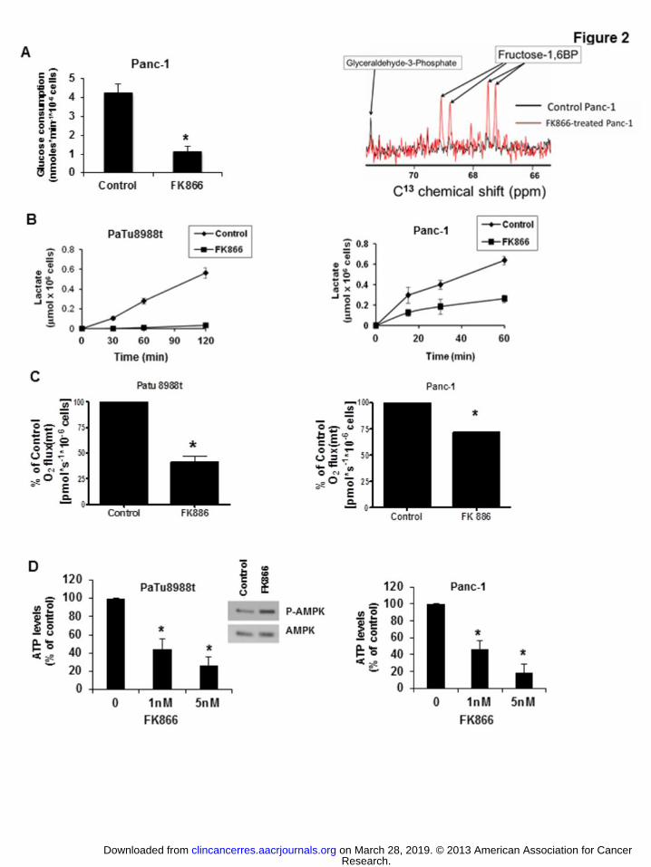
To determine if FK866 can cause a decrease in overall metabolism in pancreatic cancer
cells, we measured several metabolic parameters. First, we observed that glucose
consumption was severely impaired in cells treated with FK866 (Fig. 2A). NAD is
important for several biochemical processes including oxi-reduction reactions of the
glycolytic pathway (27). We observed an accumulation of the glycolytic intermediary
Fructose 1,6 biphosphate in cells treated with FK866 (Figure 2A), indicating that FK866
inhibits glycolytic fluxes and may cause cellular metabolic collapse. To confirm these
effects, we measured lactate production in FK866 treated cells and found thatFK866
inhibited lactate release, in both PaTu8988t cells Panc-1 cells (Figure 2B). We further
Research. on March 28, 2019. © 2013 American Association for Cancerclincancerres.aacrjournals.org Downloaded from
Author manuscripts have been peer reviewed and accepted for publication but have not yet been edited. Author Manuscript Published OnlineFirst on September 11, 2013; DOI: 10.1158/1078-0432.CCR-13-0150
12
investigated the effect of FK866 on mitochondrial oxygen consumption, ATP levels, and
the activation of AMP-activated protein kinase (AMPK). FK866 treatment decreased
mitochondrial maximum respiratory capacity (Fig. 2C and Supplementary Fig. S1), ATP
levels (Fig. 2D), and induced phosphorylation/activation of AMPK in pancreatic cancer
cells (Fig. 2D), confirming that Nampt inhibition promotes energy collapse in pancreatic
cancer cells.
Inhibition of Nampt decreases pancreatic cancer cell growth and survival
We examined the role of Nampt in the growth and survival of the pancreatic cancer cells.
FK866 cause a dose-dependent inhibition of viability in Panc-1 and PaTu8988t cells (Fig.
3A). We observed a significant difference in the sensitivity of cells to Nampt inhibition,
with PaTu8988t cells being nearly 10 times more sensitive than other cells. The least
sensitive was the normal pancreatic cell HPDE (Fig. 3 and Supplementary Fig. S2). Since
the MTT assay determines cell viability via an NAD-dependent mechanism we further
confirmed our findings with two other complementary assays to measure cell growth and
viability (Fig. 3).
Panc-1 and PaTu8988t cells were treated with 50 nM FK866 for 72 hours and viable cells
were counted by the Trypan blue exclusion assay. Treatment with FK866 decreased the
number of viable cells in both cells lines (Fig. 3B).
We determined whether Nampt can regulate anchorage-independent cell growth, an
indicator of malignant behavior. Pancreatic cancer cells were treated with FK866 and
colony formation in soft agar was measured. Colony formation was decreased by
treatment with FK866 in PaTu8988t cells and to a lesser extent in Panc-1 cells (Fig. 3C).
Research. on March 28, 2019. © 2013 American Association for Cancerclincancerres.aacrjournals.org Downloaded from
Author manuscripts have been peer reviewed and accepted for publication but have not yet been edited. Author Manuscript Published OnlineFirst on September 11, 2013; DOI: 10.1158/1078-0432.CCR-13-0150
13
Furthermore, we knocked-down Nampt using two different siRNAs. Nampt siRNAs
inhibited cell growth in both Panc-1 and PaTu8988t cell lines (Fig3D and Supplementary
Fig. S3).
NAMPT inhibition impairs pancreatic tumor growth
We further tested the effect of FK866 in a xenograft animal model of pancreatic tumor.
Nude mice were injected with Panc-1 cells and 10 days after implantation the animals
were treated with daily intraperitoneal injections of vehicle or 15mg/kg of FK866.
FK866 treatment decreased tumor size when compare with vehicle (Fig. 4A). Tumors
from mice treated with FK866 had lower cellular NAD and ATP levels (Fig. 4B), and
increased phosphorylation of AMPK (Fig. 4C). During the treatment, no obvious
treatment-related toxicity was observed. Body weight and food intake of both groups of
animals remained similar (Fig. 4D). We conclude that FK866 inhibits pancreatic tumor
growth in vivo, by inducing tumoral metabolic collapse.
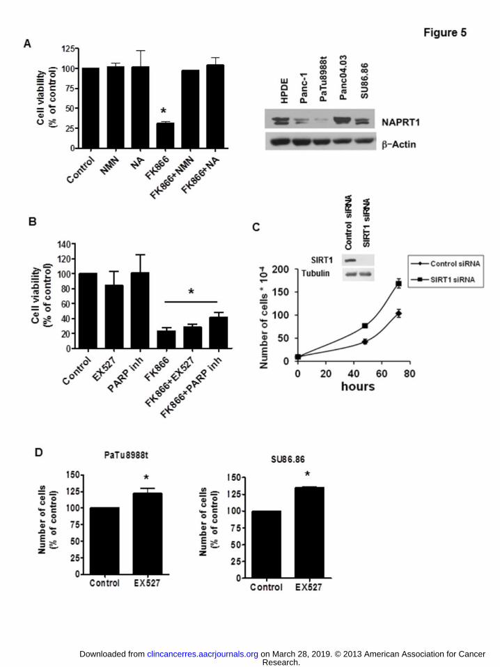
The effect of FK866 in pancreatic cancer cell viability is reversed by NMN and
Nicotinic Acid
Because in cells NAD can be synthesized by both the salvage and the de novo pathway,
we explored whether the two pathways were involved in the regulation of pancreatic
cancer cell growth. In the salvage pathway, Nampt produces nicotinamide
mononucleotide (NMN) from nicotinamide. In the de novo pathway, Nicotinic acid
phosphoribosyltransferase (Naprt1) produces nicotinic acid mononucleotide (NaMN)
from nicotinic acid. The effect of FK866 on cell viability of PaTu8988t was completely
reversed by treatment with the NAD precursors NMN and Nicotinic Acid (Fig. 5A). In
addition, the pattern of expression of Naprt1, the enzyme involved in the de novo
Research. on March 28, 2019. © 2013 American Association for Cancerclincancerres.aacrjournals.org Downloaded from
Author manuscripts have been peer reviewed and accepted for publication but have not yet been edited. Author Manuscript Published OnlineFirst on September 11, 2013; DOI: 10.1158/1078-0432.CCR-13-0150
14
pathway, was similar to the expression of Nampt in the different pancreatic cell lines (Fig
5A). These data suggest that both pathways are involved in the modulation of pancreatic
cancer cell growth. However, inhibition of the salvage pathway is sufficient to promote
energy collapse and cell death in pancreatic cancer cells.
SIRT1 and PARP-1 are not involved on the effect of FK866 in pancreatic cancer
cells.
Since NAD is necessary for both oxi-reduction and non-oxi-reduction reactions, we
further explored whether inhibition of some of the non-oxidative NAD-dependent cellular
reactions were involved on the decrease in cellular viability induced by Nampt inhibition.
Two enzymes, PARP-1 and SIRT1 catalyze crucial non-oxidative NAD-dependent
reactions. Surprisingly, neither PARP1 nor SIRT1 inhibition recapitulated or modified
the effects of Nampt inhibition on cell viability (Fig. 5B). In addition, transient SIRT1
knockdown increased cell proliferation of PaTu8988T cells (Figure 5C). The difference
in cell proliferation is shown by the decrease in doubling time in SIRT1 siRNA treated
cells compared to non-target siRNA transfected cells. The doubling time for non-target
siRNA transfected cells was 21.7 ± 1.2 hours and for SIRT1 siRNA transfected cells was
19.2 ± 0.6 hours (p value = 0.015). Moreover, pharmacological inhibition of SIRT1 using
the synthetic inhibitor EX527 resulted in a small induction of cell growth of both
PaTu8988t and SU86.86 cells, by 22 and 35 % respectively (Figures 5D). Taken together,
these results indicate that neither PARP-1 nor SIRT1-inhibiton can explain the effects of
Nampt inhibition in the pancreatic cancer cells.
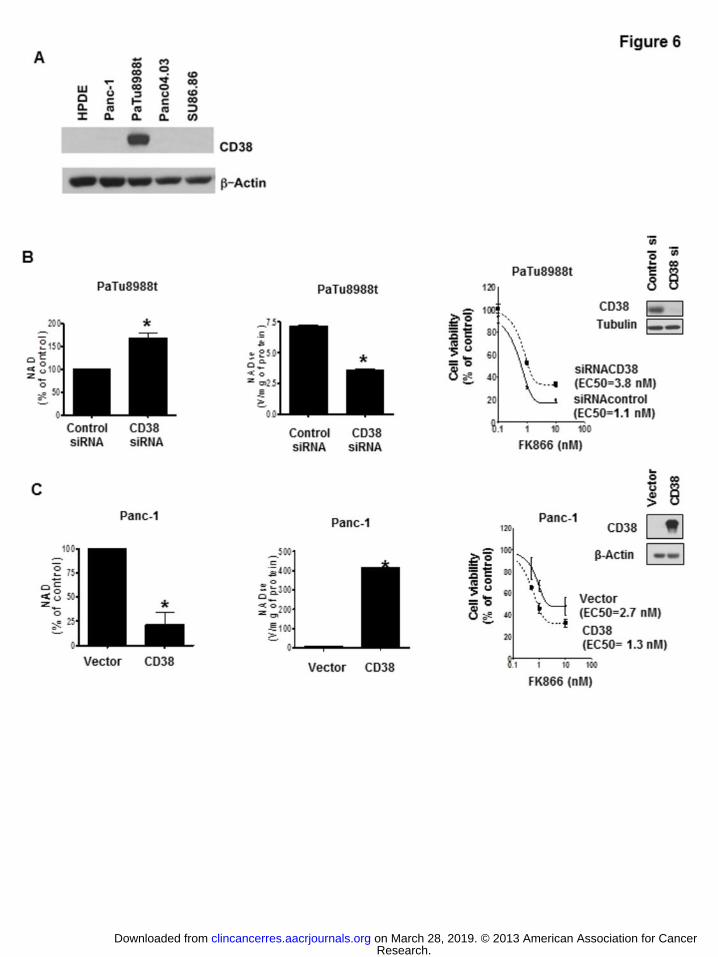
Sensitivity of cultured pancreatic cancer cells in vitro to FK866 is modulated by CD38 expression.
Research. on March 28, 2019. © 2013 American Association for Cancerclincancerres.aacrjournals.org Downloaded from
Author manuscripts have been peer reviewed and accepted for publication but have not yet been edited. Author Manuscript Published OnlineFirst on September 11, 2013; DOI: 10.1158/1078-0432.CCR-13-0150
15
Decreases in cellular NAD can be achieved by inhibition of NAD synthesis or by an
increase in its degradation. The enzyme CD38 is the main NADase in many normal
mammalian tissues (13-18). Interestingly, it has been proposed that CD38 expression
may be lost during the development of prostate cancer (28). However, the functional role
of CD38 in pancreatic cancer cells or any other solid tumor has not been explored. Here
we investigated the role of CD38 in pancreatic NAD metabolism and its role in the effect
of the Nampt inhibitor FK866 on cell viability and cell growth. Most cultured pancreatic
cancer cells tested had low levels of CD38 expression (Fig. 6A), except for PaTu8988t,
that expresses high amounts of CD38. When we knocked-down CD38 in PaTu8988t
cells, there was an increase in NAD levels and a decrease in NADase activity (Fig. 6B).
Although no significant difference on cell viability was observed after cell knockdown of
CD38 (data not shown), the cells that were knocked down for CD38 showed lower
sensitivity to FK866 than control cells (Fig. 6B, and Supplementary Fig. S4).
In contrast, Panc-1 cells have nearly undetectable levels of CD38 (Fig. 6A). Transient
expression of CD38 increased NADase activity, decreased NAD levels, and increased
sensitivity to the effect of FK866 on cell viability (Fig. 6C). Expression of CD38 in
Panc-1 cells did not affect mitochondrial respiration by itself, but it sensitized the cells to
the inhibitory effect of FK866 in mitochondrial function (Supplementary Fig. S5A). Also,
overexpression of CD38 in SU86.86 promoted a decrease in NAD levels (data not
shown) and a significant decrease in cell viability compared to vector-transfected cells
(Supplementary Fig. S5B and C).
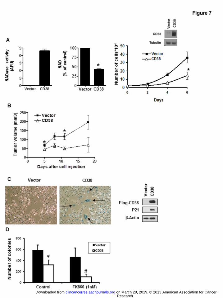
To further explore the role of CD38 in NAD metabolism and cell growth we generated a
Panc-1-CD38 stable cell line. This cell line has increased NADase activity and lower
Research. on March 28, 2019. © 2013 American Association for Cancerclincancerres.aacrjournals.org Downloaded from
Author manuscripts have been peer reviewed and accepted for publication but have not yet been edited. Author Manuscript Published OnlineFirst on September 11, 2013; DOI: 10.1158/1078-0432.CCR-13-0150
16
NAD levels than the control cell line (Fig. 7A). In addition, the Panc-1-CD38 cells
showed cell growth arrest both in vitro and also in an in vivo xenograft mouse model
(Fig. 7A and B). These cells also exhibit senescent markers, as measured by β-
galactosidase staining and increased p21 protein levels (Fig. 7C). Moreover, in colony
formation assays, Panc-1-CD38 cells showed an increase in sensitivity to the FK866
effect in comparison to the control cells (Fig. 7D), confirming that CD38 has an
important effect in regulating the sensitivity to Nampt inhibition.
Heterogeneous expression of NAD metabolizing enzymes in pancreatic tumor
tissues.
We further explored the expression of Nampt and CD38 in pancreatic cancer tumor
samples from patients. There was a significant variability in the expression of CD38 and
Nampt in pancreatic cancer samples, but, in general, the expression of both Nampt and
CD38 were higher in samples from pancreatic tumors than in normal tissue
(Supplementary Fig.6A and B). To compare these results with data from the pancreatic
cancer cell lines, we performed real time PCR in samples from pancreatic cancer cell
lines and the control pancreatic cell HPDE. All cells express Nampt and Naprt1, but the
expression levels differ between cell lines, with some cancer cells expressing similar
levels and others lower levels than the control pancreatic cell (Supplementary Fig.6C and
D). In contrast, the only pancreatic cancer cell that expresses high amounts of CD38 is
PaTu8988t (Supplementary Fig.6E). For all the cell lines, the patterns of expression of
protein and mRNA had a high correlation with all genes tested (Fig. 1A, 5B, 6A).
Since we found that the expression of CD38 is low in pancreatic cancer cells in culture,
the relatively high expression of CD38 in the pancreatic tumor tissues may be mediated
Research. on March 28, 2019. © 2013 American Association for Cancerclincancerres.aacrjournals.org Downloaded from
Author manuscripts have been peer reviewed and accepted for publication but have not yet been edited. Author Manuscript Published OnlineFirst on September 11, 2013; DOI: 10.1158/1078-0432.CCR-13-0150
17
by either inflammatory cell infiltration or by stromal cells. However, it is also possible
that pancreatic cancer cells in vivo may express CD38 differently than in vitro.
Discussion
Pancreatic cancer is one of the top five causes of cancer-related deaths around the globe,
with an extremely poor prognosis (about 5% survival in 5 years) and a median survival
for metastatic disease of about 6 months (29). New and effective therapies are urgently
needed for this disease. The studies described here clearly show a role for the salvage
pathway as potential therapeutic target in pancreatic cancer and also identified NAD
catabolic pathways that modulate the sensitivity to Nampt inhibitors in cancer models.
Recently, it has been proposed that NAD metabolism may be a potential target for the
treatment of cancers (6, 12, 21-24, 30). NAD is a crucial co-factor in redox reactions in
metabolic pathways of nearly every cell (6, 12). NAD can be found in two states,
oxidized (NAD+) or reduced (NADH). Oxidized NAD is necessary for the initial steps of
the glycolytic pathway that is important for cancer cell survival and growth (6, 12).
Intracellular NAD metabolism in cancer cells can be manipulated in several different
ways. For example, the equilibrium between the oxi-reduction states of NAD (the
NAD/NADH ratio) can be shifted one way or the other to either support or inhibit
glycolysis (31). Recently, Lu et al have elegantly shown that the enzyme NADP(H)
oxidase (NOX), plays a key role in supporting increased glycolysis in pancreatic cancer
cells by oxidizing NAD (31).
Yet, another way to modulate NAD-dependent reactions in cancer cells is to modify its
total pool via its anabolism and catabolism. To date, the functional role of the NAD
anabolic pathway in pancreatic cancer cells has not been investigated. In fact, the only
Research. on March 28, 2019. © 2013 American Association for Cancerclincancerres.aacrjournals.org Downloaded from
Author manuscripts have been peer reviewed and accepted for publication but have not yet been edited. Author Manuscript Published OnlineFirst on September 11, 2013; DOI: 10.1158/1078-0432.CCR-13-0150
18
information published about NAD synthetic pathways in pancreatic cancer cells is a study
that showed that colo357 pancreatic cancer cells express Nampt in response to IL-1
treatment (9). In this regard, characterization of the metabolic pathways for synthesis was
one of the goals of our study. We describe for the first time that pancreatic cancer cells
use the salvage pathway and rely on the enzyme Nampt for NAD synthesis, and that
inhibition of the Nampt enzyme causes pancreatic cancer cell death. The mechanism by
which NAD collapse mediated by Nampt inhibition causes cancer cell death has been
proposed to be mediated by inhibition of the glycolytic pathway and /or inhibition of the
NAD-dependent deacetylase SIRT1 (7, 8, 22, 27, 32). In our experiments, inhibition of
SIRT1 was neither necessary nor sufficient to explain the cellular effects of Nampt-
inhibition in pancreatic cancer cells.
Tumor cells have a highly active “aerobic glycolysis” known as the “Warburg effect” (1-
2). Mechanistically, the first reaction in the glycolytic pathway that is dependent on
NAD(H) is the conversion of glyceraldehyde 3 phosphate to 1.3 biphosphoglycerate
catalyzed by GAPDH (27). However, the reaction preceding this step, the interconversion
of glyceraldehyde 3 phosphate and Fructose-1,6 BP is reversible, and we expected that
inhibition of the GAPDH reaction could lead to accumulation of one of these metabolites.
Treatment with FK866 leads to decrease in lactate production, metabolic fluxes and
accumulation of Fructose-1,6 BP in pancreatic cancer cells. Our data supports a model
where Nampt inhibition promotes cell death via a decrease in NAD that leads to
inhibition of glycolytic metabolism, ATP depletion, and energy collapse, and not via
SIRT1 dependent mechanisms.
Research. on March 28, 2019. © 2013 American Association for Cancerclincancerres.aacrjournals.org Downloaded from
Author manuscripts have been peer reviewed and accepted for publication but have not yet been edited. Author Manuscript Published OnlineFirst on September 11, 2013; DOI: 10.1158/1078-0432.CCR-13-0150

19
Another goal of our study was to characterize the NAD catabolizing pathways in
pancreatic cancer cells. To our knowledge the mechanisms of NAD degradation in tumor
cells have not been well described. In particular, given the emerging interest in
developing NAD targeted therapies for human cancers, it is important to determine the
mechanisms that modulate NAD-degradation in the setting of inhibition of NAD
synthetic pathways.
We observed in cultured pancreatic cancer cells that expression of the NADase CD38
played a key role on the sensitivity of cells to the Nampt inhibitor FK866. In addition, we
observed that CD38 and Nampt expression are quite variable between tumors from
pancreatic cancer patients (Supplemental Figure 6), and we propose that the relative
expression of Nampt and CD38 may play a key role on the response to salvage pathway-
targeted therapy and may serve as potential biomarkers for the cellular response to Nampt
inhibition.
In conclusion, our data provides the first evidence of the role of NAD anabolism and
catabolism in pancreatic cancer cells. Specifically, we demonstrated that pancreatic
cancer cells rely on the salvage pathway and that inhibition of the enzyme Nampt causes
metabolic collapse and cell death both in vitro and in vivo. Furthermore, we provided one
of the first analyses of NAD catabolism in cancer cells. We believe that further
systematic characterization of the anabolism and catabolism of NAD in cancer cells may
have potential implications for the development of novel and more rational NAD-
targeted therapy for cancer.
Acknowledge: The project described was supported by Award Number P50CA102701
(Mayo Clinic SPORE in Pancreatic Cancer) from the National Cancer Institute. The
Research. on March 28, 2019. © 2013 American Association for Cancerclincancerres.aacrjournals.org Downloaded from
Author manuscripts have been peer reviewed and accepted for publication but have not yet been edited. Author Manuscript Published OnlineFirst on September 11, 2013; DOI: 10.1158/1078-0432.CCR-13-0150
20
content is solely the responsibility of the authors and does not necessarily represent the
official views of the National Cancer Institute or the National Institutes of Health.
Research. on March 28, 2019. © 2013 American Association for Cancerclincancerres.aacrjournals.org Downloaded from
Author manuscripts have been peer reviewed and accepted for publication but have not yet been edited. Author Manuscript Published OnlineFirst on September 11, 2013; DOI: 10.1158/1078-0432.CCR-13-0150
21
References
1. Madhok BM, Yeluri S, Perry SL, Hughes TA, Jayne DG. Targeting Glucose
Metabolism: An Emerging Concept for Anticancer Therapy. Am J Clin Oncol. 2011;
34:628-35.
2. Warburg O. On the origin of cancer cells. Science. 1956; 123:309-14.
3. Dang CV, Le A, Gao P. MYC-induced cancer cell energy metabolism and
therapeutic opportunities. Clin Cancer Res. 2009; 15:6479-83
4. Fritz V, Fajas L. Metabolism and proliferation share common regulatory
pathways in cancer cells. Oncogene. 2010; 29:4369-77.
5. Le A, Cooper CR, Gouw AM, Dinavahi R, Maitra A, Deck LM, et. al. Inhibition
of lactate dehydrogenase A induces oxidative stress and inhibits tumor progression. Proc
Natl Acad Sci U S A. 2010; 107:2037-42.
6. Nahimana A, Attinger A, Aubry D, Greaney P, Ireson C, Thougaard AV,et al.
The NAD biosynthesis inhibitor APO866 has potent antitumor activity against
hematologic malignancies. Blood. 2009; 113:3276-86
7. Garten A, Petzold S, Körner A, Imai S, Kiess W. Nampt: linking NAD biology,
metabolism and cancer. Trends Endocrinol Metab. 2009; 20:130-38.
8. Wang B, Hasan MK, Alvarado E, Yuan H, Wu H, Chen WY. NAMPT
overexpression in prostate cancer and its contribution to tumor cell survival and stress
response. Oncogene. 2011;30:907-21
9. Bauer L, Venz S, Junker H, Brandt R, Radons J. Nicotinamide
phosphoribosyltransferase and prostaglandin H2 synthase 2 are up-regulated in human
Research. on March 28, 2019. © 2013 American Association for Cancerclincancerres.aacrjournals.org Downloaded from
Author manuscripts have been peer reviewed and accepted for publication but have not yet been edited. Author Manuscript Published OnlineFirst on September 11, 2013; DOI: 10.1158/1078-0432.CCR-13-0150
22
pancreatic adenocarcinoma cells after stimulation with interleukin-1. Int J Oncol. 2009;
35:97-07
10. Okumura S, Sasaki T, Minami Y, Ohsaki Y.J. Nicotinamide
Phosphoribosyltransferase: A Potent Therapeutic Target in Non-small Cell Lung Cancer
with Epidermal Growth Factor Receptor-Gene Mutation. Thorac Oncol. 2012; 7:49-56.
11. Shackelford RE, Bui MM, Coppola D, Hakam A. Over-expression of
nicotinamide phosphoribosyltransferase in ovarian cancers. Int J Clin Exp Pathol. 2010;
3:522-527.
12. Khan JV, Forouhar F, Tao X and Tong L. Nicotinamide adenine dinucleotide
metabolism as an attractive target for drug discovery. Expert Opin. Ther. Targets. 2007;
11:695-05
13. Chini EN. CD38 as a regulator of cellular NAD: a novel potential
pharmacological target for metabolic conditions. Curr Pharm Des. 2009; 15:57-63.
14. Barbosa MT, Soares SM, Novak CM, Sinclair D, Levine JA, Aksoy P, et al. The
enzyme CD38 (a NAD glycohydrolase, EC 3.2.2.5) is necessary for the development of
diet-induced obesity. FASEB J. 2007; 21:3629-39.
15. Malavasi F, Deaglio S, Zaccarello G, Horenstein AL, Chillemi A, Audrito V, et
al. The hidden life of NADC-consuming ectoenzymes in the endocrine system. Journal of
Molecular Endocrinology 2010; 45:183–91
16. Aksoy P, White TA, Thompson M, Chini EN. Regulation of intracellular levels of
NAD: a novel role for CD38. Biochem Biophys Res Commun. 2006; 345:1386-92.
PMID: 16730329
Research. on March 28, 2019. © 2013 American Association for Cancerclincancerres.aacrjournals.org Downloaded from
Author manuscripts have been peer reviewed and accepted for publication but have not yet been edited. Author Manuscript Published OnlineFirst on September 11, 2013; DOI: 10.1158/1078-0432.CCR-13-0150

23
17. Hartman WR, Pelleymounter LL, Moon I, Kalari K, Liu M, Wu TY, et al. CD38
expression, function, and gene resequencing in a human lymphoblastoid cell line-based
model system. Leuk Lymphoma. 2010; 51:1315-25.
18. Sahar S, Nin V, Barbosa MT, Chini EN, Sassone-Corsi P. Altered behavioral and
metabolic circadian rhythms in mice with disrupted NAD+ oscillation. Aging (Albany
NY). 2011;3:794-02.
19. Yang H, Yang T, Baur JA, Perez E, Matsui T, Carmona JJ, et al. Nutrient-sensitive
mitochondrial NAD+ levels dictate cell survival. Cell. 2007; 130:1095-07.
20. van der Veer E, Ho C, O'Neil C, Barbosa N, Scott R, Cregan SP, et al. Extension of
human cell lifespan by nicotinamide phosphoribosyltransferase. J Biol Chem. 2007;
282:10841-845
21. Olesen UH, Thougoord AV, Josen PB, and Sehested M. A Preclinical Study on
the Rescue of Normal Tissue by Nicotinic Acid in High-Dose Treatment with APO866, a
Specific Nicotinamide Phosphoribosyltransferase Inhibitor Mol Cancer Ther. 2010;
9:1609-17.
22. Boniface J.J. MPC-9528, a cancer metabolism inhibitor, demonstrates greater
therapeutic index in a Naprt1 deficient cancer xenograft model with co-administration of
nicotinic acid. Cancer and Metabolism: Pathways to the Future Symposium, September
19-21, 2010, Edinburgh, Scotland.
23. Holen K, Saltz LB, Hollywood E, Burk K, and Hanauske AR. The
pharmacokinetics, toxicities, and biological effects of FK866, a nicotinamide adenine
dinucleotide biosynthesis inhibitor. Invest New Drugs. 2008; 26:45-51
Research. on March 28, 2019. © 2013 American Association for Cancerclincancerres.aacrjournals.org Downloaded from
Author manuscripts have been peer reviewed and accepted for publication but have not yet been edited. Author Manuscript Published OnlineFirst on September 11, 2013; DOI: 10.1158/1078-0432.CCR-13-0150
24
24. von Heideman A, Berglund A, Larsson R, Nygren P. Safety and efficacy of NAD
depleting cancer drugs: results of a phase I clinical trial of CHS 828 and overview of
published data. Cancer Chemother Pharmacol. 2010; 65:1165-72
25. Hamilton SD, Pardue HL. Quantitation of lactate by a kinetic method with an
extended range of linearity and low dependence on experimental variables. Clin Chem.
1984; 30:226-29.
26. Hütter E, Renner K, Pfister G, Stöckl P, Jansen-Dürr P, Gnaiger E. Senescence-
associated changes in respiration and oxidative phosphorylation in primary human
fibroblasts. Biochem. J.2004; 380: 919-28.
27 Tan B, Young DA, Lu ZH, Wang T, Meier TI, Shepard RL, et al. Pharmacological
inhibition of nicotinamide phosphoribosyltransferase (NAMPT), an enzyme essential for
NAD+ biosynthesis, in human cancer cells: metabolic basis and potential clinical
implications. J Biol Chem. 2013; 288: 3500-11
28. Kramer G, Steiner G, Födinger D, Fiebiger E, Rappersberger C, Binder S, et al. High
expression of a CD38-like molecule in normal prostatic epithelium and its differential
loss in benign and malignant disease. J Urol. 1995; 154:1636-41.
29. Siegel R, Naishadham D, Jemal A. Cancer statistics, 2012. CA Cancer J Clin. 2012;
62:10-29.
30. Chiarugi A, Dölle C, Felici R, and Ziegler M. The NAD metabolome — a key
determinant of cancer cell biology. Nature Reviews Cancer. 2012; 12:741-52.
31. Lu W, Hu Y, Chen G, Chen Z, Zhang H, Wang F, et al. Novel Role of NOX in
Supporting Aerobic Glycolysis in Cancer Cells with Mitochondrial Dysfunction and as a
Potential Target for Cancer Therapy. PLoS. Biol. 2012; 10: e1001326.
Research. on March 28, 2019. © 2013 American Association for Cancerclincancerres.aacrjournals.org Downloaded from
Author manuscripts have been peer reviewed and accepted for publication but have not yet been edited. Author Manuscript Published OnlineFirst on September 11, 2013; DOI: 10.1158/1078-0432.CCR-13-0150
25
32. Bowlby SC, Thomas MJ, D'Agostino RB Jr, Kridel SJ. Nicotinamide phosphoribosyl
transferase (Nampt) is required for de novo lipogenesis in tumor cells. PLoS One. 2012;
7:e40195.
Research. on March 28, 2019. © 2013 American Association for Cancerclincancerres.aacrjournals.org Downloaded from
Author manuscripts have been peer reviewed and accepted for publication but have not yet been edited. Author Manuscript Published OnlineFirst on September 11, 2013; DOI: 10.1158/1078-0432.CCR-13-0150
26
Figure legends
Figure 1. Nampt inhibition by FK866 or Nampt knockdown by siRNA reduces NAD
levels in pancreatic cancer cells. A, Different pancreatic cancer cell lines were
imunoblotted for Nampt and Actin. B, NAD levels were measured in pancreatic cancer
cells that were maintained in growth medium containing 1% FBS for 48 hours and then
treated with FK866 or vehicle for 24 hours. C and D, Cells were transfected with Nampt
siRNA or control siRNA analyzed by immunoblotting 72 hours later (insets). 24 hours
after transfection cells were re-plated in growth medium containing 1% FBS and NAD
levels were determined 48 hours later. * indicates p<0.05.
Figure 2. FK866 produces metabolic collapse in pancreatic cancer cells. A, Panc-1
cells were kept in 1% FBS for 48 hours and then treated with FK866 (10 nM) or vehicle.
48 hours later media was changed to glucose-free DMEM supplemented with 5 mM D-
[C13] glucose. Glucose consumption and glycolytic intermediates were determined using
NMR spectroscopy, and glycolytic intermediates determined. B, cells were kept in 1%
FBS for 48 hours and then treated with FK866 (10 nM) or vehicle for 48 hours. The
medium was replaced by RPMI phenol red and serum-free and aliquots of cultured
medium were collected for lactate release evaluation. C, O2 consumption rates were
measured by high resolution respirometry. After 48 hours treatment with 50 nM of
FK866 or vehicle, the cells resuspended in serum-free DMEM, and ETS-stimulated
respiration (maximum respiration) was measured. Results represent the mean ± SD of
three independent experiments. D, Cells were kept in 1% FBS for 48 hours then treated
Research. on March 28, 2019. © 2013 American Association for Cancerclincancerres.aacrjournals.org Downloaded from
Author manuscripts have been peer reviewed and accepted for publication but have not yet been edited. Author Manuscript Published OnlineFirst on September 11, 2013; DOI: 10.1158/1078-0432.CCR-13-0150

27
with FK866 or vehicle for 48 hours before ATP measurements. Values are means ± SD
of three independent experiments. Cell lysates of PaTu8988t cells treated with 10nM
FK866 for 24 hours were immunoblotted with anti-p-AMPK and AMPK antibodies. *
indicates p<0.05
Figure 3. Nampt inhibition decreased pancreatic cancer cell survival and growth. A,
Cells were treated with vehicle or FK866 and submitted to MTT analysis 72 hours later.
Values are means ± SD of three independent experiments. B, Cells were plated and
treated with vehicle or FK866 24 hours later. Cells were counted by trypan blue dye
exclusion assay 72 hours after treatment. C, Cells were grown in triplicates in soft agar
containing different concentrations of FK866 for 7 days. Results represent the mean ± SD
of three independent experiments. D, Cells were transfected with Nampt siRNA or a non-
target siRNA (Control). 24 hours after transfection the cells were re-plated in growth
medium containing 1% FBS and viable cells were counted by Trypan blue exclusion
assay 48 hours later. Efficiency of knockdown was determined by immunoblotting. *
indicates p<0.05
Figure 4. NAMPT inhibition prevents pancreatic tumor growth in vivo. Panc-1 cells
were used in an in vivo xenograft mouse model. Starting at 10 days after implantation the
animals were treated with daily intraperitoneal injections of vehicle or FK866. Tumors
were measured for an additional 40 days. A, Graph shows the average size of 16 vehicle
and 16 FK866-treated tumors measured during the experiment. Picture shows size of
representative tumors. B, presents NAD and ATP levels from 6 vehicle and 6 FK866-
Research. on March 28, 2019. © 2013 American Association for Cancerclincancerres.aacrjournals.org Downloaded from
Author manuscripts have been peer reviewed and accepted for publication but have not yet been edited. Author Manuscript Published OnlineFirst on September 11, 2013; DOI: 10.1158/1078-0432.CCR-13-0150
28
treated tumors. C, immunostaining for p-AMPK (Thr172) shows activation of AMPK (p-
AMPK) in tumors collected from vehicle and FK866-treated animals. D, mice were
weighted during the treatment period.* indicates p<0.05
Figure 5. FK866 effect in pancreatic cancer cell viability is blocked by NMN and
Nicotinic Acid, but not by SIRT1 or PARP-1 inhibitors. A, PaTu8988t cells were
treated with vehicle (control), 25 μm NMN or 25 μM Nicotinic Acid (NA) for 6 hours
before addition of 2 nM FK866. Cells were submitted to MTT analysis 72 hours later.
Values are mean ± SD of three independent experiments. Different pancreatic cell lines
were imunoblotted for NAPRT1 and actin. B, PaTu8988t cells were treated with vehicle
(control), 10 μM of the SIRT1 inhibitor (EX527) or 10 μM of the PARP inhibitor (4-
Amino-1,8-naphthalimide) for 6 hours before addition of 2 nM FK866. Cells were
submitted to MTT analysis 72 hours after treatment. Values are mean ± SD of three
independent experiments. * indicates conditions that are significantly different than
control, but are not significantly different from each other. C, Cells were transfected with
siRNA specific for SIRT1 or a non-target siRNA. 24 hour after transfection the cells
were re-plated and viable cells were counted at different time points. Values are mean ±
SD of triplicates. Plot is representative of three independent experiments. D, PaTu8988t
and SU86.86 cells were treated with the SIRT1 inhibitor EX527 (10 µM) or vehicle.
Cells were counted 72 hours after treatment by trypan blue dye exclusion assay. Values
are mean ± SD of three independent experiments. * indicates p < 0.05
Figure 6. Sensitivity of pancreatic cancer cells to FK866 depends on CD38 levels. A,
Different pancreatic cell lines were imunoblotted for CD38 and actin. B, PaTu8988t cells
Research. on March 28, 2019. © 2013 American Association for Cancerclincancerres.aacrjournals.org Downloaded from
Author manuscripts have been peer reviewed and accepted for publication but have not yet been edited. Author Manuscript Published OnlineFirst on September 11, 2013; DOI: 10.1158/1078-0432.CCR-13-0150
29
were transfected with CD38 siRNA or a non-targeting siRNA. 72 hours after transfection,
we measured NAD levels and NADase activity. For MTT analysis, cells were re-plated
24 hours after transfection, and treated with vehicle control or FK866 24 hours later.
MTT analysis was performed 48 hours after treatment. C, Panc-1 cells were transfected
with a Flag-CD38 or empty vector. NAD levels and NADase activity were measured 72
hours later. For MTT analysis cells were re-plated 24 hours after transfection, and
treated with vehicle control or FK866 24 hours later. MTT analysis was performed 48
hours after treatment. Efficiency of transfections was determined by immunoblotting 72
hours after transfection (insets). Values are mean ± SD of three independent experiments.
* indicates p<0.05
Figure 7. CD38 stable overexpression inhibits anchorage-dependent and anchorage-
independent cell growth of Panc-1 cells and increases the inhibitory effect of FK866.
A, intracellular NAD levels, NADase activity, and cell growth were determined in Panc-1
control stable clone and a Panc-1-CD38 clone. The expression of CD38 was evaluated by
immunoblotting. B, Panc-1 cells and Panc-1-CD38 cells were used in an in vivo
xenograft mouse model and the size of the tumors was measured during a three week
period (n=5). * indicates p<0.05. C, Panc-1 control cells and Panc-1-CD38 cells were
stained for β-Galactosidase. The expression of CD38 and P21 was evaluated in these cells
by immunoblotting for Flag and P21. D, Panc-1 control cells and Panc-1-CD38 cells
were grown in soft agar containing 1 nM FK866 for 7-10 days in triplicates. Results
represent the mean ± SD of three independent experiments. Quantitative analysis of
colony formation shows significant reduction (* p< 0.05, n=3) in colony number upon
Research. on March 28, 2019. © 2013 American Association for Cancerclincancerres.aacrjournals.org Downloaded from
Author manuscripts have been peer reviewed and accepted for publication but have not yet been edited. Author Manuscript Published OnlineFirst on September 11, 2013; DOI: 10.1158/1078-0432.CCR-13-0150
30
CD38 overexpression in comparison to empty vector; and (# p< 0.05, n=3) upon FK866
treatment in Panc-1-CD38 but not in Panc-1 control cells.
Research. on March 28, 2019. © 2013 American Association for Cancerclincancerres.aacrjournals.org Downloaded from
Author manuscripts have been peer reviewed and accepted for publication but have not yet been edited. Author Manuscript Published OnlineFirst on September 11, 2013; DOI: 10.1158/1078-0432.CCR-13-0150
Published OnlineFirst September 11, 2013.Clin Cancer Res Claudia CS Chini, Anatilde Gonzalez Guerrico, Veronica Nin, et al. potential novel therapy for pancreatic tumorsTargeting of NAD metabolism in pancreatic cancer cells:
Updated version
10.1158/1078-0432.CCR-13-0150doi:
Access the most recent version of this article at:
Material
Supplementary
http://clincancerres.aacrjournals.org/content/suppl/2014/01/06/1078-0432.CCR-13-0150.DC2
http://clincancerres.aacrjournals.org/content/suppl/2013/09/10/1078-0432.CCR-13-0150.DC1Access the most recent supplemental material at:
Manuscript
Authoredited. Author manuscripts have been peer reviewed and accepted for publication but have not yet been
E-mail alerts related to this article or journal.Sign up to receive free email-alerts
Subscriptions
Reprints and
To order reprints of this article or to subscribe to the journal, contact the AACR Publications
Permissions
Rightslink site. Click on "Request Permissions" which will take you to the Copyright Clearance Center's (CCC)
.http://clincancerres.aacrjournals.org/content/early/2013/09/10/1078-0432.CCR-13-0150To request permission to re-use all or part of this article, use this link
Research. on March 28, 2019. © 2013 American Association for Cancerclincancerres.aacrjournals.org Downloaded from
Author manuscripts have been peer reviewed and accepted for publication but have not yet been edited. Author Manuscript Published OnlineFirst on September 11, 2013; DOI: 10.1158/1078-0432.CCR-13-0150