Targeting Innate Immunity in Cancer Therapy
26
Review Targeting Innate Immunity in Cancer Therapy Srikrishnan Rameshbabu † , Brian W. Labadie † , Anna Argulian and Akash Patnaik * Citation: Rameshbabu, S.; Labadie, B.W.; Argulian, A.; Patnaik, A. Targeting Innate Immunity in Cancer Therapy. Vaccines 2021, 9, 138. https://doi.org/10.3390/ vaccines9020138 Academic Editor: Sumit Agarwal Received: 19 December 2020 Accepted: 2 February 2021 Published: 9 February 2021 Publisher’s Note: MDPI stays neutral with regard to jurisdictional claims in published maps and institutional affil- iations. Copyright: © 2021 by the authors. Licensee MDPI, Basel, Switzerland. This article is an open access article distributed under the terms and conditions of the Creative Commons Attribution (CC BY) license (https:// creativecommons.org/licenses/by/ 4.0/). Section of Hematology/Oncology, Department of Medicine, University of Chicago, Chicago, IL 60637, USA; [email protected] (S.R.); [email protected] (B.W.L.); [email protected] (A.A.) * Correspondence: [email protected] † These authors contributed equally to this work. Abstract: The majority of current cancer immunotherapy strategies target and potentiate antitumor adaptive immune responses. Unfortunately, the efficacy of these treatments has been limited to a fraction of patients within a subset of tumor types, with an aggregate response rate of approximately 20% to date across all malignancies. The success of therapeutic inhibition of programmed death protein 1 (PD-1), protein death ligand 1 (PD-L1) and cytotoxic T-lymphocyte-associated antigen 4 (CTLA-4) with immune checkpoint inhibitors (ICI) has been limited to “hot” tumors characterized by preexisting T cell infiltration, whereas “cold” tumors, which lack T cell infiltration, have not achieved durable benefit. There are several mechanisms by which “cold” tumors fail to generate spontaneous immune infiltration, which converge upon the generation of an immunosuppressive tumor microenvironment (TME). The role of the innate immune system in tumor immunosurveillance and generation of antitumor immune responses has been long recognized. In recent years, novel strategies to target innate immunity in cancer therapy have emerged, including therapeutic stimulation of pattern recognition receptors (PRRs), such as Toll-like receptors (TLRs); the DNA sensing cGAS/STING pathway; nucleotide-binding oligomerization domain-like receptors (NLRs), such as NLRP3; and the retinoic acid-inducible gene-I (RIG-I)-like receptors (RLRs). In addition, therapeutic modulation of key innate immune cell types, such as macrophages and natural killer cells, has been investigated. Herein, we review therapeutic approaches to activate innate immunity within the TME to enhance antitumor immune responses, with the goal of disease eradication in “cold” tumors. In addition, we discuss rational immune-oncology combination strategies that activate both innate and adaptive immunity, with the potential to enhance the efficacy of current immunotherapeutic approaches. Keywords: cancer immunotherapy; STING; NLRP3; tumor-associated macrophages; RIG-I; TLRs; CD40; NK cells; oncolytic viruses; pattern recognition receptors; innate immunity; cancer 1. Introduction There is growing evidence that successful immune-mediated elimination of can- cer requires coordination between the innate and adaptive arms of the immune system (Figure 1). Innate immune cells, such as dendritic cells (DCs), detect early cancers by a vari- ety of mechanisms, including presentation of tumor-associated neoantigens or through sens- ing tumor-derived pathogen or damage-associated molecular patterns (PAMP/DAMPs) by pattern recognition receptors (PRRs) [1–4]. These mechanisms trigger proinflammatory programs and the release of proinflammatory cytokines, chemokines and type I interferons with accompanying DC maturation and trafficking to lymph nodes, where they engage the adaptive immune system and prime and activate antigen-specific T cells. T cells traffic and infiltrate the tumor bed through chemokine and cytokine gradients and mediate tumor elimination following interaction with their cognate antigen [5]. In addition, innate im- mune cells, such as macrophages and natural killer cells, contribute to tumor elimination through direct tumor cell killing by phagocytosis and cytotoxic mechanisms, respectively. Vaccines 2021, 9, 138. https://doi.org/10.3390/vaccines9020138 https://www.mdpi.com/journal/vaccines
Transcript of Targeting Innate Immunity in Cancer Therapy
Review
Targeting Innate Immunity in Cancer Therapy
Srikrishnan Rameshbabu †, Brian W. Labadie †, Anna Argulian and Akash Patnaik *
�����������������
Citation: Rameshbabu, S.; Labadie,
B.W.; Argulian, A.; Patnaik, A.
Targeting Innate Immunity in Cancer
Therapy. Vaccines 2021, 9, 138.
https://doi.org/10.3390/
vaccines9020138
Academic Editor: Sumit Agarwal
Received: 19 December 2020
Accepted: 2 February 2021
Published: 9 February 2021
Publisher’s Note: MDPI stays neutral
with regard to jurisdictional claims in
published maps and institutional affil-
iations.
Copyright: © 2021 by the authors.
Licensee MDPI, Basel, Switzerland.
This article is an open access article
distributed under the terms and
conditions of the Creative Commons
Attribution (CC BY) license (https://
creativecommons.org/licenses/by/
4.0/).
Section of Hematology/Oncology, Department of Medicine, University of Chicago, Chicago, IL 60637, USA;[email protected] (S.R.); [email protected] (B.W.L.);[email protected] (A.A.)* Correspondence: [email protected]† These authors contributed equally to this work.
Abstract: The majority of current cancer immunotherapy strategies target and potentiate antitumoradaptive immune responses. Unfortunately, the efficacy of these treatments has been limited to afraction of patients within a subset of tumor types, with an aggregate response rate of approximately20% to date across all malignancies. The success of therapeutic inhibition of programmed death protein1 (PD-1), protein death ligand 1 (PD-L1) and cytotoxic T-lymphocyte-associated antigen 4 (CTLA-4)with immune checkpoint inhibitors (ICI) has been limited to “hot” tumors characterized by preexistingT cell infiltration, whereas “cold” tumors, which lack T cell infiltration, have not achieved durablebenefit. There are several mechanisms by which “cold” tumors fail to generate spontaneous immuneinfiltration, which converge upon the generation of an immunosuppressive tumor microenvironment(TME). The role of the innate immune system in tumor immunosurveillance and generation of antitumorimmune responses has been long recognized. In recent years, novel strategies to target innate immunityin cancer therapy have emerged, including therapeutic stimulation of pattern recognition receptors(PRRs), such as Toll-like receptors (TLRs); the DNA sensing cGAS/STING pathway; nucleotide-bindingoligomerization domain-like receptors (NLRs), such as NLRP3; and the retinoic acid-inducible gene-I(RIG-I)-like receptors (RLRs). In addition, therapeutic modulation of key innate immune cell types,such as macrophages and natural killer cells, has been investigated. Herein, we review therapeuticapproaches to activate innate immunity within the TME to enhance antitumor immune responses, withthe goal of disease eradication in “cold” tumors. In addition, we discuss rational immune-oncologycombination strategies that activate both innate and adaptive immunity, with the potential to enhancethe efficacy of current immunotherapeutic approaches.
Keywords: cancer immunotherapy; STING; NLRP3; tumor-associated macrophages; RIG-I; TLRs;CD40; NK cells; oncolytic viruses; pattern recognition receptors; innate immunity; cancer
1. Introduction
There is growing evidence that successful immune-mediated elimination of can-cer requires coordination between the innate and adaptive arms of the immune system(Figure 1). Innate immune cells, such as dendritic cells (DCs), detect early cancers by a vari-ety of mechanisms, including presentation of tumor-associated neoantigens or through sens-ing tumor-derived pathogen or damage-associated molecular patterns (PAMP/DAMPs)by pattern recognition receptors (PRRs) [1–4]. These mechanisms trigger proinflammatoryprograms and the release of proinflammatory cytokines, chemokines and type I interferonswith accompanying DC maturation and trafficking to lymph nodes, where they engage theadaptive immune system and prime and activate antigen-specific T cells. T cells traffic andinfiltrate the tumor bed through chemokine and cytokine gradients and mediate tumorelimination following interaction with their cognate antigen [5]. In addition, innate im-mune cells, such as macrophages and natural killer cells, contribute to tumor eliminationthrough direct tumor cell killing by phagocytosis and cytotoxic mechanisms, respectively.
Vaccines 2021, 9, 138. https://doi.org/10.3390/vaccines9020138 https://www.mdpi.com/journal/vaccines

Vaccines 2021, 9, 138 2 of 26
Vaccines 2021, 9, x FOR PEER REVIEW 2 of 26
tion, innate immune cells, such as macrophages and natural killer cells, contribute to tu‐
mor elimination through direct tumor cell killing by phagocytosis and cytotoxic mecha‐
nisms, respectively.
Figure 1. Cancer Immunity Cycle. Innate immune cells facilitate the immune system’s response against a recognized path‐
ogen. This process is initiated by detection of pathogen‐associated molecular patterns (PAMPs) and damage‐associated
molecular patterns (DAMPs) and other unique tumor antigens by innate immune cells which result in antigen presentation
and activation of antigen‐specific T cells in tumor draining lymph nodes. These T cells traffic to the tumor and mediate
tumor elimination. Abbreviations: PAMPs, pathogen‐associated molecular patterns; DAMPs, damage‐associated molecu‐
lar patterns; PRRs, pattern‐recognizing‐receptors.
In recent years, evasion of antitumor immune response has been recognized as a hall‐
mark of cancer and can be mediated by multiple mechanisms [6]. In the context of solid
tumors, the absence of T cell infiltration on histopathologic analysis indicates one mecha‐
nism for evasion of antitumor immunity [7]. Gene expression analyses of the TME in solid
tumors has identified genomic signatures which correlate with the presence or absence of
T cell infiltration, referred to as “hot” and “cold” tumors, respectively [8–10]. More com‐
prehensive classification has suggested four categories of TMEs: hot, altered‐excluded,
altered‐immunosuppressed and cold [11]. These immune phenotypes have been observed
Figure 1. Cancer Immunity Cycle. Innate immune cells facilitate the immune system’s response against a recognizedpathogen. This process is initiated by detection of pathogen-associated molecular patterns (PAMPs) and damage-associatedmolecular patterns (DAMPs) and other unique tumor antigens by innate immune cells which result in antigen presentationand activation of antigen-specific T cells in tumor draining lymph nodes. These T cells traffic to the tumor and mediatetumor elimination. Abbreviations: PAMPs, pathogen-associated molecular patterns; DAMPs, damage-associated molecularpatterns; PRRs, pattern-recognizing-receptors.
In recent years, evasion of antitumor immune response has been recognized as ahallmark of cancer and can be mediated by multiple mechanisms [6]. In the context ofsolid tumors, the absence of T cell infiltration on histopathologic analysis indicates onemechanism for evasion of antitumor immunity [7]. Gene expression analyses of the TMEin solid tumors has identified genomic signatures which correlate with the presence orabsence of T cell infiltration, referred to as “hot” and “cold” tumors, respectively [8–10].More comprehensive classification has suggested four categories of TMEs: hot, altered-excluded, altered-immunosuppressed and cold [11]. These immune phenotypes have beenobserved to exist in a distribution, with certain cancer types having a higher proportionof “hot” immune phenotype, such as lung adenocarcinoma and clear cell renal cell carci-noma [12]. In addition, these immune phenotypes deploy distinct mechanisms to avoidimmune-mediated elimination. For example, in response to T cell infiltration, “hot” tumors

Vaccines 2021, 9, 138 3 of 26
upregulate immune checkpoints, such as PD-1/PD-L1, within the TME, which directlysuppress T cell effector mechanisms [13]. In contrast, “cold” tumors fail to generate sponta-neous immune infiltration altogether through either “lack of antigenicity”, which resultsfrom defects in antigen processing or presentation, or “lack of immunogenicity” due toabsence of tumor antigens capable of stimulating the immune system [14]. Of increasinglyrecognized significance, “cold” tumors prohibit T cell infiltration through orchestrationof an immunosuppressive TME characterized by cell types, such as tumor-associatedmacrophages (TAMs) and myeloid-derived suppressor cells (MDSCs) [14–16].
“Hot” and “cold” tumor immune phenotypes carry prognostic significance, as solidtumors with T cell infiltration have been observed to have improved outcomes in multipletreatment settings [7,9,17–19]. To date, the success of therapeutic inhibition of PD-1/PD-L1and CTLA-4 with immune checkpoint inhibitors (ICI) has been limited to “hot” tumors,while “cold” tumors have typically failed to benefit from ICI [9,19–22]. A large percentage ofcommon malignancies, including prostate, breast and pancreatic cancers, are characterizedas having “cold” TMEs and have historically not benefited from ICI. As such, there remainsa critical unmet need to develop therapeutic approaches which drive innate immuneactivation and promote T cell infiltration in immunologically “cold” tumors. Herein,we review the roles of key innate immune cell types and discuss emerging therapeuticstrategies to target these cell types to enhance antitumor immune responses, with the goalof disease eradication in “cold” tumors.
2. Key Cellular Components of Innate Immunity
Innate immunity comprises a diverse cadre of cell types which function as the body’s“first defense” against microbes. Each distinct cell type has a unique role; however, thereexists overlap in function and cellular machinery, including expression of PRRs and proin-flammatory response to detection of PAMPs/DAMPs. The expression of PRR and otherinnate immune pathways by innate immune cell type is summarized in (Table 1).
Table 1. Innate immune cell expression of PRRs and PAMP/DAMP-sensing pathways.
DC TAM Mo-MDSC PMN-MDSC Neutrophils NK Cells Basophils Eosinophils Mast CellsTLR7/8
TLR9RIG-I
cGAS-STINGNLRP3CSF-1RCD40
NKG2DGreen = expression of innate immune pathway or receptor is observed in human cell types based on literature cited in main body of text.This is a conceptual and binary illustration which does not reflect contextual and dynamic nuances in expression.
2.1. Dendritic Cells (DCs)
DCs are a family of antigen-presenting cells (APC) that perform critical functionsin the initiation of antigen-specific immunity and tolerance. DCs express a diverse arrayof PRRs which, upon detecting PAMPs and DAMPs, lead to the upregulation of MajorHistocompatibility Complex (MHC), costimulatory molecules required for T cell activationand CCR7 expression, the latter a key chemokine receptor that directs migration into tumordraining lymph nodes (TDLN) [23,24]. In TDLNs, DCs present tumor-associated antigensand generate antigen-specific CD8+ T cells as part of the cancer immunity cycle.
Dendritic cells derive from common myeloid progenitor cells and can differentiate intofour primary DC phenotypes, which include conventional type I DCs (cDC1s), conventionaltype 2 DCs (cDC2s), plasmacytoid DCs (pDCs) and monocyte-derived DCs (MoDCs) [25].cDC1s, characterized by BATF3 and IRF8 expression, are the most effective inducers ofcell-mediated immunity due to their adept antigen processing and cross presentation.Multiple studies have demonstrated that BATF3-deficient mice are unable to eliminate

Vaccines 2021, 9, 138 4 of 26
immunogenic tumors or respond to immune-mediated therapies [4,26]. cDC2s have beenfound to enrich intratumoral CD4+ T cell density, which supports CD8+ T cell activity [27].pDCs are a rare subtype of DCs that were initially termed interferon-producing cells (IPCs)due to their capacity to produce interferons and stimulate innate immunity when exposedto viral stimuli [28]. pDCs also function to directly regulate T cell activity; however, theyhave a comparatively blunted capacity to prime naïve T cells compared to conventionalDC subtypes. More recent research suggests that pDCs may play a protumor role, astheir infiltration into tumors is a poor prognostic factor in multiple cancers. The role ofMoDCs in antitumor immunity is less clear; however, they have been shown to contributeto sustaining immunity following chemotherapy- or radiotherapy-driven cell death [24].
2.2. Macrophages
Macrophages are a type of myeloid cell that reside in healthy tissues throughoutthe body and perform critical functions to maintain tissue homeostasis and orchestrateinnate immune responses [29]. TAMs exist in a continuum of polarization states betweenprotumorigenic M2 macrophages and antitumorigenic M1 macrophages which correspondto dynamic gene expression programs [30–33]. In many cancer types, macrophages aredriven to a M2 functional program which supports tumor growth, analogous to their rolein tissue remodeling/wound healing, rather than anticancer immune activation. M2 TAMsmediate local immunosuppression via the production of IL-10 and TGF-β, suppressionof T cell proliferation via extracellular arginine-depletion and enrichment of regulatoryT cells via secretion of CCL2 [34–36]. Differentiation of a monocyte precursor to an M2macrophage phenotype is promoted by hypoxia and immunosuppressive cytokines, suchas IL-4, IL-10 and IL-13 [37,38]. In contrast, antitumorigenic M1 macrophages facilitatetumor control via multiple mechanisms, including phagocytosis and secretion of proinflam-matory cytokines, such as IFN-γ, IFN-β and IFN-α [39]. Recent efforts in single-cell RNAsequencing of solid tumors have identified multiple subclusters of TAMs, which suggeststhat macrophage functionality exists across a continuum of states, and therefore, binaryclassification inadequately represents complex TAM phenotypes. For example, one study inpancreatic adenocarcinoma identified five subsets of TAMs each with distinct gene expres-sion profiles which correlate with the diverse immunosuppressive or immune-stimulatoryfunctions listed above [40,41]. Histologic analysis of clinical specimens demonstrated thattumor-associated macrophages (TAMs) are associated with worse overall survival [42].In preclinical models, TAM depletion enhanced efficacy of radiation, chemotherapy andICI [43–45].
2.3. Neutrophils
Neutrophils are circulating myeloid cells which function in the innate immune sys-tem’s response to bacterial infection. Neutrophils have been observed in high proportionswithin the immune infiltrate of many solid tumors, and elevations in both tumor infiltratingneutrophils (or tumor-associated neutrophils, TANs) and peripheral blood neutrophils as-sociate with unfavorable outcomes [46–48]. Protumorigenic mechanisms of TANs includepromotion of neoangiogenesis, tumor migration and invasion and local immunosuppres-sion [49–51]. In contrast, TANs have been observed to exert antitumorigenic effects viadirect tumor cell cytotoxicity, production of reactive oxygen species and secretion of proin-flammatory cytokines [52–54]. The functional plasticity of TANs has led to a bipolarclassification similar to that of TAMs with protumorigenic N2 TANs and antitumorigenicN1 TANs. Research has asserted that N1 TANs predominate in early tumor development.However, TGF-β, IL-10 and IL-6 signaling supports protumorigenic N2 TANs differen-tiation over time [55–57]. Despite recent advances, the tumor immunobiology of TANsremains largely under active investigation.
Vaccines 2021, 9, 138 5 of 26
2.4. Myeloid Derived Suppressor Cells (MDSCs)
MDSCs are a heterogenous group of myeloid cells distinguished from other myeloidcell types by their predominantly immunosuppressive properties. Differentiation to MDSCphenotype is associated with chronic inflammation and low-level exposure to growthfactors and inflammatory mediators responsible for normal maturation of myeloid cells [58].Morphologically, MDSCs exist as mononuclear or polymorphonuclear subsets referredto as Mo-MDSC and PMN-MDSC, respectively. Compared to classical neutrophils andmonocytes, MDSCs have an increased expression of immunosuppressive molecules, suchas nitric oxide (NO) and IL-10, weaker phagocytic abilities and a higher expression ofthe immunosuppressive enzyme arginase-1 [59]. In many solid tumor types, MDSCshave been observed to suppress both innate and adaptive arms of the immune systemand contribute to a “cold” TME [60,61]. MDSC infiltration into the tumor site correlateswith increased cancer stage and tumor burden as well as worse prognosis [59,62]. Theseproperties highlight the relevance of MDSCs to therapeutic strategies aimed at overcomingan immunosuppressive TME.
2.5. Mast Cells
Mast cells are granulated innate immune cells that reside in peripheral tissues andsecrete a wide array of signaling molecules that facilitate tissue repair and local immuneresponses. The presence of mast cells in the tumor stroma of several solid tumors hasbeen associated with poor prognosis. However, in breast cancer, the presence of mastcells was found to be a positive prognostic factor [63–65]. Precise localization of mast cellswithin the TME may also impact tumor development [66]. Molecules secreted by mastcells, such as vascular endothelial growth factor (VEGF) and matrix metalloproteases, havebeen observed to support tumor growth and metastatic potential through the promotion ofangiogenesis and lymphangiogenesis and modification of the extracellular matrix [67,68].Tryptase secreted by mast cells acts as an agonist of proteinase-activated receptor-2 (PAR-2)to further stimulate endothelial cell proliferation and has been associated with tumor cellmigration [69]. Anticancer activities of tumor-associated mast cells include mediatingtumor cell apoptosis through inflammatory mediators and the peroxidase system [70,71].The comprehensive understanding of the function of mast cells in the TME remains an areaof active investigation.
2.6. Natural Killer (NK) Cells
NK cells belong to a heterogenous family of innate lymphoid cells which lack geneti-cally rearranged antigen receptors and do not require APC-dependent antigen presentationand selection for their cytotoxic activity. NK cells detect stress-induced molecules andaltered or downregulated MHC class-I and exert perforin and granzyme-dependent anti-tumor cytotoxicity similar to CD8+ T cells [72]. In addition, NK cells are activated uponsimultaneous binding of multiple receptors, including NKp46, NKG2D, 2B4, CD2 andDNAM, all of which are upregulated in the presence of cellular stress [72]. In addition tocytolytic functions, NK cells can also mediate Fas-ligand-induced target cell apoptosis [73].It has been long known that NK cells exert early control of transformed cells, therebyserving an important role in tumor immunosurveillance [74]. Early work exploring theantitumor activity of NK cells revealed that NK cell depletion in MCA-induced fibrosar-coma led to significant tumor growth [75]. More recent studies have shown that the releaseof IFN-γ and chemokines CCL5 and CXCL1/2 by NK cells can potentiate adaptive im-mune responses through DC activation and induction of M1 macrophages [76–78]. Studieshave also revealed that NKG2D activation stimulates macrophages and CD8+ T cells [79].Histologic analysis of human pulmonary squamous cell carcinoma and adenocarcinomaspecimens revealed an inverse correlation between NK cells and metastatic disease burdenand mortality [80–82]. Taken together, the biological rationale and preclinical findingsdescribed above suggest a significant potential of NK cell-directed therapies in generatingmeaningful anticancer responses.
Vaccines 2021, 9, 138 6 of 26
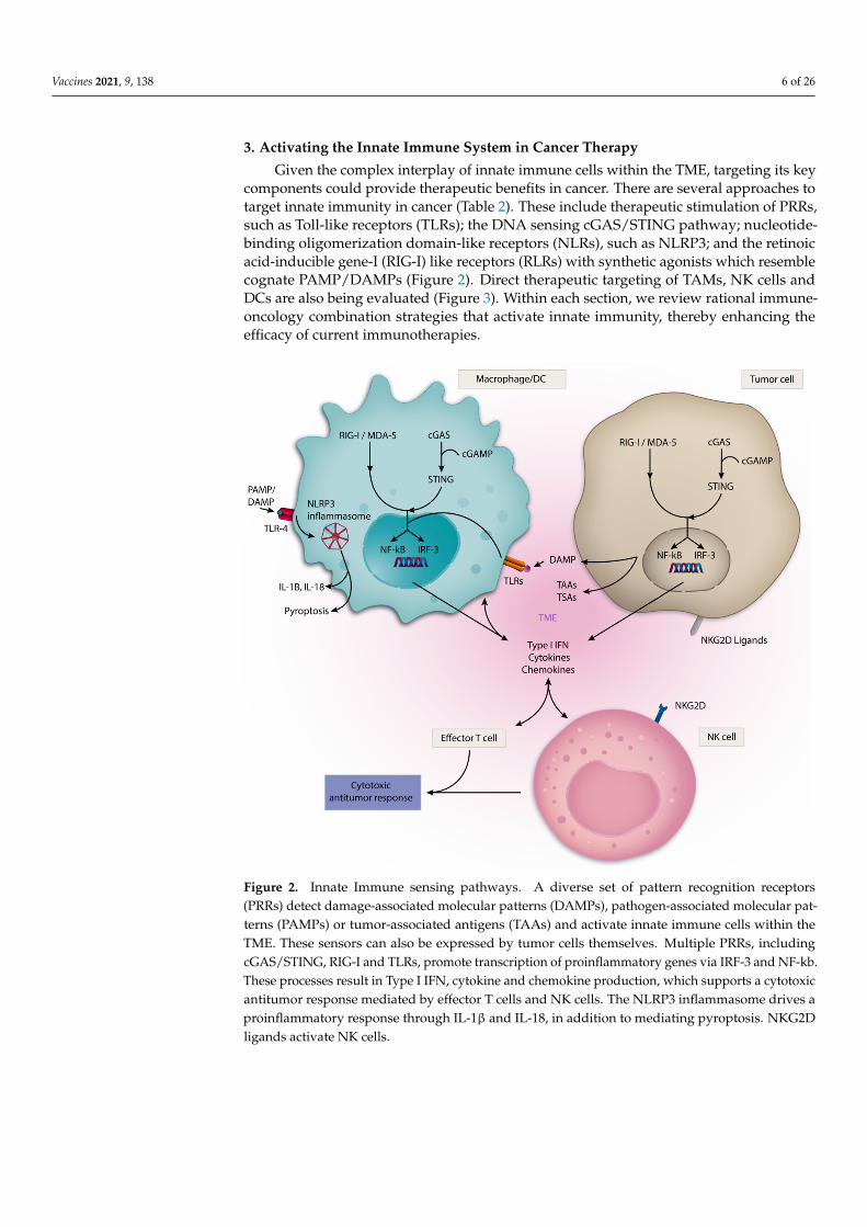
3. Activating the Innate Immune System in Cancer Therapy
Given the complex interplay of innate immune cells within the TME, targeting its keycomponents could provide therapeutic benefits in cancer. There are several approaches totarget innate immunity in cancer (Table 2). These include therapeutic stimulation of PRRs,such as Toll-like receptors (TLRs); the DNA sensing cGAS/STING pathway; nucleotide-binding oligomerization domain-like receptors (NLRs), such as NLRP3; and the retinoicacid-inducible gene-I (RIG-I) like receptors (RLRs) with synthetic agonists which resemblecognate PAMP/DAMPs (Figure 2). Direct therapeutic targeting of TAMs, NK cells andDCs are also being evaluated (Figure 3). Within each section, we review rational immune-oncology combination strategies that activate innate immunity, thereby enhancing theefficacy of current immunotherapies.
Vaccines 2021, 9, x FOR PEER REVIEW 7 of 26
Selicrelumab Monoclonal antibody Intravenous Solid tumors I, II
PI3K (delta) Inhibitors IPI‐549 Small molecule Oral Solid tumors I, II
Class IIa histone
deacetylase inhibitor TMP‐195 Small molecule Oral Solid Tumors Preclinical
IDO inhibitors Indoximod Small molecule Oral Solid tumors I, II
STAT3 inhibitors Siltuximab Monoclonal antibody Intravenous Solid tumors I, II
WP1066 Small molecule Oral Solid tumors I
TT‐101 Small molecule Oral Solid tumors I
* BMS‐986301 is being evaluated for systemic intramuscular administration. The novel NLRP3 agonist BMS‐986299 is be‐
ing studied in a phase I clinical trial as mon‐otherapy and in combination with nivolumab and ipilimumab in advanced
solid tumors [NCT03444753].
Figure 2. Innate Immune sensing pathways. A diverse set of pattern recognition receptors (PRRs) detect damage‐associ‐
ated molecular patterns (DAMPs), pathogen‐associated molecular patterns (PAMPs) or tumor‐associated antigens (TAAs)
and activate innate immune cells within the TME. These sensors can also be expressed by tumor cells themselves. Multiple
PRRs, including cGAS/STING, RIG‐I and TLRs, promote transcription of proinflammatory genes via IRF‐3 and NF‐kb.
These processes result in Type I IFN, cytokine and chemokine production, which supports a cytotoxic antitumor response
mediated by effector T cells and NK cells. The NLRP3 inflammasome drives a proinflammatory response through IL‐1β
and IL‐18, in addition to mediating pyroptosis. NKG2D ligands activate NK cells.
Figure 2. Innate Immune sensing pathways. A diverse set of pattern recognition receptors(PRRs) detect damage-associated molecular patterns (DAMPs), pathogen-associated molecular pat-terns (PAMPs) or tumor-associated antigens (TAAs) and activate innate immune cells within theTME. These sensors can also be expressed by tumor cells themselves. Multiple PRRs, includingcGAS/STING, RIG-I and TLRs, promote transcription of proinflammatory genes via IRF-3 and NF-kb.These processes result in Type I IFN, cytokine and chemokine production, which supports a cytotoxicantitumor response mediated by effector T cells and NK cells. The NLRP3 inflammasome drives aproinflammatory response through IL-1β and IL-18, in addition to mediating pyroptosis. NKG2Dligands activate NK cells.

Vaccines 2021, 9, 138 7 of 26
Table 2. Summary of agents in clinical development.
PRR Agent Molecule Type Route of Ad-ministration Cancer Type(s) Clinical Phase of Development
TLR8 VTX-2337(motilomod) Small molecule Intratumoral ovarian,
HNSCC I, II
dTLR7/8 NKTR-262 Small molecule Intratumoral Solid tumors I, II
TLR9 SD-101 CpG-C classODN Intratumoral Solid tumors I, II
EMD 1201081 Synthetic ODN SubcutaneousInjection HNSCC I, II
CPG 7909 CpG ODN SubcutaneousInjection Lymphomas I, II
IMO-2125(Tilsotolimod) Synthetic ODN Intratumoral melanoma,
Solid tumors I, II
CMP-001 CpG ODN Intratumoral Solid tumors I, II
RIG-I SLR-14 Synthetic stemloop RNA Intratumoral Solid tumors Pre-clinical
RGT-100(MK-4621)
Syntheticoligonucleotide Intratumoral Solid tumors
MDA-5 BO-112(poly(I:C))
SyntheticdsRNA Intratumoral Solid tumors I
STING E7766Novel
macrocycle-bridged
Intravenous Solid tumors I
GSK3745417 Small molecule Intravenous Solid tumors IMIW815
(ADU-S100) Synthetic CDN Intratumoral Solid tumors I, II
MK1454 Small molecule Intratumoral Solid tumors I, IIBMS-986301 Small molecule Intratumoral * Solid tumors I
NLRP3 BMS-986299 First in classagonist * Intratumoral * I
Other innate immune targets
CSF-1R Cabiralizumab Monoclonalantibody Intravenous Solid tumors I, II
JNJ-40346527 Monoclonalantibody Intravenous Advanced
prostate cancer I, II
PLX3397 Small molecule Oral Solid tumors I, IIMCS110 Solid tumors I, II
IMC-CS4 Monoclonalantibody Intravenous Solid tumors I
CD40 APX005M Monoclonalantibody Intravenous Solid tumors I, II
CP-870,893 Monoclonalantibody Intravenous Solid tumors I
Selicrelumab Monoclonalantibody Intravenous Solid tumors I, II
PI3K (delta)Inhibitors IPI-549 Small molecule Oral Solid tumors I, II
Class IIahistone
deacetylaseinhibitor
TMP-195 Small molecule Oral Solid Tumors Preclinical
IDO inhibitors Indoximod Small molecule Oral Solid tumors I, IISTAT3
inhibitors Siltuximab Monoclonalantibody Intravenous Solid tumors I, II
WP1066 Small molecule Oral Solid tumors ITT-101 Small molecule Oral Solid tumors I
* BMS-986301 is being evaluated for systemic intramuscular administration. The novel NLRP3 agonist BMS-986299 is being studied in aphase I clinical trial as mon-otherapy and in combination with nivolumab and ipilimumab in advanced solid tumors [NCT03444753].

Vaccines 2021, 9, 138 8 of 26Vaccines 2021, 9, x FOR PEER REVIEW 8 of 26
Figure 3. Strategies targeting TAMs. Multiple therapeutic strategies exist to target tumor‐associated macrophages (TAMs)
in cancer immunotherapy. CSF‐1R inhibition and CD40 agonism promote TAM polarization to proinflammatory M1 phe‐
notype. CD47‐SIRPα blockade restores TAM‐mediated phagocytosis. PD‐1 blockade reverses TAM‐mediated immuno‐
suppression. CCL5 and CCL2 blockade attenuate TAM recruitment.
3.1. Toll‐like Receptors (TLRs)
TLRs are highly conserved transmembrane and intracellular PRRs found in a variety
of cell types and play a critical role in the detection of microbial pathogens by innate im‐
mune cells [83]. In humans, ten TLRs have been identified, and they are expressed by T
cells, B‐cells, APCs as well as many non‐immune cells, including epithelial and endothe‐
lial cells. TLRs localize to two different regions of the cell: the plasma membrane and the
endosome. TLR 1, 2, 5 and 6 are found specifically in the plasma membrane. TLR 3, 4, 7, 8
and 9 are found specifically in the endosomes, while TLR4 can signal at both locations
[84]. TLRs located on the cell membrane recognize lipids and proteins, and TLRs located
on the membrane of intracellular endosomes recognize nucleic acids. TLR signaling oc‐
curs through activation of adaptor proteins that enter the nucleus and regulate the expres‐
sion of proinflammatory mediators. These adaptor proteins include MyD88, interferon
regulatory factor 3 (IRF‐3), NF‐kB and activator protein‐1 (AP‐1) [85].
TLRs are expressed by both cancer cells and immune cells within tumors, and there‐
fore, have pleiotropic effects on immunomodulation of the TME. When activated on im‐
mune cells, TLRs mediate a broad range of immunostimulatory effects that promote anti‐
tumor T cell responses [86]. Studies demonstrated that chemotherapy‐induced DAMP re‐
lease activated TLR4 and resulted in DC maturation and other immune activating effects
[87]. TLR9 signaling has been shown to activate pDCs and result in the secretion of high
levels of type I interferon [28]. TLR7 and TLR8 activation led to the reprogramming of
tumor promoting M2 TAM phenotype to antitumor M1 phenotype [88,89]. On T cell pop‐
ulations, TLRs have diverse effects, including reduced suppressive function of T‐regula‐
tory (Treg) cells and enhancement of the survival, proliferation and cytokine production
of CD8+ T cells [90–92]. Within cancer cells, TLR activation has been shown to trigger both
apoptosis and cell survival [93]. In addition, constitutive activation of TLRs can lead to
Figure 3. Strategies targeting TAMs. Multiple therapeutic strategies exist to target tumor-associated macrophages (TAMs) incancer immunotherapy. CSF-1R inhibition and CD40 agonism promote TAM polarization to proinflammatory M1 phenotype.CD47-SIRPα blockade restores TAM-mediated phagocytosis. PD-1 blockade reverses TAM-mediated immunosuppression.CCL5 and CCL2 blockade attenuate TAM recruitment.
3.1. Toll-like Receptors (TLRs)
TLRs are highly conserved transmembrane and intracellular PRRs found in a varietyof cell types and play a critical role in the detection of microbial pathogens by innateimmune cells [83]. In humans, ten TLRs have been identified, and they are expressed by Tcells, B-cells, APCs as well as many non-immune cells, including epithelial and endothelialcells. TLRs localize to two different regions of the cell: the plasma membrane and theendosome. TLR 1, 2, 5 and 6 are found specifically in the plasma membrane. TLR 3, 4, 7, 8and 9 are found specifically in the endosomes, while TLR4 can signal at both locations [84].TLRs located on the cell membrane recognize lipids and proteins, and TLRs located onthe membrane of intracellular endosomes recognize nucleic acids. TLR signaling occursthrough activation of adaptor proteins that enter the nucleus and regulate the expression ofproinflammatory mediators. These adaptor proteins include MyD88, interferon regulatoryfactor 3 (IRF-3), NF-kB and activator protein-1 (AP-1) [85].
TLRs are expressed by both cancer cells and immune cells within tumors, and therefore,have pleiotropic effects on immunomodulation of the TME. When activated on immunecells, TLRs mediate a broad range of immunostimulatory effects that promote antitumorT cell responses [86]. Studies demonstrated that chemotherapy-induced DAMP releaseactivated TLR4 and resulted in DC maturation and other immune activating effects [87]. TLR9signaling has been shown to activate pDCs and result in the secretion of high levels of type Iinterferon [28]. TLR7 and TLR8 activation led to the reprogramming of tumor promoting M2TAM phenotype to antitumor M1 phenotype [88,89]. On T cell populations, TLRs have diverseeffects, including reduced suppressive function of T-regulatory (Treg) cells and enhancementof the survival, proliferation and cytokine production of CD8+ T cells [90–92]. Within cancercells, TLR activation has been shown to trigger both apoptosis and cell survival [93]. Inaddition, constitutive activation of TLRs can lead to chronic inflammatory states which areassociated with the recruitment of immunosuppressive cell types, such as myeloid derivedsuppressor cells (MDSCs), leading to tumor progression [94–96].

Vaccines 2021, 9, 138 9 of 26
The diverse immunostimulatory functions of TLR activation provide ample ratio-nale for therapeutic targeting for the treatment of tumors with a “cold” TME. Thera-peutic administration of synthetic TLR agonists by intratumoral injection have revealedmultiple anticancer effects in preclinical models. Activation of TLR9 by synthetic CpG-oligodeoxynucleotides was shown to revert resistance to PD-1 blockade by expandingmultifunctional CD8+ T cells [97,98]. TLR9 agonist candidates SD-101 and CMP-001 werewell tolerated in early phase clinical trials and demonstrated clinical activity in combina-tion with anti-PD-1 treatment in melanoma and head and neck squamous cell carcinoma(HNSCC) [99,100]. These candidates are currently undergoing phase 2 trials for the treat-ment of solid tumors in combination with other forms of immunotherapy [NCT01042379,NCT04050085, NCT03007732, NCT03084640, NCT02554812, NCT03438318]. TLR7/8 ag-onist NKTR-262, in combination with the systemic CD122-biased IL-2 pathway agonist,bempegaldesleukin, promoted antigen presentation and CD8+ T cell infiltration in preclini-cal models [101]. This combination was well tolerated in early phase clinical trials and iscurrently being investigated in combination with anti-PD-1 therapy [NCT03435640]. An-other TLR7/8 agonist, MEDI9197, was shown to activate pDCs and macrophages leadingto interferon-α (IFN-α), IL-12 and IFN-γ release and subsequent antitumor T cell responseand tumor regression in syngeneic murine models [102]. This molecule is currently beingevaluated in early phase clinical trials [102]. Motilomod, a small molecule TLR8 agonist, isundergoing investigation in combination with nivolumab for HNSCC [NCT03906526].
Three TLR agonists are currently FDA-approved and in clinical use. Bacillus Calmette-Guerin (BCG), an attenuated strain of Mycobacterium bovis is a TLR2/4 ligand that isapproved for treatment of superficial, non-muscle invasive bladder cancer [103]. In addi-tion, the TLR4 agonist monophosphoryl A is approved as a vaccine adjuvant and TLR7agonist imiquimod is approved for the treatment of genital warts and basal cell carci-noma [104–106].
3.2. cGAS/STING Pathway
The cGAS-STING pathway detects cytosolic DNA associated with viral infectionand tumorigenesis. The cellular mechanisms by which the pathway mediates immuneactivation have been previously well described [107–109]. In brief, cGAS senses cytosolicDNA and activates Stimulator of IFN genes (STING) through synthesis of the cyclicdinucleotide, cyclic GMP-AMP (cGAMP) [110,111]. Upon activation at the endoplasmicreticulum and subsequent translocation to the golgi, STING activates IRF-3 and NF-kBtranscriptional programs, resulting in the expression and release of type I IFN [112,113].STING is expressed by multiple immune and non-immune cells, and its ability to sensetumor-derived DNA can be harnessed for cancer therapeutic purposes. In murine tumormodels, STING-dependent cytosolic DNA sensing by tumor-resident DCs was found toinduce type I IFN production and was required for CD8+ T cell infiltration and rejection ofimmunogenic tumors [108]. Intratumoral injection of STING agonists in preclinical modelsrecapitulated these proinflammatory effects and induced profound tumor regression [114].
Therapeutic STING activation has been most successful with synthetic cyclic dinu-cleotides (CDNs) due to their structural versatility and ability to bind prevalent allelic variantsin human STING [114]. Currently, there are multiple ongoing clinical trials with syntheticCDN STING agonists, which are comprehensively reviewed elsewhere [115]. MK1454 isundergoing clinical evaluation as monotherapy or in combination with pembrolizumab for thetreatment of advanced solid tumors [NCT03010176, NCT04220866] [116]. ADU-S100/MIW815is being investigated in phase II clinical trials in combination with ICI as a first line treatmentfor HNSCC [NCT03937141] [117]. Exploration of the dose-dependent effects of ADU-S100revealed that while higher doses were more effective at clearing injected tumors, lower doseselicited IFN-γ-driven CD8+ T cell expansion and demonstrated synergy with ICI [118]. Whilemost STING agonists have been administered via intratumoral injection, BMS-986301 isbeing evaluated for systemic intramuscular administration. Additionally, GSK3745417 isan intravenous STING agonist being evaluated alone and in combination with ICI in ad-

Vaccines 2021, 9, 138 10 of 26
vanced solid tumors [NCT03956680, NCT03843359] [119]. A newly developed non-CDNamidobenzimidazole-based small molecule given intravenously displayed potent STINGagonism and durable preclinical antitumor activity, thus representing an important step inadapting STING modulators for systemic administration [120]. Clinical evaluation of tol-erability and safety is ongoing. Other emerging approaches include engineered liposomalnanoparticle packaging, ex vivo loading of STING into exosomes and bacterial modificationto optimize treatment delivery [121].
The mechanistic underpinnings of the cGAS-STING pathway make STING agonists anattractive adjuvant to cancer vaccines. PancVAX is a vaccine composed of synthetic peptidesand a STING agonist-based adjuvant, ADU-V19, which when used in combination with anOX40 agonist and anti-PD-1 therapy in preclinical models leads to significant tumor regressionand improved survival [122]. The STINGVAX vaccine consists of a CDN ligand formulatedwith GM-CSF, which has potent antitumor activity as monotherapy across multiple murinemodels. It was shown to upregulate PD-L1 on the TME and resulted in combinatorial regres-sion of tumors resistant to anti-PD-1 monotherapy [123]. STING agonists used in combinationwith chemotherapy and radiation therapy have been shown to amplify antitumor immuneresponses in both preclinical and clinical settings [124,125]. Interestingly, preclinical evaluationof poly ADP-ribose polymerase (PARP) inhibitors demonstrated a STING-dependent immuneresponse which enhanced the efficacy of ICI [126].
Despite the encouraging framework for STING agonism in the treatment of cancer,some nuances with respect to context and dose-dependent effects are emerging. Recentresearch has demonstrated that cancers with high chromosomal instability (CIN) haveincreased levels of cytosolic DNA which contributes to endogenous cGAS/STING acti-vation which, if sustained over time, can promote tumorigenesis, immune evasion andmetastasis [111,127,128]. The existence of this phenotype represents an important consider-ation in developing rational strategies that incorporate STING agonists and pave the waytowards mechanistic evaluation of intermittent vs. continuous STING pathway activationin generating durable antitumor responses.
3.3. Retinoic Acid Inducible Gene-I-like Receptors (RLRs) and RIG-I
RLRs are PRRs that detect cytosolic RNA physiologically in the context of viralinfections [129]. The best studied RLRs are RIG-I, MDA5 and LGP2. RIG-I primarily bindsshorter dsRNA, whereas MDA-5 interacts with longer fragments [130]. Upon bindingof RNA, RLRs undergo a conformational change that exposes a CARD domain, whichactivates downstream effectors that promote the transcription factors IRF-1, IRF-3, IRF-7,NF-kb and IFN response elements, similar to the cGAS/STING pathway [127,128]. Inaddition, the CARD domain plays a role in inflammasome activation [131]. RIG-I mediatesa wide range of immunostimulatory functions, including DC maturation, priming of Tcells and enhancement of NK cell degranulation and cytolytic activity [132,133]. RIG-I alsoinitiates programmed cell death (PCD) through both the intrinsic and extrinsic apoptoticpathways as well as pyroptosis, an inflammatory variant of PCD. The factors determiningwhich form of cell death is driven by RIG-I is very context dependent and not clearlydefined [132]. Furthermore, recent work has revealed that RIG-I is required for adequateresponse to anti-CTLA-4 treatment by inducing caspase-3-mediated tumor cell death,following which the tumor-associated antigens are processed and presented, leading to arobust CD8+ T cell-mediated adaptive immune response [134].
These proinflammatory mechanisms provide rationale for RIG-I as an innate im-munotherapeutic target. Synthetic agonists specific to RIG-I or to RLRs more broadly,classified as RLR mimetics, are being investigated in multiple cancer types [135,136]. In-tralesional RGT100 (MK-4621), a synthetic RIG-I agonist, demonstrated tolerability in aPhase I clinical trial and is now being studied as monotherapy or in combination withpembrolizumab for the treatment of advanced solid tumors [NCT03739138] [137]. In addi-tion, synthetic stem-loop RNA (SLR) sequences that are highly specific for the RIG-I RNAbinding pocket have been developed. SLRs represent a potent strategy for stimulating

Vaccines 2021, 9, 138 11 of 26
RIG-I due to precise structural optimizations and resistance to nucleases [138]. In vivostudies have shown that intratumoral delivery of SLR14 activated both NK cell and CD8+ Tcell populations, resulting in significant antitumor effects [139]. A novel oral RIG-I agonist,SB9200, has demonstrated strong antiviral activity against resistant hepatitis C infectionthrough induction of type I IFNs, and as such may have a role in future immunotherapeuticstrategies [132,140].
Methods to stimulate RLRs more broadly include the use of poly-ICLC, a syntheticdouble-stranded RNA that activates TLR-3; MDA5; and, to a lesser extent, RIG-I [141,142]. Ina pilot trial conducted with patients with refractory HNSCC and melanoma, an “autovaccina-tion” strategy utilizing intralesional and intramuscular administration of poly-ICLC was welltolerated and led to the generation of antitumor T cell activation. This novel approach is nowbeing further investigated in a phase II clinical trial [NCT02423863] [143].
3.4. CD40
Activation of an antigen-specific T cell response requires T cell receptor-mediated anti-gen presentation and interaction between costimulatory molecules, such as CD28 and theB7 family. CD40 is a well-characterized costimulatory molecule and member of the tumornecrosis factor (TNF) receptor superfamily and is highly expressed on APCs, includingDCs, macrophages, B cells and many non-immune cells [144]. CD40 ligand (CD40L) isexpressed by T cells and other non-immune cells. Upon cross-linking, CD40 triggers cellproliferation, upregulation of MHC and other costimulatory molecules and secretion ofcytokines, including IFN-γ and IL-12a [145–148]. CD40-mediated CD8+ T cell activationhas been shown to be independent of CD4+ T cells and innate immune sensors [149,150].In addition, CD40 activates tumor-associated macrophages to the activated M1 phenotype.In one study, this resulted in the secretion of matrix metalloproteinases that modifiedtumor stroma and enhanced the effects of chemotherapy in a murine model of pancreaticadenocarcinoma [151,152].
Preclinical and early clinical experience has highlighted the efficacy of combiningCD40 agonist with chemotherapy, radiation and peptide vaccines [153–155]. In an immuno-logically cold pancreatic ductal adenocarcinoma (PDA) murine model, treatment with aCD40 agonist alone yielded minimal response. However, the addition of chemotherapyresulted in a vaccine-like effect in which potent antigen-specific T cells were generatedas a consequence of chemotherapy-induced cell death and antigen release [150]. Of note,CD40 agonism in combination with gemcitabine caused lethal hepatotoxicity in mice,ameliorated by giving CD40 agonist five days or more prior to chemotherapy [156]. Thisdata established a rationale for a clinical trial of neoadjuvant and adjuvant selicrelumab, aCD40 agonist, in combination with nab-paclitaxel in resectable PDA [NCT02588443]. Theaddition of anti-PD-1 treatment further enhanced tumor regression, and a clinical trial incombination with anti-PD-1 therapy in PDA is underway and has observed encouragingearly phase results [NCT02482168, NCT02304393] [157,158].
In recent preclinical studies utilizing murine models of non-immunogenic solid malig-nancies, triple therapy with a CD40 agonist, an anti-PD-1 antibody and a T cell activatingvaccine stimulated macrophages and DCs reduced T cell exhaustion and generated effectormemory CD4+ T cells [155]. In murine tumor models with high PD1+ T cells, CD40 agonismreversed T cell exhaustion and enhanced response to anti-PD-1 and anti-CTLA-4 immunecheckpoint inhibition, suggesting a re-sensitization benefit for patients who experienceresistance to ICI [159]. Furthermore, CD40 agonism and anti-CTLA-4 antibody achievedpromising response rates and demonstrated increased T cell infiltration and activation inpatients with melanoma [160]. From a clinical translational standpoint, multiple CD40agonists are in early phase clinical trials for solid tumors, with each agonist exhibitingunique properties [161,162]. In CD40-expressing malignancies, such as chronic lympho-cytic leukemia, CD40 monoclonal antibodies (mAbs) mediate direct tumor cell death viaantibody-dependent cellular cytotoxicity [163,164].

Vaccines 2021, 9, 138 12 of 26
3.5. NLRP3-Inflammasome
Inflammasomes are large cytosolic multiprotein complexes that mediate critical in-flammatory innate immune responses in the host defense against microbial pathogens. Thenucleotide-binding oligomerization domain-like receptors, or NOD-like receptors (NLRs)are a diverse family of intracellular PRRs. The nucleotide-binding domain and leucine-richrepeat family pyrin domain (NLRP) is a subfamily of NLRs and an important componentof the inflammasome. The most well-characterized NLRP is NLRP3, which is expressed inmacrophages, DCs and lymphocytes, in addition to non-immune populations, such as ep-ithelial cells [165]. NLRP3 inflammasome activation is driven by the recognition of PAMPsor DAMPs generated in response to cellular stress. Examples include ATP, extracellularglucose and reactive oxidative species (ROS) [166–170]. When activated, NLRP3 forms theNLRP3 inflammasome by a multistep sequential process of priming and activation [166].NLRP3 oligomerizes and cleaves procaspase-1 to caspase-1 and culminates in the releaseand proteolysis of the cytokines IL-18 and IL-1β. Gasdermin-D, a critical component ofthe inflammasome complex, permeabilizes the cell membrane and facilitates pyroptosis,as opposed to apoptosis [171–173]. Inflammasome-induced pyroptosis is canonically acaspase-1-dependent, immunostimulatory form of PCD in which target cell cytoplasmiccontents are released to induce inflammation [174].
The impact of the NLRP3 inflammasome activation on tumorigenesis is complex. Pre-clinical evaluation has demonstrated that NLRP3 inflammasome mediated IL-1β and IL-18release results in IFN-γ production and CD8+ T cell-dependent tumor regression [175,176].In a preclinical model of hepatocellular carcinoma (HCC), estrogen receptor signaling in-creased cancer cell death through NLRP3 inflammasome-initiated, caspase-1-dependent py-roptosis and inhibition of autophagy in cancer cells [177]. In a preclinical model of colorectalcancer liver metastases, mice deficient in NLRP3 inflammasome had increased metastaticgrowth due to impaired IL-18 signaling and maturation of hepatic NK cells [178]. In theclinical setting, analysis of HCC patient tissue samples revealed an association betweenlow expression of NLRP3 inflammasome components and more advanced HCC [179].
Dysregulated NLRP3 inflammasome activation has also been observed to promote tu-morigenesis in multiple solid tumor murine models. 3′methylcholanthrene (MCA)-inducedsarcomas in mice deficient in NLRP3 had decreased tumor burden compared to wild-typemice, attributed to NLRP3-mediated suppression of NK cell immune surveillance [180].In oral squamous cell carcinoma cell lines, NLRP3 inflammasome activation and IL-1βsecretion were associated with tumor progression, metastases and infiltration of immunesuppressive myeloid cells, such as TAMs and MDSCs into the TME [181–183]. In addition,recent studies have shown that NLRP3 inflammasome activation within tumor cells candrive resistance to anti-PD-1 checkpoint inhibitor treatment. In anti-PD-1 treated solidtumor murine models, activated CD8+ T cells induced NLRP3 inflammasome activationwithin tumor cells, which resulted in downstream Wnt5a-mediated CXCR2 ligand expres-sion and MDSC recruitment into the tumor tissue. This effect was abrogated by geneticand pharmacologic inhibition of NLRP3 [184].
Therapeutic targeting of NLRP3 is an emerging strategy, and a rationale exists for bothNLRP3 inflammasome activation and inhibition. The novel NLRP3 agonist BMS-986299 isbeing studied in a phase I clinical trial as monotherapy and in combination with nivolumaband ipilimumab in advanced solid tumors [NCT03444753]. Saponins, derived from treebark, contain molecules with strong proinflammatory properties that can activate theNLRP3 inflammasome and are being investigated for use as a vaccine adjuvant [185,186].Though preclinical evaluations of NLRP3 inhibitors are expanding, they have not yetentered clinical trial testing [187]. In the HNSCC murine model, a novel NLRP3 inhibitorMCC950 delayed tumor growth, reduced MDSCs, Tregs and TAMs, while also increasing Tcell infiltration within the TME [188].
Several recent studies have explored the mechanistic relationship between pyrop-tosis core proteins in cancer. Decreased expression of a primary mediator of pyroptosis,gasdermin D, is linked to enhanced cancer cell proliferation in vitro and tumor growth

Vaccines 2021, 9, 138 13 of 26
in vivo, whereas increased gasdermin E expression enhanced drug sensitivity of tumorcells [189,190]. These findings have led to therapeutic interest in activating pyroptosis asan antitumor strategy [191].
3.6. Dendritic Cell Directed Strategies
DCs are a key component of the innate immune system’s antitumor response. Strate-gies targeting PRRs, such as TLRs and RIG-I agonism, mediate their antitumor effectthrough activation of DCs [24]. Other DC activating and mobilizing agents, such asFLT3 ligands, have shown preclinical promise and are being evaluated in clinical trials[NCT03789097] [192]. Alternatively, activation of DCs can be achieved by blunting suppres-sive programs, which has led to the exploration of STAT3 and indoleamine 2,3-dioxygenase(IDO) inhibitors, now in various phases of clinical evaluation [193,194]. An increasinglynuanced understanding of DC biology has led to clinical breakthroughs in many DC-basedtherapeutic strategies that have earned FDA approval or advanced in clinical trials for thetreatment of solid cancers [24].
3.7. Adoptive DC Strategies
Ex vivo vaccination of DCs with tumor-associated antigens (TAAs) and personalizedtumor-specific antigens (TSAs) have also been explored. In these methods, autologous DCsubsets are isolated from blood, activated ex vivo and loaded with antigen and reintroducedinto the patient. Such DC vaccination strategies have long been an area of interest, withupwards of 200 clinical trials exploring their potential. Sipuleucel-T is an FDA-approvedautologous DC vaccine in which DCs are incubated ex vivo with a fusion protein ofGM-CSF and prostate-specific antigen (PSA) to treat metastatic, castrate-resistant prostatecancer [195]. Novel methods for optimizing the adoptive transfer of autologous antigen-loaded DCs are currently being developed, including personalized antigen selection and exvivo activation with adjuvants [196]. In addition, combination with ICI is currently beingevaluated to help overcome hostile immunosuppressive TMEs.
3.8. TAM Directed Strategies
Multiple therapeutic strategies have been found to drive polarization of protumori-genic M2 TAMs towards antitumor M1 TAMs, which include activation of CD40, TLR3,TLR4, TLR7/8 and TLR9 [89,90,151,155,197], covered in prior sections. Here, we discussadditional strategies being pursued in clinical trials.
3.8.1. Colony Stimulating Factor 1 (CSF-1)
Blockade of macrophage CSF-1 binding to its cognate receptor (CSF-1R) resulted in thedepletion of M2 TAMs, enrichment in M1 TAMs and tumor control in murine preclinicalmodels [198–200]. Synergy in combination with anti-PD-1 and anti-CTLA-4 therapy hasalso been demonstrated [201]. Clinical trials of antibodies and small molecule inhibitorstargeting CSF-1R are ongoing as monotherapy, and in combination with chemotherapyand ICI [197,202,203].
3.8.2. PI3K-γ Inhibition
PI3K activation via the p110γ isoform within macrophages drives polarization toa protumorigenic M2 phenotype [204]. Consistent with this observation, inhibition ofPI3K-γ isoform has been demonstrated to promote M1 polarization of TAMs and increaseproinflammatory cytokines, resulting in antitumor immune activation and suppression oftumor growth in multiple solid tumor cancer models [204–206]. PI3K-γ inhibitor IPI-549 iscurrently undergoing clinical development for the treatment of solid tumors [207].
3.8.3. CD47- Signal-Recognition Protein Alpha (SIRPα)
Therapeutic targeting of CD47, a surface glycoprotein and “don’t eat me” signalexpressed on immune and tumor cells, which interacts with SIRPα on macrophages to

Vaccines 2021, 9, 138 14 of 26
suppress phagocytosis, is also being investigated. CD47-SIRPα blockade using CD47antibodies was shown to restore phagocytosis of tumor cells by TAMs in vitro, stimulateantigen-specific T cells and limit tumor growth in murine models [208,209]. Multiple CD47antibodies are undergoing clinical development for the treatment of solid tumors [210,211].
3.8.4. Dendritic Cell-Specific Intercellular Adhesion Molecule-3-Grabbing Non-Integrin(DC-SIGN)
DC-SIGN, also known as CD209, is a marker of immunosuppressive TAMs. DC-SIGN+ TAM infiltration in clinical specimens is associated with an increased proportionof immunosuppressive regulatory T cells (Treg) and exhausted CD8+ T cells (TIGIT+
LAG3+). Treatment of human muscle-invasive bladder cancer single cell suspensions withmonoclonal antibody targeting DC-SIGN in combination with PD-1 blockade demonstratedheightened antitumor activity as compared to monotherapy, establishing a rationale forclinical development [212].
3.8.5. Other Macrophage-Directed Strategies
Inhibition of Arginase-1 (Arg1) has resulted in reduced tumor growth in preclinicalmodels, and inhibitors of this enzyme have entered clinical development for the treatmentof solid tumors [213,214]. Therapeutic blockade of CCR5, the receptor of protumoralchemokine CCL5, resulted in M2 to M1 macrophage polarization and a reduction in T-regtrafficking in a patient-derived functional in vitro organotypic culture model of hepaticcolorectal metastases. This strategy demonstrated some activity in a Phase I clinical trialin patients with hepatic metastases of refractory colorectal carcinoma [215]. Class IIahistone deacetylase inhibitor, TMP195, demonstrated tumor reduction in breast cancermurine models by increasing the abundance of M1 macrophages [216]. Finally, cancerstem cell-derived WNT paracrine signaling in an ovarian cancer model upregulated M2macrophages, which was reversed by WNT knockdown, suggesting this pathway as apotential target for TAM polarization [217]. It is noteworthy that inhibition of CCL2, animportant chemokine that recruits monocytes, demonstrated promising preclinical activity;however, minimal clinical benefit was observed in clinical trials [218–220].
3.9. NK Cell Directed Strategies
While therapeutic targeting of activating receptors on NK cells has received muchinterest, most approaches have yet to reach clinical trials.
3.9.1. Natural-Killer Group 2, Member D (NKG2D) Ligands
The most extensively studied activating receptor is NKG2D and its ligands, major his-tocompatibility complex (MHC) class I chain-related protein A (MICA) and MHC class Ichain-related protein B (MICB). MICA and MICB are polymorphic proteins that are inducedupon cell stress, damage or transformation and act as a “kill me” signal through the NKG2Dreceptor expressed on cytotoxic lymphocytes [221]. Unfortunately, many cancers shed MICAand MICB through proteolytic cleavage via ADAM10, ADAM17 and MMP14 matrix metallo-proteases [222]. To combat MICA and MICB proteolytic shedding, small molecular inhibitorsof matrix metalloproteases have been recently developed [223]. Additional strategies to reduceMICA and MICB shedding include antibody targeting of the α3 domains of MICA/B. Theseantibodies were shown to result in a significant NK cell-mediated antitumor response in animmunocompetent mouse model [224]. Similarly, proteosome inhibitors, such as bortezomib,may prevent MICA/B degradation and augment NK cell targeting of tumor cells [225]. Can-cer cells with NKG2D-resistant variants can emerge through immune editing and epigeneticmechanisms which alter NKG2D ligand expression [226,227]. Thus, histone deacetylase in-hibitors or chemotherapies have been investigated as treatments to upregulate the membraneexpression of NKG2D ligands [228,229].

Vaccines 2021, 9, 138 15 of 26
3.9.2. NK Cell Engagers (NKCEs)
Bi- and tri-specific antibody constructs can bind multiple antigens and redirect NKcells to proximity of tumor cells and trigger an immune response and tumor cell death [230].Multifunctional NKCEs with a tri-specific engager targeting two activating receptors,NKp46 and CD16, on NK cells and tumor antigen on cancer cells were observed to mediatesignificant tumor control in solid tumor murine models without notable toxicity [231].These studies establish preclinical rationale for the clinical development of these molecules.
3.9.3. NKG2A Inhibition
NKG2A, an inhibitory immune checkpoint molecule expressed on NK cells, and asubset of α/β T cells, has recently emerged as a potential therapeutic target for cancertherapy [232–234]. Monalizumab, a first in class ICI that targets the NKG2A receptorand simultaneously activates both NK cells and CD8+ T cells, has recently advanced tophase III clinical trials for head and neck squamous carcinoma, signifying an importantadvancement in NK cell therapy [235].
3.9.4. Adoptive NK Cell Strategies
Due to the potent physiologic role of NK cells in tumor immunosurveillance, adop-tive NK therapy (ACT) strategies have been an area of active investigation. Most currenttrials use allogeneic NK cells which are isolated from peripheral blood, propagated ex vivoand reinfused after lymphodepleting chemotherapy [236]. Genetic engineering of activatingreceptors on NK cells, such as the NKG2D receptor, has been used to optimize antitumoractivity. NK cell ACT has demonstrated robust control of early metastasis, tumor specificityand more favorable toxicity profiles, when compared to adoptive T cell strategies. However,concerns over efficacy in solid tumors for NK cell ACT exist, including limited proliferativecapacity, ability to infiltrate the tumor and presence of local immunosuppressive mechanismswithin the TME [237,238]. Methods to increase the persistence of NK cells after infusion arebeing investigated, such as HLA knockdown to prevent rejection by the recipient’s immunesystem [239]. Tumors can escape NK cell-driven cytotoxicity by secreting immunosuppressivefactors, such as TGF-β and adenosine, increasing immunosuppressive tryptophan metabolitesvia upregulating IDO, shedding MICA and MICB proteins and recruiting suppressive popula-tions, such as Tregs, MDSCs and M2 TAMs [240–243]. Therapies targeting these mechanismsmay also serve to reinvigorate a suppressed NK cell compartment.
4. Conclusions and Future Directions
There have been significant advances made in our understanding of the innate im-mune system’s contribution and response to tumorigenesis. Rational immuno-oncologycombination strategies to modulate these pathways and activate both innate and adap-tive immunity carry the potential to improve immunotherapy outcomes in all patients,particularly those with immunologically “cold” tumors.
Enthusiasm over novel innate immune strategies must be met with appropriateconsideration regarding the risk of immune-related adverse events (irAEs). By virtueof their desired antigen-independent on-target effects to stimulate highly potent andconserved proinflammatory mechanisms, the risk of off-target toxicity is not insignificant.As clinical use expands, it will be important to distinguish irAEs specific to innate immuneinvestigational agents from potentiation of irAEs caused by ICI, as many investigationalstrategies involve combination with ICI. There is an unmet clinical need to develop novelevidence-based protocols for toxicity management, acknowledging the expanding role ofcombinatorial approaches.
Despite considerable scientific and clinical progress, there remain gaps in our un-derstanding of the dynamic interplay between cancer and immune cells within the TMEfollowing therapeutic intervention. Of critical importance will be to further define context-dependent roles of innate immune pathways in different tumor and genomic subtypes.For example, activation of innate immunity can be a “double-edged sword” and have pro

Vaccines 2021, 9, 138 16 of 26
and antitumorigenic roles in tumor development and progression, depending on tumortype, genetic/epigenetic, metabolic and microenvironmental context. However, the selec-tive context-specific targeting of the innate immune system has the potential to become acornerstone of immunotherapy strategies for the treatment of solid tumors.
Author Contributions: S.R., B.W.L., A.A., A.P. contributed to conceptualization, original draft prepara-tion, review and editing. All authors have read and agreed to the published version of the manuscript.
Funding: This research received no external funding.
Institutional Review Board Statement: Not applicable.
Informed Consent Statement: Not applicable.
Conflicts of Interest: The authors declare no conflict of interest.
References1. Schreiber, R.D.; Old, L.J.; Smyth, M.J. Cancer immunoediting: Integrating immunity’s roles in cancer suppression and promotion.
Science 2011, 331, 1565–1570. [CrossRef]2. Kaplan, D.H.; Shankaran, V.; Dighe, A.S.; Stockert, E.; Aguet, M.; Old, L.J.; Schreiber, R.D. Demonstration of an interferon
gamma-dependent tumor surveillance system in immunocompetent mice. Proc. Natl. Acad. Sci. USA 1998, 95, 7556–7561.[CrossRef]
3. Fuertes, M.B.; Kacha, A.K.; Kline, J.; Woo, S.R.; Kranz, D.M.; Murphy, K.M.; Gajewski, T.F. Host type I IFN signals are required forantitumor CD8+ T cell responses through CD8{alpha}+ dendritic cells. J. Exp. Med. 2011, 208, 2005–2016. [CrossRef]
4. Spranger, S.; Dai, D.; Horton, B.; Gajewski, T.F. Tumor-Residing Batf3 Dendritic Cells Are Required for Effector T Cell Traffickingand Adoptive T Cell Therapy. Cancer Cell 2017, 31, 711–723.e714. [CrossRef] [PubMed]
5. Chen, D.S.; Mellman, I. Oncology meets immunology: The cancer-immunity cycle. Immunity 2013, 39, 1–10. [CrossRef] [PubMed]6. Hanahan, D.; Weinberg, R.A. Hallmarks of cancer: The next generation. Cell 2011, 144, 646–674. [CrossRef]7. Galon, J.; Costes, A.; Sanchez-Cabo, F.; Kirilovsky, A.; Mlecnik, B.; Lagorce-Pages, C.; Tosolini, M.; Camus, M.; Berger, A.; Wind, P.; et al.
Type, density, and location of immune cells within human colorectal tumors predict clinical outcome. Science 2006, 313, 1960–1964.[CrossRef]
8. Harlin, H.; Meng, Y.; Peterson, A.C.; Zha, Y.; Tretiakova, M.; Slingluff, C.; McKee, M.; Gajewski, T.F. Chemokine expression inmelanoma metastases associated with CD8+ T-cell recruitment. Cancer Res. 2009, 69, 3077–3085. [CrossRef]
9. Fridman, W.H.; Pages, F.; Sautes-Fridman, C.; Galon, J. The immune contexture in human tumours: Impact on clinical outcome.Nat. Rev. Cancer 2012, 12, 298–306. [CrossRef]
10. Gajewski, T.F.; Schreiber, H.; Fu, Y.X. Innate and adaptive immune cells in the tumor microenvironment. Nat. Immunol. 2013, 14,1014–1022. [CrossRef] [PubMed]
11. Galon, J.; Bruni, D. Approaches to treat immune hot, altered and cold tumours with combination immunotherapies. Nat. Rev.Drug Discov. 2019, 18, 197–218. [CrossRef]
12. Spranger, S.; Luke, J.J.; Bao, R.; Zha, Y.; Hernandez, K.M.; Li, Y.; Gajewski, A.P.; Andrade, J.; Gajewski, T.F. Density of immunogenicantigens does not explain the presence or absence of the T-cell–inflamed tumor microenvironment in melanoma. Proc. Natl. Acad.Sci. USA 2016, 113, E7759–E7768. [CrossRef]
13. Spranger, S.; Spaapen, R.M.; Zha, Y.; Williams, J.; Meng, Y.; Ha, T.T.; Gajewski, T.F. Up-regulation of PD-L1, IDO, and T(regs) inthe melanoma tumor microenvironment is driven by CD8(+) T cells. Sci. Transl. Med. 2013, 5, 200ra116. [CrossRef]
14. Beatty, G.L.; Gladney, W.L. Immune escape mechanisms as a guide for cancer immunotherapy. Clin. Cancer Res. 2015, 21, 687–692.[CrossRef]
15. Vinay, D.S.; Ryan, E.P.; Pawelec, G.; Talib, W.H.; Stagg, J.; Elkord, E.; Lichtor, T.; Decker, W.K.; Whelan, R.L.; Kumara, H.; et al.Immune evasion in cancer: Mechanistic basis and therapeutic strategies. Semin. Cancer Biol. 2015, 35, S185–S198. [CrossRef][PubMed]
16. Ugel, S.; De Sanctis, F.; Mandruzzato, S.; Bronte, V. Tumor-induced myeloid deviation: When myeloid-derived suppressor cellsmeet tumor-associated macrophages. J. Clin. Investig. 2015, 125, 3365–3376. [CrossRef] [PubMed]
17. Barnes, T.A.; Amir, E. HYPE or HOPE: The prognostic value of infiltrating immune cells in cancer. Br. J. Cancer 2018, 118, e5.[CrossRef] [PubMed]
18. Gajewski, T.F.; Louahed, J.; Brichard, V.G. Gene signature in melanoma associated with clinical activity: A potential clue to unlockcancer immunotherapy. Cancer J. 2010, 16, 399–403. [CrossRef] [PubMed]
19. Ji, R.-R.; Chasalow, S.D.; Wang, L.; Hamid, O.; Schmidt, H.; Cogswell, J.; Alaparthy, S.; Berman, D.; Jure-Kunkel, M.; Siemers, N.O.; et al.An immune-active tumor microenvironment favors clinical response to ipilimumab. Cancer Immunol. Immunother. 2012, 61, 1019–1031.[CrossRef]
20. Tumeh, P.C.; Harview, C.L.; Yearley, J.H.; Shintaku, I.P.; Taylor, E.J.; Robert, L.; Chmielowski, B.; Spasic, M.; Henry, G.; Ciobanu, V.; et al.PD-1 blockade induces responses by inhibiting adaptive immune resistance. Nature 2014, 515, 568–571. [CrossRef]
21. Ribas, A.; Wolchok, J.D. Cancer immunotherapy using checkpoint blockade. Science 2018, 359, 1350–1355. [CrossRef] [PubMed]

Vaccines 2021, 9, 138 17 of 26
22. Taube, J.M.; Klein, A.; Brahmer, J.R.; Xu, H.; Pan, X.; Kim, J.H.; Chen, L.; Pardoll, D.M.; Topalian, S.L.; Anders, R.A. Association ofPD-1, PD-1 ligands, and other features of the tumor immune microenvironment with response to anti-PD-1 therapy. Clin. CancerRes. 2014, 20, 5064–5074. [CrossRef] [PubMed]
23. Roberts, E.W.; Broz, M.L.; Binnewies, M.; Headley, M.B.; Nelson, A.E.; Wolf, D.M.; Kaisho, T.; Bogunovic, D.; Bhardwaj, N.;Krummel, M.F. Critical Role for CD103(+)/CD141(+) Dendritic Cells Bearing CCR7 for Tumor Antigen Trafficking and Priming ofT Cell Immunity in Melanoma. Cancer Cell 2016, 30, 324–336. [CrossRef]
24. Wculek, S.K.; Cueto, F.J.; Mujal, A.M.; Melero, I.; Krummel, M.F.; Sancho, D. Dendritic cells in cancer immunology andimmunotherapy. Nat. Rev. Immunol. 2020, 20, 7–24. [CrossRef]
25. Mildner, A.; Jung, S. Development and function of dendritic cell subsets. Immunity 2014, 40, 642–656. [CrossRef]26. Sanchez-Paulete, A.R.; Cueto, F.J.; Martinez-Lopez, M.; Labiano, S.; Morales-Kastresana, A.; Rodriguez-Ruiz, M.E.; Jure-Kunkel, M.;
Azpilikueta, A.; Aznar, M.A.; Quetglas, J.I.; et al. Cancer Immunotherapy with Immunomodulatory Anti-CD137 and Anti-PD-1Monoclonal Antibodies Requires BATF3-Dependent Dendritic Cells. Cancer Discov. 2016, 6, 71–79. [CrossRef]
27. Binnewies, M.; Mujal, A.M.; Pollack, J.L.; Combes, A.J.; Hardison, E.A.; Barry, K.C.; Tsui, J.; Ruhland, M.K.; Kersten, K.;Abushawish, M.A.; et al. Unleashing Type-2 Dendritic Cells to Drive Protective Antitumor CD4(+) T Cell Immunity. Cell 2019,177, 556–571.e516. [CrossRef]
28. Liu, Y.J. IPC: Professional type 1 interferon-producing cells and plasmacytoid dendritic cell precursors. Annu. Rev. Immunol. 2005,23, 275–306. [CrossRef] [PubMed]
29. Wynn, T.A.; Chawla, A.; Pollard, J.W. Macrophage biology in development, homeostasis and disease. Nature 2013, 496, 445–455.[CrossRef]
30. Mantovani, A.; Sica, A.; Allavena, P.; Garlanda, C.; Locati, M. Tumor-associated macrophages and the related myeloid-derivedsuppressor cells as a paradigm of the diversity of macrophage activation. Hum. Immunol. 2009, 70, 325–330. [CrossRef]
31. Franklin, R.A.; Liao, W.; Sarkar, A.; Kim, M.V.; Bivona, M.R.; Liu, K.; Pamer, E.G.; Li, M.O. The cellular and molecular origin oftumor-associated macrophages. Science 2014, 344, 921–925. [CrossRef] [PubMed]
32. Murray, P.J.; Allen, J.E.; Biswas, S.K.; Fisher, E.A.; Gilroy, D.W.; Goerdt, S.; Gordon, S.; Hamilton, J.A.; Ivashkiv, L.B.; Lawrence, T.; et al.Macrophage activation and polarization: Nomenclature and experimental guidelines. Immunity 2014, 41, 14–20. [CrossRef] [PubMed]
33. van Dalen, F.J.; van Stevendaal, M.; Fennemann, F.L.; Verdoes, M.; Ilina, O. Molecular Repolarisation of Tumour-AssociatedMacrophages. Molecules 2018, 24, 9. [CrossRef]
34. Lin, E.Y.; Pollard, J.W. Tumor-associated macrophages press the angiogenic switch in breast cancer. Cancer Res. 2007, 67, 5064–5066.[CrossRef] [PubMed]
35. Qian, B.Z.; Pollard, J.W. Macrophage diversity enhances tumor progression and metastasis. Cell 2010, 141, 39–51. [CrossRef]36. Rodriguez, P.C.; Quiceno, D.G.; Zabaleta, J.; Ortiz, B.; Zea, A.H.; Piazuelo, M.B.; Delgado, A.; Correa, P.; Brayer, J.; So-
tomayor, E.M.; et al. Arginase I production in the tumor microenvironment by mature myeloid cells inhibits T-cell receptor expressionand antigen-specific T-cell responses. Cancer Res. 2004, 64, 5839–5849. [CrossRef] [PubMed]
37. Lawrence, T.; Natoli, G. Transcriptional regulation of macrophage polarization: Enabling diversity with identity. Nat. Rev.Immunol. 2011, 11, 750–761. [CrossRef]
38. Dehne, N.; Mora, J.; Namgaladze, D.; Weigert, A.; Brune, B. Cancer cell and macrophage cross-talk in the tumor microenvironment.Curr. Opin. Pharmacol. 2017, 35, 12–19. [CrossRef]
39. Zaidi, N.E.; Shazali, N.A.H.; Chor, A.L.T.; Osman, M.A.; Ibrahim, K.; Jaoi-Edward, M.; Afizan Nik Abd Rahman, N.M. Time-Lapse2D Imaging of Phagocytic Activity in M1 Macrophage-4T1 Mouse Mammary Carcinoma Cells in Co-cultures. J. Vis. Exp. 2019.[CrossRef]
40. Chung, W.; Eum, H.H.; Lee, H.O.; Lee, K.M.; Lee, H.B.; Kim, K.T.; Ryu, H.S.; Kim, S.; Lee, J.E.; Park, Y.H.; et al. Single-cellRNA-seq enables comprehensive tumour and immune cell profiling in primary breast cancer. Nat. Commun. 2017, 8, 15081.[CrossRef] [PubMed]
41. Peng, J.; Sun, B.F.; Chen, C.Y.; Zhou, J.Y.; Chen, Y.S.; Chen, H.; Liu, L.; Huang, D.; Jiang, J.; Cui, G.S.; et al. Single-cell RNA-seqhighlights intra-tumoral heterogeneity and malignant progression in pancreatic ductal adenocarcinoma. Cell Res. 2019, 29,725–738. [CrossRef] [PubMed]
42. Zhang, Q.W.; Liu, L.; Gong, C.Y.; Shi, H.S.; Zeng, Y.H.; Wang, X.Z.; Zhao, Y.W.; Wei, Y.Q. Prognostic significance of tumor-associated macrophages in solid tumor: A meta-analysis of the literature. PLoS ONE 2012, 7, e50946. [CrossRef] [PubMed]
43. Shiao, S.L.; Ruffell, B.; DeNardo, D.G.; Faddegon, B.A.; Park, C.C.; Coussens, L.M. TH2-Polarized CD4(+) T Cells and MacrophagesLimit Efficacy of Radiotherapy. Cancer Immunol. Res. 2015, 3, 518–525. [CrossRef] [PubMed]
44. Santarpia, M.; Karachaliou, N. Tumor immune microenvironment characterization and response to anti-PD-1 therapy. Cancer Biol.Med. 2015, 12, 74–78. [CrossRef]
45. Mitchem, J.B.; Brennan, D.J.; Knolhoff, B.L.; Belt, B.A.; Zhu, Y.; Sanford, D.E.; Belaygorod, L.; Carpenter, D.; Collins, L.; Piwnica-Worms, D.; et al. Targeting tumor-infiltrating macrophages decreases tumor-initiating cells, relieves immunosuppression, andimproves chemotherapeutic responses. Cancer Res. 2013, 73, 1128–1141. [CrossRef]
46. Rao, H.L.; Chen, J.W.; Li, M.; Xiao, Y.B.; Fu, J.; Zeng, Y.X.; Cai, M.Y.; Xie, D. Increased intratumoral neutrophil in colorectalcarcinomas correlates closely with malignant phenotype and predicts patients’ adverse prognosis. PLoS ONE 2012, 7, e30806.[CrossRef]

Vaccines 2021, 9, 138 18 of 26
47. Shen, M.; Hu, P.; Donskov, F.; Wang, G.; Liu, Q.; Du, J. Tumor-associated neutrophils as a new prognostic factor in cancer:A systematic review and meta-analysis. PLoS ONE 2014, 9, e98259. [CrossRef]
48. Schmidt, H.; Bastholt, L.; Geertsen, P.; Christensen, I.J.; Larsen, S.; Gehl, J.; von der Maase, H. Elevated neutrophil and monocytecounts in peripheral blood are associated with poor survival in patients with metastatic melanoma: A prognostic model. Br. J.Cancer 2005, 93, 273–278. [CrossRef]
49. Glodde, N.; Bald, T.; van den Boorn-Konijnenberg, D.; Nakamura, K.; O’Donnell, J.S.; Szczepanski, S.; Brandes, M.; Eickhoff, S.;Das, I.; Shridhar, N.; et al. Reactive Neutrophil Responses Dependent on the Receptor Tyrosine Kinase c-MET Limit CancerImmunotherapy. Immunity 2017, 47, 789–802.e789. [CrossRef] [PubMed]
50. Benevides, L.; da Fonseca, D.M.; Donate, P.B.; Tiezzi, D.G.; De Carvalho, D.D.; de Andrade, J.M.; Martins, G.A.; Silva, J.S.IL17 Promotes Mammary Tumor Progression by Changing the Behavior of Tumor Cells and Eliciting Tumorigenic NeutrophilsRecruitment. Cancer Res. 2015, 75, 3788–3799. [CrossRef]
51. Coffelt, S.B.; Kersten, K.; Doornebal, C.W.; Weiden, J.; Vrijland, K.; Hau, C.S.; Verstegen, N.J.M.; Ciampricotti, M.; Hawinkels, L.;Jonkers, J.; et al. IL-17-producing gammadelta T cells and neutrophils conspire to promote breast cancer metastasis. Nature 2015,522, 345–348. [CrossRef]
52. Gerrard, T.L.; Cohen, D.J.; Kaplan, A.M. Human neutrophil-mediated cytotoxicity to tumor cells. J. Natl. Cancer Inst. 1981, 66,483–488.
53. Granot, Z.; Henke, E.; Comen, E.A.; King, T.A.; Norton, L.; Benezra, R. Tumor entrained neutrophils inhibit seeding in thepremetastatic lung. Cancer Cell 2011, 20, 300–314. [CrossRef]
54. Fridlender, Z.G.; Sun, J.; Mishalian, I.; Singhal, S.; Cheng, G.; Kapoor, V.; Horng, W.; Fridlender, G.; Bayuh, R.; Worthen, G.S.; et al.Transcriptomic analysis comparing tumor-associated neutrophils with granulocytic myeloid-derived suppressor cells and normalneutrophils. PLoS ONE 2012, 7, e31524. [CrossRef]
55. Fridlender, Z.G.; Sun, J.; Kim, S.; Kapoor, V.; Cheng, G.; Ling, L.; Worthen, G.S.; Albelda, S.M. Polarization of tumor-associatedneutrophil phenotype by TGF-beta: “N1” versus “N2” TAN. Cancer Cell 2009, 16, 183–194. [CrossRef]
56. Bird, L. Controlling neutrophil plasticity. Nat. Rev. Immunol. 2010, 10, 752. [CrossRef] [PubMed]57. Sagiv, J.Y.; Michaeli, J.; Assi, S.; Mishalian, I.; Kisos, H.; Levy, L.; Damti, P.; Lumbroso, D.; Polyansky, L.; Sionov, R.V.; et al.
Phenotypic diversity and plasticity in circulating neutrophil subpopulations in cancer. Cell Rep. 2015, 10, 562–573. [CrossRef]58. Condamine, T.; Gabrilovich, D.I. Molecular mechanisms regulating myeloid-derived suppressor cell differentiation and function.
Trends Immunol. 2011, 32, 19–25. [CrossRef] [PubMed]59. Veglia, F.; Perego, M.; Gabrilovich, D. Myeloid-derived suppressor cells coming of age. Nat. Immunol. 2018, 19, 108–119.
[CrossRef] [PubMed]60. Law, A.M.K.; Valdes-Mora, F.; Gallego-Ortega, D. Myeloid-Derived Suppressor Cells as a Therapeutic Target for Cancer. Cells
2020, 9, 561. [CrossRef]61. Pawelec, G.; Verschoor, C.P.; Ostrand-Rosenberg, S. Myeloid-Derived Suppressor Cells: Not Only in Tumor Immunity. Front.
Immunol. 2019, 10, 1099. [CrossRef] [PubMed]62. Lv, M.; Wang, K.; Huang, X.J. Myeloid-derived suppressor cells in hematological malignancies: Friends or foes. J. Hematol. Oncol.
2019, 12, 105. [CrossRef]63. Johansson, A.; Rudolfsson, S.; Hammarsten, P.; Halin, S.; Pietras, K.; Jones, J.; Stattin, P.; Egevad, L.; Granfors, T.; Wikstrom, P.;
et al. Mast cells are novel independent prognostic markers in prostate cancer and represent a target for therapy. Am. J. Pathol.2010, 177, 1031–1041. [CrossRef] [PubMed]
64. Welsh, T.J.; Green, R.H.; Richardson, D.; Waller, D.A.; O’Byrne, K.J.; Bradding, P. Macrophage and mast-cell invasion of tumor cellislets confers a marked survival advantage in non-small-cell lung cancer. J. Clin. Oncol. 2005, 23, 8959–8967. [CrossRef]
65. Rajput, A.B.; Turbin, D.A.; Cheang, M.C.; Voduc, D.K.; Leung, S.; Gelmon, K.A.; Gilks, C.B.; Huntsman, D.G. Stromal mast cells ininvasive breast cancer are a marker of favourable prognosis: A study of 4444 cases. Breast Cancer Res. Treat. 2008, 107, 249–257.[CrossRef]
66. Varricchi, G.; Galdiero, M.R.; Loffredo, S.; Marone, G.; Iannone, R.; Marone, G.; Granata, F. Are Mast Cells MASTers in Cancer?Front. Immunol. 2017, 8, 424. [CrossRef] [PubMed]
67. Toth-Jakatics, R.; Jimi, S.; Takebayashi, S.; Kawamoto, N. Cutaneous malignant melanoma: Correlation between neovasculariza-tion and peritumor accumulation of mast cells overexpressing vascular endothelial growth factor. Hum. Pathol. 2000, 31, 955–960.[CrossRef]
68. Malfettone, A.; Silvestris, N.; Saponaro, C.; Ranieri, G.; Russo, A.; Caruso, S.; Popescu, O.; Simone, G.; Paradiso, A.; Mangia, A.High density of tryptase-positive mast cells in human colorectal cancer: A poor prognostic factor related to protease-activatedreceptor 2 expression. J. Cell. Mol. Med. 2013, 17, 1025–1037. [CrossRef] [PubMed]
69. Morris, D.R.; Ding, Y.; Ricks, T.K.; Gullapalli, A.; Wolfe, B.L.; Trejo, J. Protease-activated receptor-2 is essential for factor VIIa andXa-induced signaling, migration, and invasion of breast cancer cells. Cancer Res. 2006, 66, 307–314. [CrossRef]
70. Gooch, J.L.; Lee, A.V.; Yee, D. Interleukin 4 inhibits growth and induces apoptosis in human breast cancer cells. Cancer Res. 1998,58, 4199–4205.
71. Henderson, W.R.; Chi, E.Y.; Jong, E.C.; Klebanoff, S.J. Mast cell-mediated tumor-cell cytotoxicity. Role of the peroxidase system. J.Exp. Med. 1981, 153, 520–533. [CrossRef]

Vaccines 2021, 9, 138 19 of 26
72. Kim, R.; Emi, M.; Tanabe, K. Cancer immunoediting from immune surveillance to immune escape. Immunology 2007, 121, 1–14.[CrossRef]
73. Hashimoto, W.; Osaki, T.; Okamura, H.; Robbins, P.D.; Kurimoto, M.; Nagata, S.; Lotze, M.T.; Tahara, H. Differential antitumoreffects of administration of recombinant IL-18 or recombinant IL-12 are mediated primarily by Fas-Fas ligand- and perforin-induced tumor apoptosis, respectively. J. Immunol. 1999, 163, 583–589. [PubMed]
74. Trinchieri, G. Biology of natural killer cells. Adv. Immunol. 1989, 47, 187–376. [CrossRef] [PubMed]75. Smyth, M.J.; Crowe, N.Y.; Godfrey, D.I. NK cells and NKT cells collaborate in host protection from methylcholanthrene-induced
fibrosarcoma. Int. Immunol. 2001, 13, 459–463. [CrossRef]76. Bottcher, J.P.; Bonavita, E.; Chakravarty, P.; Blees, H.; Cabeza-Cabrerizo, M.; Sammicheli, S.; Rogers, N.C.; Sahai, E.; Zelenay, S.;
Reis e Sousa, C. NK Cells Stimulate Recruitment of cDC1 into the Tumor Microenvironment Promoting Cancer Immune Control.Cell 2018, 172, 1022–1037.e1014. [CrossRef] [PubMed]
77. O’Sullivan, T.; Saddawi-Konefka, R.; Vermi, W.; Koebel, C.M.; Arthur, C.; White, J.M.; Uppaluri, R.; Andrews, D.M.; Ngiow, S.F.;Teng, M.W.; et al. Cancer immunoediting by the innate immune system in the absence of adaptive immunity. J. Exp. Med. 2012,209, 1869–1882. [CrossRef]
78. Cai, G.; Kastelein, R.A.; Hunter, C.A. IL-10 enhances NK cell proliferation, cytotoxicity and production of IFN-γ when combinedwith IL-18. Eur. J. Immunol. 1999, 29, 2658–2665. [CrossRef]
79. Jamieson, A.M.; Diefenbach, A.; McMahon, C.W.; Xiong, N.; Carlyle, J.R.; Raulet, D.H. The Role of the NKG2D Immunoreceptorin Immune Cell Activation and Natural Killing. Immunity 2002, 17, 19–29. [CrossRef]
80. Takanami, I.; Takeuchi, K.; Giga, M. The prognostic value of natural killer cell infiltration in resected pulmonary adenocarcinoma.J. Thorac. Cardiovasc. Surg. 2001, 121, 1058–1063. [CrossRef] [PubMed]
81. Villegas, F.R.; Coca, S.; Villarrubia, V.G.; Jiménez, R.; Chillón, M.a.J.; Jareño, J.; Zuil, M.; Callol, L. Prognostic significance of tumorinfiltrating natural killer cells subset CD57 in patients with squamous cell lung cancer. Lung Cancer 2002, 35, 23–28. [CrossRef]
82. Lopez-Soto, A.; Gonzalez, S.; Smyth, M.J.; Galluzzi, L. Control of Metastasis by NK Cells. Cancer Cell 2017, 32, 135–154. [CrossRef][PubMed]
83. Kawasaki, T.; Kawai, T. Toll-like receptor signaling pathways. Front. Immunol. 2014, 5, 461. [CrossRef] [PubMed]84. Gangloff, M. Different dimerisation mode for TLR4 upon endosomal acidification? Trends Biochem. Sci. 2012, 37, 92–98. [CrossRef]85. Satoh, T.; Akira, S. Toll-Like Receptor Signaling and Its Inducible Proteins. Microbiol. Spectr. 2016, 4. [CrossRef]86. Adams, S. Toll-like receptor agonists in cancer therapy. Immunotherapy 2009, 1, 949–964. [CrossRef]87. Apetoh, L.; Ghiringhelli, F.; Tesniere, A.; Obeid, M.; Ortiz, C.; Criollo, A.; Mignot, G.; Maiuri, M.C.; Ullrich, E.; Saulnier, P.; et al.
Toll-like receptor 4-dependent contribution of the immune system to anticancer chemotherapy and radiotherapy. Nat. Med. 2007,13, 1050–1059. [CrossRef] [PubMed]
88. Rodell, C.B.; Arlauckas, S.P.; Cuccarese, M.F.; Garris, C.S.; Li, R.; Ahmed, M.S.; Kohler, R.H.; Pittet, M.J.; Weissleder, R. TLR7/8-agonist-loaded nanoparticles promote the polarization of tumour-associated macrophages to enhance cancer immunotherapy.Nat. Biomed. Eng. 2018, 2, 578–588. [CrossRef] [PubMed]
89. Vidyarthi, A.; Khan, N.; Agnihotri, T.; Negi, S.; Das, D.K.; Aqdas, M.; Chatterjee, D.; Colegio, O.R.; Tewari, M.K.; Agrewala, J.N.TLR-3 Stimulation Skews M2 Macrophages to M1 Through IFN-alphabeta Signaling and Restricts Tumor Progression. Front.Immunol. 2018, 9, 1650. [CrossRef]
90. Yang, Y.; Huang, C.T.; Huang, X.; Pardoll, D.M. Persistent Toll-like receptor signals are required for reversal of regulatory Tcell-mediated CD8 tolerance. Nat. Immunol. 2004, 5, 508–515. [CrossRef] [PubMed]
91. Zhang, Y.; Luo, F.; Cai, Y.; Liu, N.; Wang, L.; Xu, D.; Chu, Y. TLR1/TLR2 agonist induces tumor regression by reciprocalmodulation of effector and regulatory T cells. J. Immunol. 2011, 186, 1963–1969. [CrossRef]
92. Jin, B.; Sun, T.; Yu, X.H.; Yang, Y.X.; Yeo, A.E. The effects of TLR activation on T-cell development and differentiation. Clin. Dev.Immunol. 2012, 2012, 836485. [CrossRef] [PubMed]
93. Salaun, B.; Coste, I.; Rissoan, M.C.; Lebecque, S.J.; Renno, T. TLR3 can directly trigger apoptosis in human cancer cells. J. Immunol.2006, 176, 4894–4901. [CrossRef] [PubMed]
94. Dajon, M.; Iribarren, K.; Petitprez, F.; Marmier, S.; Lupo, A.; Gillard, M.; Ouakrim, H.; Victor, N.; Vincenzo, D.B.; Joubert, P.E.;et al. Toll like receptor 7 expressed by malignant cells promotes tumor progression and metastasis through the recruitment ofmyeloid derived suppressor cells. Oncoimmunology 2019, 8, e1505174. [CrossRef]
95. Hao, B.; Chen, Z.; Bi, B.; Yu, M.; Yao, S.; Feng, Y.; Yu, Y.; Pan, L.; Di, D.; Luo, G.; et al. Role of TLR4 as a prognostic factor forsurvival in various cancers: A meta-analysis. Oncotarget 2018, 9, 13088–13099. [CrossRef]
96. Jouhi, L.; Renkonen, S.; Atula, T.; Makitie, A.; Haglund, C.; Hagstrom, J. Different Toll-Like Receptor Expression Patterns inProgression toward Cancer. Front. Immunol. 2014, 5, 638. [CrossRef] [PubMed]
97. Wang, S.; Campos, J.; Gallotta, M.; Gong, M.; Crain, C.; Naik, E.; Coffman, R.L.; Guiducci, C. Intratumoral injection of a CpGoligonucleotide reverts resistance to PD-1 blockade by expanding multifunctional CD8+ T cells. Proc. Natl. Acad. Sci. USA 2016,113, E7240–E7249. [CrossRef]
98. Sato-Kaneko, F.; Yao, S.; Ahmadi, A.; Zhang, S.S.; Hosoya, T.; Kaneda, M.M.; Varner, J.A.; Pu, M.; Messer, K.S.; Guiducci, C.; et al.Combination immunotherapy with TLR agonists and checkpoint inhibitors suppresses head and neck cancer. JCI Insight 2017, 2.[CrossRef]

Vaccines 2021, 9, 138 20 of 26
99. Ribas, A.; Medina, T.; Kummar, S.; Amin, A.; Kalbasi, A.; Drabick, J.J.; Barve, M.; Daniels, G.A.; Wong, D.J.; Schmidt, E.V.; et al.SD-101 in Combination with Pembrolizumab in Advanced Melanoma: Results of a Phase Ib, Multicenter Study. Cancer Discov.2018, 8, 1250–1257. [CrossRef]
100. Cohen, E.E.W.; Nabell, L.; Wong, D.J.L.; Day, T.A.; Daniels, G.A.; Milhem, M.M.; Deva, S.; Jameson, M.B.; Guntinas-Lichius, O.;Almubarak, M.; et al. Phase 1b/2, open label, multicenter study of intratumoral SD-101 in combination with pembrolizumab inanti-PD-1 treatment naïve patients with recurrent or metastatic head and neck squamous cell carcinoma (HNSCC). J. Clin. Oncol.2019, 37, 6039. [CrossRef]
101. Kivimae, S.; Hennessy, M.; Pena, R.; Kirksey, Y.; Nieves, W.; Quatch, P.; Cetz, J.; Ren, Z.; Cai, H.; Deng, B.L.; et al. Comprehensiveantitumor immune activation by a novel TLR7/8 targeting agent NKTR-262 combined with CD122-biased immunostimulatorycytokine NKTR-214. Proc. Am. Assoc. Cancer Res. Annu. Meet. 2018 2018, 78, 3755.
102. Mullins, S.R.; Vasilakos, J.P.; Deschler, K.; Grigsby, I.; Gillis, P.; John, J.; Elder, M.J.; Swales, J.; Timosenko, E.; Cooper, Z.; et al.Intratumoral immunotherapy with TLR7/8 agonist MEDI9197 modulates the tumor microenvironment leading to enhancedactivity when combined with other immunotherapies. J. Immunother. Cancer 2019, 7, 244. [CrossRef] [PubMed]
103. Morales, A. Long-term results and complications of intracavitary bacillus Calmette-Guerin therapy for bladder cancer. J. Urol.1984, 132, 457–459. [CrossRef]
104. Beutner, K.R.; Spruance, S.L.; Hougham, A.J.; Fox, T.L.; Owens, M.L.; Douglas, J.M., Jr. Treatment of genital warts with animmune-response modifier (imiquimod). J. Am. Acad. Dermatol. 1998, 38, 230–239. [CrossRef]
105. Casella, C.R.; Mitchell, T.C. Putting endotoxin to work for us: Monophosphoryl lipid A as a safe and effective vaccine adjuvant.Cell Mol. Life Sci. 2008, 65, 3231–3240. [CrossRef] [PubMed]
106. Oldfield, V.; Keating, G.M.; Perry, C.M. Imiquimod: In superficial basal cell carcinoma. Am. J. Clin. Dermatol. 2005, 6, 195–200;discussion 192–201. [CrossRef] [PubMed]
107. Ishikawa, H.; Ma, Z.; Barber, G.N. STING regulates intracellular DNA-mediated, type I interferon-dependent innate immunity.Nature 2009, 461, 788–792. [CrossRef]
108. Woo, S.R.; Fuertes, M.B.; Corrales, L.; Spranger, S.; Furdyna, M.J.; Leung, M.Y.; Duggan, R.; Wang, Y.; Barber, G.N.; Fitzger-ald, K.A.; et al. STING-dependent cytosolic DNA sensing mediates innate immune recognition of immunogenic tumors. Immunity2014, 41, 830–842. [CrossRef]
109. Kwon, J.; Bakhoum, S.F. The Cytosolic DNA-Sensing cGAS-STING Pathway in Cancer. Cancer Discov. 2020, 10, 26–39. [CrossRef]110. Wu, J.; Sun, L.; Chen, X.; Du, F.; Shi, H.; Chen, C.; Chen, Z.J. Cyclic GMP-AMP is an endogenous second messenger in innate
immune signaling by cytosolic DNA. Science 2013, 339, 826–830. [CrossRef] [PubMed]111. Wu, J.J.; Zhao, L.; Hu, H.G.; Li, W.H.; Li, Y.M. Agonists and inhibitors of the STING pathway: Potential agents for immunotherapy.
Med. Res. Rev. 2020, 40, 1117–1141. [CrossRef]112. Liu, S.; Cai, X.; Wu, J.; Cong, Q.; Chen, X.; Li, T.; Du, F.; Ren, J.; Wu, Y.T.; Grishin, N.V.; et al. Phosphorylation of innate immune
adaptor proteins MAVS, STING, and TRIF induces IRF3 activation. Science 2015, 347, aaa2630. [CrossRef]113. Ishikawa, H.; Barber, G.N. STING is an endoplasmic reticulum adaptor that facilitates innate immune signalling. Nature 2008,
455, 674–678. [CrossRef]114. Corrales, L.; Glickman, L.H.; McWhirter, S.M.; Kanne, D.B.; Sivick, K.E.; Katibah, G.E.; Woo, S.R.; Lemmens, E.; Banda, T.;
Leong, J.J.; et al. Direct Activation of STING in the Tumor Microenvironment Leads to Potent and Systemic Tumor Regression andImmunity. Cell Rep. 2015, 11, 1018–1030. [CrossRef] [PubMed]
115. Ding, C.; Song, Z.; Shen, A.; Chen, T.; Zhang, A. Small molecules targeting the innate immune cGAS–STING–TBK1 signalingpathway. Acta Pharm. Sin. B 2020, in press, pre-proof. [CrossRef]
116. Harrington, K.J.; Brody, J.; Ingham, M.; Strauss, J.; Cemerski, S.; Wang, M.; Tse, A.; Khilnani, A.; Marabelle, A.; Golan, T.Preliminary results of the first-in-human (FIH) study of MK-1454, an agonist of stimulator of interferon genes (STING), asmonotherapy or in combination with pembrolizumab (pembro) in patients with advanced solid tumors or lymphomas. Ann.Oncol. 2018, 29. [CrossRef]
117. Meric-Bernstam, F.; Sandhu, S.K.; Hamid, O.; Spreafico, A.; Kasper, S.; Dummer, R.; Shimizu, T.; Steeghs, N.; Lewis, N.;Talluto, C.C.; et al. Phase Ib study of MIW815 (ADU-S100) in combination with spartalizumab (PDR001) in patients (pts) withadvanced/metastatic solid tumors or lymphomas. J. Clin. Oncol. 2019, 37, 2507. [CrossRef]
118. Sivick, K.E.; Desbien, A.L.; Glickman, L.H.; Reiner, G.L.; Corrales, L.; Surh, N.H.; Hudson, T.E.; Vu, U.T.; Francica, B.J.; Banda, T.;et al. Magnitude of Therapeutic STING Activation Determines CD8(+) T Cell-Mediated Anti-tumor Immunity. Cell Rep. 2019, 29,785–789. [CrossRef]
119. Schieven, G.; Brown, J.; Swanson, J.; Stromko, B.S.C.; Ho, C.-P.; Zhang, R.; Li, W.B.; Qiu, H.; Sun, H.; Fink, B.; et al. Preclinicalcharacterization of BMS-986301, a differentiated STING agonist with robust antitumor activity as monotherapy or in combinationwith anti-PD-1. In Proceedings of the 33rd Annual Meeting & Pre-Conference Programs of the Society for Immunotherapy ofCancer (SITC 2018), Washington, DC, USA, 7–11 November 2018.
120. Ramanjulu, J.M.; Pesiridis, G.S.; Yang, J.; Concha, N.; Singhaus, R.; Zhang, S.Y.; Tran, J.L.; Moore, P.; Lehmann, S.; Eberl, H.C.; et al.Design of amidobenzimidazole STING receptor agonists with systemic activity. Nature 2018, 564, 439–443. [CrossRef] [PubMed]
121. Flood, B.A.; Higgs, E.F.; Li, S.; Luke, J.J.; Gajewski, T.F. STING pathway agonism as a cancer therapeutic. Immunol. Rev. 2019, 290,24–38. [CrossRef]

Vaccines 2021, 9, 138 21 of 26
122. Kinkead, H.L.; Hopkins, A.; Lutz, E.; Wu, A.A.; Yarchoan, M.; Cruz, K.; Woolman, S.; Vithayathil, T.; Glickman, L.H.; Ndubaku,C.O.; et al. Combining STING-based neoantigen-targeted vaccine with checkpoint modulators enhances antitumor immunity inmurine pancreatic cancer. JCI Insight 2018, 3. [CrossRef]
123. Fu, J.; Kanne, D.B.; Leong, M.; Glickman, L.H.; McWhirter, S.M.; Lemmens, E.; Mechette, K.; Leong, J.J.; Lauer, P.; Liu, W.; et al.STING agonist formulated cancer vaccines can cure established tumors resistant to PD-1 blockade. Sci. Transl. Med. 2015, 7,283ra252. [CrossRef]
124. Deng, L.; Liang, H.; Xu, M.; Yang, X.; Burnette, B.; Arina, A.; Li, X.D.; Mauceri, H.; Beckett, M.; Darga, T.; et al. STING-DependentCytosolic DNA Sensing Promotes Radiation-Induced Type I Interferon-Dependent Antitumor Immunity in Immunogenic Tumors.Immunity 2014, 41, 843–852. [CrossRef] [PubMed]
125. Ghaffari, A.; Peterson, N.; Khalaj, K.; Vitkin, N.; Robinson, A.; Francis, J.A.; Koti, M. STING agonist therapy in combination withPD-1 immune checkpoint blockade enhances response to carboplatin chemotherapy in high-grade serous ovarian cancer. Br. J.Cancer 2018, 119, 440–449. [CrossRef] [PubMed]
126. Shen, J.; Zhao, W.; Ju, Z.; Wang, L.; Peng, Y.; Labrie, M.; Yap, T.A.; Mills, G.B.; Peng, G. PARPi Triggers the STING-DependentImmune Response and Enhances the Therapeutic Efficacy of Immune Checkpoint Blockade Independent of BRCAness. CancerRes. 2019, 79, 311–319. [CrossRef] [PubMed]
127. Seth, R.B.; Sun, L.; Ea, C.K.; Chen, Z.J. Identification and characterization of MAVS, a mitochondrial antiviral signaling proteinthat activates NF-kappaB and IRF 3. Cell 2005, 122, 669–682. [CrossRef] [PubMed]
128. Kawai, T.; Takahashi, K.; Sato, S.; Coban, C.; Kumar, H.; Kato, H.; Ishii, K.J.; Takeuchi, O.; Akira, S. IPS-1, an adaptor triggeringRIG-I- and Mda5-mediated type I interferon induction. Nat. Immunol. 2005, 6, 981–988. [CrossRef]
129. Loo, Y.M.; Gale, M., Jr. Immune signaling by RIG-I-like receptors. Immunity 2011, 34, 680–692. [CrossRef]130. Kato, H.; Takeuchi, O.; Sato, S.; Yoneyama, M.; Yamamoto, M.; Matsui, K.; Uematsu, S.; Jung, A.; Kawai, T.; Ishii, K.J.; et al.
Differential roles of MDA5 and RIG-I helicases in the recognition of RNA viruses. Nature 2006, 441, 101–105. [CrossRef] [PubMed]131. Poeck, H.; Bscheider, M.; Gross, O.; Finger, K.; Roth, S.; Rebsamen, M.; Hannesschlager, N.; Schlee, M.; Rothenfusser, S.; Barchet,
W.; et al. Recognition of RNA virus by RIG-I results in activation of CARD9 and inflammasome signaling for interleukin 1 betaproduction. Nat. Immunol. 2010, 11, 63–69. [CrossRef] [PubMed]
132. Elion, D.L.; Cook, R.S. Harnessing RIG-I and intrinsic immunity in the tumor microenvironment for therapeutic cancer treatment.Oncotarget 2018, 9, 29007–29017. [CrossRef] [PubMed]
133. Jiang, X.; Kinch, L.N.; Brautigam, C.A.; Chen, X.; Du, F.; Grishin, N.V.; Chen, Z.J. Ubiquitin-induced oligomerization of the RNAsensors RIG-I and MDA5 activates antiviral innate immune response. Immunity 2012, 36, 959–973. [CrossRef]
134. Heidegger, S.; Wintges, A.; Stritzke, F.; Bek, S.; Steiger, K.; Koenig, P.A.; Gottert, S.; Engleitner, T.; Ollinger, R.; Nedelko, T.; et al.RIG-I activation is critical for responsiveness to checkpoint blockade. Sci. Immunol. 2019, 4. [CrossRef]
135. Terheyden, P.; Weishaupt, C.; Heinzerling, L.; Klinkhardt, U.; Krauss, J.; Mohr, P.; Kiecker, F.; Becker, J.C.; Dähling, A.; Döner, F.;et al. Phase I dose-escalation and expansion study of intratumoral CV8102, a RNA-based TLR- and RIG-1 agonist in patients withadvanced solid tumors. Ann. Oncol. 2018, 29, viii466. [CrossRef]
136. Barsoum, J.; Renn, M.; Schuberth, C.; Jakobs, C.; Schwickart, A.; Schlee, M.; van den Boorn, J.; Hartmann, G. Abstract B44:Selective stimulation of RIG-I with a novel synthetic RNA induces strong anti-tumor immunity in mouse tumor models. CancerImmunol. Res. 2017. [CrossRef]
137. Middleton, M.R.; Wermke, M.; Calvo, E.; Chartash, E.; Zhou, H.; Zhao, X.; Niewel, M.; Dobrenkov, K.; Moreno, V. Phase I/II,multicenter, open-label study of intratumoral/intralesional administration of the retinoic acid–inducible gene I (RIG-I) activatorMK-4621 in patients with advanced or recurrent tumors. Ann. Oncol. 2018, 29, viii712. [CrossRef]
138. Linehan, M.M.; Dickey, T.H.; Molinari, E.S.; Fitzgerald, M.E.; Potapova, O.; Iwasaki, A.; Pyle, A.M. A minimal RNA ligand forpotent RIG-I activation in living mice. Sci. Adv. 2018, 4, e1701854. [CrossRef] [PubMed]
139. Jiang, X.; Muthusamy, V.; Fedorova, O.; Kong, Y.; Kim, D.J.; Bosenberg, M.; Pyle, A.M.; Iwasaki, A. Intratumoral delivery of RIG-Iagonist SLR14 induces robust antitumor responses. J. Exp. Med. 2019, 216, 2854–2868. [CrossRef]
140. Jones, M.; Cunningham, M.E.; Wing, P.; DeSilva, S.; Challa, R.; Sheri, A.; Padmanabhan, S.; Iyer, R.P.; Korba, B.E.; Afdhal, N.; et al.SB 9200, a novel agonist of innate immunity, shows potent antiviral activity against resistant HCV variants. J. Med. Virol. 2017, 89,1620–1628. [CrossRef]
141. Kyi, C.; Roudko, V.; Sabado, R.; Saenger, Y.; Loging, W.; Mandeli, J.; Thin, T.H.; Lehrer, D.; Donovan, M.; Posner, M.; et al.Therapeutic Immune Modulation against Solid Cancers with Intratumoral Poly-ICLC: A Pilot Trial. Clin. Cancer Res. 2018, 24,4937–4948. [CrossRef]
142. Dauletbaev, N.; Cammisano, M.; Herscovitch, K.; Lands, L.C. Stimulation of the RIG-I/MAVS Pathway by Polyi-nosinic:Polycytidylic Acid Upregulates IFN-beta in Airway Epithelial Cells with Minimal Costimulation of IL-8. J. Immunol. 2015,195, 2829–2841. [CrossRef]
143. Salazar, A.M.; Erlich, R.B.; Mark, A.; Bhardwaj, N.; Herberman, R.B. Therapeutic in situ autovaccination against solid cancerswith intratumoral poly-ICLC: Case report, hypothesis, and clinical trial. Cancer Immunol. Res. 2014, 2, 720–724. [CrossRef]
144. van Kooten, C.; Banchereau, J. CD40-CD40 ligand. J. Leukoc. Biol. 2000, 67, 2–17. [CrossRef]145. Todryk, S.M.; Tutt, A.L.; Green, M.H.; Smallwood, J.A.; Halanek, N.; Dalgleish, A.G.; Glennie, M.J. CD40 ligation for immunother-
apy of solid tumours. J. Immunol. Methods 2001, 248, 139–147. [CrossRef]

Vaccines 2021, 9, 138 22 of 26
146. van Mierlo, G.J.; den Boer, A.T.; Medema, J.P.; van der Voort, E.I.; Fransen, M.F.; Offringa, R.; Melief, C.J.; Toes, R.E. CD40stimulation leads to effective therapy of CD40(-) tumors through induction of strong systemic cytotoxic T lymphocyte immunity.Proc. Natl. Acad. Sci. USA 2002, 99, 5561–5566. [CrossRef]
147. Buhtoiarov, I.N.; Lum, H.; Berke, G.; Paulnock, D.M.; Sondel, P.M.; Rakhmilevich, A.L. CD40 ligation activates murinemacrophages via an IFN-gamma-dependent mechanism resulting in tumor cell destruction in vitro. J. Immunol. 2005, 174,6013–6022. [CrossRef] [PubMed]
148. Elgueta, R.; Benson, M.J.; de Vries, V.C.; Wasiuk, A.; Guo, Y.; Noelle, R.J. Molecular mechanism and function of CD40/CD40Lengagement in the immune system. Immunol. Rev. 2009, 229, 152–172. [CrossRef] [PubMed]
149. Bennett, S.R.; Carbone, F.R.; Karamalis, F.; Flavell, R.A.; Miller, J.F.; Heath, W.R. Help for cytotoxic-T-cell responses is mediated byCD40 signalling. Nature 1998, 393, 478–480. [CrossRef] [PubMed]
150. Byrne, K.T.; Vonderheide, R.H. CD40 Stimulation Obviates Innate Sensors and Drives T Cell Immunity in Cancer. Cell Rep. 2016,15, 2719–2732. [CrossRef]
151. Beatty, G.L.; Chiorean, E.G.; Fishman, M.P.; Saboury, B.; Teitelbaum, U.R.; Sun, W.; Huhn, R.D.; Song, W.; Li, D.; Sharp, L.L.;et al. CD40 agonists alter tumor stroma and show efficacy against pancreatic carcinoma in mice and humans. Science 2011, 331,1612–1616. [CrossRef] [PubMed]
152. Long, K.B.; Gladney, W.L.; Tooker, G.M.; Graham, K.; Fraietta, J.A.; Beatty, G.L. IFNgamma and CCL2 Cooperate to RedirectTumor-Infiltrating Monocytes to Degrade Fibrosis and Enhance Chemotherapy Efficacy in Pancreatic Carcinoma. Cancer Discov.2016, 6, 400–413. [CrossRef]
153. Vonderheide, R.H.; Burg, J.M.; Mick, R.; Trosko, J.A.; Li, D.; Shaik, M.N.; Tolcher, A.W.; Hamid, O. Phase I study of the CD40agonist antibody CP-870,893 combined with carboplatin and paclitaxel in patients with advanced solid tumors. Oncoimmunology2013, 2, e23033. [CrossRef]
154. Nowak, A.K.; Cook, A.M.; McDonnell, A.M.; Millward, M.J.; Creaney, J.; Francis, R.J.; Hasani, A.; Segal, A.; Musk, A.W.;Turlach, B.A.; et al. A phase 1b clinical trial of the CD40-activating antibody CP-870,893 in combination with cisplatin and pemetrexedin malignant pleural mesothelioma. Ann. Oncol. 2015, 26, 2483–2490. [CrossRef] [PubMed]
155. Ma, H.S.; Poudel, B.; Torres, E.R.; Sidhom, J.W.; Robinson, T.M.; Christmas, B.; Scott, B.; Cruz, K.; Woolman, S.; Wall, V.Z.; et al. A CD40Agonist and PD-1 Antagonist Antibody Reprogram the Microenvironment of Nonimmunogenic Tumors to Allow T-cell-MediatedAnticancer Activity. Cancer Immunol. Res. 2019, 7, 428–442. [CrossRef]
156. Byrne, K.T.; Leisenring, N.H.; Bajor, D.L.; Vonderheide, R.H. CSF-1R-Dependent Lethal Hepatotoxicity When Agonistic CD40Antibody Is Given before but Not after Chemotherapy. J. Immunol. 2016, 197, 179–187. [CrossRef] [PubMed]
157. Winograd, R.; Byrne, K.T.; Evans, R.A.; Odorizzi, P.M.; Meyer, A.R.; Bajor, D.L.; Clendenin, C.; Stanger, B.Z.; Furth, E.E.;Wherry, E.J.; et al. Induction of T-cell Immunity Overcomes Complete Resistance to PD-1 and CTLA-4 Blockade and ImprovesSurvival in Pancreatic Carcinoma. Cancer Immunol. Res. 2015, 3, 399–411. [CrossRef]
158. O’Hara, M.H.; O’Reilly, E.M.; Rosemarie, M.; Varadhachary, G.; Wainberg, Z.A.; Ko, A.; Fisher, G.A.; Rahma, O.; Lyman, J.P.;Cabanski, C.R.; et al. A phase 1b study of CD40 agonistic monoclonal antibody APX005M together with gemcitabine and nab-paclitaxel with or without nivolumab in untreated metastatic pancreatic ductal adenocarcinoma (PDAC) patients. In Proceedingsof the American Association for Cancer Research Annual Meeting, Atlanta, GA, USA, 29 March–3 April 2019.
159. Ngiow, S.F.; Young, A.; Blake, S.J.; Hill, G.R.; Yagita, H.; Teng, M.W.; Korman, A.J.; Smyth, M.J. Agonistic CD40 mAb-Driven IL12Reverses Resistance to Anti-PD1 in a T-cell-Rich Tumor. Cancer Res. 2016, 76, 6266–6277. [CrossRef]
160. Bajor, D.L.; Mick, R.; Riese, M.J.; Huang, A.C.; Sullivan, B.; Richman, L.P.; Torigian, D.A.; George, S.M.; Stelekati, E.; Chen, F.; et al.Long-term outcomes of a phase I study of agonist CD40 antibody and CTLA-4 blockade in patients with metastatic melanoma.Oncoimmunology 2018, 7, e1468956. [CrossRef] [PubMed]
161. Yu, X.; Chan, H.T.C.; Orr, C.M.; Dadas, O.; Booth, S.G.; Dahal, L.N.; Penfold, C.A.; O’Brien, L.; Mockridge, C.I.; French, R.R.; et al.Complex Interplay between Epitope Specificity and Isotype Dictates the Biological Activity of Anti-human CD40 Antibodies.Cancer Cell 2018, 33, 664–675.e664. [CrossRef]
162. Beatty, G.L.; Li, Y.; Long, K.B. Cancer immunotherapy: Activating innate and adaptive immunity through CD40 agonists. ExpertRev. Anticancer. Ther. 2017, 17, 175–186. [CrossRef] [PubMed]
163. Luqman, M.; Klabunde, S.; Lin, K.; Georgakis, G.V.; Cherukuri, A.; Holash, J.; Goldbeck, C.; Xu, X.; Kadel, E.E., 3rd; Lee, S.H.; et al.The antileukemia activity of a human anti-CD40 antagonist antibody, HCD122, on human chronic lymphocytic leukemia cells.Blood 2008, 112, 711–720. [CrossRef]
164. Khubchandani, S.; Czuczman, M.S.; Hernandez-Ilizaliturri, F.J. Dacetuzumab, a humanized mAb against CD40 for the treatmentof hematological malignancies. Curr. Opin. Investig. Drugs 2009, 10, 579–587.
165. Franchi, L.; Warner, N.; Viani, K.; Nuñez, G. Function of Nod-like receptors in microbial recognition and host defense. Immunol.Rev. 2009, 227, 106–128. [CrossRef]
166. Swanson, K.V.; Deng, M.; Ting, J.P. The NLRP3 inflammasome: Molecular activation and regulation to therapeutics. Nat. Rev.Immunol. 2019, 19, 477–489. [CrossRef]
167. Gong, T.; Yang, Y.; Jin, T.; Jiang, W.; Zhou, R. Orchestration of NLRP3 Inflammasome Activation by Ion Fluxes. Trends Immunol.2018, 39, 393–406. [CrossRef] [PubMed]
168. Hughes, M.M.; O’Neill, L.A.J. Metabolic regulation of NLRP3. Immunol. Rev. 2018, 281, 88–98. [CrossRef] [PubMed]

Vaccines 2021, 9, 138 23 of 26
169. Martinez-Garcia, J.J.; Martinez-Banaclocha, H.; Angosto-Bazarra, D.; de Torre-Minguela, C.; Baroja-Mazo, A.; Alarcon-Vila, C.;Martinez-Alarcon, L.; Amores-Iniesta, J.; Martin-Sanchez, F.; Ercole, G.A.; et al. P2X7 receptor induces mitochondrial failure inmonocytes and compromises NLRP3 inflammasome activation during sepsis. Nat. Commun. 2019, 10, 2711. [CrossRef] [PubMed]
170. Karki, R.; Man, S.M.; Kanneganti, T.D. Inflammasomes and Cancer. Cancer Immunol. Res. 2017, 5, 94–99. [CrossRef]171. Martinon, F.; Burns, K.; Tschopp, J. The Inflammasome. Mol. Cell 2002, 10, 417–426. [CrossRef]172. He, W.T.; Wan, H.; Hu, L.; Chen, P.; Wang, X.; Huang, Z.; Yang, Z.H.; Zhong, C.Q.; Han, J. Gasdermin D is an executor of
pyroptosis and required for interleukin-1beta secretion. Cell Res. 2015, 25, 1285–1298. [CrossRef]173. Salcedo, R.; Worschech, A.; Cardone, M.; Jones, Y.; Gyulai, Z.; Dai, R.M.; Wang, E.; Ma, W.; Haines, D.; O’HUigin, C.; et al.
MyD88-mediated signaling prevents development of adenocarcinomas of the colon: Role of interleukin 18. J. Exp. Med. 2010, 207,1625–1636. [CrossRef]
174. Broz, P.; Dixit, V.M. Inflammasomes: Mechanism of assembly, regulation and signalling. Nat. Rev. Immunol. 2016, 16, 407–420.[CrossRef]
175. Allen, I.C.; TeKippe, E.M.; Woodford, R.-M.T.; Uronis, J.M.; Holl, E.K.; Rogers, A.B.; Herfarth, H.H.; Jobin, C.; Ting, J.P.Y. TheNLRP3 inflammasome functions as a negative regulator of tumorigenesis during colitis-associated cancer. J. Exp. Med. 2010, 207,1045–1056. [CrossRef]
176. Ghiringhelli, F.; Apetoh, L.; Tesniere, A.; Aymeric, L.; Ma, Y.; Ortiz, C.; Vermaelen, K.; Panaretakis, T.; Mignot, G.; Ullrich, E.; et al.Activation of the NLRP3 inflammasome in dendritic cells induces IL-1beta-dependent adaptive immunity against tumors. Nat.Med. 2009, 15, 1170–1178. [CrossRef] [PubMed]
177. Wei, Q.; Zhu, R.; Zhu, J.; Zhao, R.; Li, M. E2-Induced Activation of the NLRP3 Inflammasome Triggers Pyroptosis and InhibitsAutophagy in HCC Cells. Oncol. Res. 2019, 27, 827–834. [CrossRef]
178. Dupaul-Chicoine, J.; Arabzadeh, A.; Dagenais, M.; Douglas, T.; Champagne, C.; Morizot, A.; Rodrigue-Gervais, I.G.; Breton, V.;Colpitts, S.L.; Beauchemin, N.; et al. The Nlrp3 Inflammasome Suppresses Colorectal Cancer Metastatic Growth in the Liver byPromoting Natural Killer Cell Tumoricidal Activity. Immunity 2015, 43, 751–763. [CrossRef]
179. Wei, Q.; Mu, K.; Li, T.; Zhang, Y.; Yang, Z.; Jia, X.; Zhao, W.; Huai, W.; Guo, P.; Han, L. Deregulation of the NLRP3 inflammasomein hepatic parenchymal cells during liver cancer progression. Lab. Investig. 2014, 94, 52–62. [CrossRef] [PubMed]
180. Chow, M.T.; Sceneay, J.; Paget, C.; Wong, C.S.; Duret, H.; Tschopp, J.; Moller, A.; Smyth, M.J. NLRP3 suppresses NK cell-mediatedresponses to carcinogen-induced tumors and metastases. Cancer Res. 2012, 72, 5721–5732. [CrossRef] [PubMed]
181. Wang, H.; Luo, Q.; Feng, X.; Zhang, R.; Li, J.; Chen, F. NLRP3 promotes tumor growth and metastasis in human oral squamouscell carcinoma. BMC Cancer 2018, 18, 500. [CrossRef] [PubMed]
182. Daley, D.; Mani, V.R.; Mohan, N.; Akkad, N.; Pandian, G.S.D.B.; Savadkar, S.; Lee, K.B.; Torres-Hernandez, A.; Aykut, B.;Diskin, B.; et al. NLRP3 signaling drives macrophage-induced adaptive immune suppression in pancreatic carcinoma. J. Exp.Med. 2017, 214, 1711–1724. [CrossRef]
183. Guo, B.; Fu, S.; Zhang, J.; Liu, B.; Li, Z. Targeting inflammasome/IL-1 pathways for cancer immunotherapy. Sci. Rep. 2016, 6, 36107.[CrossRef] [PubMed]
184. Theivanthiran, B.; Evans, K.S.; DeVito, N.C.; Plebanek, M.; Sturdivant, M.; Wachsmuth, L.P.; Salama, A.K.; Kang, Y.; Hsu, D.;Balko, J.M.; et al. A tumor-intrinsic PD-L1/NLRP3 inflammasome signaling pathway drives resistance to anti-PD-1 immunotherapy.J. Clin. Investig. 2020, 130, 2570–2586. [CrossRef] [PubMed]
185. Marty-Roix, R.; Vladimer, G.I.; Pouliot, K.; Weng, D.; Buglione-Corbett, R.; West, K.; MacMicking, J.D.; Chee, J.D.; Wang, S.;Lu, S.; et al. Identification of QS-21 as an Inflammasome-activating Molecular Component of Saponin Adjuvants. J. Biol. Chem. 2016,291, 1123–1136. [CrossRef]
186. Vermaelen, K. Vaccine Strategies to Improve Anti-cancer Cellular Immune Responses. Front. Immunol. 2019, 10, 8. [CrossRef][PubMed]
187. Kopalli, S.R.; Kang, T.-B.; Lee, K.-H.; Koppula, S. NLRP3 Inflammasome Activation Inhibitors in Inflammation-Associated CancerImmunotherapy: An Update on the Recent Patents. Recent. Pat Anticancer Drug Discov. 2018, 13, 106–117. [CrossRef] [PubMed]
188. Chen, L.; Huang, C.-F.; Li, Y.-C.; Deng, W.-W.; Mao, L.; Wu, L.; Zhang, W.-F.; Zhang, L.; Sun, Z.-J. Blockage of the NLRP3inflammasome by MCC950 improves anti-tumor immune responses in head and neck squamous cell carcinoma. Cell. Mol. LifeSci. 2018, 75, 2045–2058. [CrossRef]
189. Xia, X.; Wang, X.; Cheng, Z.; Qin, W.; Lei, L.; Jiang, J.; Hu, J. The role of pyroptosis in cancer: Pro-cancer or pro-”host”? Cell DeathDis. 2019, 10, 650. [CrossRef]
190. Gao, J.; Qiu, X.; Xi, G.; Liu, H.; Zhang, F.; Lv, T.; Song, Y. Downregulation of GSDMD attenuates tumor proliferation via theintrinsic mitochondrial apoptotic pathway and inhibition of EGFR/Akt signaling and predicts a good prognosis in nonsmall celllung cancer. Oncol. Rep. 2018, 40, 1971–1984. [CrossRef]
191. Nagarajan, K.; Soundarapandian, K.; Thorne, R.F.; Li, D.; Li, D. Activation of Pyroptotic Cell Death Pathways in Cancer:An Alternative Therapeutic Approach. Transl. Oncol. 2019, 12, 925–931. [CrossRef]
192. Saito, T.; Takayama, T.; Osaki, T.; Nagai, S.; Suzuki, T.; Sato, M.; Kuwano, H.; Tahara, H. Combined mobilization and stimulation oftumor-infiltrating dendritic cells and natural killer cells with Flt3 ligand and IL-18 in vivo induces systemic antitumor immunity.Cancer Sci. 2008, 99, 2028–2036. [CrossRef]
193. Nefedova, Y.; Cheng, P.; Gilkes, D.; Blaskovich, M.; Beg, A.A.; Sebti, S.M.; Gabrilovich, D.I. Activation of dendritic cells viainhibition of Jak2/STAT3 signaling. J. Immunol. 2005, 175, 4338–4346. [CrossRef]
Vaccines 2021, 9, 138 24 of 26
194. Moon, Y.W.; Hajjar, J.; Hwu, P.; Naing, A. Targeting the indoleamine 2,3-dioxygenase pathway in cancer. J. Immunother. Cancer2015, 3, 51. [CrossRef]
195. Cheever, M.A.; Higano, C.S. PROVENGE (Sipuleucel-T) in prostate cancer: The first FDA-approved therapeutic cancer vaccine.Clin. Cancer Res. 2011, 17, 3520–3526. [CrossRef]
196. Carreno, B.M.; Magrini, V.; Becker-Hapak, M.; Kaabinejadian, S.; Hundal, J.; Petti, A.A.; Ly, A.; Lie, W.R.; Hildebrand, W.H.;Mardis, E.R.; et al. Cancer immunotherapy. A dendritic cell vaccine increases the breadth and diversity of melanoma neoantigen-specific T cells. Science 2015, 348, 803–808. [CrossRef]
197. Autio, K.A.; Klebanoff, C.A.; Schaer, D.; Kauh, J.S.; Slovin, S.F.; Blinder, V.S.; Comen, E.A.; Danila, D.C.; Hoffman, D.M.J.; Kang, S.; et al.Phase 1 study of LY3022855, a colony-stimulating factor-1 receptor (CSF-1R) inhibitor, in patients with metastatic breast cancer (MBC)or metastatic castration-resistant prostate cancer (MCRPC). J. Clin. Oncol. 2019, 37, 2548. [CrossRef]
198. Jeannin, P.; Paolini, L.; Adam, C.; Delneste, Y. The roles of CSFs on the functional polarization of tumor-associated macrophages.FEBS J. 2018, 285, 680–699. [CrossRef] [PubMed]
199. Lin, E.Y.; Nguyen, A.V.; Russell, R.G.; Pollard, J.W. Colony-stimulating factor 1 promotes progression of mammary tumors tomalignancy. J. Exp. Med. 2001, 193, 727–740. [CrossRef]
200. Escamilla, J.; Schokrpur, S.; Liu, C.; Priceman, S.J.; Moughon, D.; Jiang, Z.; Pouliot, F.; Magyar, C.; Sung, J.L.; Xu, J.; et al. CSF1receptor targeting in prostate cancer reverses macrophage-mediated resistance to androgen blockade therapy. Cancer Res. 2015,75, 950–962. [CrossRef] [PubMed]
201. Zhu, Y.; Knolhoff, B.L.; Meyer, M.A.; Nywening, T.M.; West, B.L.; Luo, J.; Wang-Gillam, A.; Goedegebuure, S.P.; Linehan, D.C.;DeNardo, D.G. CSF1/CSF1R blockade reprograms tumor-infiltrating macrophages and improves response to T-cell checkpointimmunotherapy in pancreatic cancer models. Cancer Res. 2014, 74, 5057–5069. [CrossRef] [PubMed]
202. Papadopoulos, K.P.; Gluck, L.; Martin, L.P.; Olszanski, A.J.; Tolcher, A.W.; Ngarmchamnanrith, G.; Rasmussen, E.; Amore, B.M.;Nagorsen, D.; Hill, J.S.; et al. First-in-Human Study of AMG 820, a Monoclonal Anti-Colony-Stimulating Factor 1 ReceptorAntibody, in Patients with Advanced Solid Tumors. Clin. Cancer Res. 2017, 23, 5703–5710. [CrossRef] [PubMed]
203. Wang-Gillam, A.; O’Reilly, E.M.; Bendell, J.C.; Wainberg, Z.A.; Borazanci, E.H.; Bahary, N.; O’Hara, M.H.; Beatty, G.L.; Pant, S.;Cohen, D.J.; et al. A randomized phase II study of cabiralizumab (cabira) + nivolumab (nivo) ± chemotherapy (chemo) in advancedpancreatic ductal adenocarcinoma (PDAC). J. Clin. Oncol. 2019, 37, TPS465. [CrossRef]
204. Vergadi, E.; Ieronymaki, E.; Lyroni, K.; Vaporidi, K.; Tsatsanis, C. Akt Signaling Pathway in Macrophage Activation and M1/M2Polarization. J. Immunol. 2017, 198, 1006–1014. [CrossRef]
205. Kaneda, M.M.; Messer, K.S.; Ralainirina, N.; Li, H.; Leem, C.J.; Gorjestani, S.; Woo, G.; Nguyen, A.V.; Figueiredo, C.C.; Foubert, P.; et al.PI3Kgamma is a molecular switch that controls immune suppression. Nature 2016, 539, 437–442. [CrossRef] [PubMed]
206. Kaneda, M.M.; Cappello, P.; Nguyen, A.V.; Ralainirina, N.; Hardamon, C.R.; Foubert, P.; Schmid, M.C.; Sun, P.; Mose, E.; Bouvet,M.; et al. Macrophage PI3Kgamma Drives Pancreatic Ductal Adenocarcinoma Progression. Cancer Discov. 2016, 6, 870–885.[CrossRef] [PubMed]
207. Varner, J.A. Taming the beast: Strategies to target the immune-suppressive macrophage to enhance cancer immune therapy. Proc.Am. Assoc. Cancer Res. Annu. Meet. 2019, 2019, 79.
208. Liu, J.; Wang, L.; Zhao, F.; Tseng, S.; Narayanan, C.; Shura, L.; Willingham, S.; Howard, M.; Prohaska, S.; Volkmer, J.; et al.Pre-Clinical Development of a Humanized Anti-CD47 Antibody with Anti-Cancer Therapeutic Potential. PLoS ONE 2015, 10,e0137345. [CrossRef]
209. Tseng, D.; Volkmer, J.P.; Willingham, S.B.; Contreras-Trujillo, H.; Fathman, J.W.; Fernhoff, N.B.; Seita, J.; Inlay, M.A.; Weiskopf, K.;Miyanishi, M.; et al. Anti-CD47 antibody-mediated phagocytosis of cancer by macrophages primes an effective antitumor T-cellresponse. Proc. Natl. Acad. Sci. USA 2013, 110, 11103–11108. [CrossRef]
210. Petrova, P.S.; Viller, N.N.; Wong, M.; Pang, X.; Lin, G.H.; Dodge, K.; Chai, V.; Chen, H.; Lee, V.; House, V.; et al. TTI-621(SIRPalphaFc): A CD47-Blocking Innate Immune Checkpoint Inhibitor with Broad Antitumor Activity and Minimal ErythrocyteBinding. Clin. Cancer Res. 2017, 23, 1068–1079. [CrossRef]
211. Sikic, B.I.; Lakhani, N.; Patnaik, A.; Shah, S.A.; Chandana, S.R.; Rasco, D.; Colevas, A.D.; O’Rourke, T.; Narayanan, S.;Papadopoulos, K.; et al. First-in-Human, First-in-Class Phase I Trial of the Anti-CD47 Antibody Hu5F9-G4 in Patients WithAdvanced Cancers. J. Clin. Oncol. 2019, 37, 946–953. [CrossRef]
212. Hu, B.; Wang, Z.; Zeng, H.; Qi, Y.; Chen, Y.; Wang, T.; Wang, J.; Chang, Y.; Bai, Q.; Xia, Y.; et al. Blockade of DC-SIGN+tumor-associated macrophages reactivates anti-tumor immunity and improves immunotherapy in muscle-invasive bladdercancer. Cancer Res. 2020. [CrossRef] [PubMed]
213. Colegio, O.R.; Chu, N.Q.; Szabo, A.L.; Chu, T.; Rhebergen, A.M.; Jairam, V.; Cyrus, N.; Brokowski, C.E.; Eisenbarth, S.C.;Phillips, G.M.; et al. Functional polarization of tumour-associated macrophages by tumour-derived lactic acid. Nature 2014, 513,559–563. [CrossRef] [PubMed]
214. Steggerda, S.M.; Bennett, M.K.; Chen, J.; Emberley, E.; Huang, T.; Janes, J.R.; Li, W.; MacKinnon, A.L.; Makkouk, A.; Marguier, G.; et al.Inhibition of arginase by CB-1158 blocks myeloid cell-mediated immune suppression in the tumor microenvironment. J. Immunother.Cancer 2017, 5, 101. [CrossRef] [PubMed]
215. Halama, N.; Zoernig, I.; Berthel, A.; Kahlert, C.; Klupp, F.; Suarez-Carmona, M.; Suetterlin, T.; Brand, K.; Krauss, J.; Lasitschka, F.; et al.Tumoral Immune Cell Exploitation in Colorectal Cancer Metastases Can Be Targeted Effectively by Anti-CCR5 Therapy in CancerPatients. Cancer Cell 2016, 29, 587–601. [CrossRef] [PubMed]
Vaccines 2021, 9, 138 25 of 26
216. Guerriero, J.L.; Sotayo, A.; Ponichtera, H.E.; Castrillon, J.A.; Pourzia, A.L.; Schad, S.; Johnson, S.F.; Carrasco, R.D.; Lazo, S.;Bronson, R.T.; et al. Class IIa HDAC inhibition reduces breast tumours and metastases through anti-tumour macrophages. Nature2017, 543, 428–432. [CrossRef]
217. Raghavan, S.; Mehta, P.; Xie, Y.; Lei, Y.L.; Mehta, G. Ovarian cancer stem cells and macrophages reciprocally interact through theWNT pathway to promote pro-tumoral and malignant phenotypes in 3D engineered microenvironments. J. Immunother. Cancer2019, 7, 190. [CrossRef]
218. Fang, W.B.; Yao, M.; Brummer, G.; Acevedo, D.; Alhakamy, N.; Berkland, C.; Cheng, N. Targeted gene silencing of CCL2 inhibitstriple negative breast cancer progression by blocking cancer stem cell renewal and M2 macrophage recruitment. Oncotarget 2016,7, 49349–49367. [CrossRef]
219. Pienta, K.J.; Machiels, J.P.; Schrijvers, D.; Alekseev, B.; Shkolnik, M.; Crabb, S.J.; Li, S.; Seetharam, S.; Puchalski, T.A.; Takimoto, C.; et al.Phase 2 study of carlumab (CNTO 888), a human monoclonal antibody against CC-chemokine ligand 2 (CCL2), in metastaticcastration-resistant prostate cancer. Investig. New Drugs 2013, 31, 760–768. [CrossRef]
220. Nywening, T.M.; Wang-Gillam, A.; Sanford, D.E.; Belt, B.A.; Panni, R.Z.; Cusworth, B.M.; Toriola, A.T.; Nieman, R.K.; Worley, L.A.;Yano, M.; et al. Targeting tumour-associated macrophages with CCR2 inhibition in combination with FOLFIRINOX in patientswith borderline resectable and locally advanced pancreatic cancer: A single-centre, open-label, dose-finding, non-randomised,phase 1b trial. Lancet Oncol. 2016, 17, 651–662. [CrossRef]
221. Diefenbach, A.; Jensen, E.R.; Jamieson, A.M.; Raulet, D.H. Rae1 and H60 ligands of the NKG2D receptor stimulate tumourimmunity. Nature 2001, 413, 165–171. [CrossRef]
222. Chitadze, G.; Lettau, M.; Bhat, J.; Wesch, D.; Steinle, A.; Furst, D.; Mytilineos, J.; Kalthoff, H.; Janssen, O.; Oberg, H.H.; et al.Shedding of endogenous MHC class I-related chain molecules A and B from different human tumor entities: Heterogeneousinvolvement of the “a disintegrin and metalloproteases” 10 and 17. Int. J. Cancer 2013, 133, 1557–1566. [CrossRef]
223. Cathcart, J.; Pulkoski-Gross, A.; Cao, J. Targeting Matrix Metalloproteinases in Cancer: Bringing New Life to Old Ideas. Genes Dis.2015, 2, 26–34. [CrossRef] [PubMed]
224. Ferrari de Andrade, L.; Tay, R.E.; Pan, D.; Luoma, A.M.; Ito, Y.; Badrinath, S.; Tsoucas, D.; Franz, B.; May, K.F., Jr.; Harvey, C.J.;et al. Antibody-mediated inhibition of MICA and MICB shedding promotes NK cell-driven tumor immunity. Science 2018, 359,1537–1542. [CrossRef]
225. Vales-Gomez, M.; Chisholm, S.E.; Cassady-Cain, R.L.; Roda-Navarro, P.; Reyburn, H.T. Selective induction of expression of aligand for the NKG2D receptor by proteasome inhibitors. Cancer Res. 2008, 68, 1546–1554. [CrossRef]
226. Lopez-Soto, A.; Folgueras, A.R.; Seto, E.; Gonzalez, S. HDAC3 represses the expression of NKG2D ligands ULBPs in epithelialtumour cells: Potential implications for the immunosurveillance of cancer. Oncogene 2009, 28, 2370–2382. [CrossRef] [PubMed]
227. O’Sullivan, T.; Dunn, G.P.; Lacoursiere, D.Y.; Schreiber, R.D.; Bui, J.D. Cancer immunoediting of the NK group 2D ligand H60a.J. Immunol. 2011, 187, 3538–3545. [CrossRef] [PubMed]
228. Armeanu, S.; Bitzer, M.; Lauer, U.M.; Venturelli, S.; Pathil, A.; Krusch, M.; Kaiser, S.; Jobst, J.; Smirnow, I.; Wagner, A.; et al.Natural killer cell-mediated lysis of hepatoma cells via specific induction of NKG2D ligands by the histone deacetylase inhibitorsodium valproate. Cancer Res. 2005, 65, 6321–6329. [CrossRef]
229. Hervieu, A.; Rebe, C.; Vegran, F.; Chalmin, F.; Bruchard, M.; Vabres, P.; Apetoh, L.; Ghiringhelli, F.; Mignot, G. Dacarbazine-mediated upregulation of NKG2D ligands on tumor cells activates NK and CD8 T cells and restrains melanoma growth. J. Investig.Dermatol. 2013, 133, 499–508. [CrossRef]
230. Anagnostou, E.; Kosmopoulou, M.N.; Chrysina, E.D.; Leonidas, D.D.; Hadjiloi, T.; Tiraidis, C.; Zographos, S.E.; Gyorgydeak, Z.;Somsak, L.; Docsa, T.; et al. Crystallographic studies on two bioisosteric analogues, N-acetyl-beta-D-glucopyranosylamine andN-trifluoroacetyl-beta-D-glucopyranosylamine, potent inhibitors of muscle glycogen phosphorylase. Bioorg. Med. Chem. 2006, 14,181–189. [CrossRef]
231. Gauthier, L.; Morel, A.; Anceriz, N.; Rossi, B.; Blanchard-Alvarez, A.; Grondin, G.; Trichard, S.; Cesari, C.; Sapet, M.; Bosco, F.; et al.Multifunctional Natural Killer Cell Engagers Targeting NKp46 Trigger Protective Tumor Immunity. Cell 2019, 177, 1701–1713.[CrossRef] [PubMed]
232. Andre, P.; Denis, C.; Soulas, C.; Bourbon-Caillet, C.; Lopez, J.; Arnoux, T.; Blery, M.; Bonnafous, C.; Gauthier, L.; Morel, A.; et al.Anti-NKG2A mAb Is a Checkpoint Inhibitor that Promotes Anti-tumor Immunity by Unleashing Both T and NK Cells. Cell 2018,175, 1731–1743.e1713. [CrossRef]
233. van Montfoort, N.; Borst, L.; Korrer, M.J.; Sluijter, M.; Marijt, K.A.; Santegoets, S.J.; van Ham, V.J.; Ehsan, I.; Charoentong, P.;Andre, P.; et al. NKG2A Blockade Potentiates CD8 T Cell Immunity Induced by Cancer Vaccines. Cell 2018, 175, 1744–1755.[CrossRef]
234. Creelan, B.C.; Antonia, S.J. The NKG2A immune checkpoint - a new direction in cancer immunotherapy. Nat. Rev. Clin. Oncol.2019, 16, 277–278. [CrossRef]
235. van Hall, T.; Andre, P.; Horowitz, A.; Ruan, D.F.; Borst, L.; Zerbib, R.; Narni-Mancinelli, E.; van der Burg, S.H.; Vivier, E.Monalizumab: Inhibiting the novel immune checkpoint NKG2A. J. Immunother. Cancer 2019, 7, 263. [CrossRef] [PubMed]
236. Miller, J.S.; Soignier, Y.; Panoskaltsis-Mortari, A.; McNearney, S.A.; Yun, G.H.; Fautsch, S.K.; McKenna, D.; Le, C.; Defor, T.E.;Burns, L.J.; et al. Successful adoptive transfer and in vivo expansion of human haploidentical NK cells in patients with cancer.Blood 2005, 105, 3051–3057. [CrossRef] [PubMed]
237. Miller, J.S.; Lanier, L.L. Natural Killer Cells in Cancer Immunotherapy. Annu. Rev. Cancer Biol. 2019, 3, 77–103. [CrossRef]

Vaccines 2021, 9, 138 26 of 26
238. Bald, T.; Krummel, M.F.; Smyth, M.J.; Barry, K.C. The NK cell-cancer cycle: Advances and new challenges in NK cell-basedimmunotherapies. Nat. Immunol. 2020, 21, 835–847. [CrossRef]
239. Torikai, H.; Reik, A.; Soldner, F.; Warren, E.H.; Yuen, C.; Zhou, Y.; Crossland, D.L.; Huls, H.; Littman, N.; Zhang, Z.; et al. Towardeliminating HLA class I expression to generate universal cells from allogeneic donors. Blood 2013, 122, 1341–1349. [CrossRef]
240. Baginska, J.; Viry, E.; Paggetti, J.; Medves, S.; Berchem, G.; Moussay, E.; Janji, B. The critical role of the tumor microenvironment inshaping natural killer cell-mediated anti-tumor immunity. Front. Immunol. 2013, 4, 490. [CrossRef]
241. Trzonkowski, P.; Szmit, E.; Mysliwska, J.; Dobyszuk, A.; Mysliwski, A. CD4+CD25+ T regulatory cells inhibit cytotoxic activity ofT CD8+ and NK lymphocytes in the direct cell-to-cell interaction. Clin. Immunol. 2004, 112, 258–267. [CrossRef]
242. Li, H.; Han, Y.; Guo, Q.; Zhang, M.; Cao, X. Cancer-expanded myeloid-derived suppressor cells induce anergy of NK cells throughmembrane-bound TGF-beta 1. J. Immunol. 2009, 182, 240–249. [CrossRef] [PubMed]
243. Labadie, B.W.; Bao, R.; Luke, J.J. Reimagining IDO Pathway Inhibition in Cancer Immunotherapy via Downstream Focus on theTryptophan-Kynurenine-Aryl Hydrocarbon Axis. Clin. Cancer Res. 2019, 25, 1462–1471. [CrossRef] [PubMed]