Targeted tumor detection: guidelines for developing ...
4
2154 | Chem. Commun., 2017, 53, 2154--2157 This journal is © The Royal Society of Chemistry 2017 Cite this: Chem. Commun., 2017, 53, 2154 Targeted tumor detection: guidelines for developing biotinylated diagnostics† Joo Hee Jang,‡ a Woo Ri Kim,‡ b Amit Sharma,‡ a Suk Hee Cho, b Tony D. James,* c Chulhun Kang* b and Jong Seung Kim* a The challenge in achieving precision medicine relies on how to advance and/or enhance new as well as old therapeutic strategies. Here, we highlight the significant role hydrophilicity of biotinylated fluorescent probe’s plays on their cellular uptake behaviour. Cancer is a near-term objective of the precision medicine initiative (2015) which aims to revolutionize current health programs by tailoring therapeutics towards individual patients. 1 The overarching goal behind this initiative is for medicinal care providers to decrease the cancer modalities and morbidities, regardless of the cancer subtype, contingent on the results obtained from reliable assays for relevant markers from patients, that are pertinent for disease prevention. 2 However, this monumental task presents numerous biological challenges such as pathological and physiological complications, non-targeted delivery due to genetic heterogeneity within tumors, dynamic drug resistance, problems with cancer subtype classification and associated off-target side effects. With the revolution in cancer genomics, immense effort has been directed towards the development of non-invasive biomarkers for assessing and transferring genetic information into clinical practices in order to facilitate diagnosis, monitor the therapeutic response and patient stratifications. 3 Generally, development programs are endorsed under regulatory guide- lines for better outcomes from various clinical processes and implementations. 4 However, regardless of these huge endeavors and resources provided, the real hurdles confronting the clinical development and endorsement of current precision based regimens are selectivity, tissue penetration with adequate uptake followed by pathways for drug clearing. In order to take full advantage of advanced cancer genomics, we require a better understanding of the mechanisms responsible for better targeted diagnostics leading to improved therapeutics. Biotin (vitamin H), a critical cofactor for carboxylase activity, is involved in fatty acid synthesis, branched amino acids catabolism and gluconeogenesis 5,6 and is preferentially delivered into rapidly proliferating cells including cancer cells (ovarian, colorectral, etc.), through the overexpressed sodium dependent multivitamin trans- porter (SMVT) on the cell surface, whose activity is additionally regulated by protein kinase C (PKC). 6,7 Biotin-conjugation is one of the most plausible choices in developing various cancer selective prodrugs, polymeric carriers for drug delivery, and theranostic systems. 8,9 Recently, biotin has been extensively used as a protein labeling tool in order to identify various protein interactions. 10 In theranostic biotin-conjugates, a biotin unit is linked through a self-immolative linker to the drug molecule, which may vary from lipophilic drug 11 to highly polar peptide. 12 In general, cellular uptake of hydrophilic molecules may be aided by the corresponding membrane proteins, whereas the hydro- phobic molecules, for instance, steroid hormones, simply diffuse through the membrane. 13 In this context, despite the incorporation of a biotin moiety for SMVT targeting, one may ask whether or not the hydrophobicity of biotin-conjugates plays an adverse role in their cellular uptake. Moreover, the biological pathway responsible for theranostic biotin-conjugate’s uptake still remains elusive. These factors are critical for the development of smarter biotin-conjugates in order to widen the tenets of precision medicine with an emphasis on disease prevention. In order to investigate the role of hydrophilicity in biotinylated conjugate towards cellular uptake, we designed biotin-conjugated fluorescent probes (4–6) (Scheme 1), possessing dodecyl, hexyl, diethylene glycol to adjust their overall hydrophobicity and the corresponding non-biotin analogs (1–3) were used as controls (Scheme 1). The 4-amino-1,8-naphthalimide fluorophore was chosen due to its strong emission. Additionally, the two photon properties can afford better cell imaging efficiency with minimal background and enhanced light penetration. The in vitro cellular uptake behavior and mechanism were investigated against HeLa cells. a Department of Chemistry, Korea University, Seoul, 136-701, Korea. E-mail: [email protected] b The School of East-West Medical Science, Kyung Hee University, Yongin, 446-701, Korea. E-mail: [email protected] c Department of Chemistry, University of Bath, Bath, BA2 7AY, UK. E-mail: [email protected] † Electronic supplementary information (ESI) available. See DOI: 10.1039/c7cc00311k ‡ These authors contributed equally. Received 15th January 2017, Accepted 25th January 2017 DOI: 10.1039/c7cc00311k rsc.li/chemcomm ChemComm COMMUNICATION Open Access Article. Published on 25 January 2017. Downloaded on 3/14/2022 12:28:34 AM. This article is licensed under a Creative Commons Attribution 3.0 Unported Licence. View Article Online View Journal | View Issue
Transcript of Targeted tumor detection: guidelines for developing ...

2154 | Chem. Commun., 2017, 53, 2154--2157 This journal is©The Royal Society of Chemistry 2017
Cite this:Chem. Commun., 2017,
53, 2154
Targeted tumor detection: guidelines fordeveloping biotinylated diagnostics†
Joo Hee Jang,‡a Woo Ri Kim,‡b Amit Sharma,‡a Suk Hee Cho,b Tony D. James,*c
Chulhun Kang*b and Jong Seung Kim*a
The challenge in achieving precision medicine relies on how to
advance and/or enhance new as well as old therapeutic strategies.
Here, we highlight the significant role hydrophilicity of biotinylated
fluorescent probe’s plays on their cellular uptake behaviour.
Cancer is a near-term objective of the precision medicineinitiative (2015) which aims to revolutionize current healthprograms by tailoring therapeutics towards individual patients.1
The overarching goal behind this initiative is for medicinal careproviders to decrease the cancer modalities and morbidities,regardless of the cancer subtype, contingent on the results obtainedfrom reliable assays for relevant markers from patients, that arepertinent for disease prevention.2 However, this monumental taskpresents numerous biological challenges such as pathological andphysiological complications, non-targeted delivery due to geneticheterogeneity within tumors, dynamic drug resistance, problemswith cancer subtype classification and associated off-target sideeffects. With the revolution in cancer genomics, immense efforthas been directed towards the development of non-invasivebiomarkers for assessing and transferring genetic informationinto clinical practices in order to facilitate diagnosis, monitorthe therapeutic response and patient stratifications.3 Generally,development programs are endorsed under regulatory guide-lines for better outcomes from various clinical processes andimplementations.4 However, regardless of these huge endeavorsand resources provided, the real hurdles confronting the clinicaldevelopment and endorsement of current precision based regimensare selectivity, tissue penetration with adequate uptake followedby pathways for drug clearing. In order to take full advantage ofadvanced cancer genomics, we require a better understanding of
the mechanisms responsible for better targeted diagnosticsleading to improved therapeutics.
Biotin (vitamin H), a critical cofactor for carboxylase activity, isinvolved in fatty acid synthesis, branched amino acids catabolismand gluconeogenesis5,6 and is preferentially delivered into rapidlyproliferating cells including cancer cells (ovarian, colorectral, etc.),through the overexpressed sodium dependent multivitamin trans-porter (SMVT) on the cell surface, whose activity is additionallyregulated by protein kinase C (PKC).6,7 Biotin-conjugation isone of the most plausible choices in developing various cancerselective prodrugs, polymeric carriers for drug delivery, andtheranostic systems.8,9 Recently, biotin has been extensively usedas a protein labeling tool in order to identify various proteininteractions.10 In theranostic biotin-conjugates, a biotin unit islinked through a self-immolative linker to the drug molecule,which may vary from lipophilic drug11 to highly polar peptide.12
In general, cellular uptake of hydrophilic molecules may be aidedby the corresponding membrane proteins, whereas the hydro-phobic molecules, for instance, steroid hormones, simply diffusethrough the membrane.13
In this context, despite the incorporation of a biotin moiety forSMVT targeting, one may ask whether or not the hydrophobicityof biotin-conjugates plays an adverse role in their cellular uptake.Moreover, the biological pathway responsible for theranosticbiotin-conjugate’s uptake still remains elusive. These factorsare critical for the development of smarter biotin-conjugatesin order to widen the tenets of precision medicine with anemphasis on disease prevention.
In order to investigate the role of hydrophilicity in biotinylatedconjugate towards cellular uptake, we designed biotin-conjugatedfluorescent probes (4–6) (Scheme 1), possessing dodecyl, hexyl,diethylene glycol to adjust their overall hydrophobicity and thecorresponding non-biotin analogs (1–3) were used as controls(Scheme 1). The 4-amino-1,8-naphthalimide fluorophore was chosendue to its strong emission. Additionally, the two photon propertiescan afford better cell imaging efficiency with minimal backgroundand enhanced light penetration. The in vitro cellular uptakebehavior and mechanism were investigated against HeLa cells.
a Department of Chemistry, Korea University, Seoul, 136-701, Korea.
E-mail: [email protected] The School of East-West Medical Science, Kyung Hee University, Yongin, 446-701,
Korea. E-mail: [email protected] Department of Chemistry, University of Bath, Bath, BA2 7AY, UK.
E-mail: [email protected]
† Electronic supplementary information (ESI) available. See DOI: 10.1039/c7cc00311k‡ These authors contributed equally.
Received 15th January 2017,Accepted 25th January 2017
DOI: 10.1039/c7cc00311k
rsc.li/chemcomm
ChemComm
COMMUNICATION
Ope
n A
cces
s A
rtic
le. P
ublis
hed
on 2
5 Ja
nuar
y 20
17. D
ownl
oade
d on
3/1
4/20
22 1
2:28
:34
AM
. T
his
artic
le is
lice
nsed
und
er a
Cre
ativ
e C
omm
ons
Attr
ibut
ion
3.0
Unp
orte
d L
icen
ce.
View Article OnlineView Journal | View Issue

This journal is©The Royal Society of Chemistry 2017 Chem. Commun., 2017, 53, 2154--2157 | 2155
Compared to other conjugates, biotin fluorescent probe 5 withlog Poct B 1 exhibited preferential cell membrane uptake viaSMVT proteins under PKC-mediation using intracellular ATPs.
Compounds 1–6 were synthesized according to Scheme S1 (ESI†)in good yield. Spectroscopic studies of the probes were undertakenusing UV/Vis absorption and fluorescence spectroscopy. Theseprobes exhibit a broad absorption band at 430 (in toluene), 433(in acetonitrile), and 440 nm (in PBS) respectively, (Fig. S1, ESI†) witha bandwidth of B75 nm. The corresponding fluorescence emissionbands of the probes in toluene and acetonitrile were slightly blue-shifted with enhanced intensities (505 and 524 nm respectively)when compared to the PBS (545 nm) system. Since the internalcharge-transfer (ICT) excited state phenomenon in these probesoffers a substantial excited-state dipole moment, which can bestabilized/destabilized depending upon the choice of solvent.The lower fluorescent emission intensity of the probes in PBS isascribed to their hydrogen bond donor (HBD) or hydrogenbond acceptor (HBA) ability.14
To examine the cell uptake behavior of biotin-conjugates,we performed fluorescent confocal microscopic experiments inHeLa cells for each conjugate (1–6). As shown in Fig. S2a (ESI†),probe 1 with a dodecyl group displayed the strongest fluores-cence intensity among the non-biotin probes (1–3) and itsintensity is expected to keep on increasing even after 60 min.Due to the more hydrophobic tail of this molecule, it penetratesinto the cell membrane via diffusion. Conversely, among thebiotin-conjugates (4–6), the largest intensity was observed for thesystem with a hexyl moiety (5), while one with a more hydrophobicdodecyl (4) and a hydrophilic hydroxylethyl–oxyethyl moiety (6)do not display comparable fluorescence intensity (Fig. S2b,ESI†). These results indicate that the biotin-conjugates enterthe cells through an alternate pathway from that of the non-biotin counterparts.
In order to investigate the detailed cellular uptake mechanismof the biotin-conjugates (4–6), a time course analysis of thefluorescence intensity in the cells was carried out for probes 2and 5 at various concentrations. We have found that the enhance-ment of fluorescence displays both time and concentrationdependent behavior (Fig. 1). And, probe 5 shows 6 times higherfluorescence intensity than 2 incorporating the same alkyl chain,indicating that the biotin moiety plays a significant role in thecellular uptake. However, what makes the dramatic difference
of uptake behavior among the biotin-conjugates shown inFig. S2b (ESI†). The most apparent difference among the conjugatestructures was the probes’ hydrophilicity. Thereby, we measuredthe partition-coefficients (log Poct) of the probes according to thesolubility in n-octanol and MOPS buffer (Table S1, ESI† and Fig. 2).The log Poct values for 4–6 are 1.29, 1.03, and 0.807, respectively.These results suggest an optimum hydrophobicity for thebiotin-conjugates with a log Poct value of about 1.0.
Subsequently, in order to identify the intracellular location of2 and 5, a series of colocalization experiments were performedusing commercially available organelle selective markers. As seenin Fig. S3 (ESI†), the green-channel fluorescence intensity of 5showed an excellent overlay with the red-channel fluorescence ofER (Pearson’s correlation coefficient (PCC) is 0.9729), whereas theoverlap with other trackers were relatively poor (Lyso; 0.4617 andMito; 0.5576). Its preference for the ER could be explained by
Scheme 1 Chemical structure of the non-biotin (1–3) and biotin-conjugatedprobes (4–6).
Fig. 1 Confocal microscopy images of HeLa cells upon treatment withprobes 2 and 5. (a) The fluorescent images were obtained at variable time(2 mM each); (b) and at variable probe’s concentration for 10 min. The cellswere incubated in high glucose serum free DMEM media at 37 1C, lex =458 nm, bandpath filter (505–550 nm). (c and d) Fluorescence intensities(a.u.) per cell in the images of the panel (a) and (b) respectively. The imageswere obtained using Image J software. The data are presented as mean �SD (n = 5).
Fig. 2 Lipophilicity of probes (1–6) according to log Poct value.
Communication ChemComm
Ope
n A
cces
s A
rtic
le. P
ublis
hed
on 2
5 Ja
nuar
y 20
17. D
ownl
oade
d on
3/1
4/20
22 1
2:28
:34
AM
. T
his
artic
le is
lice
nsed
und
er a
Cre
ativ
e C
omm
ons
Attr
ibut
ion
3.0
Unp
orte
d L
icen
ce.
View Article Online
2156 | Chem. Commun., 2017, 53, 2154--2157 This journal is©The Royal Society of Chemistry 2017
the fact that the ER is the metabolic center of various lipophiliccompounds such as xenobitics and lipids with the largestmembrane area among the organelles. In contrast, the non-biotin-conjugate, 2 was localized in the ER as well as mitochon-dria (PCC = 0.9373 and 0.7809, respectively).
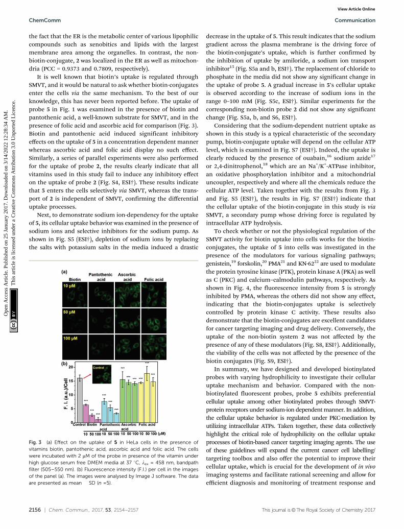
It is well known that biotin’s uptake is regulated throughSMVT, and it would be natural to ask whether biotin-conjugatesenter the cells via the same mechanism. To the best of ourknowledge, this has never been reported before. The uptake ofprobe 5 in Fig. 1 was examined in the presence of biotin andpantothenic acid, a well-known substrate for SMVT, and in thepresence of folic acid and ascorbic acid for comparison (Fig. 3).Biotin and pantothenic acid induced significant inhibitoryeffects on the uptake of 5 in a concentration dependent mannerwhereas ascorbic acid and folic acid display no such effect.Similarly, a series of parallel experiments were also performedfor the uptake of probe 2, the results clearly indicate that allvitamins used in this study fail to induce any inhibitory effecton the uptake of probe 2 (Fig. S4, ESI†). These results indicatethat 5 enters the cells selectively via SMVT, whereas the trans-port of 2 is independent of SMVT, confirming the differentialuptake processes.
Next, to demonstrate sodium ion-dependency for the uptakeof 5, its cellular uptake behavior was examined in the presence ofsodium ions and selective inhibitors for the sodium pump. Asshown in Fig. S5 (ESI†), depletion of sodium ions by replacingthe salts with potassium salts in the media induced a drastic
decrease in the uptake of 5. This result indicates that the sodiumgradient across the plasma membrane is the driving force ofthe biotin-conjugate’s uptake, which is further confirmed bythe inhibition of uptake by amiloride, a sodium ion transportinhibitor15 (Fig. S5a and b, ESI†). The replacement of chloride tophosphate in the media did not show any significant change inthe uptake of probe 5. A gradual increase in 5’s cellular uptakeis observed according to the increase of sodium ions in therange 0–100 mM (Fig. S5c, ESI†). Similar experiments for thecorresponding non-biotin probe 2 did not show any significantchange (Fig. S5a, b, and S6, ESI†).
Considering that the sodium-dependent nutrient uptake asshown in this study is a typical characteristic of the secondarypump, biotin-conjugate uptake will depend on the cellular ATPlevel, which is examined in Fig. S7 (ESI†). Indeed, the uptake isclearly reduced by the presence of ouabain,16 sodium azide17
or 2,4-dinitrophenol,18 which are an Na+/K+-ATPase inhibitor,an oxidative phosphorylation inhibitor and a mitochondrialuncoupler, respectively and where all the chemicals reduce thecellular ATP level. Taken together with the results from Fig. 3and Fig. S5 (ESI†), the results in Fig. S7 (ESI†) indicate thatthe cellular uptake of the biotin-conjugate in this study is viaSMVT, a secondary pump whose driving force is regulated byintracellular ATP hydrolysis.
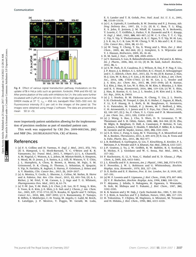
To check whether or not the physiological regulation of theSMVT activity for biotin uptake into cells works for the biotin-conjugates, the uptake of 5 into cells was investigated in thepresence of the modulators for various signaling pathways;genistein,19 forskolin,20 PMA21 and KN-6222 are used to modulatethe protein tyrosine kinase (PTK), protein kinase A (PKA) as wellas C (PKC) and calcium–calmodulin pathways, respectively. Asshown in Fig. 4, the fluorescence intensity from 5 is stronglyinhibited by PMA, whereas the others did not show any effect,indicating that the biotin-conjugates uptake is selectivelycontrolled by protein kinase C activity. These results alsodemonstrate that the biotin-conjugates are excellent candidatesfor cancer targeting imaging and drug delivery. Conversely, theuptake of the non-biotin system 2 was not affected by thepresence of any of these modulators (Fig. S8, ESI†). Additionally,the viability of the cells was not affected by the presence of thebiotin conjugates (Fig. S9, ESI†).
In summary, we have designed and developed biotinylatedprobes with varying hydrophilicity to investigate their cellularuptake mechanism and behavior. Compared with the non-biotinylated fluorescent probes, probe 5 exhibits preferentialcellular uptake among other biotinylated probes through SMVT-protein receptors under sodium-ion dependent manner. In addition,the cellular uptake behavior is regulated under PKC-mediation byutilizing intracellular ATPs. Taken together, these data collectivelyhighlight the critical role of hydrophilicity on the cellular uptakeprocesses of biotin-based cancer targeting imaging agents. The useof these guidelines will expand the current cancer cell labelling/targeting toolbox and also offer the potential to improve theircellular uptake, which is crucial for the development of in vivoimaging systems and facilitate rational screening and allow forefficient diagnosis and monitoring of treatment response and
Fig. 3 (a) Effect on the uptake of 5 in HeLa cells in the presence ofvitamins biotin, pantothenic acid, ascorbic acid and folic acid. The cellswere incubated with 2 mM of the probe in presence of the vitamin underhigh glucose serum free DMEM media at 37 1C, lex = 458 nm, bandpathfilter (505–550 nm). (b) Fluorescence intensity (F.I.) per cell in the imagesof the panel (a). The images were analysed by Image J software. The dataare presented as mean � SD (n =5).
ChemComm Communication
Ope
n A
cces
s A
rtic
le. P
ublis
hed
on 2
5 Ja
nuar
y 20
17. D
ownl
oade
d on
3/1
4/20
22 1
2:28
:34
AM
. T
his
artic
le is
lice
nsed
und
er a
Cre
ativ
e C
omm
ons
Attr
ibut
ion
3.0
Unp
orte
d L
icen
ce.
View Article Online
This journal is©The Royal Society of Chemistry 2017 Chem. Commun., 2017, 53, 2154--2157 | 2157
more importantly patient satisfaction allowing for the implementa-tion of precision medicine as part of standard patient care.
This work was supported by CRI (No. 2009-0081566, JSK)and NRF (No. 2015R1A5A1037656, CK) of Korea.
References1 (a) F. S. Collins and H. Varmus, N. Engl. J. Med., 2015, 372, 793;
(b) S. Hawgood, I. G. Hook-Barnard, T. C. O’Brien and K. R.Yamamoto, Sci. Transl. Med., 2015, 7, 300ps17; (c) L. A. Chantrill,A. M. Nagrial, C. Watson, A. L. Johns, M. Martyn-Smith, S. Simpson,S. Mead, M. D. Jones, J. S. Samra, A. J. Gill, N. Watson, V. T. Chin,J. L. Humphris, A. Chou, B. Brown, A. Morey, M. Pajic, S. M.Grimmond, D. K. Chang, D. Thomas, L. Sebastian, K. Sjoquist,S. Yip, N. Pavlakis, R. Asghari, S. Harvey, P. Grimison, J. Simes andA. V. Biankin, Clin. Cancer Res., 2015, 21, 2029–2037.
2 (a) A. Monica, V. Cecile, L. Sherene, L. Celine, M. Stefan, B. Herveand A. Fabrice, Nat. Rev. Clin. Oncol., 2015, 12, 693–704; (b) A. L.Richer, J. M. Friel, V. M. Carson, L. J. Inge and T. G. Whitsett,Pharmgenomics Pers. Med., 2015, 8, 63–79.
3 (a) Y.-W. Jun, Y.-M. Huh, J.-S. Choi, J.-H. Lee, H.-T. Song, S. Kim,S. Yoon, K.-S. Kim, J.-S. Shin, J.-S. Suh and J. Cheon, J. Am. Chem.Soc., 2005, 127, 5732–5733; (b) N. Kosaka, H. Iguchi and T. Ochiya,Cancer Sci., 2010, 101, 2087–2092; (c) S. Ramaswamy, P. Tamayo,R. Rifkin, S. Mukherjee, C.-H. Yeang, M. Angelo, C. Ladd, M. Reich,E. Latulippe, J. P. Mesirov, T. Poggio, W. Gerald, M. Loda,
E. S. Lander and T. R. Golub, Proc. Natl. Acad. Sci. U. S. A., 2001,98, 15149–15154.
4 (a) C. A. Lipinski, F. Lombardo, B. W. Dominy and P. J. Feeney, Adv.Drug Delivery Rev., 1997, 23, 3–25; (b) R. C. Bast, T. L. Klug,E. S. John, E. Jenison, J. M. Niloff, H. Lazarus, R. S. Berkowitz,T. Leavitt, C. T. Griffiths, L. Parker, V. R. Zurawski and R. C. Knapp,N. Engl. J. Med., 1983, 309, 883–887; (c) W. C. S. Cho, T. T. C. Yip,C. Yip, V. Yip, V. Thulasiraman, R. K. C. Ngan, T.-T. Yip, W.-H. Lau,J. S. K. Au, S. C. K. Law, W.-W. Cheng, V. W. S. Ma and C. K. P. Lim,Clin. Cancer Res., 2004, 10, 43–52.
5 (a) W. Yang, Y. Cheng, T. Xu, X. Wang and L. Wen, Eur. J. Med.Chem., 2009, 44, 862–868; (b) J. Zempleni, S. S. Wijeratne andY. I. Hassan, BioFactors, 2009, 35, 36–46.
6 H. M. Said, J. Nutr., 1999, 129, 490S–493S.7 (a) V. Kansara, S. Luo, B. Balasubrahmanyam, D. Pal and A. K. Mitra,
Int. J. Pharm., 2006, 312, 43–52; (b) H. M. Said, Subcell. Biochem.,2012, 56, 1.
8 (a) S. W. Park, D. E. Casalena, D. J. Wilson, R. Dai, P. P. Nag, F. Liu,J. P. Boyce, J. A. Bittker, S. L. Schreiber, B. C. Finzel, D. Schnappingerand C. C. Aldrich, Chem. Biol., 2015, 22, 76–86; (b) R. Kumar, J. Han,H.-J. Lim, W. X. Ren, J.-Y. Lim, J.-H. Kim and J. S. Kim, J. Am. Chem.Soc., 2014, 136, 17836–17843; (c) M. H. Lee, J. L. Sessler andJ. S. Kim, Acc. Chem. Res., 2015, 48, 2935–2946; (d) R. Kumar,E.-J. Kim, J. Han, H. Lee, W. S. Shin, H. M. Kim, S. Bhuniya, J. S. Kimand K. S. Hong, Biomaterials, 2016, 104, 119–128; (e) W. S. Shin,J. Han, R. Kumar, G. G. Lee, J. L. Sessler, J.-H. Kim and J. S. Kim,Sci. Rep., 2016, 6, 29018.
9 (a) J. I. Stuckey, B. M. Dickson, N. Cheng, Y. Liu, J. L. Norris,S. H. Cholensky, W. Tempel, S. Qin, K. G. Huber, C. Sagum, K. Black,F. Li, X.-P. Huang, B. L. Roth, B. M. Baughman, G. Senisterra,S. G. Pattenden, M. Vedadi, P. J. Brown, M. T. Bedford, J. Min,C. H. Arrowsmith, L. I. James and S. V. Frye, Nat. Chem. Biol., 2016,12, 180–187; (b) J. Su, F. Chen, V. L. Cryns and P. B. Messersmith,J. Am. Chem. Soc., 2011, 133, 11850–11853.
10 (a) J. Wang, S. Rao, J. Chu, X. Shen, D. N. Levasseur, T. W.Theunissen and S. H. Orkin, Nature, 2006, 444, 364–368; (b) H. Zhu,M. Bilgin, R. Bangham, D. Hall, A. Casamayor, P. Bertone, N. Lan,R. Jansen, S. Bidlingmaier, T. Houfek, T. Mitchell, P. Miller, R. A. Dean,M. Gerstein and M. Snyder, Science, 2001, 293, 2101–2105.
11 (a) S. R. Sirsi, C. Fung, S. Garg, M. Y. Tianning, P. A. Mountford andM. A. Borden, Theranostics, 2013, 3, 409–419; (b) X. Liu, B. Testa andA. Fahr, Pharm. Res., 2011, 28, 962–977.
12 J. B. Rothbard, S. Garlington, Q. Lin, T. Kirschberg, E. Kreider, P. L.McGrane, P. A. Wender and P. A. Khavari, Nat. Med., 2000, 6, 1253–1257.
13 J.-P. Gratton, J. Yu, J. W. Griffith, R. W. Babbitt, R. S. Scotland,R. Hickey, F. J. Giordano and W. C. Sessa, Nat. Med., 2003, 9,357–362.
14 P. Kucheryavy, G. Li, S. Vyas, C. Hadad and K. D. Glusac, J. Phys.Chem. A, 2009, 113, 6453–6461.
15 J. L. Kinsella and P. S. Aronson, Am. J. Physiol., 1981, 241, F374–F379.16 F. Proverbio, J. W. L. Robinson and G. Whittembury, Biochim.
Biophys. Acta, Biomembr., 1970, 211, 327–336.17 D. E. Keilin and E. F. Hartree, Proc. R. Soc. London, Ser. B, 1939, 127,
167–191.18 (a) W. F. Loomis and F. Lipmann, J. Biol. Chem., 1948, 173, 807–808;
(b) V. P. Skulachev, Biochim. Biophys. Acta, 1998, 1363, 100–124.19 T. Akiyama, J. Ishida, S. Nakagawa, H. Ogawara, S. Watanabe,
N. Itoh, M. Shibuya and Y. Fukami, J. Biol. Chem., 1987, 262,5592–5595.
20 K. B. Seamon and J. W. Daly, J. Cyclic Nucleotide Res., 1981, 7, 201–224.21 T. J. Rink, A. Sanchez and T. J. Hallam, Nature, 1983, 305, 317–319.22 H. Tokumitsu, T. Chijiwa, M. Hagiwara, A. Mizutani, M. Terasawa
and H. Hidaka, J. Biol. Chem., 1990, 265, 4315–4320.
Fig. 4 Effect of various signal transduction pathway modulators on theuptake of 5 in HeLa cells such as genistein, forskolin, PMA and KN-62. (a)After preincubation of the cells with inhibitors for 1 h. the cells were furtherincubated with 2 mM of a probe for 10 min. Under high glucose serum freeDMEM media at 37 1C. lex = 458 nm, bandpath filter (505–550 nm). (b)Fluorescence intensity (F.I.) per cell in the images of the panel (a). Theimages were obtained using Image J software. The data are presented asmean � SD (n =5).
Communication ChemComm
Ope
n A
cces
s A
rtic
le. P
ublis
hed
on 2
5 Ja
nuar
y 20
17. D
ownl
oade
d on
3/1
4/20
22 1
2:28
:34
AM
. T
his
artic
le is
lice
nsed
und
er a
Cre
ativ
e C
omm
ons
Attr
ibut
ion
3.0
Unp
orte
d L
icen
ce.
View Article Online