A List of Candidate Cancer Biomarkers for Targeted Proteomics

Targeted Proteomics Applied in Clinical Diagnostics and
Doping Analysis
- Immuno-MS based Determination of Human Chorionic
Gonadotropin
Thesis for the degree of Philosophiae Doctor
by
Hanne Lund
Department of Pharmaceutical Chemistry
School of Pharmacy
University of Oslo
Norway

© Hanne Lund, 2013 Series of dissertations submitted to the Faculty of Mathematics and Natural Sciences, University of Oslo No. 1325 ISSN 1501-7710 All rights reserved. No part of this publication may be reproduced or transmitted, in any form or by any means, without permission. Cover: Inger Sandved Anfinsen. Printed in Norway: AIT Oslo AS. Produced in co-operation with Akademika publishing. The thesis is produced by Akademika publishing merely in connection with the thesis defence. Kindly direct all inquiries regarding the thesis to the copyright holder or the unit which grants the doctorate.
CONTENTS
ACKNOWLEDGEMENTS ........................................................................................................................................... 1
LIST OF PAPERS ......................................................................................................................................................... 3
ABBREVATIONS ......................................................................................................................................................... 4
ABSTRACT ................................................................................................................................................................... 6
1. INTRODUCTION ................................................................................................................................................. 9
1.1 Proteomics .................................................................................................................................................... 9
1.2 Targeted proteomics approach by SRM ..................................................................................................... 10
Principle and background .................................................................................................................. 10 1.2.1
Tryptic digestion and signature peptides............................................................................................ 11 1.2.2
Analytical technique: LC-MS/MS ..................................................................................................... 12 1.2.3
Sample preparation: Immunoaffinity extraction proceeding MS analysis ......................................... 12 1.2.4
Quantification of proteins .................................................................................................................. 14 1.2.5
Immuno-MS strategy ......................................................................................................................... 17 1.2.6
1.3 Human chorionic gonadotropin: a diverse biomarker ................................................................................. 17
Molecular structure and biochemistry ................................................................................................ 18 1.3.1
Clinical proprieties ............................................................................................................................. 22 1.3.2
hCG in doping analysis ...................................................................................................................... 24 1.3.3
hCG detection and immunoassays ..................................................................................................... 25 1.3.4
hCG and mass spectrometry .............................................................................................................. 26 1.3.5
2. AIM OF THE STUDY ........................................................................................................................................ 27
3 RESULTS AND DISCUSSION .......................................................................................................................... 28
3.1 Identification and qualitative differentiation between hCG variants using LC-MS ................................... 28
Theoretical selection of signature peptides ........................................................................................ 28 3.1.1
LC-MS analysis: hCG peptide mapping and detection of signature peptides .................................... 32 3.1.2
Pregnyl as hCG source ....................................................................................................................... 35 3.1.3
Multiplexing hCG identification through LC-MS based detection .................................................... 35 3.1.4
3.2 Compatibility of immunoaffinity extraction with mass spectrometric detection ........................................ 36
3.3 Optimizing method sensitivity and specificity ........................................................................................... 38
Tailored selected reaction monitoring design .................................................................................... 38 3.3.1
Immunoextraction using beads in stead of wells ............................................................................... 42 3.3.2
3.4 Validation of method for quantitative determination of hCG ..................................................................... 45
3.5 hCG immuno-MS in clinical diagnostics .................................................................................................... 48
Pregnancy and cancer diagnostics ...................................................................................................... 48 3.5.1
Evaluation of anti-hCG antibody selectivity and specificity .............................................................. 52 3.5.2

3.6 hCG immuno-MS in doping analysis ......................................................................................................... 56
Clinical study ..................................................................................................................................... 56 3.6.1
Comparison of hCG immuno-MS method to immunometric assay ................................................... 60 3.6.2
3.7 Future perspectives ..................................................................................................................................... 65
CONCLUDING REMARKS ....................................................................................................................................... 67
REFERENCES............................................................................................................................................................. 68

1
ACKNOWLEDGEMENTS
The work presented in this thesis is based on research performed at Department of
Pharmaceutical Chemistry, School of Pharmacy, University of Oslo, in the period between
January 2008 and December 2012.
When setting out on a PhD project, one can hardly imagine that one day the project will actually
end. Strangely, it does. This long and bumpy road would have seemed endless and completely
impossible had it not been for rock solid support along the way. Therefore, to my outstanding,
inspiring, challenging, steady, wise and warm supervisors, Trine and Leon, thank you for your
support. I’ve appreciated it more than I’ve been able to express. Also, I’d like to thank Elisabeth
at Radiumhospitalet for providing excellent antibodies, and for always giving me a different
perspective when stuck on scientific issues. Your contribution to my work has been priceless, and
it is greatly appreciated.
Of course, what is a PhD student without her colleagues and fellow students? There are so many
of you, and I’ve enjoyed working with every single one of you. Bjørn, Håvard, Astrid, Stig, Knut,
Finn, Marte P and Peter Hemmersbach. Great colleagues. Cecilia, Silje, Lars Erik, Siri V, Siri H
and Knut F. From you I’ve learnt that coffee and a good sense of humor will solve basically any
problem. Furthermore, the years I’ve spent sharing office with Marte has been brilliant. I think
we might have come up with the perfect work/talk ratio; do you care to make a publication on the
subject?
My dear master students (some of you now PhD students yourself, yeah!); Silje and Siri V once
more, Karoline, Ann Helene and Tamara, the amount of lab work you guys have carried out on
the notorious hCG molecules is tremendous. So thank you.
For the co-authors not mentioned in any section above, Kjell Nustad, Peter Berger, and Ulf-
Håkan Stenman, I’d like to extend my grateful regards for your valuable contributions to my
papers.
My family and friends, what you’ve endured these past years, listening to me going on and on
about subjects that I suspect might not have been crystal clear to you. Thank you for listening,
comforting, and encouraging me when necessary, and for sharing my joy when papers are

2
accepted, lab experiments have paid off, or other scientific successes have gone to my head. You
mean the world to me.
The best way to let a tired head rest is to get home to Oliver, my fantastic son who really doesn’t
care about my thesis, unless is can do a dance, sing a song or for some reason is eatable. Your
perspectives are truly… refreshing! Andreas, you have had (unrealistic) faith in my abilities to
conduct research, and have backed me up from the day I started my PhD studies. Particularly the
final months have been a challenge for the both of us, but somehow you’ve multitasked dinner,
day-care and your own job effortlessly. Being at home with you is my free zone. Life with you is
easy, and I love you for it.
Oslo, December 2012
Hanne Lund

3
LIST OF PAPERS
This thesis is based on the following papers which will be referred to by their roman numeral in
the text:
I H. Lund, S.B. Torsetnes, E. Paus, K. Nustad, L. Reubsaet, T.G. Halvorsen “Exploring
the complementary selectivity of immunocapture and MS detection for the differentiation between
hCG isoforms in clinically relevant samples”, J. Prot. Res, (2009)
II H. Lund, K. Løvsletten, E. Paus, T.G. Halvorsen, L. Reubsaet “Immuno-MS based
targeted proteomics: Highly specific, sensitive, and reproducible human chorionic gonadotropin
determination for clinical diagnostics and doping analysis”, Anal. Chem. (2012)
III H. Lund, A.H. Snilsberg, E. Paus, T.G. Halvorsen, P. Hemmersbach, L. Reubsaet
“Sports drug testing using immuno-MS: clinical study comprising administration of human
chorionic gonadotropin to males” Anal. Bioanal. Chem. (2012)
IV H. Lund, A.H. Snilsberg, T.G. Halvorsen, P. Hemmersbach, L. Reubsaet “Comparison of
newly developed immuno-MS method with existing DELFIA immunoassay for human chorionic
gonadotropin determination in doping analysis” Bioanalysis (2013)
V H. Lund, E. Paus, P. Berger, U.H. Stenman, T. Torcellini, T.G. Halvorsen, L. Reubsaet
“Epitope Analysis and Detection of human chorionic gonadotropin (hCG) Variants by
Monoclonal Antibodies and Mass Spectrometry” To be submitted to Tumor Biology
Papers not included in the dissertation:
VI M. Balchen, H. Lund, L. Reubsaet, S. Pedersen-Bjergaard “Fast, selective, and sensitive
analysis of low-abundance peptides in human plasma by electromembrane extraction“, Anal.
Chim. Acta (2012)
VII S.B. Torsetnes, S.G. Løvbak, H. Lund, M.S. Nordlund, E. Paus, T.G. Halvorsen, L.
Reubsaet “Immunocapture LC-MS/MS for selective quantification and differentiations of the
isozymes of the biomarker NSE” Submitted for publication

4
ABBREVATIONS
ABC Ammonium Bicarbonate
AQUA Absolute Quantification Peptides
BLAST Basic Local Alignment Search Tool
BSA Bovine Serum Albumin
CID Collision Induced Dissociation
CTP Carboxy-terminal peptide
ELISA Enzyme-Linked Immunosorbent Assay
EMEA European Medicines Agency
ESI Electrospray Ionization
FSH Follicle-Stimulating Hormone
HCl Hydrochloric Acid
HCOOH Formic Acid
HPLC High-Performance Liquid Chromatography
IS Internal Standard
IEF Isoelectric focusing
LC Liquid Chromatography
LOD Limit of Detection
LH Luteinizing Hormone
LLOQ Lower Limit of Quantification
MALDI Matrix-Assisted Laser Desorption Ionization
MeCN Acetonitirile
MRM Multiple Reaction Monitoring
MS Mass Spectrometry
MS/MS Tandem Mass Spectrometry
m/z Mass-to-charge Ratio
NBCI National Center for Biotechnology Information
NSE Neuron-Specific Enolase
QqQ Triple Quadrupole Detector
ProGRP Progastrin Releasing Peptide
PSAQ Protein Standards for Absolute Quantification

5
r Correlation Coefficient
R Recovery
RP-HPLC Reversed-Phase High-Performance Liquid Chromatography
RSD Relative Standard Deviation
SID Stable Isotope Dilution
SIM Selected Ion Monitoring
S/N Signal-to-Noise
SQ Single Quadrupole
SPE Solid Phase Extraction
SRM Selected Reaction Monitoring
TSH Thyroid-Stimulating Hormone
TFA Trifluoroacetic Acid
UniProtKB UniProt Knowledgebase
WADA World Anti-Doping Agency

6
ABSTRACT
The principal objective of this thesis was to develop a liquid chromatography (LC) mass
spectrometry (MS) based method using the targeted proteomics approach to determine low
abundance protein biomarkers in complex matrixes. To enable adequate sensitivity of the method,
immunoaffinity extraction was intended as sample preparation strategy preceding the LC-MS
analysis. Furthermore, the developed method’s application in clinically relevant scenarios was
shown. For this purpose the human chorionic gonadotropin (hCG) family of macromolecules
was chosen, as this diverse biomarker group is currently utilized in both pregnancy and cancer
diagnostics, in addition to being a sports drug exploited for doping purposes.
In Paper I the proof of principle for the immuno-MS methodology was demonstrated. First,
unique signature peptides representing the respective hCG proteins were established theoretically,
using protein data bases and search algorithms ensuring the specificity of each peptide.
Furthermore, following enzymatic cleavage of the hCG proteins into peptides using trypsin, the
experimentally obtained LC-MS analysis enabled the multiplexed separation and detection of
various hCG proteins in one single run, using a single quadrupole (SQ) detector in the selected
ion monitoring (SIM) mode. Thereafter, specific immunoaffinity extraction of the target hCG
proteins using a monoclonal antibody immobilized to the walls 96 wells microtiter plates
followed by in-well tryptic digestion, a solid phase extraction (SPE) clean-up step, and LC-MS
analysis demonstrated the proof of principle for combining the complementary techniques of
immunoaffinity extraction and mass spectrometric detection. This allowed the identification of
hCG from complex matrixes such as serum and urine. The developed immuno-MS method was
finally used for the analysis of authentic cancer patient serum samples, and urine samples from a
pregnant woman. Qualitative hCG differentiation of various hCG forms in these samples
indicated the potential of the method to provide complementary diagnostic information to that
derived from the conventional immunoassays.
In Paper II, quantitative validation of the method was described, along with its potential for use
in clinical settings. First, the preliminary LC-MS analysis developed on a SQ detector was
transferred to a triple quadrupole (QqQ) detector to enable selective, specific and sensitive
selected reaction monitoring (SRM) detection of the hCG signature peptides. Furthermore, the
antibodies used for immunocapture were transferred to magnetic beads for the selective

7
extraction of target molecules from large sample volumes. This resulted in substantial
improvement of the methods sensitivity and specificity. Subsequently, validation of the method
was performed according to key elements and recommendations defined by the European
Medicines Agency in Guideline on Validation of Bioanalytical Methods, demonstrating robust,
reproducible and accurate quantification of hCG from serum and urine, with satisfying limit of
detection (LOD) of 5 IU/L for serum and 2 IU/L for urine. The main hCG variant responsible for
the biological activity associated with the hCG molecules was quantified, whereas the other hCG
degradation variants were qualitatively detected and differentiated. The clinical potential of the
method was tested and resulted in quantitative measurements of hCG in cancer patients serum
samples, and of hCG in urine and serum samples from pregnant women.
Besides it quantitative ability, the developed method also allowed the evaluation of antibody
selectivity, thus showing a complementary utility for the developed method in clinical diagnostics
(Paper V). This was done since defined antibody selectivity is crucial in immunoassays to ensure
accurate detection of target compounds whilst discriminating other compounds. Defined hCG
standards were extracted using 30 different anti-hCG antibodies, and analyzed using the tailored
hCG LC-MS/MS detection. The obtained results were further compared to those generated by an
hCG radioimmunoassay performed in a parallel study, and the antibodies were classified in
groups according to epitope recognition. The purity of the hCG standards was evaluated as well.
To investigate the method’s applicability in doping analysis a clinical study comprising the
administration of one out of two hCG containing pharmaceutical to 24 males was conducted
(Paper III). Serum and urine samples were collected prior to hCG injection, and for a period of
14 days following hCG injection. The analysis of the samples using the developed hCG immuno-
MS method showed that hCG could, in average, be detected for 7 days in serum following hCG
injection. The window of detection in urine was 10 days following hCG injection. This was
accomplished at LOQs as defined by World Anti-Doping Agency (WADA). Thereafter, hCG
variant patterns a function of injected hCG drug was studied, followed by interpretation of hCG
pattern complexity related to biological matrix.
Paper IV describes the comparison of the developed hCG immuno-MS method’s performance to
that of the existing reference immunometric method; the DELFIA assay. Windows of detection

8
and differences in hCG measurements were compared and discussed as a function of method
selectivity and choice of matrix.

9
1. INTRODUCTION
1.1 Proteomics
The field of proteomics entails the study of all aspects of protein properties, from expression and
profiling, to modifications and interactions. The information obtained from the identification and
quantification of proteins can impact broadly on biology and medicine when used for clinical
applications or biomedical research [1]. The enormous potential of protein biomarkers to
revolutionize clinical practice and thus improve patient care through molecular based diagnostics
has been thoroughly described [2,3]. Also the rapidly evolving peptide and protein drugs can be
characterized and their pharmacokinetics effects studied based on applied proteomics [4,5].
Mass spectrometry (MS) has increasingly become the method of choice for analysis of complex
protein samples, largely due to the development of soft ionization techniques such as electrospray
ionization (ESI) and matrix-assisted laser desorption/ionization (MALDI) [1]. The suitability of
MS analysis to complex proteomics studies is related to the unique features of the instruments in
terms of resolution and mass accuracy, combined with sensitivity and the ability to generate
specific mass spectra for the detected peptide ions [1]. In general two MS-based proteomics
approaches applied for protein analysis are the top-down approach where intact proteins are
analyzed, and the bottom-up approach which involves the enzymatic conversion of proteins into
their constituent peptides [6]. Whereas the analysis of intact proteins is greatly challenged by the
lack of sensitivity as a function of charge distribution and thus poor efficiency of ionization, the
bottom-up approach has gained territory as substantially better sensitivity is provided by
chromatographic separation of the generated peptides prior to MS analysis [7], hereby enabling
efficient ionization of the separated peptides. This bottom-up strategy can further be divided into
discovery or targeted experiments. Proteomics discovery experiments aim at identifying the
detectable protein content of a sample and are referred to as “shotgun” proteomics. This is carried
out through the interpretation of the generated mass spectra by database searching, and requires
thus high performance of the analysis in order to provide proteomics data that can be fully
interpreted to generate testable conclusions, regarding i.e. biomarker discovery, identification and
characterization [1]. Figure 1.1 summarizes a general path for bottom-up proteomic analysis.
However, targeted proteomics is increasingly used in clinical validation and diagnostic method
development, and for efficient and rational applicability in this setting some of the high

10
performance of the analysis is exchanged for higher throughput, robustness and simplicity [8].
For the targeted study of the behavior of known sets of proteins sensitive and accurate
quantification can be obtained through the selected reaction monitoring (SRM) technique [9,10].
Figure 1.1 General flow scheme for proteomic analysis (From reference [11], with permission)
1.2 Targeted proteomics approach by SRM
Principle and background 1.2.1
In targeted proteomics by SRM the MS is programmed to detect a preselected protein or group of
proteins. This technique is comparable to the selected ion monitoring (SIM) technique, in which
the intensities of several preselected specific m/z values are recorded rather than the entire mass
spectrum, resulting in increased sensitivity of the MS analysis. Further improvement of
sensitivity is obtained through the extension of SIM detection into SRM detection.
The development of the SRM technique [12,13] came along with the development of the first
triple quadrupole instruments in the late 1970s [14]. In clinical applications this has been a
reference quantitative technique for the analysis of smaller molecules for 30 years [9]. However
the recent years methodological developments have made targeted proteomics analysis by SRM-
MS a method of increasing interest [15]. SRM assays are developed on triple quadrupole
instruments (QqQ) to detect peptide ions diagnostic for the parent proteins, hereby enabling

11
sensitive, reproducible and quantitatively accurate measurements for the specific identification
and quantification of target proteins [10,16,17]. The procedure entails the enzymatic conversion
of proteins into peptides, followed by LC separation prior to SRM-MS analysis by the QqQ.
Peptides entering the first quadrupole are mainly doubly or triply charged. In the collision cell
they are further subjected to fragmentation, resulting in fragment ions. A few specific fragments
are further monitored for detection and quantification purposes [9]. This will be more thoroughly
explained in the following sections.
Tryptic digestion and signature peptides 1.2.2
The enzymatic conversion of proteins into peptides can be effectuated by trypsin, which is the
most widely used approach [11]. The specific cleaving at the C-terminal of the amino acids lysine
and arginine in the protein backbone generally results in peptide sequences of shorter size. In
addition to the charge of the N-terminal of the peptides, the lysine and arginine residues impose a
second charge on the peptides, making them suitable for electrospray ionization. These
characteristics are favorable for MS detection of peptides.
Amongst the peptides generated from enzymatic cleaving of a protein, certain peptides are
composed of unique amino acid sequences that are specific for the parent protein. These
signature peptides serve as such as diagnostic representatives for the detection of the parent
protein. Additionally, for accurate quantification of the target proteins, these signature peptides
also have to be stoichiometric representatives. This requires that the proteolysis is complete, or at
least has reached an end-point that is consistent among samples [18,19].
This detection of a unique structural component of the target molecules replaces the far more
complicated detection of intact proteins, hereby enabling sensitivity and selectivity crucial for the
quantification of low abundance proteins in complex biological matrixes. As such, this principle
has been demonstrated by several research groups [16,20-23,18,19,24]. The advantages related to
this strategy might lead to the increasing application of the SRM technology to larger molecules,
such as proteins, for analyses wherein the specific identification and quantification of the target
macromolecule is important.

12
Analytical technique: LC-MS/MS 1.2.3
The LC-SRM-based analysis of peptides is based on the complementary features and sequential
organization of LC separation of peptides, electrospray conversion of charged peptides in the
liquid phase to ions in the gas phase, and the selection and fragmentation of target peptides in the
triple quadrupole mass spectrometer.
Of particular relevance in peptide analysis is the ability of the ESI, which is a soft ionization
technique, to leave the peptides intact prior to entering the mass analyzer. Furthermore, the first
quadrupole selects target peptide ions based on m/z values programmed to the QqQ (Figure 1.2
A), and introduces these to the second quadrupole where they are subjected to fragmentation by
collision induced dissociation (CID). This fragmentation technique produces mainly cleavages
along the protein backbone between the carbonyl oxygen and the amide nitrogen, resulting in b-
fragment ions (counting from the C-terminal of the peptide) and complementary y-fragment ions
(counting from the N-terminal of the peptide) (Figure 1.2 B). Depending on signal intensity and
specificity of the generated fragment ions in the method optimization process, a selection of
fragment ions to be monitored by the third quadrupole is made.
The discriminating character of the QqQ combined with the LC separation of peptides enhances
selectivity and sensitivity of the LC-MS/MS method, which is fundamental in biomarker
determination. An additional and highly important feature of the complementary features of LC-
MS is the potential to multiplex the detection of a preselected group of proteins in one single run
[25,26].
Sample preparation: Immunoaffinity extraction proceeding MS analysis 1.2.4
Although the discriminating ability of the MS to selectively lock on to preprogrammed m/z
values and hereby exclude components of diverging m/z values is indeed effective, a sample
preparation strategy is needed to enable accurate and sensitive analysis of low abundance target
molecules in complex matrixes such as serum and urine. A traditional sample preparation
technique much applied in proteomics is sample fractionation using either one- or two-
dimensional gel electrophoresis, where proteins are separated according to molecular weight
and/or on the basis of isoelectric point by isoelectric focusing (IEF) [11]. A protein depletion step
can either be carried out independently or be combined with sample fraction depending on the
abundance concentration of the target analyte. In protein depletion abundant serum proteins are

13
A
B
Figure 1.2 A) SRM analysis on QqQ MS. Selection and filtering of co-eluting compounds
according to m/z values, followed by fragmentation and m/z based selection of fragments for
specific detection. (From reference [10], with permission) B) Representation of a peptide as a
construct of amino acid building blocks. The specific cleavage of the protein backbone by CID
generates complementary b- and y- ions
removed using immunoaffinity depletion columns [27], hereby greatly reducing serum
complexity. However, immunoaffinity enrichment of target analytes short-circuits the need for
abundant protein depletion and fractionation before SRM-MS, and has emerged as a selective and
compatible technique suitable for coupling to mass spectrometric detection allowing enrichment
of a target protein as much as 1000-fold [28-30].
In a historical perspective the development of immunoaffinity-based MS approaches followed the
demonstration of the first MALDI and ESI-MS protein analysis in the late 1980s [31-33] as a
promising strategy for solving the obvious sensitivity issues related to the MS detection of
macromolecules. Selective isolation and extraction of proteins is possible when selective and
specific antibodies directed towards epitopes on the target protein exist. If the target consists of a

14
group of different proteins, this can be achieved by combining a selection of antibodies targeted
towards the different proteins [34]. Another approach is the use one antibody that is targeted
towards a common epitope on all the target proteins [35].
Different formats can be applied for immunoaffinity sample preparation preceding MS analysis,
depending on whether the method aims at an on-line or off-line set-up. Immunoaffinity columns
can be used for both set-ups. However, since tryptic digestion is a feature of the MS-based
analysis of larger proteins, the on-line approach will be complicated. The off-line mode has been
demonstrated for the purification of proteins prior to tryptic digestion and MS detection [36,37].
Other formats are the use of conventional 96-wells format plates, as applied for immunometric
assays [35], and the immobilization of monoclonal antibodies to magnetizable particles (beads)
for the extraction from larger samples volumes [38]. The enrichment of tryptic peptides by anti-
peptide antibodies immobilized in nano-affinity columns has also been demonstrated [39]. This
approach entitled SISCAPA (Stable Isotope Standards and Capture by Anti-Peptide Antibodies)
is however restricted by the tryptic fragment selected for analysis, leaving no possibility to
change or include other signature peptides to the method. Furthermore the antibodies targeted
towards tryptic protein fragments in stead of proteins tend to be less antigenic based on size
considerations.
Successful applications of quantitative peptide hormone analysis enabled by immunoaffinity
extraction and MS detection (hereafter referred to as immuno-MS) have been demonstrated, such
as the analysis of peptide hormones in doping analysis [34]. Other applications involving
immunoextraction and MS detection of target proteins through diagnostic signature peptides have
been shown for the analysis of protein biomarkers in clinical diagnostics [40-42,22].
The above argues the potential of the immuno-MS strategy as analytical methodology for the
determination of target proteins in complex biological matrixes.
Quantification of proteins 1.2.5
Due to differences in ionization efficiency between compounds it is not possible to give accurate
quantitative measurements of target compounds based on their MS signal intensities alone. As
such MS is not inherently a quantitative technique, and to achieve accurate and precise

15
quantitative measurements in targeted proteomics the established principle of stable-isotope
dilution (SID)-MS can be employed [43,44].
SID-MS entails the addition of a known amount of isotopically labeled standard to the sample,
having the same physicochemical proprieties as the analyte. Chromatographic co-elution and
equal efficiency of ionization is thus obtained for the internal standard (IS) and the target analyte.
They are however separated in the mass spectrometer due to differences in mass (Figure 1.3).
The appropriate point in the workflow to add an IS to the sample is dependent on the nature of
the standard used (Figure 1.3). Proteins standards for absolute quantification (PSAQ) are
recombinant isotope-labeled protein analogues to the target proteins [45], and should thus be
added immediately (or as soon as possible), in defined amounts, to the sample. This is also the
case for the FLEXIQuant method, where labeled protein analogues are flanked by a FLEX
peptide which is used for their calibration [46]. Both these strategies will completely overcome
any problems associated with differential digestion, which is a major issue for many
quantification strategies. However, they require quantification of each standard separately, which
limits their strength as multiplexed strategy, increasing costs and decreasing throughput [43].
Multiplexed quantification using artificial QCAT (concatenation of tryptic peptides) proteins
(QconCAT) [47] and the Protein Epitope Signature Tag (PrEST) [48] quantification strategy are
techniques designed for multiplexed quantification. Their construction requires addition of
standard prior to digestion, in order to release the signature peptide to the sample (Figure 1.3).
Although both standards contain the target signature peptide in their protein backbone, they are
not protein analogues to the target proteins; the QconCAT protein is designed and synthetized to
contain the desired signature peptides in one larger protein [47], whereas the PrEST peptides are
shorter protein fragments produced by the Human Protein Atlas (http://www.proteinatlas.org/)
where they are used as antigens for antibody production [48]. Their relatively simple structure
(linear protein/peptide sequences) ensures near complete digestion. This, however, might not be
the case for the target proteins. Together with the strategy of the absolute quantification peptides
(AQUA), these strategies thus require strict control of completion of digestion in order to provide
accurate quantitative ratios for the added amount of IS and the generated signature peptide being
a stoichiometric representative of the target protein.

16
The AQUA peptides are isotopologes to the target signature peptides [49,50], and are thus
primarily added before LC-MS analysis. Given that the process of digestion is thoroughly
explored and validated, and is further proven to reach an end-point, the use AQUA peptides can
provide advantages such as limited in-house expertise in the preparation as these can be
purchased from several commercial companies. All in all the simplicity of use combined with
reliable quality, reasonable costs and accessibility are advantages that should be taken into
account when choosing internal standards for assays designed with inter-laboratory utility in
mind.
Figure 1.3 Strategies for absolute quantification of target proteins by various SID techniques.
The addition of the five different types of standards to the sample is dependent on the nature of
the standard; if the signature peptide needs to be released from the standard prior to LC-MS/MS
analysis, then the standard is added prior to enzymatic digestion (PSAQ, FLEXIQuant, QconCAT
and PrEST). Only standards that are protein analogues to the target proteins can be added prior
to prefractionation of the proteins (PSAQ and FLEXIQuant).The synthetic isotopically labeled
signature peptides (AQUA peptides) are added prior to LC-MS/MS analysis

17
Immuno-MS strategy 1.2.6
On the basis of what has been presented above, a strategy for absolute quantification of multiple
target proteins based on immunoaffinity extraction and bottom-up MS detection can be outlines
as summarized in Figure 1.4. This strategy involves SID-MS using AQUA peptides for
quantification purposes, prior to LC separation and tailored SRM-MS detection of signature
peptides.
Figure 1.4 Immuno-MS strategy for quantification of low-abundance protein biomarkers in
complex biological matrixes
1.3 Human chorionic gonadotropin: a diverse biomarker
Effective management of patients is funded on the ability to provide early and reliable diagnosis
and prognosis. Accurate determination of established and reliable biomarkers aims at providing
clinical information valuable for the interpretation of disorders. Further monitoring of disease
progression and response to therapy can often be measured in terms of biomarker presence, up-
or down-regulation. Since many biomarkers are proteins the use of targeted proteomics applied in

18
a clinical setting is promising, for all the reasons stressed in previous sections. However, the
application of MS-based clinical diagnostics on macromolecules has only been tailored for a few
proteins. The human chorionic gonadotropin family represents a diverse group of proteins whose
diagnostic value has been well documented. As such this is an interesting candidate for tailored
MS-based detection using the targeted proteomics approach.
Molecular structure and biochemistry 1.3.1
hCG is a highly glycosylated protein (37.5 kDa), and is part of the glycoprotein hormone family
which also includes luteinizing hormone (LH), follicle-stimulating hormone (FSH), and thyroid-
stimulating hormone (TSH). These hormones are heterodimers comprising a common α-subunit
(92 amino acids) and a specific β-subunit. The latter confers biological activity, and is specific for
each hormone. The hCG β-subunit is 145 amino acids long. The assembly (Figure 1.5) of the two
hCG subunits is non-covalent, and dissociation of the subunits might thus occur. Internal
stabilization is conferred by three disulfide bonds forming a cysteine knot. One-third of the
molecular mass is made up by eight carbohydrate moieties, of which six are attached to the β-
subunit and two to the α-subunit (Figure 1.7). These sugar moieties are either N-linked (linked to
asparagine residues) or O-linked (linked to serine residues), and vary in size, possibly resulting in
hyperglycosylated variants. Within each subunit, several disulfide bonds contribute to the tertiary
molecular structure. This results in the formation of several loops in the protein backbone, which
have been given designations according to their position counting from the N-terminal (Figure
1.5). The c-terminal peptide (CTP) constitutes the part from amino acids 114-145 in the hCG β-
subunit backbone, and this is a highly glycosylated region in the protein with four O-linked
carbohydrate groups [51,52].
The hCG molecule is not a single molecule although it is often referred to as one. It is a
heterogeneous molecule that can be diverted into variants having different cell origins and thus
different biological effects [53]. They all share the same protein backbone core, referred to as
hCG β-core fragment (Figure 1.7), but dissociation of heterodimers into free subunits, differences
in glycosylation, and nicking in the protein backbone might occur, resulting in a variety of hCG
molecules. These variants are schematically presented in figure 1.7.

19
Figure 1.5 A) Assembly of α- and β-subunit to form hCG heterodimer B) Spatial representation
of the subunit assembly in which the hCG α carboxy-terminal extension penetrates the hCG β and
is locked in a seatbelt configuration by the β-subunit. (From reference [54], with permission)
The main hCG variant is the intact hCG (αβ heterodimer), which also displays the main
biological function provided by hCG. Degradation of this molecule by metabolism is mainly the
cause of variation in hCG structure. Once released from the cell to the circulation (Figure 1.6)
degradation processes occur, resulting in degraded hCG variants that are rapidly cleared from the
circulation [52,55]. Whereas the intact hCG has a metabolic clearance half-life of ~ 36 hours, the
corresponding half-life of the free hCG β-subunit is ~ 4 hours. The degradation variants are thus
more likely to be found in urine than in serum [55].
When cleaving of the bond linking two amino acids in the protein backbone occurs as part of the
degradation process, this is called nicking. This occurs mainly in the amino acids 44-48 in the
backbone, and the most common variants are the ones displaying nick between amino acids 47/48
and 44/45 [35]. Nicking of hCG happens in both normal and pathological conditions, and seems
to be executed by proteases present in the circulation and the kidneys. The precise types of

20
proteases, however, remain yet to be elucidated [56-58]. Further degradation of the molecule by
proteases results in the ultimate hCG degradation variant, the hCG β-core fragment.
Figure 1.6 hCG variants in placenta, blood and urine. Degradation pathways, dissociation and
nicking of the hCG molecules. Large free α refers to hyperglycosylated α-subunit. (From
reference [54], with permission)

21
Figure 1.7 Outline of the structure of the 15 common hCG variants present in serum and urine
samples in either pregnancy, gestational disease or other malignancy. Numbers refer amino acid
numbers, O refers to O-linked and N to N-linked oligosaccharides. OO and NN refer to large or
hyperglycosylated oligosaccharides. βCTP is the C-terminal segment (residues 93-145) on the
hCGβ-subunit (From reference [55], with permission)

22
The different hCG variants presented in Figure 1.7 include all possible variants as a function of
dissociation, nicking, degradation and hyperglycosylation. This extensive variation is not
particularly relevant per se. Furthermore, from a methodological point of view the differentiation
based on variation of the sugar groups is quite complicated to execute. The structural variation is
thus often limited to that of the differences in the protein backbone, resulting in the pragmatic
classification of hCG molecules into the intact hCG, the free hCG β-subunit, the hCG β-core
fragment, and the nicked variants [59,60]. This classification has been recognized, and standards
have been produced for these hCG variants including the α-subunit [59].
The area of a molecule that is specifically recognized by antibodies is referred to as the epitope of
the molecule, and several epitopes might exist on the same compound. For hCG, the different
epitopes are classified according to the epitope cluster that they are in the proximity of, primarily
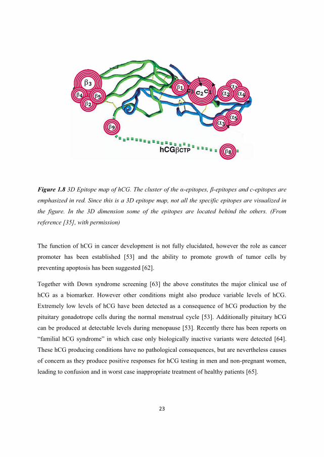
dividing the epitopes into groups of α-epitopes, β-epitopes or c-epitopes (Figure 1.8) [35]. Within
these α-, β- and c-epitope groups further sub-classification more precisely defines the epitopes’
exact position. The molecular epitope structure of hCG has been a topic of interest for some years
[35] since this family of diverse molecules poses challenges in terms of antibody-based detection.
This is related to the large number of hCG epitopes as a function of structure and subunit
assembly diversity. This will be more thoroughly discussed below.
Clinical proprieties 1.3.2
The function of hCG is mainly to maintain the progesterone production of corpus luteum during
early pregnancy. As such hCG is principally produced during the full course of pregnancy,
hereby controlling placenta, uterine and fetal growth and/or differentiation [53]. It is thus the
hormone that is used for pregnancy detection and monitoring of pregnancy. Quantitative
determinations of hCG are also used for assessment of disorders of pregnancy, hereunder the
prediction of complications especially in early pregnancy, e.g. pregnancy loss and ectopic
pregnancy [52].
hCG is also an extremely sensitive and specific marker for gestational throphoblastic disease
(GTD, pregnancy-related tumors) and for some germ cell tumors of the testis. The sensitivity and
specificity of hCG measurements in GTD approach 100%, while approximately 50% of patients
with testicular germ cell tumors have elevated hCG levels. Furthermore, for patients with various
non-trophoblastic neoplasms, elevation of free hCG β-subunit occurs in 30-70% [61].
23
Figure 1.8 3D Epitope map of hCG. The cluster of the α-epitopes, β-epitopes and c-epitopes are
emphasized in red. Since this is a 3D epitope map, not all the specific epitopes are visualized in
the figure. In the 3D dimension some of the epitopes are located behind the others. (From
reference [35], with permission)
The function of hCG in cancer development is not fully elucidated, however the role as cancer
promoter has been established [53] and the ability to promote growth of tumor cells by
preventing apoptosis has been suggested [62].
Together with Down syndrome screening [63] the above constitutes the major clinical use of
hCG as a biomarker. However other conditions might also produce variable levels of hCG.
Extremely low levels of hCG have been detected as a consequence of hCG production by the
pituitary gonadotrope cells during the normal menstrual cycle [53]. Additionally pituitary hCG
can be produced at detectable levels during menopause [53]. Recently there has been reports on
“familial hCG syndrome” in which case only biologically inactive variants were detected [64].
These hCG producing conditions have no pathological consequences, but are nevertheless causes
of concern as they produce positive responses for hCG testing in men and non-pregnant women,
leading to confusion and in worst case inappropriate treatment of healthy patients [65].

24
In addition to the biomarker proprieties of endogenously produced hCG, the pharmacological
effects of hCG injections are mostly exploited to induce ovulation in infertility treatment [52].
For men, a few more rare indications exist where to ability to stimulate testosterone production
has been used for selected cases of hypogonadotropic hypogonadism, prepubertal cryptorchidism
not due to anatomical obstruction, and in combination with other drugs for male infertility
treatment [66].
hCG in doping analysis 1.3.3
The most “famous” benefit of hCG injections is the stimulation of testosterone production in
males. The anabolic effect of the increased endogenous androgen production generates physical
advantages in male athletes, particularly in power sports, and is as such a means of indirect
androgen doping [67,68]. Additionally this effect can be exploited to increase endogenous
testosterone production that has been suppressed during and after prolonged use of anabolic
steroids [67-70]. hCG is thus included in the World Anti-Doping Agency (WADA) list of
prohibited substances, both for in- and out-of-competition testing [71].
The natural presence of this hormone during pregnancy combined with the lack of proven
beneficial effects in females make the use of hCG illegal only for male athletes [67-70]. WADA
states that the finding of hCG in the urine of male athletes at concentrations higher than 5 IU/L
may be an indicator of hCG use for doping purposes, and should thus be reported as adverse
findings [72]. Due to the complexity of hCG isoform composition in urine and the reported
association of some hCG molecular forms with pathophysiological conditions such as cancer,
consideration must be given to plausible causes, other than doping, that can produce elevated
hCG concentrations in urine samples from male athletes [72].
The WADA guidelines for reporting and management of hCG finding indicate that for the initial
testing procedure (screening) laboratories should apply immunoassays capable of detecting the
total hCG content in urine, which should include many of the molecular forms of hCG found in
urine (e.g. intact hCG, free hCG β-subunit, nicked hCG and hCG β-core fragment). The
confirmation procedure should in contrast apply immunoassays that specifically detect the intact
hCG exclusively [72].

25
hCG detection and immunoassays 1.3.4
For both cancer diagnostics and doping analysis the hCG detection is currently based on
immunometric methods [73], such as the conventional sandwich principle based immunoassays
[74-77]. The sandwich principle entails the combination of a capture and a tracer antibody
forming a complex with the target analyte. The tracer antibody is often linked to an enzyme;
when the enzyme's substrate is added to the antibody-antigen complex the subsequent reaction
produces a detectable signal, most commonly a color change in the substrate; hence enzyme-
linked immunosorbent assays (ELISA). This signal is thus proportional to the amount of target
analyte in the samples. If the assays are targeted towards several analytes the generated response
will represent the sum of all detected analytes. The uniform character of the response prevents the
differentiation between the various molecules that are captured and thus detected, including any
unspecific binding of interfering proteins.
Depending of the hCG fingerprint expressed during various clinical conditions, different
immunoassays are selected for adequate hCG detection. For regular pregnancy detection, simple
over-the-counter (OTC) assays are well suited since the production of hCG during pregnancy
development is quite high [78]. In cancer diagnostics, tailored selectivity is often required in
order to discriminate certain cancer conditions from others [75-77,61]. The selectivity of
immunoassays is dependent of the selectivity characteristics of the antibodies used; the capture
and the tracer antibody have to recognize different non-overlapping epitopes on the target
molecule. In this context, the family of hCG molecules has been shown to be greatly challenging
due to the large variability in structure of these molecules. This has lead to reporting of
substantial inter-assay variability [79,80,75,81-83], non-standardized hCG measurements
[84,85,83] and known cases of false positive and false negative hCG measurements [86-89]
leading to inappropriate patient management [86,65]. Additionally the phenomenon of “phantom”
hCG [87] and cases of interfering heterophilic antibodies [90,86] have been described, further
adding insecurity to the hCG measurements effectuated by immunoassays. The recognition of
these problems related to hCG measurements has resulted in mobilization of efforts in order to
standardize hCG measurements through generating a clear nomenclature [35] and providing
adequate hCG standards for the different variants [59,60,91]. Furthermore, the epitope mapping
of the hCG molecules [35] in combination with the characterization of the selectivity of various
anti-hCG antibodies towards the different hCG molecules [35] is currently in process. These

26
combined efforts are hoped to generate answers allowing standardization and tailoring of
immunoassays for proper hCG measurements.
Although immunometric assays are currently used for hCG detection in doping analysis, it is
indicated by WADA’s International Standard for Laboratories that MS should be the analytical
technique of choice for confirmation of prohibited substances. Furthermore the limitations of the
application to anti-doping testing of the currently commercially available hCG immunoassays,
mainly developed for pregnancy testing and cancer biomarker detection, are also recognized in
the doping testing arena. A method that can be implemented in a harmonized way and which
allow not only the quantification but also the specific identification of the target analyte brings
obvious benefits.
hCG and mass spectrometry 1.3.5
The work of characterizing the hCG β-subunit by both MALDI-MS [92,93] and LC-MS/MS
[94,95] have been previously described by others. This was succeeded by the LC-MS (/MS)
analysis of the carbohydrate groups of the hCG molecule [96,97]. Thereafter the principle of
immunoaffinity extraction of hCG prior to LC-MS/MS analysis for use in doping analysis was
demonstrated by Gam et al. [36,37]. They described the development of a method based on
immunoextraction using an immunoaffinity column in the off-line mode, followed by LC-MS
detection of the intact hCG, intended for use as confirmatory hCG test in doping analysis [36].
The specificity of the method was limited to the identification of the intact hCG molecule and the
method was not validated for quantification measurements. Additionally, the experimental set-up
employing an immunoaffinity column in the off-line mode was complicated and time-consuming,
leaving inter-laboratory implementation of the method practically impossible.
All in all the above constitutes a solid foundation for the development of a MS-based method for
determination of various hCG variants in complex biological matrixes.

27
2. AIM OF THE STUDY
The clinical impact of the hCG molecules and corresponding necessity of accurate hCG
measurements, combined with the problems associated with immunometric hCG detection, make
hCG an interesting candidate for tailored and differentiating MS detection. The intention of the
present study was thus to develop a highly specific MS based method for determination of hCG
and related molecules using the targeted proteomics approach. In order to achieve this, the
following challenges had to be addressed:
� Establishment of adequate signature peptides (Paper I)
� Reduction of proteome complexity dominating the biological matrixes in order to grant
access for the MS to the low abundance target proteins (Paper I)
� Design of selective and specific MS detection (Paper II)
� Implementation of adequate quantification strategy and validation of developed method
(Paper II)
� Demonstration of method applicability in clinical relevant scenarios
o Clinical diagnostics (Paper II)
o Evaluation of anti-hCG antibodies selectivity and specificity for tailored assays
(Paper V)
o Doping analysis (Paper III)
� Comparison of the developed methods performance to existing reference-quality method
(Paper IV)
28
3 RESULTS AND DISCUSSION
3.1 Identification and qualitative differentiation between hCG variants using LC-MS
The targeted proteomics approach is based on the detection of unique signature peptides that are
stoichiometric representatives of their respective parent proteins. When the target proteins are
subjected to tryptic digestion, a vast number of peptides are produced. For proper selection of
adequate signature peptides from this complex peptide mixture, theoretical data base selection of
candidate peptides precedes the experimental peptide selection (Paper I).
Theoretical selection of signature peptides 3.1.1
For the hCG molecules the theoretical selection of signature peptides entailed two important
considerations; first, the careful selection of signature peptides that enabled differentiation
between the hCG molecules and other structurally similar proteins. Second, the differentiation
between one hCG variant from another. This could only be possible if the structural differences
in the respective hCG molecules were in fact represented in the signature peptides.
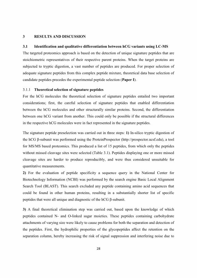
The signature peptide preselection was carried out in three steps: 1) In-silico tryptic digestion of
the hCG β-subunit was performed using the ProteinProspector (http://prospector.ucsf.edu), a tool
for MS/MS based proteomics. This produced a list of 15 peptides, from which only the peptides
without missed cleavage sites were selected (Table 3.1). Peptides displaying one or more missed
cleavage sites are harder to produce reproducibly, and were thus considered unsuitable for
quantitative measurements.
2) For the evaluation of peptide specificity a sequence query in the National Center for
Biotechnology Information (NCBI) was performed by the search engine Basic Local Alignment
Search Tool (BLAST). This search excluded any peptide containing amino acid sequences that
could be found in other human proteins, resulting in a substantially shorter list of specific
peptides that were all unique and diagnostic of the hCG β-subunit.
3) A final theoretical elimination step was carried out, based upon the knowledge of which
peptides contained N- and O-linked sugar moieties. These peptides containing carbohydrate
attachments of varying size were likely to cause problems for both the separation and detection of
the peptides. First, the hydrophilic properties of the glycopeptides affect the retention on the
separation column, hereby increasing the risk of signal suppression and interfering noise due to

29
co-elution with the injection front. Additionally, the selective character of the MS would exclude
any peptide having m/z values different to those preprogrammed to the instrument, as would be
the case for the peptides with varying carbohydrate composition. This consideration excluded
five peptides of specific protein backbones that all contained at least one carbohydrate group. All
in all, these elimination processes resulted in a list of candidate signature peptides (Table 3.1) that
would be suitable for detection of the hCG β-subunit, from a theoretical point of view.
Table 3.1 List of peptides generated from in-silico digest of the specific hCG β-subunit. All
peptides are fully digested (contains no missed cleavage sites) and are designated by peptide
number counted from the N-terminus of the protein backbone. The evaluation of each peptide as
candidate signature peptide is specified in the column to the right.
Peptide Amino acid position
Amino acid sequence Selection
T1 1-2 SK Unspecific
T2 3-8 EPLRPR Unspecific
T3 9-20 CRPINATLAVEK Specific, but N-linked carbohydrate
T4 21-43 EGCPVCITVNTTICAGYCPTMTR Specific, but N-linked carbohydrate
T5 44-60 VLQGVLPALPQVVCNYR Signature peptide candidate
T6 61-63 DVR Unspecific
T7 64-68 FESIR Unspecific
T8 69-74 LPGCPR Unspecific
T9 75-94 GVNPVVSYAVALSCQCALCR Signature peptide candidate
T10 95 R Unspecific
T11 96-104 STTDCGGPK Signature peptide candidate
T12 105-114 DHPLTCDDPR Signature peptide candidate
T13 115-122 FQDSSSSK Specific, but O-linked carbohydrate
T14 123-133 APPPSLPSPSR Specific, but O-linked carbohydrate
T15 134-145 LPGPSDTPILPQ Specific, but O-linked carbohydrate

30
However, as the family of hCG molecules comprises known variations in the structure of the
specific β-subunit, signature peptides representing this intra-variability had to be deduced. This
was addressed through a study of the structure in the protein backbone where the structural
differences appeared. Since the hCG α-subunit in unspecific (can be found in other hormones as
well), and since the intact hCG (α and β heterodimer) and the free hCG β-subunit share the same
unmodified hCG β-subunit, these two variants cannot be differentiated. They will thus be
detected through the same signature peptide. Similarly, the nicked hCG heterodimers will not be
distinguished from the free nicked hCG β-subunits. The nicked variants, however, might display
nicking in the protein backbone at two known sites: between amino acid number 44 and 45, and
between amino acid number 47 and 48. Each nicked molecule normally appears with one nick.
These nicking sites had to be expressed in the signature peptides, as to properly detect all nicked
variants, and to distinguish between non-nicked and nicked hCG. As for the smallest of the hCG
molecules, the hCG β-core fragment consists of two amino acid chains (amino acid position 6-40
and 55-92), covalently connected by four disulfide bonds. This hCG variant should therefore
have a signature peptide that is cleaved from the end of one of these sequences. The final
candidate signature peptides that theoretically allowed differentiation between the described hCG
molecules are listed in Table 3.2.
The challenge of differentiating structurally similar proteins is visualized in this short list of
candidate signature peptides; apart from the hCG β-core fragment that is represented by two
potential signature peptides, only one signature peptide will fully distinguish the hCG β-subunit
and the two nicked variants. This is due to the single nick (at two potential locations) in the
protein backbone that constitutes the only structural difference between these proteins; their total
protein masses and total amino acid sequences/compositions are identical. As the difference in
protein structure increases amongst a group of proteins, so will the number of candidate signature
peptides.

31
Table 3.2 List of candidate signature peptides for the described hCG variants. Each signature
peptide has been designated according to protein origin, location in the tryptic sequence
generated when counting from the N-terminus, and protein modification. The respective parent
proteins are listed in the column to the right.
Signature peptide Amino acid
position
Amino acid sequence Parent protein
βT5 44-60 VLQGVLPALPQVVCNYR Intact hCG
Free hCG β-subunit
nβT5 44/45 45-60 LQGVLPALPQVVCNYR Nicked hCG 44/45
Free nicked hCG 44/45
nβT5 47/48 48-60 VLPALPQVVCNYR Nicked hCG 47/48
Free nicked hCG 47/48
cfβT5 55-60 VVCNYR hCG β-core fragment
cfβT9 75-92 GVNPVVSYAVALSCQCAL hCG β-core fragment
αT2 36-42 AYPTPLR hCG α-subunit
When it comes to the differentiation between heterodimers and their dissociated free subunits, as
for the intact hCG and its free hCG β-subunit, this cannot be performed directly with the chosen
strategy. However, there are indirect approaches within this strategy that can be explored, such as
the establishment of the ratio of the detected β- and α-subunit of a heterodimer. If, for a certain
sample, the detected β- and α-subunit signal produces a ratio that exceeds this known heterodimer
ratio, then the excess signal of detected β-subunit can be contributed to free β-subunit. To explore
this approach, a signature peptide of the hCG α-subunit had to be established (listed at the bottom
of Table 3.2). It must be emphasized that since the α-subunit of the hCG molecules is the same as
that for the luteinizing hormone, follicle-stimulating hormone and thyroid-stimulating hormone,
there is always a possibility that parts of the detected αT2-signal might result from these
structurally similar molecules, unless this has been prevented one way or another. This approach
is therefore best suited for heterodimers of unique subunits. This will be further explored later in
the thesis.

32
LC-MS analysis: hCG peptide mapping and detection of signature peptides 3.1.2
A solution containing most hCG variants (intact hCG, free hCG β-subunit, nicked hCG, and hCG
β-core fragment) was subjected to tryptic digestion in order to produce peptides that were
subsequently analyzed in a gradient run on the LC-SQ system using 20 mM formic acid and
MeCN on a BioBasic C8 (50 x 1 mm) column. Since the hCG molecules contain several disulfide
bonds, reduction and alkylation of the cysteine residues had to be performed prior to tryptic
digestion, resulting in the addition of a carboxy-methyl group to each cysteine residue.
Peptide mapping of the analyzed hCG protein digest mixture was performed essentially by
matching observed peptide masses detected through a broad scan performed by the SQ (m/z
interval 350-1250) to the theoretical in-silico m/z values generated by ProteinProspector (Figure
3.1). This in-silico search was set to include 0, 1 and 2 missed cleavage peptides, in order to
enable identification of as many eluting peaks as possible. This preliminary peptide mapping was
succeeded by MS/MS experiments using an ion trap in order to verify the assumed identities of
the hCG peptides. Observed b- and y-fragment ions were matched against theoretical fragment
ions generated by ProteinProspector (Figure 3.2). A total of 13 peptides (containing 0-2 missed
cleavages) were identified, covering most of the amino acid sequence 1-114 of the hCG β-subunit
(Figure 3.1). This included the T5 signature peptide which is shown in Figure 3.2 accompanied
by its recorded MS/MS spectrum. Additionally the two nicked signature peptides nT44/45 and
nT47/48, the cfT9 signature peptide of the hCG β-core fragment, and the αT2 peptide of the hCG
α-subunit were identified. The ionextraction chromatogram of the identified signature peptides
derived from the broad scan is visualized at the bottom of Figure 3.1.
When it comes to the C-terminal of the hCG β-subunit (amino acids 115-145), this part will
theoretically generate three tryptically derived peptides (T13, T14 and T15), which are highly
glycosylated as they contain serine residues with O-linked sugar moieties attached. The theoretic
tryptic peptides T3 and T4 are also glycosylated, but contain N-linked sugar groups. The size of
these carbohydrate groups is not constant, and the ProteinProspector thus generates m/z values
based on the protein backbone, i.e. disregarding the glyco-masses. These peptides were thus of
unknown m/z values, and as such too complicated to identify. However, since all the theoretically
selected signature peptides had been identified, no further effort was made to complete the
identification of the peptides that had not been identified.

33

34
Figure 3.1 (On page 36) Top: Peptide mapping of hCG. Chromatogram of a broad scan (m/z
350-1250) of the tryptic peptides derived from proteolysis of a mixture of hCG. All peptides of the
hCG β-subunit that did not contain sugar groups attached to the protein backbone were
identified. Only one peptide of the hCG α-subunit was identified. Bottom: Ion extraction
chromatogram derived from the broad scan presented in the chromatogram above. of the
selected signature peptides of the hCG β-subunit, the hCG α-subunit, the hCG β-core fragment,
and the two nicked hCG variants
Figure 3.2 MS/MS spectrum obtained from the fragmentation of the hCG β-subunit signature
peptide, βT5. The most abundant b- and y-fragment ions are annotated. The amino acid sequence
with the corresponding b- and y-fragment ions is shown at the top of the figure.

35
Based on these experiments, an MS detection program in the selected ion monitoring (SIM) mode
was set up. The SIM detection of each peptide was made on the basis of either doubly or triply
charged peptides, depending on what charge state generated the highest signal response in the SQ.
This varied amongst the different peptides analyzed by the same SQ mass spectrometer, but also
varied for each individual peptide when transferring the LC separation to precede the ion trap
detection for the MS/MS experiments.
Pregnyl as hCG source 3.1.3
The available hCG source was the pharmaceutical formulation Pregnyl (Organon). This is an
hCG containing drug which is manufactured from the purification of the urine of pregnant
woman. The intact hCG heterodimer is the dominant hCG variant present in the drug, and is
responsible for the pharmacological benefits obtained from an injection of Pregnyl. If present, the
free hCG β-subunit also display some biological activity, but to a much smaller extent [52]. The
Pregnyl formulation also contains hCG degradation variants, namely the two nicked hCG
molecules and the hCG β-core fragment. These degradation variants do not display any relevant
biological activity. Since the developed MS method detected both intact hCG and free hCG β-
subunit through the same signature peptide (βT5) the detection of this signature peptide was
related to the corresponding international units (IU) concentration of each Pregnyl ampoule.
When analyzing a digest of hCG (diluted in ammonium bicarbonate (ABC) buffer prior to
proteolysis), it was observed that the peaks representing the detection of the nicked variants and
the hCG β-core fragment were, as expected, substantially lower in signal intensity than the peak
of the intact hCG/hCG β-subunit that related to the IU concentration. The use of Pregnyl as hCG
standard has thus limitations in terms of quantification, as this will only be possible for the sum
of intact hCG plus free hCG β-subunit. However, this formulation does contain the desired
specter of hCG variants which reflects the hCG molecular diversity that might be present in a
biological matrix, and was as such suitable for the LC-MS design and development of the method.
Multiplexing hCG identification through LC-MS based detection 3.1.4
The gradient used for hCG peptide mapping described in section 3.1.2 was 32 minutes long,
resulting in a total analysis time of 50 minutes when including washing and regeneration of the
analytical column. Further optimization of the LC gradient was thus performed in order to
36
achieve adequate separation of the signature peptides combined with short analysis time, and this
resulted in a total analysis time of 27 minutes (Paper II). The specific retention time of all the
signature peptides was thus established. Chromatograms are presented in following sections.
Combined with the dimension of retention time, the simultaneous SIM MS detection of various
target hCG proteins through their diagnostic signature peptides could be performed (Paper I).
Provided that adequate signature peptides are generated from any target proteins, this separation
and detection of peptides using LC-MS offers a means of multiplexing the specific identification
and differentiation of several proteins in one single run, as exemplified with the hCG molecules.
However, whereas multiplexed detection of proteins is often defined as the simultaneous
determination of structurally different proteins in one single run, this definition must be regarded
with some caution in relation to the hCG molecules. This family of molecules is structurally
related as they all share the hCG β-core fragment part. Furthermore, most hCG variants can be
considered degradation variants of the intact hCG molecule. Nevertheless, the differences in
molecular weight range from 15 kDa for the hCG β-core fragment to 37.5 kDa for the intact hCG.
This justifies the use of the term “multiplexed detection” for the simultaneous and differentiated
detection of these hCG molecules.
The acknowledged potential of LC-MS based multiplexing to be extremely specific and time-
efficient makes this an interesting analytical technique for clinical biomarker analysis. Combined
with an effective sample preparation strategy multiplexing of proteins in low abundance can be
enabled.
3.2 Compatibility of immunoaffinity extraction with mass spectrometric detection
For the selective and specific extraction of target hCG molecules from complex matrixes
adequate antibodies have to be carefully chosen. These will further have to be immobilized to a
solid to enable isolation of the antibody-antigen complex that is formed during extraction.
Following this, the biological matrix will be removed.
For this reason, the hCG specific monoclonal antibody E27 was selected as it is directed towards
the core of the hCG β-subunit that is common to all hCG variants [35]. It will thus recognize and
bind all hCG molecules. The antibody was immobilized to the walls of the wells in a 96 –well
microtiter plate (Paper I), and serum samples containing spiked hCG were applied to the wells.

37
Immunoextraction of the target molecules was succeeded by extensive washing, followed by in-
well reduction and alkylation. The subsequent addition of trypsin directly to the wells generated a
peptide mixture that was subjected to a solid phase extraction (SPE) step prior to the final LC-MS
analysis.
In the chromatograms resulting from these experiments the peaks of the signature peptides βT5,
nβT5 44/45, nβT5 47/48 and cfβT9 eluted at the previously established retention time hereby
demonstrating the successful extraction and detection of the hCG β-subunit (present in both intact
hCG and as free hCG β-subunit), the two nicked hCG variants, and the hCG β-core fragment. For
the detection of the hCG β-subunit linearity was observed for the concentration range of 100 to
2000 IU/L with a limit of detection (LOD) of 100 IU/L. The other hCG variants were
qualitatively detected and differentiated, but as they were present in unknown and relatively low
amounts no linearity or LOD were provided for these. The proof of the immuno-LC-MS principle
was thus established, and this was further demonstrated by the analysis of serum samples from
male patients previously diagnosed with testicular cancer. These experiments demonstrated the
detection of intact hCG and free hCG β-subunit in a number of samples (n=20), in addition to the
detection of nicked hCG in one particular sample. The varying β/α ratio in the different samples
provided evidence that the free hCG β-subunit was present in addition to the intact hCG.
Furthermore, the urine samples of pregnant women (at various stages in the pregnancy) were
analyzed, although no prior experiments on urine samples had been carried out. The distinct
detection of both the intact hCG/free hCG β-subunit and the main urine metabolite, the hCG β-
core fragment, was demonstrated in all samples (n=6), indicating that the adoption of the
developed immuno-LC-MS method to urine samples would not pose great problems.
What could be observed from the chromatograms of the serum analyses was that the signal-to-
noise (S/N) ratio of the target peaks was relatively low compared to that of the peaks resulting
from the analysis of the digest of hCG dissolved in buffer. This increase in noise at the base line
strongly influenced the sensitivity of the method, and was explained by the unspecific binding of
interfering serum proteins that were not removed by the washing steps. Although the washing
procedure was optimized the level of noise remained relatively constant. It was therefore
concluded that as the inference of serum proteins during immunoextraction clearly was
problematic to exclude, other strategies had to be considered in order to increase the signal of the

38
target proteins and thus of the immuno-LC-MS based detection sensitivity. It was clear that the
relatively limited sample capacity of the immuno-wells of 200 µL was not sufficient to provide
adequate preconcentration of the target molecules to overcome the effect of the increase in noise
following immunoextraction. As the LOD of the hCG method would need to be as low as 5 IU/L
(a factor 20 lower than the current LOD of 100 IU/L), further improvement of the sample
preparation strategy using this selective immunocapture had to be explored.
3.3 Optimizing method sensitivity and specificity
There are different ways to enable lower LODs for the immuno-LC-MS approach. Additionally,
the use of complementary strategies will greatly enhance the effects of the individual strategy.
For this reason the application of two different strategies was explored. First, it was assumed that
a more selective and specific MS design would reduce the signal effect of the interfering co-
eluting peptides on the detection of the target peptides (3.3.1). Secondly it was hypothesized that
extraction of hCG molecules using antibodies coated to beads applied to larger sample volumes
(3.3.2) would generate increased signal response of the target peptides through the increase of
preconcentration factor (Paper II).
Tailored selected reaction monitoring design 3.3.1
By using a triple quadrupole detector in the SRM mode, MS detection can be performed enabling
high structural specificity, sensitivity, and reproducibility. The transition from SQ detection to
QqQ detection would as such provide a dual improvement to a method by decreasing the
method’s LOD and enhancing the analytical evidence for the identification of the hCG molecules.
The principle was employed to hCG, and for all hCG signature peptides specific SRM transitions
were made based on the following criteria:
� The fragment ion signal intensity should be as high as possible
� Two or more diagnostic fragment ions should be included in the SRM transitions for each
signature peptide
� The ratio of the two (or more) diagnostic fragment ions should be constant for every
analysis performed under established conditions

39
The CID energy was thus optimized according to fragment ion intensity and abundance, and scan
segments with optimized scan times generating at least six detection points per analytical peak
were created.
With regards to quantitative measurements, an isotopically labeled internal standard was
incorporated to the analysis to correct for intra- and inter-day variability produced by the MS.
The selected internal standard AQUA peptide was an isotopically labeled synthetic analogue to
the signature peptide βT5 of the hCG β-subunit, and was therefore named isβT5. This peptide
contained an isotopically labeled arginine residue at the end of the sequence that resulted in a
mass shift of +10 Da. In Figure 3.3 the detected mass difference of +10 Da between the selected
fragment ions of the signature peptide βT5 and the isβT5 can be observed. The ratio of the
fragment ions y11 and y8 was approximately 1:2, and constant (established ratio of 0.47 for n=20
and with RSD of 4%). The detection of the established abundance ratio of the diagnostic
fragment ions increases the specificity of the method.
The AQUA peptide contained one cysteine residue that needed to be reduced and alkylated as to
have the exact same physical-chemical proprieties as the signature peptide βT5. For this reason
the labeled peptide is not a “classical” AQUA peptide; indeed the labeled peptide is calibrated
prior to reduction and alkylation. The yield of alkylation thus has to be 100 % in order to ensure
quantification accuracy. This was shown by full MS scan analysis in addition to tailored SRM
analysis for both alkylated and non-alkylated labeled peptide. The alkylation yield proved to be
100 %.
Since the βT5 and the isβT5 are not separated in time, it is important that the diagnostic
fragments ions used in the SRM program are y-ions. Although the precursor ions have different
m/z values, the possibility of cross-talk phenomenon is present, as all b-ions generated do not
contain the heavy arginine residue responsible for the mass shift in the fragments ions. Effects of
cross-talk phenomenon were revealed by analyzing a test solution containing high amounts of
isβT5 exclusively, and by using an SRM program in which transitions for both βT5 and isβT5
contained the identical b9-ion m/z value. From this experiment a response in the signal of the βT5
was demonstrated, although this peptide was not present in the solution. Cross-talk is most likely
a theoretical problem, but in extreme cases such as samples containing high amounts of

40
isotopically labeled IS and no target protein/peptide, this could potentially have provoked a false
positive response.
By increasing the selectivity of the detection, the LOD was extended from 100 IU/L (using MS
detection) to 20 IU/L (using MS/MS detection). As such, the conversion from SQ-enabled SIM
detection to QqQ-enabled SRM detection provided an increase in LOD by a factor 5. This
approach included immunoextraction with antibodies coated to wells prior to MS detection.
Although improved, the LOD had to be further extended.

41
Figure 3.3 Signals from the selected fragment ions of the signature peptide βT5 and the
corresponding isotopically labeled internal standard, isβT5. For both peptides the amino acid
sequence is shown, and the site of fragmentation for the selected y-ions is emphasized. The heavy
arginine residue of the internal standard is visualized in red.

42
Immunoextraction using beads in stead of wells 3.3.2
In order to explore the potential of increase in preconcentration factor as a function of sample
volume, the antibody E27 was immobilized to magnetizable particles (beads) rather than wells.
Different serum volumes were tested, and lowest LOD was obtained from the extraction of 1 mL
serum sample. The resulting LOD was 5 IU/L, and although the sample volume was increased to
2 mL no further improvement in LOD was observed. Furthermore, as the amount of beads used
for each extraction would determine the capacity of the method and as such define the linear
range, varying amounts of beads were applied to 1 mL sample volume. For 20 µL of beads
solution (10 mg beads/mL) linearity was observed for the concentration range of 5 to 5000 IU/L,
compared to the range of 100 to 2000 IU/L, which was obtained when using antibodies coated to
wells and SQ detection. All in all, the combination of selective MS/MS detection combined with
immunoextraction using beads in stead of wells generated an improvement in LOD of a factor 20.
The resulting chromatogram from the immuno-MS/MS analysis of the various hCG molecules
spiked to serum is shown in Figure 3.4A, whereas that on the analysis of digested hCG diluted in
buffer is shown in Figure 3.4B.
When comparing the two chromatograms in Figure 3.4 a second peak is observed in the cell of
the hCG β-core fragment. Due to the difference in retention time this “interfering” peak does not
compromise the detection of the authentic hCG β-core fragment signature peptide. Since this
peak was not observed in the chromatogram of the digested hCG diluted in buffer, this must thus
be an unknown peptide resulting either from interfering serum proteins or from the capture
antibodies. The immunoaffinity extraction procedure using antibodies coated to beads is
summarized in Figure 3.5. Since this procedure includes the reduction, alkylation, and tryptic
digestion of the captured hCG molecules without releasing them from the antibody-bead complex
first, the simultaneous trypsination of capture antibodies and unspesifically bound interfering
serum proteins will also happen. However, interfering matrix analytes that are not completely
removed during the wash procedure is a well-known problem of the immunoaffinity extraction
strategy, and has to be overcome [34]. In this case this was accomplished through an increase in
preconcentration factor combined with a selective MS detection SRM program.

43
Fig
ure
3.4
SRM
chr
omat
ogra
ms
show
ing
the
peak
s of
the
diffe
rent
sig
natu
re p
eptid
es a
nd th
eir
resp
ectiv
e re
tent
ion
times
. The
sig
nal
inte
nsity
is
norm
aliz
ed,
and
the
corr
espo
ndin
g de
tect
ed h
CG
var
iant
s ar
e gi
ven
for
each
sig
natu
re p
eptid
e. A
) Th
e re
sulti
ng
chro
mat
ogra
m f
rom
the
ext
ract
ion,
dig
estio
n an
d LC
-SRM
ana
lysi
s of
a s
erum
sam
ple
spik
ed w
ith h
CG
B)
The
resu
lting
chro
mat
ogra
m fr
om th
e di
gest
ion
and
LC-S
RM a
naly
sis o
f hC
G d
isso
lved
and
dilu
ted
in b
uffe
r

44
Figure 3.5 The immunocapture procedure using antibodies immobilized on magnetic beads in
stead of wells. The procedure can be summarized as follows: 1) Antibody coated beads are
washed prior to extraction 2) Beads are isolated by magnet, and wash solution is removed 3)
Sample is added, hCG bind to antibodies 4) Beads with antibodies and captured hCG are
isolated by magnet. Interfering substances are removed by extensive washing 5) hCG has been
extracted from serum, and will further be subjected to reduction, alkylation, and tryptic digestion
directly in the tube without prior release from capture antibodies
For other immunoaffinity extraction approaches combined with MS detection, other than the
bottom-up approach, the release of target analytes from the capture antibodies is necessary prior
to LC-MS analysis. This is often carried out using high molar concentrations of detergents,
denaturants or acids that have to be removed prior to MS detection. This is time-consuming, and
it is often seen that the recovery of analytes from the releasing procedure can vary to a great
extent depending on the antigen-antibody interaction, hereby affecting the sensitivity of the
method. The hCG bottom-up approach is not dependent on a pre-digestion release of the
substrate as tryptic digestion of the entire antibody-substrate complex will occur (Paper I).
However, it is possible that the dissociation of the antibody-antigen complex is necessary to
obtain complete digestion, which is crucial for accurate quantification. Therefore, when the
previously established reduction and alkylation step proved to dissociate the complex, there was
thus no need to include an additional sample preparation step in order to release hCG from
antibodies by other means.

45
The developed immuno-LC-MS/MS method for hCG determination, hereafter referred to as hCG
immuno-MS method, had demonstrated a serum LOD of 5 IU/L. For quantification purposes, it
had to be further established that the method generated reproducible, accurate and reliable results
for every analysis. Additionally, the methods application to urine samples had shown great
potential, and therefore had to be thoroughly tested.
3.4 Validation of method for quantitative determination of hCG
A thorough validation of a new methods analytical performance is essential for accurate
quantitative analysis, and constitutes as such the foundation for implementation into routine
analysis. The European Medicines Agency (EMEA) has defined a Guideline on Validation of
Bioanalytical Methods, which defines key elements necessary for the validation of bioanalytical
methods [98]. The hCG immuno-MS method was validated for both serum and urine (Paper II),
and by using Pregnyl as standard the method was challenged to perform under realistic conditions
as Pregnyl contains primarily the intact hCG molecule, but also smaller amounts of hCG
degradation variants, as previously described in section 3.1.3. This implied that as quantitative
measurements were provided based on the hCG β-subunit through the detection of signature
peptide βT5, the sum of both intact hCG and free hCG β-subunit would constitute the quantitative
response generated. Additionally, the presence of all other degradation hCG variants were
simultaneously detected, although no validation data were sought for these as their quantitative
amounts in Pregnyl are not known.
In Paper II all validation results were reported in international units (IU) since the clinical world
is most familiar with hCG levels reported in IUs. However, in order to provide comparable
sensitivity data that can be compared in a targeted proteomics setting, a conversion factor has
been applied for translation of detected hCG β-subunit to molar concentration. According to
Stenman (2006) will 1 IU of intact hCG correspond to 2.9 pmol [52], and since Pregnyl contains
mainly intact hCG (and free hCG β-subunit to a much smaller extent), this conversion factor was
chosen. It must be noted that a conversion factor the free hCG β-subunit also exist, but compared
to intact hCG this molecule display a minor biological activity (1 IU free hCG β-subunit
correspond to 42.5 pmol) [52].
The validation results for the quantitative detection of hCGβ using the developed immuno-MS
method based on the targeted proteomics approach are summarized in Table 3.2. Furthermore,

46
the individual results are thoroughly discussed in Paper II. Sensitivity has long been the bottle
neck preventing mass spectrometry from making the transition from discovery tool into the world
of routine analysis. As can be deduced from these validation results, the successful cross-
fertilization of the complementary techniques of immunoaffinity extraction and MS detection
allows quantification of macromolecules in complex matrixes in the low picomolar ranges. In our
research group this sensitivity-range has also been obtained for other clinically relevant tumor
markers, such as neuron-specific enolase (NSE, Mw of 47.3 kDa, γ-subunit) and progastrin
releasing peptide (ProGRP, Mw of 13 kDa) by using the bottom-up targeted proteomics approach
[99]. This proves that the challenging procedure involving the enzymatically conversion of
proteins into their constituent peptides can in fact be standardized to meet the criteria of
bioanalytical validation guidelines. With this, mass spectrometry is brought one step closer to
clinical diagnostics and routine laboratories.
For the validation of hCG it can be argued that the use of a proper international reference reagent
(IRR) for the intact hCG molecule should have been used. However, satisfying validation was
accomplished through the use of Pregnyl as standard although this posed more challenging
conditions. Furthermore it must be noted that there are no obstacles for replacing the calibration
and standard curves of the developed method with existing IRRs for hCG and hCG variants. The
full quantification of all hCG variants was however beyond the scope of this project, since most
clinical utility of hCG as biomarker today is based on the quantification of the intact molecule.
Clinically relevant complementary information is nevertheless provided through the qualitative
ratio generated through MS determination of other hCG variants.

47
Tabl
e 3.
3 Su
mm
ary
of v
alid
atio
n pa
ram
eter
s ob
tain
ed fo
r se
rum
and
uri
ne s
ampl
es. T
he L
OD
s fo
r se
rum
and
uri
ne w
ere
estim
ated
from
a si
gnal
to n
oise
(S/N
) rat
io o
f 3:1
. The
exp
erim
enta
l LLO
Qs w
ere
defin
ed a
s the
con
cent
ratio
n at
whi
ch v
aria
tion
of le
ss th
an 2
0%
was
obt
aine
d
Val
idat
ion
para
met
er
Lin
eari
ty
(r2 )
n=5
LO
D
n=
5
LL
OQ
n=5
Sele
ctiv
ity
(mat
rix)
n=
3
Sele
ctiv
ity
(inte
rnal
stan
dard
) n=
3
Mat
rix
effe
cts
Seru
m
˃ 0.
997
15 p
mol
/L
5 IU
/L
29 p
mol
/L
10 IU
/L
No
inte
rfer
ing
sign
al
from
bla
nk m
atrix
N
o in
terf
erin
g si
gnal
fr
om in
tern
al st
anda
rd
No
sign
al
supp
ress
ion/
enha
ncem
ent
Uri
ne
0.99
9 6
pmol
/L
2 IU
/L
15 p
mol
/L
5 IU
/L
No
inte
rfer
ing
sign
al
from
bla
nk m
atrix
N
o in
terf
erin
g si
gnal
fr
om in
tern
al st
anda
rd
No
sign
al
supp
ress
ion/
enha
ncem
ent
Val
idat
ion
para
met
er
With
-in d
ay p
reci
sion
(%)
n=5
Bet
wee
n-da
y pr
ecis
ion
(%)
n=5
Acc
urac
y (%
) n=
5 R
ecov
ery
(%)
n=3
Mat
rix
Seru
m
Urin
e Se
rum
U
rine
Seru
m
Urin
e Se
rum
U
rine
10 IU
/L
19
9 13
14
95
10
4 39
65
100
IU/L
17
12
11
30
87
98
43
57
1000
IU/L
4
10
7 19
99
10
0 32
57
48
3.5 hCG immuno-MS in clinical diagnostics
A tailored biomarker immuno-MS tool can have multiple functions in a clinical scenario; it can
be used to measure quantitative levels of well-established biomarkers in matrixes such as serum
and urine; this will be demonstrated using the tailored hCG immuno-MS method for the analysis
of clinical samples (3.5.1). Furthermore, the specificity of the MS analysis can be exploited to
evaluate the performance and compatibility of other conventional immuno-based methods which
are designed and tailored for defined pathological conditions. By characterizing the selectivity
and specificity of various anti-hCG antibodies differentiating information regarding the various
antibodies ability to recognize and isolate various target hCG analytes is provided, as will be
presented below (3.5.2).
Pregnancy and cancer diagnostics 3.5.1
The validated method was used for quantification of hCGβ and differentiation between other
hCG variants in serum and urine of a pregnant woman (Paper II). The serum hCG concentration
was measured to 2.6 x 10⁴ IU/L, which corresponded well with reported hCG levels at 21 weeks
of pregnancy [52], and for the corresponding hCG concentration in urine was measured to 6280
IU/L. One of the resulting chromatograms is visualized in Figure 3.6, showing the detected hCG
variants for both matrixes. The hCG α-subunit and hCG β-subunit were present in both matrixes,
but the ratio of β/α was higher in the urine sample indicating that a proportion of the intact hCG
had dissociated into free subunits, which is known to occur in urine. Additionally, the urinary
degradation variants hCGβ-core fragment was observed in urine but not in serum, due to the well
known degradation and excretion of intact hCG. For standard pregnancy testing it is obvious that
this need not and should not be done by other means than standard pregnancy tests. However, for
disorders of pregnancy the develop method can provide accurate quantification of the hCGβ
(intact hCG and free hCG β-subunit), and hCG fingerprints differentiating the degradation
variants (nicked hCG and hCG β-core fragment) from the unmodified hCGβ. It must also be
noted that although eventual hyperglycosylated variants are not differentiated from the normally
glycosylated variants, they are nevertheless captured and detected through the signature peptides
of the protein backbone. Furthermore, since the free hCG β-subunit is an important marker for
disorders of pregnancy, a more thorough exploration of the β/α ratio enabling differentiating
quantitative measurements for both intact hCG and free hCG β-subunit, simultaneously, could
result in increasing interest for the MS approach on this arena.

49
Fig
ure
3.6
Chr
omat
ogra
m o
f the
ana
lysi
s of
ser
um le
ftt c
hrom
atog
ram
s) a
nd u
rine
(rig
ht c
hrom
atog
ram
s) s
ampl
es fr
om a
pre
gnan
t
wom
an (w
eek
22),
take
n on
the
sam
e da
y. T
he sa
mpl
es w
ere
extr
acte
d, d
iges
ted
and
anal
yzed
by
LC-S
RM u
sing
the
deve
lope
d ta
ilore
d
hCG
imm
uno-
MS
met
hod.
The
ana
lysi
s sh
owed
det
ectio
n of
inta
ct h
CG
and
free
hC
G β
-sub
unit
in b
oth
mat
rixe
s, w
here
as th
e hC
G β
-
core
frag
men
t was
det
ecte
d in
uri
ne e
xclu
sive
ly

50
The results from the analysis of the cancer patient serum samples generated quantitative
measurements of this pathologically produced hCG that varied between ~200 and 20 000 IU/L.
Additionally no other hCG variants other than the α- and β-subunit were observed. The
chromatograms from some of these analyses are presented in Figure 3.7. In these chromatograms
the hCG β-signals have been normalized whereas the α-signals are relative to the respective β-
signals. It is also apparent that the β/α ratio vary from sample to sample, indicating that each
sample contains various amounts of free hCG β-subunit in addition to intact hCG. This is
supported by the literature that describes the free hCG β-subunit as an important tumor marker
for certain types of cancer, although for many other types the intact hCG is often the dominant
form. Other variants might also be produced for certain tumors, but these are mainly observed in
urine as degradation variants of the intact hCG and the free hCG β-subunit [52,100-103,61].
Figure 3.7 Analysis of four serum samples of patients previously diagnosed with testicular
cancer. The signal of the hCG β-subunit has been normalized whereas that of the hCG α-subunit
is represented relative to the β-subunit. No other hCG variants were observed

51
The potential of mass spectrometry to quantitate certain variants and differentiate others for use
in a clinical setting has been demonstrated through the data presented on pregnancy and cancer
samples. The method ensures that all hCG variants (both normally- and hyperglycosylated ones,
heterodimers and free subunits) are detected. However, in a clinical setting this method should
preferentially also enable the quantitative differentiation between intact hCG and free hCG β-
subunit. This was also attempted, with partial success; following the establishment of the
signature peptides for hCGα and hCGβ, a pure intact hCG standard was spiked to serum and
extracted. This was done in order to establish the β/α signature peptide ratio for intact hCG when
no free hCG β-subunit is present. The same ratio was to be established for defined increasing
amounts of free hCG β-subunit added to the sample. However, for serum samples, adequate
separation of the hydrophilic αT2 from other co-eluting hydrophilic compounds was not achieved,
hereby preventing the absolute quantification of this substance (data not shown). These
components were assumed to be interfering peptides resulting from the tryptic digestion of
unspecifically bound serum proteins that were not removed during washing of the beads. This
interfering signal could be ignored for high substance concentrations, but not for lower
concentrations. Quite some effort was laid down on solving these chromatographic issues, but
was not rewarded. For future prospects it was concluded that the use of nano-LC systems can be
a suitable strategy for enabling improved separation. From the chromatograms of the urine
sample, the same interfering signal was not observed. It was nevertheless decided not to explore
this path any further since the quantification of the sum if intact hCG and free hCG β-subunit did
not interfere with the principal scope of this project.
The possibility of tuning the selectivity of the method, in order to differentiate heterodimer from
subunits, is most easily carried out through the selection of antibodies of known selectivity and
specificity towards the different hCG variants [35]. This will limit the multiplexing feature of the
method, but can be done if this differentiation is crucial for accurate hCG determination of
certain clinical samples. The immuno-MS analysis will then have to be run several times for the
same samples using the different antibodies. However, this is currently how the conventional
immunoassays solve the differentiating issues.

52
Evaluation of anti-hCG antibody selectivity and specificity 3.5.2
The use of tailored MS detection to evaluate the selectivity of various anti-hCG antibodies to the
different hCG variants is valuable in a clinical setting as this can provide information that is
important for the tailoring of immunoassays towards specific biomarkers diagnostic of defined
pathological conditions. This utility of the developed hCG MS detection was thus exploited to
characterize various anti-hCG antibodies according to hCG epitope recognition (Paper V).
The International Society of Oncology and BioMarkers (ISOBM) initiated two Tissue
Differentiation (TD-7) Workshops (WS) on hCG and related molecules. The project described in
this thesis participated in the 2nd WS where 69 mAbs were tested for specificity and
corresponding epitope recognition. From this antibody group, a panel of 30 mAbs was chosen for
evaluation by MS (Figure 3.8).
International reference reagents (IRR) standards for intact hCG, nicked hCG, free hCG β-subunit,
nicked free hCG β-subunit, hCG β-core fragment were spiked to separate bovine serum albumin
(BSA) solutions, and extracted one by one using the 30 anti-hCG mAbs. For one mAb in
particular, the hCG α-subunit IRR was tested since this mAb was known to be targeted towards
the α-subunit. The detected signals were processed and presented in charts showing the various
antibodies recognition of the different hCG molecules/standards, together with their classification
according to specific epitope recognition. The outcome of these analyses is represented in Figure
3.8, where the results are compared to those obtained using a parallel method; the direct binding
radioimmunoassay (DB-RIA) with 125I-labeled hCG and hCG variant tracers.
For a majority of the antibodies the obtained MS data were highly concordant to the parallel
study performed by DB-RIA, and supported thus the epitope group affiliation that had been
previously suggested. The MS method can thus serve as a confirmatory method providing
specific MS based verification of recognized hCG variants. Furthermore, also more complex data
were obtained. These will be presented below.

53
Fig
ure
3.8
Com
para
tive
clas
sific
atio
n of
ISO
BM-m
Abs
by D
B-RI
A an
d Im
mun
o-LC
-MS/
MS.
The
ant
ibod
ies
are
clas
sifie
d on
the
y-
axis
acc
ordi
ng to
epi
tope
gro
up a
ffilia
tion.
The
mea
sure
d no
rmal
ized
MS
sign
al in
tens
ity is
rep
rese
nted
by
the
ratio
gen
erat
ed b
y th
e
MS
sign
al
inte
nsity
of
th
e de
tect
ed
hCG
va
rian
t di
vide
d by
th
at
of
the
inte
rnal
st
anda
rd.
54
Through the differentiating signal generated by the MS it became apparent that there existed a
cross-contamination in one of the standards; the IRR of the nicked hCG heterodimer (99/642).
Figure 3.9 shows the categorization of the tested anti-hCG mAbs and their respective recognition
of the nicked hCG standard. In this figure the signals of the two nicked variants as well as that of
the intact hCG cross-contamination are differentiated. From this chart it appears that the amount
of intact hCG cross-contamination is approximately 20%. This correlates with what has
previously been demonstrated by RP-HPLC analysis, indicating the presence of ~23.5% of
nonnicked hCG [104]. As there is an apparent tendency for the antibodies that recognize intact
hCG to recognize nicked hCG as well, this cross-contamination can be problematic for antibody-
based assays that generate a signal that is the undifferentiated sum of all recognized hCG variants.
However, the MS signal is not compromised as it clearly differentiates which part of the signal
stems from authentic nicked hCG and which stems from cross-contamination.
Since a major criterion for the classification of hCG c-mAbs is the importance of nicks in the β-
subunit, the ability of the MS to discriminate cross-contamination was particularly exploited in
the characterization of these. Nicking in this area of amino acids number 44-48 disrupts some
epitopes whereas others are unaffected. As can be seen in Figure 3.9 the MS showed that all
signals elicited from the mAbs categorized in the c2-epitope group stemmed from the intact hCG
cross-contamination. For some of these antibodies this had only been assumed previously, but
was now demonstrated by the MS data (425, 387, 436). For others, this led to the precise sub-
classification within this group (411). Furthermore the MS data showed that c3-mAbs recognize
both nicked hCG and intact hCG (mAb 446), hereby contradicting the previous assumption that
c3-mAbs are specific and selective for intact hCG.
There were however conflicting results observed as well; the c1-antibodies are also assumed not
to recognize nicked hCG (such as mAb 447), and the mAb 414 had been thoroughly established
as c1-antibody by other assays. The discordant results provided by the MS showed that this mAb
did de facto recognize nicked hCG as well, leading to the conclusion that there appears to be an
exception to the rule that c1-antibodes are specific for intact hCG. It could further be deduced
that there seems to be some variability in the epitope cluster of the c-area, resulting in a
continuum of c-epitopes with slightly different specificity patterns. The same tendency also exists
for some of the β-epitopes (β2 to β6), but this knowledge has not been deduced from MS data.

55
One of the pit-falls of this MS based approach for characterization of antibody selectivity might
be in the case of a signature peptide position being the same as for the current epitope. For two of
the antibodies no signals were generated form the MS data whereas they had shown good signal
responses in other assays. When looking at the suggested epitopes of these antibodies it appeared
that they could very well be situated at the same location in the β-subunit as the signature
peptides (amino acids 44-60 for hCG β, 47-60 for nicked hCG 47/48 and 45-60 for nicked hCG
44/45). It was thus hypothesized that the interaction between the capture mAb and the signature
peptides for the target analytes might prevent trypsin from converting this part of the molecule to
peptides, due to steric shielding. This has not been demonstrated.
Figure 3.9 Chart showing the various antibodies recognition of the nicked hCG. The antibodies
are classified on the x-axis according to epitope recognition. The measured signal intensity (y-
axis) is represented by the ratio generated by the MS signal of the detected target hCG molecule
divided by that of the internal standard. The signal of the nicked hCG 47/48 is light blue, the
signal of the nicked hCG 44/45 is dark blue, and the signal of the intact hCG cross-
contamination is red.

56
Assessing the analytical selectivity of mAbs through MS data offers an additional dimension to
the analytical proof, which is sought through the well considered use of different methods for
mAb selectivity evaluation. This is clinically relevant since it is crucial that assays tailored for
defined clinical scenarios provide results that reflect the presence of the target analytes in the
matrix, and not other structurally similar or otherwise interfering compounds.
3.6 hCG immuno-MS in doping analysis
In order to show the applicability of a developed method in a given analytical scenario, such as
the arena of doping analysis, the method has to be challenged to perform on adequate realistic
samples. For this reason a clinical study was initiated involving administration of hCG containing
pharmaceuticals to males.
Clinical study 3.6.1
Through the conduct of a clinical study (Paper III) various aspects regarding detection of illicit
hCG administration were to be studied, such as window of detection, differences in hCG
concentration in serum versus urine, and molecular diversity of the hCG molecules as a function
of matrix. The project was approved by the Norwegian Regional Committee for Medical
Research Ethics (REK, http://helseforskning.etikkom.no), and a liability assurance was signed
with the Drug Liability Association (http://www.laf.no). Furthermore, prior to entering the study
each participant signed a letter of consent stating that they participated on a voluntary basis, were
self-declared healthy, and were not competing athletes. Since hCG pharmaceuticals are currently
available on the marked as both recombinant hCG formulations and urinary hCG formulations
eventual differences related to type of hCG pharmaceutical were also of interest. Both variants
were thus included in the study, and 12 healthy males received one injection of Ovitrelle
(recombinant hCG, one dose corresponding to 6500 IU) whereas 12 others received one injection
of Pregnyl (urinary hCG, one dose corresponding to 5000 IU). Serum and urine samples were
collected prior to and following injection of drug to the abdominal fat tissue, for a period of
fourteen days.
The window of detection was defined as the period of time for which hCG could be detected at
concentrations above LOQ following injection of one single dose. In order to predict the window
of detection for the two drugs theoretical pharmacokinetic elimination profiles were estimated for
serum. This was based on the pharmacokinetic parameters provided by the SPC (summary of

57
product characteristics) of the respective drugs. The resulting elimination profiles are shown in
Figure 3.10A and from this a window of detection of 6-7 days could be deduced, for both
pharmaceuticals. No estimation was made for the urine samples.
The experimentally derived hCG elimination curves for both serum and urine (Figure 3.10B and
C) were produced based on the average detected hCG concentration (in the sample group) as a
function of day following injection. The quantitative measurement was based on the detection of
the hCG β-subunit. The elimination curve resulting from the analysis of the serum samples
(Figure 3.10B) harmonized very well with the estimated curve, showing that the experimentally
derived window of detection was, for both drugs, 7 days after administration of one single dose.
Interpretation of the chromatograms further demonstrated that only hCG β- and α-subunits were
detected in serum. Since hCG degradation variants, such as dissociated subunits, nicked and
degraded variants, are mostly observed in urine this was not unexpected [52]. Furthermore, the
presence of free hCG β-subunit in serum is mainly due to excess production of this subunit
relative to the production of the α-subunit, and this process is related to the development of
certain types of cancer [105,102,103,106,107]. The hCG β-subunit is further rapidly cleared form
the circulation due to less efficient biological activity [55,52]. From this it was considered likely
that the quantified hCG variant in serum was in fact the intact hCG molecule, and not the free
hCG β-subunit.
For the urine samples (Figure 3.10C) hCG could be detected above LOQ (of 5 IU/L) until day ten
following injection, generating a window of detection that is 3 days longer than what was shown
for the serum samples. This was valid for both drugs. The broader window of detection observed
for urine can be explained by the preconcentration of analytes in the morning urine samples. The
chromatograms resulting from these analyses displayed the occurrence of hCG molecular
variations through the detection of both nicked variants and the hCG β-core fragment in addition
to the β- and α-subunit. The signal of these degradation variants was lower than the signal of the
two main subunits, and in the samples taken on day five following injection the presence of the
degradation variants could no longer be observed in the chromatograms. It was therefore
concluded that the MS based detection of all hCG variants did not extend the window of
detection for one injection of hCG containing drug. This was consistent with what has been
reported in a previous study [108]. However, the repeatable injection of drug over a certain

58
period of time might alter the metabolic hCG trace and its respective window of detection. When
comparing the respective molecular hCG pattern detected in the two groups having received
Pregnyl and Ovitrelle, no unambiguous conclusions could be made allowing clear distinction
between these. However a tendency towards more complex hCG variation could be indicated for
the Pregnyl group.
The strategy of WADA to target the intact hCG is founded on targeting the relevant and active
molecular form of hCG, and by doing so minimizing the possible association of the hCG finding
with pathophysiological conditions. The ability to identify injected hCG would be optimal. Still,
as there are no predefined hCG fingerprints that can exclude cancer as being the source (most
cancer forms mainly produce intact hCG), this strategy of employing predefined method
selectivity does not solve the clinical issues related to a positive hCG finding. Therefore, by
giving a qualitative metabolic fingerprint in addition to the quantitative measurements more
information is provided, possibly resulting in a direct benefit for the athlete affected. However, if
the strategy of targeting the intact hCG molecule remains, this developed immuno-MS method
can easily be tuned towards quantifying this molecule exclusively. By replacing the current
antibody of broad selectivity and specificity towards all hCG variants by an antibody selective
towards the intact hCG, the MS method is able to quantify the target exclusively. This way, the
hCG immuno-MS method offers quantification at sensitivity as defined by WADA in addition to
the specific identification of the relevant doping agent. This greatly reduces the risks of false
positive and false negative responses associated with the conventional immunoassays.

59
60
Figure 3.10 (On page 62) Pharmacokinetic elimination profiles of serum and urine hCG A)
Estimated profile for Pregnyl and Ovitrelle in serum, produced on the basis of the
pharmacokinetic parameters of the drugs B) Detected profile in serum using immuno-MS C)
Detected profile in urine using immuno-MS. The profiles in B) and C) are produced based on the
average detected hCG concentration as a function of day following injection. The variation
displayed by the error bars is presented as standard error of the mean ( ). For all plots: The y-
axis hCG concentration refers to the sum of the intact hCG molecule plus the free hCGβ-subunit.
The Pregnyl curve is marked in blue, the Ovitrelle curve is marked in red. For serum: LLOQ is
10 IU/L. For urine: LLOQ is 5 IU/L
Comparison of hCG immuno-MS method to immunometric assay 3.6.2
An important aspect regarding a newly developed methods potential implementation to routine
analysis is the comparison of its performance to one of the reference methods that is currently
used by routine laboratories. For a MS based method this comparison will often be towards the
conventional immunoassays, as these are widely used due to their high throughput, sensitivity
and cost-efficiency.
In doping analysis the hCG immuno-MS method showed potential as confirmatory method
following a positive hCG finding in the screening phase. Its comparison was thus carried out
towards one of the confirmatory immunometric assays currently used by WADA accredited
laboratories, the DELFIA® hCG assay [109] (Paper IV).
A selection of 149 samples from the available biobank of the clinical study constituted the
foundation for the comparison. These had already been analyzed with regards to hCG content by
the hCG immuno-MS method, and the outcome had been described in Paper III. At the time of
the analyses performed by the DELFIA assay the samples had been stored at -32˚C for six
additional months, and had been subjected to an additional cycle of freezing and thawing.
Admittedly, this is not ideal when comparing method performance but at the time being it was
what was at hand, and the idea was to get an indication of the methods performance compared to
a reference method, in terms of quantitative measurements and window of detection.

61
Whereas stability issues related to urine hCG diversity complicated the interpretation of the
comparison between the methods, the interpretation of the serum results was relatively straight
forward. As can be seen in the Bland-Altman plot in Figure 3.11A good agreement between the
two methods was observed, with a correlation coefficient of 0.0096. This was further supported
by the superposed serum hCG elimination curves produced on the basis of average detected hCG
values for the two methods (as a function of days following injection of hCG), as shown in
Figure 3.12A.
The window of detection was here defined as the number of days for which quantification of
illicit hCG levels is possible after injection of a single dose of hCG. What could also be deduced
from the elimination curves in Figure 3.12A were comparable windows of detection for the two
methods. The consensus that thus was derived from the serum hCG measurements proved the
agreement between the two methods and hereby their ability to generate comparable serum hCG
responses. This consensus was most likely achieved in serum due to the relatively stable and
homogenous content of intact hCG, that would not provoke any differences in measurements as a
function of method selectivity.
For the urine measurements a systematic difference could be deduced from the proportional error
observed as a positive linear slope above the mean. When comparing the biological hCG
elimination pattern produced by the two methods (Figure 3.12) lower measurements were
reported by the DELFIA assay explaining the observed difference from the Bland-Altman plot.
This also affected the window of detection, which was two days shorter than what was reported
by the immuno-MS method.
The discrepancies in the urine measurements were attributed to deterioration of the intact hCG
content in urine during storage combined with the difference in selectivity of the two methods.
The dissociation of intact hCG heterodimer into its free subunits in urine upon storage has been
studied and described elsewhere [110,111]. As the DELFIA assay targets the intact hCG whilst
discriminating all other hCG variants including the free hCG β-subunit will this heterodimer
dissociation clearly affect the measurements provided. Since the urine samples had been stored
for a much longer time than what is normal in a doping analysis scenario and further since the
analyses were not performed at the same time for the two methods, no conclusion could be drawn
for the urinary hCG window of detection. However, the immuno-MS method circumvents the

62
potential problem of heterodimer dissociation as it quantifies the sum of the free hCG β-subunit
and the intact hCG, hereby avoiding reporting falsely low intact hCG levels.
Since agreement of the methods was demonstrated for the serum measurements, as well as
comparable windows of detection, it is likely that this agreement would be valid for urine
measurements as well. This is provided that the methods are allowed to perform on the same
sample material containing the same hCG content, i.e. that the samples have been subjected to the
same time of storage and the same processes of freezing and thawing. The broader selectivity of
the immuno-MS method enabling quantification of both intact hCG and free hCG β-subunit
might even allow a longer window of detection since a certain amount of intact hCG is likely to
dissociate in urine during shorter storage times as well. Nevertheless, the specific identification of
target analyte will only be provided by the MS-based method.

63
A
B
Figure 3.11 Bland-Altman plots showing the agreement between the DELFIA assay results and
the MS method results from the analysis of A) the serum samples and B) the urine samples. The
average detected hCG concentration by the two methods is given on the x-axis, and the difference
reported by the two methods is represented on the y-axis. The limits of agreement are specified as
bias ± 1.96 STD (average difference ± 1.96 standard deviation of the difference.

64
A
B
Figure 3.12 The average detected hCG concentrations generated by the two methods as a
function of day following injection, in A) serum and B) urine. The average concentration is
provided from the analysis of ~12 samples per point in the serum curve, and ~8 samples per
point in the urine curve. The curves representing the results of the analysis performed by the
DELFIA assay are purple, whereas those of the MS method are green.

65
3.7 Future perspectives
LC-MS/MS has experienced substantial growth in clinical laboratories during the last 10-15 years,
but this has been limited to the analysis of smaller molecules due to the challenges related to MS-
based peptide/protein analysis [25]. The advantage of mass spectrometry is first and foremost the
strength of the analytical evidence related to the absolute identification of the target analytes.
Problems related to false positive responses and the accompanying consequences for the patients
are thus eliminated. Furthermore the ability to multiplex and differentiate the simultaneous
detection of several target analytes through the dimension of LC separation has shown great
potential in terms of time- and cost-efficient screening procedures, and the advantages related to
multi-biomarker profiles tailored for various diseases is undisputed [25]. This applies for
diagnostic utility but also for monitoring of disease development and response to therapy. In a
future where tailored therapeutics for the individual patient is aimed for, it will be of utter
importance that the analytical techniques employed to evaluate the effects of the pharmaceutical
interventions provide detailed and differentiating data of the pathological status quo. For this to
be feasible in clinical laboratory every-day-life, the clinical results have to be achieved the easiest
and most reliable way possible. The presented results have partially shown the clinical
application of this targeted immuno-LC-MS/MS approach to the hCG molecules.
For the hCG immuno-MS method to fulfill its potential for optimal use in diagnostics and disease
management, the sensitivity has to be extended even further since the reference hCG
concentrations in men and non-pregnant women is as low as 0.7-5.4 IU/L [52]. This can likely be
achieved by converting to nano-scale LC-MS/MS, where the main advantage of using smaller i.d.
columns in HPLC is the increased detection sensitivity that can be obtained as a result of reduced
sample dilution. In addition, the increased efficiency of ionization that results as a function of the
low flow rate further extends the sensitivity of the analysis. As such the main application of
nano-LC is the separation of biomolecules in proteomics and biotechnology. Such an
instrumental set-up is however fairly complicated to manage and less robust, and requires thus a
great level of expertise from the operators. This might consequently hamper the implementation
of hCG-MS into a clinical setting, and the benefits must be weighted compared to the
disadvantages.
66
To further improve the methods applicability in a clinical setting, a natural progression would be
to use the 1st IRR standards to enable the quantification of all hCG variants. With regards to this
it could be of interest to explore other quantification strategies, to overcome economical and
practical issues related to the multiplexed quantification of several hCG variants in one run. A
QconCAT standard that includes all hCG signature peptides is a relevant alternative. This
standard will provide equimolar concentrations of the various signature peptides and thus do not
require quantification of each standard separately. As such the QconCAT is well suited for
multiplexed strategy, and decreases costs whilst increasing throughput.
Additionally, and as pointed out in the discussion in section 3.5.1, the quantitative differentiation
between heterodimers and free subunits should also be enabled. The establishment of the
suggested β/α ratio is a cleaver solution to this problem, provided that there are no other
heterodimer variants (nicked variants) present in the sample. The latter might actually confuse the
interpretation of data regarding the belonging of the α-subunit; does it stem from intact hCG or
nicked hCG? Therefore a more robust differentiation can be achieved through the use of mAbs
selective for the heterodimers exclusively, namely c-mAbs (their selectivity is shown in Figure
3.8). This will require two analyses for the same sample, but will generate robust and reliable
data.
In modern sports drug testing, the development of MS based strategies for detection of the
continuously emerging peptide- and protein-based pharmaceuticals is likely to present a rapid and
solid alternative to the time-consuming development of antibody-based methods. In its most
effective extent this comprises what has in part been demonstrated by the developed hCG
immuno-MS method; the simultaneous screening of a group of different proteins in one single
run. As the luteinizing hormone is also prohibited for males whilst in competition, the developed
immuno-MS method could gain effectiveness and interest by incorporating the detection of this
structurally similar hormone in the same multiplexed run.

67
CONCLUDING REMARKS
The presented thesis describes how the targeted proteomics strategy has been utilized in
combination with immunoaffinity extraction to enable sensitive and specific MS determination of
various hCG proteins in one multiplexed run. Through immunoaffinity extraction of target
proteins the MS has been granted access to the low abundance target proteins, enabling
unsurpassed LOQs for MS determination of macromolecules from complex matrixes such as
serum and urine. As such the presented results argue the potential of immuno-MS to have a
future in clinical chemistry, and thus make the transition from discovery tool to routine analysis.
The unmet specificity of the MS data is particularly valuable for biomarker identification in
different arenas such as routine clinical diagnostics and in doping analysis, but also for
experiments assessing antibody selectivity. Additionally, there is currently a critical demand for
rapid and robust methods to evaluate the vast number of potential biomarkers emerging from
whole proteome experiments, carried out by different shotgun proteomics strategies. This
biomarker validation requires feasible, time- and cost-efficient analysis of a limited number of
candidate proteins on a large number of samples. As shown in the presented work, the
multiplexing immuno-MS method can enable high throughput through the simultaneous
determination of several low abundance proteins.
The surrogate detection of a unique structural component in the place of a macromolecule makes
the targeted proteomics approach by SRM MS detection exquisitely specific and sensitive, and
this detection technology is highly compatible with accurate quantification strategies based on the
SID principle. As shown in this work the combined used of an AQUA peptide and an external
standard has allowed accurate quantification of proteins from complex proteomes at picomolar
levels. It is thus argued that targeted proteomics based methods can be designed in a standardized
and reproducible manner to facilitate genuine comparison of data between laboratories.
Furthermore, by using commercially available reagents that do not require particular in-house
expertise, methods can be implemented in a harmonized manner across different laboratories.

68
REFERENCES
1. Aebersold R, Mann M (2003) Mass spectrometry-based proteomics. Nature 422 (6928):198-207 2. Etzioni R, Urban N, Ramsey S, McIntosh M, Schwartz S, Reid B, Radich J, Anderson G, Hartwell L (2003) The case for early detection. Nat Rev Cancer 3 (4):243-252 3. Trusheim MR, Berndt ER, Douglas FL (2007) Stratified medicine: strategic and economic implications of combining drugs and clinical biomarkers. Nat Rev Drug Discovery 6 (4):287-293 4. Beck A, Sanglier-Cianférani S, Van Dorsselaer A (2012) Biosimilar, Biobetter, and Next Generation Antibody Characterization by Mass Spectrometry. Anal Chem 84 (11):4637-4646 5. Mesmin C, Fenaille F, Ezan E, Becher F (2011) MS-based approaches for studying the pharmacokinetics of protein drugs. Bioanalysis 3 (5):477-480 6. Walsh GM, Rogalski JC, Klockenbusch C, Kast J (2010) Mass spectrometry-based proteomics in biomedical research: emerging technologies and future strategies. Expert Rev Mol Med 12:null-null 7. Neverova I, Van Eyk JE (2005) Role of chromatographic techniques in proteomic analysis. J Chromatogr B 815 (1–2):51-63 8. Palmblad M, Tiss A, Cramer R (2009) Mass spectrometry in clinical proteomics - from the present to the future. Proteomics Clin Appl 3 (1):6-17 9. Gallien S, Duriez E, Domon B (2011) Selected reaction monitoring applied to proteomics. J Mass Spectrom 46 (3):298-312 10. Lange V, Picotti, P., Domon, B., & Aebersold, R. (2008) Selected reaction monitoring for quantitative proteomics: A tutorial. Mol Systems Biol 4 (222-222) 11. Liebler DC (2002) Introduction to proteomics: Tools for the new biology. Humana Press, 12. Baty JD, Robinson PR (1977) Single and multiple ion recording techniques for the analysis of diphenylhydantoin and its major metabolite in plasma. Biomedical mass spectrometry 4 (1):36-41 13. Zakett D, Flynn RGA, Cooks RG (1978) Chlorine isotope effects in mass spectrometry by multiple reaction monitoring. J Phys Chem 82 (22):2359-2362 14. Yost RA, Enke CG (1979) Triple quadrupole mass spectrometry for direct mixture analysis and structure elucidation. Analytical chemistry 51 (12):1251-1264 15. Doerr A (2011) Targeted proteomics. Nat Meth 8 (1):43-43 16. Addona TA, Abbatiello SE, Schilling B, Skates SJ, Mani DR, Bunk DM, Spiegelman CH, Zimmerman LJ, Ham A-JL, Keshishian H, Hall SC, Allen S, Blackman RK, Borchers CH, Buck C, Cardasis HL, Cusack MP, Dodder NG, Gibson BW, Held JM, Hiltke T, Jackson A, Johansen EB, Kinsinger CR, Li J, Mesri M, Neubert TA, Niles RK, Pulsipher TC, Ransohoff D, Rodriguez H, Rudnick PA, Smith D, Tabb DL, Tegeler TJ, Variyath AM, Vega-Montoto LJ, Wahlander A, Waldemarson S, Wang M, Whiteaker JR, Zhao L, Anderson NL, Fisher SJ, Liebler DC, Paulovich AG, Regnier FE, Tempst P, Carr SA (2009) Multi-site assessment of the precision and reproducibility of multiple reaction monitoring-based measurements of proteins in plasma. Nat Biotech 27 (7):633-641 17. Stahl-Zeng J, Lange V, Ossola R, Eckhardt K, Krek W, Aebersold R, Domon B (2007) High Sensitivity Detection of Plasma Proteins by Multiple Reaction Monitoring of N-Glycosites. Mol Cell Proteomics 6 (10):1809-1817 18. Arsene C, Ohlendorf R, Burkitt W, Pritchard C, Henrion A, O'Connor G, Bunk D, Gättler B (2008) Protein quantification by isotope dilution mass spectrometry of proteolytic fragments: cleavage rate and accuracy. Anal Chem 80 (11):4154-4160 19. Cao J, Gonzalez Covarrubias V, Covarrubias V, Straubinger R, Wang H, Duan X, Yu H, Qu J, Blanco J (2010) A rapid, reproducible, on-the-fly orthogonal array optimization method for targeted protein quantification by LC/MS and its application for accurate and sensitive quantification of carbonyl reductases in human liver. Anal Chem 82 (7):2680-2689

69
20. Barnidge DR, Goodmanson MK, Klee GG, Muddiman DC (2004) Absolute Quantification of the Model Biomarker Prostate-Specific Antigen in Serum by LC−MS/MS Using Protein Cleavage and Isotope Dilution Mass Spectrometry. J Proteome Res 3 (3):644-652 21. Keshishian H, Addona T, Burgess M, Kuhn E, Carr SA (2007) Quantitative, Multiplexed Assays for Low Abundance Proteins in Plasma by Targeted Mass Spectrometry and Stable Isotope Dilution. Mol Cell Proteomics 6 (12):2212-2229 22. Kulasingam V, Smith CR, Batruch I, Buckler A, Jeffery DA, Diamandis EP (2008) "Product Ion Monitoring"; Assay for Prostate-Specific Antigen in Serum Using a Linear Ion-Trap. J Proteome Res 7 (2):640-647 23. Winther B, Moi P, Paus E, Reubsaet JLE (2007) Targeted determination of the early stage SCLC specific biomarker pro-gastrin-releasing peptide (ProGRP) at clinical concentration levels in human serum using LC-MS. J Sep Sci 30 (16):2638-2646 24. Hu XT, Owens MA (2011) Multiplexed Protein Quantification in Maize Leaves by Liquid Chromatography Coupled with Tandem Mass Spectrometry: An Alternative Tool to Immunoassays for Target Protein Analysis in Genetically Engineered Crops. J Agr Food Chem 59 (8):3551-3558 25. Grebe S, Singh R (2011) LC-MS/MS in the Clinical Laboratory - Where to From Here? Clin Biochem Rev 32 (1):5-31 26. Seger C (2012) Usage and limitations of liquid chromatography-tandem mass spectrometry (LC–MS/MS) in clinical routine laboratories. Wien Med Wochenschr:1-6 27. Gillette MA, Carr SA (2013) Quantitative analysis of peptides and proteins in biomedicine by targeted mass spectrometry. Nat Meth 10 (1):25-34 28. Ackermann BL (2012) Understanding the Role of Immunoaffinity-Based Mass Spectrometry Methods for Clinical Applications. Clin Chem. doi:10.1373/clinchem.2012.193714 29. Ackermann BL, Berna MJ (2007) Coupling immunoaffinity techniques with MS for quantitative analysis of low-abundance protein biomarkers. Expert Rev Proteomic 4 (2):175-186 30. Nedelkov D (2006) Mass spectrometry-based immunoassays for the next phase of clinical applications. Expert Rev Proteomic 3 (6):631-640 31. Fenn JB, Mann M, Meng CK, Wong SF, Whitehouse CM (1989) Electrospray ionization for mass spectrometry of large biomolecules. Science 246 (4926):64-71 32. Karas M, Hillenkamp F (1988) Laser desorption ionization of proteins with molecular masses exceeding 10,000 daltons. Anal Chem 60 (20):2299-2301 33. Tanaka K, Waki H, Ido Y, Akita S, Yoshida Y, Yoshida T, Matsuo T (1988) Protein and polymer analyses up to m/z 100 000 by laser ionization time-of-flight mass spectrometry. Rapid Commun Mass Sp 2 (8):151-153 34. Thomas A, Schänzer W, Delahaut P, Thevis M (2012) Immunoaffinity purification of peptide hormones prior to liquid chromatography–mass spectrometry in doping controls. Methods 56 (2):230-235 35. Berger P, Sturgeon C, Bidart JM, Paus E, Gerth R, Niang M, Bristow A, Birken S, Stenman UH (2002) The ISOBM TD-7 workshop on hCG and related molecules - Towards user-oriented standardization of pregnancy and tumor diagnosis: Assignment of epitopes to the three-dimensional structure of diagnostically and commercially relevant monoclonal antibodies directed against human chorionic gonadotropin and derivatives. Tumor Biol 23 (1):1-38 36. Gam L-H, Tham S-Y, Latiff A (2003) Immunoaffinity extraction and tandem mass spectrometric analysis of human chorionic gonadotropin in doping analysis. J Chromatogr B 792 (2):187-196 37. Liu CL, Bowers LD (1996) Immunoaffinity trapping of urinary human chorionic gonadotropin and its high-performance liquid chromatographic-mass spectrometric confirmation. J Chromatogr B 687 (1):213-220

70
38. Paus E, Nustad K (1989) Immunoradiometric assay for alpha gamma- and gamma gamma-enolase (neuron-specific enolase), with use of monoclonal antibodies and magnetizable polymer particles. Clin Chem 35 (10):2034-2038 39. Anderson NL, Anderson NG, Haines LR, Hardie DB, Olafson RW, Pearson TW (2004) Mass Spectrometric Quantitation of Peptides and Proteins Using Stable Isotope Standards and Capture by Anti-Peptide Antibodies (SISCAPA). J Proteome Res 3 (2):235-244 40. Whiteaker JR, Zhao L, Zhang HY, Feng L-C, Piening BD, Anderson L, Paulovich AG (2007) Antibody-based enrichment of peptides on magnetic beads for mass-spectrometry-based quantification of serum biomarkers. Anal Biochem 362 (1):44-54 41. Hoofnagle AN, Becker JO, Wener MH, Heinecke JW (2008) Quantification of Thyroglobulin, a Low-Abundance Serum Protein, by Immunoaffinity Peptide Enrichment and Tandem Mass Spectrometry. Clin Chem 54 (11):1796-1804 42. Winther B, Nordlund M, Paus E, Reubsaet L, Halvorsen TG (2009) Immuno-capture as ultimate sample cleanup in LC-MS/MS determination of the early stage biomarker ProGRP. J Sep Sci 32 (17):2937-2943 43. Holman S, Sims PFG, Eyers C (2012) The use of selected reaction monitoring in quantitative proteomics. Bioanalysis 4 (14):1763-1786 44. Brun V, Masselon C, Garin J, Dupuis A (2009) Isotope dilution strategies for absolute quantitative proteomics. J Proteomics 72 (5):740-749 45. Picard G, Lebert D, Louwagie M, Adrait A, Huillet C, Vandenesch F, Bruley C, Garin J, Jaquinod M, Brun V (2012) PSAQ™ standards for accurate MS–based quantification of proteins: from the concept to biomedical applications. J Mass Spectrom 47 (10):1353-1363 46. Singh S, Springer M, Steen J, Kirschner MW, Steen H (2009) FLEXIQuant: A Novel Tool for the Absolute Quantification of Proteins, and the Simultaneous Identification and Quantification of Potentially Modified Peptides. J Prot Res 8 (5):2201-2210 47. Beynon R, Doherty M, Pratt J, Gaskell S (2005) Multiplexed absolute quantification in proteomics using artificial QCAT proteins of concatenated signature peptides. Nature Methods 2 (8):587-589 48. Zeiler M, Straube WL, Lundberg E, Uhlen M, Mann M (2012) A Protein Epitope Signature Tag (PrEST) Library Allows SILAC-based Absolute Quantification and Multiplexed Determination of Protein Copy Numbers in Cell Lines. Mol Cell Proteomics 11 (3) 49. Gerber SA, Rush J, Stemman O, Kirschner MW, Gygi SP (2003) Absolute quantification of proteins and phosphoproteins from cell lysates by tandem MS. Proc Natl Acad Sci 100 (12):6940-6945 50. Kettenbach A, Rush J, Gerber S (2011) Absolute quantification of protein and post-translational modification abundance with stable isotope-labeled synthetic peptides. Nature protocols 6 (2):175-186 51. Pierce JG, Parsons TF (1981) Glycoprotein Hormones: Structure and Function. Annu Rev Biochem 50 (1):465-495 52. Stenman U-H, Tiitinen A, Alfthan H, Valmu L (2006) The classification, functions and clinical use of different isoforms of HCG. Hum Reprod Update 12 (6):769-784 53. Cole LA (2012) hCG, five independent molecules. Clin Chim Acta 413 (1–2):48-65 54. de Medeiros SF, Norman RJ (2009) Human choriogonadotrophin protein core and sugar branches heterogeneity: basic and clinical insights. Hum Reprod Update 15 (1):69-95 55. Cole L (2009) New discoveries on the biology and detection of human chorionic gonadotropin. Reprod Biol Endocrin 7 (1):8 56. Birken S, Gawinowicz MA, Kardana A, Cole LA (1991) The Heterogeneity of Human Chorionic Gonadotropin (hCG). II. Characteristics and Origins of Nicks in hCG Reference Standards. Endocrinology 129 (3):1551-1558 57. Cole LA, Kardana A, Park SY, Braunstein GD (1993) The deactivation of hCG by nicking and dissociation. J Clin Endocrinol Metab 76 (3):704-710
71
58. Puisieux A, Bellet D, Troalen F, Razafindratsita A, Lhomme C, Bohuon C, Bidart J-M (1990) Occurrence of Fragmentation of Free and Combined Forms of the β-Subunit of Human Chorionic Gonadotropin. Endocrinology 126 (2):687-694 59. Birken S, Berger P, Bidart J-M, Weber M, Bristow A, Norman R, Sturgeon C, Stenman U-H (2003) Preparation and Characterization of New WHO Reference Reagents for Human Chorionic Gonadotropin and Metabolites. Clin Chem 49 (1):144-154 60. Bristow A, Berger P, Bidart J-M, Birken S, Norman R, Stenman U-H, Sturgeon C, on behalf of the IFCC Working Group on hCG (2005) Establishment, Value Assignment, and Characterization of New WHO Reference Reagents for Six Molecular Forms of Human Chorionic Gonadotropin. Clin Chem 51 (1):177-182 61. Stenman U-H, Alfthan H, Hotakainen K (2004) Human chorionic gonadotropin in cancer. Clin Biochem 37 (7):549-561 62. Butler SA, Ikram MS, Mathieu S, Iles RK (2000) The increase in bladder carcinoma cell population induced by the free beta subunit of human chorionic gonadotrophin is a result of an anti-apoptosis effect and not cell proliferation. Brit J Cancer 82 (9):1553-1556 63. Wald NJ, Rodeck C, Hackshaw AK, Rudnicka A (2005) SURUSS in Perspective++. Semin Perinatol 29 (4):225-235 64. Cole L (2012) Familial HCG syndrome. J Reproduct Immunol 93 (1):52-57 65. Cole LA (2004) Inappropriate management of women with persistent low hCG results. J Reprod Med 49 (6):423-432 66. Norwegian Medicines Agency (2011) Preparatsøk. Preparatomtale. Retrieved 25 Sept from http://www.legemiddelverket.no/custom/Preparatsok/prepSearch____80333.aspx 67. Handelsman DJ (2006) The Rationale for Banning Human Chorionic Gonadotropin and Estrogen Blockers in Sport. J Clin Endocrinol Metab 91 (5):1646-1653 68. Handelsman DJ, Goebel C, Idan A, Jimenez M, Trout G, Kazlauskas R (2009) Effects of recombinant human LH and hCG on serum and urine LH and androgens in men. Clin Endocrin 71 (3):417-428 69. Kicman A, Gower DB (2003) Anabolic steroids in sport: biochemical, clinical and analytical perspectives. Annals of Clinical Biochemistry 40 (4):321-356 70. Stenman UH, Hotakainen K, Alfthan H (2008) Gonadotropins in doping: pharmacological basis and detection of illicit use. Br J Pharmacol 154 (3):569-583 71. World Anti-Doping Agency (2012) The 2012 Prohibited List. Retrieved October 2012 from http://www.wada-ama.org/documents/world_anti-doping_program/wadp-prohibited-list/2012/wada_prohibited_list_2012_en.pdf 72. World Anti-Doping Agency (2011) Guidelines: Reporting and managment of human chorionic gonadotropin (hCG) findings Version 10 September 2011 (Version 1.0 September 2011):1-8. Retrieved October 2012 from http://www.wada-ama.org/Documents/Resources/Guidelines 73. Madersbacher S, Berger P (2000) Antibodies and Immunoassays. Methods 21 (1):41-50 74. Berger P, Sturgeon C, Bidart JM, Paus E, Bristow A, Zenzmair C, Birken S, Stenman UH (2007) Human Chorionic Gonadotropin (HCG) isoforms and their epitopes: Physiological occurrence and diagnostic roles in pregnancy and oncology. Tumor Biol 28:14-14 75. Cole LA (2009) Human chorionic gonadotropin tests. Expert Rev Mol Diagn 9 (7):721-747 76. Cole LA, Shahabi S, Butler SA, Mitchell H, Newlands ES, Behrman HR, Verrill HL (2001) Utility of Commonly Used Commercial Human Chorionic Gonadotropin Immunoassays in the Diagnosis and Management of Trophoblastic Diseases. Clin Chem 47 (2):308-315 77. Cole LA, Sutton JM (2004) Selecting an Appropriate hCG Test for Managing Gestational Trophoblastic Disease and Cancer. J Reprod Med (49):545-553

72
78. Cervinski MA, Lockwood CM, Ferguson AM, Odem RR, Stenman UH, Alfthan H, Grenache DG, Gronowski AM (2009) Qualitative point-of-care and over-the-counter urine hCG devices differentially detect the hCG variants of early pregnancy. Clin Chim Acta 406 (1-2):81-85 79. Bagshawe KD (2000) Limitations of tests for human chorionic gonadotropin. Lancet 355 (9205):671 80. Cole LA (2006) The need for an hCG assay that appropriately detects trophoblastic disease and other hCG-producing cancers. J Reprod Med 51 (10):793-811 81. Cole LA, Kardana A (1992) Discordant results in human chorionic gonadotropin assays. Clin Chem 38 (2):263-270 82. Cole LA, Sutton JM, Higgins TN, Cembrowski GS (2004) Between-Method Variation in Human Chorionic Gonadotropin Test Results. Clin Chem 50 (5):874-882 83. Sturgeon CM, Berger P, Bidart J-M, Birken S, Burns C, Norman RJ, Stenman U-H, on behalf of the IFCC Working Group on hCG (2009) Differences in Recognition of the 1st WHO International Reference Reagents for hCG-Related Isoforms by Diagnostic Immunoassays for Human Chorionic Gonadotropin. Clin Chem 55 (8):1484-1491 84. Cao Z, Rej R (2008) Are Laboratories Reporting Serum Quantitative hCG Results Correctly? Clin Chem 54 (4):761-764 85. Stenman U-H (2001) Immunoassay Standardization: Is It Possible, Who Is Responsible, Who Is Capable? Clin Chem 47 (5):815-820 86. Butler SA, Cole LA (2002) Falsely elevated human chorionic gonadotropin leading to unnecessary therapy. Obstet Gynecol 99 (3):516-517 87. Cole LA (1998) Phantom hCG and Phantom Choriocarcinoma. Gynecol Oncol 71 (2):325-329 88. Gronowski AM, Cervinski M, Stenman U-H, Woodworth A, Ashby L, Scott MG (2009) False-Negative Results in Point-of-Care Qualitative Human Chorionic Gonadotropin (hCG) Devices Due to Excess hCG{beta} Core Fragment. Clin Chem 55 (7):1389-1394 89. Rotmensch S, Cole LA (2000) False diagnosis and needless therapy of presumed malignant disease in women with false-positive human chorionic gonadotropin concentrations. Lancet 355 (9205):712-715 90. Berglund L, Holmberg NG (1989) Heterophilic antibodies against rabbit serum causing falsely elevated gonadotropin-levels. Acta Obstet Gyn Scan 68 (4):377-378 91. Gronowski AM, Grenache DG (2009) Characterization of the hCG Variants Recognized by Different hCG Immunoassays: An Important Step Toward Standardization of hCG Measurements. Clin Chem 55 (8):1447-1449 92. Kicman AT, Parkin MC, Iles RK (2007) An introduction to mass spectrometry based proteomics--Detection and characterization of gonadotropins and related molecules. Mol Cell Endocrinol 260-262:212-227 93. Laidler P, Cowan DA, Hider RC, Keane A, Kicman AT (1995) Tryptic mapping of human chorionic-gonadotropin by matrix-assisted laser-desorption ionization mass-spectrometry. Rapid Commun Mass Sp 9 (11):1021-1026 94. Liu CL, Bowers LD (1997) Mass spectrometric characterization of nicked fragments of the beta-subunit of human chorionic gonadotropin. Clin Chem 43 (7):1172-1181 95. Liu CL, Bowers LD (1997) Mass spectrometric characterization of the beta-subunit of human chorionic gonadotropin. J Mass Spectrom 32 (1):33-42 96. Toll H, Berger P, Hofmann A, Hildebrandt A, Oberacher H, Lenhof HP, Huber CG (2006) Glycosylation patterns of human chorionic gonadotropin revealed by liquid chromatography-mass spectrometry and bioinformatics. Electrophoresis 27 (13):2734-2746 97. Valmu L, Alfthan H, Hotakainen K, Birken S, Stenman UH (2006) Site-specific glycan analysis of human chorionic gonadotropin beta-subunit from malignancies and pregnancy by liquid chromatography-electrospray mass spectrometry. Glycobiology 16 (12):1207-1218 98. European Medicines Agency (EMEA) (2009) Guideline on validation of bioanalytical methods

73
99. Torsetnes SB, Nordlund MS, Paus E, Halvorsen TG, Reubsaet L (2012) Digging Deeper into the Field of the Small Cell Lung Cancer Tumor Marker ProGRP: A Method for Differentiation of Its Isoforms. Journal of Proteome Research. doi:10.1021/pr300751j 100. Cole LA, Butler SA, Khanlian SA, Giddings A, Muller CY, Seckl MJ, Kohorn EI (2006) Gestational trophoblastic diseases: 2. Hyperglycosylated hCG as a reliable marker of active neoplasia. Gynecol Oncol 102 (2):151-159 101. Cole LA, Dai D, Butler SA, Leslie KK, Kohorn EI (2006) Gestational trophoblastic diseases: 1. Pathophysiology of hyperglycosylated hCG. Gynecol Oncol 102 (2):145-150 102. Cole LA, Khanlian SA, Muller CY, Giddings A, Kohorn E, Berkowitz R (2006) Gestational trophoblastic diseases: 3. Human chorionic gonadotropin-free [beta]-subunit, a reliable marker of placental site trophoblastic tumors. Gynecol Oncol 102 (2):160-164 103. Lempiainen A, Stenman U-H, Blomqvist C, Hotakainen K (2008) Free {beta}-Subunit of Human Chorionic Gonadotropin in Serum Is a Diagnostically Sensitive Marker of Seminomatous Testicular Cancer. Clin Chem 54 (11):1840-1843 104. National Institute of Biological Standards and Controls WHO Reference Reagent Human Chorionic Gonadotrophin, nicked form (purified) hCG-n. NIBSC code: 99/642. 105. Alfthan H, Haglund C, Roberts P, Stenman UH (1992) Elevation of free beta-subunit of human choriogonadotropin and core beta-fragment of human choriogonadotropin in the serum and urine of patients with malignant pancreatic and biliary disease. Cancer Res 52 (17):4628-4633 106. Marcillac I, Cottu P, Theodore C, Terrierlacombe MJ, Bellet D, Droz JP (1993) Free hCG-beta subunit as tumor-marker in urothelial cancer. Lancet 341 (8856):1354-1355 107. Marcillac I, Troalen F, Bidart JM, Ghillani P, Ribrag V, Escudier B, Malassagne B, Droz JP, Lhomme C, Rougier P, Duvillard P, Prade M, Lugagne PM, Richard F, Poynard T, Bohuon C, Wands J, Bellet D (1992) Free human chorionic gonadotropin beta-subunit in gonadal and nongonadal neoplasms. Cancer Res 52 (14):3901-3907 108. Stenman U-H, Unkila-Kallio L, Korhonen J, Alfthan H (1997) Immunoprocedures for detecting human chorionic gonadotropin: clinical aspects and doping control. Clin Chem 43 (7):1293-1298 109. A007-101 DELFIA® hCG Time-resolved fluoroimmunoassay. Instructions for use. PerkinElmer, Wallac Oy, Turku, Finland, March 2011. Retrieved October 2012 from http://www.perkinelmer.co.jp/tech/tech_ls/protocol_collection/A007-101_DELFIA_hCG.pdf 110. Lempiäinen A, Hotakainen K, Alfthan H, Stenman U-H (2012) Loss of human chorionic gonadotropin in urine during storage at −20°C. Clin Chim Acta 413 (1–2):232-236 111. Robinson N, Sottas P-E, Saugy M (2010) Evaluation of two immunoassays for the measurement of human chorionic gonadotropin in urine for anti-doping purposes. Clin Lab 56 (5-6):197-206


I


II


III


IV


V


1
Epitope Analysis and Detection of hCG Variants by Monoclonal Antibodies and Mass 1
Spectrometry 2
3
Hanne Lunda, Elisabeth Pausb, Peter Bergerc, Ulf-Håkan Stenamnd, Tamara Torcelliniae, Trine 4
Grønhaug Halvorsena, Léon Reubsaeta* 5
6
aDepartment of Pharmaceutical Chemistry, School of Pharmacy, University of Oslo, Oslo, 7
Norway 8
bDepartment of Medical Biochemistry, Radiumhospitalet, Oslo University Hospital, Oslo, 9
Norway 10
cInstitute for Biomedical Aging Research, University of Innsbruck, Innsbruck, Austria 11
dHormone and Tumor Marker Laboratories, Helsinki University Central Hospital, Finland 12
13
14
Keywords: Mass spectrometry, human chorionic gonadotropin, targeted proteomics, reference 15
reagents, ISOBM 16
2
ABSTRACT 17
The reactivity of 30 monoclonal antibodies to six hCG variants was measured using an 18
immunoselection technique combined with LC-MS/MS detection. Each of the antibodies had 19
been characterized with regard to epitope specificity during the 2nd TD-7 Workshop. For most 20
mAbs directed against hCGβ the MS data was highly concordant to that obtained using the 21
standard immunological methods employed during the workshop. The ability of MS to 22
simultaneously perform epitope analysis and identification of the hCG variant facilitated the 23
detection of intact hCG cross-contamination in the international nicked-hCG 99/642 standard. 24
This led to fine-tuning of the previous sub-classification of the heterodimeric hCG-reactive c-25
mAbs; c2 mAbs did not recognize hCGn as the signal observed for the hCGn standard was 26
strictly that of the cross-contamination. Furthermore, evidence was obtained for c3-mAb 27
recognition of both hCG and hCGn. One mAb, belonging to c1-mAb subgroup, was unique, in 28
being able to recognize nicked as well as intact hCG. Complementary MS-based analytical 29
evidence thus proved useful in the characterization of anti-hCG mAbs, contributing to their 30
precise classification according to epitope recognition. 31
32

3
INTRODUCTION 33
Human Chorionic Gonadotropin (hCG) is a placental hormone produced by trophoblastic cells 34
during the entire course of pregnancy and serves as a biomarker for pregnancy detection (1). It is 35
also used clinically as a marker for disorders of pregnancy (gestational trophoblastic diseases) 36
(1), nontrophoblastic neoplasms (i.e. germ cell tumors) (2-6) and Down syndrome screening (7). 37
The term hCG describes a heterogeneous group of variant molecules. Intact hCG, is a 38
heterodimeric glycoprotein (37.5 kDa) which is composed of two subunits; hCGα (92 amino 39
acids, 14 kDa) and hCGβ (145 amino acids, 23.5 kDa). The subunits are non-covalently 40
associated and internally stabilized by a cystine knot consisting of three disulfide bonds (8, 9). 41
From this central cystine knot two beta-sheet protein loops (1 & 3) protrude from one side. They 42
are opposed by the single larger loop 2. Structural differences in hCGβ result in variants of the 43
hCG molecule. These variants comprise free hCGβ subunit, nicked hCG (hCGn) and nicked 44
hCGβ (hCGβn) which are both cleaved between hCGβ amino acids 44/45 or 47/48, metabolic 45
variants like hCGβ core fragment (hCGβcf) comprising amino acids 6-40 and 55-92, and 46
differentially glycosylated variants (10, 11). The heterogeneity of related hCG molecules 47
challenges precise hCG detection; what lies within the total hCG response that is provided by 48
tradiotional used immunometric assays? Clearly, not all anti-hCG antibodies (Abs) recognize the 49
same hCG variants, or to the same extent. 50
Monoclonal Abs (mAbs) against hCG can be classified according to their major subunit 51
specificities. (i) mAbs that recognize free and/or assembled hCGβ and derivatives (hCGβ-mAbs), 52
(ii) mAbs that recognize free and/or assembled hCGα (hCGα-mAbs) and (iii) mAbs that bind to 53
heterodimeric hCG (c-mAbs) but do not recognize free subunits or variants thereof. 54

4
The International Society of Oncology and BioMarkers (ISOBM) initiated two Tissue 55
Differentiation (TD-7) Workshops (WS) on hCG and related molecules. The 1st TD-7 WS 56
comprised 27 mAbs (11), whilst the 2nd TD-7 characterized the epitope specificity of 69 mAbs, 57
(reported back-to-back in this issue,). 58
Mass spectrometry (MS) has emerged as a state-of-the-art technique for in-depth 59
characterization of therapeutic antibodies, and is being used at all stages of mAb production, and 60
preclinical and clinical mAb development (12). Additionally, valuable insights in structure-61
function relationships have also been gained (12). However, a complementary dimension of MS 62
utility in relation to mAbs is the MS based characterization of mAb selectivity and specificity 63
(13). Specific and tailored hCG detection based on mass spectrometry following the extraction of 64
five different hCG variants by 30 anti-hCG mAbs enables differentiating data regarding the 65
various capture Abs ability to recognize and isolate target hCG analytes. This differentiated MS 66
response complements the uniform response representing the sum of all captured and detected 67
molecules provided by other conventional immunometric detection methods. 68
This targeted proteomics approach using MS for detection of hCG and related molecules is based 69
on enzymatic cleavage of proteins into their constituent peptides. The peptide mixture is 70
subsequently separated using liquid chromatography (LC) prior to MS/MS detection. Unique 71
signature peptides are generated which are characteristic for their parent hCG proteins and related 72
molecules (14, 15), and these signature peptides are represented in figure 1 along with their 73
corresponding parent protein. Specific and unique mass fingerprints for each hCG variant enable 74
the reliable identification and simultaneous differentiation between the captured molecules (14). 75
This method thus provides evidence for which hCG variants are recognized by the various 76
capture anti-hCG Abs. Finally, and perhaps most importantly, the MS detection will can reveal 77

5
impurities in the standards, i.e. cross-contamination of other hCG variants in the current standard. 78
In this paper, we used LC-MS/MS to comparatively characterize 30 anti-hCG mAbs from the 2nd 79
ISOBM workshop with respect to their recognition of intact hCG, hCGn, hCGβ, hCGβn and 80
hCGβcf. 81
82

6
MATERIAL AND METHODS 83
Monoclonal antibodies 84
Sixty nine mAbs were submitted by eight different participants to the scientific coordinator of the 85
ISOBM workshops. The mAbs were coded and forwarded to the working groups. A panel of 30 86
anti-hCG monoclonal antibodies (table 1) was used for immunoextraction and liquid 87
chromatography tandem mass spectrometry (LC-MS/MS) experiments. Different mAbs were 88
chosen to represent the different epitope groups. 89
Reference reagents for hCG and related molecules 90
Reference reagents for hCG were obtained from the National Institute of Biological Standards 91
and Controls (NIBSC). These included the 1st International Reference Reagents (IRRs) for intact 92
hCG (99/688), hCGn (99/642), hCGβ (99/650), hCGβn (99/692), hCGβcf (99/708) and hCGα 93
(99/720). The 1st IRRs were assigned values in substance concentrations (mol) (16, 17). The 94
lyophilized content of each ampoule was reconstituted with the appropriate volume of phosphate 95
buffer saline (PBS) containing 10 g/L BSA, generating a concentration of 1 nmol/mL for each 96
IRR. Each preparation was aliquoted and stored at -20 ˚C. 97
Coupling of antibodies to magnetizable particles 98
Coupling of antibodies to magnetizable particles was performed essentially as previously 99
described (18) using 20 mg of antibody to 1 g of Dynabeads (dynabeads M280 tosylactivated, 100
Life Technology, Invitrogen Dynal, Oslo, Norway). This resulted in approximately 15 µg of 101
immobilized antibody/mg of bead suspention. The solution was diluted to 10 mg of beads/mL 102
using PBS containing 0.1 % BSA (pH 7.4). 103

7
Immunoaffinity extraction 104
The immunoaffinity extraction procedure was performed essentially as previously described (14). 105
Each hCG IRR aliquot was thawed and diluted to 0.5 pmol/mL, (except for IRR 99/708 which 106
was diluted to 5 pmol/mL) using 50 g/L BSA in PBS?. In the experimental set-up for the 107
evaluation of the performance of the 30 monoclonal antibodies, triplicates of 1 mL sample 108
volume were performed (n=3) for each extraction of the respective IRRs. All antibodies were 109
tested for specificities towards five 1st IRRs (99/688, 99/642, 99/950, 99/692 and 99/708), except 110
for mAb 404 for which also the IRR for hCGα (99/720) was included. 111
The antibody-coated beads (20 uL per extraction) were washed using 500 uL PBS containing 112
0.005% Tween 20 prior to extraction. To facilitate antigen-antibody interaction the samples were 113
placed in a HulaMixer (Invitrogen) for 1 h. Then the beads were washed three times with PBS 114
(0.005% Tween 20; 500 uL), PBS (500 uL) and finally 10 mM Tris-HCl (pH 7.4). A magnetic 115
rack was used for isolation of the magnetic beads whilst removing wash solution from the 116
eppendorf tubes. l 117
Tryptic digestion 118
Freshly prepared 50 mM ammonium bicarbonate buffer (200 uL) was added to the tubes 119
containing the antibody beads with the captured hCG molecules. Reduction was performed with 120
dithiotreitol (DTT) prior to alkylation with iodoacetic acid (IAA), followed by addition of trypsin 121
to the tubes, as described (14). The beads were not removed from the tubes. Tryptic digestion was 122
performed at 37˚ C for 18 hrs with gentle agitaion. The peptide digests were then subjected to 123
solid phase extraction (SPE), and the beads discarded. Evaporation of the eluate and further 124
reconstitution of the dried peptides in 20 mM formic acid followed the SPE (14). 125

8
Liquid Chromatography-Mass Spectrometry 126
The samples were loaded on the liquid chromatographic mass spectrometric (LC-MS/MS) system 127
for separation of peptides using reversed phase chromatography, followed by MS detection of 128
preselected signature peptides. The signature peptides representing the various hCG molecules 129
are schematically represented in figure 1 along with their corresponding parent proteins. 130
The Dionex UltiMate 3000 chromatographic system consisted of an autosampler, two pumps, and 131
a flow manager, all Dionex (Sunnyvale, CA, U.S.A.). The processing was managed through 132
Chromeleon software, version 6.80 SR6. The triple-quadrupole MS detector (TSQ) was a Thermo 133
Scientific TSQ Quantum Access detector. Data acquisition and processing were carried out using 134
Xcalibur software, version 2.0.7 SP1. The chromatographic separation was carried out on a 135
Biobasic-C8 column from Thermo Scientific (Rockford, IL, U.S.A.) as described earlier (14). 136
The electrospray ionization (ESI) source was operated in the positive ionization mode, and 137
experiments were performed in the SRM mode, using predefined specific m/z values for hCG 138
signature peptides (14, 15). 139
Method validation 140
The analytical sensitivity was determined to be 5 IU/L. The validation of precision indicates a 141
total variation of between 4 and 19 % for concentrations of 10, 100 and 1000 IU/L. All samples 142
were analyzed in triplicate. 143
144

9
RESULTS AND DISCUSSION 145
MAb specificities towards the 1st IRRs for hCG and variants 146
The detected signal responses for each hCG variant recognized by the ISOBM-mAbs are 147
summarized in figure 2A-E. For a majority of the antibodies the LC-MS/MS data are highly 148
concordant to the parallel study performed by direct binding radioimmunoassay (DB-RIA) with 149
125I-labeled hCG and hCG variant tracers (reference), and thus supports the epitope group 150
affiliation that has been previously suggested (reference). The following mAbs (25/30) have been 151
reliably epitope typed: epitope β1 (ISOBM-403 and -435), epitope β2 (ISOBM-408, -417 and -152
449), epitope β3 (ISOBM-405), β4 (ISOBM-419 and -445), β5 (ISOBM-428), β6 (ISOBM-437), β7 153
(ISOBM-407, -409 and -439), β8 (ISOBM-450; type 1 and -420; type 2), βcf (ISOBM-384), β11 154
(ISOBM-444), β13 (ISOBM-443), c1 (ISOBM-414 and -447), c2 (ISOBM-411, -425, -387 and -155
436) and c3 (ISOBM-446). For the following antibodies, no specific epitope sub-classification 156
has been established: hCGα-mAb ISOBM-404 (α6?), hCGβ-mAb ISOBM-392, and c-mAb 157
ISOBM-389. No signal was generated in LC-MS/MS for ISOBM-386 and -422. 158
hCGα-mAbs 159
According to DB-RIA ISOBM-404 is a specific mAb for non-assembled hCGα. Although the 160
exact molecular epitope localization has not been investigated using synthetic peptides it is 161
assumed that ISOBM-404 is directed against epitope α6 (amino acids hCGα 33-41) since this is 162
the only region of hCGα that is known to elicit high affinity Abs specific for free hCGα. As can 163
be seen from figure 2F the complementary information retrieved from the LC-MS/MS data 164
confirms that ISOBM-404 de facto is specific for hCGα and does not recognize hCG or hCGn 165
(Fig.2F). 166
10
hCGβ-mAbs 167
Most mAbs directed against hCGβ have been reliably epitope typed using techniques like DB-168
RIA (see accompanying paper). The LC-MS/MS data confirmed these classifications (see above). 169
MAbs against epitopes β1-5 recognized hCG and the four hCG/hCGβ related variants, whereas 170
mAbs against epitopes β6 and β7 predominantly recognized free hCGβ, hCGβn and hCGβcf. 171
MAbs against the linear epitope β8, which occurs at the very end of the hCGβ specific carboxyl-172
terminal peptide (CTP), recognized all hCG variants except for hCGβcf. MAbs targeted against 173
epitopes β11, β13 and βcf all recognized hCGβcf exclusively. 174
LC-MS/MS confirmed the non-involvement of hCGβ loop 2 in epitopes β1-β5 and the CTP-175
located epitopes as neither nick 44/45 nor 47/48 influenced binding of the mAbs (Fig3). However, 176
a few uncertainties related to epitope sub-classification remained. The LC-MS/MS data showed, 177
that in accordance with the DB-RIA results, ISOBM-392 predominantly recognized hCGβ and to 178
a minor extent intact hCG. Interestingly the LC-MS/MS signal generated by using 179
immunoaffinity extraction of hCGn originated from the cross-contamination in the 1st IRR with 180
intact hCG. Thus this mAb is targeted against hCGβ, however, the specific epitope localization 181
remained unresolved. Signal responses generated by ISOBM-392 were fairly low as observed in 182
the LC-MS/MS, the DB-RIA methods and affinity analyses by Forster Resonance Energy 183
Transfer (FRET; see also accompanying publication, Ref). Thus ISOBM-392 displays low 184
affinity thereby limiting the sensitivity and the significance of data by any method. Final epitope 185
characterization of this mAb seems to be of minor relevance due to its moderate to low affinity, 186
and it is thus unlikely that this mAb will be clinically useful. 187
Furthermore, some disparity of the LC-MS/MS data as compared to the DB-RIA results was 188
observed regarding mAb ISOBM-407. In sandwich assays this mAb been grouped with Abs that 189
11
recognized the cystine knot related epitope β7 (reference), although in DB-RIA it showed lower 190
cross-reactivity with hCGβcf (4%) as compared to other β7 mAbs (ISOBM-409 and -439). The 191
LC-MS/MS data revealed no reactivity with hCGβcf for this mAb (Fig.2E). However, the lack of 192
signal in the MS can be explained by the low signal for the hCGβcf in LC-MS/MS. This is due to 193
poor extent of ionization in the electro-spray which greatly influences the signal intensity and 194
thus method sensitivity, and is the reason why all hCGβcf (99/708) extractions were performed at 195
5 pmol/L concentration in stead of 0.5 pmol/L as for all other variants. Based on the recognition 196
of both free and nicked hCGβ, together with the previously demonstrated hCGβcf reactivity, this 197
antibody was classified as β7 antibody. 198
Both mAbs ISOBM-403 and -435 targeted towards epitope β1 were in fact the same antibody, 199
and unwittingly served as internal controls. Their true identity was revealed after breaking the 200
code and as can be seen in figure 2A-E, the results are highly concordant and display minimal 201
variability. 202
hCG cross-contamination in hCGn (IRR 99/642) 203
LC-MS/MS of hCGn (1st IRR 99/642) confirmed a significant cross-contamination of approx. 20% 204
by the intact i.e. non-nicked hCG molecule and to a smaller extent the hCGβ. This can be seen in 205
figures 2C and 3 which show the differentiated hCG signals of the hCGn standard (99/642) for 206
the various Abs. Thus, LC-MS/MS clearly differentiates which part of the signal actually 207
originates from hCGn and which stems from the contamination with intact hCG. The 208
chromatogram resulting from the analysis of the extraction of hCGn (99/642) by ISOBM-445 209
(epitope β4-antibody) is shown in figure 4, that visualizes different peaks of the detected hCG 210
variants. In addition to the two nicked hCG variants hCGn and hCGβn, intact hCG generates an 211
unambiguous signal which confirms its presence as a cross-contamination. Since the affinity of 212

12
the anti epitope β1-5 Abs towards the intact molecule seems to be slightly higher than the affinity 213
towards the nicked variants, this method can only give a rough estimate of the cross-214
contamination content in hCGn (99/642). However, cross-contaminations in hCGn has previously 215
been estimated by NIBSC to be approximately 23,5 % (17.5 % hCG plus 5.1 % hCGβ) (19). 216
It is apparent that there is a strong tendency for antibodies against intact hCG and hCG β-subunit 217
to recognize both nicked heterodimer and nicked free β-subunit (figure 1C and 1D) (figure 1A) as 218
well (figure 1B). For the evaluation of mAb performance towards the nicked variant containing 219
the intact hCG contamination, this can be problematic for detection methods that generate a 220
uniform signal that represents the sum of all captured variants. However, for the differentiating 221
MS based detection this causes no problem. 222
c-mAbs 223
c-mAbs uniquely recognize the hCGαβ heterodimers but neither free subunit. Parts of their 224
epitopes are located on beta-sheet loop 2 of hCGβ. Apart from sandwich assay compatibility a 225
major criterion for the sub-classification is the recognition of hCGn, thus the influence of nicks 226
between amino acids 44/45 and 47/48 in peptide loop 2 of assembled hCGβ. Although located in 227
the same molecular region, epitopes c1-3 can be either dependent on the intact loop (c1 and c2) or 228
not (c3). The value of LC-MS/MS data was particularly demonstrated when characterizing c-229
epitopes; by differentiating the signal of nicked and non-nicked hCG the MS signal permitted 230
recognition of both nicked and non-nicked (cross-contamination) hCG from the nicked hCG 231
standard (99/642) (figure 2C). This resulted in the specific sub-classification of the ISOBM-411 232
to recognize epitope c2, which is not present on the nicked hCG. This sub-classification had 233
previously been assumed, but was now verified by the MS data. The same specificity and 234

13
selectivity toward intact hCG was also demonstrated for the other c2 Abs (ISOBM-425, -387, -235
436). 236
In the case of ISOBM-414, which has clearly been established as an Ab recognizing epitope c1 by 237
sandwich assays (reference), discordant results were observed by LC-MS/MS analyses. These 238
showed that in addition to intact hCG, this Ab actually recognizes hCGn, and not just the intact 239
hCG contamination in the nicked standard (Fig.2C). Thus it appears that ISOBM-414 is an 240
exception to the rule that c1 mAbs are specific for intact hCG (such as ISOBM-447). These data 241
tell us that similar to what is observed for the epitope cluster of hCGβ loops 1 and 3 (epitopes β2 242
to β6), there seems to be some variability in epitopes of the c-domain, and thus a continuum of c-243
epitopes with slightly different Ab specificity patterns. 244
LC-MS/MS permitted the classification of mAbs which did not work in sandwich assays, such as 245
the ISOBM-389. This mAb was known to be directed against a c-epitope, but its sub-246
classification was not established. As can be deduced from figure 1C the signal from hCGn 247
proved to be derived from the hCG cross-contamination and this explained the apparent response 248
to hCGn generated in DB-RIA (reference). Thus ISOBM-389 can’t be directed against epitope c3 249
like reference Ab ISOBM-446 since such Abs recognize both hCG and hCGn. It might however 250
be an Ab directed against epitope c2, such as mAb 411, or alternatively against epitope c1, which 251
is known to be specific for intact hCG, but the lack of hLH cross-reactivity contradicts such 252
classification. 253
In general it appeared that c-mAbs recognizing hCGn did not differentiate between nicks 44/45 254
and 47/48 (Fig.3). Thus c1 and c2 epitopes are sensitive to either nick. Consequently neither 255
epitope was dependent on just one of the nicks. This can most likely be extrapolated to be valid 256

14
for all nicking that might occur in the area of amino acids 44-48 of the hCGβ.that has been 257
described (Ref) 258
Limitations of MS detection and discordant results 259
For two Abs, ISOBM-422 (epitope c2) and ISOBM-386 (epitope β7), no signals were generated in 260
LC-MS/MS although the DB-RIA method provided appropriate signals. Furthermore it was 261
observed that ISOBM-411 and -436, both directed against epitope c2, elicited minor signal 262
responses. We have observed this phenomenon for other Abs as well when using the MS based 263
targeted proteomics approach combined with Ab capture of proteins (data not published). This 264
might be related to interaction between the capture Ab and the signature peptides for the target 265
analytes, although antigen-Ab dissociation should have occurred due to the relatively harsh 266
conditions (boiling, reducing conditions). The signature peptides of the hCG and hCGn as well as 267
hCGβ and hCGβn is situated in the area of amino acids 44-60 corresponding to loop 2 (14). If the 268
epitope was part of the signature peptides, binding of the Ab could have prevented trypsin 269
cleavage and consequently detection of the signature peptide in LC-MS/MS. 270
A clear limitation of this MS based detection method is the inability to distinguish heterodimers 271
from free subunits when these are detected through the same signature peptide. If hCG and hCGβ 272
are mixed together, their differentiation will not be possible as they are both detected through the 273
signature peptide βT5. This also applies for other variants; the nicked heterodimers cannot be 274
distinguished from the nicked free subunits as long as the detection is based on the same 275
signature peptide. 276
277
278

15
CONCLUDING REMARKS 279
In this report MS based evidence is provided regarding the epitope specificity of thirty hCG Abs 280
from the 2nd ISOMB Workshop. In the majority of cases (25/30) the data confirmed their 281
previously established epitope classification. For some Abs a more precise sub-classification was 282
possible using the LC-MS/MS data. Furthermore, the high resolution has allowed us to confirm 283
the presence of an hCG cross-contamination in the hCGn standard (1st IRR 99/642). This feature 284
has had a notable impact, particularly for the evaluation of c-mAb specificity, since some of these 285
recognize both hCG and hCGn whereas others recognize only hCG. Since cross-contamination of 286
the hCGn standard by hCG does not compromise the MS detection, the unambiguous hCG-origin 287
of c-mAb responses enables more precise sub-classification into c1, c2 or c3 epitope group for 288
certain c-mAbs. The presented results provide complementary information on hCG mAb 289
specificity and selectivity that can facilitate and strengthen the assumption of what is the best 290
selection. 291

16
REFERENCES 292
1. Stenman U-H, Tiitinen A, Alfthan H, Valmu L. The classification, functions and clinical use of 293 different isoforms of HCG. Hum Reprod Update. 2006 November 1, 2006;12(6):769-84. 294 2. Stenman U-H, Alfthan H, Hotakainen K. Human chorionic gonadotropin in cancer. Clin Biochem. 295 2004;37(7):549-61. 296 3. Lempiainen A, Stenman U-H, Blomqvist C, Hotakainen K. Free {beta}-Subunit of Human Chorionic 297 Gonadotropin in Serum Is a Diagnostically Sensitive Marker of Seminomatous Testicular Cancer. Clin 298 Chem. 2008 November 1, 2008;54(11):1840-3. 299 4. Marcillac I, Cottu P, Theodore C, Terrierlacombe MJ, Bellet D, Droz JP. Free hCG-beta subunit as 300 tumor-marker in urothelial cancer. Lancet. 1993 May;341(8856):1354-5. PubMed PMID: 301 ISI:A1993LD25300055. 302 5. Marcillac I, Troalen F, Bidart JM, Ghillani P, Ribrag V, Escudier B, et al. Free human chorionic 303 gonadotropin beta-subunit in gonadal and nongonadal neoplasms. Cancer Res. 1992 Jul;52(14):3901-7. 304 PubMed PMID: ISI:A1992JC58600012. 305 6. Saller B, Clara R, Spottl G, Siddle K, Mann K. Testicular cancer secretes intact human 306 choriogonadotropin (hCG) and its free beta-subunit: evidence that hCG (+hCG-beta) assays are the most 307 reliable in diagnosis and follow-up. Clin Chem. 1990 February 1, 1990;36(2):234-9. 308 7. Wald NJ, Rodeck C, Hackshaw AK, Rudnicka A. SURUSS in Perspective++. Semin Perinatol. 309 2005;29(4):225-35. 310 8. Lapthorn AJ, Harris DC, Littlejohn A, Lustbader JW, Canfield RE, Machin KJ, et al. Crystal structure 311 of human chorionic gonadotropin. Nature. 1994;369(6480):455-61. 312 9. Pierce JG, Parsons TF. Glycoprotein Hormones: Structure and Function. Annu Rev Biochem. 313 1981;50(1):465-95. 314 10. Cole LA. Human chorionic gonadotropin and associated molecules. Expert Rev Mol Diagn. 315 2009;9:51-73. 316 11. Berger P, Sturgeon C, Bidart JM, Paus E, Gerth R, Niang M, et al. The ISOBM TD-7 workshop on 317 hCG and related molecules - Towards user-oriented standardization of pregnancy and tumor diagnosis: 318 Assignment of epitopes to the three-dimensional structure of diagnostically and commercially relevant 319 monoclonal antibodies directed against human chorionic gonadotropin and derivatives. Tumor Biol. 2002 320 Jan-Feb;23(1):1-38. PubMed PMID: ISI:000174879200001. 321 12. Beck A, Sanglier-Cianférani S, Van Dorsselaer A. Biosimilar, Biobetter, and Next Generation 322 Antibody Characterization by Mass Spectrometry. Anal Chem. 2012 2012/06/05;84(11):4637-46. 323 13. Laha T, Strathmann F, Wang Z, de Boer I, Thummel K, Hoofnagle A. Characterizing Antibody 324 Cross-reactivity for Immunoaffinity Purification of Analytes prior to Multiplexed Liquid Chromatography-325 Tandem Mass Spectrometry. Clin Chem. 2012;58(12):1711-6. 326 14. Lund H, Løvsletten K, Paus E, Halvorsen TG, Reubsaet L. Immuno MS-based targeted proteomics: 327 Highly specific, sensitive and reproducible hCG determination for clinical diagnostics and doping analysis. 328 Anal Chem. 2012 2012/09/18;84(18):7926-32. 329 15. Lund H, Torsetnes SB, Paus E, Nustad K, Reubsaet Lo, Halvorsen TG. Exploring the 330 Complementary Selectivity of Immunocapture and MS Detection for the Differentiation between hCG 331 Isoforms in Clinically Relevant Samples. Journal of Proteome Research. 2009 2009/11/06;8(11):5241-52. 332 16. Birken S, Berger P, Bidart J-M, Weber M, Bristow A, Norman R, et al. Preparation and 333 Characterization of New WHO Reference Reagents for Human Chorionic Gonadotropin and Metabolites. 334 Clin Chem. 2003 January 1, 2003;49(1):144-54. 335

17
17. Bristow A, Berger P, Bidart J-M, Birken S, Norman R, Stenman U-H, et al. Establishment, Value 336 Assignment, and Characterization of New WHO Reference Reagents for Six Molecular Forms of Human 337 Chorionic Gonadotropin. Clin Chem. 2005 January 1, 2005;51(1):177-82. 338 18. Paus E, Nustad K. Immunoradiometric assay for alpha gamma- and gamma gamma-enolase 339 (neuron-specific enolase), with use of monoclonal antibodies and magnetizable polymer particles. Clin 340 Chem. 1989;35(10):2034-8. 341 19. National Institute of Biological Standards and Controls. WHO Reference Reagent Human 342 Chorionic Gonadotrophin, nicked form (purified) hCG-n. NIBSC code: 99/642. 343
344
345

18
LEGENDS TO FIGURES 346
Figure 1. Schematic presentation of the hCGβ protein backbone of the various hCG molecules 347
and their respective signature peptides including the signature peptide of hCGα. The cysteine 348
residues which constitute the disulfide bonds are highlighted in blue. The hCG and hCGβ 349
signature peptide (βT5) is marked in orange, the signature peptides of hCGn and hCGβn (nβT5’ 350
and nβT5) as well as that of hCGβcf (cfβT9) are marked in green. The signature peptide of hCGα 351
(αT2) is marked in red. 352
Figure 2. Comparative classification of ISOBM-mAbs by DB-RIA and Immuno-LC-MS/MS. 353
The results from classification of mAbs according to their main epitope specificities by DB-RIA 354
are taken from the workshop report published in this issue of TumorBiology (ref.) The measured 355
normalized MS signal intensity is represented by the ratio generated by the MS signal intensity of 356
the detected hCG variant divided by that of the internal standard. The IRR antigens are: hCG 357
(hCG 99/688), hCGn (hCGn 99/642), hCG� (hCGβ99/650, hCG�� (hCGβn 99/692), hCG�cf 358
(hCGβcf 99/708) and hCG� (hCGα 99/720). 359
Figure 3 Mass spectrometric analysis of hCGn (99/642). The signal response detected for the two 360
nicked hCG variants (44/47 and 47/48) has been differentiated. The signal of the hCG cross-361
contamination is marked in red, the signal of hCGn 44/45 is dark blue, and the signal of hCGn 362
47/48 is light blue. 363
Figure 4 LC-MS chromatogram from the analysis of the extraction of IRR 642 (hCGn) by 364
ISOBM-445. Only the peaks representing the detected hCG variants are shown, at their 365
respective retention times. All signal responses are normalized. A thorough explanation of MS 366

19
based analytical evidence for identification of the hCG variant identification is reported in a 367
previous study (14). 368
369
370

20
371
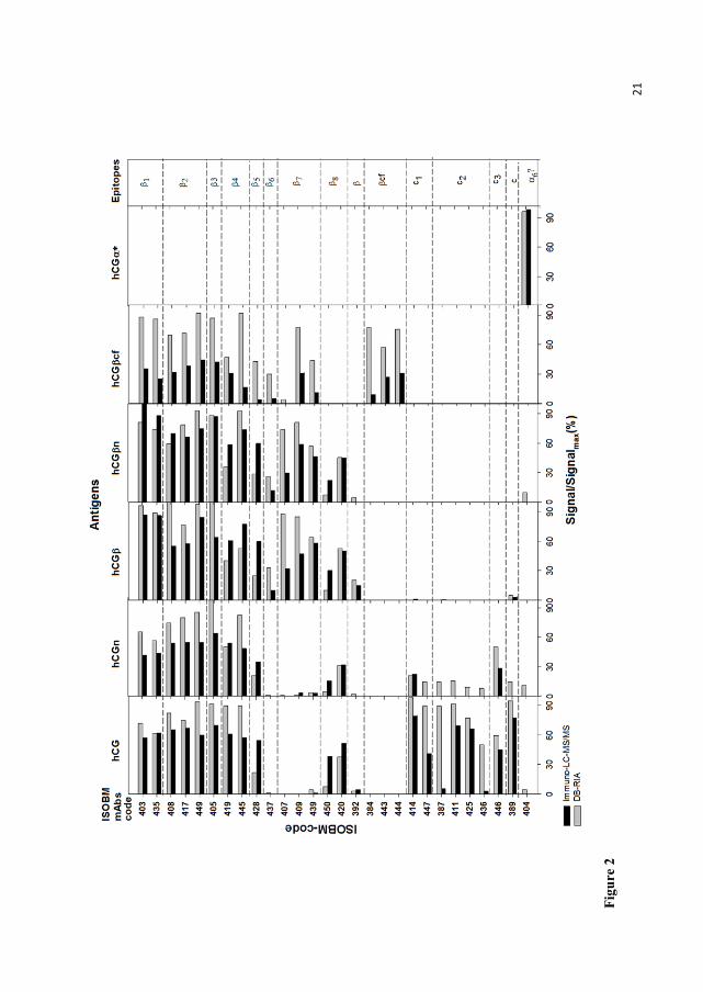
21
Figu
re 2

22
Figure 3

23
Figure 4