Faculty of Engineering and Technology SRM University SRM Nagar
Upload
nguyendieuCategory
view
218download
0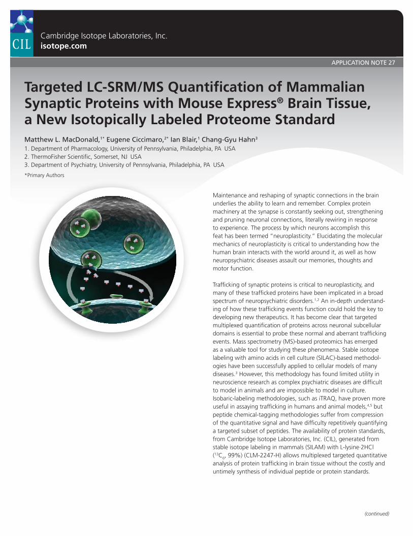
Targeted LC-SRM/MS Quantification of Mammalian Synaptic Proteins with Mouse Express® Brain Tissue,a New Isotopically Labeled Proteome StandardMatthew L. MacDonald,1* Eugene Ciccimaro,2* Ian Blair,1 Chang-Gyu Hahn3
1. Department of Pharmacology, University of Pennsylvania, Philadelphia, PA USA2. ThermoFisher Scientific, Somerset, NJ USA3. Department of Psychiatry, University of Pennsylvania, Philadelphia, PA USA
*Primary Authors
Maintenance and reshaping of synaptic connections in the brain underlies the ability to learn and remember. Complex protein machinery at the synapse is constantly seeking out, strengthening and pruning neuronal connections, literally rewiring in response to experience. The process by which neurons accomplish this feat has been termed “neuroplasticity.” Elucidating the molecular mechanics of neuroplasticity is critical to understanding how the human brain interacts with the world around it, as well as how neuropsychiatric diseases assault our memories, thoughts and motor function.
Trafficking of synaptic proteins is critical to neuroplasticity, and many of these trafficked proteins have been implicated in a broad spectrum of neuropsychiatric disorders.1,2 An in-depth under stand-ing of how these trafficking events function could hold the key to developing new therapeutics. It has become clear that targeted multiplexed quantification of proteins across neuronal subcellular domains is essential to probe these normal and aberrant trafficking events. Mass spectrometry (MS)-based proteomics has emerged as a valuable tool for studying these phenomena. Stable isotope labeling with amino acids in cell culture (SILAC)-based methodol-ogies have been successfully applied to cellular models of many diseases.3 However, this methodology has found limited utility in neuroscience research as complex psychiatric diseases are difficult to model in animals and are impossible to model in culture. Isobaric-labeling methodologies, such as iTRAQ, have proven more useful in assaying trafficking in humans and animal models,4,5 but peptide chemical-tagging methodologies suffer from compression of the quantitative signal and have difficulty repetitively quantifying a targeted subset of peptides. The availability of protein standards, from Cambridge Isotope Laboratories, Inc. (CIL), generated from stable isotope labeling in mammals (SILAM) with L-lysine·2HCl (13C6, 99%) (CLM-2247-H) allows multiplexed targeted quantitative analysis of protein trafficking in brain tissue without the costly and untimely synthesis of individual peptide or protein standards.
Cambridge Isotope Laboratories, Inc.isotope.com
(continued)
APPLICATION NOTE 27

In this note, we describe a liquid chromatography-selected reaction monitoring LC-SRM / MS approach for the targeted quantification of synaptic peptides in subcellular fractions of mammalian brain tissue utilizing CIL’s Mouse Express® Brain Tissue L-Lysine (13C6, 97%) (MT-LYS6-MB). This method utilizes membrane preparations from SILAM brain homogenate as an internal standard facilitating the quantification of over 100 proteins across three neuronal subfractions: vesicular, presynaptic and postsynaptic density, isolated from mouse brain tissue.
Experimental Design SRM design: Discovery data from 2D LC-MS / MS analyses of synaptic fractions from mouse and human tissue were mined for tryptic peptides from 200 synaptic proteins of interest.4,6 Pinpoint™ (ThermoFisher Scientific) was utilized to select peptides containing at least one lysine with homology between mouse and human sequences. Peptide homology allows this method to be utilized in clinical subjects as well as additional animal models.
Fractionation and sample preparation: “Light” (unlabeled) vesicular, presynaptic and postsynaptic density fractions were prepared from the cortex of two mice by sucrose density gradient centrifugation and pH-specific Triton X-100 precipitation (Figure 1). “Heavy” (labeled) membrane fractions were prepared from 40 mg Mouse Express® Brain Tissue (male) L-Lysine (13C6, 97%) (MT-LYSC6)
using an abbreviated version of the fractionation method (Figure 1). “Light” fractions were mixed with Mouse Express® brain tissue preparations at a ratio of 2:1 (Figure 1). The mixed proteomes were separated on a 4-12% bis-Tris gel, divided into two fractions: (i.e. < 64 kDa and between 64 – 250 kDa, reduced, alkylated and digested with trypsin (Figure 1)).
LC-SMR / MS: The resulting peptides were desalted offline and analyzed on a TSQ Vantage QqQ (ThermoFisher Scientific) interfaced to a NanoLC-Ultra 2D (Eksigent) and CaptiveSpray source (Michrom). A 5 μL peptide sample was loaded onto a 150 mm × 100 μm Magic C18 column (Michrom) at 1 μl / min 97% mobile phase A (H2O with 0.1% (v/ v) formic acid) 3% mobile phase B (acetonitrile with 0.1% (v/ v) formic acid) for 12 min, and eluted at 750 nl / min over a 25 min gradient from 3-35% mobile phase B. Retention windows were 1 – 1.5 min, depending on the number of SRMs to be analyzed. In this manner, SRM Dwell times are dynamic, based on the number of analytes eluting at a given time, and range from 5 to 60 msec per transition monitored in these experiments.
Data AnalysisFirst, light / heavy ratios were calculated for each peptide in Pinpoint™ using the integrated area under the curve (Figure 2). Where multiple peptides from a single protein were monitored, Pinpoint™ averaged the ratio values. To gain a global view of the data, the proteins were grouped into functional categories by gene ontology (GO) terms within ProteinCenter™ (ThermoFisher Scientific). Relative amounts of representative proteins from four of these categories are reported in Figure 3.
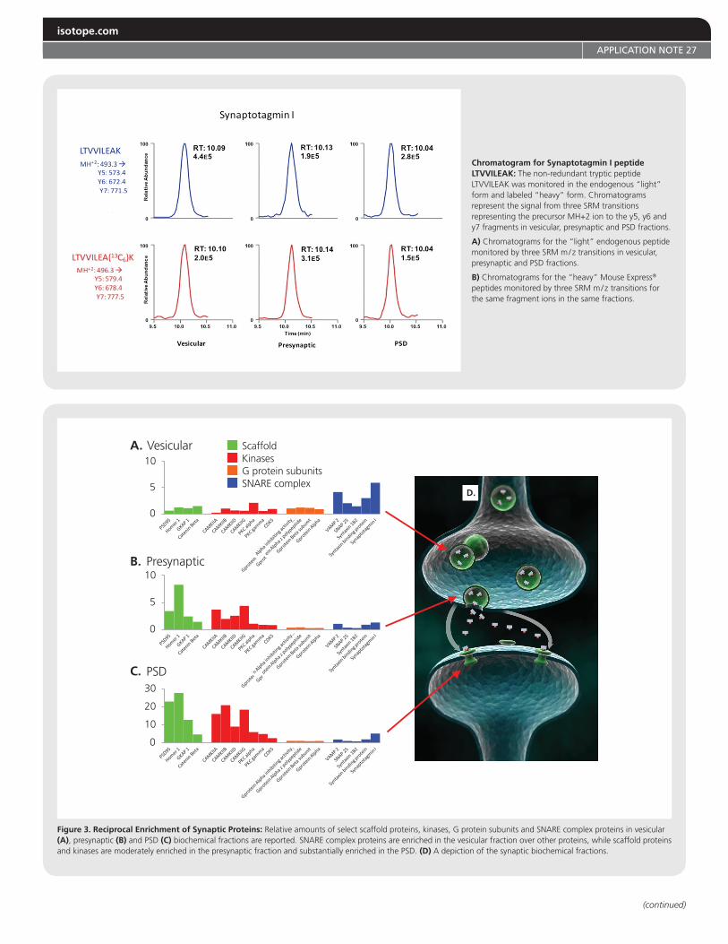
ResultsWe observed a significant interaction between microdomain partitioning and functions performed in those domains. A subset of the data for four families is reported in Figure 3. The postsynaptic density (PSD) fraction is highly enriched in kinases and scaffolding proteins, such as PSD-95. Scaffolding proteins link glutamate receptors to each other and kinases in PSD facilitating amplification and propagation of neurotransmitter signals. The synaptic vesicles, on the other hand, are highly enriched for soluble N-ethylmaleimide sensitive factor attachment protein receptor (SNARE) complex proteins, responsible for mediating vesicular fusion, and display a moderate enrichment of G-Protein subunits. The SNARE complex is responsible for fusing vesicles to the presynaptic membrane for neurotransmitter release.
DiscussionQuantitative analyses of subcellular proteomes has become increasingly important in neuroscience. Intracellular signaling pathways in the brain are no longer seen as a linear process from a molecule to the next, but are thought to occur in the context of numerous other protein interactions. This shift in paradigm has begun to impact the study of neuropsychiatric illnesses and thus, it is critically important to have the ability to quantify many proteins simultaneously with microdomain specificity.
A.
Mouse Brain300 mg
Homogenize0.32 M sucrose
Homogenize0.32 M sucrose
18,000 RPM15 min
1,000 G, 15 min
Supernatant
Synaptic Vesicles
Supernatant
Supernatant
µg MouseExpress ® ISTD
µg Brain Preparation
TotalHomogenate
Pellet
25mM Tris, pH 61% Triton X-100
18,000 RPM30 min
25mM Tris, pH 81% Triton X-100
36,000 RPM45 min
1-1.25 M sucrosedensity gradient14,000 G, 30 min
191
97
64
51
39
28
19
Synaptic Vesicles
Presynaptic Membranes
Postsynaptic Density
1-1.25 M sucrosedensity gradient
28,000 RPM, 3 hrs
MouseExpress® Mouse Brain40 mg
B.
C.
kDa
SynapticMembrane
MembranePellet
PostsynapticDensity
PresynapticMembrane
TotalHomogenate
Membrane
SynapticMembrane
Pellet
20
20
20
10
10
10
Figure 1. Brain Tissue Fractionation and Sample Preparation: A) Synaptic vesicle, presynaptic membrane and postsynaptic density enrichments were prepared from 300 mg mouse brain tissue using a variation of the method described in Hahn et al. 2009. A membrane fraction was prepared from 40 mg of Mouse Express® Brain Tissue for use as an internal standard (ISTD). B) The “light” fractions were mixed 2:1 with the “heavy” Mouse Express® membrane internal standard (ISTD). C) The mixed proteomes were separated in the first dimension by gel electrophoresis and divided into two fractions: 19-64 kDa and 64-250 kDa for on-gel trypsin digestion.
Cambridge Isotope Laboratories, Inc.
APPLICATION NOTE 27
(continued)
C.
B. Presynaptic
PSD
A. Vesicular ScaffoldKinasesG protein subunitsSNARE complex
0
10
20
30
0
5
10
0
5
10
D.
Chromatogram for Synaptotagmin I peptide LTVVILEAK: The non-redundant tryptic peptide LTVVILEAK was monitored in the endogenous “light” form and labeled “heavy” form. Chromatograms represent the signal from three SRM transitions representing the precursor MH+2 ion to the y5, y6 and y7 fragments in vesicular, presynaptic and PSD fractions.
A) Chromatograms for the “light” endogenous peptide monitored by three SRM m / z transitions in vesicular, presynaptic and PSD fractions.
B) Chromatograms for the “heavy” Mouse Express® peptides monitored by three SRM m / z transitions forthe same fragment ions in the same fractions.
Figure 3. Reciprocal Enrichment of Synaptic Proteins: Relative amounts of select scaffold proteins, kinases, G protein subunits and SNARE complex proteins in vesicular (A), presynaptic (B) and PSD (C) biochemical fractions are reported. SNARE complex proteins are enriched in the vesicular fraction over other proteins, while scaffold proteins and kinases are moderately enriched in the presynaptic fraction and substantially enriched in the PSD. (D) A depiction of the synaptic biochemical fractions.
isotope.com
APPLICATION NOTE 27
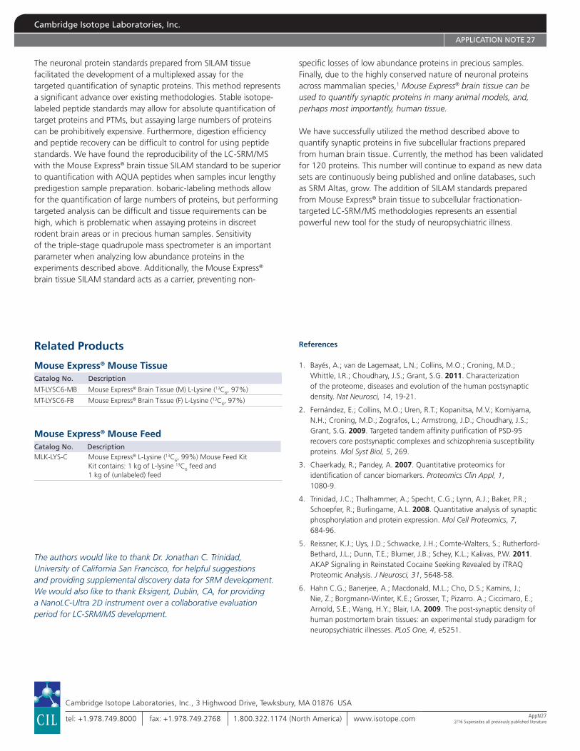
The neuronal protein standards prepared from SILAM tissue facilitated the development of a multiplexed assay for the targeted quantification of synaptic proteins. This method represents a significant advance over existing methodologies. Stable isotope-labeled peptide standards may allow for absolute quantification of target proteins and PTMs, but assaying large numbers of proteins can be prohibitively expensive. Furthermore, digestion efficiency and peptide recovery can be difficult to control for using peptide standards. We have found the reproducibility of the LC-SRM / MS with the Mouse Express® brain tissue SILAM standard to be superior to quantification with AQUA peptides when samples incur lengthy predigestion sample preparation. Isobaric-labeling methods allow for the quantification of large numbers of proteins, but performing targeted analysis can be difficult and tissue requirements can be high, which is problematic when assaying proteins in discreet rodent brain areas or in precious human samples. Sensitivity of the triple-stage quadrupole mass spectrometer is an important parameter when analyzing low abundance proteins in the experiments described above. Additionally, the Mouse Express® brain tissue SILAM standard acts as a carrier, preventing non-
specific losses of low abundance proteins in precious samples. Finally, due to the highly conserved nature of neuronal proteins across mammalian species,1 Mouse Express® brain tissue can be used to quantify synaptic proteins in many animal models, and, perhaps most importantly, human tissue.
We have successfully utilized the method described above to quantify synaptic proteins in five subcellular fractions prepared from human brain tissue. Currently, the method has been validated for 120 proteins. This number will continue to expand as new data sets are continuously being published and online databases, such as SRM Altas, grow. The addition of SILAM standards prepared from Mouse Express® brain tissue to subcellular fractionation-targeted LC-SRM / MS methodologies represents an essential powerful new tool for the study of neuropsychiatric illness.
References
1. Bayés, A.; van de Lagemaat, L.N.; Collins, M.O.; Croning, M.D.; Whittle, I.R.; Choudhary, J.S.; Grant, S.G. 2011. Characterization of the proteome, diseases and evolution of the human postsynaptic density. Nat Neurosci, 14, 19-21.
2. Fernández, E.; Collins, M.O.; Uren, R.T.; Kopanitsa, M.V.; Komiyama, N.H.; Croning, M.D.; Zografos, L.; Armstrong, J.D.; Choudhary, J.S.; Grant, S.G. 2009. Targeted tandem affinity purification of PSD-95 recovers core postsynaptic complexes and schizophrenia susceptibility proteins. Mol Syst Biol, 5, 269.
3. Chaerkady, R.; Pandey, A. 2007. Quantitative proteomics for identification of cancer biomarkers. Proteomics Clin Appl, 1, 1080-9.
4. Trinidad, J.C.; Thalhammer, A.; Specht, C.G.; Lynn, A.J.; Baker, P.R.; Schoepfer, R.; Burlingame, A.L. 2008. Quantitative analysis of synaptic phosphorylation and protein expression. Mol Cell Proteomics, 7, 684-96.
5. Reissner, K.J.; Uys, J.D.; Schwacke, J.H.; Comte-Walters, S.; Rutherford-Bethard, J.L.; Dunn, T.E.; Blumer, J.B.; Schey, K.L.; Kalivas, P.W. 2011. AKAP Signaling in Reinstated Cocaine Seeking Revealed by iTRAQ Proteomic Analysis. J Neurosci, 31, 5648-58.
6. Hahn C.G.; Banerjee, A.; Macdonald, M.L.; Cho, D.S.; Kamins, J.; Nie, Z.; Borgmann-Winter, K.E.; Grosser, T.; Pizarro. A.; Ciccimaro, E.; Arnold, S.E.; Wang, H.Y.; Blair, I.A. 2009. The post-synaptic density of human postmortem brain tissues: an experimental study paradigm for neuropsychiatric illnesses. PLoS One, 4, e5251.
Related Products
Mouse Express® Mouse TissueCatalog No. Description
MT-LYSC6-MB Mouse Express® Brain Tissue (M) L-Lysine (13C6, 97%)
MT-LYSC6-FB Mouse Express® Brain Tissue (F) L-Lysine (13C6, 97%)
Mouse Express® Mouse FeedCatalog No. DescriptionMLK-LYS-C Mouse Express® L-Lysine (13C6, 99%) Mouse Feed Kit Kit contains: 1 kg of L-lysine 13C6 feed and 1 kg of (unlabeled) feed
The authors would like to thank Dr. Jonathan C. Trinidad, University of California San Francisco, for helpful suggestions and providing supplemental discovery data for SRM development. We would also like to thank Eksigent, Dublin, CA, for providing a NanoLC-Ultra 2D instrument over a collaborative evaluation period for LC-SRM/MS development.
Cambridge Isotope Laboratories, Inc., 3 Highwood Drive, Tewksbury, MA 01876 USA
tel: +1.978.749.8000 fax: +1.978.749.2768 1.800.322.1174 (North America) www.isotope.com AppN272/16 Supersedes all previously published literature
Cambridge Isotope Laboratories, Inc.
APPLICATION NOTE 27