Targeted Ablation, Silencing, and Activation Establish...
17
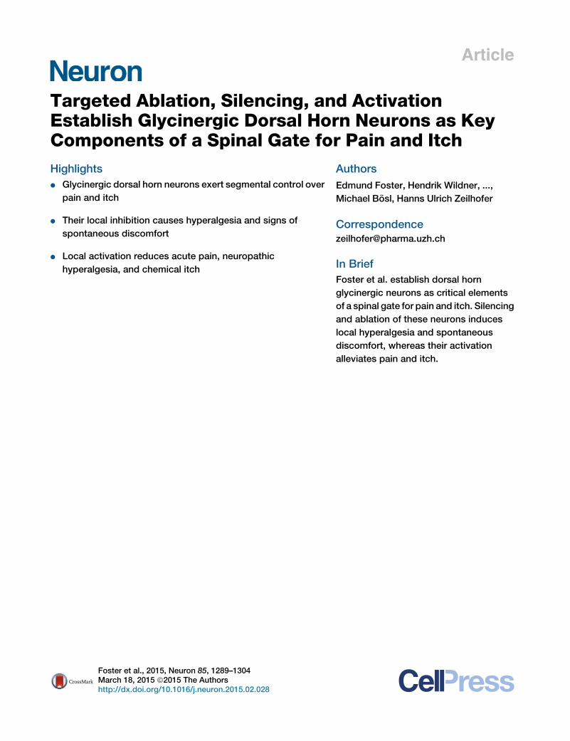
Article Targeted Ablation, Silencing, and Activation Establish Glycinergic Dorsal Horn Neurons as Key Components of a Spinal Gate for Pain and Itch Highlights d Glycinergic dorsal horn neurons exert segmental control over pain and itch d Their local inhibition causes hyperalgesia and signs of spontaneous discomfort d Local activation reduces acute pain, neuropathic hyperalgesia, and chemical itch Authors Edmund Foster, Hendrik Wildner, ..., Michael Bo ¨ sl, Hanns Ulrich Zeilhofer Correspondence [email protected] In Brief Foster et al. establish dorsal horn glycinergic neurons as critical elements of a spinal gate for pain and itch. Silencing and ablation of these neurons induces local hyperalgesia and spontaneous discomfort, whereas their activation alleviates pain and itch. Foster et al., 2015, Neuron 85, 1289–1304 March 18, 2015 ª2015 The Authors http://dx.doi.org/10.1016/j.neuron.2015.02.028
Transcript of Targeted Ablation, Silencing, and Activation Establish...
Article
Targeted Ablation, Silenci
ng, and ActivationEstablish Glycinergic Dorsal Horn Neurons as KeyComponents of a Spinal Gate for Pain and ItchHighlights
d Glycinergic dorsal horn neurons exert segmental control over
pain and itch
d Their local inhibition causes hyperalgesia and signs of
spontaneous discomfort
d Local activation reduces acute pain, neuropathic
hyperalgesia, and chemical itch
Foster et al., 2015, Neuron 85, 1289–1304March 18, 2015 ª2015 The Authorshttp://dx.doi.org/10.1016/j.neuron.2015.02.028
Authors
Edmund Foster, Hendrik Wildner, ...,
Michael Bosl, Hanns Ulrich Zeilhofer
In Brief
Foster et al. establish dorsal horn
glycinergic neurons as critical elements
of a spinal gate for pain and itch. Silencing
and ablation of these neurons induces
local hyperalgesia and spontaneous
discomfort, whereas their activation
alleviates pain and itch.

Neuron
Article
Targeted Ablation, Silencing, and ActivationEstablish Glycinergic Dorsal Horn Neuronsas Key Components of a Spinal Gate for Pain and ItchEdmund Foster,1,5 Hendrik Wildner,1,5 Laetitia Tudeau,1,4 Sabine Haueter,1,4 William T. Ralvenius,1 Monika Jegen,1,4
Helge Johannssen,1 Ladina Hosli,1 Karen Haenraets,1,4 Alexander Ghanem,2 Karl-Klaus Conzelmann,2 Michael Bosl,3
and Hanns Ulrich Zeilhofer1,4,*1Institute of Pharmacology and Toxicology, University of Zurich, Winterthurerstrasse 190, 8057 Zurich, Switzerland2Gene Center, Ludwig Maximilians University Munich, Max von Pettenkofer Institute of Virology, Feodor Lynen Strasse 25, 81377 Munich,
Germany3Experimental Biomedicine, University of Wurzburg, Josef Schneider Strasse 2, 97080 Wurzburg, Germany4Institute of Pharmaceutical Sciences, Swiss Federal Institute of Technology (ETH) Zurich, Vladimir Prelog Weg 4, 8093 Zurich, Switzerland5Co-first author
*Correspondence: [email protected]
http://dx.doi.org/10.1016/j.neuron.2015.02.028
This is an open access article under the CC BY-NC-ND license (http://creativecommons.org/licenses/by-nc-nd/4.0/).
SUMMARY
The gate control theory of pain proposes that inhibi-tory neurons of the spinal dorsal horn exert criticalcontrol over the relay of nociceptive signals to higherbrain areas. Here we investigated how the glyciner-gic subpopulation of these neurons contributesto modality-specific pain and itch processing. Wegenerated a GlyT2::Cre transgenic mouse line suit-able for virus-mediated retrograde tracing studiesand for spatially precise ablation, silencing, and acti-vation of glycinergic neurons. We found that theseneurons receive sensory input mainly from myelin-ated primary sensory neurons and that their localtoxin-mediated ablation or silencing induces local-ized mechanical, heat, and cold hyperalgesia; spon-taneous flinching behavior; and excessive lickingand biting directed toward the corresponding skinterritory. Conversely, local pharmacogenetic activa-tion of the same neurons alleviated neuropathic hy-peralgesia and chloroquine- and histamine-induceditch. These results establish glycinergic neurons ofthe spinal dorsal horn as key elements of an inhibi-tory pain and itch control circuit.
INTRODUCTION
Pain evoked in response to potentially tissue-damaging stimuli is
essential for the maintenance of our physical integrity. It can,
however, have a severe impact on our well-being when it occurs
spontaneously or in response to inappropriate stimuli. It is widely
accepted that the spinal dorsal horn, which constitutes the first
site for synaptic processing in the pain pathway, serves a critical
role for maintaining a physiological level of pain sensitivity.
Conversely, maladaptive changes at this site contribute critically
to a wide variety of pain pathologies (Kuner, 2010; Sandkuhler,
2009;Woolf and Salter, 2000; Zeilhofer et al., 2012). The concept
of the dorsal horn as a first site to control the modality and inten-
sity of sensory signals conveyed to higher CNS centers dates
back to Wall and Melzack’s gate control theory of pain (Melzack
and Wall, 1965). According to their theory, inhibitory dorsal
horn interneurons would constitute the physical basis of this
pain gate. They would become activated primarily by input
from (non-nociceptive) low-threshold myelinated sensory nerve
fibers and would, in turn, control the activity of spinal output
neurons.
Early work with antagonists of inhibitory neurotransmitter
receptors has demonstrated that exaggerated pain responses
can be triggered by blocking inhibitory neurotransmission in
the spinal cord (Beyer et al., 1985; Loomis et al., 2001; Sherman
and Loomis, 1994; Sivilotti and Woolf, 1994; Yaksh, 1989). More
recent work has suggested that diminished inhibitory control
over dorsal horn sensory circuits leads to increased excitability
and spontaneous activity of dorsal horn neurons (Drew et al.,
2004; Sorkin et al., 1998) and allows the excitation of normally
pain-specific neurons by inappropriate (non-nociceptive) signals
(Baba et al., 2003; Keller et al., 2007; Torsney and MacDermott,
2006; for a review see Sandkuhler, 2009). A large body of
evidence, meanwhile, indicates that diminished synaptic inhi-
bition also occurs naturally in the course of inflammatory and
neuropathic syndromes through various mechanisms (Zeilhofer
et al., 2012).
Inhibitory interneurons make up about one-third of the total
neuronal population in the spinal dorsal horn. These neurons
use either g-aminobutyric acid (GABA) or glycine or both for
fast neuronal inhibition. In the superficial dorsal horn, i.e., in the
termination area of nociceptors, the majority of inhibitory neu-
rons are purely GABAergic, whereas those in the deep dorsal
horn are mainly mixed GABA/glycinergic neurons (Todd and
Spike, 1993; for a review, see Zeilhofer et al., 2012). Despite
recent progress (Duan et al., 2014), the circuits and interneuron
types contributing to spinal pain control are still not completely
understood. A detailed and comprehensive analysis has so far
Neuron 85, 1289–1304, March 18, 2015 ª2015 The Authors 1289

(legend on next page)
1290 Neuron 85, 1289–1304, March 18, 2015 ª2015 The Authors

been hampered by the lack of appropriate molecular tools. Such
studies became feasible with the advent of sophisticated viral
vector-based approaches and Cre transgenic mouse lines.
These technologies allow the functional andmorphological map-
ping of neuronal circuits through retrograde tracing starting from
precisely defined neuron populations (Wickersham et al., 2007)
and the manipulation of specific subsets of neurons through
ablation, silencing, and excitation in a spatially highly precise
manner (Betley and Sternson, 2011).
To specifically address the role of the glycinergic neuron
population in the dorsal horn, we generated a bacterial artificial
chromosome (BAC) transgenic mouse line that expresses the
Cre recombinase selectively in glycinergic neurons. Retrograde
tracing experiments showed that glycinergic dorsal horn neu-
rons receive sensory input mainly from low-threshold myelin-
ated dorsal root ganglion (DRG) neurons. Local unilateral
ablation of glycinergic neurons with diphtheria toxin or
silencing of these neurons with tetanus toxin over three lumbar
segments induced mechanical, heat, and cold hyperalgesia
and signs of spontaneous aversive behavior, including exten-
sive localized biting reminiscent of chronic itch. Accordingly,
selective and local activation of glycinergic neurons alleviated
nocifensive responses to acute noxious stimuli, hyperalgesia
in neuropathic mice, and chloroquine- and histamine-induced
itch responses.
RESULTS
Generation of GlyT2::Cre BAC Transgenic MiceTo specifically and locally manipulate glycinergic neurons, we
generated a BAC transgenic mouse that expresses the Cre
recombinase under the transcriptional control of the GlyT2
gene, a marker of glycinergic neurons (Poyatos et al., 1997).
We used the same strategy as employed previously for a
GlyT2::eGFP BAC transgenic mouse (Zeilhofer et al., 2005; Fig-
ure 1A), whose eGFP expression pattern precisely recapitulated
the distribution of glycinergic neurons (Hossaini et al., 2007;
Zeilhofer et al., 2005). To characterize the spinal pattern
of GlyT2::Cre-dependent recombination, we generated triple
transgenic mice carrying the GlyT2::eGFP and GlyT2::Cre
transgenes together with a ROSA26LacZ reporter. We found a
nearly complete match of b-galactosidase (b-gal) expression
with GlyT2::eGFP in the deep dorsal horn, whereas the majority
of superficial dorsal horn b-gal+ neurons lacked GlyT2::eGFP
Figure 1. Generation and Characterization of GlyT2::Cre BAC Transge
(A) GlyT2::Cre BAC transgenic mice were generated from a BAC DNA clone conta
of a Cre expression cassette.
(B) Comparison of Cre-mediated recombination and GlyT2::eGFP expression in G
and anti-eGFP (green) antibodies.
(C) Percentage of b-gal and eGFP double-positive neurons (mean ± SD).
(D) Same as (B) but analysis of co-expression of Cre with eGFP.
(E) Quantification of (D).
(F) Same as (B) but analysis of co-expression of Cre with Pax2.
(G) Quantification of (F). The two left columns refer to the entire spinal cord cro
superficial dorsal horn (laminae I/II) and the rest of the spinal cord (RIII) separate
(H) Higher magnification of the lamina II/III border (the dashed area indicated in F
Scale bars, 100 mm (B) and 50 mm (F).
(Figures 1B and 1C). However, when the actual Cre protein
expression pattern was analyzed in adult mice, an almost com-
plete overlap of Cre and eGFP expression was found throughout
the dorsal horn. Both were most abundant in the deep dorsal
horn but largely absent from the superficial layers, suggesting
that expression of the b-gal reporter in the superficial dorsal
horn was caused by transient Cre expression during develop-
ment (Figures 1D and 1E). To further confirm eutopic Cre ex-
pression, we demonstrate that nearly all (93.7% ± 1.5%) Cre+
neurons also stain positive for Pax2, a transcription factor
expressed by more than 90% of adult spinal Gad67eGFP and
GlyT2::eGFP neurons (Figures 1F–1H; Figure S1). In the deep
dorsal and ventral horn, almost 90% of Pax2+ neurons also
expressed Cre, whereas this coexpression dropped to 20% in
laminae I/II (Figures 1F–1H), which corresponds well with the
spinal distribution of glycinergic neurons (Todd and Spike,
1993). The 20% Cre+ neurons of laminae I/II included most of
the neuronal nitric oxide synthase (nNOS)+ inhibitory neurons
in these laminae (Figure S2).
Primary Sensory Input onto Dorsal Horn GlycinergicInterneuronsThe preferential localization of glycinergic neurons in the deep
dorsal horn (i.e., in the termination areas of non-nociceptive
primary sensory neurons) is consistent with what has been
proposed in the gate control theory, but unequivocal proof for
the innervation by non-nociceptive primary sensory neurons is
lacking. Here we used a two-step rabies virus-based retrograde
monosynaptic tracing strategy involving an EnvA pseudotyped
eGFP rabies virus and TVA expression fromaCre-dependent ad-
eno-associated virus (AAV) tomap neurons presynaptic to dorsal
horn glycinergic neurons (Figures 2A and 2B). Seven days after
rabies virus injection, we foundbetween 30 and 40primary rabies
virus-infected (TVA+/eGFP+) cells per spinal cord section (Fig-
ure S3) and numerous eGFP+ neurons in three to four adjacent
lumbar DRGs ipsilateral to the virus injection site (Figure 2C),
demonstrating the presence of direct synaptic input fromprimary
sensory neurons onto glycinergic dorsal horn neurons. Ninety-
one percent of these neurons (851 of 934) were myelinated
(NF200+), including 7.6% (49 of 641) peptidergic (CGRP+/
NF200+) neurons. Only 4.2% were NF200�/CGRP+ (27 of 641)
and only 0.15% (1 of 664) were NF200+/IB4+ (Figures 2D–2F).
These results are consistent with strong innervation of dorsal
horn glycinergic neurons by non-nociceptive sensory neurons.
nic Mice
ining the mouseGlyT2 locus, of which exon 2 (E2) wasmodified by the insertion
lyT2::Cre;GlyT2::eGFP;Rosa26LacZ triple-transgenic mice using anti-b-gal (red)
ss section (laminae I–X), whereas the two right columns show results for the
ly.
).
Neuron 85, 1289–1304, March 18, 2015 ª2015 The Authors 1291
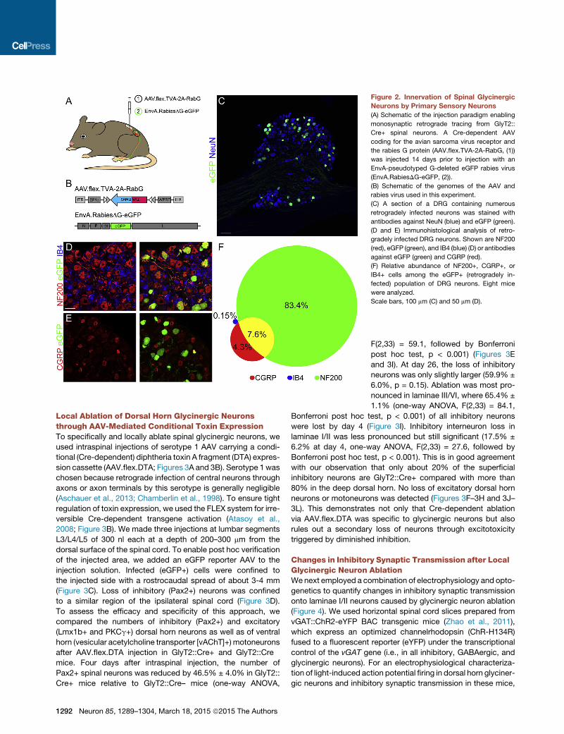
Figure 2. Innervation of Spinal Glycinergic
Neurons by Primary Sensory Neurons
(A) Schematic of the injection paradigm enabling
monosynaptic retrograde tracing from GlyT2::
Cre+ spinal neurons. A Cre-dependent AAV
coding for the avian sarcoma virus receptor and
the rabies G protein (AAV.flex.TVA-2A-RabG, (1))
was injected 14 days prior to injection with an
EnvA-pseudotyped G-deleted eGFP rabies virus
(EnvA.RabiesDG-eGFP, (2)).
(B) Schematic of the genomes of the AAV and
rabies virus used in this experiment.
(C) A section of a DRG containing numerous
retrogradely infected neurons was stained with
antibodies against NeuN (blue) and eGFP (green).
(D and E) Immunohistological analysis of retro-
gradely infected DRG neurons. Shown are NF200
(red), eGFP (green), and IB4 (blue) (D) or antibodies
against eGFP (green) and CGRP (red).
(F) Relative abundance of NF200+, CGRP+, or
IB4+ cells among the eGFP+ (retrogradely in-
fected) population of DRG neurons. Eight mice
were analyzed.
Scale bars, 100 mm (C) and 50 mm (D).
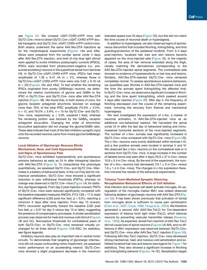
Local Ablation of Dorsal Horn Glycinergic Neuronsthrough AAV-Mediated Conditional Toxin ExpressionTo specifically and locally ablate spinal glycinergic neurons, we
used intraspinal injections of serotype 1 AAV carrying a condi-
tional (Cre-dependent) diphtheria toxin A fragment (DTA) expres-
sion cassette (AAV.flex.DTA; Figures 3A and 3B). Serotype 1was
chosen because retrograde infection of central neurons through
axons or axon terminals by this serotype is generally negligible
(Aschauer et al., 2013; Chamberlin et al., 1998). To ensure tight
regulation of toxin expression, we used the FLEX system for irre-
versible Cre-dependent transgene activation (Atasoy et al.,
2008; Figure 3B). We made three injections at lumbar segments
L3/L4/L5 of 300 nl each at a depth of 200–300 mm from the
dorsal surface of the spinal cord. To enable post hoc verification
of the injected area, we added an eGFP reporter AAV to the
injection solution. Infected (eGFP+) cells were confined to
the injected side with a rostrocaudal spread of about 3-4 mm
(Figure 3C). Loss of inhibitory (Pax2+) neurons was confined
to a similar region of the ipsilateral spinal cord (Figure 3D).
To assess the efficacy and specificity of this approach, we
compared the numbers of inhibitory (Pax2+) and excitatory
(Lmx1b+ and PKCg+) dorsal horn neurons as well as of ventral
horn (vesicular acetylcholine transporter [vAChT]+) motoneurons
after AAV.flex.DTA injection in GlyT2::Cre+ and GlyT2::Cre�mice. Four days after intraspinal injection, the number of
Pax2+ spinal neurons was reduced by 46.5% ± 4.0% in GlyT2::
Cre+ mice relative to GlyT2::Cre– mice (one-way ANOVA,
1292 Neuron 85, 1289–1304, March 18, 2015 ª2015 The Authors
F(2,33) = 59.1, followed by Bonferroni
post hoc test, p < 0.001) (Figures 3E
and 3I). At day 26, the loss of inhibitory
neurons was only slightly larger (59.9% ±
6.0%, p = 0.15). Ablation was most pro-
nounced in laminae III/VI, where 65.4% ±
1.1% (one-way ANOVA, F(2,33) = 84.1,
Bonferroni post hoc test, p < 0.001) of all inhibitory neurons
were lost by day 4 (Figure 3I). Inhibitory interneuron loss in
laminae I/II was less pronounced but still significant (17.5% ±
6.2% at day 4, one-way ANOVA, F(2,33) = 27.6, followed by
Bonferroni post hoc test, p < 0.001). This is in good agreement
with our observation that only about 20% of the superficial
inhibitory neurons are GlyT2::Cre+ compared with more than
80% in the deep dorsal horn. No loss of excitatory dorsal horn
neurons or motoneurons was detected (Figures 3F–3H and 3J–
3L). This demonstrates not only that Cre-dependent ablation
via AAV.flex.DTA was specific to glycinergic neurons but also
rules out a secondary loss of neurons through excitotoxicity
triggered by diminished inhibition.
Changes in Inhibitory Synaptic Transmission after LocalGlycinergic Neuron AblationWenext employed a combination of electrophysiology and opto-
genetics to quantify changes in inhibitory synaptic transmission
onto laminae I/II neurons caused by glycinergic neuron ablation
(Figure 4). We used horizontal spinal cord slices prepared from
vGAT::ChR2-eYFP BAC transgenic mice (Zhao et al., 2011),
which express an optimized channelrhodopsin (ChR-H134R)
fused to a fluorescent reporter (eYFP) under the transcriptional
control of the vGAT gene (i.e., in all inhibitory, GABAergic, and
glycinergic neurons). For an electrophysiological characteriza-
tion of light-induced action potential firing in dorsal horn glyciner-
gic neurons and inhibitory synaptic transmission in these mice,
see Figure S4. We crossed vGAT::ChR2-eYFP mice with
GlyT2::Cre+mice to obtain GlyT2::Cre+;vGAT::ChR2-eYFP dou-
ble transgenic and GlyT2::Cre-;vGAT::ChR2-eYFP control mice.
Both strains underwent the same AAV.flex.DTA injections as
for the morphological experiments (Figures 4Aa and 4Ab).
Slices were prepared from the lumbar spinal cords 4 days
after AAV.flex.DTA injection, and brief (4-ms) blue light stimuli
were applied to evoke inhibitory postsynaptic currents (IPSCs).
IPSCs were recorded from photocurrent-negative (i.e., pre-
sumed excitatory) neurons in the superficial dorsal horn (laminae
I/II). In GlyT2::Cre-;vGAT::ChR2-eYFP mice, IPSCs had mean
amplitudes of 1.39 ± 0.41 nA (n = 21), whereas those in
GlyT2:Cre+;vGAT::ChR2-eYFP mice were only 0.62 ± 0.10 nA
(n = 23) (Figures 4Ac and 4Ad). To test whether the remaining
IPSCs originated from purely GABAergic neurons, we deter-
mined the relative contribution of glycine and GABA to the
IPSC in GlyT2::Cre+ and GlyT2::Cre– mice after AAV.flex.DTA
injection (Figure 4B). We found that, in both strains of mice, the
glycine receptor antagonist strychnine blocked on average
more than 70% of the total IPSC amplitude (73.0% ± 3.5%,
n = 12, and 78.5% ± 3.9%, n = 15 for GlyT2::Cre– and GlyT2::
Cre+ mice, respectively; p < 0.05, unpaired t test), whereas
the remaining portion was blocked by the GABAA receptor
antagonist bicuculline. Changing the order of strychnine/
bicuculline application did not change the glycine/GABA ratio.
These data indicate that most of the fast inhibitory synaptic input
onto the recorded neurons came frommixed glycine/GABAergic
neurons.
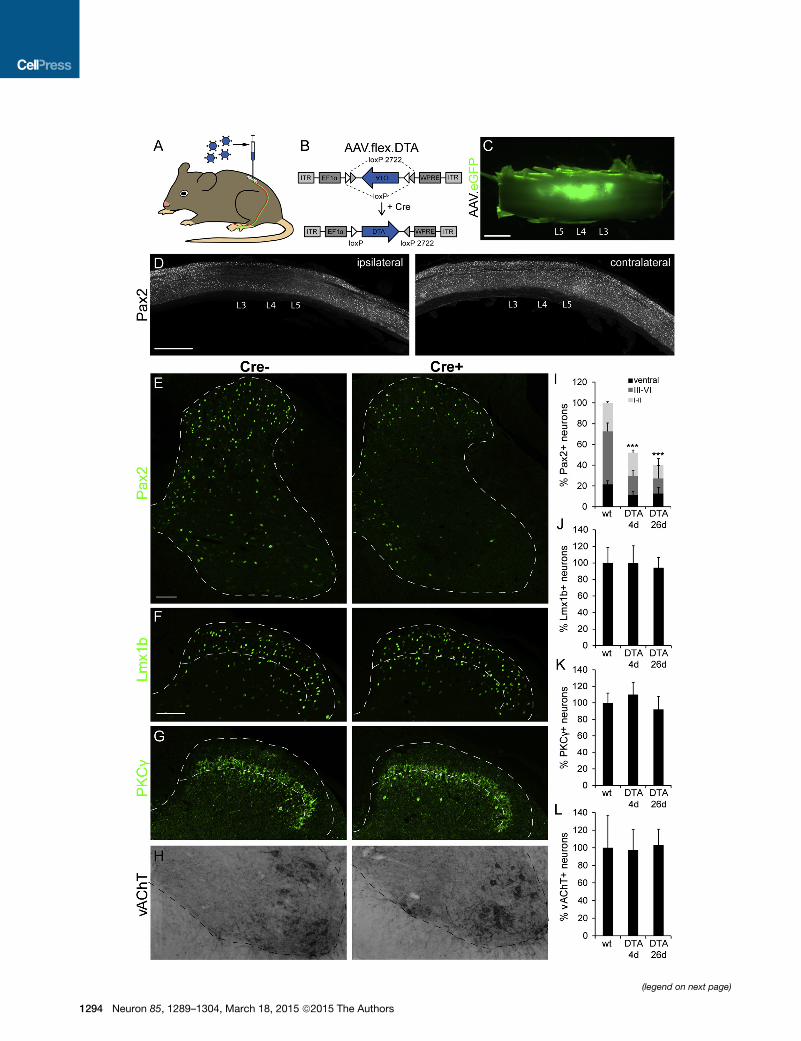
Local Ablation of Glycinergic Neurons ElicitsMechanical, Heat, and Cold Hypersensitivityand Signs of Spontaneous PainGlyT2::Cre+ mice exhibited hypersensitivity and spontaneous
aversive behaviors as early as 24 hr after intraspinal injection
with AAV.flex.DTA (Figure 5). To characterize this phenotype
quantitatively, we assessed GlyT2::Cre+ and GlyT2::Cre– litter-
mates in a battery of behavioral tests. In the von Frey test for me-
chanical sensitization, GlyT2::Cre+ mice showed a significant
reduction in paw withdrawal thresholds (PWTs), whereas no
change was observed in GlyT2::Cre– mice (Figure 5A; for statis-
tics, see figure legend). From day 2 post-injection onward, PWTs
of GlyT2::Cre+ mice were reduced significantly compared with
the baseline (repeated measures ANOVA, F(13,130) = 17.5; least
significant difference (LSD) post hoc test, p < 0.01), reaching a
minimum 5 days after virus injection. From day 13 onward,
PWTs recovered significantly toward the baseline (LSD post
hoc test, p < 0.01 for day 5 versus day 13 or later), suggesting
the presence of compensatory processes. A similar sensitization
process was observed for heat and noxious cold stimuli (Figures
5B and 5C). Nociceptive thresholds for contralateral paws of
both GlyT2::Cre+ or GlyT2::Cre– mice remained virtually un-
changed for all three stimuli (Figures S5A–S5C; for statistics,
see figure legends).
Inhibitory neurons also play an important role in ventral motor
circuits. To demonstrate that targeting of glycinergic interneu-
rons did not cause confounding motor impairment, we assessed
motor performance on an accelerating rotarod. GlyT2::Cre+
mice showed a slight progressive decrease in the maximum
tolerated speed over 25 days (Figure 5D), but this did not match
the time course of neuronal death or hyperalgesia.
In addition to hyperalgesia, we also observed signs of sponta-
neous discomfort that included flinching, licking/biting, and limb
guarding/protection of the ipsilateral hindlimb. From 3–4 days
post-injection, localized hair loss and skin lesions became
apparent on the virus-injected side (Figure 5E). In the majority
of cases, the area of hair removal extended along the thigh,
closely matching the dermatomes corresponding to the
AAV.flex.DTA-injected spinal segments. The contralateral side
showed no evidence of hypersensitivity or hair loss and lesions.
Similarly, AAV.flex.DTA-injected GlyT2::Cre– mice remained
completely normal. To assess spontaneous aversive behaviors,
we quantified paw flinches in AAV.flex.DTA-injected mice and
the time the animals spent licking/biting the affected limb.
In GlyT2::Cre+mice, we observed a significant increase in flinch-
ing and the time spent licking/biting, which peaked around
4 days after injection (Figure 5F). After day 4, the frequency of
flinching decreased over the course of the remaining experi-
ment, mirroring the recovery from thermal and mechanical
hyperalgesia.
We next investigated the expression of c-fos, a marker of
neuronal activation, in AAV.flex.DTA-injected mice as an
additional non-behavioral readout. We harvested the spinal
cords 24 hr after the last day of behavioral testing and immu-
nostained horizontal sections of the virus-injected segments.
The number of c-fos+ somata was significantly increased in
GlyT2::Cre+ mice compared with GlyT2::Cre– mice (Figure 5G).
The c-fos+ neurons were evenly distributed within laminae I–IV,
and a few positive somata were located in laminae V and VI.
We observed few c-fos+ neurons on the contralateral side or in
sections from GlyT2::Cre– mice. A large increase in the number
of labeled soma was seen after 4 days (76.6 ± 37 in Cre+ versus
10.8 ± 5.4 in Cre– mice). By the end of the experiment, the num-
ber of c-fos+ neurons had decreased (19.1 ± 9 in Cre+ versus
1.6 ± 1.4 in Cre– mice). The changes in c-fos expression there-
fore mirrored the results of the behavioral experiments.
Tetanus Toxin-Mediated Synaptic SilencingRecapitulates Behavioral Effects of Cell AblationViral infection and neuronal cell death activate microglia. An up-
regulation of the microglia marker IBA1 was indeed observed
following ablation of glycinergic neurons with AAV.flex.DTA (Fig-
ure S6). It has been shown previously that activation of dorsal
horn microglia alone is sufficient to cause pain sensitization
(Clark et al., 2007; Coyle, 1998; Tanga et al., 2004). We therefore
generated a second AAV (AAV.flex.TeLC) for Cre-dependent
expression of tetanus toxin light chain (TeLC), which silences
neurons by preventing vesicular transmitter release (Sweeney
et al., 1995). As expected, dorsal horn injection of AAV.flex.TeLC
did not cause loss of spinal neurons (Figures 6A–6H), and no dif-
ference in IBA1 expression was observed between GlyT2::Cre+
and GlyT2::Cre– mice after AAV.flex.TeLC injection (Figure S6).
Following AAV.flex.TeLC injection, GlyT2::Cre+ mice developed
strong mechanical, heat, and noxious cold sensitization and ex-
hibited localized hair loss and lesions (see legend for Figure 7 for
statistics). They also showed a significant increase in flinching
and licking/biting behavior (Figures 7A–7E). Sensitization began
Neuron 85, 1289–1304, March 18, 2015 ª2015 The Authors 1293
(legend on next page)
1294 Neuron 85, 1289–1304, March 18, 2015 ª2015 The Authors

to decrease from day 10 onward. We also found a significant
increase in the number of c-fos+ cells 4 days and 26 days after
injection of AAV.flex.TeLC (Figure 7F). No changes were seen
in the contralateral paw (Figure S5). The results obtained with
tetanus toxin-mediated silencing therefore closely mirrored the
effects of AAV.flex.DTA-mediated cell ablation.
Local Pharmacogenetic Activation of Dorsal HornGlycinergic Neurons Mitigates NeuropathicHyperalgesiaOur observation that glycinergic neurons serve a critical role in
maintaining a physiological level of pain sensitivity prompted us
to test whether exogenous activation of glycinergic neurons
ameliorates chronic pain. To this end, we employed a phar-
macogenetic approach (Armbruster et al., 2007) and injected
GlyT2::Cre;GlyT2::eGFP double-transgenic mice with AAV.flex.
hM3Dq-mCherry, targeted to the left L3–L5 spinal segments.
AAV.flex.hM3Dq encodes an engineered G protein-coupled
receptor that renders Cre-expressing neurons responsive to
activation by clozapine N-oxide (CNO) (Alexander et al., 2009;
Figure 8A). We verified expression of hM3Dq-mCherry in the
somata and neurites of GlyT2::eGFP+ neurons 2 weeks post-
injection (Figure 8B). Transduced neurons were largely confined
to lamina III/VI of the dorsal horn. No mCherry+ cell bodies
were found in the ipsilateral ventral horn or on the contralateral
side.
One week after virus injection, a unilateral neuropathic
sensitization of the hindpaw ipsilateral to the side of virus
injection was induced through a chronic constriction injury
(CCI) of the sciatic nerve (Bennett and Xie, 1988). On day 7 after
CCI surgery, we injected CNO (1 mg/kg, i.p.]) or vehicle and
assessed mechanical (von Frey) paw withdrawal thresholds
for 5 hr. Within 1-2 hr after CNO administration, mechanical
sensitivity decreased significantly in drug-treated but not
vehicle-treated GlyT2::Cre+ mice (Figure 8C). Vehicle-injected
mice showed no improvement over the testing period. We
also tested whether activation of glycinergic neurons would
reduce responses to acute noxious stimuli (Figure 8D). CNO
prolonged withdrawal latencies upon heat and noxious cold
stimulation more than 2-fold in naive AAV.flex.hM3Dq-injected
GlyT2::Cre mice. Similarly, CNO reduced the number of escape
responses upon pinprick stimulation of the hindpaw. To
exclude muscle relaxation as a confounder, we assessed the
Figure 3. Specific Loss of Inhibitory Interneurons after AAV.flex.DTA In
(A) AAV.flex.DTA was injected into the lumbar dorsal horn of GlyT2::Cre+ GlyT2:
(B) The AAV.flex.DTA genome. The expression of DTA is driven by the EF1a pro
coding sequence.
(C) Green fluorescence illustrates virus spread (of an AAV.eGFP) following three
(D) Sagittal sections of a spinal cord after injection of AAV.flex.DTA illustrate amar
the contralateral side (right).
(E) Compared with AAV.flex.DTA-injected GlyT2::Cre� control mice, a clear los
injected side of GlyT2::Cre+ mice. Fluorescence signals detected by confocal m
(F–H) Same as (E) but analysis of Lmx1b+ excitatory dorsal horn neurons (F), PK
(I–L) Quantification (mean ± SD). At least three to four horizontal sections per mo
(I) ***p < 0.001 versus baseline, one-way ANOVA, F(2,33) = 59.1, followed by Bo
(I–K) No significant differences in cell counts were found for Lmx1b+, PKCg+, or v
borders of lamina II (F and G).
Scale bars, 1 mm (C and D) and 100 mm (E and H).
performance of CNO and vehicle-injected CCI mice in the
horizontal wire test and found no difference (Figure 8E). Taken
together, these results demonstrate that activation of dorsal
horn glycinergic neurons effectively alleviates neuropathic
hyperalgesia.
Dorsal Horn Glycinergic Neurons Control Histamine- orChloroquine-Induced ItchGlyT2::Cre+ mice injected with AAV.flex.DTA or AAV.flex.TeLC
not only developed long-lasting thermal and mechanical
hypersensitivity but also displayed excessive localized licking
and biting behavior leading to hair removal and skin lesions.
This behavior is reminiscent of changes typically seen with
chronic itch (Akiyama and Carstens, 2013; Ross et al., 2010).
We therefore tested whether mice would respond more
strongly to pruritic stimuli after local spinal ablation of glyciner-
gic neurons. These experiments did not yield conclusive re-
sults, probably because the spontaneous aversive behavior
was too intense to detect further increases. As an alternative
strategy, we examined whether pharmacogenetic activation
of spinal glycinergic neurons would attenuate responses to
puritic stimuli. To this end, we again injected GlyT2::Cre+
mice with AAV.flex.hM3Dq-mCherry. Two weeks after virus
injection, their ipsilateral hindpaws were injected intracutane-
ously with either chloroquine or histamine (Figure 8F). Com-
pared with vehicle, CNO significantly reduced the number of
flinching and biting bouts induced by the injection of histamine
or chloroquine. This final set of experiments indicates that
spinal glycinergic neurons control not only pain but also itch
transmission.
DISCUSSION
In this study, we used viral vector-mediated neuronmanipulation
to address a possible contribution of local glycinergic dorsal
horn neurons to pain and itch control. The results from the
comprehensive set of experiments involving local ablation or
silencing of these neurons demonstrate that compromising
their function induces an exaggerated abnormal sensitivity to
mechanical, heat, and cold stimuli and triggers behavioral
changes reminiscent of spontaneous pain and itch. Conversely,
pharmacogenetic excitation of the same neurons alleviated
neuropathic pain and itch.
jection into the Dorsal Horn of GlyT2::Cre+ Mice
:Cre� littermates.
motor (EF1a) and depends on Cre-mediated irreversible inversion of the DTA
separate unilateral injections into the lumbar spinal cord at levels L3–L5.
ked reduction in the number of Pax2+ cells on the ipsilateral side (left) but not on
s of Pax2+ cells was observed 4 days after injection of AAV.flex.DTA on the
icroscopy were false-colored in green.
Cg+ excitatory dorsal horn neurons (G), and vAChT+ motoneurons (H).
use centered around the L4 injection site were used for quantification.
nferroni post hoc test.
AChT+ neurons (p > 0.20). Dashed lines indicate gray matter border (E–H) and
Neuron 85, 1289–1304, March 18, 2015 ª2015 The Authors 1295

Figure 4. Inhibitory Synaptic Transmission after Local Glycinergic Interneuron Ablation(Aa–Ad) IPSCs evoked by 4-ms, 473-nm light stimulation recorded in slices prepared from mice 4 days after AAV.flex.DTA injection.
(Aa) Schematic illustrating the recording situation.
(Ab) Distribution of mCherry expression observed after unilateral injection of AAV.mCherry together with the AAV.flex.DTA.
(Ac) Average IPSC traces per cell (gray) of 21 neurons from GlyT2::Cre� (left) and 23 neurons from GlyT2::Cre+ mice (right). Black traces represent the average
IPSC for each genotype.
(Ad) Statistics. Dots represent average IPSC amplitudes of individual cells. Horizontal and vertical lines indicate mean values and SEM. **p < 0.01 (unpaired t test).
(Ba–Bc) Contribution of glycine and GABA to the IPSC amplitude in AAV.flex.DTA-injected GlyT2::Cre+ and GlyT2::Cre� mice.
(Ba) Time course of the experiment. Strychnine (stry, 0.5 mM) and a combination of strychnine (0.5 mM) and bicuculline (bic, 20 mM)were applied 6 and 12min after
the recording started.
(Bb) The top traces show averages of 10 consecutive traces recorded under control conditions during application of strychnine and in the combined presence of
strychnine and bicuculline. The normalized traces below illustrate the difference in the decay kinetics of the GABAergic and glycinergic IPSC components.
(Bc) Statistics. Dots represent the glycinergic IPSC component in individual cells. Horizontal and vertical lines indicate mean values and SEM, p = 0.49 (unpaired
t test), n = 12 and 15 for GlyT2::Cre- and GlyT2::Cre+ mice, respectively.
Vector-Mediated Ablation as a Tool of InterneuronManipulationClostridial toxins are highly versatile tools to manipulate cell
function. In this study, we used the catalytic subunits of diph-
theria toxin or tetanus toxin to locally ablate or silence glycinergic
neurons. Cell-type-specific manipulation through diphtheria
toxin or tetanus toxin can be achieved through different strate-
gies. Expression as Cre-dependent germline transgenes is well
established (Palmiter et al., 1987) but would have led to global
ablation of glycinergic neurons early in development and likely
to subsequent early death (Gomeza et al., 2003). Buch et al.
(2005) introduced a method for inducible cell-type-specific abla-
tion involving Cre-dependent expression of the diphtheria toxin
1296 Neuron 85, 1289–1304, March 18, 2015 ª2015 The Authors
receptor followed by local injection of diphtheria toxin. However,
because GlyT2::Cre is transiently active in superficial dorsal
horn neurons that are not glycinergic in the adult, this approach
would still have lacked specificity. To circumvent these prob-
lems, we chose the viral approach, which enables local manipu-
lation and avoids undesired effects originating from transient
Cre expression during development.
Glycine Is the Dominant Inhibitory Neurotransmitterin Pain- and Itch-Controlling Spinal CircuitsGlycinergic neurons of the spinal dorsal horn are most abun-
dant in lamina III and deeper, whereas only relatively few glyci-
nergic neurons are found in the superficial layers (lamina I/II)

Figure 5. Mechanical, Heat, and Cold Hyperalgesia and Spontaneous Aversive Behaviors Induced after Local Ablation of Glycinergic Dorsal
Horn Neurons
(A) Reduction in mechanical PWT (mean ± SEM) over time after unilateral injection of AAV.flex.DTA into GlyT2::Cre+ mice and GlyT2::Cre� littermates. Two-way
repeated measures ANOVA revealed a significant time x genotype interaction (F(13,221) = 13.9, p < 0.001), and post hoc comparisons show significant
differences between genotypes at 3–25 days (p < 0.01, n = 11 and 8 for GlyT2::Cre+ and GlyT2::Cre� mice, respectively). baseline.
(B) Heat hyperalgesia. Two-way repeatedmeasures ANOVA revealed a significant time x genotype interaction (F(13,221) = 4.04, p < 0.001). Significant differences
between genotypes were found for time points 3–10 days (p < 0.01, n = 11 and 8 for GlyT2::Cre+ and GlyT2::Cre� mice, respectively).
(C) Cold hyperalgesia. Two-way repeated measures ANOVA revealed a significant time x genotype interaction (F(13,143) = 2.81, p = 0.001). Significant
differences were found between genotypes for time points 7–16 days (p < 0.01, n = 6 and 7 for GlyT2::Cre+ and GlyT2::Cre� mice, respectively).
(D) Accelerating rotarod performance (maximum tolerated rounds perminute [RPM]). Repeatedmeasures ANOVA, F(4,40) = 4.14; p = 0.007 (n = 11). Post hoc LSD
test revealed a significant change from the baseline for day 25 only.
(E) GlyT2::Cre+ mice, but not GlyT2::Cre� littermates, exhibited localized hair loss on the thigh ipsilateral to the virus injection (depicted here 26 days after virus
injection).
(F) Spontaneous aversive behaviors (flinches, left, and time spent licking/biting, right) in GlyT2::Cre+ mice and GlyT2::Cre� littermates (n = 7 each). Two-way
repeated measures ANOVA revealed a significant time x genotype interaction for flinching (F(4,48) = 7.02, p < 0.001), and time spent licking/biting (F(4,48) = 7.02,
p < 0.001). Significant differences between genotypes were found for days 4 and 10 (p% 0.01 for both readouts). On day 25, the number of flinches and the time
spent licking/biting were reduced significantly compared with day 4 (p < 0.05).
(G) c-fos expression in spinal cords of GlyT2::Cre+mice and GlyT2::Cre� littermates 4 days after dorsal horn AAV.flex.DTA injection. ***p < 0.001, unpaired t test,
n R 9 sections from 3-4 different animals. Scale bar, 200 mm.
Error bars indicate SEM in (A–D) and (F) and SD in (G).
(Hossaini et al., 2007; Polgar et al., 2013; Todd and Spike,
1993; Zeilhofer et al., 2005). They are therefore relatively sparse
in the laminae innervated by primary nociceptive fibers and
much more frequent in the deep dorsal horn, where mainly
non-nociceptive mechanosensitive fibers terminate. Indeed,
our results demonstrated strong innervation by myelinated pre-
sumed non-nociceptive neurons (Figure 2). The concentration
of glycinergic neurons in the deep dorsal horn has led to the
suggestion that their dysfunction might primarily induce
touch-evoked pain (also known as allodynia) (Lu et al., 2013;
Miraucourt et al., 2009). Our results also point to a critical
role of glycinergic neurons in the processing of noxious thermal
and mechanical input. Such a more general role in pain control
is consistent with the results from our optogenetic experiments,
which demonstrate that most of the inhibitory input onto
superficial dorsal horn neurons is glycinergic (see also Keller
et al., 2001). The presence of strong glycinergic inhibition in
the superficial dorsal horn is also consistent with a dense
expression of glycine receptors of the a3 subtype at this site
(Harvey et al., 2004) and the abundance of GlyT2-positive neu-
ropil (Zeilhofer et al., 2005). In fact, it has been shown recently
that positive allosteric modulators of dorsal horn glycine
Neuron 85, 1289–1304, March 18, 2015 ª2015 The Authors 1297

Figure 6. Expression of Tetanus Toxin Light Chain in Glycinergic Dorsal Horn Interneurons Does Not Cause Neuronal Death
(A) No loss of Pax2 immunoreactive cells following AAV.flex.TeLC injection into GlyT2::Cre+ mice (images taken 4 days after virus injection).
(B–D) No apparent loss of Lmx1b+ excitatory dorsal horn neurons (B), of PKCg+ excitatory dorsal horn neurons (C), or of vAChT+motoneurons (D) was detected.
(E–H) Quantifications (percent relative to GlyT2::Cre�, mean ± SD). Scale bars, 100 mm (A and B).
receptors effectively reverse pathological pain states (Lu et al.,
2013; Xiong et al., 2011, 2012).
Activation of Spinal Glycinergic Neurons EffectivelyAlleviates Neuropathic PainChronic pain is accompanied by states of diminished synaptic
inhibition of dorsal horn neurons. This disinhibition is widely
believed to contribute to pain hypersensitivity and to sponta-
1298 Neuron 85, 1289–1304, March 18, 2015 ª2015 The Authors
neous pain sensations (Zeilhofer et al., 2012). A variety of
different mechanisms, including reduced expression of the
GABA synthesizing enzyme GAD-65 (Moore et al., 2002; Scholz
et al., 2005), reduced release of GABA and/or glycine (Muller
et al., 2003) and reduced responsiveness of postsynaptic glycine
receptors (Ahmadi et al., 2002; Harvey et al., 2004), have been
proposed as mechanisms of diminished inhibition. Our finding
that glycinergic neuron ablation or silencing induces thermal
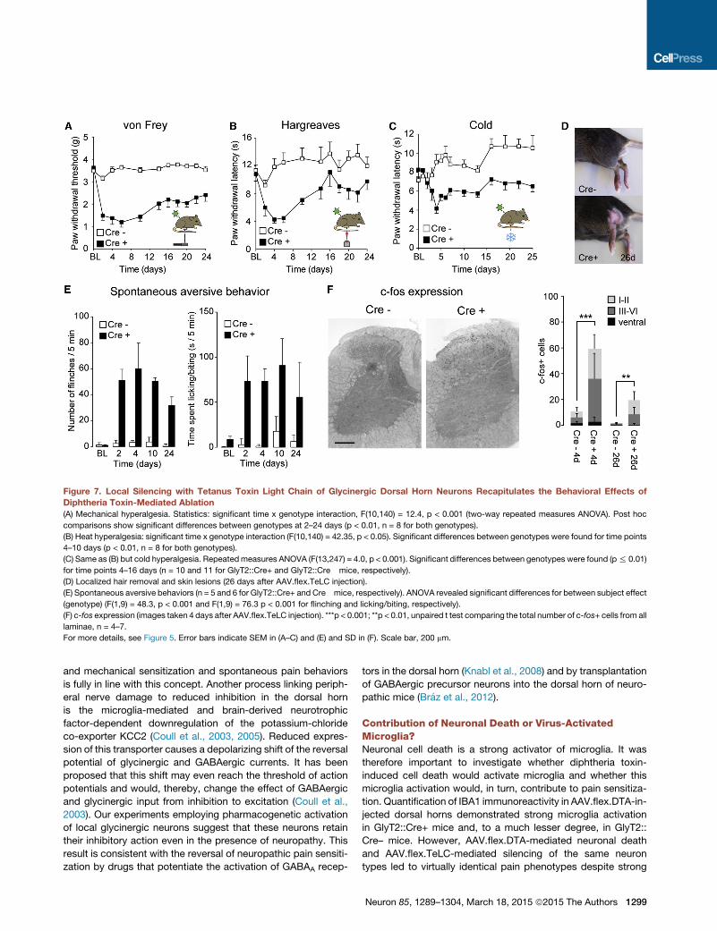
Figure 7. Local Silencing with Tetanus Toxin Light Chain of Glycinergic Dorsal Horn Neurons Recapitulates the Behavioral Effects of
Diphtheria Toxin-Mediated Ablation
(A) Mechanical hyperalgesia. Statistics: significant time x genotype interaction, F(10,140) = 12.4, p < 0.001 (two-way repeated measures ANOVA). Post hoc
comparisons show significant differences between genotypes at 2–24 days (p < 0.01, n = 8 for both genotypes).
(B) Heat hyperalgesia: significant time x genotype interaction (F(10,140) = 42.35, p < 0.05). Significant differences between genotypes were found for time points
4–10 days (p < 0.01, n = 8 for both genotypes).
(C) Same as (B) but cold hyperalgesia. Repeatedmeasures ANOVA (F(13,247) = 4.0, p < 0.001). Significant differences between genotypes were found (p% 0.01)
for time points 4–16 days (n = 10 and 11 for GlyT2::Cre+ and GlyT2::Cre� mice, respectively).
(D) Localized hair removal and skin lesions (26 days after AAV.flex.TeLC injection).
(E) Spontaneous aversive behaviors (n = 5 and 6 for GlyT2::Cre+ and Cre�mice, respectively). ANOVA revealed significant differences for between subject effect
(genotype) (F(1,9) = 48.3, p < 0.001 and F(1,9) = 76.3 p < 0.001 for flinching and licking/biting, respectively).
(F) c-fos expression (images taken 4 days after AAV.flex.TeLC injection). ***p < 0.001; **p < 0.01, unpaired t test comparing the total number of c-fos+ cells from all
laminae, n = 4–7.
For more details, see Figure 5. Error bars indicate SEM in (A–C) and (E) and SD in (F). Scale bar, 200 mm.
and mechanical sensitization and spontaneous pain behaviors
is fully in line with this concept. Another process linking periph-
eral nerve damage to reduced inhibition in the dorsal horn
is the microglia-mediated and brain-derived neurotrophic
factor-dependent downregulation of the potassium-chloride
co-exporter KCC2 (Coull et al., 2003, 2005). Reduced expres-
sion of this transporter causes a depolarizing shift of the reversal
potential of glycinergic and GABAergic currents. It has been
proposed that this shift may even reach the threshold of action
potentials and would, thereby, change the effect of GABAergic
and glycinergic input from inhibition to excitation (Coull et al.,
2003). Our experiments employing pharmacogenetic activation
of local glycinergic neurons suggest that these neurons retain
their inhibitory action even in the presence of neuropathy. This
result is consistent with the reversal of neuropathic pain sensiti-
zation by drugs that potentiate the activation of GABAA recep-
tors in the dorsal horn (Knabl et al., 2008) and by transplantation
of GABAergic precursor neurons into the dorsal horn of neuro-
pathic mice (Braz et al., 2012).
Contribution of Neuronal Death or Virus-ActivatedMicroglia?Neuronal cell death is a strong activator of microglia. It was
therefore important to investigate whether diphtheria toxin-
induced cell death would activate microglia and whether this
microglia activation would, in turn, contribute to pain sensitiza-
tion. Quantification of IBA1 immunoreactivity in AAV.flex.DTA-in-
jected dorsal horns demonstrated strong microglia activation
in GlyT2::Cre+ mice and, to a much lesser degree, in GlyT2::
Cre– mice. However, AAV.flex.DTA-mediated neuronal death
and AAV.flex.TeLC-mediated silencing of the same neuron
types led to virtually identical pain phenotypes despite strong
Neuron 85, 1289–1304, March 18, 2015 ª2015 The Authors 1299

Figure 8. Pharmacogenetic Activation of Spinal Glycinergic Neurons Ameliorates CCI-Induced Neuropathic Pain and Chloroquine- orHistamine-Induced Itch
(A) Diagram illustrating the AAV genome containing the flex.hM3Dq-mCherry cassette.
(B) Expression of hM3Dq in the spinal cord of GlyT2::Cre;GlyT2::eGFP double-transgenic mice is indicated by mCherry fluorescence. Expression is limited to the
ipsilateral dorsal horn and is present in somata (see detail, bottom) and neurites of eGFP+ neurons. Scale bars, 200 mm (top) and 50 mm (bottom).
(C) Antihyperalgesic effects. All experiments weremade in GlyT2::Cre+mice. AAV.flex.hM3Dq-mCherry was injected at day 0, CCI surgery was performed on day
7 on virus-injected mice, and vehicle or CNO (1 mg/kg, i.p.) was injected on day 14. Mechanical PWTs (g) were assessed using electronic von Frey filaments
before CCI surgery (pre-CCI), after CCI surgery immediately before CNO/vehicle injection (post-CCI), for 5 hr after CNO/vehicle injection, and 1 day later (post-
drug). Repeated measures ANOVA, F(6,66) = 4.47; p = 0.001 for treatment x time interaction. Post hoc comparisons revealed significant differences between
CNO and vehicle-treated groups for time points 2 and 3 hr (n = 6 and 7 for vehicle and CNO, respectively).
(D) Acute antinociceptive effects (repeated-measures ANOVA). Significant treatment effects were observed in the Hargreaves test (F(1,9) = 43.1, p < 0.001
[n = 5 and 6 for CNO and vehicle, respectively]), for cold hyperalgesia (F(1,9) = 56.2, p < 0.001 [n = 6 and 5]), and for pinprick stimulation (F(1,8) = 21.0, p = 0.002
[n = 5 each]).
(E) Horizontal wire test. Repeated measures ANOVA (F(1,7) = 0.001, p = 0.98).
(F) Blockade of chloroquine- and histamine-induced itch in AAV.flex.hM3Dq-mCherry-injected GlyT2::Cre+mice by CNO. ***p < 0.001; **p < 0.01, unpaired t test,
n = 7 for all four groups.
All error bars indicate SEM.
differences in the degree of IBA1 upregulation. Hence, the
pain was due to the ablation or silencing of glycinergic neurons
rather than due to secondary microglia activation. Microinjec-
tions into the spinal cord are alone sufficient to induce microglia
activation (Hutson et al., 2012). We found similar degrees of
1300 Neuron 85, 1289–1304, March 18, 2015 ª2015 The Authors
microglial IBA1 upregulation in AAV.flex.TeLC-injected GlyT2::
Cre– and GlyT2::Cre+ mice. However, pain sensitization
occurred only in GlyT2::Cre+ mice; i.e., under conditions in
which the glycinergic neurons were silenced. Pain sensitization
by microglia may therefore require a specific microglial

activation state (Graeber and Christie, 2012; Hanisch and Ket-
tenmann, 2007; Taves et al., 2013).
Spinal Plasticity Ameliorating Long-Lasting Pain?An unexpected finding in our behavioral experiments was the
partial recovery from pain sensitization after glycinergic neuron
ablation or silencing. The results discussed above largely rule
out recovery from microglia-induced pain sensitization as the
underlying mechanism. Virus inactivation, e.g., through DNA
methylation (Brooks et al., 2004), could have accounted for
part of the recovery from TeLC-mediated neuronal silencing.
However, recovery occurred similarly in ablation experiments,
leaving neuronal and synaptic plasticity as the most likely mech-
anisms. Such compensatory plasticity may involve a downregu-
lation of excitatory input, changes in descending pain control, or
the sprouting of inhibitory axons surviving in the ablated area. It is
certainly appealing to think that a facilitation of such mecha-
nisms might be exploited therapeutically in chronic pain states.
Control of Itch-Transmitting Circuits by GlycinergicNeuronsThe extensive licking and biting behavior and hair loss after gly-
cinergic neuron ablation or silencing together with the suppres-
sion of itch responses by activation of local glycinergic neurons
indicate that these neurons control not only pain but also itch
processing. The intense innervation of glycinergic dorsal horn
neurons by myelinated primary sensory neurons fits nicely with
the concept of the gate control theory for pain, where light touch
has been proposed to reduce transmission of painful signals.
However, 9% of the retrogradely labeled primary sensory neu-
rons were peptidergic or bound IB4. These presumed nocicep-
tors likely innervate the small population of glycinergic neurons
in the superficial dorsal horn. It is tempting to speculate that
these primary sensory fibers and glycinergic neurons underlie
the reduction of itch sensations by painful stimulation (Davidson
et al., 2009; Ward et al., 1996).
Two previous reports (Kardon et al., 2014; Ross et al., 2010)
have identified subpopulations of inhibitory dorsal horn neurons
as a source of spinal itch inhibition. These subpopulations
depend on the transcription factor Bhlhb5 and express galanin
and/or nNOS (Kardon et al., 2014). In the rat, many of the inhib-
itory nNOS-positive neurons are also glycinergic (Laing et al.,
1994), whereas most of the galanin+ neurons are likely purely
GABAergic (Simmons et al., 1995). Our studies (Figure S2) indi-
cate that this is also true for the mouse. Therefore, the ablation
of the nNOS+ subpopulation of glycinergic neuronsmay underlie
the induction of itch behavior observed in our study. On the
other hand, Kardon et al. (2014) provided evidence that the inhi-
bition of itch by dorsal horn interneurons depends on the action
of the opioid peptide dynorphin, which is found in the galanin+
subpopulation of purely GABAergic (non-glycinergic) interneu-
rons (Sardella et al., 2011). The latter scenario would suggest
the presence of at least two different subsets of dorsal horn
inhibitory neurons controlling itch.
Future ImplicationsThis study establishes glycinergic neurons of the dorsal horn as
critical elements of a spinal gate for pain and itch signals. It is
likely that inhibitory dorsal horn neurons contribute to other
spinal cord functions in addition to pain and itch control. Such
functions may include improvement of tactile acuity through
lateral inhibition (Isaacson and Scanziani, 2011) or of posture
and gait control through feedback and feedforward inhibition in
motor circuits (Goulding, 2009). It will be interesting to investi-
gate whether these different functions involve distinct glycinergic
subpopulations. The virus-based techniques established here
constitute a versatile set of tools to address these questions.
EXPERIMENTAL PROCEDURES
Mice
GlyT2::eGFP (Tg(Scl6a5-EGFP)1Uze) and vGAT::ChR2-eYFP (Tg(Slc32a1-
COP4*H134R/EYFP)8Gfng) mice have been described previously (Zeilhofer
et al., 2005; Zhao et al., 2011). GlyT2::Cre (Tg(Slc6a5-Cre)1Uze) mice
were generated following the same strategy as used previously for GlyT2::
eGFP mice (Zeilhofer et al., 2005). Control experiments were performed in
Gad67eGFP (Gad1tm1Tama) mice (Tamamaki et al., 2003) and ROSA26lacZ
(Gt(ROSA)26Sor) mice (Soriano, 1999). For details, see Supplemental Experi-
mental Procedures. All procedures involving animals were approved by
the Veterinaramt des Kantons Zurich (license numbers 64/2010, 75/2010,
75/2013, and 86/2013).
Immunohistochemistry and Image Analysis
Immunohistochemistry was performed as described previously (Punnakkal
et al., 2014). For information on antibodies, see Supplemental Experimental
Procedures. Fluorescent images were acquired on a Zeiss LSM710 Pascal
confocal microscope. For quantitative analyses, sections were prepared
from at least three animals, and at least three sections were analyzed per
animal. Numbers of immunoreactive cells were determined using the ImageJ
Cell Counter plugin. Controlled mean fluorescent intensity (CMTF) was quan-
tified using ImageJ.
Virus Production and Injections
AAV.flex.hM3Dq-mCherry and AVV.eGFPwere obtained from the University of
North Carolina (UNC) vector core and Penn Vector Core (Perelman School of
Medicine, University of Pennsylvania), respectively. All other AAV vectors were
cloned in-house and packaged at the Penn Vector Core. The cell lines required
for production of EnvA.RabiesDG-eGFP were obtained from Dr. Edward
Callaway (Salk Institute). Propagation was done following the protocol by
Osakada and Callaway (2013).
Virus injections weremade inmice anesthetized with 2%–5% isoflurane and
immobilized on a motorized stereotaxic frame (David Kopf Instruments and
Neurostar). Lumbar vertebrae L4 and L5 were exposed, and the vertebral
column was fixed using a pair of spinal adaptors. Injections were made at a
rate of 30 nl/min with glass micropipettes (tip diameter, 30–40 mm) attached
to a 10-ml Hamilton syringe.
Electrophysiology and Optogenetics
Transverse lumbar spinal cord slices were cut from 25- to 28-day-old
vGAT::ChR2-eYFP;GlyT2::Cre double-transgenic and vGAT::ChR2-eYFP
control (GlyT2::Cre–) mice injected at lumbar levels L3–L5 with a combination
of AAV.flex.DTA and AAV.mCherry. Whole-cell patch-clamp recordings were
made at room temperature from excitatory superficial dorsal horn neurons.
Light-evoked IPSCswere elicited at a frequency of 5min by wide-field illumina-
tion of the dorsal horn (473 ± 5 nm wavelength, 2.7 mW, 4 ms).
Behavioral Analyses
Adult male GlyT2::Cre+ and GlyT2::Cre– mice (7–8 weeks old) received
three unilateral injections at lumbar levels L3–L5 with AAV.flex.DTA, AAV.
flex.TeLC, or AAV.flex.hM3Dq-mCherry. In GlyT2::Cre+ and GlyT2::Cre�mice injected with AAV.flex.DTA and AAV.flex.TeLC, mechanical, heat,
and noxious cold sensitivity were assessed for up to 26 days after virus injec-
tion. Pharmacogenetic experiments with CNO (1 mg/kg, i.p.) were done in
Neuron 85, 1289–1304, March 18, 2015 ª2015 The Authors 1301

AAV.flex.hM3Dq-mCherry-injectedmice 7 days after CCI surgery (i.e., 14 days
after virus injection). Itch responses (flinches and biting bouts) were elicited
through intracutaneous injections of chloroquine (10 ml of 8 mg/ml) or hista-
mine (10 ml of 10 mg/ml) into one hindpaw. Animals were videotaped for
30 min, and responses were analyzed offline. The horizontal wire test and
the rotarod test were used to assess muscle relaxation and motor impairment,
respectively.
SUPPLEMENTAL INFORMATION
Supplemental Information includes Supplemental Experimental Procedures
and six figures and can be found with this article online at http://dx.doi.org/
10.1016/j.neuron.2015.02.028.
AUTHOR CONTRIBUTIONS
E.F. performed all virus injections and surgeries and performed and analyzed
the behavioral experiments except for the pharmacogenetic study. H.W.
designed the viral vectors and performed and analyzed the morphological
experiments. M.B. performed the pronucleus injections. M.J. and H.W.
characterized the GlyT2::Cre mice. H.J. and L.T. performed and analyzed
the optogenetic experiments. L.H. contributed to the itch experiments. K.H.
established pseudotyped rabies virus production in our group and provided
pseudotyped rabies virus for the experiments. W.T.R. and S.H. performed
and analyzed the pharmacogenetic experiments. K.K.C. and A.G. provided
first stocks of rabies viruses for the retrograde labeling experiments. E.F.,
H.W., and H.U.Z. designed experiments and wrote the manuscript. All authors
commented on the manuscript.
ACKNOWLEDGMENTS
This work was supported by an ERC Advanced Investigator Grant (DHISP
250128) and by grants from the Swiss National Research Foundation
(131093 and 156393) (to H.U.Z.) and by the DFG (SFB 870 Neuronal Circuits)
(to K.K.C. and A.G.). Dr. Ron Yu (Stowers Institute for Medical Research)
provided the plasmid containing the TeLC cds (pGEMT-EZ-TeTx), Dr. Karl
Deisseroth (StanfordUniversity) provided the pAAV-Ef1a-DIO-hChR2(H134R)-
mCherry-WPRE-pA plasmid, and Dr. Edward M. Callaway (Salk Institute)
provided the material required for amplification of G-deficient rabies virus.
Received: September 30, 2014
Revised: January 26, 2015
Accepted: February 12, 2015
Published: March 18, 2015
REFERENCES
Ahmadi, S., Lippross, S., Neuhuber, W.L., and Zeilhofer, H.U. (2002). PGE2
selectively blocks inhibitory glycinergic neurotransmission onto rat superficial
dorsal horn neurons. Nat. Neurosci. 5, 34–40.
Akiyama, T., and Carstens, E. (2013). Neural processing of itch. Neuroscience
250, 697–714.
Alexander, G.M., Rogan, S.C., Abbas, A.I., Armbruster, B.N., Pei, Y., Allen,
J.A., Nonneman, R.J., Hartmann, J., Moy, S.S., Nicolelis, M.A., et al. (2009).
Remote control of neuronal activity in transgenic mice expressing evolved G
protein-coupled receptors. Neuron 63, 27–39.
Armbruster, B.N., Li, X., Pausch, M.H., Herlitze, S., and Roth, B.L. (2007).
Evolving the lock to fit the key to create a family of G protein-coupled receptors
potently activated by an inert ligand. Proc. Natl. Acad. Sci. USA 104, 5163–
5168.
Aschauer, D.F., Kreuz, S., and Rumpel, S. (2013). Analysis of transduction
efficiency, tropism and axonal transport of AAV serotypes 1, 2, 5, 6, 8 and 9
in the mouse brain. PLoS ONE 8, e76310.
1302 Neuron 85, 1289–1304, March 18, 2015 ª2015 The Authors
Atasoy, D., Aponte, Y., Su, H.H., and Sternson, S.M. (2008). A FLEX switch
targets Channelrhodopsin-2 to multiple cell types for imaging and long-range
circuit mapping. J. Neurosci. 28, 7025–7030.
Baba, H., Ji, R.R., Kohno, T., Moore, K.A., Ataka, T., Wakai, A., Okamoto, M.,
and Woolf, C.J. (2003). Removal of GABAergic inhibition facilitates polysyn-
aptic A fiber-mediated excitatory transmission to the superficial spinal dorsal
horn. Mol. Cell. Neurosci. 24, 818–830.
Bennett, G.J., and Xie, Y.K. (1988). A peripheral mononeuropathy in rat that
produces disorders of pain sensation like those seen in man. Pain 33, 87–107.
Betley, J.N., and Sternson, S.M. (2011). Adeno-associated viral vectors for
mapping, monitoring, and manipulating neural circuits. Hum. Gene Ther. 22,
669–677.
Beyer, C., Roberts, L.A., and Komisaruk, B.R. (1985). Hyperalgesia induced by
altered glycinergic activity at the spinal cord. Life Sci. 37, 875–882.
Braz, J.M., Sharif-Naeini, R., Vogt, D., Kriegstein, A., Alvarez-Buylla, A.,
Rubenstein, J.L., and Basbaum, A.I. (2012). Forebrain GABAergic neuron pre-
cursors integrate into adult spinal cord and reduce injury-induced neuropathic
pain. Neuron 74, 663–675.
Brooks, A.R., Harkins, R.N., Wang, P., Qian, H.S., Liu, P., and Rubanyi, G.M.
(2004). Transcriptional silencing is associated with extensive methylation of
the CMV promoter following adenoviral gene delivery to muscle. J. Gene
Med. 6, 395–404.
Buch, T., Heppner, F.L., Tertilt, C., Heinen, T.J., Kremer, M., Wunderlich, F.T.,
Jung, S., and Waisman, A. (2005). A Cre-inducible diphtheria toxin receptor
mediates cell lineage ablation after toxin administration. Nat. Methods 2,
419–426.
Chamberlin, N.L., Du, B., de Lacalle, S., and Saper, C.B. (1998). Recombinant
adeno-associated virus vector: use for transgene expression and anterograde
tract tracing in the CNS. Brain Res. 793, 169–175.
Clark, A.K., Gentry, C., Bradbury, E.J., McMahon, S.B., and Malcangio, M.
(2007). Role of spinal microglia in rat models of peripheral nerve injury and
inflammation. Eur. J. Pain 11, 223–230.
Coull, J.A., Boudreau, D., Bachand, K., Prescott, S.A., Nault, F., Sık, A., De
Koninck, P., and De Koninck, Y. (2003). Trans-synaptic shift in anion gradient
in spinal lamina I neurons as a mechanism of neuropathic pain. Nature 424,
938–942.
Coull, J.A., Beggs, S., Boudreau, D., Boivin, D., Tsuda, M., Inoue, K., Gravel,
C., Salter, M.W., and De Koninck, Y. (2005). BDNF from microglia causes
the shift in neuronal anion gradient underlying neuropathic pain. Nature 438,
1017–1021.
Coyle, D.E. (1998). Partial peripheral nerve injury leads to activation of astroglia
and microglia which parallels the development of allodynic behavior. Glia 23,
75–83.
Davidson, S., Zhang, X., Khasabov, S.G., Simone, D.A., and Giesler, G.J., Jr.
(2009). Relief of itch by scratching: state-dependent inhibition of primate spi-
nothalamic tract neurons. Nat. Neurosci. 12, 544–546.
Drew, G.M., Siddall, P.J., and Duggan, A.W. (2004). Mechanical allodynia
following contusion injury of the rat spinal cord is associated with loss of
GABAergic inhibition in the dorsal horn. Pain 109, 379–388.
Duan, B., Cheng, L., Bourane, S., Britz, O., Padilla, C., Garcia-Campmany, L.,
Krashes, M., Knowlton, W., Velasquez, T., Ren, X., et al. (2014). Identification
of spinal circuits transmitting and gating mechanical pain. Cell 159, 1417–
1432.
Gomeza, J., Ohno, K., Hulsmann, S., Armsen,W., Eulenburg, V., Richter, D.W.,
Laube, B., and Betz, H. (2003). Deletion of the mouse glycine transporter 2 re-
sults in a hyperekplexia phenotype and postnatal lethality. Neuron 40,
797–806.
Goulding, M. (2009). Circuits controlling vertebrate locomotion: moving in a
new direction. Nat. Rev. Neurosci. 10, 507–518.
Graeber, M.B., and Christie, M.J. (2012). Multiple mechanisms of microglia: a
gatekeeper’s contribution to pain states. Exp. Neurol. 234, 255–261.
Hanisch, U.K., and Kettenmann, H. (2007). Microglia: active sensor and versa-
tile effector cells in the normal and pathologic brain. Nat. Neurosci. 10, 1387–
1394.
Harvey, R.J., Depner, U.B., Wassle, H., Ahmadi, S., Heindl, C., Reinold, H.,
Smart, T.G., Harvey, K., Schutz, B., Abo-Salem, O.M., et al. (2004). GlyR a3:
an essential target for spinal PGE2-mediated inflammatory pain sensitization.
Science 304, 884–887.
Hossaini, M., French, P.J., and Holstege, J.C. (2007). Distribution of glyciner-
gic neuronal somata in the rat spinal cord. Brain Res. 1142, 61–69.
Hutson, T.H., Verhaagen, J., Yanez-Munoz, R.J., and Moon, L.D. (2012).
Corticospinal tract transduction: a comparison of seven adeno-associated
viral vector serotypes and a non-integrating lentiviral vector. Gene Ther. 19,
49–60.
Isaacson, J.S., and Scanziani, M. (2011). How inhibition shapes cortical activ-
ity. Neuron 72, 231–243.
Kardon, A.P., Polgar, E., Hachisuka, J., Snyder, L.M., Cameron, D., Savage,
S., Cai, X., Karnup, S., Fan, C.R., Hemenway, G.M., et al. (2014). Dynorphin
acts as a neuromodulator to inhibit itch in the dorsal horn of the spinal cord.
Neuron 82, 573–586.
Keller, A.F., Coull, J.A., Chery, N., Poisbeau, P., and De Koninck, Y. (2001).
Region-specific developmental specialization of GABA-glycine cosynapses
in laminas I-II of the rat spinal dorsal horn. J. Neurosci. 21, 7871–7880.
Keller, A.F., Beggs, S., Salter, M.W., and De Koninck, Y. (2007).
Transformation of the output of spinal lamina I neurons after nerve injury and
microglia stimulation underlying neuropathic pain. Mol. Pain 3, 27.
Knabl, J., Witschi, R., Hosl, K., Reinold, H., Zeilhofer, U.B., Ahmadi, S.,
Brockhaus, J., Sergejeva, M., Hess, A., Brune, K., et al. (2008). Reversal of
pathological pain through specific spinal GABAA receptor subtypes. Nature
451, 330–334.
Kuner, R. (2010). Central mechanisms of pathological pain. Nat. Med. 16,
1258–1266.
Laing, I., Todd, A.J., Heizmann, C.W., and Schmidt, H.H. (1994).
Subpopulations of GABAergic neurons in laminae I-III of rat spinal dorsal
horn defined by coexistence with classical transmitters, peptides, nitric oxide
synthase or parvalbumin. Neuroscience 61, 123–132.
Loomis, C.W., Khandwala, H., Osmond, G., and Hefferan, M.P. (2001).
Coadministration of intrathecal strychnine and bicuculline effects synergistic
allodynia in the rat: an isobolographic analysis. J. Pharmacol. Exp. Ther.
296, 756–761.
Lu, Y., Dong, H., Gao, Y., Gong, Y., Ren, Y., Gu, N., Zhou, S., Xia, N., Sun, Y.Y.,
Ji, R.R., and Xiong, L. (2013). A feed-forward spinal cord glycinergic neural cir-
cuit gates mechanical allodynia. J. Clin. Invest. 123, 4050–4062.
Melzack, R., and Wall, P.D. (1965). Pain mechanisms: a new theory. Science
150, 971–979.
Miraucourt, L.S., Moisset, X., Dallel, R., and Voisin, D.L. (2009). Glycine
inhibitory dysfunction induces a selectively dynamic, morphine-resistant,
and neurokinin 1 receptor- independent mechanical allodynia. J. Neurosci.
29, 2519–2527.
Moore, K.A., Kohno, T., Karchewski, L.A., Scholz, J., Baba, H., andWoolf, C.J.
(2002). Partial peripheral nerve injury promotes a selective loss of GABAergic
inhibition in the superficial dorsal horn of the spinal cord. J. Neurosci. 22, 6724–
6731.
Muller, F., Heinke, B., and Sandkuhler, J. (2003). Reduction of glycine recep-
tor-mediated miniature inhibitory postsynaptic currents in rat spinal lamina I
neurons after peripheral inflammation. Neuroscience 122, 799–805.
Osakada, F., and Callaway, E.M. (2013). Design and generation of recombi-
nant rabies virus vectors. Nat. Protoc. 8, 1583–1601.
Palmiter, R.D., Behringer, R.R., Quaife, C.J., Maxwell, F., Maxwell, I.H., and
Brinster, R.L. (1987). Cell lineage ablation in transgenic mice by cell-specific
expression of a toxin gene. Cell 50, 435–443.
Polgar, E., Durrieux, C., Hughes, D.I., and Todd, A.J. (2013). A quantitative
study of inhibitory interneurons in laminae I-III of the mouse spinal dorsal
horn. PLoS ONE 8, e78309.
Poyatos, I., Ponce, J., Aragon, C., Gimenez, C., and Zafra, F. (1997). The
glycine transporter GLYT2 is a reliable marker for glycine-immunoreactive
neurons. Brain Res. Mol. Brain Res. 49, 63–70.
Punnakkal, P., von Schoultz, C., Haenraets, K., Wildner, H., and Zeilhofer, H.U.
(2014). Morphological, biophysical and synaptic properties of glutamatergic
neurons of the mouse spinal dorsal horn. J. Physiol. 592, 759–776.
Ross, S.E., Mardinly, A.R., McCord, A.E., Zurawski, J., Cohen, S., Jung, C.,
Hu, L., Mok, S.I., Shah, A., Savner, E.M., et al. (2010). Loss of inhibitory inter-
neurons in the dorsal spinal cord and elevated itch in Bhlhb5 mutant mice.
Neuron 65, 886–898.
Sandkuhler, J. (2009). Models and mechanisms of hyperalgesia and allodynia.
Physiol. Rev. 89, 707–758.
Sardella, T.C., Polgar, E., Garzillo, F., Furuta, T., Kaneko, T., Watanabe, M.,
and Todd, A.J. (2011). Dynorphin is expressed primarily by GABAergic neu-
rons that contain galanin in the rat dorsal horn. Mol. Pain 7, 76.
Scholz, J., Broom, D.C., Youn, D.H., Mills, C.D., Kohno, T., Suter, M.R., Moore,
K.A., Decosterd, I., Coggeshall, R.E., andWoolf, C.J. (2005). Blocking caspase
activity prevents transsynaptic neuronal apoptosis and the loss of inhibition
in lamina II of the dorsal horn after peripheral nerve injury. J. Neurosci. 25,
7317–7323.
Sherman, S.E., and Loomis, C.W. (1994). Morphine insensitive allodynia is pro-
duced by intrathecal strychnine in the lightly anesthetized rat. Pain 56, 17–29.
Simmons, D.R., Spike, R.C., and Todd, A.J. (1995). Galanin is contained in
GABAergic neurons in the rat spinal dorsal horn. Neurosci. Lett. 187, 119–122.
Sivilotti, L., and Woolf, C.J. (1994). The contribution of GABAA and glycine
receptors to central sensitization: disinhibition and touch-evoked allodynia in
the spinal cord. J. Neurophysiol. 72, 169–179.
Soriano, P. (1999). Generalized lacZ expression with the ROSA26 Cre reporter
strain. Nat. Genet. 21, 70–71.
Sorkin, L.S., Puig, S., and Jones, D.L. (1998). Spinal bicuculline produces
hypersensitivity of dorsal horn neurons: effects of excitatory amino acid antag-
onists. Pain 77, 181–190.
Sweeney, S.T., Broadie, K., Keane, J., Niemann, H., and O’Kane, C.J. (1995).
Targeted expression of tetanus toxin light chain in Drosophila specifically
eliminates synaptic transmission and causes behavioral defects. Neuron 14,
341–351.
Tamamaki, N., Yanagawa, Y., Tomioka, R., Miyazaki, J., Obata, K., and
Kaneko, T. (2003). Green fluorescent protein expression and colocalization
with calretinin, parvalbumin, and somatostatin in the GAD67-GFP knock-in
mouse. J. Comp. Neurol. 467, 60–79.
Tanga, F.Y., Raghavendra, V., and DeLeo, J.A. (2004). Quantitative real-time
RT-PCR assessment of spinal microglial and astrocytic activation markers in
a rat model of neuropathic pain. Neurochem. Int. 45, 397–407.
Taves, S., Berta, T., Chen, G., and Ji, R.R. (2013). Microglia and spinal cord
synaptic plasticity in persistent pain. Neural Plast. 2013, 753656.
Todd, A.J., and Spike, R.C. (1993). The localization of classical transmitters
and neuropeptides within neurons in laminae I-III of the mammalian spinal
dorsal horn. Prog. Neurobiol. 41, 609–645.
Torsney, C., and MacDermott, A.B. (2006). Disinhibition opens the gate to
pathological pain signaling in superficial neurokinin 1 receptor-expressing
neurons in rat spinal cord. J. Neurosci. 26, 1833–1843.
Ward, L., Wright, E., andMcMahon, S.B. (1996). A comparison of the effects of
noxious and innocuous counterstimuli on experimentally induced itch and
pain. Pain 64, 129–138.
Wickersham, I.R., Lyon, D.C., Barnard, R.J., Mori, T., Finke, S., Conzelmann,
K.K., Young, J.A., and Callaway, E.M. (2007). Monosynaptic restriction of
transsynaptic tracing from single, genetically targeted neurons. Neuron 53,
639–647.
Woolf, C.J., and Salter, M.W. (2000). Neuronal plasticity: increasing the gain
in pain. Science 288, 1765–1769.
Neuron 85, 1289–1304, March 18, 2015 ª2015 The Authors 1303
Xiong, W., Cheng, K., Cui, T., Godlewski, G., Rice, K.C., Xu, Y., and Zhang, L.
(2011). Cannabinoid potentiation of glycine receptors contributes to cannabis-
induced analgesia. Nat. Chem. Biol. 7, 296–303.
Xiong, W., Cui, T., Cheng, K., Yang, F., Chen, S.R., Willenbring, D., Guan, Y.,
Pan, H.L., Ren, K., Xu, Y., and Zhang, L. (2012). Cannabinoids suppress inflam-
matory and neuropathic pain by targeting a3 glycine receptors. J. Exp. Med.
209, 1121–1134.
Yaksh, T.L. (1989). Behavioral and autonomic correlates of the tactile evoked
allodynia produced by spinal glycine inhibition: effects of modulatory receptor
systems and excitatory amino acid antagonists. Pain 37, 111–123.
1304 Neuron 85, 1289–1304, March 18, 2015 ª2015 The Authors
Zeilhofer, H.U., Studler, B., Arabadzisz, D., Schweizer, C., Ahmadi, S., Layh,
B., Bosl, M.R., and Fritschy, J.M. (2005). Glycinergic neurons expressing
enhanced green fluorescent protein in bacterial artificial chromosome trans-
genic mice. J. Comp. Neurol. 482, 123–141.
Zeilhofer, H.U., Wildner, H., and Yevenes, G.E. (2012). Fast synaptic inhibition
in spinal sensory processing and pain control. Physiol. Rev. 92, 193–235.
Zhao, S., Ting, J.T., Atallah, H.E., Qiu, L., Tan, J., Gloss, B., Augustine, G.J.,
Deisseroth, K., Luo, M., Graybiel, A.M., and Feng, G. (2011). Cell type–specific
channelrhodopsin-2 transgenic mice for optogenetic dissection of neural cir-
cuitry function. Nat. Methods 8, 745–752.


![SURFACE MELTING AND THERMAL ABLATION ...chalcogen.ro/1161_Preoteasa.pdfwhitening [9] and activation of teeth bleaching agents [10], and caries prevention by surface modifications of](https://static.fdocuments.us/doc/165x107/5f7b962af18fdc1987195599/surface-melting-and-thermal-ablation-whitening-9-and-activation-of-teeth-bleaching.jpg)