T Usneoides Billings 1904
29
A Study of Tillandsia usneoides Author(s): Frederick H. Billings Source: Botanical Gazette, Vol. 38, No. 2 (Aug., 1904), pp. 99-121 Published by: The University of Chicago Press Stable URL: http://www.jstor.org/stable/2465941 Accessed: 20/01/2010 13:36 Your use of the JSTOR archive indicates your acceptance of JSTOR's Terms and Conditions of Use, available at http://www.jstor.org/page/info/about/policies/terms.jsp . JSTOR's Terms and Conditions of Use provides, in part, that unless you have obtained prior permission, you may not download an entire issue of a journal or multiple copies of articles, and you may use content in the JSTOR archive only for your personal, non-commercial use. Please contact the publisher regarding any further use of this work. Publisher contact information may be obtained at http://www.jstor.org/action/showPublisher?publisherCode=ucpress . Each copy of any part of a JSTOR transmission must contain the same copyright notice that appears on the screen or printed page of such transmission. JSTOR is a not-for-profit service that helps scholars, researchers, and students discover, use, and build upon a wide range of content in a trusted digital archive. We use information technology and tools to increase productivity and facilitate new forms of scholarship. For more information about JSTOR, please contact [email protected]. The University of Chicago Press is collaborating with JSTOR to digitize, preserve and extend access to Botanical Gazette. http://www.jstor.org
-
Upload
henriquetg -
Category
Documents
-
view
220 -
download
0
Transcript of T Usneoides Billings 1904

8/8/2019 T Usneoides Billings 1904
http://slidepdf.com/reader/full/t-usneoides-billings-1904 1/28
A Study of Tillandsia usneoidesAuthor(s): Frederick H. BillingsSource: Botanical Gazette, Vol. 38, No. 2 (Aug., 1904), pp. 99-121Published by: The University of Chicago PressStable URL: http://www.jstor.org/stable/2465941
Accessed: 20/01/2010 13:36
Your use of the JSTOR archive indicates your acceptance of JSTOR's Terms and Conditions of Use, available at
http://www.jstor.org/page/info/about/policies/terms.jsp. JSTOR's Terms and Conditions of Use provides, in part, that unless
you have obtained prior permission, you may not download an entire issue of a journal or multiple copies of articles, and youmay use content in the JSTOR archive only for your personal, non-commercial use.
Please contact the publisher regarding any further use of this work. Publisher contact information may be obtained at
http://www.jstor.org/action/showPublisher?publisherCode=ucpress.
Each copy of any part of a JSTOR transmission must contain the same copyright notice that appears on the screen or printed
page of such transmission.
JSTOR is a not-for-profit service that helps scholars, researchers, and students discover, use, and build upon a wide range of
content in a trusted digital archive. We use information technology and tools to increase productivity and facilitate new forms
of scholarship. For more information about JSTOR, please contact [email protected].
The University of Chicago Press is collaborating with JSTOR to digitize, preserve and extend access to
Botanical Gazette.
http://www.jstor.org
8/8/2019 T Usneoides Billings 1904
http://slidepdf.com/reader/full/t-usneoides-billings-1904 2/28
A STUDY OF TILLANDSIA USNEOIDES.FREDERICK H. BILLINGS.
(WITH ONE FIGURE AND PLATES VIII-XI)
Tillandsia usneoides, popularly called "long moss," "black
moss," or "Spanish moss," is the most widely distributed representa-
tive of the tropical and subtropical family Bromeliaceae. Accord-
ing to SCHIMPER (I) it extends from southern Virginia, its northern
limit, as far southward as the Argentine Confederation. It formseverywhere a conspicuous and characteristic object of the landscape,
its long gray festoons adorning not only trees of the virgin forest but
many cultivated ones as well. Although the beauty of the landscape
is enhanced by its presence, its growth upon ornamental trees is
regarded often with apprehension, a common impression being that it
lives parasitically. A most casual examination, however, will reveal
the fact that the moss is in no way connected with the tree, but merely
wraps its dead, wiry stems loosely around the twigs in order to support
itself. Old festoons which have hung in the same place for years
occasionally show a connection with the bark, the annual growths of
the limb finally enclosing some of the decorticated moss stems; much
in the same way that an old horseshoe hung astride a branch and left
unmoved for a long time will be partially enclosed.
An indirect cause of the popular belief in the parasitism of Til-
landsia is its preference for sunny exposures. This habit would tendto keep it from trees having a dense shade. In dark forests it hangs
suspended from the higher limbs of tall trees, especially those that
are dead. Many a cultivated tree when in perfectly healthy condition
possesses too dense foliage to serve as a host for Tillandsia, but if for
some reason the supply of leaves should be reduced, the light condi-
tions might be such as to make the presence of the epiphyte possible.
Should it make its appearance, the owner of the tree would be very
apt to regardthe moss as the cause rather than the result of the reducedfoliage. A proof of the true epiphytism of the plant is its long-
continued and vigorous growth upon decorticated limbs of dead trees.
Near Baton Rouge are many such trees, killed by girdling long ago,
1904] 99

8/8/2019 T Usneoides Billings 1904
http://slidepdf.com/reader/full/t-usneoides-billings-1904 3/28
100 BOTANICAL GAZETTE [AUGUST
yet supporting a large quantity of moss. In order to demonstrate
experimentally that the moss can live solely on what it derives from
air and rain, some festoons were supported by twine and hung fromsome branches of a tree upon which moss was already growing. As
was expected, the festoons produced normal flowers, gave rise to new
growth, and at the end of eighteen months looked as vigorous as any
on the tree, though they came at no time in contact with it.
Because Tillandsia has no influence as a parasite, it does not
follow that it exerts none in other ways, yet to just what extent it affects
a host tree is at present difficult to say. Aside from the slight damage
done in breaking twigs and small branches by its weight, it is doubt-ful whether such objections as shading and cutting off the supply of
air are really worthy of consideration. It is almost certain that these
objections are not sufficient to explain a reduction in foliage that
people so often ascribe to the presence of the moss. It is realized,
however, that this problem can only be answered satisfactorily by
experiments extending over a considerable number of years.
The problem of the distribution of T. usneoides upon the various
species of trees is one of the first to force itself upon the observation.
That certain trees of a given locality are abundantly supplied while
others not far distant are not, is a well-known fact. One factor in
the case has already been mentioned, and that is the light relation.
But there are others to be considered, and the most important perhaps
is concerned with the method of dissemination. The epiphyte is not
usually propagated by seeds but by fragments of festoons, which
being somewhat heavy cannot be carried far except in a very highwind, or by birds, which according to SCHIMPER (I) in some regions
utilize the plant in building their nests. There is a good chance,
therefore, for a tree a little distant from others bearing the moss not
to receive its first detachment of the epiphyte.
The character of the foliage also plays a part, in that a tree with
leaves densely crowded on the outermost twigs would scarcely permit
a wind-blown fragment of moss to hook itself to the branches, but
would shed it. SCHIMPER (i) observes in this connection that "Biume
mit sehr dichtem Haube entbehren der Sonnenepiphyten beinahe
ganzlich." According to PEIRCE (2) Rarnalina reticulata, a lichen
having a habit and mode of dissemination similar to T. usneoides,

8/8/2019 T Usneoides Billings 1904
http://slidepdf.com/reader/full/t-usneoides-billings-1904 4/28
I904] BILLINGS-TILLANDSIA USNEOIDES I O
is found more frequently on deciduous than on evergreen trees,
because, as he explains, the foliage of the evergreen trees interferes
with its reaching the branches. The umbrella tree (Melia Azederac1l)has a remarkably dense foliage and is almost universally devoid of
moss, yet near the university is a tree of this species with a scanty
supply of foliage and an abundance of moss. It is reasonable to con-
clude that any tree furnishing proper conditions for attachment and
growth may become a host of the epiphyte.
The source of the water supply of Tillandsia is atmospheric pre-
cipitation, as in all epiphytes. Dissolved in the water are the neces-
sary salts which have been dissolved by the rain from the dust in theair. Perhaps an equally fruitful source of salts is in many cases the
washings from the tree, which in dry weather may accumulate much
earthy material in the form of dust upon its branches. The plant
itself even serves in collecting dust on account of the scaly surface, so
that when wet the deposits beneath the scales yield a small amount
of soluble material.
A most remarkable characteristic of Tillandsia is its ability to
retain water. The absorption of water is accomplished over the
entire surface of the living parts by means of scales, as will be described
further on, its retention being accomplished also by the scales, and of
course by the cuticularized epidermis. It is much easier to under-
stand how a melon cactus with its globose form and consequent
minimum surface and enormously developed water-storage tissue
can resist prolonged drouth than it is to see how Tillandsia with its
small cylindrical leaves, much greater surface exposure, and compara-tively small storage facility can, without any water supply, endure
drouth. A small festoon was hung in a closed dry room for nineteen
days without water. It lost 23 per cent. in weight during the time,
but when placed in water it absorbed as much as it had lost, and
remained a healthy plant, showing that it had not really suffered
injury by exposure to the drouth. There is occasionally, of course,
a similar drying process in the open air when drouth occurs. During
the dry spell in the spring of 1902, moss plants were known to have
been subjected to two months of rainless exposure without injury.
From an economic standpoint, Tillandsia is of some commercial
value on account of its mechanical tissue. This forms a central

8/8/2019 T Usneoides Billings 1904
http://slidepdf.com/reader/full/t-usneoides-billings-1904 5/28
102 BOTANICAL GAZETTE [AUGUST
cylindricalstrand composed of reduced phloem and xylem, surrounded
by a mass of thick-walled sclerenchyma fibers. When the paren-
chymatous cortex is removed, the sclerenchymatous axis remains asa tough elastic fiber, which serves as a packing in upholstery. The
so-called curing process is a means of eliminating the parenchyma.
One method largely employed is that of burying the moss in trenches
or pits, allowing it to remain till the cortex is dead and in a condition
to be removed easily.
DEVELOPMENT OF THE EMBRYO SAC.
The primordia of the ovules arise on the innermost wall of each
loculus of the tricarpellate, superior ovary. By a one-sided growth
each primordium becomes bent toward the base of the ovary, develop-
ing into the anatropous type of ovule. When the bending has reached
an angle of about go', the nucellus appears as a hemispherical mass
of cells, at the base of which can be seen the beginning of the inner
integument. Imbedded under two layers of nucellus cells, the single
archesporial cell becomes differentiated in the usual way, by its
slightly larger size and greater staining capacity (fig. 2). As theovule increases in size, the nucellus elongates, the outer integument
appears, and the archesporial cell enlarges considerably, especially
in length. There is no parietal cell formed, but by multiplication
of cells the nucellus over the archesporial cell forms an additional
layer, making three (fig. 3). The archesporial cell is now much
elongated, and occupies the central region of the nucellus. It is
filled with granular, longitudinally-striated cytoplasm, and has a
relatively large nucleus. The first and second divisions of this
nucleus probably give rise to the gametophyte generation. Only
one spindle of the first division was observed, and it was but little
more than one-third the length of the cell (fig. 4). The chromosomes
were short, and closely crowded at the equatorial plate. The con-
ditions were altogether unfavorable for ascertaining their number
on account of the small size of the figure. The number, however,
was definitely made out from the second division of the pollen mothercells, and was found to be sixteen. A protracted search failed to
yield a nuclear figure which definitely showed the chromosome
number in the sporophyte, though considerably over sixteen were
observed.
8/8/2019 T Usneoides Billings 1904
http://slidepdf.com/reader/full/t-usneoides-billings-1904 6/28
1904] BILLINGS-TILLANDSIA USNEOIDES 103
The first division of the archesporial cell is usually followed by
a transverse wall and a resting condition of the nuclei (fig. 5); but a
single case was observed, as reported by SMITH 3) for Eichhorniacrassipes, in which a row of four nuclei was formed without sepa-
rating walls (fig. 7). In Eichhornia the absence of the walls is said
to be the rule, but in Tillandsia it is the exception. The division
which gives rise to the third and fourth megaspores, thus completing
the axial row, will be seen from fig. 6 to be in the cell nearest the
micropyle. In the meantime, the basal of the two proximal mega-
spores begins to elongate, and is destined to develop into the embryo
sac. A vacuole is formed in this cell as it pushes outwards crushingthe other three megaspores, whose contents soon show evidence of
breaking down. The remaining stages in development are the
familiar ones of complete absorption of the non-functional mega-
spores by the functional, and the internal division of the latter into
eight cells. The two cells that are to form the synergids soon come
to possess larger nuclei than does the egg cell. The egg nucleus
in fact is smaller than is customarily observed. In the completed
embryo sac, the egg often lies against the wall of the sac near one
synergid, but may occupy a position between the synergids. The
polar nuclei usually approach each other and fuse near the antipodal
region (fig. 14). The antipodals occupy a pocket at the extreme
end of the sac.FERTILIZATION.
The pollen tube passes through the micropyle, penetrates the
nucellus, and enlarges as it enters the embryo sac. It does notappear to pass between the synergids, but to one side of them, one
synergid being disorganized in the process. The two male nuclei
which have arisen from the generative nucleus during the develop-
ment of the pollen tube lie near together and a little in advance of the
tube nucleus. In no case observed did the male nuclei show the
much elongated, spermatozoid-like form so often described for other
plants. In fig. I5, which represents the tube before its rupture,
they are elliptical; but when discharged they are slightly more elon-
gated and may have pointed ends. The place of discharge may be
either at the end of the tube or lateral, though near the end (figs.
I6-i9). The tube nucleus is usually to be seen at the time of dis-
8/8/2019 T Usneoides Billings 1904
http://slidepdf.com/reader/full/t-usneoides-billings-1904 7/28
I04 BOTANICAL GAZETTE [AUGUST
charge of the male nuclei, but may be absent later, which would
indicate that it too was ejected. In one instance (fig. 19) the nucleus
was observed after ejectment. The male nuclei are of about thesame size and appearance, and leave the pollen tube at about the
same time. The nucleus which is to fuse with the endosperm nucleus
can be seen in various stages of its passage to the antipodal end of
the embryo sac. There is no evidence that either nucleus increases
in size after leaving the pollen tube. The time of fusion with the
polars may be either before or after their complete union with each
other; in fig. iS it is before. In fig. IS the fusion of the two male
nuclei with the egg and polar nuclei respectively is seen to be simul-taneous. After fertilization the egg secretes a wall about itself and
rests for a time.
The occurrence of darkly-stained bodies so frequently seen in
pollen tubes has been noted in Tillandsia. They were observed in
the microspores before germination, which would account for their
presence in the pollen tube.
THE SEED.
The most noticeable change that results from fertilization is the
extensive elongation of the entire ovule. Part of the growth is due
to enlargement of the embryo sac and its surrounding integuments,
while the remainder is traceable to elongation of that part of the
outer integument which is prolonged above the body of the ovule.
The inner integument does not appear to elongate at all, hence the
opening of the micropylar canal comes to lie far below the openingof the canal formed by the outer integument (fig. 22). A similar
elongation of the outer integument was observed in Puya chilensis
by HOFMEISTER (4).
Accompanying the growth of the embryo sac is the development
of the endosperm. It begins to form at once after fertilization, and
the nuclei resulting from the first divisions of the endosperm nucleus
take position at either end of the sac, leaving, however, a few to form
a thin parietal layer between. At the antipodal end, cell formationwith walls begins at once, and a number of large cells form a tissue
which stands out conspicuously in the cavity of the sac, which other-
wise contains only a few free endosperm nuclei. At first this tissue
8/8/2019 T Usneoides Billings 1904
http://slidepdf.com/reader/full/t-usneoides-billings-1904 8/28
1904] BILLIANGS-TILLANDSIA USNEOIDES I05
was taken as an extraordinary development of antipodals, but cases
were found where the three degenerate cells were lying beneath the
tissue in the small pocket at the end of the embryo sac. The freeendosperm nuclei gradually gather in increasing numbers against
the endosperm tissue, finally forming walls about themselves but
remaining readily distinguishable from the other tissue (fg. 24).
The functions of the two tissues appear to be somewhat different.
The originally formed cell-compact retains its richness of protoplasmic
contents during the development of the embryo, probably serving
in the conduction of food materials to the later formed tissue adjoining
it, which soon shows signs of containing food deposits. The reservematerials thus laid down are not utilized by the embryo before seed
germination, but exist as the endosperm of the ripe seed. The endo-
sperm at the micropylar end of the embryo sac does not develop in
large quantity, forming a tissue about the embryo only after the
latter attains a considerable size.
The egg cell remains dormant for a time after fertilization. In
1903 the period of blossoming lasted (at Baton Rouge) for a month
following the middle of May. Material gathered about the first
of July showed egg cells undivided, as well as embryos of only a few
cells. Growth during the summer is slow, small embryos being
found in material gathered about the tenth of August. It was not
till the middle of September that large ones were observed, and even
then there was much diversity in size.
The first wall formed in the division of the egg cell is transverse,
as is the second one also. The proembryo of three superimposedcells is therefore not different from the type that holds in so many
monocotyledons. The divisions immediately following, however,
vary considerably in sequence.
The middle segment may divide sooner than the terminal (fig.
28), or the reverse may be true (fig. 27). The basal segment divides
sooner or later by longitudinal walls into four cells-a variation from
the Alisma-type, in which the segment is unicellular and vesicular.
The terminal segment divides by longitudinal walls to form the
quadrant, and by transverse walls to form the octant. The latter
walls instead of being precisely transverse may be oblique (fig. 34).
In many older embryos the arrangement of the cells in this segment

8/8/2019 T Usneoides Billings 1904
http://slidepdf.com/reader/full/t-usneoides-billings-1904 9/28
io6 BOTANTICAL GAZETTE [AUGUST
indicates that the walls in question were originally oblique or else
became so by unequal growth in different parts of the embryo (fig.
36). The dermatogen usually forms first in the terminal segment.To distinguish the middle from the terminal segment soon becomes a
difficult matter, but from the position of the concavity in which the
stem apex is developed, it is safe to say that the apex arises from
the middle and the cotyledon from the terminal segment, as in
Alisma. The middle segment also gives rise to the root-tip, hypo-
cotyl, and part of the suspensor. A short time before the differentia-
tion of the stem tip in the lateral depression, the region adjoining and
outside of the area where the stem tip is to appear grows upward intoa ridge of tissue, which in the mature embryo encloses the growing
point completely. If the figure of the embryo of Guzmannia, as
shown by WITTMACKn Engler and Prantl's Nathirlichen Pflanzen-
Jamilien be compared with that of T. usneoides (fig. 40), the resem-
blance will at once be apparent. It will be noticed that what I have
called cotyledon in Tillandsia is called scutellum in Guzmannia,
the term cotyledon' being reserved by WITTMACKor the small out-
growth labeled c, near the stem apex. It is probable that the author
in thus naming the two organs scutellum and cotyledon only wished
to emphasize the difference in function, one as an organ of absorp-
tion, the other as a rudimentary leaf, at the same time recognizing
the two as homologous with the cotyledons of the dicotyledons.
From a study of the seed germination of T. usneoides, however, it
will be seen that it is extremely doubtful if the organ named cotyledon
in Guzmannia is really such. Further discussion of this point, how-ever, will be postponed till seed germination is considered.
When the embryo of Tillandsia is about three-fourths grown,
there occurs a degradation of certain cortical cells of either the root
or the end of the hypocotyl nearest the root-tip. The cells in question
show at first a contracted protoplast, with incapacity to stain deeply,
and by the time the embryo has reached its full size almost a complete
absence of cell contents (fig. 42). This phenomenon undoubtedly
stands in intimate relation with the complete atrophy of root that
obtains in the mature plant.
'The index letter c in the description of fig. 19, G of the Bromeliaceae has been
found through correspondence to indicate cotyledon.

8/8/2019 T Usneoides Billings 1904
http://slidepdf.com/reader/full/t-usneoides-billings-1904 10/28
1904] BILLINGS-TILLANDSIA USNEOIDES 107
Dispersal of seeds in the Tillandsia is accomplished by the assist-
ance of long delicate hairs that beset the seed coat. These arise by
elongation of the cells of that part of the outer integument whichforms a portion of the body of the seed, and also from that part which
extends to the funiculus. The hairs not only assist in wind trans-
portation, but are also of use to the seed in enabling it to adhere to
bark or festoons of moss. The adaptation for effective adherence
consists in closely appressed barbs attached to the hairs at inter-
vals (fig. 44). Soon after the opening of the capsules, numerous
instances of seeds clinging tightly to limbs and to moss festoons
may be observed.The time of discharge of seeds is in March (at Baton Rouge).
I have no data as to possible variation of this time in localities widely
distant, but suppose it is nearly uniform for the southern states.
March, of course, is an unusual month for dehiscence of fruits in the
north temperate zone, but in Tillandsia it stands in close relation
to another property not generally possessed by seeds in temperate
climates, that is, quick germination. Though lack of facts forbids
positive statement, it may be conjectured that this relationship
originated from ancestors living in tropical lowlands, where a dormant
period to withstand unfavorable conditions is unnecessary.
GERMINATION OF THE SEED.
Tillandsia produces seed in considerable quantity each year.
Just what proportion contains fully-matured embryos has not been
ascertained, but -there is no doubt that a large percentage have them.The embryos appear perfectly normal, with the exception of the dead
cortical cells in the root or hypoctyl, and show no apparent reason
why they should not give rise to seedlings. The experience of inves-
tigators, however, has been that seeds produced by the epiphyte are
worthless, a condition which has arisen through the introduction of
a vegetative mode of reproduction, whereby seed-production has
degenerated. Nevertheless, I made efforts to induce seeds to germi-
nate by placing them in a germinator, but without success. MEEHAN
(5) reports having found the seed germinating in the hollow crotch
of a tree in which vegetable mold had collected. He says that from
the seedlings or young plants proceed stolons or runners, having buds
8/8/2019 T Usneoides Billings 1904
http://slidepdf.com/reader/full/t-usneoides-billings-1904 11/28
io8 BOTANICAL GAZETTE [AUGUST
every few inches, which push out into leaves and stems to form the
gray-green moss. SCHIMPERI) succeeded in finding one seedling,
but he gives no description of it. MEZ (6) states he was unable toobtain any seedling at all. Realizing that the observations of MEEHAN
were worth consideration, I searched crotches of moss-laden trees, in
which plenty of vegetable mold had collected, but without success.
* . :^A.~4.. . .. .. .......... ....
twu'~ ~ ~ "::::f .. ....
ill, iii a ll mji!,W !'! ok 000 ;; 0 0 ' ; ;:00 i; 0 .00000 ?.Hit'~~~~~~~~~~~~~fs: i' 'i 'i ii . . ......... . .... ..... ... ..
~~~~~~~~~~~~~~~~~~~~~~ ff~;.-
r~'-:.- . ,:r
.itll..
. ... .....
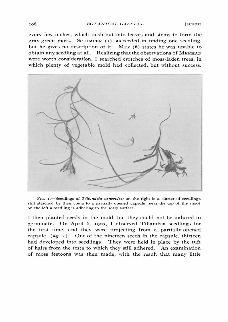
FIG. I.-Seedlings of Tillandsia usneoides; on the right is a cluster of seedlings
still attached by their coma to a partially opened capsule; near the top of the shoot
on the left a seedling is adhering to the scaly surface.
I then planted seeds in the mold, but they could not be induced to
germinate. On April 6, 1903, I observed Tillandsia seedlings for
the first time, and they were projecting from a partially-openedcapsule (fIig.). Out of the nineteen seeds in the capsule, thirteen
had developed into seedlings. They were held in place by the tuft
of hairs from the testa to which they still adhered. An examination
of moss festoons was then made, with the result that many little

8/8/2019 T Usneoides Billings 1904
http://slidepdf.com/reader/full/t-usneoides-billings-1904 12/28
1904] BILLINGS-TILLANVDSIA USNEOIDES 0og
seedlings were found either still attached to the capsules, or else
hanging to the scaly stems and leaves of the mother plants. In every
case the seed coat still adhered to the base, or root-end of the seed-lings, so as to enable the coma to keep them from falling to the ground,
which they certainly would have done without this provision. When
it is remembered that the capsules dehisced in March, and the seed-
lings were found early in April, it will be seen that germination fol-
lowed dehiscence quite closely. Of course the early growth was
attained at the expense of the endosperm, but when it was exhausted,
continued growth, which would naturally.be expected from healthy
looking seedlings, failed to occur. Material gathered in the summerand autumn yielded the usual crop of seedlings, but in no case were
any found that were larger than those found in April. Festoons
gathered the middle of January, nearly a year after the capsules
opened, had numerous little seedlings hanging to them, all healthy
looking, but no larger than any observed before them. It is expected
that when the warm weather of spring comes, when Tillandsia puts
forth its most vigorous growth, the seedlings also will increase in size.
The question naturally arises here, why Tillandsia seedlings are not
to be seen in all stages developing into mature plants, counting of
course those which germinated previous years. As such is not the
case, it can only be. conjectured that, as the spring of I903 was an
unusually rainy one, the conditions for germination were especially
favorable.
Seedlings exhibiting various stages in germination were imbedded
in paraffin and longitudinally sectioned. In the earliest stage (fig.45) the first leaf shows only a slight growth, the stem apex is still
undifferentiated, while from the axil of the ridge of tissue that enclosed
the stem apex, or else from its inner surface, a pair of organs have
arisen. It is believed that the presence of these organs throws some
light upon the morphological nature of the ridge of tissue. If a sec-
tion is made through the nodal region of a mature plant (fig. 49),
it will be seen that the leaf sheath which encloses the lateral shoot
and main axis is double. The doubling is not due to splitting of a
tissue once entire, but to bifurcation. A section through a very young
sheath (fig. 49a) reveals an outgrowth, one to several cells in extent,
from which a double layer of cells arises. These soon separate to

8/8/2019 T Usneoides Billings 1904
http://slidepdf.com/reader/full/t-usneoides-billings-1904 13/28
I O BOTANICAL GAZETTE [AUGUST
form the double sheath. In older stages the base of the sheath is
composed of many cells in width, so that the sheath appears no longer
to originate as a bifurcation of a single organ, but rather as two dis-tinct organs. Both organs or portions of the sheath may develop
equally, though it more often happens that one portion becomes
larger than the other. Occasionally, the inner scarcely develops at
all, but remains a tiny rudiment.
The sheaths which arise in the seedling develop precisely like those
in the mature plant and differ from them in no respect. The two
organs that originate on the ridge of tissue, therefore, may be regarded
without hesitation as the first sheath, and as every sheath appears inconnection with a leaf, that leaf must be the cotyledon. From the
section of the mature plant it will be noticed that the bases of each
leaf and its sheath are at the same level on the axis. If a difference
in level should occur, however, whereby the base of the sheath were
elevated above that of the corresponding leaf, the cell growth pro-
ducing that elevation would originate from the cortical parenchyma
lying immediately under the sheath. The parenchyma would give
rise to a ridge bearing the sheath upon its summit. Such an occur-
rence does not of course actually take place in the mature plant, but
it is believed that it is in such a way that the ridge of tissue originates
in the embryo. Reasons for coming to this conclusion are based upon
the position of the first sheath. While the inner portion of the sheath
may grow from the crotch at the base of the ridge of tissue, the outer,
and sometimes the inner also, is attached to the ridge upon its inner
surface. The outer portion may in fact arise from the summit ofthe ridge. The base of the sheath, therefore, is on the whole raised
above that of the cotyledon, the elevation being accomplished through
growth of the subjacent parenchyma. Thus there develops a special
organ which serves a special purpose, perhaps as protection to the
stem apex, and which must therefore be regarded as an embryonic
structurewithout an exact counterpart in the adult plant. It cannot
be a leaf, or a cotyledon, because a leaf does not bear such a relation
to its sheath. A leaf and its sheath always develop with a growing
point between them, so that they can never join in a median section.
CAMPBELL (7) calls a similarly placed though less extensive outgrowth
in the embryo of Sparganium a sheath. While it does not require a

8/8/2019 T Usneoides Billings 1904
http://slidepdf.com/reader/full/t-usneoides-billings-1904 14/28
1904] BILLINGS-TILLANDSIA USNEOIDES III
stretch of the imagination to consider the growth in question a sheath,
there is at least one objection to this solution of the problem. The
development of the sheath shows that it appears as a bifurcatedorgan almost from its incipiency, and that the base, at first narrow,
subsequently increases greatly in width. Quite the reverse would
be true in the embryo if the organ enclosing the growing point were
regarded as a sheath, for the basal portion is first enormously devel-
oped, leaving the upper bifurcated portion to appear comparatively
late.
The stages in germination are shown in figs. 45-48, which should
be compared with fig. 49. The latter exhibits a difference in rela-tive time of differentiation of stem and leaf apex as compared with
the seedling. In the mature plant the leaf is still quite small when
the stem apex becomes distinguishable at its base, while in the seed-
ling the leaf first attains considerable size.
THE FLOWER.
The flowers, which are produced in considerable quantity in May
and June, present little of special interest. Each flower has a calyxof three sepals, and a corolla of three green petals. Having a fra-
grant odor, it is possible that it is visited by insects, though no infor-
mation has been collected by me on the subject. Thrips, however,
inhabit many of the flowers and puncture the style in order to
deposit an egg at its base. It is possible, therefore, that they may
serve in cross pollination.
Although the flower appears to be terminal, it is regarded by
MEZ (6) as a reduced indeterminate inflorescence. An examination
of preparations made longitudinally through buds bears him out in
his statement, for a growing point of considerable size is present,
though having dead meristem tissue.
THE LEAVES.
The leaves of T. usneoides are acicular and with an approximately
semicircular cross section. The epidermal cells do not have specially
heavy walls, nor are the inner ones thicker than the outer, as in certain
other Bromeliaceae. Sections through the leaf show it to have three
fibrovascularbundles, each surrounded by a tissue composed of thick-
walled sclerenchyma fibers (figs. 50, si). The principal portion
8/8/2019 T Usneoides Billings 1904
http://slidepdf.com/reader/full/t-usneoides-billings-1904 15/28
II2 BOTANICAL GAZETTE [AUGUST
of the leaf is composed of parenchyma cells which do not show any
differentiatiation at all into palisade and spongy tissue. While the
cells have the shape of those in typical spongy tissue, the large inter-cellular air spaces characteristic of most mesophytic leaves are here
replaced by small ones, giving the whole tissue a much more compact
appearance. Not all of the parenchyma cells contain chloroplasts,
for there are interspersed cells without them, whose function is that
of water-storage, having walls provided with large pits which facilitate
the passage of water from one cell to another.
Aside from acting in the capacity of mechanical tissue, the vascular
system has undergone a process of degeneration. The necessityfor a functional xylem with its transpiration stream is eliminated by
the fact that there is a complete absence of roots, and also by the fact
that the water-absorbing organs, the scales, are found over the entire
exposed surface with the exception of some of the floral organs.
There would appear also to be no need for a functional phloem since
all living cells either contain chlorophyll and are exposed to light, or
else are approximate to those containing chlorophyll.
THE CHLOROPLASTS.
One of the most interesting features of the leaf is the structure and
behavior of the chloroplasts. These bodies, instead of exhibiting
the more or less homogeneous structure observed in most chloroplasts,
are seen to be composed of masses of smaller chloroplasts, measuring
about 2 p long and about a third as wide (fig. 52). While a very
few cells in every cross section of the living leaf contain chloroplastsof the usual type, the vast majority of them contain such as have
been described above. The little chlorophyll bodies have almost,
if not quite, the minuteness of bacteria, and for convenience will be
spoken of as microchloroplasts; he larger bodies, of which they appear
to form a part, being distinguished as megachloroplasts. The true
significance of the formation of the microchloroplasts will be readily
seen when it is stated that they may not remain in bunches (fig. 52),
but can and often do separate from one another till the entire cyto-
plasm of the cell becomes dotted with them (fig. 53). Under a low
magnification such a cell appears uniformly green throughout. They
even enter the vacuoles, where a lively Brownian movement is set up.
8/8/2019 T Usneoides Billings 1904
http://slidepdf.com/reader/full/t-usneoides-billings-1904 16/28
1904] BILLINGS-TILLANDSIA USNEOIDES II3
It was at once suspected that the various phases in distribution of the
microchloroplasts were conditioned by the light intensity, and hence
their movements could be made subject to control. Festoons ofTillandsia accordingly were placed under different conditions varying
from darkness to direct sunlight. Those placed in darkness were
allowed to remain there 24 to 3o hours, and a similar period of expo-
sure was allotted to festoons hung in the shade. Those exposed to
direct sunlight were hung up early in the morning. All were exam-
ined duringthe hours between11: 30 A. M. and 3:00 P. M.
The examination was made by sectioning numerous leaves of vari-
ous ages, and from as many different regions of each festoon as pos-sible. Plants were also sectioned at different times of day and also at
night. The results in every instance were approximately the same.
Sections were obtained from plants under the varying conditions of
light intensity used in the experiment; sections in which the mega-
chloroplasts were present; in which they were in the process of disin-
tegration into microchloroplasts; in which there was distribution of the
microchloroplasts uniformly through the cell; and in which all the
foregoing stages were present in the same section. In fact, the same
leaf varied in these respects in its different portions. There seemed
to be no method of telling before examination just what condition the
chloroplasts would be in. One of the best instances of complete
uniformity of distribution of the microchloroplasts throughout the
cytoplasm was obtained from the tiny leaf of a seedling. That the
disintegration of the mega- into microchloroplasts is not the result of
injury due to sectioning may be proven by an examination of the entireleaf through the epidermis. Sections also cut thick contain in their
centers cells untouched by the razor.
Homogeneous chloroplasts of the usual type were found which
showed evidence of undergoing division. Megachloroplasts, in which
the microchloroplasts were distinctly visible, were also found showing
a deep constriction as though they too were undergoing fission.
Owing to the difficulty of observing well the interior of the leaf
through the overlapping scales, it was not ascertained whether the
microchloroplasts return to form megachloroplasts or not; but if so
it seems certain that the latter would not be constructed of identically
the same microchloroplasts a second time.
8/8/2019 T Usneoides Billings 1904
http://slidepdf.com/reader/full/t-usneoides-billings-1904 17/28
II4 BOTANICAL GAZETTE [AUGUST
It is offered in explanation of this interesting condition of affairs
that the supply of light of Tillandsia is considerably diminished by
the presence of the overlapping scales, which are necessary for waterabsorption and for protection against too rapid transpiration. In
order to meet this diminution, it not only prefers sunny exposures,
but has modified its chlorophyll-bearing apparatus by causing it to
occupy a much larger area in order to utilize to better advantage such
light as penetrates to the interior of the leaf.
It may be stated here that precautions were taken to examine
healthy festoons removed directly from moss-laden trees. In some
instances these were examined immediately after such removal, lestconfinement in the laboratory should in some way induce pathologi-
cal conditions.THE SCALES.
The scales cover the entire living exposed portion of the plant with
the exception of the corolla, stamens, ovary, and a portion of the
calyx. Each scale develops from a single epidermal cell, the early
divisions of which occur while the young leaves and stems are included
within the leaf sheath. The first division is transverse (fig. 55).The proximal cell thus produced remains undivided, the distal
dividing transversely till four cells are produced, of which the lower
three form the stalk of the scale (fig. 57). The outermost hemi-
spherical cell becomes divided into four cells by two longitudinal walls
perpendicular to one another (figs. 58 and 63). By periclinal walls
a central group of four cells becomes separated from four outer ones
(fig. 64). The central cells divide no further. The outer ones divideby periclinal walls to form two concentric rows (fig. 65). The cells
of both rows become eight in number by anticlinal walls, the inner
row undergoing no further division, but the outer, by another set of
anticlinals, finally has sixteen. A fourth concentric row is then
formed by periclinal walls from the outermost sixteen cells. The
three inner layers consist of four, eight, and sixteen cells respectively,
which numbers remain constant, but the fourth layer undergoes
repeated divisions till a large number of cells are produced (fig. 67).
These last lengthen greatly and form the wing of the scale. The
surface view of the mature scale is seen in fig. 68, the longitudinal
section in fig. 70. All of the cells but the stalk cells and the original

8/8/2019 T Usneoides Billings 1904
http://slidepdf.com/reader/full/t-usneoides-billings-1904 18/28
I904] BILLINGS-TILLANDSIA USNEOIDES I I5
basal cells undergo thickening of their walls in certain portions and
lose their cell contents.
SCHIMPER (I) was the first to call attention to the water absorptivefunction of the scales, and his experiments along this line were so
complete as to leave little else to be done. That the leaves of
Tillandsia can absorb water is easily demonstrated either by wetting
them with water and then watching it disappear, or by noting the
weight before and after allowing them to remain a short time in water.
That the channel of absorption is through the scales is shown by using
colored water, which stains the stalk cells. Unlike most similar
appendages of the epidermis, the scales do not hinder the leaf frombecoming wet, but actually conduct water into the interstices beneath
them. When dry, the leaf is of a gray color, due to the air enclosed
by the scales, but when wet, the air is replaced by water, and a deep
green color results. From an examination of fig. 70 it will be seen
that the outer walls of the scale are thickened. When water is
absorbed by the cells with thickened walls, they become turgid,
expand below, and raise the wing of the scale well above the epider-
mis (fig. 69). The water absorbed by the outer cells of the scale
passes to the stalk cells, which have thin walls and rich protoplasmic
contents. Through these it passes through the basal cell to the water-
storage cells of the parenchyma. If the plant be soaked in dilute
potassium iodid solution for a day, the walls of the stalk, basal, and
neighboring parenchyma cells will be stained. It should be noticed
that no ordinary type of epidermal cell with its thickened cuticularized
wall separates the scale from the parenchyma. The cell that repre-sents the epidermis beneath the scale is the basal cell resulting from
the first division of the epidermal cell that gave rise to the scale. The
walls of this basal cell are thin and uncuticularized. If a scale whose
wing is raised well above the epidermis by the turgescence of its cells
be treated with glycerin, the contraction due to loss of turgescence
will draw the scale close down against the epidermis. This illus-
trates the process that takes place when scales become dry from
evaporation, as occurs in nature. Such a process cannot but assist
the epidermis in checking transpiration, so that the scales may be
considered not only as organs of absorption, but as serving to prevent
too rapid escape of the water they have been instrumental in bring-
ing into the plant.
8/8/2019 T Usneoides Billings 1904
http://slidepdf.com/reader/full/t-usneoides-billings-1904 19/28
ii6 BOTANICAL GAZETTE [AUGUST
The effect of an absorptive system extending over the entire sur-
face has already been mentioned in the reduction of the mechanical
and conductive tissues. As such reduction is found mostly in sub-merged hydrophytes, it will be seen that T. usneoides behaves in these
respects much like such plants.
The scales stand in connection with the water-storage tissue. The
cells of this tissue lie well distributed among the chlorophyll-bearing
cells and keep them in a state of turgescence. Even after a plant has
lost one-fourth of its weight by transpiration, and the leaves have
become grooved by contraction, the chlorophyll-bearing parenchyma
is unhurt. It is believed that the leaf shrinkage is due to a partialcollapse of the storage tissue upon loss of water, rather than by
decrease in turgescence of the green parenchyma. There is no evi-
dence that the plant undergoes desiccation and subsequent revival,
as in the case of Polypodium vulgare.'
THE STOMATA.
In addition to protection afforded by scales, hairs, and thick-
walled epidermal cells, xerophytes sometimes guard against toorapid transpiration by means of the position and structure of the
stomata. Sunken stomata, or those vestibuled by an epidermal air
space, itself with a narrow opening to the exterior, are all well known.
In some xerophytic plants the usual closing of the pore by the guard
cells is assisted in its function of checking transpiration by modifica-
tions in neighboring parenchymatous or epidermal cells. In Kingia
australis, for instance, there is, according to TSCHIRCH,2a large inter-
cellular space adjoining the stoma, partially filled with coiled cellular
' Since this paper went to press, one by MEZ (9) has appeared on the physiology
of water absorption in certain species of Tillandsia, among them T. usneoides. MEZ
corrects SCHIMPER'Sobservations as to the details of the absorptive process, claiming
that the empty cells of the scale do not contain air, but are collapsed when the sur-
face of the plant is dry. The thickened part of the scale swells when wet, raising it
and causing the lumen to reappear in the collapsed cells. Water passes from exterior
capillary spaces into the partial vacuum through thin places in the cell walls, whence,
from the filled cells as reservoirs, the water is taken up and passed into the mesophyll
by the stalk cells (Aujnahmezellen) through the usual process of osmosis. MEZdescribes the scale of T. usneoides as having only one stalk cell instead of three.
While it is true that two of the cells are very thin, their presence can readily be made
out in good sections of mature scales and still more readily in sections of young ones.
2 HABERLANDT, G., Physiologische Pflanzenanatomie. 2d ed. p. 399. i896.

8/8/2019 T Usneoides Billings 1904
http://slidepdf.com/reader/full/t-usneoides-billings-1904 20/28
1904] BILLINGS-TILLANDSIA USNEOIDES I I 7
outgrowths of the parenchyma. The outgrowths do not stop, but
merely hinder transpiration. Xanthorrhoea hastilis exhibits a similar
contrivance. Camellia japonica and Prunus Laurocerasushave thefaculty of filling up the air space as a result of excessive drouth or by
death of the guard cells. In such cases tylose-like processes occur
which block up all gas interchange. Pilea elegans differs from those
mentioned above in that certain subjacent parenchyma cells develop
thickenings on their exterior walls. One of these finally pushes up
against the pore of the stoma and effectually closes it. There is no
movement of the parenchyma cell away from the stoma, hence the
aperture is permanently closed. From an examination of figs. 72
and 73 it will be apparent that Tillandsia presents a condition of
affairs not widely different from that of Pilea. The principal differ-
ence lies in the fact that in Tillandsia the parenchyma cells undergo
no thickening. Both longitudinal and cross sections through the
leaf show outgrowths from the parenchyma cells lining the sides of
the air space. The outgrowths turn upward and either stop up the
opening of the stoma or else press directly against the guard cells.
It will be seen that the enormously thickened walls of the guard cells
preclude a possibility of change in their form. To show this experi-
mentally some plants were placed in water and exposed to direct
sunlight for a few hours. The leaves were then sectioned and the
guard cells watched with a micrometer while glycerin was run under
the cover glass. There was no measurable change. According to
MEZ (6) the guard cells have lost the power of functioning, this power
having been transferred to certain cells of the subjacent tissue whichoperate the passive guard cells, thus opening and closing the stoma.
There are two cells which come in contact with the guard cell and
may therefore be the means of moving it. One is the cell to which
it is attached and which extends from the hinge to the inner face of
the guard cell. This cell is usually continuous, but may be divided
by a cross wall into two cells. Should this cell, which is epidermal,
become turgescent, it would tend to raise the guard cell, swinging its
free side outwards. Such a movement, however, would close rather
than open the pore of the stoma. The hinge is quite thick and may
be much thicker than any shown in the figures. If the epidermal cell
is divided the division wall would effectually hinder any movement of
8/8/2019 T Usneoides Billings 1904
http://slidepdf.com/reader/full/t-usneoides-billings-1904 21/28
ii8 BOTANICAL GAZETTE [AUGUST
the guard cell. From these two considerations it would appear doubt-
ful whether the guard cells move at all in either direction. Of course
the glycerin experiment was repeatedly tried, but no motion wasdiscernible. The only other cells which by contact with the guard cells
can move them are the parenchyma cells whose processes push against
the guard cells on the under side. It was at first thought that the
parenchyma cells were operated by variations in turgescence of the
epidermal cell, so that regarding the guard cells as immovable the
epidermal cell would press downward upon the subjacent paren-
chyma cell during turgescence, and lower the process, thus unstopping
the stoma. Out of a number of such processes only one reaches thecenter of the stoma, all the others being considered attempts that
from necessity have failed. This explanation of the function of the
parenchymatous outgrowth is plausible, to say the least, but it has
not been experimentally proven by the glycerin test. Numerous
instances were investigated carefully, but in not a single case did any
of the processes change their position. It is here confessed that no
reaction was noticed in any part of the stoma or adjacent tissue in
response to the action of glycerin, nor was an instance found in fresh
material where the guard cells appeared to be separated. The
experimental demonstration of the presence of a mechanism in the
stomata, therefore, has not thus far met with success.
Another explanation might be mentioned, in which the processes
are to be considered attempts on the part of the plant to close the
stomata permanently. It may be that not all the processes actually
reach the center of the stoma and close it, so that, granted that asmall opening exists between the guard cells, the number of functional
stomata would merely be reduced. The total number of stomata
per square millimeter was ascertained and found to be relatively small.
The estimate was made by counting the number of stomata in each
section of serial sections taken from a portion of leaf of known length.
For instance, a piece of leaf 3mm ong contained 52 stomata. Calcu-
lating the surface from the circumference of the cross section, there
would be 7 per square millimeter, or, in round numbers, 4,300 per
square inch.
It must of course be taken into consideration that sections of living
leaves were used for experiment and not entire ones. If variations in

8/8/2019 T Usneoides Billings 1904
http://slidepdf.com/reader/full/t-usneoides-billings-1904 22/28
I904] BILLINGS-TILLANDSIA USNEOIDES II9
the pressure of the water-storage tissue exert any influence on the
opening and closing of the stomata it is very probable that the injury
done to the tissue in sectioning would greatly interfere with the actionof the mechanism.
HABERLANDT 8) figures the stoma of Tillandsia zonata, which in
respect to guard cells, and their supporting cells, resembles that of
T. usneoides. The guard cells have greatly thickened walls, and a
thickened hinge. From Haberlandt's account it is evident that he
does not fully comprehend the mechanism. In T. zonata no subja-
cent parenchyma is mentioned as taking part in the opening or closing
of the stoma.THE STEM.
Aside from the vascular region, the stem differs in no essential
particulars from the leaves as to structure. The stem, of course, has
the added function of support, so that there is developed between and
around the bundles a thick tissue of sclerenchyma fibers (fig. 74).
The fibers measure about 750)Lun length. They do not impart
rigidity, but flexibility and power to resist longitudinal strain. If a
fragment of moss is blown from one limb of a tree to another, and
succeeds in getting a hold, the cortex of that portion of the stem that
passes over the limb dies, and then disintegrates, leaving the scleren-
chymatous axis, which holds the plant in place for several and perhaps
many years. It is upon the durability and elasticity of this tissue
that the economic value of the moss in upholstery depends.
What has already been said in regard to reduction in the function
of the xylem and phloem of the leaves could with equal truth be saidabout the stems. With a superficial absorptive system and no root,
the xylem as a conductive system is useless. The pendent habit and
method of dissemination are both closely associated with reduction in
mechanical tissue, though they are more likely to be the result than
the cause of the reduction. The parenchymatous cortex, as in
leaves, is supplied with chlorophyll-bearing cells, all of which are
exposed to light, so that a tissue like the phloem, to carry elaborated
materials to cells distant from the center of photosynthesis, would be
unnecessary.
LOUISIANA STATE UNIVERSITY,
Baton Rouge, La.
8/8/2019 T Usneoides Billings 1904
http://slidepdf.com/reader/full/t-usneoides-billings-1904 23/28
I20 BOTANICAL GAZETTE [AUGUST
LITERATURE CITED.
I. SCHIMPER, A. F. W., Ueber Bau und Lebensweise der Epiphyten Westin-
diens. Bot. Centralbl. I7: I92 et seq. I884.
--, Botanische Mittheilungen aus den Tropen. 2. Die epiphytische Vege-
tation Amerikas. pp. I62. pls. 6. Jena. i888.
2. PEIRCE,G. J., On the mode of dissemination and on the reticulations of Rama-
lina reticulate. Bot. Gazette 25: 404-4I7. I898.
3. SMITH, R. W., A contribution to the life history of the Pontederiaceae. Bot.
Gazette 25: 324-337. I898.
4. HOFMEISTER, W., Neue Beobachtungen uiber Embryobildung der Phane-
rogamen. Jahrb. Wiss. Bot. i: 82-I88. pis. 7-IO. I858.
5. MEEHAN, T., The Florida moss,Tillandsia usneoides. Proc. Acad. Nat. Sci.
Philadelphia I875: 466.
6. MEZ, C., Monographiae Phanerogamarum. Editore et pro parte auctore
Casimiro de Candolle. IX. Bromeliaceae. Paris. I896.
7. CAMPBELL, D. H., Studies on the flower and embryo of Sparganium. Proc.
California Acad. Sci. III. Bot. I: 293-328. pls. 46-48. I899.
8. HABERLANDT, G., Zur Kenntniss des Spalt6ffnungapparatus. Flora 70:
97-II0. p1. 2. I887.
9. MEZ, CARL, Physiologische Bromeliaceen-Studien. I. Die Wasser-Oekono-
mie der extrematmosphaeriichen Tillandsien. Jahrb. Wiss. Bot. 40: I57-
229. I904.
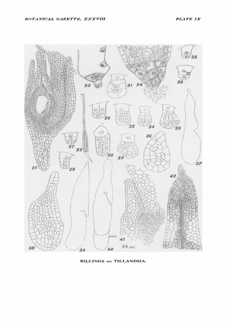
EXPLANATION OF PLATES VIII-XI.
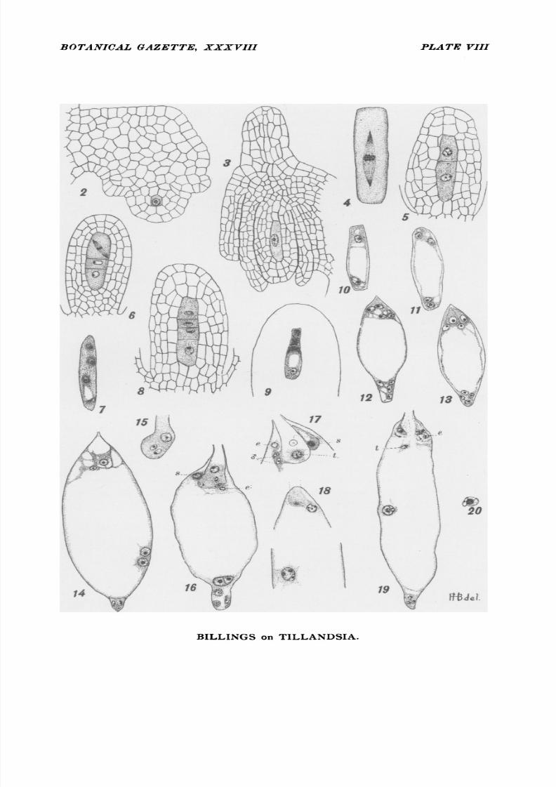
FIG. 2. Ovule fundament showing archesporial cell.
FIG. 3. Young ovule at period just before first division of archesporial cell.
FIG. 4. Spindle of first division.
FIGS. 5-6. Stages in formation of axial row of potential megaspores.
FIG. 7. Megaspores without separating walls.
FIGS. 8-9. Enlargement of basal megaspore to form embryo sac mother cell.FIGS. I0-I4. Stages in formation of embryo sac.
FIG. I5. Pollen tube just after entering embryo sac.
FIG. i6. Fusion of polars before rupture of pollen tube: s, synergid; e, egg.
FIG. I7. Lateral discharge of pollen tube: e, egg; t, tube nucleus; s, synergids.
FIG. i8. Simultaneous double fertilization.
FIG. i9. Double fertilization with discharge of tube nucleus (t); e, egg.
FIG. 20. Fusion of male and endosperm nuclei.
FIG. 2I. Ovule at time of completed embryo sac.
FIG. 22. Elongation of ovule and outer integument after fertilization.FIG. 23. First division in formation of chalazal endosperm tissue.
FIG. 24. Chalazal endosperm tissue and portion of endosperm that is to serve
as reserve material in ripe seed.
FIGS. 25-26. Two- and three-celled embryos.
8/8/2019 T Usneoides Billings 1904
http://slidepdf.com/reader/full/t-usneoides-billings-1904 24/28
BOTANICAL GAZETTE, XXXVIII PLA T rIHI
41~~1
8/8/2019 T Usneoides Billings 1904
http://slidepdf.com/reader/full/t-usneoides-billings-1904 25/28
BOTANICAL GAZETTE, XXXVHII PLATE IX

8/8/2019 T Usneoides Billings 1904
http://slidepdf.com/reader/full/t-usneoides-billings-1904 26/28
BOTANICAL GAZETTE, XxVIII PLATE X
49
And~~.1
BILING onTILAND47-cot
49.

8/8/2019 T Usneoides Billings 1904
http://slidepdf.com/reader/full/t-usneoides-billings-1904 27/28
BOTANICAL GAZETTE, XXXVIII PLA TE XI
12
60~~~~~~~~~~~6
0~~~~~~~~~0~~~~~~~~

8/8/2019 T Usneoides Billings 1904
http://slidepdf.com/reader/full/t-usneoides-billings-1904 28/28
1904] BILLINGS-TILLANDSIA USNEOIDES I2 I
FIG. 27. Formation of quadrant.
FIG. 28. Division of middle before terminalsegment.
FIG. 29. Unusually early development of basal and middle segments.
FIG. 30. An unusual formof embryo.FIGS. 3I-36. Stages in embryo development; in fig. 34, the transversewalls
in the terminal segment are oblique; the last three figures show beginning of
dermatogen.
FIG. 37. Embryo about one-fourth grown.
FIGS. 38-40. Outlines of embryos in late stages of development; fig. 30
representsa mature embryo.
FIG. 4I. Region in vicinity of growing point of a nearly ripe embryo.
FIG. 42. Root region of nearly matureembryo,showingdead corticalcells.
FIG. 43. Ripe seed.FIG. 44. Barbs on hairof coma.
FIG. 45. Early stage in germination;outline of longitudinal section.
FIGS. 46-48. Stages in developmentof seedling; outline of longitudinalsec-
tion.
FIG. 49. Longitudinal section throughthe growingpoint regions of a mature
plant: s, sheath; st, stem; 1, leaf; sa, stem apex; la, leaf apex.
FIG. 49a. Very young sheath.
FIG. 50. Cross section of leaf; p, pit in water-storagecell.
FIG. 5i. Bundle of leaf enlargedto show phloem (p) and xylem (x).
FIG. 52. Megachloroplastsshowing divisioninto microchloroplasts.FIG. 53. Stage in separation of microchloroplastsby which they become dis-
tributedthroughthe cytoplasm.
FIGS. 54-6i. Stages in developmentof the scale seen in longitudinalsection;
fig. 54 shows the epidermalcell from whichthe scale arises.
FIGS. 62-68. Stages in scale development seen from the surface; fig. 68
shows a maturescale.
FIG. 69. Scale in longitudinal section, after soaking in water for several
hours; the wing is seen to be raised considerablyabove the epidermis.
FIG. 70. Scale in longitudinalsection, drawn from a paraffinsection; it will
be seen to lie much closer to the epidermis than the one in fig. 69.
FIG. 7I. General appearanceof the surfaceof the leaf, showing the scales.FIG. 72. Section through a stoma; the guard cells are unquestionablyclosed;
in addition a process has grown up from the parenchymainto the pore of the
stoma; s, scales.
FIG. 73. Section of stoma showing slight variationfrom that in fig. 72; figs.
72 and 7,3were drawn fromsectionsthroughlivingmaterial.
FIG. 74. Cross section through the vascular region of the stem: p, phloem;
x, xylem.