
Effect of temperature on the number of selected microorganism groups and enzymatic activityof

Systems/Circuits
Activity of Tachykinin1-Expressing Pet1 Raphe NeuronsModulates the Respiratory Chemoreflex
X Morgan L. Hennessy,1 Andrea E. Corcoran,2 Rachael D. Brust,1 Yoon Jeung Chang,1 Eugene E. Nattie,2
and X Susan M. Dymecki1
1Department of Genetics, Harvard Medical School, Boston, Massachusetts 02115; and 2Department of Physiology and Neurobiology, Geisel School ofMedicine at Dartmouth, Lebanon, New Hampshire 03756
Homeostatic control of breathing, heart rate, and body temperature relies on circuits within the brainstem modulated by the neurotrans-mitter serotonin (5-HT). Mounting evidence points to specialized neuronal subtypes within the serotonergic neuronal system, borne outin functional studies, for the modulation of distinct facets of homeostasis. Such functional differences, read out at the organismal level,are likely subserved by differences among 5-HT neuron subtypes at the cellular and molecular levels, including differences in the capacityto coexpress other neurotransmitters such as glutamate, GABA, thyrotropin releasing hormone, and substance P encoded by theTachykinin-1 (Tac1) gene. Here, we characterize in mice a 5-HT neuron subtype identified by expression of Tac1 and the serotonergictranscription factor gene Pet1, referred to as the Tac1-Pet1 neuron subtype. Transgenic cell labeling showed Tac1-Pet1 soma residentlargely in the caudal medulla. Chemogenetic [clozapine-N-oxide (CNO)-hM4Di] perturbation of Tac1-Pet1 neuron activity blunted theventilatory response of the respiratory CO2 chemoreflex, which normally augments ventilation in response to hypercapnic acidosis torestore normal pH and PCO2. Tac1-Pet1 axonal boutons were found localized to brainstem areas implicated in respiratory modulation,with highest density in motor regions. These findings demonstrate that the activity of a Pet1 neuron subtype with the potential to releaseboth 5-HT and substance P is necessary for normal respiratory dynamics, perhaps via motor outputs that engage muscles of respirationand maintain airway patency. These Tac1-Pet1 neurons may act downstream of Egr2-Pet1 serotonergic neurons, which were previouslyestablished in respiratory chemoreception, but do not innervate respiratory motor nuclei.
IntroductionSerotonin (5-HT)-producing neurons in the lower brainstem arethe subject of intense investigation given their importance in
life-sustaining modulation of breathing, heart rate, and bodytemperature. These functions in rodents may be coordinated inpart by specialized subsets of 5-HT neurons, distinguishable andperhaps functionally divisible by coexpression of various neu-rotransmitters. For example, some medullary serotonergic neu-rons coexpress substance P, thyrotropin-releasing hormone,Received July 21, 2016; revised Nov. 30, 2016; accepted Dec. 30, 2016.
Author contributions: M.L.H., A.E.C., R.D.B., and S.M.D. designed research; M.L.H., A.E.C., and Y.J.C. performedresearch; M.L.H., A.E.C., E.E.N., and S.M.D. analyzed data; M.L.H., E.E.N., and S.M.D. wrote the paper.
This work was supported by the National Institutes of Health (Grant P01 HD036379 to S.M.D, E.E.N., and A.E.C.;National Institute of General Medical Sciences Grants F31NS073276 to R.D.B. and T32GM007753 to M.L.H.; andNational Institute of Neurological Disorders and Stroke Grant F31 NS083165– 02 to M.L.H). The content is solely theresponsibility of the authors and does not necessarily represent the official views of the National Institute of GeneralMedical Sciences or the National Institutes of Health. We thank B. Rood, B. Okaty, M. Freret, and R. Dosumu-Johnsonfor discussion; J.J. Mai for technical support; the Harvard Neurobiology Imaging Center for microscopy support; M.Rice for help with figure graphics; and R. Ouillette for experimental assistance.
The authors declare no competing financial interests.
A. Corcoran’s present address: John Merck Division of Science and Technology, Southern Vermont College, Ben-nington, VT.
R.D. Brust’s present address: Department of Cell Biology, Harvard Medical School, Boston, MA.Correspondence should be addressed to Susan M. Dymecki, Department of Genetics, Harvard Medical School, 77
Avenue Louis Pasteur, New Research Building, Room 358, Boston, Massachusetts 02115. E-mail:[email protected].
DOI:10.1523/JNEUROSCI.2316-16.2017Copyright © 2017 the authors 0270-6474/17/370001-•$15.00/0
Significance Statement
Serotonin (5-HT) neurons modulate physiological processes and behaviors as diverse as body temperature, respiration, aggres-sion, and mood. Using genetic tools, we characterize a 5-HT neuron subtype defined by expression of Tachykinin1 and Pet1(Tac1-Pet1 neurons), mapping soma localization to the caudal medulla primarily and axonal projections to brainstem motornuclei most prominently, and, when silenced, observed blunting of the ventilatory response to inhaled CO2. Tac1-Pet1 neuronsthus appear distinct from and contrast previously described Egr2-Pet1 neurons, which project primarily to chemosensory inte-gration centers and are themselves chemosensitive.
The Journal of Neuroscience, Month ??, 2017 • 37(xx):xxxx–xxxx • 1
rich3/zns-neusci/zns-neusci/zns99917/zns9419d17z xppws S!5 1/19/17 10:31 4/Color Figure(s): F1-F6 Art: 2316-16 Input-SMW
AQ: auAQ: or
Fn1
AQ: A

GABA, met-enkephalin, or leu-enkephalin (Glazer et al., 1981;Kachidian et al., 1991; Pilowsky, 2014). Transcriptional profilingof individual serotonergic neurons supports similar conclu-sions, revealing molecularly distinct subtypes of 5-HT neurons(Spaethling et al., 2014; Okaty et al., 2015), including subtypesthat not only express the gene encoding tryptophan hydroxylase 2(Tph2) for synthesis of 5-HT, but also neurotransmitter genessuch as Tachykinin 1 (Tac1) encoding substance P, or enzymegenes such as Gad1, encoding GAD-67 for GABA synthesis. Tac1-expressing Pet1! neurons, annotated as Tac1-Pet1 neurons,populate the medullary raphe, especially the raphe obscurus(Okaty et al., 2015). Gad1-expressing Pet1! neurons were iden-tified in a 5-HT neuronal lineage defined by embryonic expres-sion of Egr2 and by arising from rhombomere 5 (r5). Thesemature r5Egr2-Pet1 neurons have GABAergic and serotonergicfeatures, as suggested by prior anatomical studies (Stamp andSemba, 1995). Differential expression of Tac1, Egr2, and Gad1 by5-HT neurons or their precursors suggests different functional-ities and offers genetic access to subtypes of 5-HT neurons whenused as Cre drivers partnered with the Flpe driver transgene Pet1::Flpe (Jensen et al., 2008) and various intersectional reporter andeffector tools (Ray et al., 2011; Brust et al., 2014; Niederkofler etal., 2016). Here, we apply these tools to characterize the functionand projections of Tac1-Pet1 neurons.
Studies have linked the control of breathing to 5-HT, sub-stance P, its receptor, the neurokinin-1 receptor (NK1R), andmedullary raphe neurons more generally (Gray et al., 2001; Wanget al., 2001; Nattie and Li, 2006; Berner et al., 2007; Hodges andRicherson, 2008; Ptak et al., 2009; Cummings et al., 2011b; Ray etal., 2011). Only recently, though, have tools with sufficientresolving power become available to explore double-positive5-HT/substance P neurons; that is, Tac1-Pet1 neurons. Priorstudies probing raphe obscurus neurons based on anatomicallocation have implicated these neurons in respiratory control(Peever et al., 2001; Dias et al., 2008; Nuding et al., 2009; Depuy etal., 2011). For example, injection of Cre-dependent virus target-ing channelrhodopsin2-mCherry to caudal raphe Pet1-expre-ssing neurons demonstrated stimulation of breathing uponactivation and the presence of axon fibers in brainstem motornuclei and respiratory rhythm generation regions (Depuy et al.,2011). We hypothesized that the 5-HT neuron subtype responsi-ble for this effect is the Tac1-Pet1 subtype because it overlapsanatomically with the 5-HT neurons captured by viral injectionand because fibers harboring substance P, in some cases colocal-ized with 5-HT, map to many of the same targets (Thor andHelke, 1987; Holtman, 1988; Tallaksen-Greene et al., 1993; Na-kamura et al., 2006), which themselves are NK1R!.
Our previous studies showed that Egr2-Pet1 neurons are re-quired for a normal increase in ventilation in response to inspired5% CO2 via the respiratory chemoreflex, which maintains tissuePCO2 and pH within physiological range. In addition, r5Egr2-Pet1 neurons were found to be intrinsically chemosensitive, in-creasing firing in response to hypercapnic acidosis, collectivelyindicating a role as chemoreceptors that facilitate respiratorydrive (Brust et al., 2014). Downstream targets include otherchemoreceptor centers such as the retrotrapezoid nucleus; how-ever, innervation of respiratory motor nuclei under serotonergicmodulation was absent (Brust et al., 2014). The latter, we posit,may be provided by Tac1-Pet1 neurons. To explore this, we part-nered a Tac1::IRES-cre driver (Harris et al., 2014) with Pet1::Flpe(Jensen et al., 2008) and dual Cre/Flp-responsive reporter andeffector alleles (Dymecki et al., 2010; Ray et al., 2011; Brust et al.,2014; Niederkofler et al., 2016) to label Tac1-Pet1 boutons, iden-
tifying innervation targets, or to suppress Tac1-Pet1 neuron ex-citability, examining effects on the respiratory response tohypercapnia.
Materials and MethodsTransgenic mouse strains. Procedures were in accordance with Institu-tional Animal Care and Use Committee policies at Harvard MedicalSchool and Dartmouth Giesel School of Medicine. Triple-transgenic an-imals were generated by breeding Tac1::IRES-cre (Harris et al., 2014)mice to either double-transgenic Pet1::Flpe (Jensen et al., 2008), RC::FPDi (Ray et al., 2011) mice, Pet1::Flpe, RC::FrePe (Brust et al., 2014)mice, or Pet1::Flpe; RC::FPSiT (Niederkofler et al., 2016) mice. Double-and single-transgenic sex-matched littermates served as controls.
Respiratory assays. Nonanesthetized adult Tac1::IRES-cre, Pet1::Flpe,RC::FPDi male and female mice (n " 18) and littermate controls (n " 18)were acclimated individually for 20 min in a whole-body plethysmo-graph chamber filled with room air. The animals were then exposed to5% CO2 for 15 min and then switched back to room air for 15 min. Micewere removed for an intraperitoneal injection of 10 mg/kg CNO (1mg/ml in saline) and immediately returned to the chamber with room airfor 10 min, after which gas was changed to 5% CO2 for 15 min. The last5 min of each segment were used for data analyses. Rectal thermocoupletemperature was read immediately before placement in the chamber,upon removal for injection, and upon final removal and no significantdeviations in temperature were noted in these experiments. The averageof these three temperatures was used in the tidal volume calculationbelow. Tracings with high background noise due to animal movement(observed directly during the assay) were noted at the time of assay andnot included in final data analysis.
Respiratory airflow was recorded in a 140 ml of water-jacketed,temperature-controlled, glass chamber attached to a differential pressuretransducer (Validyne Engineering) and reference chamber. Water tem-perature was 35.1°C, resulting in a chamber temperature of 34°C. An AEITechnologies pump pulled air through the chamber at #325 ml/min.Volume calibrations were performed by repeated known-volume injec-tions. Humidified gas flowing into the chamber was either room air or a5% CO2 mixture balanced with medical grade air. Pressure transducerreadings were digitized at 1 kHz (PowerLab; ADInstruments) and ana-lyzed offline for peak amplitude, peak frequency, and average voltage(LabChart 6; ADInstruments). Tidal volume was determined by the fol-lowing formula:
{[(A/B) ! C] ! [(D " 273.15) ! (F # H)]}/{[(D " 273.15)
! (F # H)] # [(E " 273.15) ! (F # G)]}
Where A is the peak of breath signal (volts), B is the peak of signal forinjection volume, C is the volume injection (ml), D is the body temper-ature (°C), E is the chamber temperature (°C), F is the barometric pres-sure (mmHg), G is the" pressure of the water vapor of the mouse "1.142 ! (0.8017 $ D) % (0.012 $ D 2) ! (0.0006468 $ D 3), and H is thepressure of the water vapor of the chamber " 1.142 ! (0.8017 $ E) %(0.012 $ E 2) ! (0.0006468 $ E 3).
Results were analyzed by two-way ANOVA with Holm–Sidak correc-tion for multiple comparisons, with inspired gas and CNO administra-tion as factors (GraphPad Prism). After significance was identified intwo-way ANOVA, post hoc paired t tests were performed. The extent towhich ventilation, VE (ml/g/min) response to 5% CO2 (the percentageincrease), was reduced upon CNO/Di-induced neuronal inhibition,referred to as the “difference value,” was calculated as follows:&'VE
5%CO2/VERoom Air( ! 100% # 100%)pre-CNO # &'VE
5%CO2/VERoom Air( !
100% # 100%]post-CNO.Temperature assay. Male Tac1-IRES::Cre, Pet1::Flpe, RC::FPDi and lit-
termate control animals were implanted with IPTT-300 transponders(Bio-Medic Data Systems) subcutaneously at least 3 d before experi-ments. Temperatures were obtained via scanning with a Bio Medic DataSystems transponder reader without handling the animal. On the day ofthe experiment, animals were singly housed in cages with bedding and adlibitum access to food and water. Animals were weighed and placed into
2 • J. Neurosci., Month ??, 2017 • 37(xx):xxxx–xxxx Hennessy et al. • Tachykinin1-Expressing Pet1 Raphe Neurons
rich3/zns-neusci/zns-neusci/zns99917/zns9419d17z xppws S!5 1/19/17 10:31 4/Color Figure(s): F1-F6 Art: 2316-16 Input-SMW
AQ: B

their cages and allowed a 30 – 60 min period of acclimatization. No an-esthetic or analgesic reagents were used in these experiments. Baselinetemperature was measured at 0, 15, and 30 min. After the 30 min timepoint, an intraperitoneal injection of 10 mg/kg CNO 1 mg/ml dissolvedin saline was performed. After injection, animals remained at room tem-perature for 30 min and body temperature recorded. Animals were thentransferred to 4°C for 2 h, with body temperatures measured every 10min for the first hour and every 30 min for the second hour.
Tissue collection and sectioning. For fixed tissue, adult mice were anes-thetized with Avertin (tribromoethanol) and immediately perfused int-racardially with PBS followed by 4% paraformaldehyde (PFA) in PBS.Tissue was fixed overnight in 4% PFA at 4°C. Brains were then cryopro-tected using 30% sucrose in PBS and subsequently embedded in Tissue-Freezing Medium (Triangle Biomedical Sciences) and six sets of 20 $mon-slide or 40 $m free-floating serial coronal sections were collected inPBS. For fresh tissue, adult mice were anesthetized with Avertin anddecapitated and brains were collected and subsequently embedded inTissue-Tek Optimal Cutting Temperature Compound (Sakura Finetek).Embedded tissue was flash-frozen in 2-methylbutane on dry ice and 6sets of 20 $m on-slide coronal sections were collected.
Immunohistochemistry. For transgenic RC::FrePe tissue, free-floatingand on-slide sections were rinsed with 0.1% Triton X-100 PBS (PBS-T)and blocked in 5% normal donkey serum in PBS-T for 1 h at roomtemperature, followed by 24 – 48 h incubation at 4°C with primary anti-body in PBS-T (chicken polyclonal anti-GFP, 1:5000, ab13970, Abcamand rabbit polyclonal anti-dsRed, 1:1000, catalog #632496, Clontech).Sections were then washed with PBS-T three times and incubated withsecondary antibody (IgG-Alexa Fluor 488-conjugated donkey anti-chicken secondary antibody, 1:500, Jackson ImmunoResearch Laborato-ries) and Cy3-conjugated donkey anti-rabbit secondary antibody (1:500;Jackson ImmunoResearch Laboratories). Cell nuclei were visualized withDAPI. For transgenic RC::FPSiT tissue, free-floating sections were pre-pared as above. Each set was stained with chicken polyclonal anti-GFP(1:2000, ab13970; Abcam) and rabbit polyclonal anti-tyrosine hydroxy-lase (TH) (1:5000, AB152; Millipore), rabbit polyclonal anti-NK1R (1:2000, S8305; Sigma-Aldrich), or goat polyclonal anti-choline acetyltrans-ferase (ChAT) (1:500, AB144P; Millipore). Primary antibody incubation wasperformed for 48–72 h at 4°C in PBS-T with 5% serum (NK1R, TH) orwithout (ChAT). Sections were then washed with PBS-T 3 times and incu-bated with secondary antibody at room temperature for 2 h. Secondaryantibody staining was performed with IgG–Alexa Fluor 488-conjugateddonkey anti-chicken (1:500; Jackson ImmunoResearch Laboratories) andeither Cy3-conjugated donkey anti-rabbit (1:500; Jackson ImmunoResearchLaboratories) or Cy3-conjugated donkey anti-goat 1:500; Jackson Immu-noResearch Laboratories). Cell nuclei were visualized with DAPI.
Phox2b and ChAT staining. For transgenic RC::FPSiT tissue, on-slidecryosections were rinsed 3 times with PBS for 10 min and submitted tosteamer antigen-retrieval in 10 mM sodium citrate, pH 6.0, for 15 min.After cooling down at room temperature (RT) for 20 min, the sectionswere washed 5 times in PBS for 5 min, permeabilized with 0.5% TritonX-100 in PBS for 2 h, and quenched in 0.1 M glycine for 30 min at RT. Thesections were then rinsed 3 times with antibody buffer (0.2% gelatin, 300mM NaCl, 0.3% Triton X-100 in PBS) for 5 min, followed by incubationfor 72 h at 4°C with the primary antibodies in the same buffer. Primaryantibodies were as follows: chicken polyclonal anti-GFP (1:1000; GFP-1010; Aves Labs), mouse monoclonal anti-Phox2b (1:100, B-11, sc-376997; Santa Cruz Biotechnology), and goat polyclonal anti-ChAT (1:100, AB144P; Millipore). Sections were then washed with antibodybuffer 3 times for 5 min and incubated with secondary antibodies for 2 hat RT. Secondary antibodies were as follows: donkey anti-chicken IgG–Alexa Fluor 488 (1:500; Jackson ImmunoResearch Laboratories), donkeyanti-mouse IgG–Alexa Fluor 594 (1:500; Thermo Fisher Scientific), anddonkey anti-goat IgG–Alexa Fluor 647 (1:500; Thermo Fisher Scientific).DAPI was used for nuclear counterstaining.
RNAscope in situ hybridization. Fresh-frozen brain tissue from adultTac1::IRES-cre, Pet1::Flpe, RC::FrePe mice was collected as describedabove. Slides were dehydrated in EtOH, postfixed with 4% PFA, andstained with the RNASCOPE Multiplex Fluorescent Assay Kit (AdvancedCell Diagnostics) as described previously (Wang et al., 2012) with eGFP
(catalog #400281) and Tac1 (catalog #410351-C3) probes. Cell nucleiwere visualized with DAPI.
Image collection. Epifluorescence images were collected using an up-right Zeiss Axioplan2 microscope equipped with an Axiocam 506 cam-era. SlideScanner epifluorescence images were collected on a VS120-SLmicroscope. SlideScanner images were brightness and contrast adjusteduniformly across the entire image in FIJI (http://fiji.sc/Fiji) for ease ofvisualization. Confocal z-stacks were collected on a Zeiss LSM 780 in-verted microscope; for RC::FPSiT tissue, laser power and pinhole diam-eter were optimized for the GFP channel and kept constant for all imagescollected. Three triple-transgenic animals and double-transgenic controlanimals from independent litters, aged postnatal day 60 (P60), were used.Two equivalent anatomical regions were selected for each target areafrom each animal imaged. Efforts were made to select equivalent areasacross samples using the anatomical landmarks described here. ChATimmunostaining was used to mark motor nuclei [nucleus ambiguus(NA), trigeminal nucleus (5N), facial nucleus (7N), spinal accessory nu-cleus (11N), and hypoglossal nucleus (12N)]. In general, only one slicecontained the 11N per series, whereas the 5N, 7N, and 12N images werecollected where the region of ChAT staining was the largest, marking themiddle of their anterior–posterior extent. For the pre-Botzinger complex(preBotC), the compact NA was used as an anatomical landmark becauseboth are NK1R immunoreactive. For the parabrachial/pericoerulear re-gion, images were taken immediately adjacent to the crescent-shapedlocus ceruleus, marked by TH, where the dendrites of these neurons werevisible. For both the caudal and rostral nucleus of the solitary tract (NTS),TH-positive cell bodies were used to mark the region; rostral NTS imageswere collected adjacent to the fourth ventricle, whereas caudal NTS im-ages were collected adjacent to the area postrema and central canal, tak-ing care to avoid the dorsal motor nucleus of the vagus. The C1adrenergic nucleus was visualized with TH and was generally only pres-ent in one slice per series. Image stacks (.czi files) were imported into FIJIfor background subtraction and thresholding (using a minimum fluo-rescence intensity of 15) to generate GFP mask images. Control imagescollected from each region had negligible fluorescence intensity signalabove the selected threshold.
ResultsTac1-Pet1 intersectionally labeled neurons reside primarily inthe raphe obscurus, raphe pallidus, and lateralparagigantocellularis and express Tac1 transcriptTo ascertain whether Pet1::Flpe-expressing raphe neurons have ahistory of Tac1 expression by adulthood and to determine wherethese neurons are located, we used an intersectional genetic strat-egy that combined a Tac1::IRES-cre knock-in allele (Harris et al.,2014), a Pet1::Flpe BAC transgenic driver (Jensen et al., 2008),and the intersectional reporter allele RC::FrePe (Bang et al., 2012;Brust et al., 2014) (Fig. 1A) to mark double-positive cells witheGFP (referred to as the intersectional population, or in this case,the Tac1-Pet1 subtype) while simultaneously marking all otherPet1::Flpe-expressing raphe cells with mCherry (referred to as thesubtractive Pet1 neuron population). The majority of eGFP!neurons were found populating the full rostrocaudal extent of theraphe obscurus, as well as the immediately ventral raphe pallidus,albeit to a lesser extent (Fig. 1G). These findings are consistentwith reported transcriptional profiling studies of Pet1 neuronsisolated from these anatomical regions (Okaty et al., 2015), aswell as immunohistochemical studies localizing substance P(Ptak et al., 2009). Also in the medulla, a subset of intersectionallylabeled eGFP! cells were found in the caudal portion of theraphe magnus and the lateral paragigantocellularis (Fig. 1E,F); incontrast, in the midbrain/pons region, only a few eGFP! cellswere detected in the dorsal raphe (Fig. 1C) and none within themedian raphe (Fig. 1D). In addition to adult tissue, we examinedP8 and P15 brain tissue from triple-transgenic Tac1::IRES-cre,Pet1::Flpe, RC::FrePe animals and identified eGFP! cells in the
Hennessy et al. • Tachykinin1-Expressing Pet1 Raphe Neurons J. Neurosci., Month ??, 2017 • 37(xx):xxxx–xxxx • 3
rich3/zns-neusci/zns-neusci/zns99917/zns9419d17z xppws S!5 1/19/17 10:31 4/Color Figure(s): F1-F6 Art: 2316-16 Input-SMW
F1

dorsal raphe and the raphe obscurus at P8 and P15 (data notshown), indicating that Tac1::IRES-cre expression in Pet1::Flpe-positive cells is initiated by P8.
Dual Flpe- and Cre-mediated recombination of the RC::FrePereporter allele indicates a history and/or possible time-of-harvestexpression of the recombinases. To assess adult, time-of-harvestTac1 expression in these Pet1 neurons, we used RNAscope insitu hybridization to detect Tac1 and eGFP mRNA in triple-transgenic adult Tac1::IRES-cre, Pet1::Flpe, RC::FrePe tissue. In alleGFP! cells (Tac1-Pet1 lineage cells) examined, in this case in theraphe obscurus, Tac1 transcripts were detected, suggesting active
adult expression of Tachykinin1 and substance P. Some nearbyneurons expressed Tac1, but little to no eGFP (Fig. 1H–M), indi-cating expression of Tac1 in raphe neurons not captured by ourPet1::Flpe driver; these Tac1! neurons could represent non-5-HT neurons expressing Tac1 or Pet1 neurons missed by Pet1::Flpe, which are are known to exist (Barrett et al., 2016).
Acute silencing of Tac1-Pet1 neurons blunts the 5% CO2
respiratory chemoreflexThe respiratory chemoreflex underlies the increase in ventilationrequired to clear elevated tissue/blood CO2 levels to maintain
A
B
C D E F G
H I J
K L M
Figure 1. Tac1-Pet1 neurons are located primarily in caudal medullary raphe nuclei and express Tac1 transcript. A, Intersectional genetic strategy: a combination of expressed transgenes 1 and2 result in dual Flpe- and Cre-mediated recombination of intersectional allele, RC::FrePe (knock-in transgene 3) in cells expressing both Tac1 and Pet1. B, Sagittal and coronal schematics of mousebrainstem slices highlighting anatomical locations of 5-HT raphe nuclei. Intersectionally labeled eGFP-positive cells were found in the dorsal raphe (C), caudal raphe magnus (E), lateral paragigan-tocellularis (F ), and raphe obscurus (G), but not in the median raphe, (D); mCherry marks other (non-Tac1-expressing) Pet1 neurons. Scale bars, 200 $m. H–M, Tac1 transcript (red) was localizedwithin all eGFP transcript-positive (green) cells examined in the raphe obscurus in Tac1::IRES-cre, Pet1::Flpe, RC::FrePe tissue. Tac1 transcript (I,L), and eGFP transcript (J,M ) shown separately ingrayscale. Scale bars, 50 $m.
4 • J. Neurosci., Month ??, 2017 • 37(xx):xxxx–xxxx Hennessy et al. • Tachykinin1-Expressing Pet1 Raphe Neurons
rich3/zns-neusci/zns-neusci/zns99917/zns9419d17z xppws S!5 1/19/17 10:31 4/Color Figure(s): F1-F6 Art: 2316-16 Input-SMW

blood pH homeostasis (Dean and Nattie, 2010; Guyenet et al.,2010; Nattie and Li, 2012). 5-HT neuron function is required tomount a robust chemoreflex, both in models in which 5-HTneurons fail to develop (Hodges et al., 2008) and in models ofacute suppression of Pet1::Flpe- or Slc6a4::cre-expressing neu-rons (Ray et al., 2011). Selective suppression of just the Egr2-Pet15-HT neuron subtype comparably blunts the chemoreflex as well,whereas most other Pet1 neuron subtypes do not (Brust et al.,2014); Tac1-Pet1 neurons were not analyzed previously due tolack of a suitable driver. Now with the availability of the Tac1::IRES-cre driver (Harris et al., 2014) to couple with Pet1::Flpe,we applied our dual-recombinase intersectional RC::FPDi al-lele (Fig. 2A) to selectively and acutely perturb Tac1-Pet1neurons in vivo in the context of hypercapnia. RC::FPDi is
identical in design to the RC::FrePe allele (Bang et al., 2012;Brust et al., 2014) used above except that, in RC::FPDi, thesynthetic inhibitory receptor hM4Di (Armbruster et al., 2007)is expressed in the Cre/Flpe dual-targeted subtype, whereasmCherry marks remainder of the Pet1 neurons (the subtrac-tive population; Ray et al., 2011; Brust et al., 2014). Uponbinding of its exogenous ligand CNO, hM4Di triggers cell-autonomous hyperpolarization and reduced neuronal excit-ability (Armbruster et al., 2007; Ray et al., 2011; Brust et al.,2014). Di has been used previously to suppress excitability ofPet1 neurons in primary neuronal cultures, transgenic brain-stem slices, and transgenic mice (Ray et al., 2011; Brust et al.,2014; Teissier et al., 2015; Brust and Dymecki, unpublisheddata) and has been shown to inhibit synaptic transmission
A
B
C D E
F G H
I
Figure 2. Acute silencing of Tac1-Pet1 neurons results in a decrease in ventilation at 5% CO2. A, Intersectional DREADD allele. B, Experimental design. After an initial period of acclimation,experimental adult animals and littermate controls are exposed to 5% CO2 and then returned to room air, briefly removed from the chamber for intraperitoneal injection of 10 mg/kg CNO, and placedback in room air. Animals were then exposed to 5% CO2 a second time, followed by another period of room air breathing in 12 animals from each group. Gray boxes (a–d) indicate time intervals usedfor data analysis. C–E, Baseline measurements of VE, respiratory rate (RR), and tidal volume (VT) in control (n " 14) and Tac1::IRES-cre, Pet1::Flpe, RC::FPDi (“Tac1-Pet1 hM4Di”) animals (n " 15)before CNO injection were not significantly different. Each circle represents one animal, error bars represent SEM. F, Average percentage increase in ventilation (VE) before CNO injection (pre-CNO,VE
b/VEa ! 100%) and minutes after CNO injection (post-CNO, VE
d/VEc ! 100%) in control and Tac1-Pet1 hM4Di animals. Error bars indicate SEM. G, H, Average fractional increase in
respiratory rate (RR) or tidal volume (VT) to 5% CO2 before and after CNO injection. I, Change in room air ventilation after CNO administration compared with baseline (VEe/VE
a) in control (n " 12)and Tac1-Pet1 hM4Di animals (n " 12). See Results for numerical data and statistical tests.
Hennessy et al. • Tachykinin1-Expressing Pet1 Raphe Neurons J. Neurosci., Month ??, 2017 • 37(xx):xxxx–xxxx • 5
rich3/zns-neusci/zns-neusci/zns99917/zns9419d17z xppws S!5 1/19/17 10:31 4/Color Figure(s): F1-F6 Art: 2316-16 Input-SMW
F2

(Stachniak et al., 2014). Here, we targeted Di expression toallow CNO-triggered inhibition selectively in Tac1-Pet1neurons.
We used whole-body plethysmography to assay the ventila-tory response to inspired 5% CO2 before and during acute CNO/Di-triggered perturbation of Tac1-Pet1 neurons (Fig. 2B) asdescribed previously (Ray et al., 2011; Brust et al., 2014). Micewere assayed in quiet wakefulness because vigilance state has beenshown to affect the respiratory chemoreflex in some models (Na-kamura et al., 2007). Body temperature was also monitored andfound to be stable during these measurements; the plethysmo-graph chamber was held continuously at 34°C. To test the effectsof Tac-Pet1 neuron activity perturbation on ventilation, animalswere exposed to a series of CO2 challenges and their ventilationmeasured as diagrammed in Figure 2B. Before CNO administra-tion (at time point “a”), no significant differences were found inthe baseline respiratory rate, tidal volume, ventilation (VE), oroxygen consumption (VO2) of the Tac1-Pet1 hM4Di-expressingmice compared with littermate controls, indicating relative neu-trality of the hM4Di receptor on respiration in the absence ofCNO (Fig. 2C–E). Before CNO injection, during the first CO2
challenge, the percentage increase in VE upon 5% CO2 exposure(from time point “a” to “b”) was not significantly different be-tween Tac1-Pet1 hM4Di-expressing mice and control littermates(229% vs 207.6%, unpaired t test, p " 0.20) (Fig. 2F, pre-CNO).However, upon CNO administration, a significant blunting ofthe ventilatory response to 5% CO2 (from time point “c” to “d”)was observed in only the Tac1-Pet1 hM4Di-expressing mice(229% vs 186.8%, paired t test, p " 0.0167, t " 2.717, df " 14),whereas the ventilatory response of control littermates showedno significant difference from pre-CNO measurements (207.6%vs 222.7%, paired t test, p " 0.27).
We then examined increases in respiratory rate and tidal vol-ume separately to ascertain whether perturbation of Tac1-Pet1neuron activity affected these two components of ventilation dif-ferentially. The fractional increase in respiratory rate in responseto 5% CO2 was significantly blunted in Tac1-Pet1 hM4Di-expressing mice upon CNO exposure compared with pre-CNOlevels (1.38 vs 1.56, paired t test, p " 0.0374, t " 2.269, df " 16),whereas the fractional increase in tidal volume in response to 5%CO2 was not significantly different upon Tac1-Pet1 neuron per-turbation (Fig. 2G,H). In control littermates, the fractional in-creases in respiratory rate and tidal volume in response to 5%CO2 were not significantly altered upon CNO administration(Fig. 2G,H).
Although these experiments were not designed to best mea-sure the effect of Tac1-Pet1 neuron suppression on room air ven-tilation (rather, the time course was optimized to assess effects onthe chemoreflex), we nonetheless also examined the overall effectof CNO/hM4Di-mediated perturbation on room air ventilationin both control and Tac1-Pet1 hM4Di-expressing animals overthe course of the experiment. Room air ventilation at time point“e” and time point “a” was calculated and the e/a ratio deter-mined for each animal, importantly allowing each animal to serveas its own control (Fig. 2I). Each group, control and hM4Di-expressing, showed a similar average ratio (0.87 vs 0.92, unpairedt test, p " 0.4375), thus revealing no discernable effects on roomair ventilation.
Intact body temperature homeostasis in the face of Tac1-Pet1neuron perturbationWhereas acute silencing of all serotonergic neurons en masseresults in rapid and significant hypothermia (Ray et al., 2011),
this was not observed upon perturbation of the Tac1-Pet1 neuronsubset. Specifically, after intraperitoneal injection of 10 mg/kgCNO followed by exposure to ambient 4°C, Tac1-Pet1 hM4Di-expressing male animals (n " 8), on average, dropped a maxi-mum of 2.36 * 0.32°C body temperature, an effect similar to thatobserved for control male littermates (n " 8), which showed amaximum drop of 2.73 * 0.31°C (unpaired t test, p " 0.42).
Tac1-Pet1 neurons project to motor nuclei and the preBotCrespiratory rhythm generatorFinding that Tac1-Pet1 neuron activity perturbation blunts theventilatory response to hypercapnia (Fig. 2) and that Tac1-Pet1neurons are located primarily within the raphe obscurus, a regionknown to project to motor nuclei within the brainstem (Loewy,1981; Thor and Helke, 1987; Ellenberger et al., 1992; Depuy et al.,2011) (Fig. 1), we sought to identify brainstem and spinal cordregions receiving projections from Tac1-Pet1 neurons. We usedthe intersectional reporter allele RC::FPSiT, which expresses asynaptophysin–GFP fusion in the intersectional neuron popula-tion (Niederkofler et al., 2016), to resolve Tac1-Pet1 axonalboutons within the brainstem and cervical spinal cord. Synapto-physin is a synaptic vesicle membrane glycoprotein that has beenwidely used as a marker of synaptic-vesicle-containing boutons(Nakata et al., 1998; Pennuto et al., 2002; Li et al., 2010). Upondual recombination of the RC::FPSiT reporter allele, synapto-physin–GFP is expressed and trafficked cell autonomously to ax-onal boutons in intersectionally labeled neurons. This approachallows illumination of terminal and en passant axonal boutonsupon fluorescent staining for GFP. In previous work examiningthe projection patterns of Pet1 raphe neurons, dorsal raphe Pet1neurons have been found to project mainly to forebrain areas,whereas medullary Pet1 raphe neurons projected within thebrainstem and spinal cord (Bang et al., 2012). Although we can-not rule out that dorsal raphe Tac1-Pet1 neurons may contributeto brainstem projections, we presume that the majority of theinnervation presented here originates from medullary Tac1-Pet1neurons.
Fluorescent staining of synaptophysin–GFP labeled Tac1-Pet1boutons revealed a pattern, which can be seen in low-magnificationimages (Fig. 3), of Tac1-Pet1 projections primarily within motornuclei, some of which have roles in respiration and airway pa-tency, including the 5N, 7N, 11N, 12N, and NA. Within the cer-vical spinal cord, boutons were localized among the ventralmotor neurons (VMNs), but not sensory neurons in the dorsalhorn. A subset of these cervical spinal VMNs gives rise to thephrenic nerve, innervating the diaphragm, the main muscle ofrespiration. In contrast, the laterodorsal tegmental nucleus,which sends cholinergic projections to subcortical and corticalstructures such as the thalamus and ventral tegmental area, wasdevoid of synaptophysin–GFP signal (Fig. 3A, inset), as was thedorsal motor nucleus of the vagus nerve (10N) (Fig. 3C), whichsupplies parasympathetic motor innervation to various cellswithin the gastrointestinal tract.
After identifying this gross pattern of Tac1-Pet1 projections atlow magnification (Fig. 3), we pursued a higher-resolution anal-ysis using confocal microscopy to visualize terminals in motorregions where Tac1-Pet1 boutons were localized, as well as toexplore chemosensory regions of the brainstem. Because nearbyEgr2-Pet1 neurons were not found to send major projections tomotor centers known to receive serotonergic innervation, butrather project heavily to chemosensory integration regions, in-cluding the retrotrapezoid nucleus (Brust et al., 2014), a knownchemosensory site (Mulkey et al., 2015), our findings point to
6 • J. Neurosci., Month ??, 2017 • 37(xx):xxxx–xxxx Hennessy et al. • Tachykinin1-Expressing Pet1 Raphe Neurons
rich3/zns-neusci/zns-neusci/zns99917/zns9419d17z xppws S!5 1/19/17 10:31 4/Color Figure(s): F1-F6 Art: 2316-16 Input-SMW
AQ: C
F3

Tac1-Pet1 neurons as a source of serotonergic projections to mo-tor nuclei.
We examined each of the motor nuclei identified in Figure 3 asreceiving Tac1-Pet1 innervation, as well as chemosensory regionsimportant in respiratory modulation. Each area was queried inthree animals from independent litters; two equivalent anatom-ical regions were selected from each animal for imaging (seeMaterials and Methods). Representative images are shown todemonstrate relative density of synaptophysin–GFP-labeledterminals, alongside schematic coronal brainstem sectionshighlighting the area of interest (Fig. 4). GFP-mask images,thresholded and processed uniformly across all areas of GFP flu-orescence data as described in the Materials and Methods section,are shown for ease of terminal visualization (Fig. 4A+–E+). Tac1-Pet1 terminals were found in the trigeminal motor nucleus (Fig.4A), where serotonergic input may contribute to the modulationof rhythmic oral activity (Hsiao et al., 2002), as well as the facialmotor nucleus (Fig. 4B), which innervates skeletal muscles of theface and has been implicated in respiration in rodents (Zhang etal., 2004). Tac1-Pet1 terminal density was also found in the NA(Fig. 4C), which contains branchial motor neurons contribut-ing to the vagus glossopharyngeal nerves, as well as the hypo-glossal nucleus (Fig. 4D), which innervates the genioglossus,the main muscle of the tongue. The hypoglossal nucleus iscritical for establishing airway patency, is activated duringinspiration, and its output has been shown to be modulated byraphe obscurus neurons (Peever et al., 2001; Saboisky et al.,2006). Finally, we observed Tac1-Pet1 innervation in the spi-nal accessory nucleus (Fig. 4E), which innervates several of theaccessory muscles of respiration. Because many of these re-gions innervate muscles involved in actions distinct from res-piration, this suggests a possible broader role for Tac1-Pet1neurons in motor regulation.
Given the role of Tac1-Pet1 neurons in the respiratorychemoreflex, we hypothesized that these neurons may also inner-vate brainstem regions involved in chemosensation and rhythmgeneration, perhaps at a lower terminal density than was seen inrespiratory motor nuclei within the brainstem and spinal cord
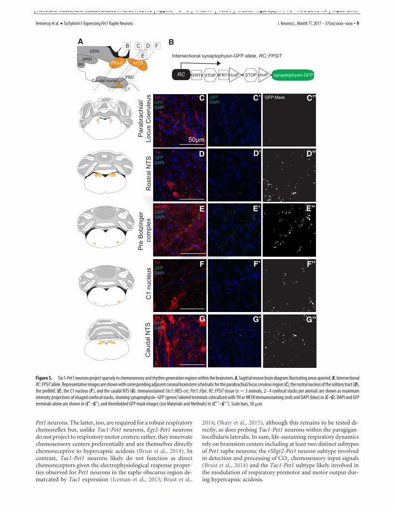
because these areas have been shown previously to receive denseinnervation from the Egr2-Pet1 neuron subtype (Brust et al.,2014). We examined Tac1-Pet1 projections in several chemosen-sory regions of the brainstem using TH to identify cell bodies asanatomical landmarks for the parabrachial/pericoerulear region,the C1 adrenergic nucleus, and nucleus of the solitary tract andNK1R staining to mark the preBotC. Representative images areshown to illustrate qualitative innervation density to these re-gions (Figs. 5, 6). The parabrachial/pericerulear region, imagedimmediately lateral to the crescent-shaped locus ceruleus andcontaining dendrites from these neurons, was devoid of Tac1-Pet1 innervation (Fig. 5A), whereas this region is densely inner-vated by the Egr2-Pet1 subtype (Brust et al., 2014). The rostraland caudal NTS, responsible for the integration of respiratorystimuli from within the brainstem and from the periphery tocoordinate cardiorespiratory control, and likely also chemo-sensitive themselves, received projections from Tac1-Pet1neurons (Fig. 5 B, E). The C1 adrenergic nucleus, which servesto modulate the sympathetic nervous system response to hy-percapnia, also receives Tac1-Pet1 innervation (Fig. 5C). ThepreBotC, the primary respiratory rhythm generator, markedby NK1R staining, receives innervation from Tac1-Pet1 neu-rons as well (Fig. 5D). Overall, these chemosensory andrhythm generation centers received qualitatively lower termi-nal density from Tac1-Pet1 neurons than did the motor re-gions examined above, as can be seen in the thresholded GFP-mask images (Fig. 5A,,–E,,). Notably, the chemosensoryretrotrapezoid nucleus localized via Phox2b-expressing cellsventral to the facial nucleus (Stornetta et al., 2006; Guyenet etal., 2010) and the associated lateral parafacial region involvedin expiratory oscillation (Huckstepp et al., 2015) both con-tained little to no Tac1-Pet1 boutons (Fig. 3D).
DiscussionDiversity among serotonergic neurons is proving to be substan-tial (Jensen et al., 2008; Kim et al., 2009; Deneris, 2011; Hale andLowry, 2011; Gaspar and Lillesaar, 2012; Andrade and Haj-Dahmane, 2013; Brust et al., 2014; Spaethling et al., 2014; Fernan-
Figure 3. Tac1-Pet1 terminals primarily target respiratory and airway motor nuclei. Coronal brainstem sections from Tac1::IRES-cre, Pet1::Flpe, RC::FPSiT animals were stained for synaptophysin-GFP (green) to visualize terminals, ChAT (red) to identify motor neurons, and Phox2b (magenta) to mark retrotrapezoid nucleus neurons. A, 5N receives Tac1-Pet1 projections, whereas thelaterodorsal tegmental nucleus (LDTg) does not. B, 7N contains Tac1-Pet1 projections. C, 10N does not receive projections, whereas 12N and NA contain synaptophysin–GFP staining. D, Retrotrap-ezoid nucleus (RTN) does not receive projections and lies ventral to the 7N, which is positive for Tac1-Pet1 projections. E, F, 11N (E) and VMNs (F ) in the cervical spinal cord both receive projectionsfrom Tac1-Pet1 neurons. In addition to marking axonal boutons, there is sufficient synaptophysin–GFP expression to mark Tac1-Pet1 cell bodies, as seen in C. Marked by asterisks, prominentautofluorescence highlights the entering vestibulocochlear nerve tract in A and the dorsolateral spinocerebellar tracts in D and E.
Hennessy et al. • Tachykinin1-Expressing Pet1 Raphe Neurons J. Neurosci., Month ??, 2017 • 37(xx):xxxx–xxxx • 7
rich3/zns-neusci/zns-neusci/zns99917/zns9419d17z xppws S!5 1/19/17 10:31 4/Color Figure(s): F1-F6 Art: 2316-16 Input-SMW
F4
F6,AQ:D
AQ: E

dez et al., 2016; Okaty et al., 2015; Niederkofler et al., 2016).Progress toward making sense of this heterogeneity has beenaided by intersectional genetic tools that permit mechanisticprobing of subsets of Pet1 raphe neurons (Jensen et al., 2008; Kimet al., 2009; Dymecki et al., 2010; Ray et al., 2011; Brust et al.,2014; Niederkofler et al., 2016); these tools are applied here toexplore a newly accessible subtype of raphe neuron, the Tac1-Pet1 subtype. We show that CNO/hM4Di manipulation of Tac1-
Pet1 neurons in vivo blunts the respiratory chemoreflex,suggesting a stimulatory effect of these neurons on breathing.Axonal boutons from Tac1-Pet1 neurons appear enriched inbrainstem and spinal cord motor nuclei, many of which are nec-essary for normal respiration and airway patency. Tac1-Pet1 neu-rons are thus likely to exert a substantial portion of their effectthrough modulation of motor output. These findings comple-ment our previously reported work on medullary raphe Egr2-
G’
A B
C C’ C”
D D’ D”
E E’ E”
F’F F”
G G”
Figure 4. Tac1-Pet1 neurons project densely to specific motor nuclei within the brainstem. A, Sagittal mouse brain schematic illustrating areas queried. B, Intersectional RC::FPSiT allele.Representative images are shown with corresponding adjacent coronal brainstem schematic for the trigeminal motor nucleus (C), facial nucleus (D), NA (E), hypoglossal nucleus (F ), and spinalaccessory nucleus (G). Immunostained Tac1::IRES-cre, Pet1::Flpe, RC::FPSiT tissue (n " 3 animals, 2– 4 confocal stacks per animal) are shown as maximum intensity projections of imaged confocalstacks, with synaptophysin–GFP puncta (green) from intersectional Tac1-Pet1 cells colocalized with ChAT immunostaining (red) and DAPI (blue) (C–G) and synaptophysin-GFP puncta with DAPIonly (C!–G!). In (C!!–G!!), thresholded GFP mask images (see Materials and Methods) are shown for ease of visualization, highlighting the relative terminal density in regions throughout thebrainstem. Scale bars, 50 $m.
8 • J. Neurosci., Month ??, 2017 • 37(xx):xxxx–xxxx Hennessy et al. • Tachykinin1-Expressing Pet1 Raphe Neurons
rich3/zns-neusci/zns-neusci/zns99917/zns9419d17z xppws S!5 1/19/17 10:31 4/Color Figure(s): F1-F6 Art: 2316-16 Input-SMW
Pet1 neurons. The latter, too, are required for a robust respiratorychemoreflex but, unlike Tac1-Pet1 neurons, Egr2-Pet1 neuronsdo not project to respiratory motor centers; rather, they innervatechemosensory centers preferentially and are themselves directlychemoreceptive to hypercapnic acidosis (Brust et al., 2014). Incontrast, Tac1-Pet1 neurons likely do not function as directchemoreceptors given the electrophysiological response proper-ties observed for Pet1 neurons in the raphe obscurus region de-marcated by Tac1 expression (Iceman et al., 2013; Brust et al.,
2014; Okaty et al., 2015), although this remains to be tested di-rectly, as does probing Tac1-Pet1 neurons within the paragigan-tocellularis lateralis. In sum, life-sustaining respiratory dynamicsrely on brainstem centers including at least two distinct subtypesof Pet1 raphe neurons: the r5Egr2-Pet1 neuron subtype involvedin detection and processing of CO2 chemosensory input signals(Brust et al., 2014) and the Tac1-Pet1 subtype likely involved inthe modulation of respiratory premotor and motor output dur-ing hypercapnic acidosis.
A B
C C’ C”
D D’ D”
E E’ E”
F’F F”
G G”G’
Figure 5. Tac1-Pet1 neurons project sparsely to chemosensory and rhythm generation regions within the brainstem. A, Sagittal mouse brain diagram illustrating areas queried. B, IntersectionalRC::FPSiT allele. Representative images are shown with corresponding adjacent coronal brainstem schematic for the parabrachial/locus ceruleus region (C), the rostral nucleus of the solitary tract (D),the preBotC (E), the C1 nucleus (F ), and the caudal NTS (G). Immunostained Tac1::IRES-cre, Pet1::Flpe, RC::FPSiT tissue (n " 3 animals, 2– 4 confocal stacks per animal) are shown as maximumintensity projections of imaged confocal stacks, showing synaptophysin–GFP (green) labeled terminals colocalized with TH or NK1R immunostaining (red) and DAPI (blue) in (C–G). DAPI and GFPterminals alone are shown in (C!–G!), and thresholded GFP mask images (see Materials and Methods) in (C!!–G!!). Scale bars, 50 $m.
Hennessy et al. • Tachykinin1-Expressing Pet1 Raphe Neurons J. Neurosci., Month ??, 2017 • 37(xx):xxxx–xxxx • 9
rich3/zns-neusci/zns-neusci/zns99917/zns9419d17z xppws S!5 1/19/17 10:31 4/Color Figure(s): F1-F6 Art: 2316-16 Input-SMW

Distinct subtypes of Pet1 raphe neurons impinge on differentcomponents of the respiratory chemoreflexCNO/hM4Di-triggered perturbation of Tac1-Pet1 neurons bluntedthe respiratory chemoreflex to 5% CO2 by a percentage differencevalue of 42 percentage points, comparable to the effect foundafter perturbation of Egr2-Pet1 neurons (difference value of 36percentage points) or all Pet1::Flpe-expressing neurons (differ-ence value of 36 percentage points) (Brust et al., 2014). Thissuggests a model of serial action; perturbation of both subtypessimultaneously, as would occur upon silencing all Pet1::Flpe neu-rons, does not lead to a more severe respiratory phenotype thanperturbing either subtype alone. This model is supported electro-physiologically and hodologically. r5Egr2-Pet1 neurons, raphe mag-nus constituents, respond directly to conditions of hypercapnicacidosis (Brust et al., 2014) and efferents restricted to brainstemchemosensory centers. In contrast, Tac1-Pet1 efferents densely in-nervate brainstem centers for respiratory rhythm generation, airwaypatency, and engagement of muscles of respiration. Moreover, indi-rect evidence suggests that Tac1-Pet1 neurons, especially those of theraphe obscurus, are likely not intrinsically chemosensitive (Brust etal., 2014). Supported, then, is a model in which r5Egr2-Pet1 neuronspotentiate the respiratory chemoreflex via chemosensory input,whereas Tac1-Pet1 neurons do so via modulation of motor outputand rhythm generation. In addition, these two Pet1 neuron subtypesare likely separate cell lineages, occupying distinct raphe territoriesalbeit with some intermingling. Whole-genome RNA sequencing ofpooled and single Egr2-Pet1 neurons and raphe obscurus neuronsshow no overlap in Tac1 expression; Tac1 transcripts are undetect-able in Egr2-Pet1 neurons and raphe obscurus Tac1! Pet1 neuronsdo not express Egr2 (Okaty et al., 2015).
Potentiation of the respiratory chemoreflex by Tac1-Pet1 neu-rons stemmed from effects on respiratory rate; in contrast, dis-ruption of Egr2-Pet1 neuron activity affected both rate and tidalvolume (Brust et al., 2014; R. Brust, personal communication).The latter may reflect a more upstream role of chemosensation
subserved by Egr2-Pet1 neurons, resulting in engagement of bothpathways (frequency and volume) to increase ventilation in re-sponse to hypercapnia. Tac1-Pet1 neurons, though, may repre-sent only one of the downstream effector groups impinging onrespiratory chemoreflex output.
Resolution of 5-HT neuron subtype-specific functions helpsto reconcile debates regarding 5-HT neurons in the respiratorychemoreflex. Reports arguing against a 5-HT neuron chemore-ceptive function queried raphe obscurus neurons (Depuy et al.,2011), albeit under isoflurane anesthesia, which is known to in-hibit 5-HT neuron activity (Massey et al., 2015). Our data alsosuggest that obscurus neurons are largely nonchemosensitive(Brust et al., 2014) and composed of Tac1-Pet1 neurons. Con-trastingly, reports arguing for serotonergic neurons as intrinsicchemoreceptors (Veasey et al., 1995; Richerson, 2004; Hodgesand Richerson, 2010; Ray et al., 2011; Iceman et al., 2013; Brust etal., 2014) queried what we now know as r5Egr2-Pet1 neurons, asubset indeed exhibiting intrinsic chemosensitivity (Brust et al.,2014). Both subtypes (Egr2-Pet1 and Tac1-Pet1) appear to becritical for the respiratory chemoreflex, but each likely influencesdifferent parts of the circuit: one perception of hypercapnic aci-dosis and transformation into respiratory drive and the otheroutput to respiratory premotor and motor centers.
Innervation profile suggests a broader role for Tac1-Pet1neurons in motor controlTac1-Pet1 boutons were found localized to somatic and auto-nomic motor regions in the brainstem, consistent with more gen-eral projection mapping data from raphe obscurus neurons(Loewy, 1981; Thor and Helke, 1987; Ellenberger et al., 1992;Depuy et al., 2011). Given this widespread innervation of motortargets, Tac1-Pet1 neurons may comprise part of the serotonergiccircuitry that modulates basal motor tone (Jacobs and Fornal,1991, 1997; Jacobs et al., 2002). Interestingly, Tac1-Pet1 neuronsinnervate face and neck muscles involved in rhythmic oral behav-
A B
C
Figure 6. Summary of Tac1-Pet1 innervation, cell body locations, and Pet1 neuron subtypes in the respiratory chemoreflex. A, Schematic illustrating regions receiving Tac1-Pet1 projectionsthroughout the brainstem and spinal cord and locations of cell bodies within the raphe nuclei. B, Contrasting roles of Egr2-Pet1 and Tac1-Pet1 neurons in respiratory chemoreflex. Egr2-Pet1 neuronshave been shown to respond directly to decreased pH, whereas direct recordings of Tac1-Pet1 neurons have not yet been performed. Both Tac1- and Egr2-Pet1 neuron subsets project tochemosensory regions, whereas only Tac1-Pet1 neurons project to motor nuclei within the brainstem. C, Effect of silencing Egr2-Pet1 and Tac1-Pet1 subsets on the respiratory rate and tidal volumecomponents of the respiratory chemoreflex.
10 • J. Neurosci., Month ??, 2017 • 37(xx):xxxx–xxxx Hennessy et al. • Tachykinin1-Expressing Pet1 Raphe Neurons
rich3/zns-neusci/zns-neusci/zns99917/zns9419d17z xppws S!5 1/19/17 10:31 4/Color Figure(s): F1-F6 Art: 2316-16 Input-SMW

iors such as sucking, licking, and chewing, which require coordi-nation of respiration to prevent aspiration of oral contents. Incats, these behaviors lead to an increase in firing rate among apopulation of dorsal raphe neurons, as well as obscurus and pal-lidus neurons (Veasey et al., 1995; Fornal et al., 1996). Therefore,Tac1-Pet1 neurons may be involved in the modulation of otherrhythmic motor behaviors, for example, those requiring coordi-nation with respiration.
Serotonergic neurons have been implicated in other respira-tory reflexes, including long-term facilitation of respiration byrepeated exposure to hypoxia. Stimulation of raphe obscurusneurons in anesthetized cats leads to long-term potentiation ofphrenic nerve output (Millhorn, 1986), which is thought to rep-resent the source of facilitation of respiration after either directstimulation of carotid body afferents (Millhorn et al., 1980a,1980b) or exposure to hypoxia (Olson et al., 2001). Further, de-pletion of spinal serotonin leads to an attenuation or even abol-ishment of long-term facilitation in anesthetized rats (Baker-Herman and Mitchell, 2002). Given their location within theraphe obscurus and projections to spinal cord motor areas, Tac1-Pet1 neurons may indeed be a source of serotonergic input mod-ulating the generation of respiratory facilitation in response tohypoxia, a subject to pursue in future studies.
Although resident largely within the raphe obscurus and para-gigantocellularis lateralis, Tac1-Pet1 neurons can be found to asmall extent within raphe pallidus, a nucleus implicated in ther-moregulation by way of projections to the spinal cord interme-diolateral cell column harboring sympathetic output neuronsthat regulate shivering thermogenesis and brown fat metabolism(Hale et al., 2011; Morrison and Nakamura, 2011). Notablythough, we observed no defect in thermoregulation upon CNO/hM4Di-mediated perturbation of Tac1-Pet1 neurons, contrast-ing the hypothermia phenotype induced upon suppression ofPet1::Flpe neurons en masse (Ray et al., 2011). These findingssuggest that a separate group of serotonergic neurons within thepallidus modulates this thermogenesis circuit.
Tac1-Pet1 neurons and sudden infant death syndromeSerotonergic abnormalities have been linked to several respi-ratory-related pathologies, including sudden infant death syn-drome (SIDS) (Kinney et al., 2001; Kinney, 2009). Serotoninreleased at respiratory motor neurons appears to be importantfor respiratory plasticity in response to repeated apneic events(Baker-Herman and Mitchell, 2002), whereas 5-HT-deficientneonatal mice have failed heart rate recovery and excess mortalityin response to hypoxia (Cummings et al., 2011a; Barrett et al.,2016). Furthermore, neonatal Tac1%/% mice have defects in long-term respiratory facilitation in response to intermittent hypoxiaand abnormal hypoxic respiratory responses (Berner et al., 2007),whereas substance P levels have been found to be elevated in SIDScases, suggesting defects in both the serotonergic and substanceP-producing systems in these infants (Bergstrom et al., 1984;Obonai et al., 1996; Ozawa and Takashima, 2002). The questionof whether Tac1-Pet1 neurons may play a role in respiratory reg-ulation not only at adult time points, but also at early postnatalages is an important one. We have found that Tac1-Pet1 neuronsare present as early as P8 (data not shown) and future studies willbe necessary to determine whether activity of these neurons isrequired for maintaining perinatal respiratory homeostasis.
In conclusion, this work confirms the importance of rapheobscurus and substance P-expressing Pet1 neurons in the controlof respiration and for the first time aligns anatomical, molecular,functional, and projection data to describe a specialized subset of
Tachykinin1-expressing Pet1 neurons with the ability to modu-late breathing.
ReferencesAndrade R, Haj-Dahmane S (2013) Serotonin neuron diversity in the dorsal
raphe. ACS Chem Neurosci 4:22–25. CrossRef MedlineArmbruster BN, Li X, Pausch MH, Herlitze S, Roth BL (2007) Evolving the
lock to fit the key to create a family of G protein-coupled receptors po-tently activated by an inert ligand. Proc Natl Acad Sci U S A 104:5163–5168. CrossRef Medline
Baker-Herman TL, Mitchell GS (2002) Phrenic long-term facilitation re-quires spinal serotonin receptor activation and protein synthesis. J Neu-rosci 22:6239 – 6246. Medline
Bang SJ, Jensen P, Dymecki SM, Commons KG (2012) Projections and in-terconnections of genetically defined serotonin neurons in mice. EurJ Neurosci 35:85–96. CrossRef Medline
Barrett KT, Dosumu-Johnson RT, Daubenspeck JA, Brust RD, Kreouzis V,Kim JC, Li A, Dymecki SM, Nattie EE (2016) Partial raphe dysfunctionin neurotransmission is sufficient to increase mortality after anoxic expo-sures in mice at a critical period in postnatal development. J Neurosci36:3943–3953. CrossRef Medline
Bergstrom L, Lagercrantz H, Terenius L (1984) Post-mortem analyses ofneuropeptides in brains from sudden infant death victims. Brain Res323:279 –285. CrossRef Medline
Berner J, Shvarev Y, Lagercrantz H, Bilkei-Gorzo A, Hokfelt T, Wickstrom R(2007) Altered respiratory pattern and hypoxic response in transgenicnewborn mice lacking the tachykinin-1 gene. J Appl Physiol 103:552–559.CrossRef Medline
Brust RD, Corcoran AE, Richerson GB, Nattie E, Dymecki SM (2014) Func-tional and developmental identification of a molecular subtype of brainserotonergic neuron specialized to regulate breathing dynamics. Cell Rep9:2152–2165. CrossRef Medline
Cummings KJ, Commons KG, Hewitt JC, Daubenspeck JA, Li A, Kinney HC,Nattie EE (2011a) Failed heart rate recovery at a critical age in 5-HT-deficient mice exposed to episodic anoxia: implications for SIDS. J ApplPhysiol 111:825– 833. CrossRef Medline
Cummings KJ, Hewitt JC, Li A, Daubenspeck JA, Nattie EE (2011b) Post-natal loss of brainstem serotonin neurones compromises the ability ofneonatal rats to survive episodic severe hypoxia. J Physiol 589:5247–5256.CrossRef Medline
Dean JB, Nattie EE (2010) Central CO2 chemoreception in cardiorespira-tory control. J Appl Physiol 108:976 –978. CrossRef Medline
Deneris ES (2011) Molecular genetics of mouse serotonin neurons acrossthe lifespan. Neuroscience 197:17–27. CrossRef Medline
Depuy SD, Kanbar R, Coates MB, Stornetta RL, Guyenet PG (2011) Controlof breathing by raphe obscurus serotonergic neurons in mice. J Neurosci31:1981–1990. CrossRef Medline
Dias MB, Li A, Nattie E (2008) Focal CO2 dialysis in raphe obscurus doesnot stimulate ventilation but enhances the response to focal CO2 dialysisin the retrotrapezoid nucleus. J Appl Physiol 105:83–90. CrossRefMedline
Dymecki SM, Ray RS, Kim JC (2010) Mapping cell fate and function usingrecombinase-based intersectional strategies. Meth Enzymol 477:183–213.CrossRef Medline
Ellenberger HH, Vera PL, Feldman JL, Holets VR (1992) Multiple putativeneuromessenger inputs to the phrenic nucleus in rat. Journal of ChemicalNeuroanatomy 5:375–382. CrossRef Medline
Fernandez SP, Cauli B, Cabezas C, Muzerelle A, Poncer JC, Gaspar P (2016)Multiscale single-cell analysis reveals unique phenotypes of raphe 5-HTneurons projecting to the forebrain. Brain Struct Funct 221:4007– 4025.Medline
Fornal CA, Metzler CW, Marrosu F, Ribiero-do-Valle LE, Jacobs BL (1996)A subgroup of dorsal raphe serotonergic neurons in the cat is stronglyactivated during oral-buccal movements. Brain Res 716:123–133.CrossRef Medline
Gaspar P, Lillesaar C (2012) Probing the diversity of serotonin neurons.Philos Trans R Soc Lond B Biol Sci 367:2382–2394. CrossRef Medline
Glazer EJ, Steinbusch H, Verhofstad A, Basbaum AI (1981) Serotonin neu-rons in nucleus raphe dorsalis and paragigantocellularis of the cat containenkephalin. J Physiol (Paris) 77:241–245. Medline
Gray PA, Janczewski WA, Mellen N, McCrimmon DR, Feldman JL (2001)
Hennessy et al. • Tachykinin1-Expressing Pet1 Raphe Neurons J. Neurosci., Month ??, 2017 • 37(xx):xxxx–xxxx • 11
rich3/zns-neusci/zns-neusci/zns99917/zns9419d17z xppws S!5 1/19/17 10:31 4/Color Figure(s): F1-F6 Art: 2316-16 Input-SMW

Normal breathing requires preBotzinger complex neurokinin-1 receptor-expressing neurons. Nat Neurosci 4:927–930. CrossRef Medline
Guyenet PG, Stornetta RL, Bayliss DA (2010) Central respiratory chemore-ception. J Comp Neurol 518:3883–3906. CrossRef Medline
Hale MW, Lowry CA (2011) Functional topography of midbrain andpontine serotonergic systems: implications for synaptic regulation of se-rotonergic circuits. Psychopharmacology (Berl) 213:243–264. CrossRefMedline
Hale MW, Dady KF, Evans AK, Lowry CA (2011) Evidence for in vivo ther-mosensitivity of serotonergic neurons in the rat dorsal raphe nucleus andraphe pallidus nucleus implicated in thermoregulatory cooling. Exp Neu-rol 227:264 –278. CrossRef Medline
Harris JA, Hirokawa KE, Sorensen SA, Gu H, Mills M, Ng LL, Bohn P, Mor-trud M, Ouellette B, Kidney J, Smith KA, Dang C, Sunkin S, Bernard A,Oh SW, Madisen L, Zeng H (2014) Anatomical characterization of Credriver mice for neural circuit mapping and manipulation. Front NeuralCircuits 8:76. CrossRef Medline
Hodges MR, Richerson GB (2008) Contributions of 5-HT neurons to respi-ratory control: neuromodulatory and trophic effects. Respir Physiol Neu-robiol 164:222–232. CrossRef Medline
Hodges MR, Richerson GB (2010) Medullary serotonin neurons and theirroles in central respiratory chemoreception. Respir Physiol Neurobiol173:256 –263. CrossRef Medline
Hodges MR, Tattersall GJ, Harris MB, McEvoy SD, Richerson DN, DenerisES, Johnson RL, Chen ZF, Richerson GB (2008) Defects in breathingand thermoregulation in mice with near-complete absence of central se-rotonin neurons. J Neurosci 28:2495–2505. CrossRef Medline
Holtman JR (1988) Immunohistochemical localization of serotonin- andsubstance P-containing fibers around respiratory muscle motoneurons inthe nucleus ambiguus of the cat. Neuroscience 26:169 –178. CrossRefMedline
Hsiao CF, Wu N, Levine MS, Chandler SH (2002) Development and sero-tonergic modulation of NMDA bursting in rat trigeminal motoneurons.J Neurophysiol 87:1318 –1328. Medline
Huckstepp RT, Cardoza KP, Henderson LE, Feldman JL (2015) Role ofparafacial nuclei in control of breathing in adult rats. J Neurosci 35:1052–1067. CrossRef Medline
Iceman KE, Richerson GB, Harris MB (2013) Medullary serotonin neuronsare CO2 sensitive in situ. J Neurophysiol 110:2536 –2544. CrossRefMedline
Jacobs BL, Fornal CA (1991) Activity of brain serotonergic neurons in thebehaving animal. Pharmacol Rev 43:563–578. Medline
Jacobs BL, Fornal CA (1997) Serotonin and motor activity. Curr Opin Neu-robiol 7:820 – 825. CrossRef Medline
Jacobs BL, Martín-Cora FJ, Fornal CA (2002) Activity of medullary seroto-nergic neurons in freely moving animals. Brain Res Brain Res Rev 40:45–52. CrossRef Medline
Jensen P, Farago AF, Awatramani RB, Scott MM, Deneris ES, Dymecki SM(2008) Redefining the serotonergic system by genetic lineage. Nat Neu-rosci 11:417– 419. CrossRef Medline
Kachidian P, Poulat P, Marlier L, Privat A (1991) Immunohistochemicalevidence for the coexistence of substance P, thyrotropin-releasing hor-mone, GABA, methionine-enkephalin, and leucin-enkephalin in the se-rotonergic neurons of the caudal raphe nuclei: a dual labeling in the rat.J Neurosci Res 30:521–530. CrossRef Medline
Kim JC, Cook MN, Carey MR, Shen C, Regehr WG, Dymecki SM (2009)Linking genetically defined neurons to behavior through a broadly appli-cable silencing allele. Neuron 63:305–315. CrossRef Medline
Kinney HC (2009) Brainstem mechanisms underlying the sudden infantdeath syndrome: evidence from human pathologic studies. Dev Psycho-biol 51:223–233. CrossRef Medline
Kinney HC, Filiano JJ, White WF (2001) Medullary serotonergic networkdeficiency in the sudden infant death syndrome: review of a 15-year studyof a single dataset. J Neuropathol Exp Neurol 60:228 –247. CrossRefMedline
Li L, Tasic B, Micheva KD, Ivanov VM, Spletter ML, Smith SJ, Luo L (2010)Visualizing the distribution of synapses from individual neurons in themouse brain. PLoS One 5:e11503. CrossRef Medline
Loewy AD (1981) Raphe pallidus and raphe obscurus projections to theintermediolateral cell column in the rat. Brain Res 222:129 –133. CrossRefMedline
Massey CA, Iceman KE, Johansen SL, Wu Y, Harris MB, Richerson GB
(2015) Isoflurane abolishes spontaneous firing of serotonin neurons andmasks their pH/CO 2chemosensitivity. J Neurophysiol 113:2879 –2888.CrossRef Medline
Millhorn DE (1986) Stimulation of raphe (obscurus) nucleus causes long-term potentiation of phrenic nerve activity in cat. J Physiol 381:169 –179.CrossRef Medline
Millhorn DE, Eldridge FL, Waldrop TG (1980a) Prolonged stimulation ofrespiration by endogenous central serotonin. Respir Physiol 42:171–188.CrossRef Medline
Millhorn DE, Eldridge FL, Waldrop TG (1980b) Prolonged stimulation ofrespiration by a new central neural mechanism. Respir Physiol 41:87–103.CrossRef Medline
Morrison SF, Nakamura K (2011) Central neural pathways for thermoreg-ulation. Front Biosci (Landmark Ed) 16:74 –104. CrossRef Medline
Mulkey DK, Hawkins VE, Hawryluk JM, Takakura AC, Moreira TS, Tzingou-nis AV (2015) Molecular underpinnings of ventral surface chemorecep-tor function: focus on KCNQ channels. J Physiol 593:1075–1081.CrossRef Medline
Nakamura M, Yasuda K, Hasumi-Nakayama Y, Sugiura M, Tomita I, Mori R,Tanaka S, Furusawa K (2006) Colocalization of serotonin and substanceP in the postnatal rat trigeminal motor nucleus and its surroundings. IntJ Dev Neurosci 24:61– 64. CrossRef Medline
Nakamura A, Zhang W, Yanagisawa M, Fukuda Y, Kuwaki T (2007) Vigi-lance state-dependent attenuation of hypercapnic chemoreflex and exag-gerated sleep apnea in orexin knockout mice. J Appl Physiol 102:241–248.CrossRef Medline
Nakata T, Terada S, Hirokawa N (1998) Visualization of the dynamics ofsynaptic vesicle and plasma membrane proteins in living axons. J Cell Biol140:659 – 674. CrossRef Medline
Nattie E, Li A (2006) Neurokinin-1 receptor-expressing neurons in the ven-tral medulla are essential for normal central and peripheral chemorecep-tion in the conscious rat. J Appl Physiol 101:1596 –1606. CrossRefMedline
Nattie E, Li A (2012) Central chemoreceptors: locations and functions.Compr Physiol 2:221–254. CrossRef Medline
Niederkofler V, Asher TE, Okaty BW, Rood BD, Narayan A, Hwa LS, Beck SG,Miczek KA, Dymecki SM (2016) Identification of serotonergic neuronalmodules that affect aggressive behavior. Cell Rep 17:1934 –1949. CrossRefMedline
Nuding SC, Segers LS, Shannon R, O’Connor R, Morris KF, Lindsey BG(2009) Central and peripheral chemoreceptors evoke distinct responsesin simultaneously recorded neurons of the raphe-pontomedullary respi-ratory network. Philos Trans R Soc Lond B Biol Sci 364:2501–2516.CrossRef Medline
Obonai T, Takashima S, Becker LE, Asanuma M, Mizuta R, Horie H, TanakaJ (1996) Relationship of substance P and gliosis in medulla oblongata inneonatal sudden infant death syndrome. Pediatr Neurol 15:189 –192.CrossRef Medline
Okaty BW, Freret ME, Rood BD, Brust RD, Hennessy ML, deBairos D, KimJC, Cook MN, Dymecki SM (2015) Multi-scale molecular deconstruc-tion of the serotonin neuron system. Neuron 88:774 –791. CrossRefMedline
Olson EB Jr, Bohne CJ, Dwinell MR, Podolsky A, Vidruk EH, Fuller DD,Powell FL, Mitchel GS (2001) Ventilatory long-term facilitation in un-anesthetized rats. J Appl Physiol (1985) 91:709 –716. Medline
Ozawa Y, Takashima S (2002) Developmental neurotransmitter pathologyin the brainstem of sudden infant death syndrome: a review and sleepposition. Forensic Sci Int 130:S53–S59. Medline
Peever JH, Necakov A, Duffin J (2001) Nucleus raphe obscurus modulateshypoglossal output of neonatal rat in vitro transverse brain stem slices.J Appl Physiol (1985) 90:269 –279. Medline
Pennuto M, Dunlap D, Contestabile A, Benfenati F, Valtorta F (2002) Flu-orescence resonance energy transfer detection of synaptophysin I andvesicle-associated membrane protein 2 interactions during exocytosisfrom single live synapses. Mol Biol Cell 13:2706 –2717. CrossRef Medline
Pilowsky PM (2014) Peptides, serotonin, and breathing: the role of the ra-phe in the control of respiration. Prog Brain Res 209:169 –189. CrossRefMedline
Ptak K, Yamanishi T, Aungst J, Milescu LS, Zhang R, Richerson GB, Smith JC(2009) Raphe neurons stimulate respiratory circuit activity by multiplemechanisms via endogenously released serotonin and substance P. J Neu-rosci 29:3720 –3737. CrossRef Medline
12 • J. Neurosci., Month ??, 2017 • 37(xx):xxxx–xxxx Hennessy et al. • Tachykinin1-Expressing Pet1 Raphe Neurons
rich3/zns-neusci/zns-neusci/zns99917/zns9419d17z xppws S!5 1/19/17 10:31 4/Color Figure(s): F1-F6 Art: 2316-16 Input-SMW

Ray RS, Corcoran AE, Brust RD, Kim JC, Richerson GB, Nattie E, DymeckiSM (2011) Impaired respiratory and body temperature control uponacute serotonergic neuron inhibition. Science 333:637– 642. CrossRefMedline
Richerson GB (2004) Serotonergic neurons as carbon dioxide sensors thatmaintain pH homeostasis. Nat Rev Neurosci 5:449 – 461. CrossRefMedline
Saboisky JP, Butler JE, Fogel RB, Taylor JL, Trinder JA, White DP, GandeviaSC (2006) Tonic and phasic respiratory drives to human genioglossusmotoneurons during breathing. J Neurophysiol 95:2213–2221. Medline
Spaethling JM, Piel D, Dueck H, Buckley PT, Morris JF, Fisher SA, Lee J, SulJY, Kim J, Bartfai T, Beck SG, Eberwine JH (2014) Serotonergic neuronregulation informed by in vivo single-cell transcriptomics. FASEB J 28:771–780. CrossRef Medline
Stachniak TJ, Ghosh A, Sternson SM (2014) Chemogenetic synaptic silenc-ing of neural circuits localizes a hypothalamus¡midbrain pathway forfeeding behavior. Neuron 82:797– 808. CrossRef Medline
Stamp JA, Semba K (1995) Extent of colocalization of serotonin and GABAin the neurons of the rat raphe nuclei. Brain Res 677:39 – 49. CrossRefMedline
Stornetta RL, Moreira TS, Takakura AC, Kang BJ, Chang DA, West GH,Brunet JF, Mulkey DK, Bayliss DA, Guyenet PG (2006) Expression ofPhox2b by brainstem neurons involved in chemosensory integration inthe adult rat. J Neurosci 26:10305–10314. CrossRef Medline
Tallaksen-Greene SJ, Elde R, Wessendorf MW (1993) Regional distributionof serotonin and substance P co-existing in nerve fibers and terminals inthe brainstem of the rat. Neuroscience 53:1127–1142. CrossRef Medline
Teissier A, Chemiakine A, Inbar B, Bagchi S, Ray RS, Palmiter RD, DymeckiSM, Moore H, Ansorge MS (2015) Activity of raphe serotonergic neu-rons controls emotional behaviors. Cell Rep 13:1965–1976. CrossRefMedline
Thor KB, Helke CJ (1987) Serotonin- and substance P-containing projec-tions to the nucleus tractus solitarii of the rat. J Comp Neurol 265:275–293. CrossRef Medline
Veasey SC, Fornal CA, Metzler CW, Jacobs BL (1995) Response of seroto-nergic caudal raphe neurons in relation to specific motor activities infreely moving cats. J Neurosci 15:5346 –5359. Medline
Wang F, Flanagan J, Su N, Wang LC, Bui S, Nielson A, Wu X, Vo HT, Ma XJ,Luo Y (2012) RNAscope: a novel in situ RNA analysis platform forformalin-fixed, paraffin-embedded tissues. J Mol Diagn 14:22–29.CrossRef Medline
Wang W, Tiwari JK, Bradley SR, Zaykin RV, Richerson GB (2001) Acidosis-stimulated neurons of the medullary raphe are serotonergic. J Neuro-physiol 85:2224 –2235. Medline
Zhang C, Yan H, Li C, Zheng Y (2004) Possible involvement of the facialnucleus in regulation of respiration in rats. Neurosci Lett 367:283–288.CrossRef Medline
Hennessy et al. • Tachykinin1-Expressing Pet1 Raphe Neurons J. Neurosci., Month ??, 2017 • 37(xx):xxxx–xxxx • 13
rich3/zns-neusci/zns-neusci/zns99917/zns9419d17z xppws S!5 1/19/17 10:31 4/Color Figure(s): F1-F6 Art: 2316-16 Input-SMW

JOBNAME: AUTHOR QUERIES PAGE: 1 SESS: 5 OUTPUT: Thu Jan 19 10:32:27 2017/rich3/zns-neusci/zns-neusci/zns99917/zns9419d17z
au—Please confirm the given-names and surnames are identified properly by the colors.Red - Given-Name, Green - Surname.The colors are for proofing purposes only; they will not appear in the published article.
or—Please carefully check the ORCID IDs (indicated by a green circle before the author’sname—the name itself is the link) when reviewing the proofs.
A—Note that abbreviations considered nonstandard by SfN should be used at least one other timeand must be expanded at first use in the abstract, significance statement, main text, figurelegends, and table footnotes. If an abbreviation is only used once in any of these sections, ithas been deleted (if expanded by you already) or expanded (expansions made by us havebeen queried for accuracy if not clear). Organization acronyms and gene/protein names areexceptions and may be used any number of times.
B—Please confirm that you meant “high background noise” here.
C—Terms such as i.p. are only allowed within parentheses with specific doses.
D—There was no callout for Figure 6 in the text, so we placed it immediately following that forFigure 5. If there is a more suitable place, pleas indicate on your proofs, keeping in mind thatfigures must appear in numerical order in the text or must be renumbered.
E—Please clarify this sentence structure for clarity.
AUTHOR QUERIES
AUTHOR PLEASE ANSWER ALL QUERIES 1


