Systemic mycoses
72
SYSTEMIC MYCOSES KIMAIGA H.O MBChB (University of Nairobi )
-
Upload
kimaiga-ho -
Category
Health & Medicine
-
view
251 -
download
3
Transcript of Systemic mycoses

SYSTEMIC MYCOSES
KIMAIGA H.O
MBChB (University of Nairobi)

Systemic Mycoses
• Fungal infections or diseases which involve the internal organs
• Two distinct categories
• Infections caused by the true pathogenic fungi
• Infections due to opportunistic fungi

TRUE PATHOGENIC FUNGI
Genus Species Clinical conditions
Histoplasma Histoplasmacapsulatum
Histoplasmosis
Coccidiodies Coccidioidesimmitis
coccidiodimycosis
Blastomyces Blastomyces dermatitidis
blastomycoses
Paracoccidioides Paracoccidioidesbraziliensis
paracoccidiomycosis

• Causes infections in immunocompetent and immunocompromised humans
• Majority have restricted geographical distribution mostly in parts of N & S america
• Organisms exhibit dimorphism • Filamentous in saprophytic state or when grown at
temperature of 25°C
• Budding yeast in the parasitic state – in tissues or when grown at 37°C
• Infections by the majority is acquired by inhalation of spores from the environment into the lungs causing a • Acute pulmonary infection (asymptomatic)
• Chronic pulmonary infection
• Disseminated infection- immunocompromised people.

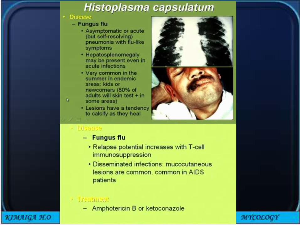
Histoplasmosis
• Causative agents – histoplasma capsulatum• 2 varieties
A. Histoplasma capsulatum capsulatum• A saprophyte which grows in the soil – is found in
birds' and bats' droppings• Spelunking (cave exploration)disease or cleaning
chicken coops especially chicken farmers• A disease which affects people in all parts of the
world – most highly in some parts of America.• Primary infection – remains asymptomatic in the
majority or manifests with non specific mild symptoms – flu-like

• Minority develop the classical illness with variation in severity and manifestations – as chronic pulmonary disease characterized by progressive cavity formation to disseminated disease
• Reactivation of the asymptomatic infections may or may not occur after several years
• Opportunistic histoplasmosis occurs rarely
• A disseminated disease with multiple organ involvement – enlarged liver, spleen with or without anaemia
• The organism is demonstrated in infected parts as an intracellular parasite of RES within phagocytic cells

B. Histoplasma capsulatum duboisii
• Causative agent of African histoplasmosis
• Encountered in some countries in Central parts of Africa
• Clinical manifestation include ulcerative lesions and swellings which involve the:
• Skin, subcutaneous tissues and bones
• Lungs are not commonly involved
• Source and the route of not clearly defined

Hyphae with micronidia and tuberculatemacronidia

Small intracellular yeasts with narrow neck on bud, no capsuleCan get 30 or more Histoplasma cells in 1 hman cell,cog wheel

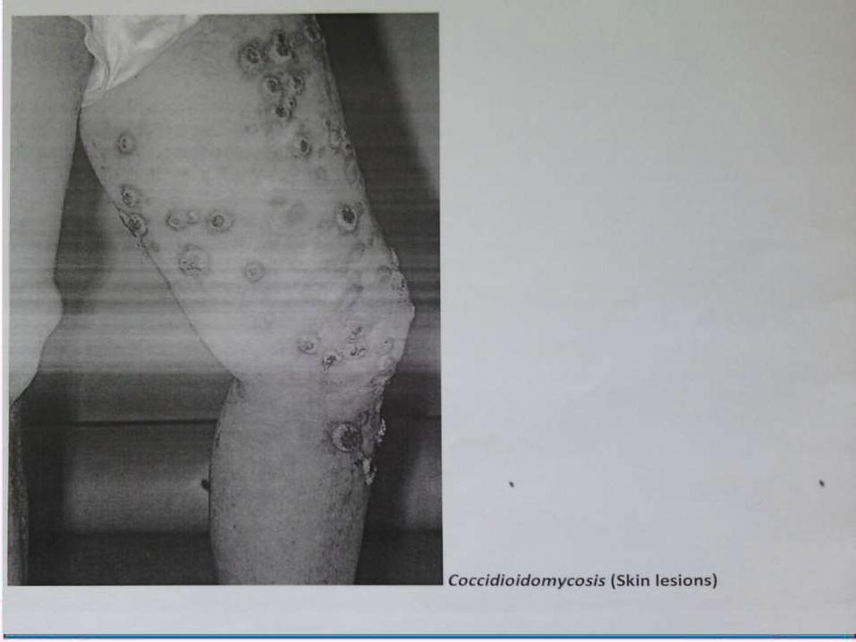
Coccidioidomycosis
• Coccidioides immitis• Desert valley fever• Arthroconidia breaks from hyphae and is found in desert
sand• Spores are inhaled – primary infections in the lungs• Spores develop into large structures in body
temperature– spherules – each spherule contains spores• Spherules is the yeast form• Can be asymptomatic or mild illness – mild pneumonia –
or severe illness in the minority associated with cavity formation in the lungs
• Dissemination occurs rarely - a military disease –Involves skin, meninges, bones, joints, subcutaneous tissue, i.e extra-pulmonary disease

Spherule formation with endospores


Blastomycosis(South American blastomycosis)• Blastomyces dermatitis• Endemic in parts of North America – also
detected in parts of Asia and Africa• Infection can – be asymptomatic
• Manifest as primary pulmonary disease• Disseminate to the skin producing chronic
swellings or ulceration on exposed parts
• Local spread – to bones & possibly internal organs
• Fungus observed in infected tissues as yeast cells - each cell produces a single bud
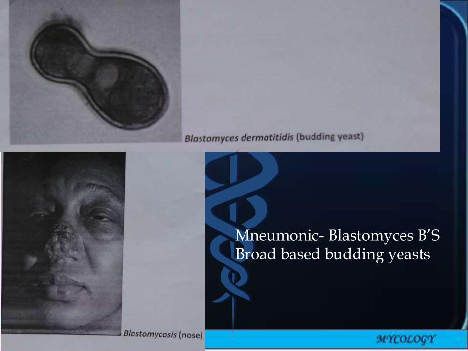
Mneumonic- Blastomyces B’SBroad based budding yeasts

Paracoccidioidomycosis(South American blastomycosis)• Infection is possibly acquired by inhalation
or by direct implantation on the oral or nasal mucous membranes
• Spreads via lymphatic and hemotogenousroutes
• Manifestation include ulceration of the mouth and nose with extension to local lymph nodes
• Recognized in infected tissues as large yeast cells with multiple budding

LAB DIAGNOSIS OF PATHOGENIC SYSTEMIC
MYCOSES• Higher risk specimens therefore examined
in labs with proper equipments and other necessary facilities
• Examination of specimens – sputum, discharges, skin scrapings, pus, infected tissue, blood for serology
• Demonstration of the yeast form of causative fungus – by direct microscopy or staining and microscopy

• Culture – on Sabouraud's medium for isolation and identifications – demonstration of dimorphism
• Serological tests in - classical histoplasmosis, coccidioidomycosis and paracoccidioidomycosis
• Associated with difficulties in interpretation of results
• The significance of antibody levels in differentiation of the illness and past exposure

INFECTIONS DUE TO OPPORTUNISTIC FUNGI
• Causative organisms are of low virulence• Disease production depends on diminished host
resistance to infections• Genera examples
• Candida- Candidiasis• Cryptococcus - Cryptococcosis• Aspergillus- Aspergillosis
• Other • Penicillin - Penicilliosis• Pneumocystis jirovecii• Species of penicillium• Mucor, Rhizopus, Absidia (Zygomycophyta)

Candida
• Small ovoid cells that reproduce by budding
• Thin walled dimorphic fungus
• Unicellular yeast- sexual form (harmless)
• Filamentous – asexual form (pathogenic)
• Can form hyphae in poorly aerated media, body
• Cell wall – polysaccharide, mannan, glucan & chitin, alone or complexed with proteins
• Fibrillar outer layer (mannan, mannoprotein)

• Over 150 spp. 9 commonly pathogenic to humans
• C. albicans – most common
• C. glabrata, C. tropicalis, C. pseudotropicalis, C. krusei, C. parapsilosis, C. dubliniensis, C. lusitaniae, C. gluilliermondii,

Epidemiology
• All of the above yeasts are considered to be normal flora for humans
• They usually inhabit oral, intestinal, and urogenital mucous membranes
• In healthy individuals, candida infections are usually due to impaired epithelial barrier functions, remain superficial and respond to rx
• Systemic candidiasis is usually seen in pts with cell mediated immune deficiency, and those receiving aggressive cancer rx, immunosuppressives, or transplantation therapy.

Predisposing factors
• Broad spectrum antibiotics – suppress normal bacterial flora – proliferation
• Chemotherapy
• IV catheters/ prosthetic implants/ GI tract surgery – provide route of entry
• Immunosuppressive conditions – HIV, steroids, pregnancy e.t.c
• Diabetes, oral contraceptives and oestrogentherapy
Virulense factors and pathogenesis
• Colonization of mucosa e.g female genital tract
• Surface molecules for adhesion to the mucosal cells:• Mannoproteins in outer fibrillar layers – adhesins
• Fibronectin and other components of extracellular matrix – receptors to mediate binding
• Ability to convert to hyphal forms• Associated with adherence and invasion – less
virulent spp do not form hyphae
• May be involved in the penetration of mucosal barriers and aids spread,

• Capacity to form strong attachments to epithelial cells
• Hyphal wall protein (Hwp1) – Found on surface of germ tubes and hyphae mediates binding
• Mannoproteins mediate binding to components of extracellular matrix(fibronectin, collagen, laminin)
• Extracellular enzymes – proteases, elastases, phospholipases (secreted by hyphae)
• Digest epithelial cells – facilitate invasiveness
• Aspartic proteinases-digest keratin and collagen enabling invasion of deep tissue
• Phenotypic switching

• Hyphae have surface proteins that resemble complement receptors on phagocytes- confuse ability of phagocytes to recognise C3b bound to candida surface
• Up regulation of receptors in conditions such as elevated glucose concentration hence resistance to phagocytes

• Colonization is 1st step in development of infections
• Kept under control by bacterial normal flora e.g lactobacilli
• Broad spectrum antibiotics
• Vaginal antiseptics and douching.
• Shift of C. albicans from yeast form to hyphal form associated with pathogenecity

• Appearance of virulence factors associated with adherence and invasion – less virulent spp do not form hyphae
• Hyphae may be involved in the penetration of mucosal barriers and aids spread,
• Extracellular enzymes – proteases, elestasesdigest epithelial cells – facilitate invasiveness
• Phenotypic switching

Clinical manifestations
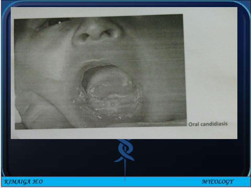
Mucocutaneous infection
• Oropharyngeal candidiasis• Infants, elderly, immunocompromised most commonly
• Perleche, Oral thrush• A common early presentation in AIDS
• 5 types• Acute pseudomembranous candidiasis
• Acute atrophic oral candidiasis
• Chronic erythematous candidiasis
• Chronic nodular candidiasis
• Chronic plaque like candidiasis/oral leukiplakia-MA LINES• May progress to oral carcinoma

• Cutaneous infections• Skin – axilla, groin, vulva, glans penis, inflammatory
folds, around umbilicus
• Napkin area in infants (diaper rash)
• Interdigital clefts, nails, skin folds around nails –hands and feet frequently in water.
• Systemic Infections• Neutropenia is a predisposing factor
• Endocarditis
• Urinary tract infections
• Disseminated infections
• GIT, pulmonary, carditis, meningitis, ocular, osteoarticular, candidemia, or disseminated candidiasis.


Vaginal candidiasis
• Normally C.albicans, also non-albicans species• Usually no underlying abnormality• No ↑ in frequency in severely
immunocompromised females, but patients with AIDS have persistent infections
• Common - pregnancy, diabetes• Acute (pseudomembranous or erythematous)
form more common• Chronic relapsing or persistent form• Secondary vaginal candidiasis - in underlying
mucosal disease

Chronic mucocutaneouscandidiasis (CMC)
• In patients with T-lymphocyte immunodeficiencies – chronic, relapsing
• In childhood or infancy - oral, nail & cutaneous candidiasis which recur despite treatment
• Oral-chronic pseudomembranous or plaque types
• Skin- crusted plaques
• Nail plates, folds & periungual skin damaged
• Defects unknown
Lab diagnosis of candida
• Specimen – exudates or epithelial scrapping, aspirates, biopsy, blood.
• Microscopic examination• KOH and/or gram stain of exudates and tissue – budding yeast
and pseudohyphae
• Culture• On Sabouraud Dextrose Agar
• 370C , 12-24 hrs• White colonies with a yeast smell.
• Germ tube tests (+ve) .Presumptive diagnosis of C. albicans. Form germ tubes at 370C
• Multiple samples may be necessary for proper interpretation of results
• Freshly collected specimens are more useful especially where quantitative cultures are required for confirmation of the diagnosis

• Blood cultures• For detection of candidaemia and endocarditis
• Species identification;• Carbohydrate assimilation and fermentation e.g
API (analytical profile index) system
• Pseudohyphea & chlamydospore production in nutritionally poor media e.g cornmeal agar
• Chrom Agar - Media gives different color changes for different fungal species → C.albicans - green, C.tropicalis - blue, C.gatata -pink e.t.c
• PCR
•Susceptibility testing

C.albicans G.stain

Candida albicans on SDA

Germ tube test (candida albicans)


Treatment• Amphotericin B – Drug of choice, IV infusion• More beneficial when used in combination
with 5-fluorocytosine (5-FC)• Synergistic effects
• lower dose of Amphotericin B – less toxic effects• delay in development of resistance to 5-FC
• Other alternatives: fluconazole, itraconazole• Relatively new antifungal agents –
voriconazole, caspofungin.• Rx followed by long term suppressive therapy
in AIDs pts – fluconazole• No specific preventive measures.

• Non albican candida – less susceptible to most antifungal agents, particularly the azoles
• C. kreusei – intrinsic resistance to fluconazole
• C. lusitaniae – usually resistance to Amphotericin B.
• Superficial – reduce moisture and chronic trauma – topical rx – nystatin or azole preparations.
• Chronic mucocutaneous infections –fluconazole
• Deeper infections – control of predisposing factors may resolve infections

Cryptococcosis
• Causative agent – crytococcocus neoformans• Yeast with a polysaccharide capsule• Reproduces by budding• Round/ovoid; 4-6 um diameter• C. neoformans-major pathogenic spp• Subclassified into 4 serotypes and 2 variants
• Serotypes based on capsular agglutination rxns –types A, B, C, D
• A & D – worldwide; B & C – tropics and subtropics. • A-variety glubii• D-var neoformans• B+C-var gatii
Epidemiology• Environmental – Detected and isolated mainly from
pigeon droppings and nesting places, other birds droppings and soil contaminated with the droppings
• Also been cultured from eucalyptus trees• Initial infection is acquired through inhalation of dust
contaminated with the yeast• Primary infection in most cases occurs in the lungs &
remains sub-clinical• Tendency to flare-up after several years as a
complication of other conditions particularly those that cause suppression of immunity
• Manifestation – meningitis commonest• Uncommon – cutaneous, mucocutaneous, bone
infections, disseminated infections

Pathogenesis
• Transmission is by inhalation of yeast from an environmental source
• Yeast is engulfed by lung macrophages and retained in them
• Circulates to other body parts in the macrophages, particular attraction to CNS, where they lodge and multiply
• Appear in CSF and blood
• Extreme immunosuppression – can cause disease in other organs.

Risk factors
• More common in pts with defects in T cell mediated immunity
• HIV/AIDS
• Post-transplantation
• Prolonged corticosteroids therapy,
• Hodgkin's disease
• Malignancies
• Sarcoidosis

Clinical manifestation
• Pulmonary cryptococcosis – usually asymptomatic
• CNS diseases-meningitis(85%) common in AIDS,meningoencephalitis,cryptococcoma
• Cutaneous cryptococcosis
• Bone and joint dx
• Ocular cryptococcosis
• Genital urinary dx-prostate
Lab diagnosis
• Specimen – CSF, blood, sputum, biopsy of infected tissues
• Procedures• Direct microscopy of CSF, homogenates of tissue
mounted in India ink for yeast cells with a capsule• CSF – raised pressure, increased protein, reduced
glucose, increase in WCC (increased lymphocytes)
• India ink or nigrosin staining of CSF deposits.• Latex agglutination tests (cryptococcal antigen-CRA
6 test) on CSF or serum. Detection of capsular antigen
• Rapid test• Detects polysaccharide capsular antigen

• Culture of CSF or other specimens on suboraud dextrose agar (SDA) for isolation and identification tests, 370C, in air, may require. 48Hrs – soft, creamy, mucoid, brown colonies
• Selective media – Birdseed agar – melanin production (brown colonies)
• Gram stain: gram positive yeast cells
• Biochemical tests:
• Urease test positive
• CHO assimilation and fermentations tests e.gAPI

C. neoformans - Gram positive

Cryptococcus neoforman on SDA

Cryptococcus neoformans using a light India ink staining preparation
Cryptococcus neoforman- Yeast with capsule

Treatment
• Amphotericin B – Drug of choice, IV infusion
• More beneficial when used in combination with 5-fluorocytosine (5-FC)
• Synergistic effects
• Lower dose of Amphotericin B – less toxic effects
• Delay in development of resistance to 5-FC
• After patient is afebrile and culture negative use fluconazole.

Aspergillosis
• A group of diseases in which the fungi belonging to the genus aspergillus are involved
• May or may not be opportunistic• Genus aspergillus consists of over 800 species –
species commonly involved in causation of disease in humans• A. fumigatus – associated with majority• A. flavus
• Fungus aspergillus and the spores are found in the environment; soil, decaying vegetation, organic debris, construction and demolition sites
• Increased chances of contamination and inhalation of airborne spores
• Aspergillosis can manifest as a disease of the respiratory system or other systems
• In the respiratory system• Basically three categories of disease• Allergic bronchopulmonary aspergillosis –
extrinsic asthma – allergic alveolitis due to hypersensitivity to the spores. Common in farmers that inhale mould in hay
• Colonizing aspergillosis – colonization of the pre-existing cavity - post-pulmonary TB in which the fungus grows as a mycelial mass – fungus ball or aspergilloma
• Invasive disease – encountered in patients with chronic debilitating illness – characterized by invasion and destruction of lung tissues and blood vessels.-Pneumonia, meningitis, Cellulitis
• Majority caused by A. fumigatus

• Other forms of aspergillosis
• Aspergilloma in the air sinuses – associated with maxillary sinus
• Cutaneous aspergillosis – involvement of external ear (otomycosis) – commonly caused by A. niger
• Mycetoma – rare – A. nidulans.

Lab confirmation of aspergillosis
• Specimen – sputum, infected tissue
• Direct microscopy examination
• Culture and identifications
• Sabouraud's medium and colonial features
• Various tests to identify the species
• To be differentiated from species not associated with disease causation
• Detection of antibodies – in the diagnosis of aspergilloma. Serologic test for circulating cell wall galactomannan antigen in serum.

Aspergillus spp culture
• Macroscopic morphology• SDA colony texture
• Fast growing
• Powdery
• Colony color• Front: may vary depending
on spp - white, yellow, yellow-brown, brown to black, shades of green
• Reverse: uncolored to pale yellow in most isolates; may be purple to olive
• Macroscopic morphology
• Lactophenol cotton blue stain
• Conidopheres:• Terminate in a vesicle
covered with either....
• Conidia:• One celled, smooth or
rough walled
• Hyaline or pigmented
• Basocatenate, forming long dry chains which may be: divergent(radiate) or in compact columns (columnar)


Aspergillus Aspergillus fumigatus

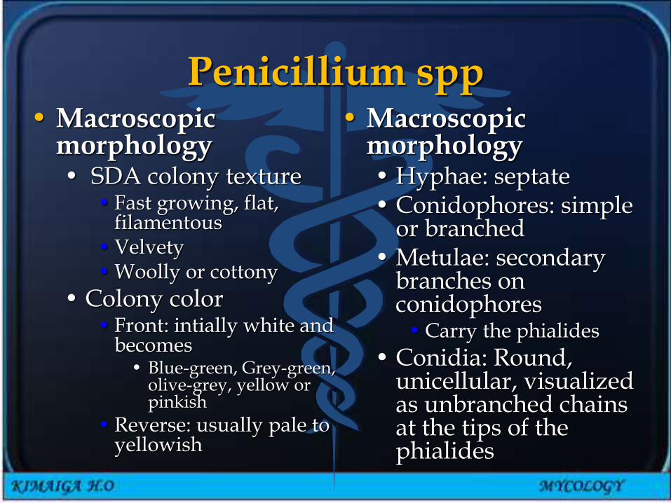
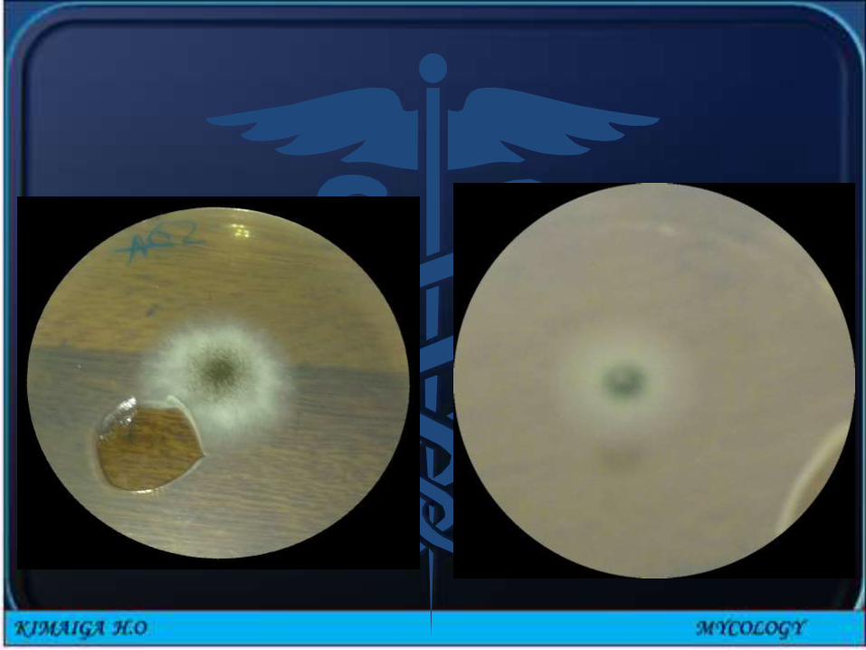
Penicillium spp• Macroscopic
morphology• SDA colony texture
• Fast growing, flat, filamentous
• Velvety• Woolly or cottony
• Colony color• Front: intially white and
becomes• Blue-green, Grey-green,
olive-grey, yellow or pinkish
• Reverse: usually pale to yellowish
• Macroscopic morphology• Hyphae: septate• Conidophores: simple
or branched• Metulae: secondary
branches on conidophores
• Carry the phialides
• Conidia: Round, unicellular, visualized as unbranched chains at the tips of the phialides


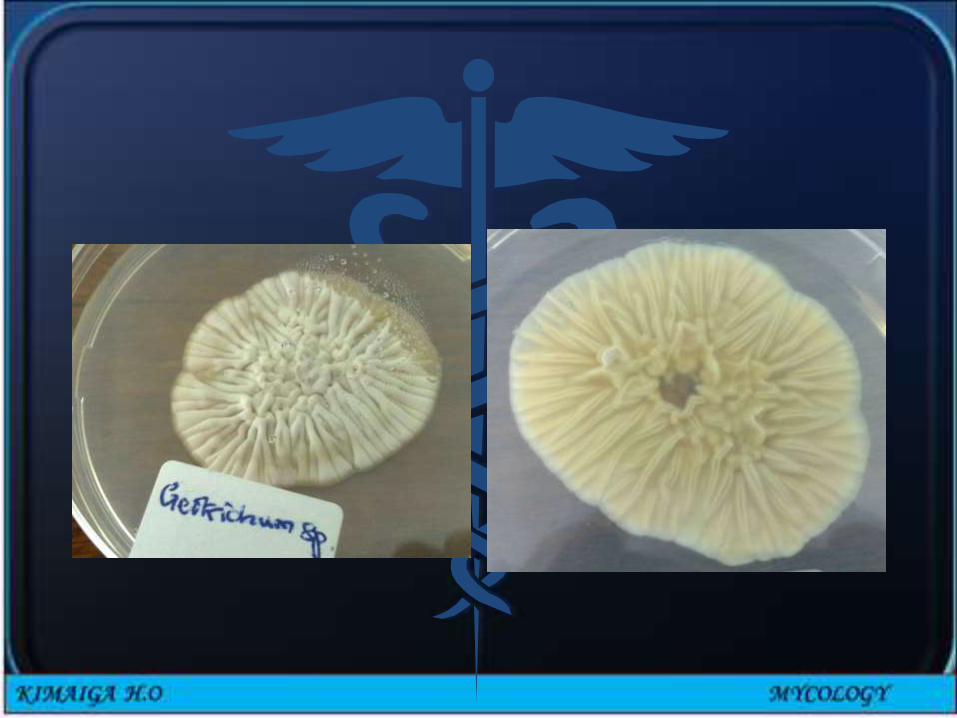
Geotrichum spp• Macroscopic
morphology• SDA colony texture
• Fast growing
• Dry
• Finely suede-like
• Colony color• Front: white to cream
• Reverse: no pigment
• Macroscopic morphology• Lactophenol cotton
blue (LCB) stain
• Hyphae
• Hyaline, septate, branched
• Break up into chains of• Hyaline, smooth, one
celled, subglobose to cylindrical → arthroconidia: release by the separation of a double septum
• Balctoconidiaproduction: not found in this genus

Aspects of management of systemic mycoses
• Management of predisposing conditions where applicable
• Antifungal agents
• Amphotericin B
• Others – itraconazole, fluconazole, ketoconazole
• Other measures - where applicable & surgical procedures