Systemic lupus erythematosus (SLE)...Systemic lupus erythematosus (SLE) - is a chronic, autoimmune...
136
Transcript of Systemic lupus erythematosus (SLE)...Systemic lupus erythematosus (SLE) - is a chronic, autoimmune...


Systemic lupus erythematosus (SLE) - is a
chronic, autoimmune inflammatory disease
with unknown etiology, disorder
characterized by an autoantibody response
to nuclear and cytoplasmic antigens,in
presence of genetic predisposition, and can
affect any organ and system.
SLE can affect any organ or system -
joints, skin, vessels, and various types
of organ-related disorders.

Epidemiology
SLE is spread worldwide,
Incidence rate - 1 case per 10,000 population
(variations as 1.8-7.6 per 100 thousand) per year
Morbidity / prevalence rate is about 500 patients
per 1,000,000 population (variations as 12 -50 cases /
100 000 population
Disease morbidity vary among various geographic
areas
The incidence rate varies between 12-50 to 100,000
people, the highest being among African Americans,
Asians, African-Caribbean’s and Hispanics
The female/male ratio is 3: 1 in children, which is
increasing to 7-15: 1 in adults.

The survival rate of patients whit 10-
years SLE now exceeds 90%. Prior to
1955, the 5-year survival rate was
less than 50%.

Race and ethnicityThe frequency of SLE rate varies
between 12-50 to 100,000 people, the
highest being among African Americans,
Asians, African-Caribbean’s and Hispanics.
The incidence of SLE in black women is
approximately 4 times higher than in
caucazian women.
SLE is more frequent in Asian women
than in white women.
The disease is rarely reported among
blacks who live in Africa.

The prevalence of SLE is highest among
women in reproductive period, bat the
age limits varied from 14 to 64 years.
For all ages, the female-to-male ratio
is 8:1 and 10:1 during the childbearing
years.
The risk of SLE development in men is similar
to that in prepubertal or postmenopausal
women.
SLE does not have an age predilection in males.
Although the specific cause of SLE is
unknown, multiple genetic predispositions
and gene-environment interactions have
been identified. This complex situation
perhaps explains the variable clinical
manifestations in persons with SLE

Multiple factors are associated with
the development of the disease,
including –
genetic,
racial,
hormonal,
and environmental factors.
Many immune disturbances, both
innate and acquired, occur in SLE.

Numerous studies have investigated the role of infectious etiologies.
Viruses may stimulate specific cells in the immune network.
Different viruses are suspected, due to the presence of antiviral antibodies (anti-ARN şi anti-ADN) including to protein regions homologous to nuclear antigens.
Epstein-Barr virus (EBV) – may perpetuate autoimmunity. Patients with SLE have higher titers of antibodies to EBV, have increased circulating EBV viral loads.
Anothers viruses are suspected: rubella, rubeola, citomegalovirus, retroviruses, but the attempts to isolate the virus from the tissues of patients with SLE fails.

Bacterial infection
Certains bacterias ( ex. Mycobacterial infections ) may induce anti-DNA antibodies (or another antibodies) even lupuslike symptoms, and lupus flares may follow bacterial infections.

A genetic predisposition is supported by the 25% concordance among monozygotic twins versus 2% in dizygotic twins. In monozygotic twins the Morbidity is
approximately 10 times higher than in dizygotic.
The immune damages in family of patients with SLE are frequent.
Detection of antinuclear, antilimfocitaryantibody and false positive test for lues at the I degree relatives.
It also proven that in the SLE patients families the incidence of other systemic connective tissue diseases is higher.

Studies of the human leukocyte antigens (HLA) reveal that HLA-A1, B8, HLA-DR2 and DR3 are more common in persons with SLE than in the general population.
the presence of antinuclear antibody Is in correlation whit HLA A2, B7 şi DR3
More than 10 gene loci are known to increase the risk of SLE.

Congenital deficiencies of complement - C1,C2,C4,C5,C8 (especially C4, C2) are also associated with an increased risk of SLE.
Complement deficiency – C2,
C3 şi C4 at the I degree relatives.

Endocrine – related factors
1. The negative influence of estrogens in the evolution of SLE (change for the worse during the pregnancy and in the post-partum period).
2. Breastfeeding is associated with a decreased risk of developing SLE.
3. SLE frequently starts at women of childbearing age, and the use of exogenous hormones has been associated with lupus onset and flares, suggesting a role for hormonal factors in the pathogenesis of the disease.
4. The protective role of androgens in SLE.

UV raysPhotosensitivity is clearly a precipitant of
skin disease. UV stimulate cells apoptosis and production of autoantigens , induce autoimune process in persones with genetics predispozition. UV radiation stimulate production of
IL-1, favorize the immune reaction
vitamin D levels
The results of some study suggest that low
vitamin D levels increase autoantibody
production in healthy individuals;

Antibacterial- Izoniazid, certain antibiotics;
Cardiovascular drugs – Procainamid,Hidralazin, Metildopa;
Antitiroid drugs- Metiltiouracil;
Contraceptives
The drugs are more likely to aggravate the evolution of pre - existent SLE

Pathophysiology
[CLOSE WINDOW]
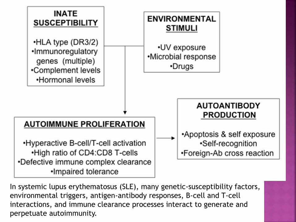
In systemic lupus erythematosus (SLE), many genetic-susceptibility factors,
environmental triggers, antigen-antibody responses, B-cell and T-cell
interactions, and immune clearance processes interact to generate and
perpetuate autoimmunity.

I.Immunological factorAt an early stage the polyclonal (B -
cell) immune activation dominates,
later on - an antigen-specific T-
cell immune reaction prevails.
II. Imparied apoptosisFundamental immune disorder underlying
lupus erythematosus - congenital or
induced defects programmed cell
death (apoptosis)

The specific cause of SLE is unknown, immune-system disorders and immune-complex tissue damage are suspected. Multiple immune disturbances may predispose to SLE :
Dysfunction of T- and B – lymphocytes (depression of T- suppressors, hiperreactivity of B lymphocytes , amplification of B lymphocytes function and in consequence - antibodies hyper production against intracellular components (ADN, nucleoproteins ribonucleoproteins ) and CIC formation .

Th lymphocytes increases, they become more active,
Thereupon occurs Th / Ts imbalance for Th,
Th cooperates with B-lymphocytes,
B-cell activation results in excessive auto -antibody production to a variety of antigens (antinuclear, anti-cytoplasmic, anti-DNA double stranded and single-stranded,
anti-RNA anti-plasma membrane) and hypergammaglobulinaemia .

anti-double-stranded DNA,
anti-Sm,
antiRNP (anti riboproteinic)
anti-nucleoproteins,
anti-Ro.
Antibodies are immunoglobulins
IgG and IgM class

Serum antinuclear antibodies
(ANAs) are found in virtually all
individuals with active SLE,
and antibodies to native
double-stranded DNA (dsDNA)
are relatively specific for the
diagnosis of SLE.
Many of the clinical manifestations of
SLE are caused by the effects of circulating immune complexes.
Antibodies are linked to antigens, forming circulating immune
complexes (CIC) -(DNA + anti-
DNA), with complement activation and
inflammation.

Antibody-antigen complexes deposit on the different tissues (ex. basement membranes of skin and kidneys, serous membranes, visceral organs) cause vaculities and various clinical manifestations.
The effects of circulating immune complexes on
various tissues or the direct effects of antibodies to
cell surface components determine:
1. complement activation2.Inflammatory reaction development,
3. neutrophyls migration
4. cytokines and other lesion substances release.

In active SLE,
this process
has been
confirmed by
demonstratio
n of
complexes of
nuclear
antigens such
as DNA,
immunoglobu
lins, and
complement
proteins at
these sites.

The inflammatory reaction are
associated with
producing proinflammatory
cytokines (Il – 1, Il -2) – with
chemotactic and vasoactive action
for neutrophyls and
proinflammatory lizozomale
enzymes.

One proposed mechanism for the development of
autoantibodies involves a defect in apoptosis that causes increased cell death and a disturbance in immune tolerance.
Recent evidence suggests that initially there is increased apoptosis of lymphoid cells. Nucleosomes (i.e. the DNA histone chromatin constituents) are released from these cells and are taken up by antigen-presenting cells via nucleosome receptors. They are presented to T cells that stimulate B cells to produce autoantibodies directed against these nucleosomes and their constituents (e.g. DNA).Different subsets of autoantibodies may be responsible for the various clinical patterns.
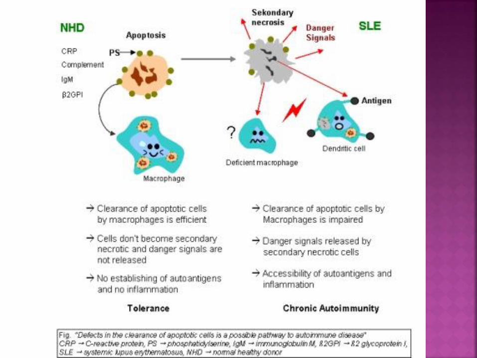


The redistribution of cellular
antigens during apoptosis leads
to a cell-surface display of
plasma and nuclear antigens in
the form of nucleosomes.
Thus, dysregulated (intolerant)
lymphocytes begin targeting
normally protected
intracellular antigens.

I. Depending on the evolution:
1.Acute,
2.Subacute,
3.Cronic.
II. Depending on the disease activity :
1.Minimal.
2.Moderate.
3.Maximal.
(Nasonova V.A.,1986.)

Systemic lupus erythematosus (SLE) is a chronic autoimmune disease that can affect almost any organ system.
SLE is characterized by: exacerbations and remissions (sometimes spontaneous).
Its onset, presentation and course are highly variable, ranging from indolent to fulminant.

The triad of fever, joint pain, and rash in a woman of childbearing age should suggest the diagnosis of SLE. This is one of the most common presentations of SLE. However, patients may present with any of the following types of manifestations:
Constitutional
Dermatologic
Musculoskeletal
Pulmonary
Cardiac
Renal
Neuropsychiatric
Gastrointestinal
Hematologic
In patients with suggestive clinical findings, a family history of autoimmune disease should raise further suspicion of SLE.


Nonspecific fatigue, fever, weight changes arthralgia, are the most common symptoms in new cases or
recurrent active SLE flares.
Constitutional symptoms associated with SLE, generally occurs in concert with other clinical and laboratory markers.

20-25% of patients with lupus have skin damage already disease onset, 60-70% - in different evolution stage, in 10-15% -cutaneous manifestations are missing
Cutaneous manifestations of SLE are very variable, comprise 4 diagnostic criteria and multiple other clues to a potential diagnosis of lupus.

Malar rash which is characterized by
an erythematous rash
(dermatitis) over the
cheeks and nasal bridge
who remember a
butterfly.
It lasts from days to
weeks and is occasionally
painful or pruritic.


The classic malar rash, also known as a butterfly rash, with
distribution over the cheeks and nasal bridge. Note that the fixed
erythema, sometimes with mild induration as seen here,
characteristically spares the nasolabial folds.


SLE is characterized by annular
erythematous and infiltrated
formations with moderate
atrophy and teleangiectasia
elements, with subsequent
formation of hypo pigmented or
hyper pigmented scars.


Photosensitivity which may be elicited from patients who are asked if they have any unusual rash or symptom exacerbation after sun exposure.

Photosensitive rash is often macular or diffusely
erythematous in sun-exposed areas of the face, arms, or
hands, as in the image below.

less-specific
cutaneous feature of
SLE. It often affects
the temporal regions
diffuse or creates a
patchlike pattern of
hair loss.

Discoid lesions often
also develop in sun-
exposed areas but are
plaquelike in
character, with
follicular plugging and
scarring. They may be
part of systemic lupus
or may represent
discoid lupus without
organ involvement,
which is a separate
diagnostic entity.


Discoid lesions occurs on sun-exposed body
parts: face, arms, neck, associated
themselves in systemic lupus
erythematosus or discoid lupus may be as
a separate entity.

related to but not specific to SLE
include Raynaud phenomenon,
livedo reticularis, panniculitis (lupus
profundus), bullous lesions,
vasculitic purpura, telangiectasias,
and urticaria

Livedo reticularis is
characterized by a
lacy, mottled,
erythematous skin
pattern

may be observed with
blue, white, and red
color change at the
distal digital tips.

It is characterized
by erythema and
discoid changes,
atrophy and
depigmentation on
the lips (cheilitis),
petechiae and
mouth ulcers.


Joint pain is one of the most common
reasons for the initial clinical
presentation in patients with SLE (90%).
Arthralgia, myalgia, and frank arthritis
may involve the small joints of the
hands, wrists, and knees.
In contrast to rheumatoid arthritis, SLE
arthritis are non-erosive,
nondeformant, with small effusion, may
be asymmetrical, with pain that is
disproportionate to swelling.

Most frequently are affected interphalangeal joints of hands,indecreasing:
- metacarpophalangeal,
- Radiocarpal
- knees and joints of the foot
- rare plants.- Femural joint necrosis may occurs in patients
with antiphospholipid syndrome or after prolonged treatment with corticosteroids.Osteoporosis is a common complication of treat-ment with GCS

Myalgia are common (15-64%), rarely meet
myiositis (5-11%), manifested by proximal
muscle fatigue and increased concentration
of enzymes (creatine fosfokinaze).
In 5-10% of patients treated with steroid
develops GCS myopathy.

Pulmonary manifestations of SLE may manifest acutely or indolently, representing many complications.
Pleurisy with pleuritic chest pain with or without pleural effusion is the most common feature of acute pulmonary involvement in SLE, often are bilateral.
Shortness of breath or dyspnea may be due to many causes : pleural or pericardial effusions, pulmonary embolism, lupus pneumonitis, interstitial lesions.

Serositis due to pericardial or pulmonary
effusions, pulmonary embolism, lupus
pneumonitis, chronic lupus interstitial lung
disease, or infection may be related to lupus
disease.
Most seriously, hemoptysis may herald
diffuse alveolar hemorrhage, a rare, acute,
life-threatening pulmonary complication of
SLE.
Lung damage in SLE is a manifestation of
pulmonary vasculitis.

is characterized by dyspnea and cough with
haemoptysis. X-ray detects increased pulmonary
draw, infiltrates, vital capacity of lungs is reduced .

Heart failure or chest pain must be
carefully examined in patients with
SLE.
All heart compartments may be
involved - pericardium, myocardium,
endocardium, valves and coronary
arteries.(in 25%).

Pericarditis , the most frequent
cardiovascular involvement, that manifests
as chest pain, manifesting as positional chest
pain that is often relieved when the patient
leans forward. Is presented by small
pericardial effusion detected during EcoCG.
After resorption of effusion between
pericardium surfaces light fibrous adhesions
is formed.

A maild myocarditis also may occurs in SLE,
giving rise to arrhythmias, with heart failure
symptomatology.
Coronary vasculitis manifesting as angina or
infarction is rarely reported. The
Framingham Offspring Study demonstrated
that women aged 35-44 years with SLE
were 50 times more likely to develop
myocardial ischemia than healthy women.

Libman – Sacks endocarditis is noninfectious but may manifest as symptoms
similar to those of infectious endocarditis .
Very rare involvement of MV and AoV.
Raynaud ‘s vasculitis arterial and venous
thromboses can occur.

The exudate in the pericardium is pale-
yellow, sometimes contains streaks of blood,
more than 20,000 leukocytes / mm 3 (mainly
polynuclear), antinuclear antibodies (ANA),
circulating immune complexes (CIC), LE-
cells, the is low-complement level .

Impairment of the peripheral vessels
occurs by livedo reticularis, Lupus-
panniculitis and thrombosis of arteries or
veins of the limbs or internal organs,
which are usually found in patients with
antifisfolipidic syndrome

The kidney is the most commonly
involved visceral organ in SLE.
Although only approximately 50%
of patients with SLE develop
clinically evident renal disease,
biopsy studies demonstrate some
degree of renal involvement in
almost all patients.

Glomerular disease usually develops within
the first few years of SLE onset and is
usually asymptomatic.
Most type of glomerulonephritis occur,
including mesangial, focal, diffuse and
membranous.

The most common symptom is proteinuria
(1g.per 24 h), associated with hematuria and
cilindruria. Proliferative glomerulonephritis is
detected (the focal or diffuse) or membrouse,
which is manifested by nephrotic syndrome.
Acute or chronic renal failure may cause
symptoms related to uremia and fluid overload.
Acute nephritic disease may manifest as
hypertension and hematuria.
Nefrotic syndrome may cause edema,
weight gain, or hyperlipidemia.

In glomeruli are detected deposits of IgG, C3
complement fraction, fibrin, IgM and less -
IgA.
The electronic microscopy is proving
subendotelial, subepitelial and
intramembranouse immune complex
deposits, these deposits are markers of lupus
nephritis.

Gastrointestinal symptoms secondary to
primary SLE and adverse effects of
medication are common among persons with
SLE, bat not major presenting feature.
Abdominal pain in SLE is significant
because it may be directly related to active
lupus, including peritonitis, pancreatitis,
mesenteric vasculitis, and bowel infarction.

Nausea and dyspepsia are common symptoms in
patients with active SLE and are sometimes
difficult to correlate with objective evidence of
gastrointestinal involvement.
Esophagus damage- dysphagia, reducing
peristalsis, dilation of the esophagus (5%).
Duodenum and stomach ulcers - caused both by
the basic disease and the adverse effects of
treatment
Jaundice due to autoimmune hepatitis may also
occur.
Pancreatitis is uncommon.
Lymphadenopaty and splenomegalia.
Involvement of the nervous system occurs in up to 60% of cases and symptoms may fluctuate.
There may be a mild depression but occasionally more severe psychiatric disturbances occur. Epilepsy, cerebral ataxia, aseptic meningitis , cranial nerve lesion, cerebrovascular accidents or a polyneuropathy may be seen.
Only seizure and psychosis are included among the diagnostic criteria.
Central nervous system is the result of cerebral vasculitisand CIC deposition (10-15% of cases) – Antineuronal, antilimphocytic, antiglial antibodies are detected.

Psychosis may manifest as paranoia or
hallucinations.
Delirium represents a spectrum of
fluctuating altered consciousness
characteristic of SLE. Delirium may be
caused by CNS vasculitis, encephalopathy,
or the manifestations previously called
organic brain syndrome.

Seizures related to SLE may be generalized
or partial and may precipitate status
epilepticus.
Coreea,aseptic meningitis, myelopathy, optic
neuropathy, or other demyelinating disorders
may also require urgent evaluation.
Transverse myelitis with spastic paraparesis
is a rare but serious complication of SLE.

Migraine headaches may also be
linked to antiphospholipid syndrome,
although this is less clear.
Headache and mood disorders may
be the most commonly reported
neurologic manifestation of SLE, but
cause and effect may be difficult to
distinguish.

Cognitive disorders may be variably apparent in patients with
SLE. Formal neuropsychiatric testing
reveals deficits in 21-67% of patients with
SLE.
Whether this represents true
encephalopathy, neurological damage,
medication effects, depression, or some
other process is unclear.

Stroke and transient ischemic attack (TIA)
may be related to antiphospholipid
antibody syndrome or vasculitis.
It can diagnose polineurite and
mononeurite, peripheral sensory
neuropathy, aseptic meningitis

A history of multiple cytopenias such as
anemia,leukopenia, lymphopenia,, or
thrombocytopenia may suggest SLE,
among other etiologies.
Leukopenia and, more specifically,
lymphopenia are common in SLE; this and
hypocomplementemia may predispose
persons with SLE to frequent infections.

Anemia is occasionally overlooked in
young menstruating women. Hypochromic
anemia (50%) - its strongly correlated
with SLE activity.
Hemolytic anemia with positive Coombs
test is rarely encountered in patients with
antiphospholipid syndrome.

Leukopenia - autoimmune origin .
Lymphopenia correlates with SLE activity
and antilymphocitic antibody.
Thrombocytopenia may be mild or part
of a thrombotic thrombocytopenic
purpura (TTP)–like syndrome or
antiphospholipid antibody syndrome.

Thrombocytopenia may be mild or part of a
thrombotic thrombocytopenic purpura (TTP)–
like syndrome or antiphospholipid antibody
syndrome.
History of recurrent early miscarriages or a
single late pregnancy loss may be clues to
lupus or isolated antiphospholipid antibody
syndrome.
A family history of autoimmune disease
should also raise further suspicion of SLE.

( ACR, 1997).

1. Malar rash: Fixed erythema, flat or raised, over the malar eminences
2. Discoid rash: Erythematous circular raised patches with adherent keratotic scaling and follicular plugging; atrophic scarring may occur
3. Photosensitivity: Exposure to ultraviolet light causes rash
4. Oral ulcers: Includes oral and nasopharyngeal ulcers, observed by physician
5. Arthritis: Nonerosive arthritis of two or more peripheral joints, with tenderness, swelling, or effusion
6. Serositis: Pleuritis or pericarditis documented by ECG or rub or evidence of effusion
7. Renal disorder: Proteinuria >0.5 g/d or 3+, or cellular casts
8. Neurologic disorder: Seizures or psychosis without other causes
Classification Criteria for the Diagnosis of
Systemic Lupus Erythematosus (SLE) (1)

9. Hematologic disorder: Hemolytic anemia or leukopenia(<4000/L) or lymphopenia (<1500/L) or thrombocytopenia (<100,000/L) in the absence of offending drugs
10. Immunologic disorder: Anti-dsDNA, anti-Sm, and/or anti-phospholipid
11. Antinuclear antibodies: An abnormal titer of ANA by immunofluorescence or an equivalent assay at any point in time in the absence of drugs known to induce ANAs
Any combination of 4 or more of 11 criteria, well-documented at any time during a patient's history, makes it likely that the patient has SLE (specificity and sensitivity are 95% and 75%, respectively).
Note: ECG, electrocardiography; dsDNA, double-stranded DNA; ANA, antinuclear antibodies.

SLEDAI SCORE

The SLEDAI (Systemic Lupus Erythematosus
Activity Index) was developed in 1985
through a nominal group process and is based
on the presence of 24 features in 9 organ
systems over the patient’s past 10 days.
The maximal score -105 points.

Wt SLEDAI
scoreDescriptor Definition
8 Seizure Recent onset. Exclude metabolic, infectious or drug
cause
8 Psychosis Altered ability to function in normal activity due to
severe disturbance in the perception of reality. Include
hallucinations, incoherence, marked loose associations,
impoverished thought content, marked illogical
thinking, bizarre, disorganized, or catatonic behavior.
Excluded uremia and drug causes.
8 Organic
Brain
Syndrome
Altered mental function with impaired orientation,
memory or other intelligent function, with rapid onset
fluctuating clinical features. Include clouding of
consciousness with reduced capacity to focus, and
inability to sustain attention to environment, plus at
least two of the following: perceptual disturbance,
incoherent speech, insomnia or daytime
drowsiness, or increased or decreased psychomotor
activity. Exclude metabolic, infectious or drug causes.

Wt SLEDAI
scoreDescriptor Definition
8 Visual
Distur -
bance
Retinal changes of SLE. Include cytoid
bodies, retinal hemorrhages,
serious exodate or hemorrhages in the
choroids, or optic neuritis. Exclude
hypertension, infection, or drug causes.
8 Cranial
Nerve
Disorder
New onset of sensory or motor neuropathy
involving cranial nerves.
Headache Severe persistent headache: may be
migrainous, but must be nonresponsive
to narcotic analgesia.
CVA New onset of cerebrovascular accident(s).
Exclude arteriosclerosis

Wt SLEDAI
scoreDescriptor Definition
8 Vasculitis Ulceration, gangrene, tender finger nodules,
periungual, infarction,splinter hemorrhages, or biopsy
or angiogram proof of vasculitis
4 Arthritis More than 2 joints with pain and signs of inflammation
(i.e. tenderness,swelling, or effusion).
4 Myositis Proximal muscle aching/weakness, associated with
elevated creatine
phosphokinase/adolase or electromyogram changes or
a biopsy showing
myositis.
4 Urinary
Casts
Heme-granular or red blood cell casts

Wt SLEDAI
scoreDescriptor Definition
4 Hematuria >5 red blood cells/high power field.
Exclude stone, infection or other
cause.
4 Proteinuri
a
>0.5 gm/24 hours. New onset or
recent increase of more than 0.5
gm/24 hours.
4 Pyuria >5 white blood cells/high power
field. Exclude infection.
2 New Rash New onset or recurrence of
inflammatory type rash.

Wt SLEDAI
scoreDescriptor Definition
2 Alopecia New onset or recurrence of abnormal, patchy or
diffuse loss of hair.
2 Mucosal Ulcers New onset or recurrence of oral or nasal
ulcerations
2 Pleurisy Pleuritic chest pain with pleural rub or effusion,
or pleural thickening.
2 Pericarditis Pericardial pain with at least 1 of the following:
rub, effusion, or electrocardiogram confirmation.
2 Low
Complement
Decrease in CH50, C3, or C4 below the lower
limit of normal for testing laboratory

Wt SLEDAI
scoreDescriptor Definition
2 Increased
DNA
binding
>25% binding by Farr assay or above
normal range for testing laboratory
1 Fever >38°C. Exclude infectious cause
1 Thrombocy
topenia
<100,000 platelets/mm3
1 Leukopenia <3,000 White blood cell/mm3. Exclude
drug causes
TOTAL SCORE (Sum of weights next to
descriptors marked present)
Check box: If descriptor is present at the
time of visit or in the proceeding 30 days

Mild or Moderate Flare □ Severe Flare □
□ Change in SLEDAI > 3 points □ Change in SLEDAI > 12
□ New/worse
discoid,photoscnsitive,
profundus,
cutaneous vasculitis, bullous lupus
Nasopharyngeal ulcers
Pleuritis
Pericarditis
Arthritis
Fever (SLE)
□ New/worse CNS-SLE
Vasculius
Nephritis
Myositis
Pk < 60.000
Home anemia: Hb <7% or decrease in Hb > 3%
Requiring: double prednisone
Prednisone>0.5 mg/kg/day hospitalization
Increase in Prednisone, but not to
>0.5 mg/kg/day
Prednisone >0.5 mg/kg/day
Added NSAID or Plaquenil New Cytoxan, Azathioprine, Methotrexate,
Hospitalization (SLE)
≥1.0 Increase in PGA, but not to
more than 2.5
□
Increase in PGA to > 2.5

Small-joint arthritis of the hands and wrists is
the most common musculoskeletal finding in SLE,
followed by arthritis of the knees. Pain reports
may be out of proportion to synovitis or swelling
upon examination.
Myositis that may manifest as weakness rarely
occurs and is more commonly related to overlap
syndromes or corticosteroid-induced myopathy.
Fibromyalgia, which should be distinguished by
myofascial tenderness without weakness, is
commonly concomitant with SLE.

Pleuropericardial friction rubs and signs of
effusions may be found.
Hypoxia, tachypnea, crackles, or gross
hemoptysis may be signs of pneumonitis.
Heart failure signs or arrhythmias may point
to ischemia or inflammatory myocarditis.
Murmurs may represent Libman-Sacks
endocarditis, superimposed infectious
endocarditis, or thromboembolic disease.

Renal findings
Hypertension.
Edema of periorbital or peripheral regions
and anasarca are common physical findings
related to nephrotic syndrome or volume
overload with renal failure.

Gastrointestinal findings
Abdominal tenderness and pain may be
observed in peritonitis, pancreatitis,
mesenteric vasculitis, or non–lupus-related
processes. Lupus peritonitis is a less-common
serositis that may be present, even in the
absence of ascites.

Neuropsychiatric findings
Focal neurological deficits may represent
stroke, or mononeuritis.
Mononeuritis may manifest as the functional
loss of one or a few isolated peripheral
nerves and is observed in some patients with
SLE vasculitis or antiphospholipid disease.

Screening laboratory studies to diagnose
possible SLE should include a CBC count with
differential, serum creatinine, urinalysis with
microscopy, ANA, and, perhaps, basic
inflammatory markers.
Laboratory
Studies

Inflammatory markers:
Levels of inflammatory markers, including
the erythrocyte sedimentation rate (ESR)
or C-reactive protein (CRP), may be
elevated in any inflammatory condition,
including SLE.
CRP levels change more acutely, and the ESR
lags behind disease changes.

A CBC count may help to screen for
anemia,
leukopenia,
lymphopenia,
and thrombocytopenia,
Urinalysis (proteinuria, haematuria, leucocyturia) and creatinine studies may be useful to screen for kidney lupus disease.
Liver test results may be mildly elevated in acute SLE or in response to therapies such as azathioprine or nonsteroidal anti-inflammatory drugs (NSAIDS).
Creatinine kinase levels may be elevated in myositis or overlap syndromes.

Elevation of the antinuclear antibody (ANA)
titer 1:100 (dilution) or higher is the most sensitive of the ACR diagnostic criteria.
More than 95% of patients with systemic lupus erythematosus have an elevated ANA titer.
Although a significant proportion of patients may have a negative ANA titer early in the disease.
However, the ANA test is not specific for systemic lupus
erythematosus. Diagnostic importance is only to detect him in high titre> 1:100.
Lupus cells (LE) is detected în70-80% of cases.
PS A study41 involving 15 international laboratories found that ANA tests in the general population were positive in 32 percent of persons at a 1:40 and in 5 percent of persons at a 1:160 dilution.
Rates of positive ANA tests were not affected by age up to 60 years (the upper age limit of the study)

In patients with high clinical
suspicion or high ANA titers,
additional testing is indicated. This
commonly includes evaluation of
antibodies to dsDNA, complement,
and ANA subtypes such as Sm, SSA,
SSB, and ribonucleoprotein (RNP)
(often called the ENA panel).

ANA - Screening test; sensitivity 95%; not diagnostic
without clinical features
Anti-dsDNA - High specificity; sensitivity only 70%;
level variable based on disease activity
Anti-Sm - Most specific antibody for SLE; only 30-
40% sensitivity
Lupus anticoagulant is detected in 25% patients.
Anti-SSA (Ro) or Anti-SSB (La) - Present in 15% of
patients with SLE and other connective-tissue
diseases such as Sjogren syndrome;
Anti-ribosomal P - Uncommon antibodies that may
correlate with lupus cerebritis
The following are autoantibody tests used in
the diagnosis of SLE: (1)

Anticardiolipin - IgG/IgM variants (ELISA) -used to screen for antiphospholipid antibody syndrome
Lupus anticoagulant - is detected in 25% patients - (SLE=antiphospholipid syndrome )
Coombs test - Coombs test–positive anemia to denote antibodies on RBCs
Anti-histone - Drug-induced lupus ANA antibodies often this type (eg, with procainamide or hydralazine; perinuclear antineutrophil cytoplasmic antibody [p-ANCA]–positive in minocycline-induced drug-induced lupus)

In most patients are detected:
circulating immune complexes,
rheumatoid factor in low titre
antibodies antiplateletes,
Cryoglobulins,
hipergamaglobulinaemia,
higher levels of IgG, IgM,
reduction of CH50 and of the
complementfractions of C3 and C4.

Complement levels: C3 and C4 levels are
often depressed in patients with active SLE
because of consumption by immune complex–
induced inflammation. In addition, some
patients have congenital complement
deficiency that predisposes them to SLE.

Changes in coagulation system is explained
by the presence of lupus anticoagulant that
inhibits the release of prostacyclin from
endothelium.
The deficit of prostacyclin, as the main
inhibitor of platelet aggregation, leads to
thrombus formation, and to intrauterine
fetal death.

X-ray
Joint radiography often provides little evidence of SLE given the absence of erosions, even in the presence of Jaccoudarthropathy with deformity or subluxations. The most common radiographic changes in SLE include periarticular osteopenia and soft-tissue swelling.
Chest radiography and chest CT scanning can be used to monitor interstitial lung disease and to assess for pneumonitis, pulmonary emboli, and alveolar hemorrhage.

Brain MRI/magnetic resonance angiography
(MRA) is used to evaluate CNS lupus for
white-matter changes, vasculitis, or stroke,
although findings are often nonspecific and
may be absent in as many as 42% of cases
with neuropsychiatric symptoms.[25]
Echocardiography is used to assess for
pericardial effusion, pulmonary
hypertension, or verrucous Libman-Sacks
endocarditis.
Angiography assesses signs of vasculitis,
cerebral stroke and other nonspecific
changes.

Lumbar puncture may be performed to exclude infection with fever or neurologic symptoms. Nonspecific elevations in cell count and protein level and decrease in glucose level may be found in the cerebrospinal fluid of patients with CNS lupus.
Renal biopsy is used to identify the specific type of glomerulonephritis, to aid in prognosis, and to guide treatment. Another benefit of renal biopsy is in distinguishing renal lupus from renal thrombosis, which may complicate antiphospholipid antibody syndrome and require anticoagulation rather than immunomodulatory therapy.
Skin biopsy can help to diagnose SLE or unusual rashes in patients with SLE. Many different rashes may herald SLE, making review by a dermatopathologist important.

Renal biopsy is used to confirm the presence of lupus nephritis, to aid in classification of SLE nephritis, and to guide therapeutic decisions. The World Health Organization classification for lupus nephritis is based on light microscopy, electron microscopy, and immunofluorescence findings.
Lupus skin rash often demonstrates inflammatory infiltrates at the dermoepidermal junction and vacuolar change in the basal columnar cells. Discoid lesions demonstrate more-significant skin inflammation, with hyperkeratosis, follicular plugging, edema, and mononuclear cell infiltration at the dermoepidermal junction. In many SLE rashes, immunofluorescent stains demonstrate immunoglobulin and complement deposits at the dermoepidermal basement membrane.

Procedures
Lumbar puncture may be performed to exclude infection with fever or neurologic symptoms. Nonspecific elevations in cell count and protein level and decrease in glucose level may be found in the cerebrospinal fluid of patients with CNS lupus.
Renal biopsy is used to identify the specific type of glomerulonephritis, to aid in prognosis, and to guide treatment. Another benefit of renal biopsy is in distinguishing renal lupus from renal thrombosis, which may complicate antiphospholipid antibody syndrome and require anticoagulation rather than immunomodulatorytherapy.
Skin biopsy can help to diagnose SLE or unusual rashes in patients with SLE. Many different rashes may herald SLE, making review by a dermatopathologist important.

Antiphospholipid SyndromeFibromyalgiaHepatitis CInfectious mononucleosisinfective endocarditisLyme DiseaseLymphoma, B-CellConnect-Tissue Disease MixedPolyarteritis nodosumpreeclampsiarheumatic FeverRheumatoid Arthritissclerodermaserum SicknessThrombotic thrombocytopenic purpuraConnect-Tissue Disease Undifferentiated


First-line medication in the treatment of SLE
are corticosteroids.
In serious organic pathology, the dose of
GCS should be 0.5 to 1 mg / kg, with
reduction to maintenance dose (5-10 mg /
day).

The intravenous methylprednisolone high
doses administration:
pulstherapya (500-1000 mg/24 hours) for
3-5 days.
Pathophysiological reasoning of high
doses GCS – immunosuppression and
inflammation ihibition.

Indications for pulse therapy at the onset of
the disease are :
young age,
lupus nephritis fulminant, progressive high
immunological activity and affection of
nervous system.
The combined intravenous puls therapy
Recommended:
1000mg methyilprednisolone for 3 days
+1000mg ciclofosfan i/v on the first day.

Indications for complex Treatment of SLE
with chemotherapy are:
Acute lupus nephritis,
vasculitis,
resistant forms to GCS,
the need for reducing the dose of GCS,
high activity of lupus, progressive or
fulminant.

It is used:
Cyclophosphamide in puls therapy 1000 mg i / v per day, then 200 mg / day (5000 mg dose summary).
Azathioprine - 2-2.5 mg / kg / day,
Methotrexate 7.5 to -10-15-20mg 10 mg each / week per os,
Mycophenolate (CellCept) 250 mg / day is useful for maintenance in lupus nephritis and other serious lupus cases. This agent inhibits inosine monophosphate dehydrogenase (IMPDH) and suppresses de novo purine synthesis by lymphocytes, thereby inhibiting their proliferation. It inhibits antibody production.

Mycophenolate (CellCept) 250 mg / day is useful for maintenance in lupus nephritis and other serious lupus cases. This agent inhibits inosine monophosphate dehydrogenase (IMPDH) and suppresses de novo purine synthesis by lymphocytes, thereby inhibiting their proliferation. It inhibits antibody production.
Another scheme:500mg per day the first week,500mg x 2 time the 2nd week
750mg x2 the 3rd week
1000mg x 2 the 4th week.

Antimalarials may work through numerous proposed mechanisms in SLE,
mediating subtle immunomodulation without causing overt immunosuppression.
They are useful in preventing and treating lupus skin rashes, constitutional symptoms, arthralgias, and arthritis.
They also help to prevent lupus flares and have been associated with reduced morbidity and mortality in SLE.
Is prescribed in cases of photosensitivity and skin damage.
Dose - 200 mg per dsy.

This agent inhibits chemotaxis of eosinophils
and locomotion of neutrophils and impairs
complement-dependent antigen-antibody
reactions.
Dose - 200 mg per day.

NonSteroidal anti-inflammatory These agents
provide symptomatic relief for arthralgias, fever,
and mild serositis. NSAIDs may cause elevated
liver function test results in patients with active
SLE. Additionally, concomitant administration
with prednisone may increase risk of GI
ulceration.
Ibuprofen
Diclofenac ..

monoclonal anti-TNF a antibody type human IgG1: Influximab (Remicade)- 3mg/kg (maxim-10mg/kg) 1-2-6 weeks, then every 8 weeks.
Adalimumab(Humira)-40mg,
s.c., every 2 weeks.
Belimumab (Benlysta) inhibits the biological activity of B-lymphocyte stimulator (BLyS);
etanercept (Enbrel);Receptors for TNFa, 25mg s.c. 2 times a week.
Belimumab inhibits the biological activity of B-lymphocyte stimulator (BLyS); BLyS is a naturally occurring protein required for survival and for development of B-lymphocyte cells into mature plasma B cells that produce antibodies. In autoimmune diseases, elevated BLyS levels are thought to contribute to production of autoantibodies.
This agent is indicated for active, autoantibody-positive SLE in patients who are receiving standard therapy, including corticosteroids, antimalarials, immunosuppressives, and nonsteroidal anti-inflammatory drugs.

anticoagulants,
antiagregan
diuretics,
preparations of Ca and K.
Extracorporeal treatment methods:
plasmapheresis, hemosorbtion.

Tratamentul lupusului ce evoluează cu unele semne
specifice
Tromboze -Aspirina,
anticoagulante
-Avorturi, moartea
intrauterină a fătului -Aspirina şi alte
remedii
-Citopenie -GCS intravenos
-Glomerulonefrită -GCS, citostatice
-Tromboze ale vaselor -Anticoagulante
-Vasculite -GCS, citostatice
-Infarcte (secundare pe
fond de vasculită) -GCS, citostatice, prostaciclina
-Trombocitopenie -gamaglobulinăin i/v

SLE carries a highly variable prognosis. The natural history of SLE ranges from relatively benign disease to rapidly progressive and even fatal disease.
The disease course is milder and survival rate higher among persons with isolated skin and musculoskeletal involvement than in those with renal and CNS disease.
Mortality in patients with SLE has decreased over the past 20 years. Prior to 1955, the 5-year survival rate in SLE was less than 50%; currently, the average 10-year survival rate exceeds 90%,and the 15-year survival rate is approximately 80%.

Infectious complications related to
active SLE and immunosuppressive
treatment are now the most
common cause of death in early
active SLE, and accelerated
arteriosclerosis is a key cause of late
mortality.