SYSTEMIC LUPUS ERYTHEMATOSUS
50
SYSTEMIC LUPUS ERYTHEMATOSUS Pardis Nematollahi MD,ACP June,2014
-
Upload
irma-sharpe -
Category
Documents
-
view
80 -
download
0
description
SYSTEMIC LUPUS ERYTHEMATOSUS. Pardis Nematollahi MD,ACP June,2014. SYSTEMIC LUPUS ERYTHEMATOSUS. Autoimmune multisystem disease characterized by widespread microvascular inflammation and production of autoantibodies This means wide spectrum of presentation - PowerPoint PPT Presentation
Transcript of SYSTEMIC LUPUS ERYTHEMATOSUS
SYSTEMIC LUPUS ERYTHEMATOSUS
Pardis Nematollahi MD,ACP
June,2014
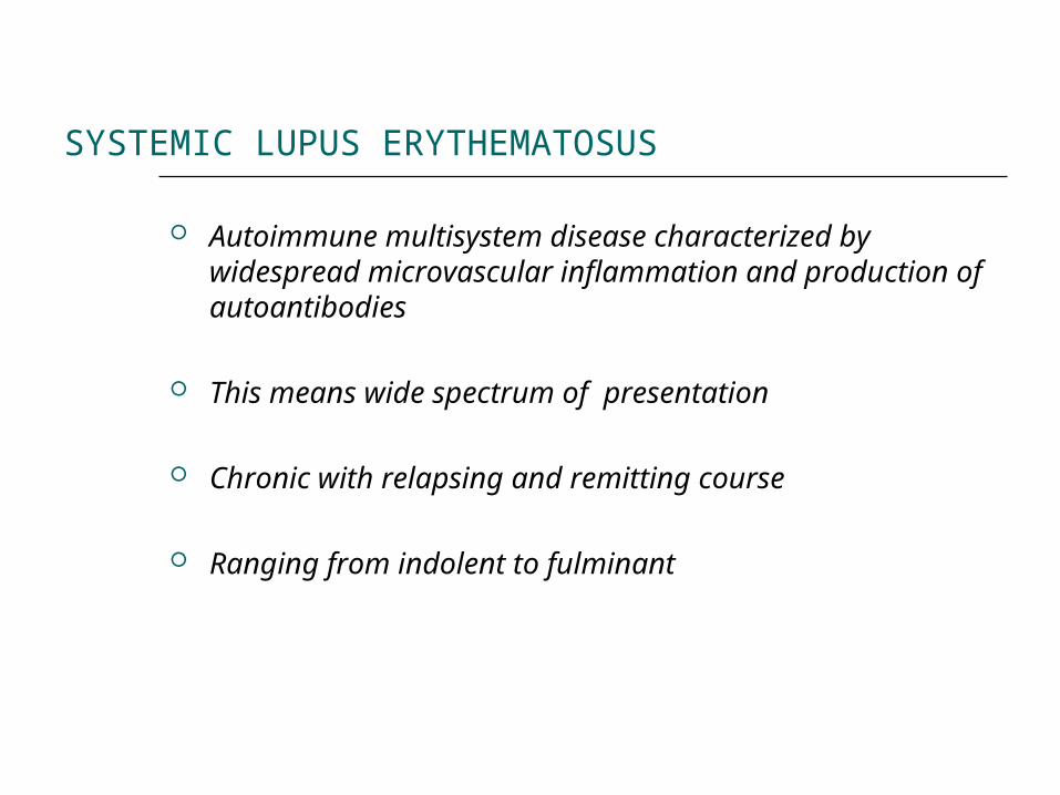
SYSTEMIC LUPUS ERYTHEMATOSUS
Autoimmune multisystem disease characterized by widespread microvascular inflammation and production of autoantibodies
This means wide spectrum of presentation
Chronic with relapsing and remitting course
Ranging from indolent to fulminant
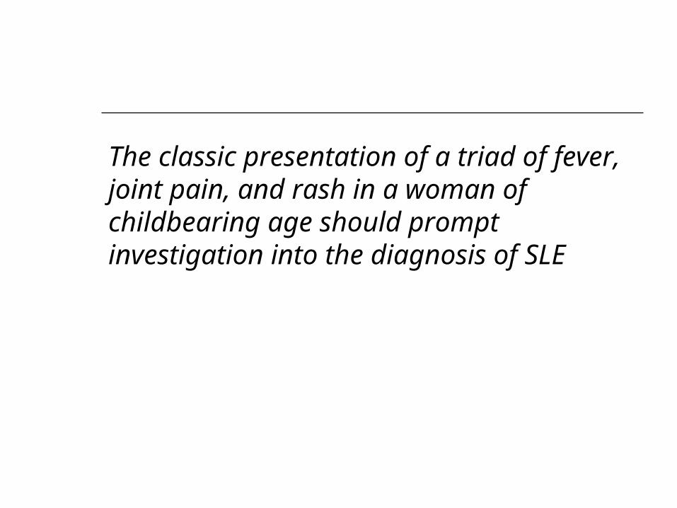
The classic presentation of a triad of fever, joint pain, and rash in a woman of childbearing age should prompt investigation into the diagnosis of SLE
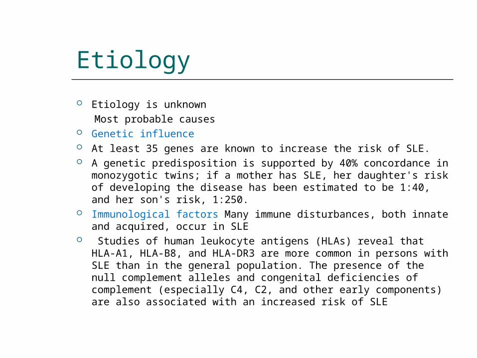
Etiology
Etiology is unknown Most probable causes Genetic influence At least 35 genes are known to increase the risk of SLE. A genetic predisposition is supported by 40% concordance in
monozygotic twins; if a mother has SLE, her daughter's risk of developing the disease has been estimated to be 1:40, and her son's risk, 1:250.
Immunological factors Many immune disturbances, both innate and acquired, occur in SLE
Studies of human leukocyte antigens (HLAs) reveal that HLA-A1, HLA-B8, and HLA-DR3 are more common in persons with SLE than in the general population. The presence of the null complement alleles and congenital deficiencies of complement (especially C4, C2, and other early components) are also associated with an increased risk of SLE
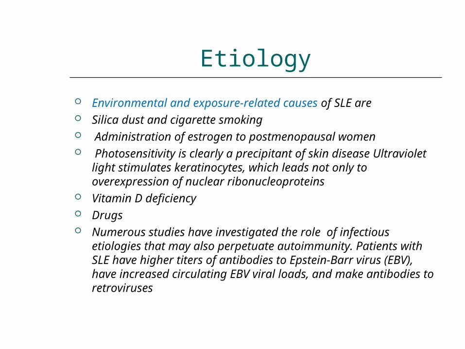
Etiology
Environmental and exposure-related causes of SLE are Silica dust and cigarette smoking Administration of estrogen to postmenopausal women Photosensitivity is clearly a precipitant of skin disease
Ultraviolet light stimulates keratinocytes, which leads not only to overexpression of nuclear ribonucleoproteins
Vitamin D deficiency Drugs Numerous studies have investigated the role of infectious
etiologies that may also perpetuate autoimmunity. Patients with SLE have higher titers of antibodies to Epstein-Barr virus (EBV), have increased circulating EBV viral loads, and make antibodies to retroviruses
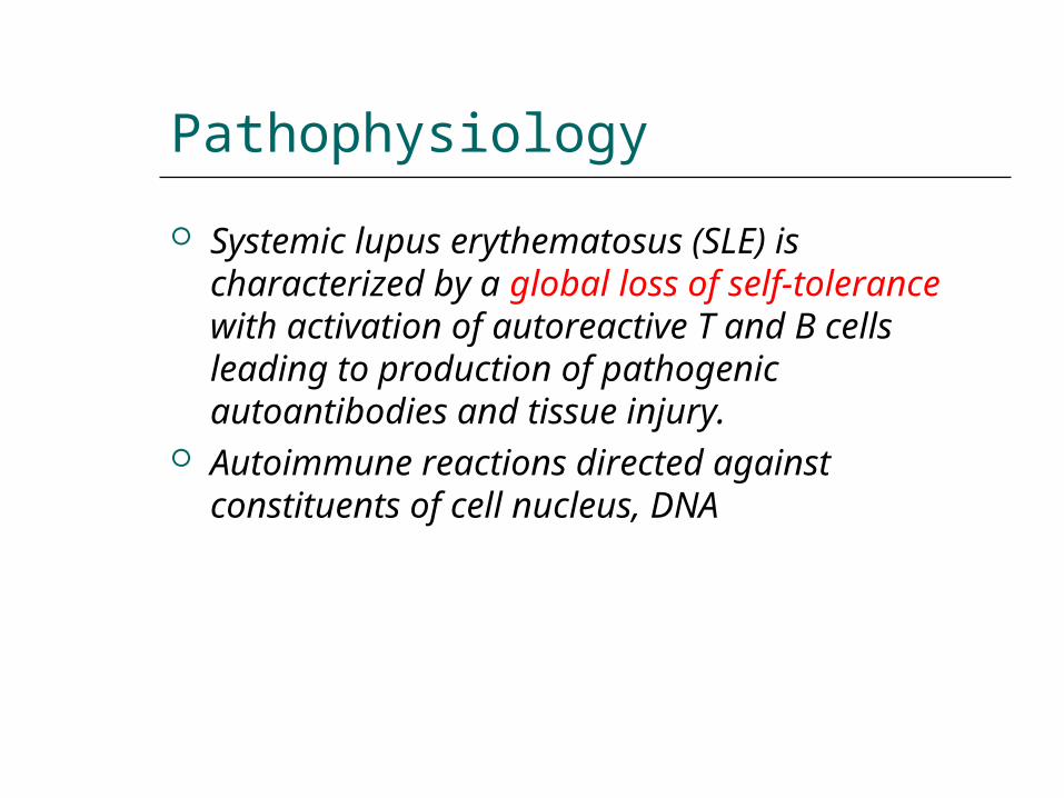
Pathophysiology
Systemic lupus erythematosus (SLE) is characterized by a global loss of self-tolerance with activation of autoreactive T and B cells leading to production of pathogenic autoantibodies and tissue injury.
Autoimmune reactions directed against constituents of cell nucleus, DNA
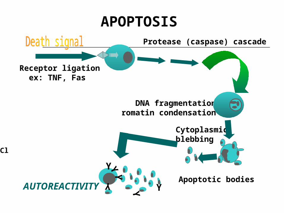
Receptor ligation ex: TNF, Fas
Protease (caspase) cascade
DNA fragmentationChromatin condensation
Cytoplasmic blebbing
Apoptotic bodies
APOPTOSIS
Clearance by phagocytesY
Y
Y
YY
YAUTOREACTIVITY
Y
YYY
Y
YY
YY
Y
Y
Y
Y
YY
Y
Y
Y
Y
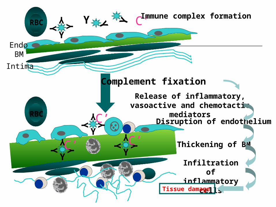
C ’ C ’
C ’Immune complex formation
C ’
EndoBM
Intima
Complement fixation
Release of inflammatory, vasoactive and chemotactic
mediatorsDisruption of endothelium
Thickening of BM
Infiltration of inflammatory
cellsTissue damage
RBC
RBC
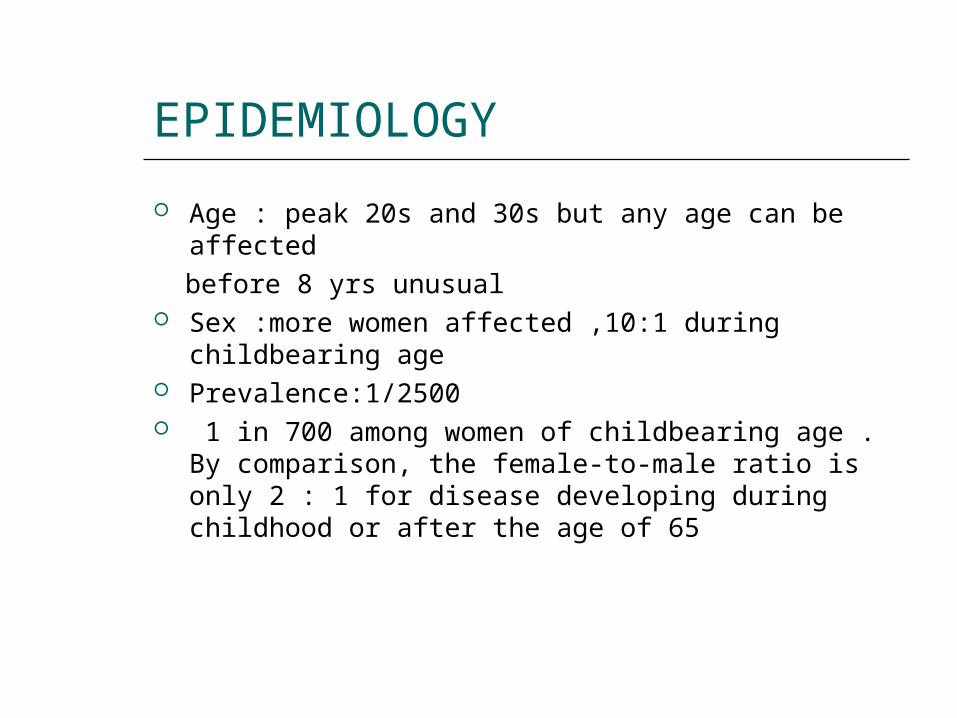
EPIDEMIOLOGY
Age : peak 20s and 30s but any age can be affected before 8 yrs unusual Sex :more women affected ,10:1 during childbearing
age Prevalence:1/2500 1 in 700 among women of childbearing age . By
comparison, the female-to-male ratio is only 2 : 1 for disease developing during childhood or after the age of 65
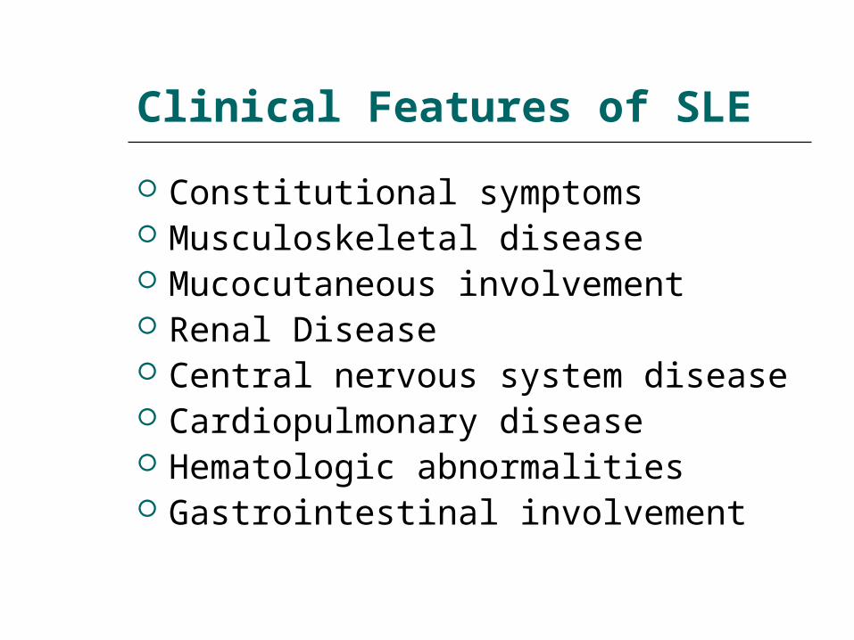
Clinical Features of SLE
Constitutional symptoms Musculoskeletal disease Mucocutaneous involvement Renal Disease Central nervous system disease Cardiopulmonary disease Hematologic abnormalities Gastrointestinal involvement
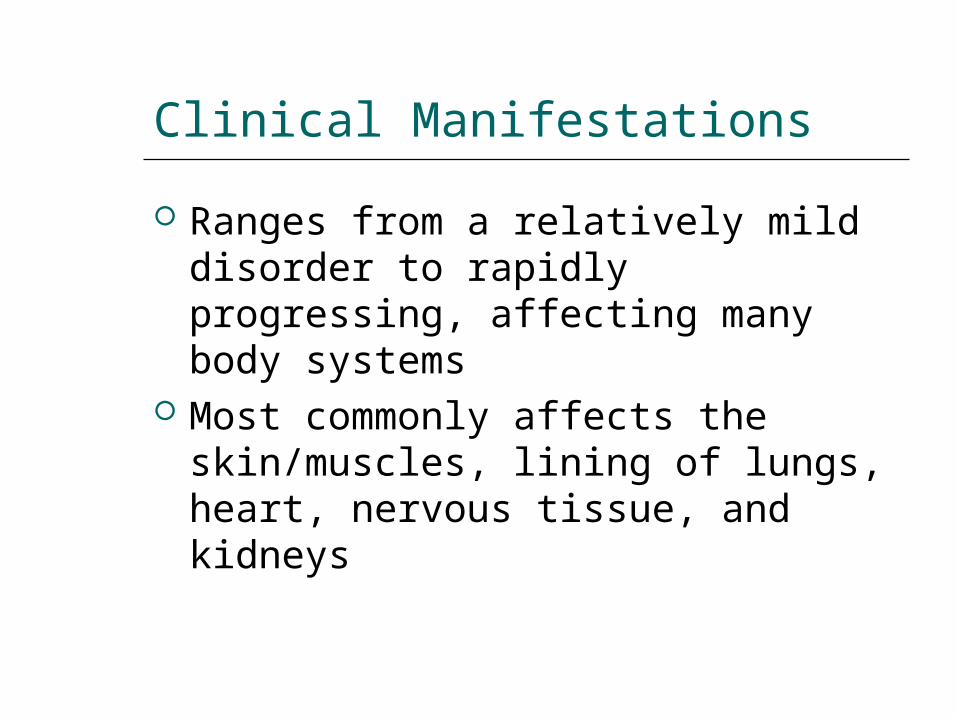
Clinical Manifestations
Ranges from a relatively mild disorder to rapidly progressing, affecting many body systems
Most commonly affects the skin/muscles, lining of lungs, heart, nervous tissue, and kidneys
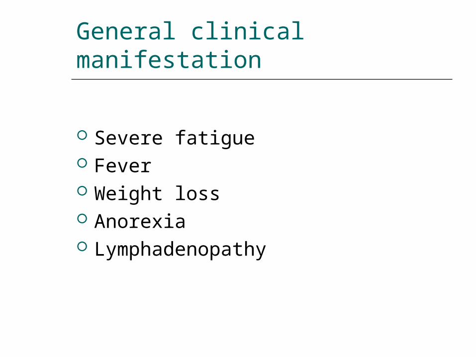
General clinical manifestation
Severe fatigue Fever Weight loss Anorexia Lymphadenopathy
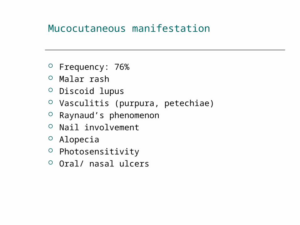
Mucocutaneous manifestation
Frequency: 76% Malar rash Discoid lupus Vasculitis (purpura, petechiae) Raynaud’s phenomenon Nail involvement Alopecia Photosensitivity Oral/ nasal ulcers
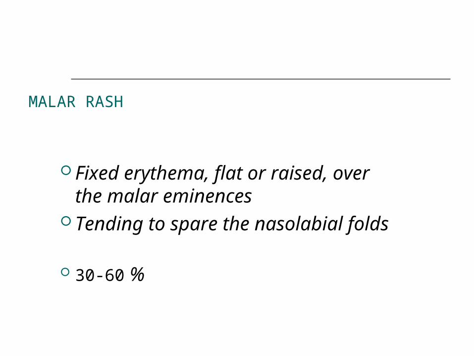
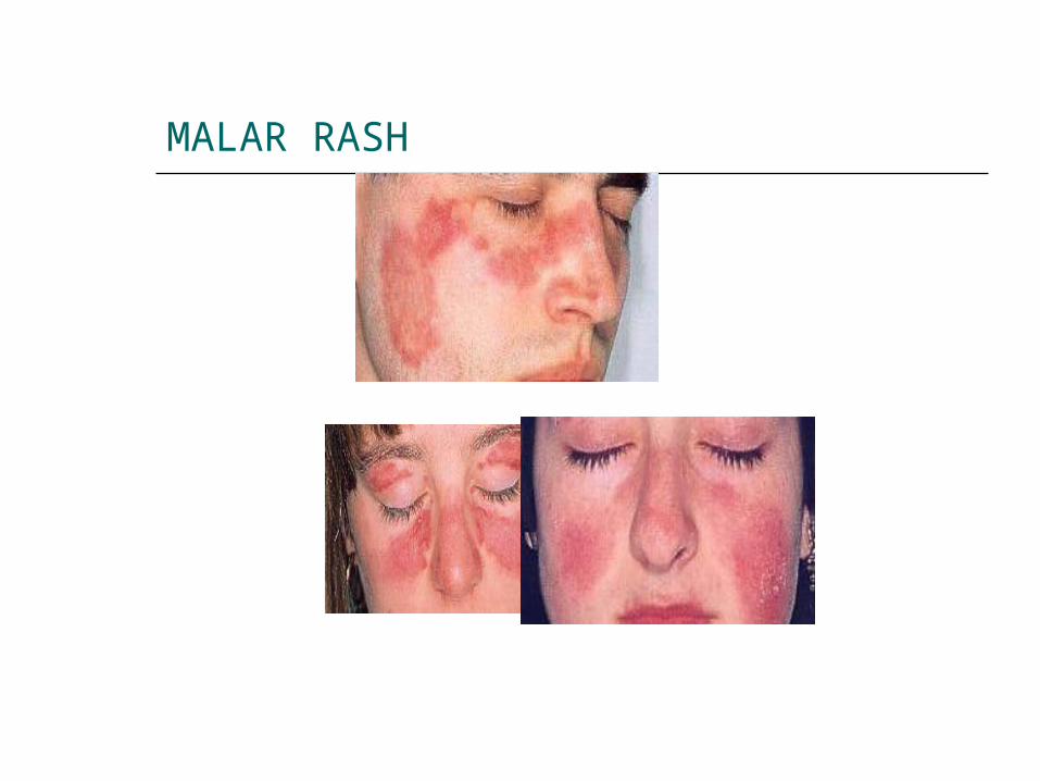
MALAR RASH
Fixed erythema, flat or raised, over the malar eminences
Tending to spare the nasolabial folds
30-60 %
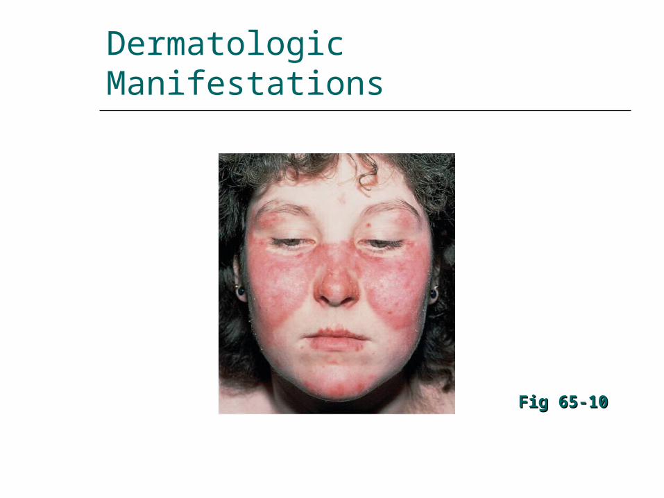
Dermatologic Manifestations
Fig 65-10Fig 65-10
MALAR RASH
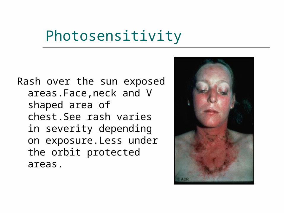
Photosensitivity
Rash over the sun exposed areas.Face,neck and V shaped area of chest.See rash varies in severity depending on exposure.Less under the orbit protected areas.
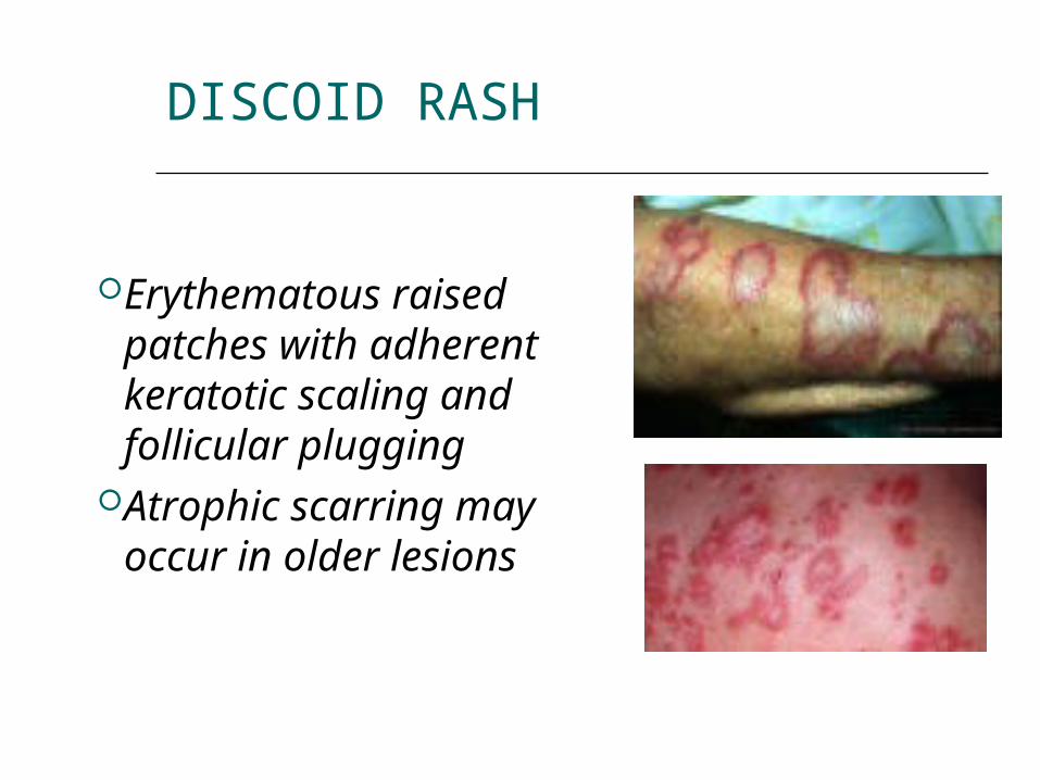
DISCOID RASH
Erythematous raised patches with adherent keratotic scaling and follicular plugging
Atrophic scarring may occur in older lesions
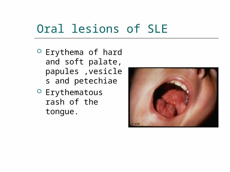
Oral lesions of SLE
Erythema of hard and soft palate, papules ,vesicles and petechiae
Erythematous rash of the tongue.
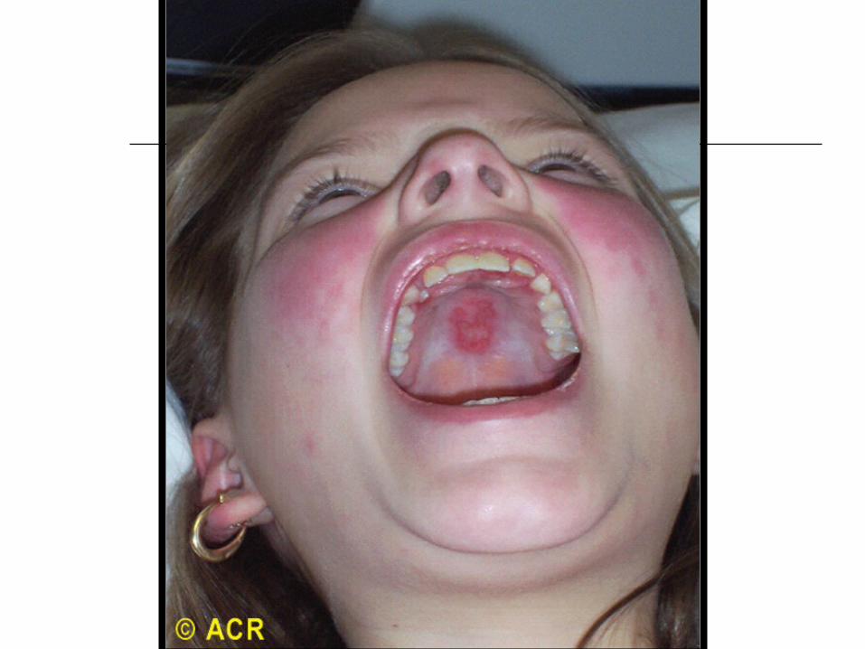
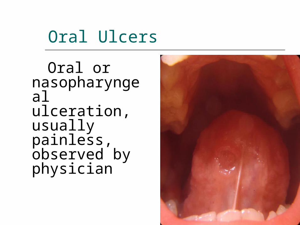
Oral Ulcers
Oral or nasopharyngeal ulceration, usually painless, observed by physician
Joint
arthralgia, arthropathy, myalgia, frank arthritis, avascular necrosis
Joint involvement is typically a nonerosive synovitis with little deformity, which contrasts with rheumatoid arthritis
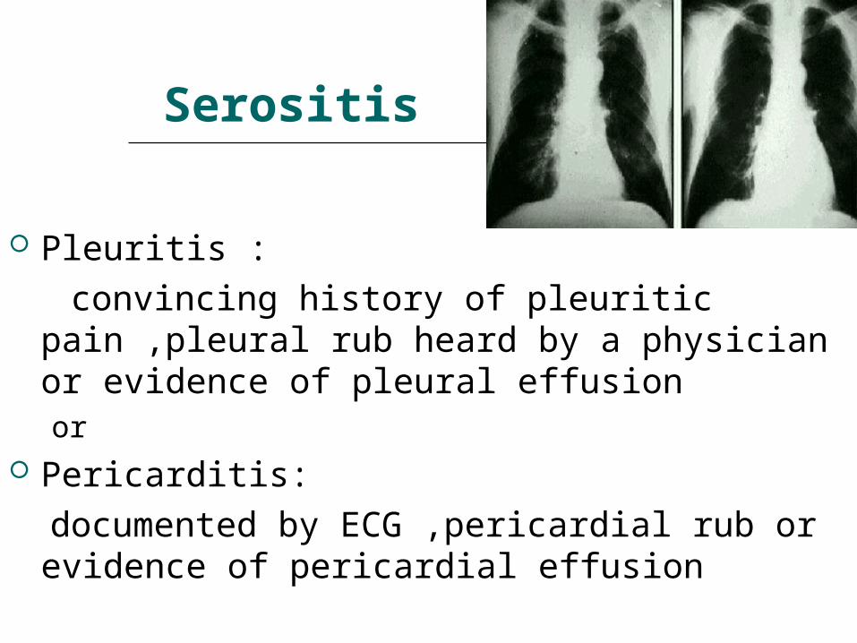
Serositis
Pleuritis : convincing history of pleuritic pain ,pleural
rub heard by a physician or evidence of pleural effusion or
Pericarditis: documented by ECG ,pericardial rub or
evidence of pericardial effusion
Pulmonary Findings In SLE
Incidence: 5-67% May be subclinical (abnormal PFTs) Pleuritis Pleural effusion Pneumonitis Pulmonary hemorrhage Pulmonary hypertension Restrictive pulmonary disease & diffusion
defects most commonly observed abnormalities on PFTs
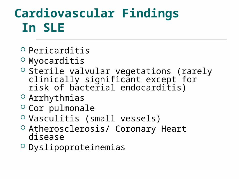
Cardiovascular Findings In SLE
Pericarditis Myocarditis Sterile valvular vegetations (rarely
clinically significant except for risk of bacterial endocarditis)
Arrhythmias Cor pulmonale Vasculitis (small vessels) Atherosclerosis/ Coronary Heart disease Dyslipoproteinemias
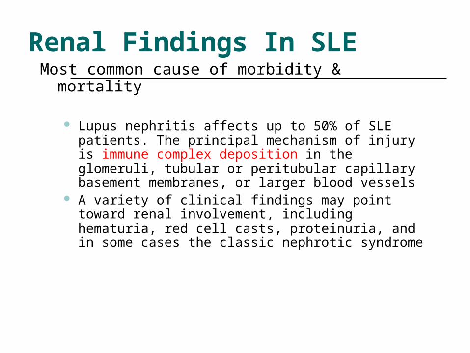
Renal Findings In SLEMost common cause of morbidity & mortality
Lupus nephritis affects up to 50% of SLE patients. The principal mechanism of injury is immune complex deposition in the glomeruli, tubular or peritubular capillary basement membranes, or larger blood vessels
A variety of clinical findings may point toward renal involvement, including hematuria, red cell casts, proteinuria, and in some cases the classic nephrotic syndrome
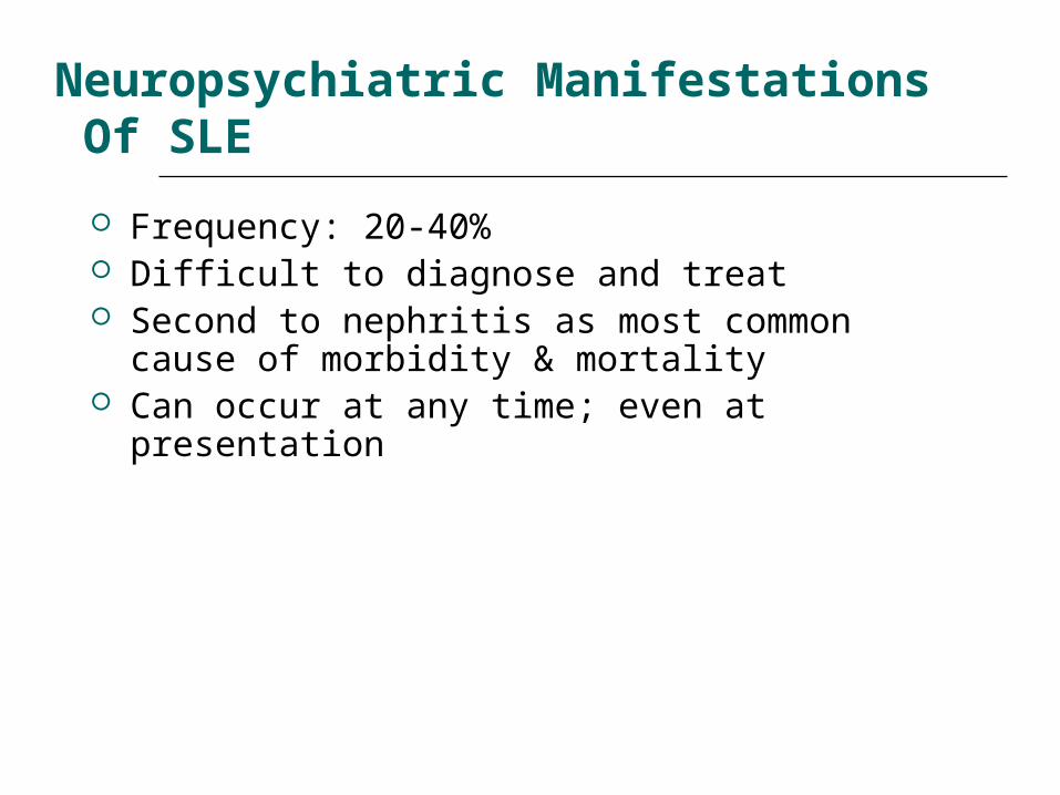
Neuropsychiatric Manifestations Of SLE
Frequency: 20-40% Difficult to diagnose and treat Second to nephritis as most common cause of
morbidity & mortality Can occur at any time; even at presentation
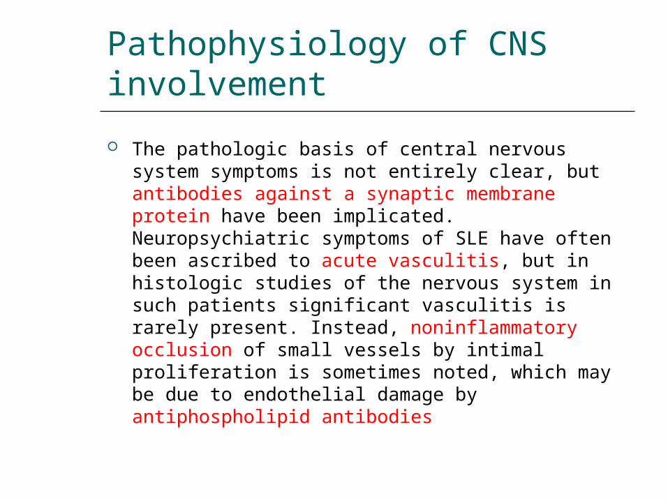
Pathophysiology of CNS involvement
The pathologic basis of central nervous system symptoms is not entirely clear, but antibodies against a synaptic membrane protein have been implicated. Neuropsychiatric symptoms of SLE have often been ascribed to acute vasculitis, but in histologic studies of the nervous system in such patients significant vasculitis is rarely present. Instead, noninflammatory occlusion of small vessels by intimal proliferation is sometimes noted, which may be due to endothelial damage by antiphospholipid antibodies
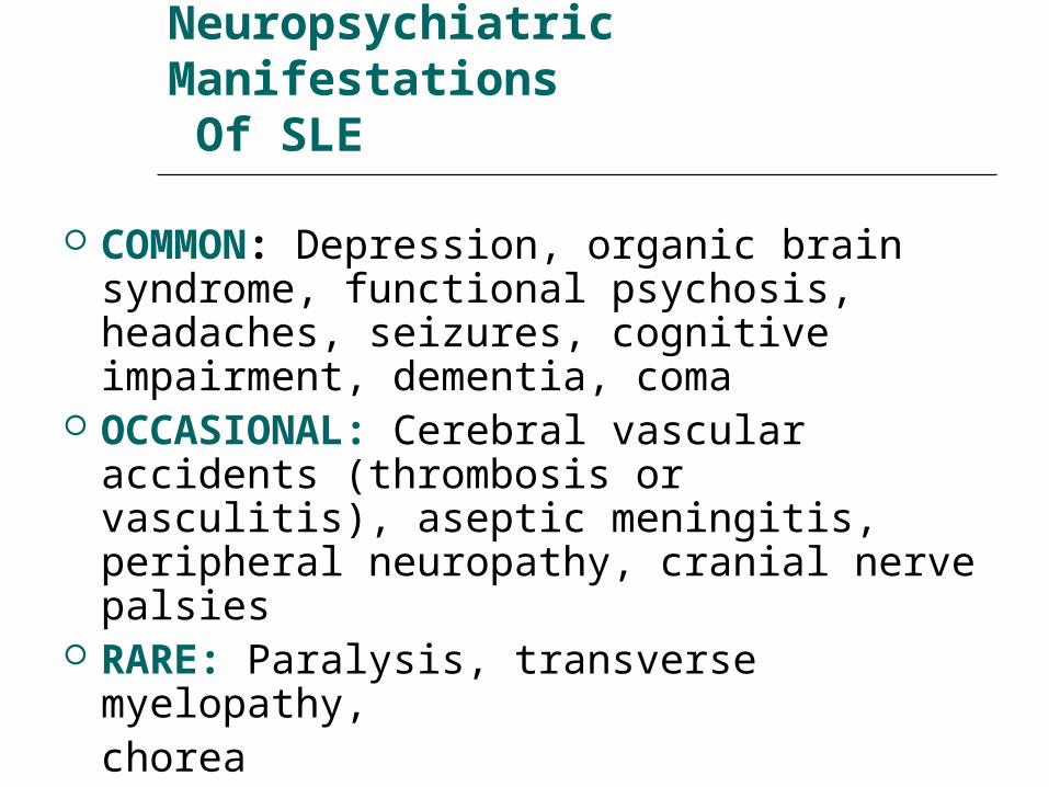
Neuropsychiatric Manifestations Of SLE
COMMON: Depression, organic brain syndrome, functional psychosis, headaches, seizures, cognitive impairment, dementia, coma
OCCASIONAL: Cerebral vascular accidents (thrombosis or vasculitis), aseptic meningitis, peripheral neuropathy, cranial nerve palsies
RARE: Paralysis, transverse myelopathy,chorea
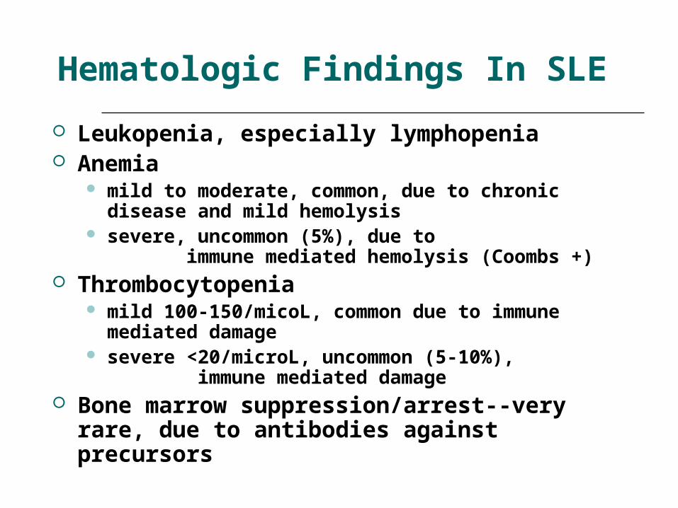
Hematologic Findings In SLE
Leukopenia, especially lymphopenia Anemia
mild to moderate, common, due to chronic disease and mild hemolysis
severe, uncommon (5%), due to immune mediated hemolysis (Coombs
+) Thrombocytopenia
mild 100-150/micoL, common due to immune mediated damage
severe <20/microL, uncommon (5-10%), immune mediated damage
Bone marrow suppression/arrest--very rare, due to antibodies against precursors
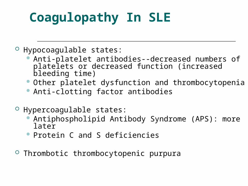
Coagulopathy In SLE
Hypocoagulable states: Anti-platelet antibodies--decreased numbers of
platelets or decreased function (increased bleeding time)
Other platelet dysfunction and thrombocytopenia Anti-clotting factor antibodies
Hypercoagulable states: Antiphospholipid Antibody Syndrome (APS): more
later Protein C and S deficiencies
Thrombotic thrombocytopenic purpura
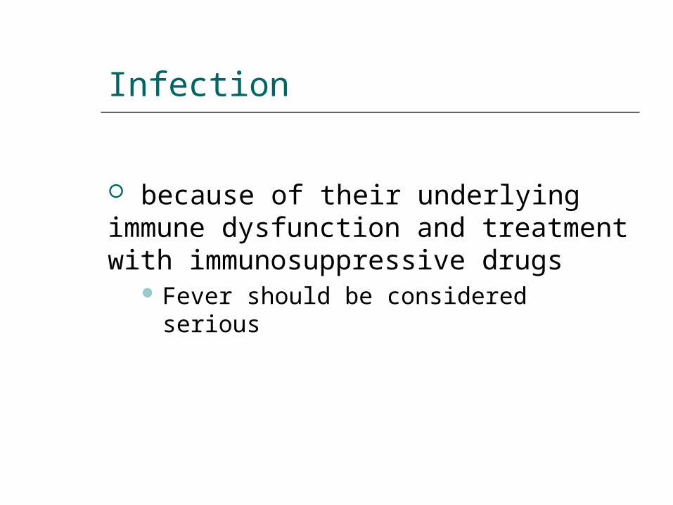
Infection
because of their underlying immune dysfunction and treatment with immunosuppressive drugs
Fever should be considered serious
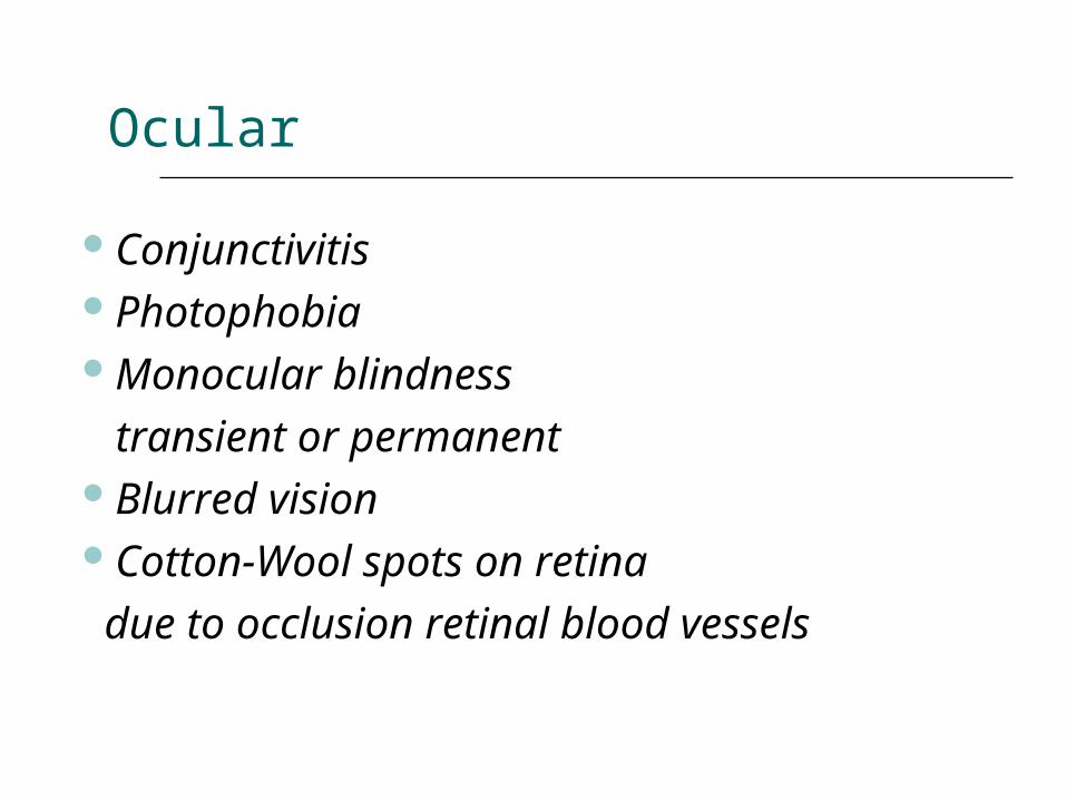
Ocular
ConjunctivitisPhotophobiaMonocular blindness transient or permanentBlurred visionCotton-Wool spots on retina due to occlusion retinal blood vessels
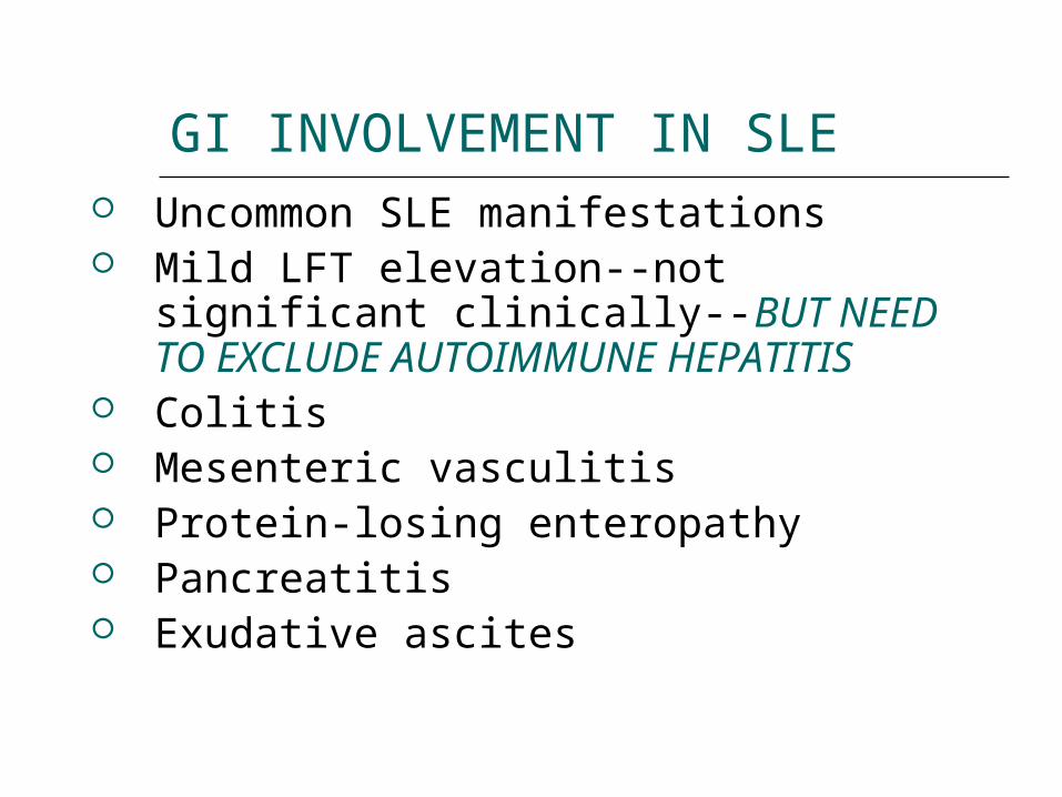
GI INVOLVEMENT IN SLE Uncommon SLE manifestations Mild LFT elevation--not significant
clinically--BUT NEED TO EXCLUDE AUTOIMMUNE HEPATITIS
Colitis Mesenteric vasculitis Protein-losing enteropathy Pancreatitis Exudative ascites
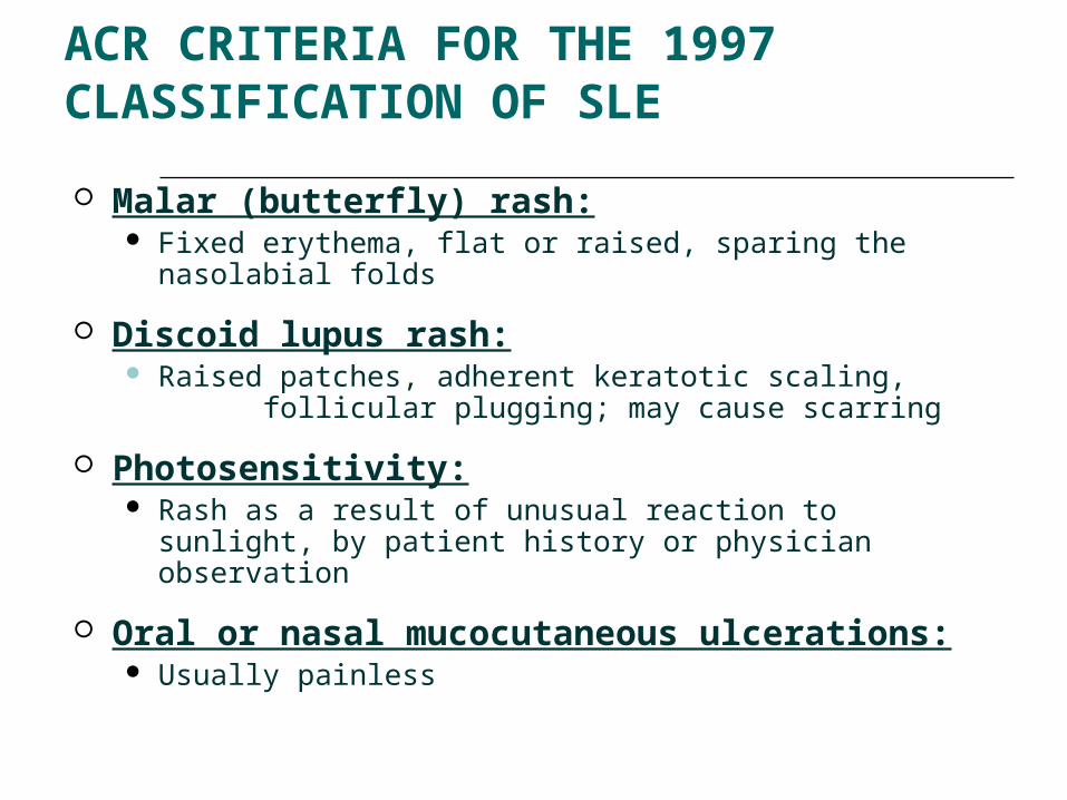
1997 ACR CRITERIA FOR THE CLASSIFICATION OF SLE
Malar (butterfly) rash: Fixed erythema, flat or raised, sparing the nasolabial
folds
Discoid lupus rash: Raised patches, adherent keratotic scaling,
follicular plugging; may cause scarring
Photosensitivity: Rash as a result of unusual reaction to sunlight, by
patient history or physician observation
Oral or nasal mucocutaneous ulcerations: Usually painless
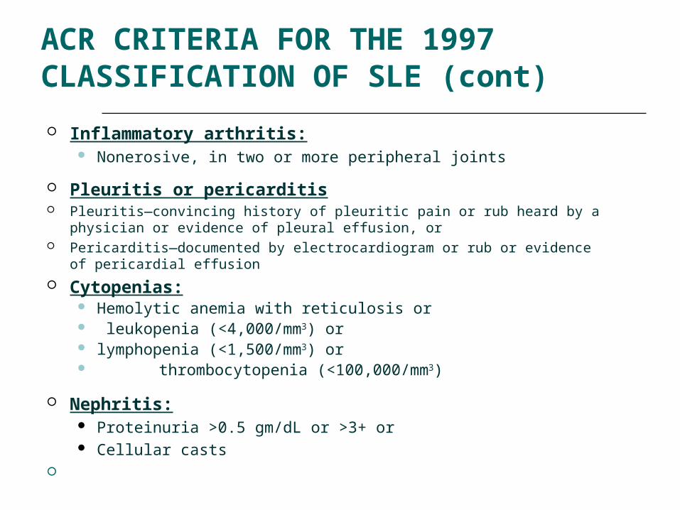
1997 ACR CRITERIA FOR THE CLASSIFICATION OF SLE (cont)
Inflammatory arthritis: Nonerosive, in two or more peripheral joints
Pleuritis or pericarditis Pleuritis—convincing history of pleuritic pain or rub heard by a physician or
evidence of pleural effusion, or Pericarditis—documented by electrocardiogram or rub or evidence of
pericardial effusion
Cytopenias: Hemolytic anemia with reticulosis or leukopenia (<4,000/mm3) or lymphopenia (<1,500/mm3) or thrombocytopenia (<100,000/mm3)
Nephritis: Proteinuria >0.5 gm/dL or >3+ or Cellular casts
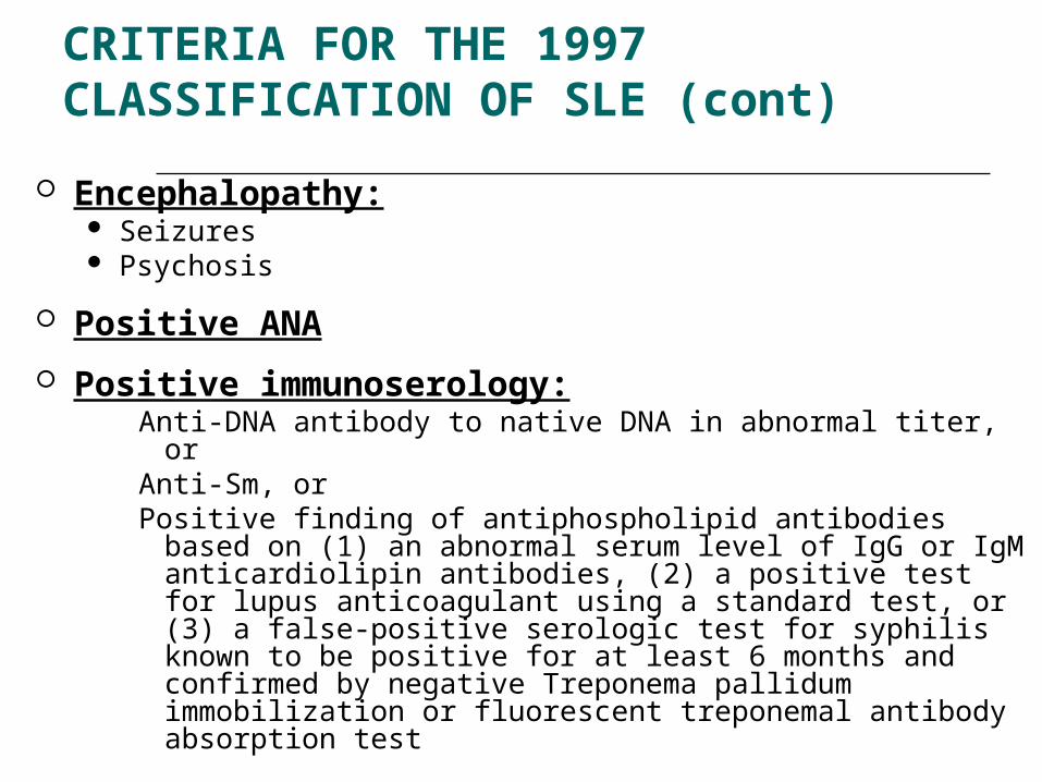
1997 CRITERIA FOR THE CLASSIFICATION OF SLE (cont)
Encephalopathy: Seizures Psychosis
Positive ANA
Positive immunoserology:Anti-DNA antibody to native DNA in abnormal titer, or Anti-Sm, or Positive finding of antiphospholipid antibodies based on (1)
an abnormal serum level of IgG or IgM anticardiolipin antibodies, (2) a positive test for lupus anticoagulant using a standard test, or (3) a false-positive serologic test for syphilis known to be positive for at least 6 months and confirmed by negative Treponema pallidum immobilization or fluorescent treponemal antibody absorption test
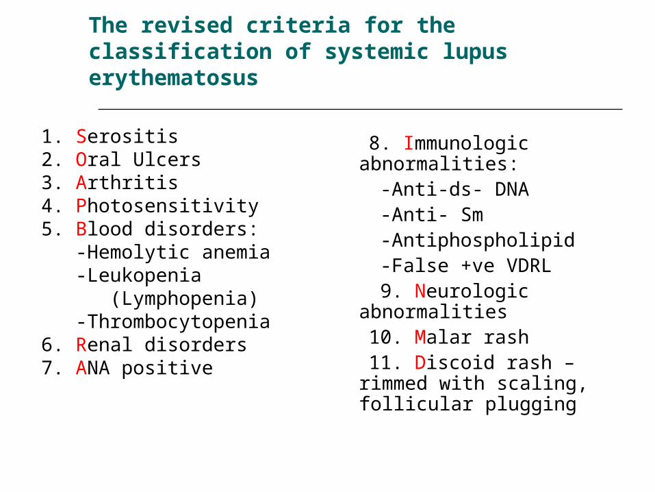
The revised criteria for the classification of systemic lupus erythematosus
1. Serositis 2. Oral Ulcers3. Arthritis4. Photosensitivity5. Blood disorders: -Hemolytic anemia -Leukopenia (Lymphopenia) -Thrombocytopenia 6. Renal disorders7. ANA positive
8. Immunologic abnormalities:
-Anti-ds- DNA -Anti- Sm -Antiphospholipid -False +ve VDRL 9. Neurologic
abnormalities 10. Malar rash 11. Discoid rash – rimmed
with scaling, follicular plugging

CLASSIFICATION CRITERIA
Must have 4 of 11 for Classification Sensitivity 96% Specificity 96%(In children 100%)
Not all “Lupus” is SLE Chronic discoid Lupus erythematosus Drug induced lupus Subacute Cutaneous Lupus
erythematosus
Chronic Discoid Lupus Erythematosus
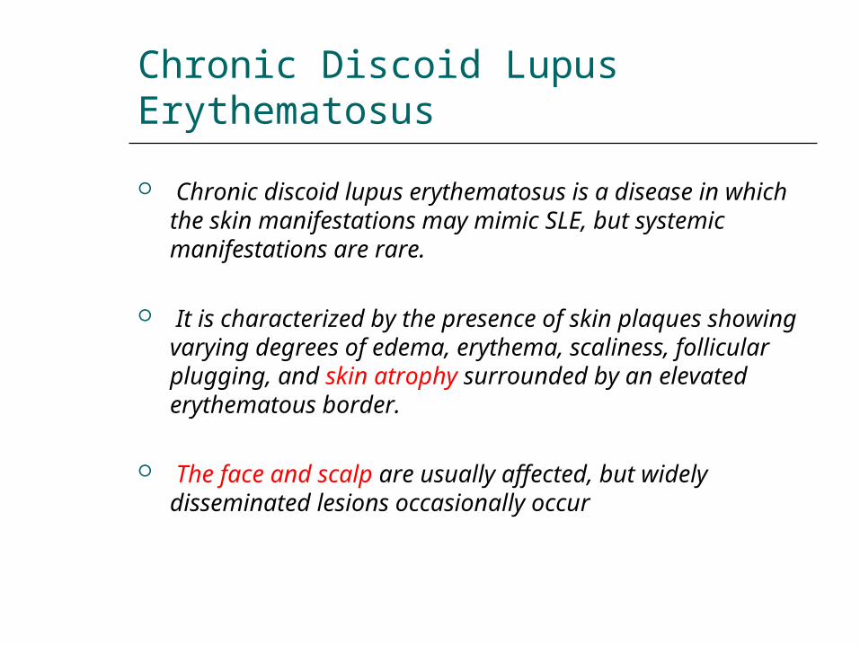
Chronic discoid lupus erythematosus is a disease in which the skin manifestations may mimic SLE, but systemic manifestations are rare.
It is characterized by the presence of skin plaques showing varying degrees of edema, erythema, scaliness, follicular plugging, and skin atrophy surrounded by an elevated erythematous border.
The face and scalp are usually affected, but widely disseminated lesions occasionally occur
Chronic Discoid Lupus Erythematosus
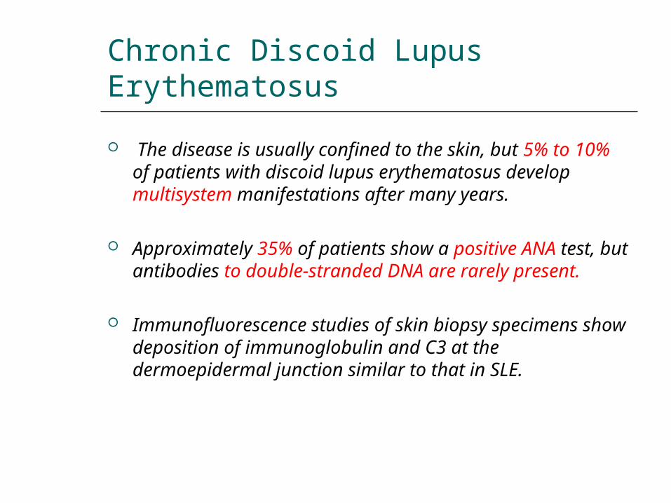
The disease is usually confined to the skin, but 5% to 10% of patients with discoid lupus erythematosus develop multisystem manifestations after many years.
Approximately 35% of patients show a positive ANA test, but antibodies to double-stranded DNA are rarely present.
Immunofluorescence studies of skin biopsy specimens show deposition of immunoglobulin and C3 at the dermoepidermal junction similar to that in SLE.
Subacute Cutaneous Lupus Erythematosus.
This condition also presents with predominant skin involvement and can be distinguished from chronic discoid lupus erythematosus by several criteria.
The skin rash in this disease tends to be widespread, superficial, and nonscarring, although scarring lesions may occur in some patients.
Most patients have mild systemic symptoms consistent with SLE. Furthermore, there is a strong association with antibodies to the SS-A antigen and with the HLA-DR3 genotype.
Thus, the term subacute cutaneous lupus erythematosus seems to define a group intermediate between SLE and lupus erythematosus localized only to skin.
Drug-Induced Lupus Erythematosus
A lupus erythematosus–like syndrome may develop in patients receiving a variety of drugs, including hydralazine, procainamide, isoniazid, and d-penicillamine, Many of these drugs are associated with the development of ANAs, but most patients do not have symptoms of lupus erythematosus.
For example, 80% of patients receiving procainamide test positive for ANAs, but only one third of these manifest clinical symptoms, such as arthralgias, fever, and serositis.
Drug-Induced Lupus Erythematosus,cont.
Although multiple organs are affected, renal and central nervous system involvement is distinctly uncommon.
There are serologic and genetic differences from classical SLE, as well. Antibodies specific for double-stranded DNA are rare, but there is an extremely high frequency of antibodies specific for histone
Persons with the HLA-DR4 allele are at a greater risk of developing lupus erythematosus after administration of hydralazine.
The disease remits after withdrawal of the offending drug.
DIFFERENTIAL DIAGNOSIS
Rheumatic: RA, Sjogren’s syndrome, systemic sclerosis, dermatomyositis
Nonrheumatic: HIV, endocarditis, viral infections, hematologic malignancies, vasculitis, ITP, other causes of nephritis
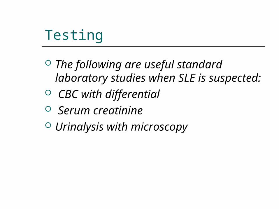
Testing
The following are useful standard laboratory studies when SLE is suspected:
CBC with differential Serum creatinine Urinalysis with microscopy
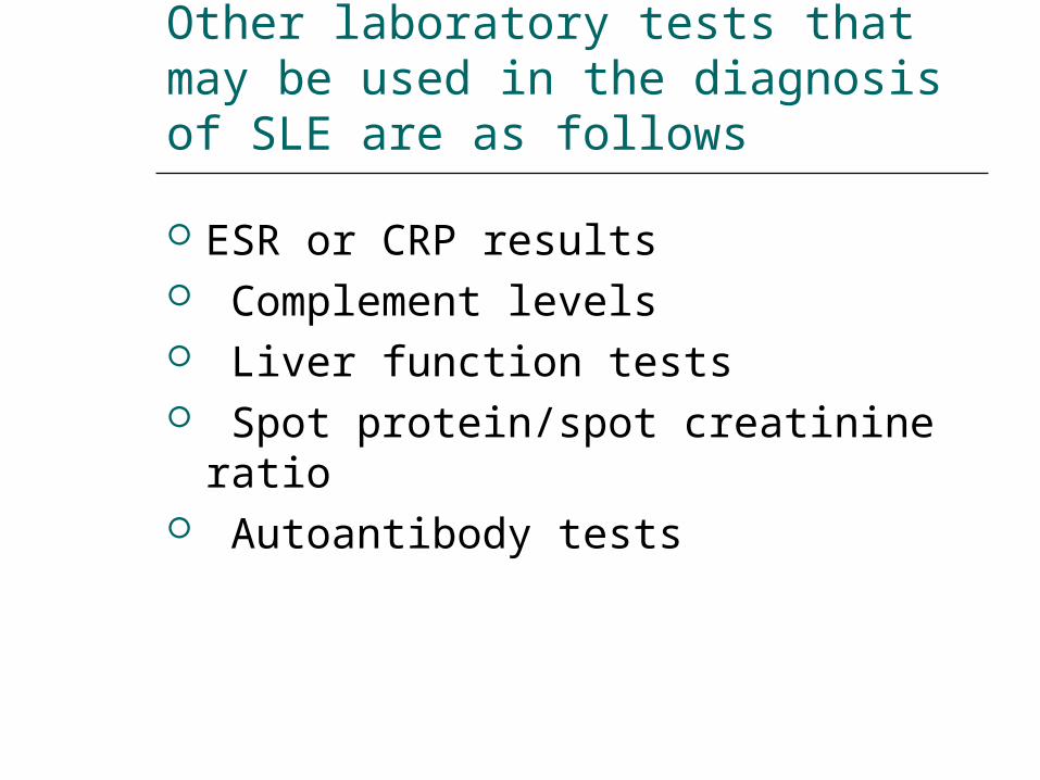
Other laboratory tests that may be used in the diagnosis of SLE are as follows
ESR or CRP results Complement levels Liver function tests Spot protein/spot creatinine ratio Autoantibody tests
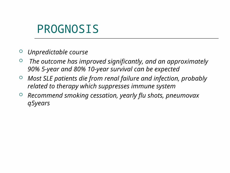
PROGNOSIS
Unpredictable course The outcome has improved significantly, and an
approximately 90% 5-year and 80% 10-year survival can be expected
Most SLE patients die from renal failure and infection, probably related to therapy which suppresses immune system
Recommend smoking cessation, yearly flu shots, pneumovax q5years
Refrences
1. Robbins and Cotran,Pathologic basis of disease,8th
edition,2010
2. http://emedicine.medscape.com/article Updated: Feb 19, 2014
3. Livingston B, Bonner A, Pope J. Differences in clinical manifestations between childhood-onset lupus and adult-onset lupus: a meta-analysis. Lupus. Nov 2011;20(13):1345-55. [Medline]
4. American College of Rheumatology. 1997 Update of the 1982 American College of Rheumatology revised criteria for classification of systemic lupus erythematosus. Available at http://tinyurl.com/1997SLEcriteria. Accessed March 15, 2012

Thank You