Systemic fungi - bpums.ac.irparamed.bpums.ac.ir/UploadedFiles/CourseFiles/... · Sputum, BAL,...
39
Systemic fungi
Transcript of Systemic fungi - bpums.ac.irparamed.bpums.ac.ir/UploadedFiles/CourseFiles/... · Sputum, BAL,...

Systemic fungi

Histoplasmosis
Thermally dimorphic fungus: Histoplasma capsulatum Two varieties: Capsulatum & Duboisii
Soil enriched with bird or bat droppings
Wide spectrum of clinical manifestations:
Asymptomatic pulmonary infection
Acute or chronic disease of the lungs
More widespread disseminated disease
Healthy individualsLow-level exposure
High-level exposureUnderlying condition

Acute pulmonary histoplasmosis Fewer than 5% develop symptomatic disease
In immunocompetent individuals:
Non-specific flu-like illness
Chest radiographs: small, scattered, bilateral, nodular infiltrates
The infiltrates: heal over several months & scattered calcifications

Chronic pulmonary histoplasmosis:
Underlying lung damage, (chronic obstructive pulmonary disease)
First manifests: transient, segmental pneumonia
Heals without treatment
Progresses to fibrosis and cavitation

As lesions heal, peripheral fibrosis and centralcalcification may occur

Disseminated histoplasmosis
older adults, infants and immunosuppressed patients
o The liver and spleen are enlarged
o
o Anaemia, leucopenia and thrombocytopenia
o Mucosal lesions can occur
o CNS involvement (10–20%)

African histoplasmosis
Histoplasma capsulatum var. duboisii
lungs
Skin and bones Painless papular lesions Osteomyelitis
Joints arthritis
liver, spleen and other organs

Sputum, BAL, Blood, urine, lymph node, bone marrow
Wet preparations of clinical material ?
Wright-stained
Stained tissue sections of lung, liver, lymph node
Yeast cells with narrow-based buds
The definitive diagnosis: isolation of the fungus
25–30◦C for 4–6 weeks on Sabouraud’s dextrose agar + cycloheximide
37◦C on brain heart infusion agar
Rapid identification: AccuProbe test
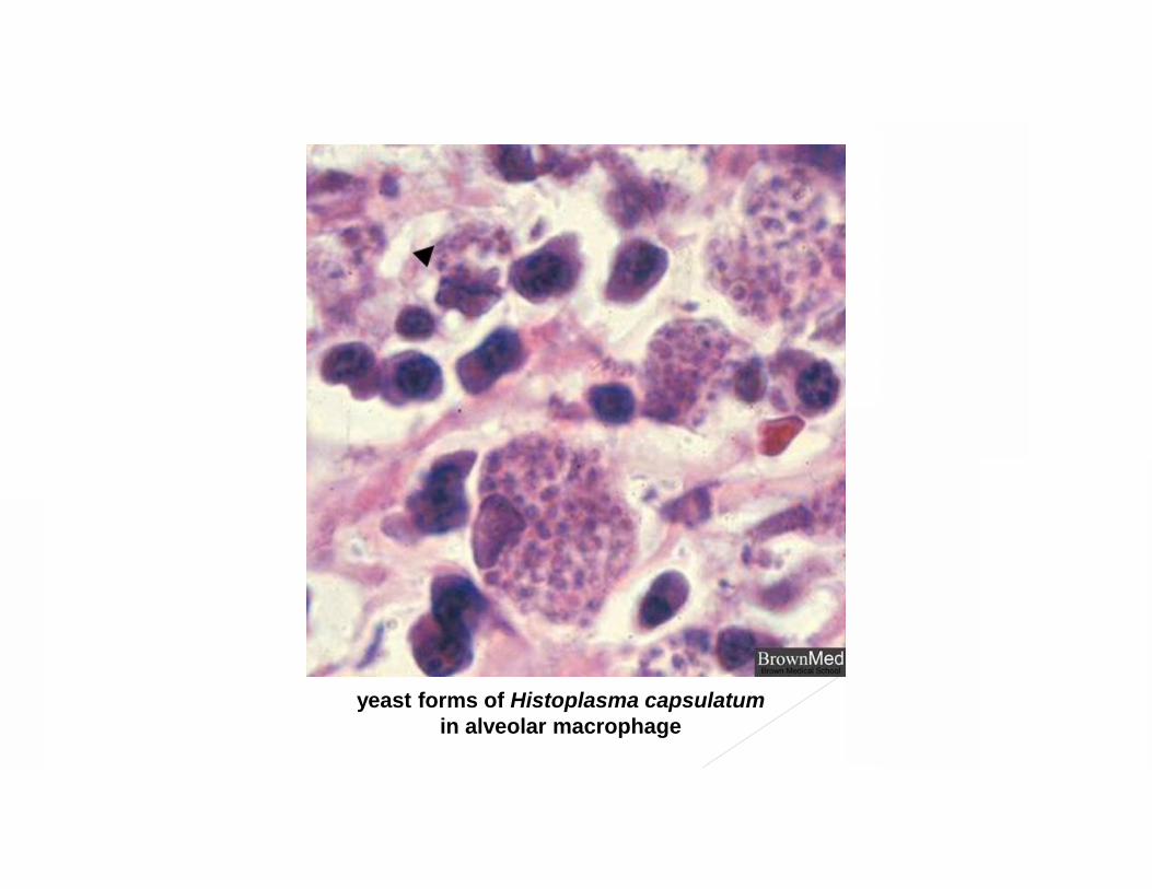
yeast forms of Histoplasma capsulatumin alveolar macrophage

Monocytes in peripheral blood withphagocytosed yeast forms of
Histoplasma capsulatum


Skin tests Not recommended for diagnosis
Serological tests Sub-acute pulmonary histoplasmosis
Chronic pulmonary histoplasmosis
Disseminated disease ?
Immunodiffusion (ID) test
Complement fixation (CF) test
Antigen detection tests For ?

Treatment
Mild symptoms: Self-limited
Progressive disseminated disease:
Oral itraconazole (200 mg once or twice daily for 6–12 weeks)
Amphotericin B (3–5 mg/kg per day for 1–2 weeks)

Blastomycosis‘‘Chicago’s disease’
Blastomyces dermatitis
In nature as a mould
In tissue as large, round budding yeast cells
Following inhalation: wide spectrum of clinical manifestations
Occasional: traumatic cutaneous inoculation
Geographical distribution
Overlap with the endemic region for histoplasmosis

Clinical manifestations
Pulmonary blastomycosisAt least 50% of individuals: asymptomatic
Symptomatic pulmonary diseaseo Similar to histoplasmosis
o Non-specific flulike illness
o The radiological findings: Hilar lymphadenopathy is uncommon
o Spontaneous resolution
Chronic infectiono Cavitation is less common

Disseminated blastomycosis
Cutaneous blastomycosis
40–80% of cases
Raised, crusted verrucous lesions with an irregular shape and sharp borders
Face, neck and scalp
Osteomyelitis & Arthritis
25–30%
The spine, pelvis, skull, ribs and long bones
Genitourinary blastomycosis
The prostate, epididymis or testis in 10–30%


Essential investigations
Microscopy Wet preparations
Yeast cells with thick refractive walls and broad-based single buds
Culture 25–30◦C for 1–3 weeks on Sabouraud’s dextrose agar + cycloheximide
37◦C on brain heart infusion agar
More rapid identification: AccuProbe test The AccuProbe test produces a positive result with P. brasiliensis

Yeast form in the lungs


Serological tests
Antibody detection tests:
Immunodiffusion (ID)
Complement fixation (CF)
o CF: cross-reactions with H. capsulatum and Coccidioides species
Antigen detection tests

Management Spontaneous resolution
Mild to moderate pulmonary disease
Oral itraconazole
Posaconazole and voriconazole
lipid formulation of amphotericin B
Disseminated blastomycosis
Amphotericin B

►Coccidioides immitis
Nature as a mould (arthrospores)
Human and animal tissue: spherules
A single arthroconidia
Arid climate, alkaline soil, hot summers
Coccidioidomycosis


Large, mature spherule containing hundreds ofendospores (H &E stain)

Clinical manifestations
Acute pulmonary coccidioidomycosis
60%: no symptoms
Moderate flu-like illness
Up to 50% of patients: erythematous rash (trunk and limbs)
Erythema nodosum or erythematous multiforme: 5% of infected persons
More common in women
In joints
Disseminated coccidioidomycosis
black, Asian or Filipino
1–5% of individuals
Following reactivation in an immunosuppressed individual
Cutaneous and subcutaneous lesions, bones, joints, and meninges

The lesion on the nose resulted from disseminationfrom the lungs

Differential diagnosis:
Diffuse lung infiltrates in AIDS patients
Pneumocystis jirovecii infection
Lung cavities
Cryptococcosis, tuberculosis
Cutaneous form
Histoplasmosis, blastomycosis, cryptococcosis, tuberculosis
Meningeal form
Cryptococcosis

Diagnostic
► Microscopy
• Stained tissue sections• Wet preparations of pus, sputum or joint fluid: less sensitive
Spherules containing Endospore
► Culture
25–30◦C on Sabouraud’s dextrose agar with cycloheximide
Arthroconidia
AccuProbe test

► Skin tests
► Serology
The immunodiffusion (ID) complement fixation (CF

Paracoccidioidomycosis
Paracoccidioides brasiliensis
In nature: as a mould
In tissue: round cells with multiple buds
Inhalation OR traumatic inoculation
Women appear to be protected from the disease, but not from infection ?
Risk factors: Alcoholism and smoking, as well as malnutrition

Clinical manifestations
chronic or adult form
Lungs (Multiple bilateral infiltrates, hilar calcified)
Spreads through the lymphatics in immunocompromised patients
Painful, Ulcerative mucocutaneous lesions(face, mouth and)
The juvenile or acute form
Generalized lymphadenopathy
Weight loss
Multiple cutaneous and/or osteolytic lesions
lung and mucous membrane involvement is uncommon

Differential diagnosis
Mucocutaneous leishmaniasis

Round yeast cells with multiple peripheral buds

Ship's wheel

Cultures
25–30◦C on Sabouraud’s dextrose agar with cycloheximide
Mycelial cultures seldom sporulate
37◦C on brain heart infusion agar with glutamine: yeast form
No commercial DNAprobe test
Serological tests: The immunodiffusion test
The complement fixation (CF) test

Without appropriate treatment
Chronic form will die within a few yearsAcute or sub-acute disease will die within a few months
Three different classes:
The sulphonamides (Sulphadiazine )
Amphotericin B
Azoles (Choice: itraconazole)