System inflammatory response syndrome and sepsis for surgery patients Surgery department №2 DSMA.
32
System inflammatory response syndrome and sepsis for surgery patients Surgery department №2 DSMA
-
Upload
amanda-goodman -
Category
Documents
-
view
217 -
download
0
Transcript of System inflammatory response syndrome and sepsis for surgery patients Surgery department №2 DSMA.

System inflammatory response syndrome and
sepsis for surgery patients
Surgery department №2 DSMA

System inflammatory response syndrome (SIRS) -
Sepsis — SIRS + septic site

SIRS
• Continuum of clinical pathophysiology and severity
• Process rather than an event
• Mild dysfunction to frank organ failure
• Changes in the function of every organ system mediated by the host immune system.

SIRS
• Systemic Inflammatory Response Syndrome Criteria (ACCP/SCCM Consensus)
– Temperature >38°C or <36°
– Heart rate >90 bpm
– Respiratory Rate>20 or PaCO2<32mmHg
– WBC>12,000/μl or <4,000/μl

Sepsis
• Sepsis: 2 or more-– Tachycardia >90bpm– Rectal temp>38°C or <36°C– Tachypnea(>20bpm)
• With 1 or more– Alteration in mental status– Hypoxemia (PaO2<72mmHG at FiO20.21)– Elevated plasma lactate– Oligouria

Sepsisclassification by ethiology
• Gram (+)• Gram (-)• Aerobic• Anaerobic• Mycobacterial• Staphylococcus• Streptococcus• Mixt-sepsis

Sepsisclassification by primary focus
• Post-traumatic:– burn– wound
• Lung• Angiogenic• Cardiogenic• Abdominal:
– Biliary– Pancreatic– Intestinal– Peritoneal– Appendicular
• Soft-tissue inglammation• Urological etc

Sepsisclassification by development with a time (stages)
• Toxemia
• Septicemia
• Septicopyemia
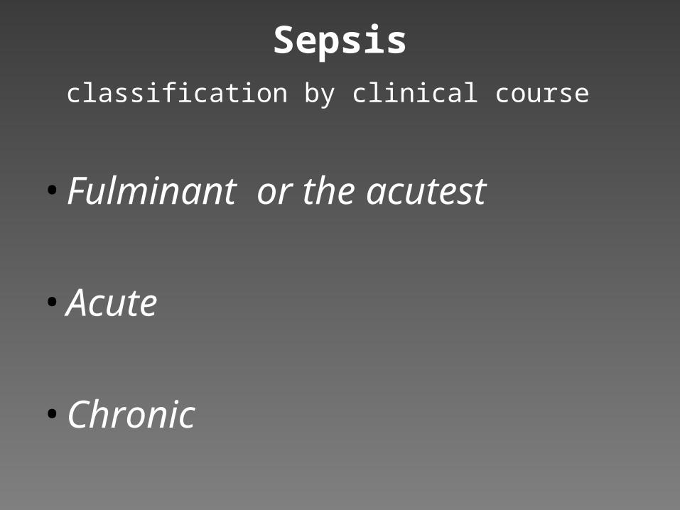
Sepsisclassification by clinical course
• Fulminant or the acutest
• Acute
• Chronic

Sepsis classification by clinical severity
Sepsis
Severe sepsis – sepsis + organ dysfunction
Septic shock – sepsis + hypotension
(Multiple organ dysfunction)

Sepsis
• Severe Sepsis– Tachycardia >90bpm– Rectal temp>38°C or <36°C
– Tachypnea(>20bpm) or PaCO2<32mmHg
– Hypotension despite fluid resuscitation– Presence of perfusion abnormalities: lactic
acidosis, oligouria, alteration in mental status

Sepsis
• Mediators of Sepsis– Lipospolysaccharide (gram-negative bacteria)
– Lipoteichoic acid (gram-positive bacteria
– Peptidoglycan
– Cytokines• IL-1 – mediates systemic effects of infection
• IL-6 – effects liver function
• TNF-α- potentiates the activation of neutrophils and macrophages
• IL-8 – regulates neutrophil function, mediates lung injury in sepsis

Sepsis
• Mediators of Sepsis– Complement– Nitric Oxide– Lipid Mediators: Chemotaxis, Cell activation,
Vascular Permeability
Phospholipase A2
PAF
Eicosanoids

Sepsis
• Mediators of Sepsis– Adhesion Molecules
• Selectins
• Leukocyte Antigens

Sepsis
• Circulatory Manifestations– Vasodilation– Tachycardia– Increased Cardiac Output– Depressed Myocardial Function– Increased Delivery– Decreased Extraction

Sepsis
• Circulatory Manifestations– Downregulation of catecholamine receptors– Increased local vasodilating substances
• Nitric oxide
• Prostacyclin
• Decreased Oxygen
• Low pH
• Increased anaerobic metabolism
• Shunting

Sepsis
• Pulmonary Dysfunction– Endothelial Injury– Interstitial Edema– Alveolar Edema– Neutrophil entrapment– Injury Type I pneumocyte– Hyperplasia Type II pneumocyte– Continued Neutrophil, monocyte, leukocyte and
platelet aggregation
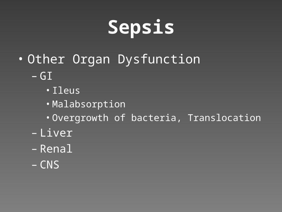
Sepsis
• Other Organ Dysfunction– GI
• Ileus
• Malabsorption
• Overgrowth of bacteria, Translocation
– Liver– Renal– CNS

Sepsis
• Organisms– Lower Respiratory Tract Infections (25%)– Urinary Tract Infections (25%)– Gastrointestinal Infections (25%)– Soft Tissue Infections (15%)– Reproductive Organs (5%)

Sepsis
• Risk Factors– Extremes of Age (<10 and >70 years)– Pre-existing Organ Dysfunction– Immunosuppression– Major Surgery, Trauma, Burns– Indwelling Devices– Prolonged Hospitalization– Malnutrition– Prior Antibiotic Treatment

Sepsis
• Principles for Management of Sepsis– Early Recognition– Early and Adequate Antibiotic Therapy– Source Control– Early Hemodynamic Resuscitation and continued
support– Drotrecogin Alpha (Apache II>25)– Tight Glycemic Control– Ventilatory Support

Sepsis
• Drotrecogin-alpha/Recombinant Human Activated Protein C– Reduced levels of anti-inflammatory mediators– Activated Protein C
• Inhibits thrombosis• Decreases inflammation• Promotes fibrinolysis
– Side Effect: Bleeding– PROWESS study group
• Lower mortality rate (24.7 vs. 30.8%)

Sepsis
• Steroids???– Older trials used high doses– Recent trials suggest low dose, with taper and tight
glycemic control may improve outcome– Vasopressor-dependent shock– Cosyntropin Stim Test-Relative Adrenal
Insufficiency (<9mcg/dL)

Sepsis
• Experimental Therapies– Dopexamine- beta 2 adrenergic and dopaminergic
effects, NO alpha adrenergic activity– Vasopressin- reduces inducible NO synthase,
upregulates endogenous catecholamine receptors– Phosphodiesterase Inhibitors-ionotropic agents
with vasodilating actions– Nitric Oxide Inhibitors- N-monomethyl-l-arginine

ARDS
• Frequent Complication in Sepsis(40%)
• Adult Respiratory Distress Syndrome– Oxygenation abnormality: PaO2/FiO2 ratio less
than 200– Bilateral opacities on CXR– PAOP <18mm Hg or no evidence of L atrial
hypertension

ARDS
• Frequent Complication in Sepsis(40%)• Adult Respiratory Distress Syndrome
– Oxygenation abnormality: PaO2/FiO2 ratio less than 200
– Bilateral opacities on CXR– PAOP <18mm Hg or no evidence of L atrial
hypertension– Frequency of ARDS in sepsis 18-38%– 16% patients die w/irreversible respiratory failure

ARDS
• Pathophysiology– Injury to Alveolocapillary unit
– Exudative Phase• Endothelial injury, immune cell infiltration, pneumocyte and
endothelial injury and necrosis
– Proliferative Phase• Organization of exudate, myofibroblast proliferation
• Conversion of exudate to fibrous tissue
– Fibrotic Phase• Remodeling of fibrosis, microcystic honeycomb formation and
traction bronchiectasis

ARDS
• Management– Lung-Protective Strategy-Reduction of
Barotrauma– TV 5ml/kg– Longer inspiratory time
– Peak Inspiratory Pressure<35-40cmH2O
– Permissive Hypercapnea– PEEP

Acute Renal Failure
• Increases Mortality in ICU 30%• Physiology
– Glomerular Filtration dependent on perfusion pressure (MAP 60-80mmHg)
– Less than 60mmHG• Decreased flow• Arterial dilation in pre-glomerular arterioles
(prostaglandins)• Constriction of post-glomerular arterioles (angiotensin
II)

Acute Renal Failure
• As Renal Perfusion Falls– Increased reabsorption in proximal tubules
• 90% water is reabsorbed (normal is 60%)
– Decreased fluid to the distal tubules• Loss of potassium elimination
– Tubular cells dependent on aerobic respiration• Ascending loop is most sensitive to ischemia

Acute Renal Failure
• Dose all drugs appropriately• Correction of Metabolic Acidosis
– Isotonic Bicarbonate – Cannot Correct Ongoing Hypoperfusion
• Renal Replacement Therapy– Absolute indication
• Acidosis• Hyperkalemia• Uremia (relative)

Sepsis
• Principles for Management of Sepsis– Early Recognition– Early and Adequate Antibiotic Therapy– Source Control– Early Hemodynamic Resuscitation and continued
support– Drotrecogin Alpha (Apache II>25)– Tight Glycemic Control– Ventilatory Support