System 1 The Respiratory - NURSING LIJAN
35
Transcript of System 1 The Respiratory - NURSING LIJAN

• Alveolar macrophages
scavenge the alveolar
surface to engulf and
remove microscopic
debris that has made it
past the “mucociliary
blanket” that traps most
foreign particles higher
in the respiratory tract.
2

Alveolar fluid• It keeps the surface between the cells and the air
moist.
• It contains surfactant which is a complex mixture ofphospholipids and lipoproteins.
• Surfactant lowers the surface tension of alveolarfluid, which reduces the tendency of alveoli tocollapse and thus maintains their patency.
3

Respiratory Membrane• Consists of four layers:
1. An alveolar wall:layer of type I andtype II alveolar cells.
2. An epithelialbasement membraneunderlying thealveolar wall
3. A capillary basementmembrane that isoften fused to theepithelial basementmembrane
4. The capillary
• Very thin membrane (0.5 μmthick) to allow rapid diffusionof gases
4

Blood Supply to the Lungs• The lungs receive blood via two sets of arteries:
1. Pulmonary arteries:• The only arteries in the body that carry deoxygenated blood.• Deoxygenated blood passes from the right ventricleb through the
pulmonary trunk, which divides into a left pulmonary artery thatenters the left lung and a right pulmonary artery that enters theright lung
• Return of the oxygenated blood to the heart occurs by way of thefour pulmonary veins, which drain into the left atrium.
2. Bronchial arteries• Deliver oxygenated blood to the lungs.• The blood mainly perfuses the muscular walls of the bronchi and
bronchioles.• Most of this blood returns to the heart via pulmonary veins.• Some blood drains into bronchial veins, and returns to the heart
via the superior vena cava
5

Ventilation-Perfusion Coupling• Ventilation-perfusion coupling is the coupling of
perfusion (blood flow) to each area of he lungs tomatch the extent of ventilation (airflow) to alveoli inthat area
• In the lungs, vasoconstriction in response to hypoxiadiverts pulmonary blood from poorly ventilatedareas of the lungs to well-ventilated regions
• In all other body tissues, hypoxia causes dilation ofblood vessels to increase blood flow
6

Respiration• Respiration is the process of gas exchange in the
body it has three basic steps:1. Pulmonary ventilation: the inflow and outflow of
air between the atmosphere and the lung alveoli.Has two parts:• Inhalation• Exhalation
2. External respiration is the exchange of gasesbetween the alveoli and the blood across therespiratory membrane. The blood gains O2 andloses CO2.
3. Internal respiration is the exchange of gasesbetween blood and tissue cells. The blood loses O2
and gains CO2.
7

Mechanics Of PulmonaryVentilation
8

Atmospheric pressure• The respiratory system depends on the medium of the
earth’s atmosphere to extract the oxygen necessaryfor life.
• The atmosphere is composed of these gases:• Nitrogen (N2) 78%• Oxygen (O2) 21%• Carbon Dioxide (CO2) 0.04%• Water Vapor ~1%
• The gases of the atmosphere have a mass and a weight• Atmospheric pressure is the force exerted against a
surface by the weight of the air above the surface.• It depends on the altitude and the temperature
• At sea level, the Atmospheric pressure is: 760 mmHg• At high altitudes, the atmospheric pressure is less;
below sea level, atmospheric pressure is greater.

10
• According toBoyle’s law, thepressure of a gasin a closedcontainer isinverselyproportional tothe volume of thecontainer.
• Gas will alwaysmove from aregion of highpressure to aregion of lowpressure.
• Applying Boyle'slaw: If the volumeinside the thoraciccavity , the

Muscles That Cause Lung Expansion AndContraction
• Atmospheric pressure is 760 mm Hg at sea level.• At rest when no air is flowing into or out of the lungs, the
pressures in all parts of the respiratory tree, all the way to thealveoli, are equal to atmospheric pressure.
• In order for air to flow in or out of the lungs the pressureinside the alveoli has to be lower or higher than atmosphericpressure, respectively.
• This is achieved by increasing or decreasing the size of thelungs.
• The lungs can be expanded and contracted in two ways:1. Downward and upward movement of the diaphragm to
lengthen or shorten the chest cavity
2. Elevation and depression of the ribs to increase anddecrease the anteroposterior diameter of the chest cavity.
11

• Contraction of the diaphragmcauses it to flatten, lowering itsdome. This increases the verticaldiameter of the thoracic cavity.
• Contraction of externalintercostals elevates the ribs andpush the sternum forward. Thisincrease the anteroposterior andlateral diameters of the chest
it
• During expiration, the diaphragmsimply relaxes, and the elasticrecoil of the lungs, chest wall, andabdominal structures compressesthe lungs and expels the air.
• Contraction of internal intercostals& abdominal recti pulls the ribsdownwards and push the sternumbackward. This decreases the
t t i d l t l
12

13

Pressure Changes during Inhalation• The most important muscle of inhalation is the
diaphragm.• Contraction of the diaphragm is responsible for
about 75% of the air that enters the lungs duringquiet breathing.
• Contraction of the external intercostals is responsiblefor about 25% of the air that enters the lungs duringnormal quiet breathing.
• During deep & forceful inhalations , the diaphragmmay descend up to 10 cm, and the accessory musclesof inspiration also participate in increasing the sizeof the thoracic cavity.
• The accessory muscles of inhalation include thesternocleidomastoid muscles the scalene muscles
14

Pressure Changes during Exhalation• Exhalation during quiet breathing is a passive
process because no muscular contractions areinvolved.
• It results from elastic recoil of the chest wall andlungs, both of which have a natural tendency tospring back after they have been stretched. Forcesthat contribute to elastic recoil:
1. The recoil of elastic fibers that were stretchedduring inhalation
2. The inward pull of surface tension due to the filmof alveolar fluid.
• Exhalation becomes active only during forcefulbreathing
• Muscles of exhalation (the abdominal recti andinternal intercostals) contract which increases
15

Intrapleural Pressure• It is the pressure between the two pleural layers in
the pleural cavity• It is always subatmospheric (lower than atmospheric
pressure).• This creates a suction force that holds the lungs open• Just before inhalation, it is about 4 mmHg less than
the atmospheric pressure• During inspiration as the size of the thoracic cavity
increases, the volume of the pleural cavity alsoincreases, which causes intrapleural pressure todecrease even more.
• This will help the lungs to expand.
16

17
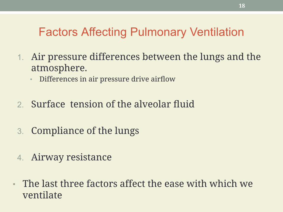
Factors Affecting Pulmonary Ventilation
1. Air pressure differences between the lungs and theatmosphere.• Differences in air pressure drive airflow
2. Surface tension of the alveolar fluid
3. Compliance of the lungs
4. Airway resistance
• The last three factors affect the ease with which weventilate
18

Surface Tension of Alveolar Fluid• A thin layer of alveolar fluid coats the luminal surface of
alveoli and exerts surface tension.• This produces an inwardly directed force.• Surface tension accounts for two-thirds of lung elastic
recoil.
• However surface tension must be overcome to expandthe lungs during each inhalation.
• The surfactant (a mixture of phospholipids andlipoproteins) present in alveolar fluid reduces its surfacetension below the surface tension of pure water.
• Without the surfactant the alveoli collapse at the end ofeach exhalation. Great effort is then needed at the nextinhalation to reopen the collapsed alveoli.
19

Surface tension arises at all air–water interfaces because the watermolecules are more strongly attracted to each other than they are to gasmolecules in the air.
20

21

Compliance of the Lungs• Compliance refers to how much effort is required to
stretch the lungs and chest wall.
• Normally the lungs and chest wall have highcompliance SO the lungs and chest wall expand easily.
• Depends on two factors: elasticity and surfacetension.
• Lung tissue is made of elastic fibers that are easilystretched this increases the compliance
• Surfactant in alveolar fluid reduces surface tensionthereby it increases the compliance
22

Airway Resistance• Airflow is governed by the basic flow equation,
which relates flow to the pressure difference &airways resistance.
• Flow = pressure difference / resistance = ΔP/R
• Airway resistance depends on the diameter of theairway any change in the diameter will have aprofound effect on the resistance
• During inhalation as the lungs expand thebronchioles enlarge because their walls are pulledoutward in all directions. This decreases theresistance.
• During exhalation airway resistance increases as thediameter of bronchioles decreases.
23

• Airway diameter is also regulated by the degree ofcontraction or relaxation of smooth muscle in the walls ofthe airways.
• Signals from the sympathetic nervous system cause relaxation ofthe smooth muscles, which results in bronchodilation anddecreased resistance.
• Signals from the parasympathetic nervous system causecontraction of the smooth muscles, which results inbronchoconstriction and increased resistance.
• Normally airway resistance is small and negligible. So ΔPis the main determinant of air flow
• Any pathology that narrows or obstructs the airwaysincreases resistance, so that more pressure difference isrequired to maintain the same airflow.
24

Measuring Ventilation• Ventilation can be measured using spirometry.• Spirometre is the apparatus commonly used to measure the
volume of air exchanged during breathing.
• The record obtained from spirometry is called a spirogram.Inhalation is recorded as an upward deflection, and exhalationis recorded as a downward deflection
• Tidal Volume (VT) is the volume of air inspired (or expired)during normal quiet breathing (500 ml).
• Inspiratory Reserve Volume (IRV) is the volume inspiredduring a very deep inhalation (3100 ml – height and genderdependent).
• Expiratory Reserve Volume (ERV) is the volume expired duringa forced exhalation (1200 ml).

26
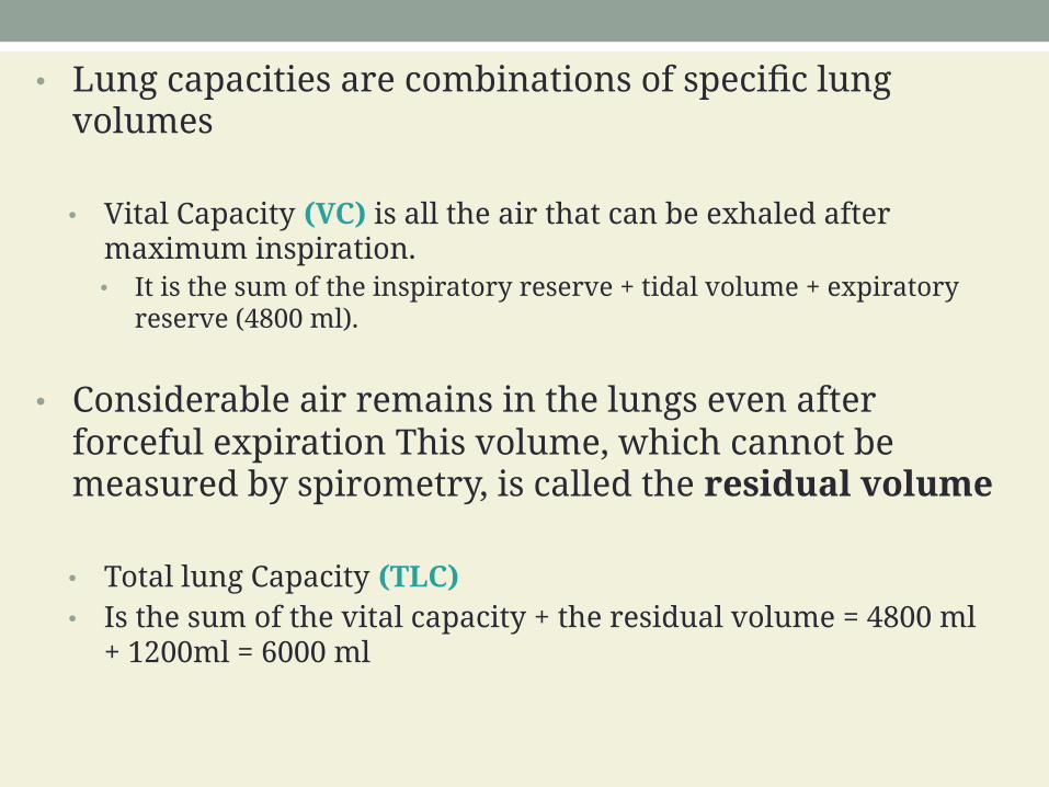
• Lung capacities are combinations of specific lungvolumes
• Vital Capacity (VC) is all the air that can be exhaled aftermaximum inspiration.• It is the sum of the inspiratory reserve + tidal volume + expiratory
reserve (4800 ml).
• Considerable air remains in the lungs even afterforceful expiration This volume, which cannot bemeasured by spirometry, is called the residual volume
• Total lung Capacity (TLC)• Is the sum of the vital capacity + the residual volume = 4800 ml
+ 1200ml = 6000 ml

Anatomic dead space• About 70% of the tidal volume (350 mL) actually
reaches the the respiratory bronchioles, alveolarducts, alveolar sacs, and alveoli and participates inexternal respiration.
• The other 30% (150 mL) remains in the conductingairways of the nose, pharynx, larynx, trachea,bronchi, bronchioles, and terminal bronchioles.
• The air that does not undergo respiratory exchangeis known as the anatomic dead space.
28

Minute & Alveolar Ventilation• The respiratory rate is the number of breaths a
person takes each minute. At rest, a healthy adulttakes 12 breaths a minute.
• The minute ventilation (MV) is the total volume ofair inhaled and exhaled each minute
• MV= respiratory rate X VT = 12 X 500 = 6000 ml/min
• The alveolar ventilation rate is the volume of airper minute that actually reaches the respiratory zone.
• The alveolar ventilation (volume of air/min that actuallyreaches the alveoli) = 12 x 350 = 4200ml.
29

Physiological dead space• Alveolar dead space refers to the alveoli that are
nonfunctional or only partially functional because ofabsent or poor blood flow through the adjacentpulmonary capillaries.
• Physiological dead space = Anatomical dead space +Alveolar dead space
30

Exchange of Oxygen and CarbonDioxide
• The exchange of oxygen and carbon dioxide betweenalveolar air and pulmonary blood occurs via passivediffusion.
• Which is governed by the behavior of gases asdescribed by two gas laws:
1. Dalton’s law which is important for understandinghow gases move down their pressure gradients bydiffusion
2. Henry’s law which helps explain how the solubilityof a gas relates to its diffusion.
31

Dalton’s Law• According to Dalton’s law, each gas in a mixture of
gases exerts its own pressure as if no other gaseswere present.
•• The pressure of a specific gas in a mixture is called its
partial pressure (Px)
• The total pressure of the mixture is calculated simplyby adding all of the partial pressures.
• Atmospheric air is a mixture of gases so
32

PN2 = 0.786 x 760 mmHg = 597.4 mmHg
PO2 = 0.209 x 760 mmHg = 158.8 mmHg
PH2O = 0.004 x 760 mmHg = 3.0 mmHg
PCO2=
0.0004 x 760 mmHg = 0.3 mmHg
Pother gases=
0.0006 x 760 mmHg = 0.5 mmHg
Total = 760.0 mmHgPartial pressures of gases in inhaled air for sea
level

• We can determine the partial pressure exerted byeach component in the mixture by multiplying thepercentage of the gas in the mixture by the totalpressure of the mixture.
• Each gas diffuses across a permeable membranefrom the area where its partial pressure is greater tothe area where its partial pressure is less.
• The greater the difference in partial pressure, thefaster the rate of diffusion.
34

Henry’s Law• Henry’s law states that the quantity of a gas that will
dissolve in a liquid is proportional to the partialpressure of the gas and its solubility.
• The solubility of CO2 is 24 times greater than that ofO2.
• Even though the air we breathe contains about 79%nitrogen, very little of it dissolves in blood plasmabecause of its low solubility at sea level pressure.
35