
Synthesis of Uniformly Sized Manganese Oxide Nanocrystals...
8
FULL PAPER DOI: 10.1002/ejic.201101193 Synthesis of Uniformly Sized Manganese Oxide Nanocrystals with Various Sizes and Shapes and Characterization of Their T 1 Magnetic Resonance Relaxivity Kwangjin An, [a] Mihyun Park, [a] Jung Ho Yu, [a] Hyon Bin Na, [a] Nohyun Lee, [a] Jongnam Park, [a,b] Seung Hong Choi, [c] In Chan Song, [c] Woo Kyung Moon, [c] and Taeghwan Hyeon* [a] Keywords: Manganese / Nanoparticles / Thermal decomposition / Heat-up process / Imaging agents We synthesized manganese oxide (MnO and Mn 3 O 4 ) nano- crystals with various sizes and shapes by the thermal reaction of a Mn II –oleate complex through a “heat-up process”. When a Mn II –oleate complex was thermally decomposed in non- coordinating hydrocarbon solvents, uniformly sized MnO nanocrystals with cubic and octahedral shapes were pro- duced. We were able to synthesize anisotropic, multi- branched MnO nanocrystals by the oriented attachment of MnO truncated-nanocube building blocks. When the Mn II – oleate complex was heated in 1-hexadecene in the presence of strongly coordinating carboxylic acid surfactants, spherical nanocrystals were generated, and their diameter was con- trolled in the range 3–13 nm by varying the chain length of the carboxylic acid. When oleyl alcohol was added to the Introduction The designed synthesis of uniformly sized nanocrystals with controlled sizes and shapes is of great importance for both fundamental science and technical applications, be- cause the properties of nanocrystals directly depend on their size and shape. [1] Recently, manganese oxide nanocrys- tals have attracted a lot of attention because of their unique magnetic properties, which result from both the variable oxidation states of their manganese ions (Mn 2+ , Mn 3+ , and Mn 4+ ) and their high surface-to-volume ratio. [2] Recent re- ports demonstrated that MnO nanocrystals were readily ox- idized to MnO/Mn 3 O 4 core–shell structures, which exhibit [a] World Class University (WCU) Program of Chemical Convergence for Energy & Environment (C2E2), Institute of Chemical Processes, and School of Chemical and Biological Engineering, Seoul National University, Seoul 151-744, Korea E-mail: [email protected] [b] Interdisciplinary School of Green Energy, School of Nano- Bioscience and Chemical Engineering, Ulsan National Institute of Science and Technology (UNIST), Ulsan 681-800, Korea [c] Department of Radiology, Seoul National University College of Medicine, Seoul National University Hospital, Seoul 110-744, Korea Supporting information for this article is available on the WWW under http://dx.doi.org/10.1002/ejic.201101193. © 2012 Wiley-VCH Verlag GmbH & Co. KGaA, Weinheim Eur. J. Inorg. Chem. 2012, 2148–2155 2148 Mn–oleate complex in phenyl ether, tetrahedral MnO nano- crystals were synthesized. The as-synthesized MnO nano- crystals were oxidized in air to Mn 3 O 4 or MnO/Mn 3 O 4 core– shell structures, which exhibited exchange coupling with shifted magnetic hysteresis loops. The effect of the size and shape of the phospholipid-capped manganese oxide nano- crystals on their applicability as T 1 contrast agents in mag- netic resonance imaging (MRI) were examined. As the size of the nanocrystals decreased, their relaxivities increased, thereby generating brighter MR images. In particular, spher- ical 3 nm-sized Mn 3 O 4 nanocrystals had a high specific re- laxivity (r 1 ) of 2.38 mM –1 s –1 , clearly demonstrating their po- tential for use as an efficient T 1 MRI contrast agent. exchange coupling at the interface of two magnetic compo- nents. [3] Various methods have been reported for the synthe- sis of manganese oxide nanocrystals with various sizes and shapes. [4] In particular, thermal decomposition of metal– oleate complexes through a “heat-up process” has been used to produce uniformly sized manganese oxide nano- crystals. [5,6] A tremendous amount of research has been performed on the applications of magnetic iron oxide nanocrystals as T 2 contrast agents in magnetic resonance imaging (MRI). [7] On the other hand, biocompatible manganese oxide nano- crystals have been developed as new T 1 MRI contrast agents for in vivo diagnosis. [8] However, there have been very few reports on the size- and shape-dependent T 1 con- trast properties of manganese oxide nanocrystals. [9] Herein, we report on the size- and shape-controlled synthesis of manganese oxide nanocrystals by thermal decomposition of a Mn–oleate complex through a heat-up process. Various manganese oxide nanocrystals were synthesized, having not only thermodynamically controlled cubic, octahedral, and spherical shapes but also kinetically controlled branched or tetrahedral shapes. Crystal structures as well as size- and shape-dependent magnetic properties of the synthesized manganese oxide nanocrystals were determined. Further- more, we investigated the T 1 relaxation properties of the
Transcript of Synthesis of Uniformly Sized Manganese Oxide Nanocrystals...
FULL PAPER
DOI: 10.1002/ejic.201101193
Synthesis of Uniformly Sized Manganese Oxide Nanocrystals with VariousSizes and Shapes and Characterization of Their T1 Magnetic Resonance
Relaxivity
Kwangjin An,[a] Mihyun Park,[a] Jung Ho Yu,[a] Hyon Bin Na,[a] Nohyun Lee,[a]
Jongnam Park,[a,b] Seung Hong Choi,[c] In Chan Song,[c] Woo Kyung Moon,[c] andTaeghwan Hyeon*[a]
Keywords: Manganese / Nanoparticles / Thermal decomposition / Heat-up process / Imaging agents
We synthesized manganese oxide (MnO and Mn3O4) nano-crystals with various sizes and shapes by the thermal reactionof a MnII–oleate complex through a “heat-up process”. Whena MnII–oleate complex was thermally decomposed in non-coordinating hydrocarbon solvents, uniformly sized MnOnanocrystals with cubic and octahedral shapes were pro-duced. We were able to synthesize anisotropic, multi-branched MnO nanocrystals by the oriented attachment ofMnO truncated-nanocube building blocks. When the MnII–oleate complex was heated in 1-hexadecene in the presenceof strongly coordinating carboxylic acid surfactants, sphericalnanocrystals were generated, and their diameter was con-trolled in the range 3–13 nm by varying the chain length ofthe carboxylic acid. When oleyl alcohol was added to the
Introduction
The designed synthesis of uniformly sized nanocrystalswith controlled sizes and shapes is of great importance forboth fundamental science and technical applications, be-cause the properties of nanocrystals directly depend ontheir size and shape.[1] Recently, manganese oxide nanocrys-tals have attracted a lot of attention because of their uniquemagnetic properties, which result from both the variableoxidation states of their manganese ions (Mn2+, Mn3+, andMn4+) and their high surface-to-volume ratio.[2] Recent re-ports demonstrated that MnO nanocrystals were readily ox-idized to MnO/Mn3O4 core–shell structures, which exhibit
[a] World Class University (WCU) Program of ChemicalConvergence for Energy & Environment (C2E2), Institute ofChemical Processes, and School of Chemical and BiologicalEngineering, Seoul National University,Seoul 151-744, KoreaE-mail: [email protected]
[b] Interdisciplinary School of Green Energy, School of Nano-Bioscience and Chemical Engineering, Ulsan National Instituteof Science and Technology (UNIST),Ulsan 681-800, Korea
[c] Department of Radiology, Seoul National University Collegeof Medicine, Seoul National University Hospital,Seoul 110-744, KoreaSupporting information for this article is available on theWWW under http://dx.doi.org/10.1002/ejic.201101193.
© 2012 Wiley-VCH Verlag GmbH & Co. KGaA, Weinheim Eur. J. Inorg. Chem. 2012, 2148–21552148
Mn–oleate complex in phenyl ether, tetrahedral MnO nano-crystals were synthesized. The as-synthesized MnO nano-crystals were oxidized in air to Mn3O4 or MnO/Mn3O4 core–shell structures, which exhibited exchange coupling withshifted magnetic hysteresis loops. The effect of the size andshape of the phospholipid-capped manganese oxide nano-crystals on their applicability as T1 contrast agents in mag-netic resonance imaging (MRI) were examined. As the sizeof the nanocrystals decreased, their relaxivities increased,thereby generating brighter MR images. In particular, spher-ical 3 nm-sized Mn3O4 nanocrystals had a high specific re-laxivity (r1) of 2.38 mM–1 s–1, clearly demonstrating their po-tential for use as an efficient T1 MRI contrast agent.
exchange coupling at the interface of two magnetic compo-nents.[3] Various methods have been reported for the synthe-sis of manganese oxide nanocrystals with various sizes andshapes.[4] In particular, thermal decomposition of metal–oleate complexes through a “heat-up process” has beenused to produce uniformly sized manganese oxide nano-crystals.[5,6]
A tremendous amount of research has been performedon the applications of magnetic iron oxide nanocrystals asT2 contrast agents in magnetic resonance imaging (MRI).[7]
On the other hand, biocompatible manganese oxide nano-crystals have been developed as new T1 MRI contrastagents for in vivo diagnosis.[8] However, there have beenvery few reports on the size- and shape-dependent T1 con-trast properties of manganese oxide nanocrystals.[9] Herein,we report on the size- and shape-controlled synthesis ofmanganese oxide nanocrystals by thermal decomposition ofa Mn–oleate complex through a heat-up process. Variousmanganese oxide nanocrystals were synthesized, having notonly thermodynamically controlled cubic, octahedral, andspherical shapes but also kinetically controlled branched ortetrahedral shapes. Crystal structures as well as size- andshape-dependent magnetic properties of the synthesizedmanganese oxide nanocrystals were determined. Further-more, we investigated the T1 relaxation properties of the

Uniformly Sized Manganese Oxide Nanocrystals and Their T1 Relaxivity
manganese oxide nanocrystals dispersed in water with re-gard to their potential applications as MRI contrast agents.
Results and Discussion
Uniformly sized MnO nanocrystals with cubic and octa-hedral shapes were synthesized by the thermal decomposi-tion of a MnII–oleate complex in non-coordinating hydro-carbon solvents. When a solution of the MnII–oleate com-plex in 1-hexadecene was slowly heated to reflux (288 °C),without the employment of any additional surfactant, andkept at that temperature for 1 h, uniformly sized MnO nano-cubes with an edge length of 16 nm were synthesized (Fig-ure 1a). When the reaction was conducted in 1-octadeceneinstead of 1-hexadecene at a higher temperature of 320 °Cfor 30 min, octahedral MnO nanocrystals with an averageedge length of 26 nm were generated (Figure 1b). When thesynthesis was conducted for a longer time of 1 h, the edgelength of the octahedral nanocrystals increased slightly to29 nm (Figure 1c). In the heat-up process, traces of waterand oxygen significantly affect not only the crystallinity andphase of the manganese oxide nanocrystals[4c,10] but alsothe uniformity of their size and shape.[6] Therefore, theMnII–oleate complex was completely dried under vacuumto yield a pink powder (Supporting Information Figure S1),and the synthesis was performed using standard Schlenktechniques. The X-ray diffraction (XRD) pattern revealedthat the as-synthesized MnO nanocrystals possess a cubicrock-salt structure (Fm3m, JCPDS No. 78–0424) withoutany contamination by Mn3O4 (Supporting Information,Figure S3).[4] Because of the extremely narrow size distribu-tion of the nanocrystals, 2D superlattices were readily gen-erated. Furthermore, the superlattice structure depended onthe shape of the MnO nanocrystals.[11] For example, thecubic MnO nanocrystals assembled to form a rectangularstructure (Figure 1a), while the octahedral MnO nanocrys-tals were hexagonally assembled (Figure 1b and c). Underthe current thermal decomposition reaction conditions, therelatively small nanocrystals (of size less than 16 nm) syn-thesized at low temperature were preferentially cubic, whilethe large nanocrystals with diameters greater than 20 nm,which were synthesized at a high temperature of 320 °C,were octahedral. In a cubic rock-salt structure, the three
Figure 1. TEM images of MnO nanocrystals with (a) cubic and (b,c) octahedral shapes. The mean lengths of their edges are (a) 16 nm,(b) 26 nm, and (c) 29 nm.
Eur. J. Inorg. Chem. 2012, 2148–2155 © 2012 Wiley-VCH Verlag GmbH & Co. KGaA, Weinheim www.eurjic.org 2149
low-energy surfaces are the {100}, {110}, and {111} crystalplanes, with a surface-energy ratio of 1/1.41/1.73.[12] There-fore, we speculate that at relatively low temperatures, thesmaller and {100}-surface-developed cubes formed to mini-mize the total surface energy, while at high temperaturesthe larger and {111}-surface-developed nano-octahedronscould be generated, as high temperatures provide sufficientenergy for nanocrystals to grow along the {100} surface.Furthermore, the formation mechanism for MnO nano-cubes is very similar to that of 5 nm-sized iron oxide nano-cubes.[5a]
When strongly coordinating carboxylic acid surfactantswith long hydrocarbon chains were added to the reactionmixture, spherical nanocrystals were generated (Figure 2).The particle size was controlled by varying the chain lengthof the carboxylic acids. For example, spherical MnO nano-crystals with particle diameters of 3 nm (Figure 2a), 5 nm(Figure 2b), 11 nm (Figure 2c), and 13 nm (Figure 2d) weresynthesized by using behenic acid (C21H43COOH), stearicacid (C17H35COOH), myristic acid (C13H27COOH), anddecanoic acid (C9H19COOH), respectively. In view of theaverage surface energy of a rock-salt-structured sphere,which is between the surface energy of nanocubes andnano-octahedrons, the experimental results show that, re-gardless of the nanocrystal size, excess carboxylic acid sur-factant further reduces the surface energy, inhibiting shape-controlled growth and resulting in the formation of spheri-cal nanocrystals.[13] The XRD patterns reveal that the as-synthesized MnO nanocrystals with sizes of 11 and 13 nmhave a pure cubic rock-salt crystal structure (JCPDS No.78-0424), whereas the nanocrystals with sizes of 3 and 5 nmpossess mixed crystal structures of cubic MnO and tetrago-nal Mn3O4 (JCPDS No. 80–0382) (see Supporting Infor-mation, Figure S4). It seems that the small MnO nanocrys-tals with a high surface-to-volume ratio are readily oxidizedin air. Recent studies demonstrated that the as-synthesizedMnO nanocrystals were transformed to MnO/Mn3O4 core–shell structures by surface oxidation in air, resulting in theformation of exchange-coupled magnetic structures withshifted magnetic hysteresis loops.[3] On the other hand,when we intentionally oxidized the 26 nm-sized MnO nano-crystals by using trimethylamine N-oxide as an oxidant,pure Mn3O4 nanocrystals were generated (Supporting In-

T. Hyeon et al.FULL PAPERformation, Figure S5).[4a,4e] The result implies that Mn3O4
nanocrystals with various sizes and shapes could be synthe-sized by further oxidation of as-synthesized MnO nanocrys-tals.
Figure 2. TEM images of spherical MnO nanocrystals with nano-crystal diameters of (a) 3 nm, (b) 5 nm, (c) 11 nm, and (d) 13 nm,synthesized by using behenic acid, stearic acid, myristic acid, anddecanoic acid, respectively.
As described above, when the Mn–oleate complex washeated to reflux in non-coordinating solvents, such as 1-hexadecene (288 °C) and 1-octadecene (320 °C), isotropicMnO nanocrystals with cubic and octahedral shapes weregenerated. However, when the Mn–oleate complex washeated at a slightly lower temperature of 280 °C in the pres-ence of excess carboxylic acid surfactants, club-shapedMnO nanorods were produced. For example, when myristicacid was used as the carboxylic acid surfactant, MnO nano-rods with club-shaped tips at both ends were generated(Figure 3a), whereas a mixture of club-shaped nanorodsand T-shaped nanorods with club-shaped tips at all ex-tremities was produced when decanoic acid was used in thesynthesis (Figure 3b). It is interesting that this little differ-ence in the reaction temperature completely changed thefinal shape of the nanocrystals.[14] Furthermore, when weperformed the synthesis at an intermediate temperature of285 °C, a mixture of spherical and rod-shaped MnO nano-crystals was generated (Figure 3c). The high-resolutiontransmission electron microscopy (HRTEM) image of aMnO nanorod revealed that truncated nanocubes of ap-proximately 10 nm in size with a {200} crystal lattice pa-rameter of 2.17 Å were aligned along the �100� direction(Figure 3d). From these results, we concluded that the an-isotropic growth of MnO nanocrystals proceeded by theoriented attachment mechanism.[15,16]
The addition of alcohol during the heat-up process isknown to significantly alter the nanocrystal growth behav-
www.eurjic.org © 2012 Wiley-VCH Verlag GmbH & Co. KGaA, Weinheim Eur. J. Inorg. Chem. 2012, 2148–21552150
Figure 3. TEM images of (a) rod-shaped and (b) T-shaped MnOnanocrystals and of (c) a mixture of spherical MnO nanocrystalsand anisotropic MnO nanocrystals. (d) High-resolution TEM im-age of rod-shaped MnO nanocrystals (inset: schematic illustrationshowing the oriented attachment of the MnO nanocrystals alongthe �100� direction).
Figure 4. TEM images of tetrahedral MnO nanocrystals with lat-eral dimensions of (a) 6 nm, (b) 8 nm, and (c) 20 nm. (d) HRTEMimage and (e) XRD pattern of 20 nm-sized nanocrystals.

Uniformly Sized Manganese Oxide Nanocrystals and Their T1 Relaxivity
ior.[17] When oleyl alcohol was injected into a Mn–oleatesolution in phenyl ether solvent at 260 °C, tetrahedral MnOnanocrystals were obtained (Figures 4 and S6). By con-trolling the heating rate and the reaction time, the edgelength of the tetrahedral nanocrystals was adjusted to 6, 8,and 20 nm (Figures 4a, b, and c, respectively). The HRTEMimage (Figure 4d) and XRD pattern (Figure 4e) clearlyshow that the tetrahedral MnO nanocrystals possess a cubicrock-salt crystal structure.[4] The well-defined lattice planeswith their interplanar distance of 2.56 Å show that tetrahe-dral MnO nanocrystals were generated with the develop-ment of the {110} surface, which has the second lowest sur-face energy in a cubic rock-salt structure.[12] Consideringthat the growth temperature of tetrahedral MnO nanocrys-tals (260 °C) is significantly lower than that of {100}-sur-face-developed cubic (288 °C) or {111}-surface-developedoctahedral (320 °C) nanocrystals, we speculate that theoleyl alcohol coordinates on the nanocrystal surface tolower the surface energy, consequently altering the nano-crystal growth behavior. Furthermore, in the formation ofthe tetrahedral MnO nanocrystals, not only oleyl alcoholbut also the ether played an important role. While phenylether could be replaced with octyl ether for the synthesisof tetrahedral MnO nanocrystals, the addition of a non-coordinating solvent, such as 1-octadecene, instead of
Figure 5. (a,b) Temperature and (c,d) field dependence of magnetizations of (a, c) spherical and (b, d) tetrahedral manganese oxidenanocrystals, which were stored under ambient conditions for one month. [All of the magnetization measurements were carried out atthe temperature range 5–300 K, after zero-field cooling (ZFC) or field cooling (FC) with an applied field of 100 Oe from 300 K, andmagnetic hysteresis loops were measured at 5 K.].
Eur. J. Inorg. Chem. 2012, 2148–2155 © 2012 Wiley-VCH Verlag GmbH & Co. KGaA, Weinheim www.eurjic.org 2151
phenyl ether, resulted in the formation of octahedral MnOnanocrystals (Supporting Information, Figure S7). To thebest of our knowledge, this is the first report on the synthe-sis of tetrahedral MnO nanocrystals. The experimental con-ditions used for the preparation of the various manganeseoxide nanocrystals and the size distribution histograms de-scribed above are summarized in Table S1 and Figure S8 ofthe Supporting Information.
We investigated the size-dependent magnetic propertiesof spherical and tetrahedral manganese oxide nanocrystals.The temperature dependence of the magnetization of man-ganese oxide nanocrystals was measured with zero-field-cooling (ZFC) and field-cooling (FC) procedures in an ap-plied magnetic field of 100 Oe in the temperature range 5–300 K (Figure 5a, b).[18] The observed blocking tempera-tures (TB) were 15, 17, 17, and 24 K for spherical nanocrys-tals with diameters of 3, 5, 11, and 13 nm, respectively, and24, 31, and 24 K for tetrahedral nanocrystals with edgelengths of 6, 8, and 20 nm, respectively. When MnO nano-crystals are exposed to air, they are spontaneously oxidizedto core–shell structured MnO/Mn3O4 mixtures or Mn3O4.[3]
The degree of surface oxidation varies with nanocrystal sizeand shape. In the case of spherical nanocrystals, MnOnanocrystals with sizes of 3 and 5 nm were fully trans-formed to Mn3O4 (Supporting Information, Figure S9).
T. Hyeon et al.FULL PAPERThe blocking temperatures of the spherical Mn3O4 nano-crystals of size 3 and 5 nm have a linear relationship withtheir particle volume, which is consistent with previous re-ports.[4a–4c] However, the nanocrystals of size 11 and 13 nmwere transformed to a MnO/Mn3O4 core–shell structure.The core–shell structure is composed of an antiferromag-netic core and a ferrimagnetic shell, and the structure isknown to exhibit exchange bias, that is, a displacement ofthe hysteresis loop along the magnetic field axis.[19] Asshown in Figure 5c, spherical MnO/Mn3O4 core–shellnanocrystals with sizes of 11 and 13 nm exhibit high ex-change bias with 250 Oe of exchange bias field (HE), whilespherical Mn3O4 nanocrystals of size 3 and 5 nm exhibitnegligible exchange bias (Supporting Information,Table S2). Likewise, the tetrahedral MnO nanocrystals withsizes of 6 and 8 nm were transformed to MnO/Mn3O4 core–shell structures, which exhibited exchange bias with 700 and300 Oe of HE, respectively (Figure 5d). On the other hand,air-oxidation of tetrahedral 20 nm-sized MnO nanocrystalswas not detected by X-ray diffraction. Consequently, theblocking temperature of the tetrahedral 20 nm-sized MnOnanocrystals was slightly lower than that of the smallerMnO/Mn3O4 core–shell structured nanocrystals. Further-more, the tetrahedral nanocrystals with edge lengths of 6and 8 nm show an anomalous hysteresis with steps, whichis presumably due to the exchange bias combined with theunique morphology of tetrahedral MnO/Mn3O4 core–shellstructures (Figure 5d).[20] The experimental results showthat the magnetic properties of manganese oxide nanocrys-tals strongly depend on their detailed crystalline structures.
Recently, biocompatible MnO nanocrystals were pro-posed as a new T1 MRI contrast agent, and their size-de-pendent T1 relaxation properties were characterized.[8] Thesmaller MnO nanocrystals exhibited brighter signal en-hancement in the T1-weighted MRI than the larger ones.[8a]
Later, it was reported that as-synthesized MnO nanocrys-tals were spontaneously oxidized to Mn3O4 nanocrystals inair.[9a] The T1 relaxation property of as-synthesized Mn3O4
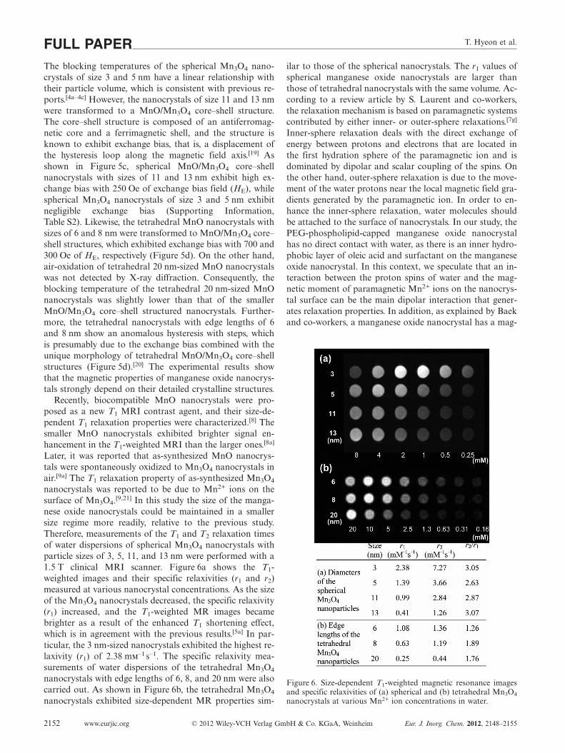
nanocrystals was reported to be due to Mn2+ ions on thesurface of Mn3O4.[9,21] In this study the size of the manga-nese oxide nanocrystals could be maintained in a smallersize regime more readily, relative to the previous study.Therefore, measurements of the T1 and T2 relaxation timesof water dispersions of spherical Mn3O4 nanocrystals withparticle sizes of 3, 5, 11, and 13 nm were performed with a1.5 T clinical MRI scanner. Figure 6a shows the T1-weighted images and their specific relaxivities (r1 and r2)measured at various nanocrystal concentrations. As the sizeof the Mn3O4 nanocrystals decreased, the specific relaxivity(r1) increased, and the T1-weighted MR images becamebrighter as a result of the enhanced T1 shortening effect,which is in agreement with the previous results.[5a] In par-ticular, the 3 nm-sized nanocrystals exhibited the highest re-laxivity (r1) of 2.38 mm–1 s–1. The specific relaxivity mea-surements of water dispersions of the tetrahedral Mn3O4
nanocrystals with edge lengths of 6, 8, and 20 nm were alsocarried out. As shown in Figure 6b, the tetrahedral Mn3O4
nanocrystals exhibited size-dependent MR properties sim-
www.eurjic.org © 2012 Wiley-VCH Verlag GmbH & Co. KGaA, Weinheim Eur. J. Inorg. Chem. 2012, 2148–21552152
ilar to those of the spherical nanocrystals. The r1 values ofspherical manganese oxide nanocrystals are larger thanthose of tetrahedral nanocrystals with the same volume. Ac-cording to a review article by S. Laurent and co-workers,the relaxation mechanism is based on paramagnetic systemscontributed by either inner- or outer-sphere relaxations.[7g]
Inner-sphere relaxation deals with the direct exchange ofenergy between protons and electrons that are located inthe first hydration sphere of the paramagnetic ion and isdominated by dipolar and scalar coupling of the spins. Onthe other hand, outer-sphere relaxation is due to the move-ment of the water protons near the local magnetic field gra-dients generated by the paramagnetic ion. In order to en-hance the inner-sphere relaxation, water molecules shouldbe attached to the surface of nanocrystals. In our study, thePEG-phospholipid-capped manganese oxide nanocrystalhas no direct contact with water, as there is an inner hydro-phobic layer of oleic acid and surfactant on the manganeseoxide nanocrystal. In this context, we speculate that an in-teraction between the proton spins of water and the mag-netic moment of paramagnetic Mn2+ ions on the nanocrys-tal surface can be the main dipolar interaction that gener-ates relaxation properties. In addition, as explained by Baekand co-workers, a manganese oxide nanocrystal has a mag-
Figure 6. Size-dependent T1-weighted magnetic resonance imagesand specific relaxivities of (a) spherical and (b) tetrahedral Mn3O4
nanocrystals at various Mn2+ ion concentrations in water.
Uniformly Sized Manganese Oxide Nanocrystals and Their T1 Relaxivity
netic moment due to incomplete cancelation of the antifer-romagnetic spins in an oxide lattice.[9d] The Mn2+ ions onthe surface of nanocrystals can increase the magnetic mo-ment by breaking the symmetry of an oxide lattice. Thehigh surface-to-volume ratio of small nanocrystals causes ahigher contrast effect through enhanced relaxation causedby the surface Mn2+ ions. The magnetic-resonance-activeMn2+ ions on the surface of the Mn3O4 nanocrystalsshorten the T1 relaxation time of water protons, thus ren-dering brighter MR images.
Conclusions
In summary, we report the size- and shape-controlledsynthesis of manganese oxide nanocrystals by the thermaldecomposition of a Mn–oleate complex through a heat-upprocess. When the thermal decomposition of the MnII–ole-ate complex was conducted in non-coordinating hydro-carbon solvents, uniformly sized MnO nanocrystals withcubic and octahedral shapes were synthesized. When thethermal decomposition was performed in the presence ofstrongly coordinating carboxylic acid surfactants, sphericalnanocrystals were generated, whose diameters were con-trolled from 3 to 13 nm by varying the chain length of thecarboxylic acids. Tetrahedral MnO nanocrystals were syn-thesized by the addition of oleyl alcohol to the reactionmixture at high temperatures. The as-synthesized MnOnanocrystals were transformed to Mn3O4 or MnO/Mn3O4
core–shell structures by oxidation in air. The air-oxidizedMnO/Mn3O4 nanocrystals exhibited exchange couplingwith shifted magnetic hysteresis loops. Finally, an investiga-tion of the T1 relaxation properties of the various manga-nese oxide nanocrystals was carried out. As the size of thenanocrystals decreased, their specific relaxivities increased,thereby generating brighter MR images. In particular, thespherical 3 nm-sized Mn3O4 nanocrystals exhibited a highspecific relaxivity (r1) of 2.38 mm–1 s–1, which clearly dem-onstrates their potential use as an efficient T1 MRI contrastagent. The current study provides valuable information forthe development of optimal nanocrystal-based T1 MRIcontrast agents by the fine tuning of the sizes and shapesof manganese oxide nanocrystals.
Experimental Section
Chemicals: Hexane, ethanol, and acetone were used as received.Manganese(II) chloride tetrahydrate (MnCl2·4H2O, 98%), oleicacid [CH3(CH2)7CH=CH(CH2)7COOH, 90%], stearic acid(C17H35COOH, 95 %), myristic acid (C13H27COOH, 99%), dec-anoic acid (C9H19COOH, 96%), and 1-octadecene [CH2=CH-(CH2)15CH3, 90%] were purchased from Aldrich Chemicals. So-dium oleate [CH3(CH2)7CH=CH(CH2)7COONa, 95%], 1-hexade-cene [CH2=CH(CH2)13CH3, 95%], oleyl alcohol [CH3(CH2)7-CH=CH(CH2)8OH, 60%], phenyl ether (C6H5OC6H5, 99%), andtrimethylamine N-oxide [(CH3)3NO, 98%] were purchased fromTCI Organic Chemicals. Behenic acid (C21H43COOH) was pur-chased from Kanto Chemicals. 1,2-distearoyl-sn-glycero-3-phos-
Eur. J. Inorg. Chem. 2012, 2148–2155 © 2012 Wiley-VCH Verlag GmbH & Co. KGaA, Weinheim www.eurjic.org 2153
phoethanolamine-N-[methoxy-(polyethylene glycol)-2000] (mPEG-2000 PE) was purchased from Avanti Polar Lipids, Inc.
Synthesis of MnII–Oleate Complex: The MnII–oleate complex wasprepared by reacting manganese chloride with sodium oleate. In atypical synthesis, manganese chloride (MnCl2·4H2O, 7.92 g,40 mmol, Aldrich, 98%) and sodium oleate (24.36 g, 80 mmol,TCI, 95%) were dissolved in a solvent mixture composed of eth-anol (40 mL), distilled water (30 mL), and hexane (70 mL). Theresulting solution was heated to 60 °C and kept at this temperaturefor 4 h. It was then transferred to a separatory funnel, and theupper hexane layer containing the Mn–oleate complex was col-lected and washed several times with distilled water. Evaporationof hexane afforded the MnII–oleate complex. When the Mn–oleatecomplex adsorbed water in air, its appearance changed from a pinkpowder to a red-brown solid (Supporting Information, Figure S1).Therefore, the collection of the hexane layer and the evaporationshould be conducted promptly to get Mn–oleate as pink powder.In addition, the MnII–oleate powder should be stored in a desicca-tor or in a dry box before use.
Synthesis of MnO Nanocrystals with Cubic, Octahedral, and Spheri-cal Shapes
For all of the experiments described here regarding the synthesisof manganese oxide nanocrystals, standard Schlenk techniqueswere used. The solution containing MnII–oleate, surfactants, andsolvents was degassed at 70 °C for 1 h under vacuum to removewater and oxygen prior to the synthesis. The resulting mixture wasthen heated to high temperatures with vigorous stirring under anargon atmosphere. In a typical synthesis of MnO nanocubes of size16 nm, Mn oleate (1.24 g, 2 mmol) was dissolved in 1-hexadecene(10 g) at room temperature. The reaction mixture was degassed,then heated to reflux (ca. 288 °C) at a rate of 2 °Cmin–1 with vigor-ous stirring under an argon atmosphere, and kept at this tempera-ture for 1 h. The color of the solution gradually changed from pinkto deep green. The solution was then cooled to room temperature,and hexane (20 mL) was added to the solution, followed by theaddition of acetone (80 mL) to precipitate the MnO nanocrystals.The waxy precipitate was retrieved by centrifugation. When thereaction was conducted in 1-octadecene instead of 1-hexadecene at320 °C for 30 min and all the other synthetic conditions were keptunchanged, octahedral MnO nanocrystals with an average edgelength of 26 nm were produced. When the reaction was performedin 1-octadecene at the same temperature for 1 h, MnO nanocrystalsof size 29 nm were generated. The as-synthesized nanocrystals werewashed with acetone several times to remove excess surfactant andthen dispersed in chloroform.
When we performed the synthesis in 1-hexadecene in the presenceof a carboxylic acid surfactant, spherical MnO nanocrystals wereproduced. When a reaction mixture composed of MnII oleate(1.24 g, 2 mmol), 1-hexadecene (10 g), and carboxylic acid(2 mmol) was heated to reflux (ca. 288 °C) at a rate of 5 °Cmin–1
with vigorous stirring under an argon atmosphere and kept at thistemperature for 2 h, spherical MnO nanocrystals with diameters of3, 5, 11, and 13 nm were generated, when the carboxylic acid usedwas behenic, stearic, myristic, and decanoic acid, respectively.
Synthesis of MnO Nanorods: In a typical synthesis of the rod-shaped MnO nanocrystals, myristic acid or decanoic acid (2 mmol)was added to a solution of MnII oleate (1.24 g) in 1-hexadecene(10 g). The resulting mixture was then heated to 280 °C at a rateof 2 °C min–1 with vigorous stirring under an argon atmosphereand kept at this temperature for 2 h. After cooling to room tem-perature, rod-shaped MnO nanocrystals were obtained.
T. Hyeon et al.FULL PAPERSynthesis of Tetrahedral MnO Nanocrystals: In a typical synthesisof tetrahedral MnO nanocrystals, Mn oleate (1.24 g) was dissolvedin phenyl ether (10 g), and the solution was heated up to 260 °C ata rate of 2 °Cmin–1 under an argon atmosphere. Then, oleylalcohol (4 mL) was added to the solution by using a glass syringe,and the color of the solution gradually changed from pale pink todeep green. After cooling to room temperature, tetrahedral nano-crystals with edge lengths of 6, 8, and 20 nm were obtained byvariation of the heating rate and reaction time (Table S1 in theSupporting Information).
Synthesis of Mn3O4 Nanocrystals from MnO Nanocrystals: As-syn-thesized MnO nanocrystals were gradually oxidized to MnO/Mn3O4 core–shell structures or Mn3O4 under ambient conditions.For an intentional oxidation of MnO nanocrystals, MnO nanocrys-tal powder (0.1 g) was added to 1-octadecene (10 g) in the presenceof dried trimethylamine N-oxide (0.45 g, 6 mmol) at room tempera-ture, and the mixture was degassed at 70 °C for 1 h under vacuum.It was then heated to 300 °C with vigorous stirring and kept at thistemperature for 2 h.
Characterization of the Materials: The manganese oxide nanocrys-tals were characterized by low- and high-resolution transmissionelectron microscopy (TEM and HRTEM), electron diffraction(ED), X-ray diffraction (XRD), and superconducting quantum in-terference device (SQUID) magnetometry. The TEM images wereobtained with a JEOL 2010 microscope operated at 200 kV. PowderXRD was performed with a Rigaku D/Max-3C diffractometer (Cu-Kα radiation, λ = 0.15418 nm). The magnetic properties were inves-tigated with zero-field-cooling (ZFC) and field-cooling (FC) pro-cedures in an applied magnetic field of 100 Oe between 5 and300 K, and hysteresis loops were measured at 5 K with a commer-cial SQUID magnetometer (Quantum Design, MPMS5XL).
Magnetic Resonance (MR) Relaxivity Measurements: Water-dis-persible and biocompatible manganese oxide nanocrystals wereprepared by a previously reported method with some modifica-tions. In a typical procedure, mPEG-2000 PE (20 mg) in chloro-form (2 mL) was added to manganese oxide nanocrystals (10 mg)dispersed in of chloroform (1 mL). After evaporating the chloro-form, the residue was incubated at 60 °C in vacuum for 1 h. Whenwater (10 mL) was added to the residue, a clear brown suspensionwas generated. After filtration (0.2 μm syringe filter, cellulose acet-ate), excess mPEG-2000 PE was removed by ultracentrifugation(40000 rpm, 1 h, 2 times). When the as-synthesized MnO nanocrys-tals were transferred into water in the presence of mPEG-2000 PE,the nanocrystals were further oxidized to Mn3O4 nanocrystals. ThePEG-phospholipid-capped nanocrystals exhibit high colloidal sta-bility and no sign of aggregation (for details, see Supporting Infor-mation, Figure S2). The T1 and T2 relaxation times of the Mn3O4
nanocrystals dispersed in water were measured with a 1.5 T clinicalMRI scanner (GE Signa Excite) at various Mn2+ ion concentra-tions. The Mn2+ ion concentration was determined by inductivelycoupled plasma atomic emission spectroscopy (ICP-AES) after allthe nanocrystals were completely etched in hydrochloric acid. Aninversion-recovery fast spin-echo (IR-FSE) sequence with 30 inver-sion time (TI) values [relaxation time (TR): 4400 ms, echo time(TE): 8.4 ms, TI: 50–4000 ms] for the T1 measurement and a con-ventional Carr–Purcell–Meiboom–Gill (CPMG) sequence with 12TE values (TR: 5000 ms, TE: 16–200 ms) for the T2 measurementwere performed with a head coil on a 1.5 T MRI scanner. The T1
and T2 relaxation times were calculated by fitting the variation ofthe signal intensity with the TE or TI values by using a monoex-ponential function, namely, |[1 – (1 – k)exp(–TI/T1)]M0|, by meansof a nonlinear least-squares fit and by utilizing the Levenberg–Marquardt algorithm.
www.eurjic.org © 2012 Wiley-VCH Verlag GmbH & Co. KGaA, Weinheim Eur. J. Inorg. Chem. 2012, 2148–21552154
Supporting Information (see footnote on the first page of this arti-cle): Photographs of pink and brown Mn–oleate complexes; sizedistribution histograms, XRD patterns, TEM images, magnetic pa-rameters, and details of the syntheses of manganese oxide nano-crystals.
Acknowledgments
T. H. acknowledges financial support by the Korean Ministry ofEducation, Science and Technology through the Global ResearchLaboratory (2011-0021628), Strategic Research (2010-0029138),and the World Class University (R31-10013) programs of theNational Research Foundation (NRF) of Korea.
[1] a) K. J. Klabunde, Nanoscale Materials in Chemistry, Wiley-Interscience, New York, 2001; b) T. Hyeon, Chem. Commun.2003, 927; c) S. Sun, Adv. Mater. 2006, 18, 393; d) Y. Xia, P.Yang, Y. Sun, Y. Wu, B. Mayers, B. Gates, Y. Yin, F. Kim, H.Yan, Adv. Mater. 2003, 15, 353; e) A.-H. Lu, E. L. Salabas, F.Schüth, Angew. Chem. 2007, 119, 1242; Angew. Chem. Int. Ed.2007, 46, 1222; f) M. Niederberger, G. Garnweitner, Chem. Eur.J. 2006, 12, 7282.
[2] a) J. Pike, J. Hanson, L. Zhang, S.-W. Chan, Chem. Mater.2007, 19, 5609; b) T. D. Schladt, T. Graf, W. Tremel, Chem.Mater. 2009, 21, 3183; c) Q. Li, J. Wang, Y. He, W. Liu, X.Qiu, Cryst. Growth Des. 2009, 9, 3100; d) P. Li, C. Nan, Z.Wei, J. Lu, Q. Peng, Y. Li, Chem. Mater. 2010, 22, 4232; e) N.Wang, X. Cao, L. He, W. Zhang, L. Guo, C. Chen, R. Wang,S. Yang, J. Phys. Chem. C 2008, 112, 365.
[3] a) G. Salazar-Alvarez, J. Sort, S. Suriñach, M. D. Baró, J.Nogués, J. Am. Chem. Soc. 2007, 129, 9102; b) A. E. Berkowitz,G. F. Rodriguez, J. I. Hong, K. An, T. Hyeon, N. Agarwal, D. J.Smith, E. E. Fullerton, Phys. Rev. B 2008, 77, 024403; c) A.López-Ortega, D. Tobia, E. Winkler, I. V. Golosovsky, G. Sala-zar-Alvarez, S. Estradé, M. Estrader, J. Sort, M. A. González,S. Suriñach, J. Arbiol, F. Peiró, R. D. Zysler, M. D. Baró, J.Nogués, J. Am. Chem. Soc. 2010, 132, 9398.
[4] a) M. Yin, S. O’Brien, J. Am. Chem. Soc. 2003, 125, 10180; b)M. A. Morales, R. Skomski, S. Fritz, G. Shelburne, J. E. Shield,M. Yin, S. O’Brien, D. L. Leslie-Pelecky, Phys. Rev. B 2007, 75,134423; c) W. S. Seo, H. H. Jo, K. Lee, B. Kim, S. J. Oh, J. T.Park, Angew. Chem. 2004, 116, 1135; Angew. Chem. Int. Ed.2004, 43, 1115; d) J. Park, E. Kang, C. J. Bae, J.-G. Park, H.-J.Noh, J.-Y. Kim, J.-H. Park, H. M. Park, T. Hyeon, J. Phys.Chem. B 2004, 108, 13594; e) Y.-P. Du, Y.-W. Zhang, L.-D.Sun, C.-H. Yan, J. Phys. Chem. C 2009, 113, 6521; f) I. Djerdj,D. Arcon, Z. Jaglicic, M. Niederberger, J. Phys. Chem. C 2007,111, 3614; g) G. H. Lee, S. H. Huh, J. W. Jeong, B. J. Choi,S. H. Kim, H.-C. Ri, J. Am. Chem. Soc. 2002, 124, 12094; h)N. Wang, L. Gao, L. He, X. Cao, C. Chen, R. Wang, S. Yang,Small 2007, 3, 606; i) N. Zhao, W. Nie, X. Liu, S. Tian, Y.Zhang, X. Ji, Small 2008, 4, 77; j) Y. Tan, L. Meng, Q. Peng,Y. Li, Chem. Commun. 2011, 47, 1172.
[5] a) J. Park, K. An, Y. Hwang, J.-G. Park, H.-J. Noh, J.-Y. Kim,J.-H. Park, N.-M. Hwang, T. Hyeon, Nat. Mater. 2004, 3, 891;b) S. G. Kwon, Y. Piao, J. Park, S. Anagappane, Y. Jo, N.-M.Hwang, J.-G. Park, T. Hyeon, J. Am. Chem. Soc. 2007, 129,12571.
[6] I. Rusakova, T. Ould-Ely, C. Hofmann, D. Prieto-Centurion,A. Kumar, C. S. Lavin, N. J. Halas, A. Lüttge, K. H. White-mire, Chem. Mater. 2007, 19, 1369.
[7] a) J.-H. Lee, Y.-M. Huh, Y.-w. Jun, J.-w. Seo, J.-t. Jang, H.-T.Song, S. Kim, E.-J. Cho, H.-G. Yoon, J.-S. Suh, J. Cheon, Nat.Med. 2007, 13, 95; b) Y.-w. Jun, J.-w. Seo, J. Cheon, Acc. Chem.Res. 2008, 41, 179; c) R. Weissleder, K. Kelly, E. Y. Sun, T.Shtatland, L. Josephson, Nat. Biotechnol. 2005, 23, 1418; d)J. W. M. Bulte, D. L. Kraitchman, NMR Biomed. 2004, 17, 484;e) H. Gu, K. Xu, C. Xu, B. Xu, Chem. Commun. 2006, 941; f)
Uniformly Sized Manganese Oxide Nanocrystals and Their T1 Relaxivity
J. Y. Park, E. S. Choi, M. J. Baek, G. H. Lee, S. Woo, Y. Chang,Eur. J. Inorg. Chem. 2009, 2477; g) S. Laurent, D. Forge, M.Port, A. Roch, C. Robic, L. V. Elst, R. N. Muller, Chem. Rev.2008, 108, 2064.
[8] a) H. B. Na, J. H. Lee, K. An, Y. I. Park, M. Park, I. S. Lee,D.-H. Nam, S. T. Kim, S.-H. Kim, S.-W. Kim, K.-H. Lim, K.-S. Kim, S.-O. Kim, T. Hyeon, Angew. Chem. 2007, 119, 5493;Angew. Chem. Int. Ed. 2007, 46, 5397; b) H. B. Na, I. C. Song,T. Hyeon, Adv. Mater. 2009, 21, 2133.
[9] a) J. Shin, R. M. Anisur, M. K. Ko, G. H. Im, J. H. Lee, I. S.Lee, Angew. Chem. 2009, 121, 327; Angew. Chem. Int. Ed. 2009,48, 321; b) D. Choi, A. Han, J. P. Park, J. K. Kim, J. H. Lee,T. H. Kim, S.-W. Kim, Small 2009, 5, 571; c) E. S. Choi, J. Y.Park, M. J. Baek, W. Xu, K. Kattel, J. H. Kim, J. J. Lee, Y.Chang, T. J. Kim, J. E. Bae, K. S. Chae, K. J. Seo, G. H. Lee,Eur. J. Inorg. Chem. 2010, 4555; d) M. J. Baek, J. Y. Park, W.Xu, K. Kattel, H. G. Kim, E. J. Lee, A. K. Patel, J. J. Lee, Y.Chang, T. J. Kim, J. E. Bae, K. S. Chae, G. H. Lee, ACS Appl.Mater. Interfaces 2010, 2, 2949; e) C.-C. Huang, N.-H. Khu,C.-S. Yeh, Biomaterials 2010, 31, 4073.
[10] J. Pike, J. Hanson, L. Zhang, S.-W. Chan, Chem. Mater. 2007,19, 5609.
[11] a) A. Puglisi, S. Mondini, S. Cenedese, A. M. Ferretti, N.Santo, A. Ponti, Chem. Mater. 2010, 22, 2804; b) R. Zheng, H.Gu, B. Xu, K. K. Fung, X. Zhang, S. P. Ringer, Adv. Mater.2006, 18, 2418; c) S. Xie, X. Zhou, X. Han, Q. Huang, M. Jin,Y. Jiang, Z. Xie, L. Zheng, J. Phys. Chem. C 2009, 113, 19107;d) Z. Quan, J. Fang, Nano Today 2010, 5, 390.
[12] a) T. Sugimoto, Monodispersed Particles, Elsevier, New York,2001; b) J. Cheon, M.-J. Kang, S.-M. Lee, J.-H. Lee, J.-H.Yoon, S. J. Oh, J. Am. Chem. Soc. 2004, 126, 1950; c) D. Kim,N. Lee, M. Park, B. H. Kim, K. An, T. Hyeon, J. Am. Chem.Soc. 2009, 131, 454; d) P. Guardia, N. Pérez, A. Labarta, X.Batlle, Langmuir 2010, 26, 5843.
[13] a) M. V. Kovalenko, M. I. Bodnarchuk, R. T. Lechner, G.Hesser, F. Schäffler, W. Heiss, J. Am. Chem. Soc. 2007, 129,6352; b) Z. Chen, H. Chen, H. Hu, M. Yu, F. Li, Q. Zhang,Z. Zhou, T. Yi, C. Huang, J. Am. Chem. Soc. 2008, 130, 3023;
Eur. J. Inorg. Chem. 2012, 2148–2155 © 2012 Wiley-VCH Verlag GmbH & Co. KGaA, Weinheim www.eurjic.org 2155
c) A. L. Willis, N. J. Turro, S. O’Brien, Chem. Mater. 2005, 17,5970; d) N. J. Turro, P. H. Lakshminarasimhan, S. Jockusch,S. P. O’Brien, S. G. Grancharov, F. X. Redl, Nano Lett. 2002,2, 325; e) L. Wang, P. Li, J. Zhuang, F. Bai, J. Feng, X. Yan,Y. Li, Angew. Chem. 2008, 120, 1070; Angew. Chem. Int. Ed.2008, 47, 1054; f) Z. L. Wang, J. Phys. Chem. B 2000, 104,1153.
[14] Y. Chen, E. Johnson, X. Peng, J. Am. Chem. Soc. 2007, 129,10937.
[15] a) Z. Tang, N. A. Kotov, M. Giersig, Science 2002, 297, 237;b) R. L. Penn, J. F. Banfield, Science 1998, 281, 969; c) C. Pach-olski, A. Kornowski, H. Weller, Angew. Chem. 2002, 114, 1234;Angew. Chem. Int. Ed. 2002, 41, 1188; d) J. H. Yu, J. Joo, H. M.Park, S.-I. Baik, Y. W. Kim, S. C. Kim, T. Hyeon, J. Am. Chem.Soc. 2005, 127, 5662; e) K.-S. Cho, D. V. Talapin, W. G.Gaschler, C. B. Murray, J. Am. Chem. Soc. 2005, 127, 7140.
[16] a) D. Zitoun, N. Pinna, N. Frolet, C. Belin, J. Am. Chem. Soc.2005, 127, 15034; b) X. Zhong, R. Xie, L. Sun, I. Lieberwirth,W. Knoll, J. Phys. Chem. B 2006, 110, 2; c) T. Ould-Ely, D.Prieto-Centurion, A. Kumar, W. Guo, W. V. Knowles, S. Aso-kan, M. S. Wong, I. Rusakova, A. Lüttge, K. H. Whitemire,Chem. Mater. 2006, 18, 1821.
[17] Y. Chen, M. Kim, G. Lian, M. B. Johnson, X. Peng, J. Am.Chem. Soc. 2005, 127, 13331.
[18] a) P. Guardia, B. Batelle-Brugal, A. G. Roca, O. Iglesias, M. P.Morales, C. J. Serna, A. Labarta, X. Batlle, J. Magn. Magn.Mater. 2007, 316, e756; b) G. F. Goya, T. S. Berquó, F. C. Fon-seca, M. P. Morales, J. Appl. Phys. 2003, 94, 3520.
[19] a) W. H. Meiklejohn, C. P. Bean, Phys. Rev. 1956, 102, 1413;b) J. Nogués, I. K. Schuller, J. Magn. Magn. Mater. 1999, 192,203; c) V. Skumryev, S. Stoyanov, Y. Zhang, G. Hadjipanayis,D. Divord, J. Nogués, Nature 2003, 423, 850.
[20] P. Petracic, Z.-P. Li, I. V. Roshchin, M. Viret, R. Morales, X.Batlle, I. K. Schuller, Appl. Phys. Lett. 2005, 87, 222509.
[21] T. Yu, J. Moon, J. Park, Y. I. Park, H. B. Na, B. H. Kim, I. C.Song, W. K. Moon, T. Hyeon, Chem. Mater. 2009, 21, 2272.
Received: October 27, 2011Published Online: February 10, 2012


