Synthesis of nanocrystalline NiO by sol-gel and homogeneous
5
Indian Journal of Chemistry Vol. 51A, April 2012, pp. 586-590 Synthesis of nanocrystalline NiO by sol-gel and homogeneous precipitation methods P Jeevanandam* & V Ranga Rao Pulimi Department of Chemistry, Indian Institute of Technology Roorkee, Roorkee 247 667, India Email: [email protected] Received 20 October 2011; revised and accepted 15 March 2012 Nanocrystalline nickel oxide has been prepared via thermal decomposition of precursors obtained using two methods, sol-gel process and homogeneous precipitation. The precursors and the oxide obtained by calcination are characterized by various analytical techniques such as powder X-ray diffraction, thermal gravimetric analysis, FT-IR spectroscopy, surface area measurements, scanning electron microscopy, transmission electron microscopy and magnetic measurements. The crystallite size of nickel oxide prepared from homogeneous precipitation method is ~ 2 nm whereas that of the nickel oxide prepared from sol-gel process is about 19 nm. The nanocrystalline NiO samples obtained by both the methods exhibit superparamagnetic behavior. Keywords: Nanocrystalline materials, Nickel oxide, Sol-gel process, Homogeneous precipitation Nanocrystalline nickel oxide (NC-NiO) is an important transition metal oxide and finds use in a variety of applications. For example, NC-NiO is used as sensors 1,2 , catalysts 3,4 , anode material in Li-ion batteries 5,6 , solid-oxide fuel cell applications 7,8 , nanoscale optoelectronic devices such as electrochromic display 9 , optical fibers, photovoltaic applications 10 , solar thermal absorbers 11 and environmental remediation 12 . Nanocrystalline NiO also possesses interesting magnetic properties related to size and surface effects 13-16 . Various chemical methods have been reported in the literature for the synthesis of nanocrystalline nickel oxide from different starting materials. Most of the methods are simple such as sol-gel and modified sol-gel processes 17-19 , thermal decomposition 20,21 , and homogeneous precipitation 22,23 . Along with these simple methods, there are others which include microwave method 24 , spray pyrolysis 25 , template process 26 and solvothermal/hydrothermal method 27 . Solution based methods are becoming more popular since they can yield high purity products at low costs, starting from easily available materials. In the present study, attempts were made to prepare nanocrystalline NiO by thermal decomposition of precursors prepared using two methods, viz., sol-gel process, and homogeneous precipitation. A comparison has been made between the results obtained by both the methods. These methods were chosen since sol-gel process and homogeneous precipitation have been reported to be very simple and give good results. First, the precursors to nanocrystalline NiO were prepared, which on heat treatment produced the nanocrystalline oxide. It was found that the synthetic method of preparation affects the size of nanocrystalline NiO and its magnetic properties. Experimental All the chemicals were used as received without further purification. The precursor was synthesized by two methods, viz., sol-gel process 19 and homogeneous precipitation 23 . In the sol-gel process, NiCl 2 .6H 2 O (1.5 g, 0.0063 mol) was transferred to a 250 mL round bottom flask and dissolved in 70 mL of absolute ethanol at room temperature, leading to a clear green colored solution. In another beaker, NaOH (0.5 g, 0.0125 mol) was dissolved in 100 mL absolute ethanol and the NaOH solution was added to the nickel chloride solution dropwise. The mixture was stirred at room temperature for 2 h. During this time, the reaction mixture was found to form a light green colored gel. After 2 h, the gel was filtered, washed thoroughly with distilled water and then finally with ethanol. The precipitate was air-dried, yielding a light green colored powder. The TGA data showed that the green powder decomposes at ~290 °C. Hence, the precursor powder was taken in a porcelain crucible and subjected to calcination at ~290 °C for 30 min in a muffle furnace leading to a black powder. In the homogeneous precipitation method, in a 250 ml beaker, nickel sulphate (5.2 mmol) was dissolved in 80 ml of distilled water. To this urea (0.083 mmol, 5 g) was added and the contents were heated to 80 °C for 4 hours. During the reaction, a green precipitate was formed. The green precipitate was filtered, washed with water several times to remove the unreacted nickel salt and urea and finally with ethyl alcohol, and dried at room temperature.
Transcript of Synthesis of nanocrystalline NiO by sol-gel and homogeneous
Indian Journal of Chemistry Vol. 51A, April 2012, pp. 586-590
Synthesis of nanocrystalline NiO by sol-gel and homogeneous precipitation methods
P Jeevanandam* & V Ranga Rao Pulimi Department of Chemistry,
Indian Institute of Technology Roorkee, Roorkee 247 667, India Email: [email protected]
Received 20 October 2011; revised and accepted 15 March 2012
Nanocrystalline nickel oxide has been prepared via thermal decomposition of precursors obtained using two methods, sol-gel process and homogeneous precipitation. The precursors and the oxide obtained by calcination are characterized by various analytical techniques such as powder X-ray diffraction, thermal gravimetric analysis, FT-IR spectroscopy, surface area measurements, scanning electron microscopy, transmission electron microscopy and magnetic measurements. The crystallite size of nickel oxide prepared from homogeneous precipitation method is ~ 2 nm whereas that of the nickel oxide prepared from sol-gel process is about 19 nm. The nanocrystalline NiO samples obtained by both the methods exhibit superparamagnetic behavior.
Keywords: Nanocrystalline materials, Nickel oxide, Sol-gel
process, Homogeneous precipitation
Nanocrystalline nickel oxide (NC−NiO) is an important transition metal oxide and finds use in a variety of applications. For example, NC−NiO is used as sensors1,2, catalysts3,4, anode material in Li-ion batteries5,6, solid-oxide fuel cell applications7,8, nanoscale optoelectronic devices such as electrochromic display9, optical fibers, photovoltaic applications10, solar thermal absorbers11 and environmental remediation12. Nanocrystalline NiO also possesses interesting magnetic properties related to size and surface effects13-16. Various chemical methods have been reported in the literature for the synthesis of nanocrystalline nickel oxide from different starting materials. Most of the methods are simple such as sol-gel and modified sol-gel processes17-19, thermal decomposition20,21, and homogeneous precipitation22,23. Along with these simple methods, there are others which include microwave method24, spray pyrolysis25, template process26 and solvothermal/hydrothermal method27. Solution based methods are becoming more popular since they can yield high purity products at low costs, starting from easily available materials. In the present
study, attempts were made to prepare nanocrystalline NiO by thermal decomposition of precursors prepared using two methods, viz., sol-gel process, and homogeneous precipitation. A comparison has been made between the results obtained by both the methods. These methods were chosen since sol-gel process and homogeneous precipitation have been reported to be very simple and give good results. First, the precursors to nanocrystalline NiO were prepared, which on heat treatment produced the nanocrystalline oxide. It was found that the synthetic method of preparation affects the size of nanocrystalline NiO and its magnetic properties. Experimental
All the chemicals were used as received without further purification. The precursor was synthesized by two methods, viz., sol-gel process19 and homogeneous precipitation23.
In the sol-gel process, NiCl2.6H2O (1.5 g, 0.0063 mol) was transferred to a 250 mL round bottom flask and dissolved in 70 mL of absolute ethanol at room temperature, leading to a clear green colored solution. In another beaker, NaOH (0.5 g, 0.0125 mol) was dissolved in 100 mL absolute ethanol and the NaOH solution was added to the nickel chloride solution dropwise. The mixture was stirred at room temperature for 2 h. During this time, the reaction mixture was found to form a light green colored gel. After 2 h, the gel was filtered, washed thoroughly with distilled water and then finally with ethanol. The precipitate was air-dried, yielding a light green colored powder. The TGA data showed that the green powder decomposes at ~290 °C. Hence, the precursor powder was taken in a porcelain crucible and subjected to calcination at ~290 °C for 30 min in a muffle furnace leading to a black powder.
In the homogeneous precipitation method, in a 250 ml beaker, nickel sulphate (5.2 mmol) was dissolved in 80 ml of distilled water. To this urea (0.083 mmol, 5 g) was added and the contents were heated to 80 °C for 4 hours. During the reaction, a green precipitate was formed. The green precipitate was filtered, washed with water several times to remove the unreacted nickel salt and urea and finally with ethyl alcohol, and dried at room temperature.
NOTES
587
From the TGA data, it was found that the green powder obtained decomposes at ~ 350 °C. Hence, the powder was subjected to calcination at ~350 °C and a black powder was obtained.
Both, the precursors as well as the nanocrystalline nickel oxide, were characterized by an array of techniques. Powder XRD measurements were carried out using a Bruker AXS D8-Advance powder X-ray diffractometer. Cu-Kα (λ = 1.5418 Å) radiation was used as the X-ray source (40 kV, 30 mA), scan speed was 2°/min, and the 2θ ranged from 5°−75°. The IR spectra were recorded using a Thermo Nicolet Nexus FT-IR spectrometer. The powder samples were made into KBr pellets using a hydraulic press and the IR spectra were recorded in the range 400−4000 cm-1. TGA was used to determine the temperature required to decompose the precursors to obtain the nanocrystalline metal oxide. The analyses were conducted using a Perkin Elmer Thermal analyzer (Pyris Diamond) under nitrogen flow with a heating rate of 10°/min from room temperature to 450 °C. A Vario EL III CHNSO elemental analyzer was used for analyzing the percentage of C, H, N and S. Atomic absorption spectroscopy was used to determine the metal content in the prepared samples using a Perkin Elmer instrument (AAnalyst 800). Surface area measurements of the powders were made by BET method using a Quantochrome Autosorb instrument (model 4000e) by nitrogen physisorption. SEM analyses were made using a FEI Quanta 200F scanning electron microscope, while TEM measurements were made using a Technai 2G electron microscope. For measuring the magnetic moment of the nanocrystalline nickel oxide samples, a vibrating sample magnetometer (PAR, model 155) was used. Approximately, 20 mg of sample powder was loaded into a sample holder and the measurements of magnetization versus applied field were made up to 10,000 Gauss at room temperature.
Results and discussion The results pertaining to the precursors and their
subsequent conversion to nanocrystalline NiO are discussed below separately.
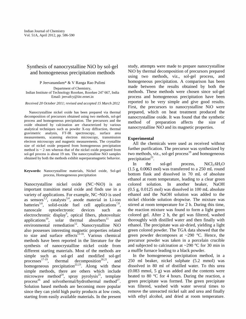
The XRD patterns recorded for the NiO precursors prepared using two different methods are shown in Fig. 1. The product formed by the sol-gel process is amorphous to X-rays, while in the case of homogeneous precipitation, the product is α-Ni(OH)2 with an interplanar distance of ~7.3 Å (refs 28, 29).
α-Ni(OH)2 is a hydroxyl deficient layered compound with intercalated anions for charge stabilization.
The IR spectra were recorded for both the NiO precursors (Supplementary data, Fig. S1). The broad band at ~3400 cm-1 is assigned to hydrogen bonded water molecules. The precursor synthesized by homogeneous precipitation method shows a band at ~2230 cm-1 due to OCN– group, which is not observed in the sample prepared using sol-gel process. The bands due to carbonate groups30 are observed in both the precursor samples, e.g. ~1400 cm-1, ~1115 cm-1 and ~840 cm-1. Ni-OH vibration is observed at ~ 770 cm-1 and the stretching frequency for Ni-O bond is also observed at ~ 475 cm-1. The weak bands observed at about ~2700-2900 cm-1 are attributed to the aliphatic stretching vibrations of the organic impurity present in the sample.
The TGA patterns for both the nickel oxide precursor samples were recorded (Supplementary data, Fig. S2). The decomposition temperature of the precursor from sol-gel process is about 290 °C, while that of the precursor from homogeneous precipitation is about 350 °C. These temperatures were used for the conversion of the precursors to NiO. The precursor sample from sol-gel process showed two weight loss steps with an overall weight loss of about 26 %. This is comparable to that for the precursor sample obtained by homogeneous precipitation (~ 28 %). On the basis of elemental analysis and atomic absorption spectroscopy results, the stoichiometry of the precursor from homogeneous precipitation was found to be Ni(OH)1.6(OCN)0.34(CO3)0.03(H2O)0.2. The
Fig. 1 Powder XRD patterns for the precursors prepared from (1) sol-gel process and (2) homogeneous precipitation.
INDIAN J CHEM, SEC A, APRIL 2012
588
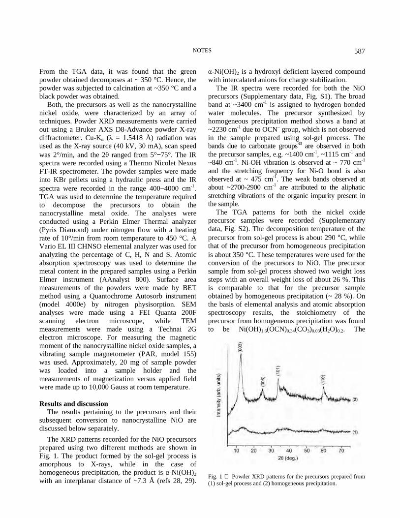
FE-SEM images recorded for the precursor samples are shown in Fig. 2. In the case of precursor sample from sol-gel method (Fig. 2a), the particles look agglomerated and the morphology is irregular, whereas in the case of precursor from homogeneous precipitation (Fig. 2b), the particles possess agglomerated sheet-like morphology.
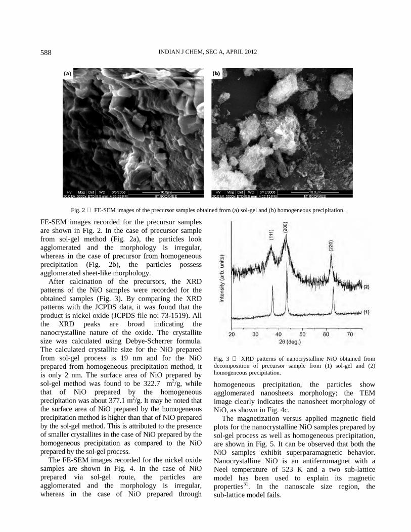
After calcination of the precursors, the XRD patterns of the NiO samples were recorded for the obtained samples (Fig. 3). By comparing the XRD patterns with the JCPDS data, it was found that the product is nickel oxide (JCPDS file no: 73-1519). All the XRD peaks are broad indicating the nanocrystalline nature of the oxide. The crystallite size was calculated using Debye-Scherrer formula. The calculated crystallite size for the NiO prepared from sol-gel process is 19 nm and for the NiO prepared from homogeneous precipitation method, it is only 2 nm. The surface area of NiO prepared by sol-gel method was found to be 322.7 m2/g, while that of NiO prepared by the homogeneous precipitation was about 377.1 m2/g. It may be noted that the surface area of NiO prepared by the homogeneous precipitation method is higher than that of NiO prepared by the sol-gel method. This is attributed to the presence of smaller crystallites in the case of NiO prepared by the homogeneous precipitation as compared to the NiO prepared by the sol-gel process.
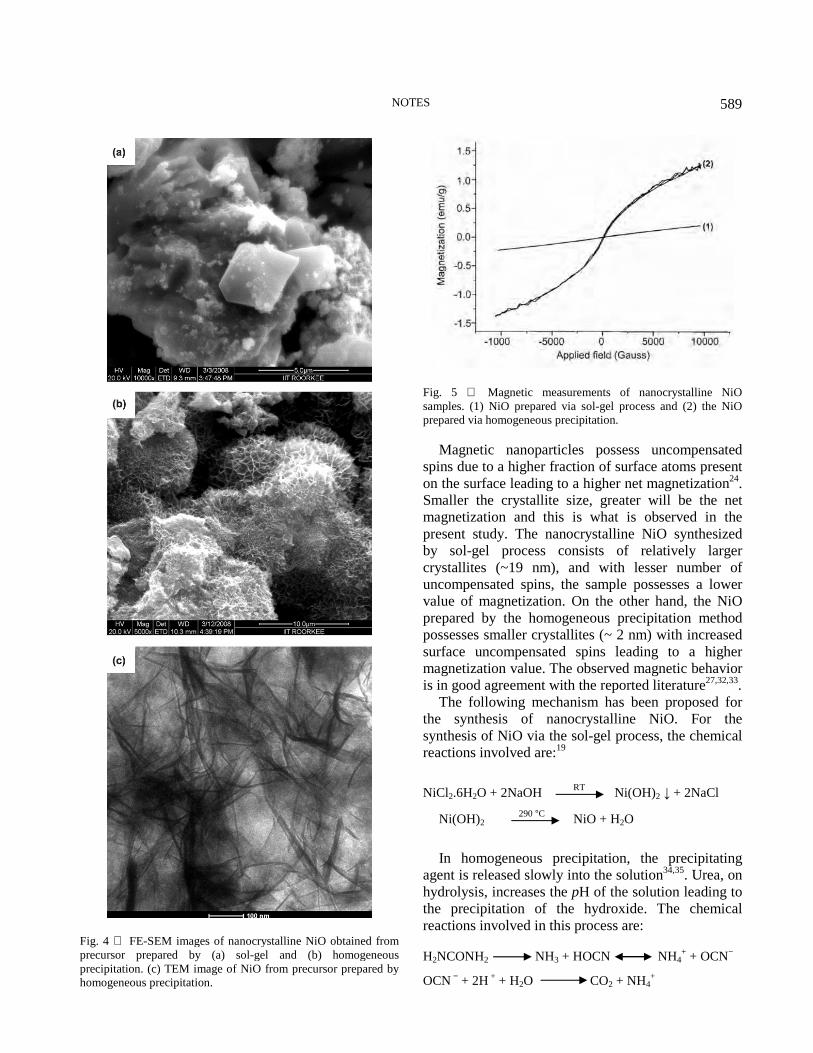
The FE-SEM images recorded for the nickel oxide samples are shown in Fig. 4. In the case of NiO prepared via sol-gel route, the particles are agglomerated and the morphology is irregular, whereas in the case of NiO prepared through
homogeneous precipitation, the particles show agglomerated nanosheets morphology; the TEM image clearly indicates the nanosheet morphology of NiO, as shown in Fig. 4c.
The magnetization versus applied magnetic field plots for the nanocrystalline NiO samples prepared by sol-gel process as well as homogeneous precipitation, are shown in Fig. 5. It can be observed that both the NiO samples exhibit superparamagnetic behavior. Nanocrystalline NiO is an antiferromagnet with a Neel temperature of 523 K and a two sub-lattice model has been used to explain its magnetic properties31. In the nanoscale size region, the sub-lattice model fails.
Fig. 2 FE-SEM images of the precursor samples obtained from (a) sol-gel and (b) homogeneous precipitation.
Fig. 3 XRD patterns of nanocrystalline NiO obtained from decomposition of precursor sample from (1) sol-gel and (2) homogeneous precipitation.
NOTES
589
Magnetic nanoparticles possess uncompensated spins due to a higher fraction of surface atoms present on the surface leading to a higher net magnetization24. Smaller the crystallite size, greater will be the net magnetization and this is what is observed in the present study. The nanocrystalline NiO synthesized by sol-gel process consists of relatively larger crystallites (~19 nm), and with lesser number of uncompensated spins, the sample possesses a lower value of magnetization. On the other hand, the NiO prepared by the homogeneous precipitation method possesses smaller crystallites (~ 2 nm) with increased surface uncompensated spins leading to a higher magnetization value. The observed magnetic behavior is in good agreement with the reported literature27,32,33.
The following mechanism has been proposed for the synthesis of nanocrystalline NiO. For the synthesis of NiO via the sol-gel process, the chemical reactions involved are:19
NiCl2.6H2O + 2NaOH RT Ni(OH)2 ↓ + 2NaCl
Ni(OH)2 290 °C NiO + H2O
In homogeneous precipitation, the precipitating
agent is released slowly into the solution34,35. Urea, on hydrolysis, increases the pH of the solution leading to the precipitation of the hydroxide. The chemical reactions involved in this process are:
H2NCONH2 NH3 + HOCN NH4+ + OCN−
OCN − + 2H + + H2O CO2 + NH4+
Fig. 4 FE-SEM images of nanocrystalline NiO obtained from precursor prepared by (a) sol-gel and (b) homogeneous precipitation. (c) TEM image of NiO from precursor prepared by homogeneous precipitation.
Fig. 5 Magnetic measurements of nanocrystalline NiO samples. (1) NiO prepared via sol-gel process and (2) the NiO prepared via homogeneous precipitation.

INDIAN J CHEM, SEC A, APRIL 2012
590
The OCN− species and the CO2 produced during the urea hydrolysis get intercalated into the interlamellar space of α-Ni(OH)2 for charge stabilization. The precipitated α-Ni(OH)2 decomposes on calcination at about 350 oC to produce nanocrystalline NiO.
In the present study, nanocrystalline nickel oxide has been prepared by the calcination of precursors prepared by two methods (sol-gel process and homogeneous precipitation). The precursors and the oxide have been characterized by powder XRD, FT-IR, TGA, elemental analysis, surface area measurements, FE-SEM, TEM and magnetic measurements. Homogeneous precipitation leads to the formation of smaller crystallites (size ~ 2 nm) as compared to the sol-gel process (size ~19 nm). The surface area of NiO prepared by homogeneous precipitation method is higher than that prepared by the sol-gel method. The effect of smaller size is reflected in the magnetic properties. The nanocrystalline NiO samples prepared by both the methods show superparamagnetism. However, the NiO from homogeneous precipitation possesses a higher net magnetization value as compared to that from the sol-gel process.
Supplementary data The supplementary data associated with this
article, i.e., Figs S1 and S2 are available in electronic form at http://www.niscair.res.in/jinfo/ijca/ IJCA 51A(04) 586-590_Suppl Data.pdf.
Acknowledgement PJ thanks the Council of Scientific and Industrial
Research, New Delhi, Govt. of India, for funding (Project No: 01 (2311) /09/EMR-II). Thanks are due to Dr. Rohit Kumar Rana, Indian Institute of Chemical Technology (IICT), Hyderabad, for his help in carrying out the surface area measurements.
References 1 Salimi A, Noorbakhsh A & Semnani A, J Solid State
Electrochem, 15 (2011) 2041. 2 Mattei G, Mazzoldi P, Post M L, Buso D, Guglielmi M &
Martucci A, Adv Mater, 19 (2007) 561. 3 Chockalingam K, Ganapathy A, Paramasivan G &
Govindasamy M, J Am Ceram Soc, 94 (2011) 2499. 4 Sietsma J R A, Meeldijk J D, den Breejen J P,
Versluijs-Helder M, van Dillen A J, de Jong P E & de Jong K P, Angew Chem Int Ed, 46 (2007) 4547.
5 Idris N H, Wang J, Chou S, Zhong C, Rahman M M & Liu H, J Mater Res, 26 (2011) 860.
6 Chiu K F, Chang C Y & Lin C M, J Electrochem Soc, 152
(2005) A1188. 7 Yoshito W K, Ussui V, Lazar D R R & Paschoal J O A,
Mater Sci Forum, 660-661 (2010) 977. 8 Marrero-Lopez D, Ruiz-Morales J C, Pena-Martinez J,
Canales-Vazquez J & Nunez P, J Solid State Chem, 181 (2008) 685.
9 Lin S H, Chen F R & Kai J J, Appl Surf Sci, 254 (2008) 2017.
10 Borgstrom M, Blart E, Boschloo G, Mukhtar E, Hagfeldt A, Hammarstrom L & Odobel F, J Phys Chem B, 109 (2005) 22928.
11 Zhao S, Ribbing C G & Wackelgard E, Solar Energy, 78 (2005) 125.
12 Ranjit K T, Medine G, Jeevanandam P, Martyanov I N & Klabunde K J, in Environmental Catalysis, edited by V H Grassian, (CRC Press, Boca Raton, FL) 2005, p. 391.
13 Tadic M, Panjan M, Markovic D, Milosevic I & Spasojevic V, J Alloys Comp, 509 (2011) 7134.
14 Bi H, Li S, Zhang Y & Du Y, J Magn Magn Mater, 277 (2004) 363.
15 Ranga Rao Pulimi V & Jeevanandam P, J Magn Magn
Mater, 321 (2009) 2556. 16 Bodker F, Hansen M F, Koch C B & Morup S, J Magn Magn
Mater, 221 (2000) 32. 17 Thota S & Kumar J, J Phys Chem Solids, 68 (2007) 1951. 18 Wu Y, He Y, Wu T, Weng W & Wan H, Mater Lett, 61
(2007) 2679. 19 Carnes C L, Stipp J, Klabunde K J & Bonevich J, Langmuir,
18 (2002)1352. 20 Wang D S, Xie T, Peng Q, Zhang S Y, Chen J & Li Y D,
Chem Eur J, 14 (2008) 2507. 21 Li Y, Afzaal M & O’Brien P, J Mater Chem, 16 (2006) 2175. 22 Xin X, Lue Z, Zhou B, Huang X, Zhu R, Sha X, Zhang Y &
Su W, J Alloys Comp, 427 (2007) 251. 23 de A A Soler-Illia G J, Jobbagy M, Regazzoni A E &
Blesa M A, Chem Mater, 11 (1999) 3140. 24 Parada C & Moran E, Chem Mater, 18 (2006) 2719. 25 Seo D J, Park S B, Kang Y C & Choy K L, J Nanopart Res,
5 (2003) 199. 26 Kanthimathi M, Dhathathreyan A & Nair B U, Mater Lett,
58 (2004) 2914. 27 Ghosh M, Biswas K, Sundaresan A & Rao C N R, J Mater
Chem, 16 (2006) 106. 28 Jeevanandam P, Koltypin Y & Gedanken A, Nano Lett,
1 (2001) 263. 29 Genin P, Delahaye-Vidal A, Portemer F, Tekaia-Elhsissen K
& Figlarz M, Eur J Solid State Inorg Chem, 28 (1991) 505. 30 Faure C, Borthomieu Y, Delmas C & Fouassier M, J Power
Sourc, 36 (1991) 113. 31 Berkowitz A E, Kodama R H, Makhlouf S A, Parker F T,
Spada F E, McNiff Jr E J & Foner S, J Magn Magn Mater, 196-197 (1999) 591.
32 Richardson J T, Yiagas D I, Turk B, Forster K & Twigg M V, J Appl Phys, 70 (1991)6977.
33 Khadar M A, Biju V & Inoue A, Mater Res Bull, 38 (2003)1341.
34 Matijevic E & Hsu W P, J Colloid Interface Sci, 118 (1987) 506. 35 Jayalakshmi M, Venugopal N, Reddy B R & Rao M M,
J Power Sourc, 150 (2005) 272.