Synthesis of bivalent lactosides and their activity as sensors for differences between lectins in...
6
Synthesis of bivalent lactosides and their activity as sensors for differences between lectins in inter- and intrafamily comparisons Sabine André a , Dilip V. Jarikote b , Dandan Yan b,c , Lisa Vincenz b , Guan-Nan Wang b , Herbert Kaltner a , Paul V. Murphy b,⇑ , Hans-Joachim Gabius a a Institute of Physiological Chemistry, Faculty of Veterinary Medicine, Ludwig-Maximilians-University Munich, Veterinärstr. 13, 80539 Munich, Germany b School of Chemistry, National University of Ireland, Galway, University Road, Galway, Ireland c School of Chemistry and Chemical Biology, University College Dublin, Belfield, Dublin 4, Ireland article info Article history: Received 18 September 2011 Revised 1 November 2011 Accepted 2 November 2011 Available online 10 November 2011 Keywords: Agglutinin Glycocluster Glycocyclophane Lectin N-Glycosyl 1,2,3-triazoles abstract The synthesis of nine bivalent lactosides (based on ditriazoles, diamides, a glycocyclophane and an acy- clic analogue of the glycocyclophane) and one monovalent lactosyl triazole facilitated the assessment of the sensitivity of plant/animal lectins to this type of ligand display. The inhibitory potency of the com- pounds was determined in two assays of increasing biorelevance. These were solid-phase and cell bind- ing set-ups. Hereby, the ability of the compounds to inhibit the binding of two plant agglutinins and the entire set of adhesion/growth-regulatory galectins from one organism (chicken) to a glycoprotein or to cell surfaces was systematically evaluated. Differential sensitivities were detected between plant and ani- mal lectins and also between distinct galectin forms within the chicken series. Two of the bivalent probes can be considered as sensors for interlectin differences. Most pronounced were the selectivities of N-gly- cosyl 1,2,3-triazole derivatives for the chimera-type galectin and its proteolytically truncated version. Ó 2011 Elsevier Ltd. All rights reserved. A broad range of physiological functions is emerging for the gly- can chains of cellular glycoconjugates via carbohydrate-protein (lectin) recognition. 1 Distinct carbohydrate structures serve as tar- get sites for receptors (exogenous agglutinins from plants or bacte- ria, tissue lectins) and their interaction begins the conversion of sugar-encoded information into cellular responses. If conse- quences of this interplay are harmful for the organism, for exam- ple, toxicity exerted by plant lectins or immune dysregulation/ tumor invasion by endogenous effectors, 2 then blocking of the docking onto cells by custom-made inhibitors becomes an attrac- tive goal for medicinal chemistry. In principle, two general struc- tural parameters can be varied with the aim of accomplishing high-level specificity: (i) the carbohydrate headgroup including any modification (additions such as sulfation or synthetic modifi- cations at distinct sites to enhance complementary binding to tar- get receptors) and (ii) the valency of presentation. Of course, interpretation of experimental results benefits from a clear design approach, avoiding too many parameter changes. Also, such stud- ies gain impact if—besides revealing affinity of a carbohydrate derivative in a binding assay for a single lectin—further compara- tive binding analyses with a range of relevant targets are per- formed to assess selectivity. Ideally, this should be accomplished with cell assays where natural ligands are in their physiological presentation. To meet these criteria we herein focused on characterizing the impact of different modes of bivalent ligand presentation of a com- mon sugar headgroup (lactose). Although the linker length (dis- tance between two lactose residues) cannot always bridge the distance between contact sites of bi- to oligovalent lectins in an intramolecular sense, such a topological (bivalent) ligand display has nonetheless previously indicated its potential for targeting galactoside-specific lectins to varying extents. 3 Three ditriazoles, four diamides, a glycocyclophane and an acylic analogue of the lat- ter were synthesized as the test panel (Fig. 1, 1–4, 6–10). Also in- cluded was a control monomer (5), which contains structural features of the aglycone found in 3 and 4, where lactose is con- nected via a triazole to a benzylated glucuronic acid derivative. A galactoside-binding plant toxin (Viscum album L., VAA; with low (Tyr)- and high (Trp)-affinity sites in the dimer separated by 87 Å or 15 Å, respectively, in the b-trefoil fold 4 ), a leguminous b-sand- wich agglutinin (Erythrina crystagalli agglutinin, ECA) and adhe- sion/growth-regulatory galectins were purified by affinity chromatography 3a to give a range of suitable lectins for the bio- chemical/cell biological part of the study. Of note, the family of galectins encompasses three topological modes for binding-site presentation (Fig. 2). Since it is appealing to comprehensively determine the structure–activity profiles of these three types for proteins from one organism, we have run the assays on the five chicken galectins (CGs). 5 Our panel thus comprises the three homodimeric proto-type galectins (CG-1A, CG-1B, CG-2), the chimera-type CG-3 along with its proteolytically truncated variant 0960-894X/$ - see front matter Ó 2011 Elsevier Ltd. All rights reserved. doi:10.1016/j.bmcl.2011.11.010 ⇑ Corresponding author. Tel.: +353 91 492465; fax: +353 91 495576. E-mail address: [email protected] (P.V. Murphy). Bioorganic & Medicinal Chemistry Letters 22 (2012) 313–318 Contents lists available at SciVerse ScienceDirect Bioorganic & Medicinal Chemistry Letters journal homepage: www.elsevier.com/locate/bmcl
-
Upload
sabine-andre -
Category
Documents
-
view
212 -
download
0
Transcript of Synthesis of bivalent lactosides and their activity as sensors for differences between lectins in...

Bioorganic & Medicinal Chemistry Letters 22 (2012) 313–318
Contents lists available at SciVerse ScienceDirect
Bioorganic & Medicinal Chemistry Letters
journal homepage: www.elsevier .com/ locate/bmcl
Synthesis of bivalent lactosides and their activity as sensors for differencesbetween lectins in inter- and intrafamily comparisons
Sabine André a, Dilip V. Jarikote b, Dandan Yan b,c, Lisa Vincenz b, Guan-Nan Wang b, Herbert Kaltner a,Paul V. Murphy b,⇑, Hans-Joachim Gabius a
a Institute of Physiological Chemistry, Faculty of Veterinary Medicine, Ludwig-Maximilians-University Munich, Veterinärstr. 13, 80539 Munich, Germanyb School of Chemistry, National University of Ireland, Galway, University Road, Galway, Irelandc School of Chemistry and Chemical Biology, University College Dublin, Belfield, Dublin 4, Ireland
a r t i c l e i n f o
Article history:Received 18 September 2011Revised 1 November 2011Accepted 2 November 2011Available online 10 November 2011
Keywords:AgglutininGlycoclusterGlycocyclophaneLectinN-Glycosyl 1,2,3-triazoles
0960-894X/$ - see front matter � 2011 Elsevier Ltd. Adoi:10.1016/j.bmcl.2011.11.010
⇑ Corresponding author. Tel.: +353 91 492465; fax:E-mail address: [email protected] (P.V.
a b s t r a c t
The synthesis of nine bivalent lactosides (based on ditriazoles, diamides, a glycocyclophane and an acy-clic analogue of the glycocyclophane) and one monovalent lactosyl triazole facilitated the assessment ofthe sensitivity of plant/animal lectins to this type of ligand display. The inhibitory potency of the com-pounds was determined in two assays of increasing biorelevance. These were solid-phase and cell bind-ing set-ups. Hereby, the ability of the compounds to inhibit the binding of two plant agglutinins and theentire set of adhesion/growth-regulatory galectins from one organism (chicken) to a glycoprotein or tocell surfaces was systematically evaluated. Differential sensitivities were detected between plant and ani-mal lectins and also between distinct galectin forms within the chicken series. Two of the bivalent probescan be considered as sensors for interlectin differences. Most pronounced were the selectivities of N-gly-cosyl 1,2,3-triazole derivatives for the chimera-type galectin and its proteolytically truncated version.
� 2011 Elsevier Ltd. All rights reserved.
A broad range of physiological functions is emerging for the gly-can chains of cellular glycoconjugates via carbohydrate-protein(lectin) recognition.1 Distinct carbohydrate structures serve as tar-get sites for receptors (exogenous agglutinins from plants or bacte-ria, tissue lectins) and their interaction begins the conversion ofsugar-encoded information into cellular responses. If conse-quences of this interplay are harmful for the organism, for exam-ple, toxicity exerted by plant lectins or immune dysregulation/tumor invasion by endogenous effectors,2 then blocking of thedocking onto cells by custom-made inhibitors becomes an attrac-tive goal for medicinal chemistry. In principle, two general struc-tural parameters can be varied with the aim of accomplishinghigh-level specificity: (i) the carbohydrate headgroup includingany modification (additions such as sulfation or synthetic modifi-cations at distinct sites to enhance complementary binding to tar-get receptors) and (ii) the valency of presentation. Of course,interpretation of experimental results benefits from a clear designapproach, avoiding too many parameter changes. Also, such stud-ies gain impact if—besides revealing affinity of a carbohydratederivative in a binding assay for a single lectin—further compara-tive binding analyses with a range of relevant targets are per-formed to assess selectivity. Ideally, this should be accomplishedwith cell assays where natural ligands are in their physiologicalpresentation.
ll rights reserved.
+353 91 495576.Murphy).
To meet these criteria we herein focused on characterizing theimpact of different modes of bivalent ligand presentation of a com-mon sugar headgroup (lactose). Although the linker length (dis-tance between two lactose residues) cannot always bridge thedistance between contact sites of bi- to oligovalent lectins in anintramolecular sense, such a topological (bivalent) ligand displayhas nonetheless previously indicated its potential for targetinggalactoside-specific lectins to varying extents.3 Three ditriazoles,four diamides, a glycocyclophane and an acylic analogue of the lat-ter were synthesized as the test panel (Fig. 1, 1–4, 6–10). Also in-cluded was a control monomer (5), which contains structuralfeatures of the aglycone found in 3 and 4, where lactose is con-nected via a triazole to a benzylated glucuronic acid derivative. Agalactoside-binding plant toxin (Viscum album L., VAA; with low(Tyr)- and high (Trp)-affinity sites in the dimer separated by 87 Åor 15 Å, respectively, in the b-trefoil fold4), a leguminous b-sand-wich agglutinin (Erythrina crystagalli agglutinin, ECA) and adhe-sion/growth-regulatory galectins were purified by affinitychromatography3a to give a range of suitable lectins for the bio-chemical/cell biological part of the study. Of note, the family ofgalectins encompasses three topological modes for binding-sitepresentation (Fig. 2). Since it is appealing to comprehensivelydetermine the structure–activity profiles of these three types forproteins from one organism, we have run the assays on the fivechicken galectins (CGs).5 Our panel thus comprises the threehomodimeric proto-type galectins (CG-1A, CG-1B, CG-2), thechimera-type CG-3 along with its proteolytically truncated variant

NOO
HOHO
OHO OHO
OH
OH
OHO
HOHO
OH O
OH
HOHO
OH
1N N
NNN
NH
OOHO
HO
OHO OHO
OH
OHO
HO
HOHO
OH
O
OH
HOHO
OH
HN
2O
O
NH
OOHO
HO
OHO OHO
OH
OHO
HO
HOHO
OH
O
OH
HOHO
OH
HN
8
O
O
NH
OOHO
HO
OHO OHO
OH
OHO
HO
HOHO
OH
O
OH
HOHO
OH
HN
O
O
7
NH
OOHO
HO
OH
O OHOOH
OHOHO
HOHO
OH
O
OH
HOHO
OH
NH
O O9
NOO
HOHO
OH
O OHOOH
OHOHO
HOHO
OH
O
OH
HOHO
OHN N
NNN
OO
5
NOO
HOHO
OH
O OHOOH
OH
OHO
HOHO
OH
O
OH
HOHO
OH
N N
NNN
O
O
6
NOO
HOHO
OHOHO
HOHO
OH
N NOO
BnOBnO
CO2H
10
O
HN
OBn
O
O
O
OBn
BnO
NHO
BnO
ON
OOHO HO
OHO
HO
HOHO
OH
N NO
OHOOH
OH
O
HO
HOHO OH
NNN
O
HN
OBn
O
O
O
OBn
BnO
NHO
BnO
ON
OOHO
HO
OHO
HO
HOHO
OH
N N
O OHOOH
OH
O
OH
HOHO
OH
NNN
3
4
Figure 1. Compounds 1–10.
314 S. André et al. / Bioorg. Med. Chem. Lett. 22 (2012) 313–318
(trCG-3) and the two versions of tandem-repeat-type CG-8 withnatural variations in linker length (either 9 or 28 amino acids)which arises from alternative splicing, together with a separate do-main (the N-terminal domain, termed Gal-8N) (Fig. 2). Orthologs ofproteins from each group are present in mammals, and the poten-tial for functional competition, documented between proto- andchimera-type galectins,6 makes this broad screening approachmandatory. Regarding potential for aggregation of the proteins insolution, CG-8 behaves as monomer in gel filtration and ultracen-trifugation, as does galectin-3, which can oligomerize in the pres-ence of multivalent ligands.3c,5d The affinity of 1–10 was assessedin inhibitory assays in two steps: firstly a measure of their potencyin interfering with lectin binding to a glycoprotein matrix(asialofetuin (ASF), a pan-galectin ligand) was evaluated; secondly
their ability to inhibit lectin binding to cultured cells was evalu-ated. The latter experiment provides an in vitro setting with in-creased relevance for the clinical situation.
The synthesis of ditriazoles began from the lactosyl azide 11(Scheme 1), which was prepared as described previously3e and re-acted with a variety of dialkynes (12–14) using copper-catalysedazide alkyne cycloaddition reactions7,8 to give protected interme-diates. Subsequent deacetylation using methoxide in methanolgave compounds 1, 6 and 7.
Next, the glycocyclophane 3 was prepared, also from the azide11 (Scheme 2). Reaction of 11 with glucuronic acid derivative 15by copper-catalysed azide alkyne cycloaddition reaction gave tria-zole 16. The coupling of 16 with p-xylylenediamine, subsequentring closure metathesis and removal of the acetates gave the final
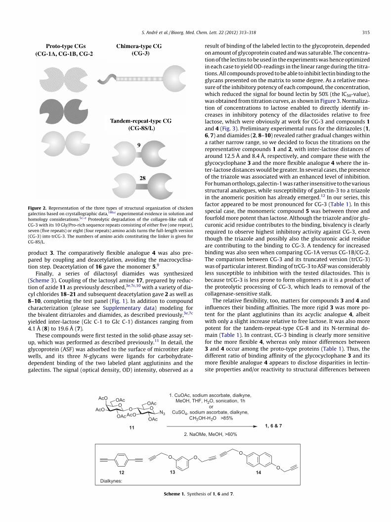
Figure 2. Representation of the three types of structural organization of chickengalectins based on crystallographic data,18b,c experimental evidence in solution andhomology considerations.5c–e Proteolytic degradation of the collagen-like stalk ofCG-3 with its 10 Gly/Pro-rich sequence repeats consisting of either five (one repeat),seven (five repeats) or eight (four repeats) amino acids turns the full-length version(CG-3) into trCG-3. The numbers of amino acids constituting the linker is given forCG-8S/L.
S. André et al. / Bioorg. Med. Chem. Lett. 22 (2012) 313–318 315
product 3. The comparatively flexible analogue 4 was also pre-pared by coupling and deacetylation, avoiding the macrocyclisa-tion step. Deacetylation of 16 gave the monomer 5.9
Finally, a series of dilactosyl diamides was synthesized(Scheme 3). Coupling of the lactosyl amine 17, prepared by reduc-tion of azide 11 as previously described,3e,7c,10 with a variety of dia-cyl chlorides 18–21 and subsequent deacetylation gave 2 as well as8–10, completing the test panel (Fig. 1). In addition to compoundcharacterization (please see Supplementary data) modeling forthe bivalent ditriazoles and diamides, as described previously,3e,7c
yielded inter-lactose (Glc C-1 to Glc C-1) distances ranging from4.1 Å (8) to 19.6 Å (7).
These compounds were first tested in the solid-phase assay set-up, which was performed as described previously.11 In detail, theglycoprotein (ASF) was adsorbed to the surface of microtiter platewells, and its three N-glycans were ligands for carbohydrate-dependent binding of the two labeled plant agglutinins and thegalectins. The signal (optical density, OD) intensity, observed as a
OOAcO
OAc
OAcOAcO
AcOOAc
OAc
N3
OO
2. NaOM
1. CuOAc, sodiuMeOH, THF,
CuSO4, sodiumCH3OH
Dialkynes:
11
12 13
Scheme 1. Synthesi
result of binding of the labeled lectin to the glycoprotein, dependedon amount of glycoprotein coated and was saturable. The concentra-tion of the lectins to be used in the experiments was hence optimizedin each case to yield OD-readings in the linear range during the titra-tions. All compounds proved to be able to inhibit lectin binding to theglycans presented on the matrix to some degree. As a relative mea-sure of the inhibitory potency of each compound, the concentration,which reduced the signal for bound lectin by 50% (the IC50-value),was obtained from titration curves, as shown in Figure 3. Normaliza-tion of concentrations to lactose enabled to directly identify in-creases in inhibitory potency of the dilactosides relative to freelactose, which were obviously at work for CG-3 and compounds 1and 4 (Fig. 3). Preliminary experimental runs for the ditriazoles (1,6, 7) and diamides (2, 8–10) revealed rather gradual changes withina rather narrow range, so we decided to focus the titrations on therepresentative compounds 1 and 2, with inter-lactose distances ofaround 12.5 Å and 8.4 Å, respectively, and compare these with theglycocyclophane 3 and the more flexible analogue 4 where the in-ter-lactose distances would be greater. In several cases, the presenceof the triazole was associated with an enhanced level of inhibition.For human orthologs, galectin-1 was rather insensitive to the variousstructural analogues, while susceptibility of galectin-3 to a triazolein the anomeric position has already emerged.12 In our series, thisfactor appeared to be most pronounced for CG-3 (Table 1). In thisspecial case, the monomeric compound 5 was between three andfourfold more potent than lactose. Although the triazole and/or glu-curonic acid residue contributes to the binding, bivalency is clearlyrequired to observe highest inhibitory activity against CG-3, eventhough the triazole and possibly also the glucuronic acid residueare contributing to the binding to CG-3. A tendency for increasedbinding was also seen when comparing CG-1A versus CG-1B/CG-2.The comparison between CG-3 and its truncated version (trCG-3)was of particular interest. Binding of trCG-3 to ASF was considerablyless susceptible to inhibition with the tested dilactosides. This isbecause trCG-3 is less prone to form oligomers as it is a product ofthe proteolytic processing of CG-3, which leads to removal of thecollagenase-sensitive stalk.
The relative flexibility, too, matters for compounds 3 and 4 andinfluences their binding affinities. The more rigid 3 was more po-tent for the plant agglutinins than its acyclic analogue 4, albeitwith only a slight increase relative to free lactose. It was also morepotent for the tandem-repeat-type CG-8 and its N-terminal do-main (Table 1). In contrast, CG-3 binding is clearly more sensitivefor the more flexible 4, whereas only minor differences between3 and 4 occur among the proto-type proteins (Table 1). Thus, thedifferent ratio of binding affinity of the glycocyclophane 3 and itsmore flexible analogue 4 appears to disclose disparities in lectin-site properties and/or reactivity to structural differences between
O
O
1, 6 & 7e, MeOH, >60%
m ascorbate, dialkyne,H2O, sonication, 1h
or ascorbate, dialkyne,-H2O >85%
14
s of 1, 6 and 7.

OOAcO
AcO
OAcOAcO
AcOAcO
OAc
N3
Cu(OAc)2, sodium ascorbatedialkyne, MeOH, THF, H2Osonication, 1h, 82%
11
O CO2HBnO O
BnO
15
+
1. p-xylylenediamine, PyBopDIPEA, DMF, 48h, 76%
2. NaOMe, MeOH, 70%
4
1. p-xylylenediamine, PyBop,DIPEA, DMF, 48h, 76%2. Grubbs-II, CH2Cl2, 91%3. NaOMe, MeOH, 70%
3
NOO
AcOAcO
OAcOAcO
AcOAcO
OAc
N NOO
BnOBnO
CO2H
16
5
NaOMeMeOH
Scheme 2. Synthesis of 3 and 4
OOAcO
OAc
OAcOAcO
AcOOAc
OAc
NH2
17
O
OCl
1. coupling with diacylchlorides
2. protecting groupremoval
ClO
O
2, 8-10
ClCl
O
OCl
Cl
O O
ClCl
18 19 20 21Diacyl chlorides
Scheme 3. Synthesis of 2 and 8–10.
Figure 3. Titration curves for extent of binding of biotinylated CG-3 (1 lg/ml) tosurface-immobilized glycoprotein (ASF) in the presence of increasing amounts oflactose (top) and compounds 1 and 4 (bottom), respectively. For compilation ofIC50-values, please see Table 1.
316 S. André et al. / Bioorg. Med. Chem. Lett. 22 (2012) 313–318
3 and 4 for the tandem-repeat-type (bivalent) CG-8 when com-pared to the homodimeric proteins. In other words, this pair ofsynthetic compounds is a sensor to distinguish between the galec-tin subgroups depicted in Figure 2. The monomeric N-terminal do-main of CG-8 (CG-8N) is rather equally well blocked bydilactosides, irrespective of the nature of the scaffold. In line withthis result, proteolytic processing of CG-3, which abolishes oligo-mer formation via the collagen-like stalk, impairs this lectin’smarked sensitivity to dilactosides (cf. CG-3 and trCG-3, Table 1).The acyclic compound 4 had highest relative potency of all the li-gands for CG-3. Lectin binding to the surface-presented glycopro-tein in this case was effectively impaired, with a reasonably highlevel of selectivity.
In order to establish whether the solid-phase assay has predic-tive value for a compounds’ ability to inhibit the binding of a lectinto cells we next performed such bioassays. On cell surfaces, a nat-ural panel of high-affinity ligands for lectins is presented in thephysiologically relevant mode. Experimentally, the labeled lectin,in the absence or presence of inhibitors, was incubated with ali-quots of cell suspensions from the same passage, in order to avoidglycophenotype changes upon prolonged periods in culture, andthe extent of binding was quantitated by cytofluorometry, as re-ported previously when testing aglyconic extensions of lactoseand dilactosides with human galectins.3e,12
The binding data obtained by cytofluorometry is documented asthe percentage of positive cells and the mean fluorescence inten-
sity for each experiment shown in Figure 4; the two determinedvariables will range between two controls, the first obtained in

Table 1IC50-values of four bivalent lactosides and free lactose (Lac) for blocking binding of biotinylated lectins to surface-immobilized ASF (in mM)a
Lectininhibitor
VAA(1.5 lg/ml)
ECA(0.2 lg/ml)
CG-1A(8 lg/ml)
CG-1B(3 lg/ml)
CG-2(4 lg/ml)
CG-3(1 lg/ml)
trCG-3(1.5 lg/ml)
CG-8S(0.75 lg/ml)
CG-8L(0.75 lg/ml)
CG-8N(15 lg/ml)
1 0.13 (4.6) 0.6 (1.8) 0.22 (2.3) 1.5 (2.7) 1.2 (5.0) 0.07 (15.7) 3.1 (1.5) 0.7 (2.6) 1.8 (2.8) 0.5 (3.2)2 0.55 (1.1) 1.2 (0.9) 0.36 (1.4) 1.3 (3.1) 1.4 (4.3) 0.6 (1.8) 4.2 (1.1) 2.5 (0.7) 4.5 (1.1) 0.8 (2.0)3 0.24 (2.5) 0.8 (1.4) 1.6 (0.3) 0.6 (6.7) 0.7 (8.6) 0.08 (13.8) 5.3 (0.8) 0.4 (4.5) 0.6 (8.3) 0.3 (5.3)4 1.3 (0.5) 1.3 (0.8) 1.4 (0.5) 0.46 (8.7) 0.65 (9.2) 0.014 (78.6) 5.0 (0.9) 3 (0.6) 2.4 (2.1) 0.4 (4.0)Lactose 0.6 (1) 1.1 (1) 0.5 (1) 4 (1) 6 (1) 1.1 (1) 4.5 (1) 1.8 (1) 5 (1) 1.6 (1)
a For structures of 1–4, see Figure 1; assays at a constant amount of 0.5 lg ASF used for coating of microtiter plate wells were routinely done in triplicates for up to fiveindependent series with standard deviations not exceeding 12.4%. The lectin concentration is given in each case. Numbers in brackets denote the inhibitory potency relativeto free lactose. Concentration values are normalized to lactose in all cases.
Figure 4. Cell surface staining (percentage of positive cells/mean fluorescenceintensity) by labeled lectins and the impact of presence of inhibitors. Binding curvesfor CG-3 (a) and trCG-3 (b) at 5 lg/ml in the absence of inhibitor (100%-value) and(listed from bottom to top) 1 mM lactose, 1 mM 2 and 0.25 mM 1 (a, gray area: 0%-value, background control in the absence of lectin) or lactose, 2 and 1 at 2 mM usingCHO cells (b). Low level of sensitivity was recorded for ECA (2 lg/ml) and humanSW480 colon adenocarcinoma cells for 2, 3, lactose and 1 at 0.2 mM (c), whereasmarked differences were present for interfering of binding of CG-8S (5 lg/ml) toCHO cells by 2, lactose and 1 tested at 1 mM (d). Aliquots of the same batch wereanalyzed in triplicates in at least three independent series with standard deviationsnot exceeding 11.3% after normalization of the data.
S. André et al. / Bioorg. Med. Chem. Lett. 22 (2012) 313–318 317
the absence of lectin (0%-value; gray area in each panel of Fig. 4)and the second obtained in the presence of the lectin without addi-tion of inhibitor (100%-value; black line in each panel of Fig. 4).Blocking lectin binding by an inhibitor will reduce the 100%-valueaccording to its potency. The results of these assays, when carriedout with the compounds, were consistently in agreement with theresults obtained from the solid-phase binding assays; this isexemplarily documented for the two forms of CG-3 (Fig. 4a, b),the leguminous lectin and its low level of sensitivity (Fig. 4c) aswell as for CG-8 (Fig. 4d). At the same time, the experiments ex-tend the data basis for differences in binding affinity toward thetested lectins to cell surfaces as the assay platform and hereby af-ford a determination of the potential of the compounds to reducelectin binding to cell surfaces.
Overall, the combined data shown in this communication leadsto the conclusion that bivalency contributes to inhibitory activity.Kinetic aspects of binding affecting on/off rates to increase affinity,as reported for mucin loading,13 may be a reasonable explanation
also in this context, whereas simultaneous reactivity (intramolec-ular binding) with extended sites, as discussed for selectins,14 ap-pears less likely, despite binding of histo-blood grouptetrasaccharides, digalactosides or poly-N-acetyllactosamine re-peats presenting two galactose units to the extended contact siteof chicken and human galectins.15 In general, density of ligand pre-sentation appears to be a key factor for galectins when targetingdistinct counter-receptors from the glycome complexity, at the le-vel of N-glycan branching and presentation in microdomains, alsoseen between CG-1A and CG-1B when testing polyvalent glycopro-teins.15a,16 The example of ‘non-spanning’ bivalent ligands and thecholera toxin B—pentamer teaches the lesson that transient, non-specific binding can also have a significant bearing on affinity.17
Dilactosides with a triazole at the anomeric carbon appeared morefavorable than those with an amide in most cases. Screening in twoassay types with increasing biorelevance were in agreement andrevealed notable differences in binding within the set of galectins,separating the two natural forms of the chimera-type galectin aswell as the tandem-repeat-type protein and the non-covalentlyassociated homodimers.
In summary, the compounds described herein block glycopro-tein and cell binding to various lectins, with differences in potencyand selectivity in some cases. This provides a basis for further re-search with a view to accomplish selectivity enhancement. Explo-ration of the impact of (i) substitutions in the sugar headgroup,with guidelines provided by chemical mapping, glycan testing,crystallographic analysis and molecular modeling15,16,18 and (ii)increasing valency would both be of interest in this regard.
Acknowledgments
The authors are grateful to the reviewers for their helpful ad-vice. The work described herein was generously funded by IRCSET(D.Y.), Science Foundation Ireland (PI/IN1/B966, 08/SRC/B1393)and the EU (MC-EIF to D.V.J.; GlycoHIT, Grant agreement no.260600 to H.-J.G.).
Supplementary data
Supplementary data (experimental details for synthesis of thecompounds and analytical data) associated with this article canbe found, in the online version, at doi:10.1016/j.bmcl.2011.11.010.
References and notes
1. (a)The Sugar Code. Fundamentals of Glycosciences; Gabius, H.-J., Ed.; Wiley-VCH:Weinheim, 2009; (b) Gabius, H.-J.; André, S.; Jiménez-Barbero, J.; Romero, A.;Solís, D. Trends Biochem. Sci. 2011, 36, 298.
2. (a) Read, R. J.; Stein, P. E. Curr. Opin. Struct. Biol. 1993, 3, 853; (b) Gabius, H.-J.;Darro, F.; Remmelink, M.; André, S.; Kopitz, J.; Danguy, A.; Gabius, S.; Salmon, I.;Kiss, R. Cancer Invest. 2001, 19, 114; (c) Ulbrich, H.; Eriksson, E. E.; Lindbom, L.Trends Pharmacol. Sci. 2003, 24, 640; (d) Hartley, M. R.; Lord, J. M. Biochim.Biophys. Acta 2004, 1701, 1; (e) Audi, J.; Belson, M.; Patel, M.; Schier, J.; Osterloh,J. J. Am. Med. Assoc. 2005, 294, 2342; (f) Villalobo, A.; Nogales-González, A.;Gabius, H.-J. Trends Glycosci. Glycotechnol. 2006, 18, 1; (g) Gabius, H.-J. Biochem.Soc. Trans. 2008, 36, 1491; (h) Norling, L. V.; Perretti, M.; Cooper, D. J. Endocrinol.

318 S. André et al. / Bioorg. Med. Chem. Lett. 22 (2012) 313–318
2009, 201, 169; (i) Osborn, H. M. I.; Turkson, A. In The Sugar Code. Fundamentalsof Glycosciences; Gabius, H.-J., Ed.; Wiley-VCH: Weinheim, 2009; pp 469–483;(j) Schwartz-Albiez, R. In The Sugar Code. Fundamentals of Glycosciences; Gabius,H.-J., Ed.; Wiley-VCH: Weinheim, 2009; pp 447–467; (k) Osorio, F.; Reis eSousa, C. Immunity 2011, 34, 651.
3. (a) André, S.; Ortega, P. J. C.; Perez, M. A.; Roy, R.; Gabius, H.-J. Glycobiology1999, 9, 1253; (b) André, S.; Liu, B.; Gabius, H.-J.; Roy, R. Org. Biomol. Chem.2003, 1, 3909; (c) Ahmad, N.; Gabius, H.-J.; André, S.; Kaltner, H.; Sabesan, S.;Roy, R.; Liu, B.; Macaluso, F.; Brewer, C. F. J. Biol. Chem. 2004, 279, 1081; (d)André, S.; Specker, D.; Bovin, N. V.; Lensch, M.; Kaltner, H.; Gabius, H.-J.;Wittmann, V. Bioconjugate Chem. 2009, 20, 1716; (e) Leyden, R.; Velasco-Torrijos, T.; André, S.; Gouin, S.; Gabius, H.-J.; Murphy, P. V. J. Org. Chem. 2009,74, 9010.
4. (a) Olsnes, S.; Stirpe, F.; Sandvig, K.; Pihl, A. J. Biol. Chem. 1982, 257, 13263; (b)Niwa, H.; Tonevitsky, A. G.; Agapov, I. I.; Saward, S.; Pfüller, U.; Palmer, R. A. Eur.J. Biochem. 2003, 270, 2739; (c) Jiménez, M.; André, S.; Siebert, H.-C.; Gabius, H.-J.; Solís, D. Glycobiology 2006, 16, 926; (d) Jiménez, M.; André, S.; Barillari, C.;Romero, A.; Rognan, D.; Gabius, H.-J.; Solís, D. FEBS Lett. 2008, 582, 2309.
5. (a) Beyer, E. C.; Zweig, S. E.; Barondes, S. H. J. Biol. Chem. 1980, 255, 4236; (b)Oda, Y.; Kasai, K.-I. Biochem. Biophys. Res. Commun. 1983, 761, 237; (c) Kaltner,H.; Solís, D.; Kopitz, J.; Lensch, M.; Lohr, M.; Manning, J. C.; Mürnseer, M.;Schnölzer, M.; André, S.; Sáiz, J. L.; Gabius, H.-J. Biochem. J. 2008, 409, 591; (d)Kaltner, H.; Solís, D.; André, S.; Lensch, M.; Manning, J. C.; Mürnseer, M.; Sáiz, J.L.; Gabius, H.-J. Biochemistry 2009, 48, 4403; (e) Kaltner, H.; Kübler, D.; López-Merino, L.; Lohr, M.; Manning, J. C.; Lensch, M.; Seidler, J.; Lehmann, W. D.;André, S.; Solís, D.; Gabius, H.-J. Anat. Rec. 2011, 294, 427.
6. (a) Kopitz, J.; von Reitzenstein, C.; André, S.; Kaltner, H.; Uhl, J.; Ehemann, V.;Cantz, M.; Gabius, H.-J. J. Biol. Chem. 2001, 276, 35917; (b) Sanchez-Ruderisch,H.; Fischer, C.; Detjen, K. M.; Welzel, M.; Wimmel, A.; Manning, J. C.; André, S.;Gabius, H.-J. FEBS J. 2010, 277, 3552.
7. (a) Kolb, H. C.; Finn, M. G.; Sharpless, K. B. Angew. Chem., Int. Ed. 2001, 40, 2004;(b) Meldal, M.; Tornoe, C. W. Chem. Rev. 2008, 108, 2952; (c) André, S.; Velasco-Torrijos, T.; Leyden, R.; Gouin, S.; Tosin, M.; Murphy, P. V.; Gabius, H.-J. Org.Biomol. Chem. 2009, 7, 4715.
8. For synthesis of other bivalent ditriazole derivatives see: Ziegler, T.; Hermann,C. Tetrahedron Lett. 2008, 49, 2166.
9. Details for preparation of 5 are provided in the Supplementary data section. Seealso: (a) Mereyala, H. B.; Gurrala, S. R.; Mohan, S. K. Tetrahedron 1999, 55,11331; (b) Jarikote, D. V.; O’Reilly, C.; Murphy, P. V. Tetrahedron Lett. 2010, 51,6776.
10. (a) Peto, C.; Batta, G.; Györgydeák, Z.; Sztaricskai, F. Liebigs Ann. Chem. 1991, 5,505; (b) Aravind, S.; Park, W. K. C.; Brochu, S.; Roy, R. Tetrahedron Lett. 1994, 35,7739.
11. (a) André, S.; Pei, Z.; Siebert, H.-C.; Ramström, O.; Gabius, H.-J. Bioorg. Med.Chem. 2006, 14, 6314; (b) André, S.; Maljaars, C. E. P.; Halkes, K. M.; Gabius, H.-J.; Kamerling, J. P. Bioorg. Med. Chem. Lett. 2007, 17, 793.
12. (a) André, S.; Giguère, D.; Dam, T. K.; Brewer, C. F.; Gabius, H.-J.; Roy, R. New J.Chem. 2010, 34, 2229; (b) Giguère, D.; André, S.; Bonin, M.-A.; Bellefleur, M.-A.;Provencal, P. C.; Pucci, B.; Roy, R.; Gabius, H.-J. Bioorg. Med. Chem. 2011, 19,3280.
13. Dam, T. K.; Brewer, C. F. Adv. Carbohydr. Chem. Biochem. 2010, 63, 139.14. Maaheimo, H.; Renkonen, R.; Turunen, J. P.; Penttilä, L.; Renkonen, O. Eur. J.
Biochem. 1995, 234, 616.15. (a) Wu, A. M.; Singh, T.; Liu, J. H.; Krzeminski, M.; Russwurm, R.; Siebert, H.-C.;
Bonvin, A. M. J. J.; André, S.; Gabius, H.-J. Glycobiology 2007, 17, 65; (b)Krzeminski, M.; Singh, T.; André, S.; Lensch, M.; Wu, A. M.; Bonvin, A. M. J. J.;Gabius, H.-J. Biochim. Biophys. Acta 2011, 1810, 150; (c) Martín-Santamaría, S.;André, S.; Buzamet, E.; Caraballo, R.; Ferná, B.; Cañada, F. J.; Menéndez, M.;Ramström, O.; Jiménez-Barbero, J.; Solís, D.; Gabius, H.-J. Org. Biomol. Chem.2011, 9, 5445; (d) Miller, M. C.; Ribeiro, J. P.; Roldós, V.; Martín-Santamaría, S.;Cañada, F. J.; Nesmelova, I.; André, S.; Pang, M.; Klyosov, A.; Baum, L. G.;Jiménez-Barbero, J.; Gabius, H.-J.; Mayo, K. H. Glycobiology 2011, in press,doi:10.1093/glycob/cwr083.
16. (a) Wu, A. M.; Wu, J. H.; Tsai, M.-S.; Kaltner, H.; Gabius, H.-J. Biochem. J. 2001,358, 529; (b) Wu, A. M.; Wu, J. H.; Liu, J.-H.; Singh, T.; André, S.; Kaltner, H.;Gabius, H.-J. Biochimie 2004, 86, 317; (c) Morelle, W.; Stechly, L.; André, S.; vanSeuningen, I.; Porchet, N.; Gabius, H.-J.; Michalski, J.-C.; Huet, G. Biol. Chem.2009, 390, 529; (d) Stechly, L.; Morelle, W.; Dessein, A. F.; André, S.; Grard, G.;Trinel, D.; Dejonghe, M. J.; Leteurtre, E.; Drobecq, H.; Trugnan, G.; Gabius, H.-J.;Huet, G. Traffic 2009, 10, 438; (e) Kopitz, J.; Bergmann, M.; Gabius, H.-J. IUBMBLife 2010, 62, 624.
17. Liu, J.; Begley, D.; Mitchell, D. D.; Verlinde, C. L. M. J.; Varani, G.; Fan, E. Chem.Biol. Drug Des. 2008, 71, 408.
18. (a) Solís, D.; Romero, A.; Kaltner, H.; Gabius, H.-J.; Díaz-Mauriño, T. J. Biol. Chem.1996, 271, 12744; (b) Varela, P. F.; Solís, D.; Díaz-Mauriño, T.; Kaltner, H.;Gabius, H.-J.; Romero, A. J. Mol. Biol. 1999, 294, 537; (c) López-Lucendo, M. F.;Solís, D.; Sáiz, J. L.; Kaltner, H.; Russwurm, R.; André, S.; Gabius, H.-J.; Romero,A. J. Mol. Biol. 2009, 386, 366.