
Synthesis, characterization and antioxidant activity of water soluble MnIII complexes of...
7
Synthesis, characterization and antioxidant activity of water soluble Mn III complexes of sulphonato-substituted Schiff base ligands Diego Moreno a , Verónica Daier a , Claudia Palopoli a , Jean-Pierre Tuchagues b,c , Sandra Signorella a, * a IQUIR (Instituto de Química Rosario) CONICET, Facultad de Ciencias Bioquímicas y Farmacéuticas, Universidad Nacional de Rosario, Suipacha 531, S2002LRK Rosario, Argentina b CNRS, LCC (Laboratoire de Chimie de Coordination), 205, route de Narbonne, F-31077 Toulouse, France c Université de Toulouse, UPS, INPT, LCC, F-31077 Toulouse, France article info Article history: Received 30 September 2009 Received in revised form 17 December 2009 Accepted 18 December 2009 Available online 6 January 2010 Keywords: Water soluble Mn complexes Schiff base ligands SOD models Catalase activity abstract Two new Mn III complexes Na[Mn(5-SO 3 -salpnOH)(H 2 O)] 5H 2 O (1) and Na[Mn(5-SO 3 -salpn)(MeOH)] 4H 2 O (2) (5-SO 3 -salpnOH = 1,3-bis(5-sulphonatosalicylidenamino)propan-2-ol, 5-SO 3 -salpn = 1,3-bis(5- sulphonatosalicylidenamino)propane) have been prepared and characterized. Electrospray ionization- mass spectrometry, UV–visible and 1 H NMR spectroscopic studies showed that the two complexes exist in solution as monoanions [Mn(5-SO 3 -salpn(OH))(solvent) 2 ] , with the ligand bound to Mn III through the two phenolato-O and two imino-N atoms located in the equatorial plane. The E 1/2 of the Mn III /Mn II couple (47.11 (1) and 77.80 mV (2) vs. Ag/AgCl) allows these complexes to efficiently catalyze the dismuta- tion of O 2 , with catalytic rate constants 2.4 10 6 (1) and 3.6 10 6 (2) M 1 s 1 , and IC 50 values of 1.14 (1) and 0.77 (2) lM, obtained through the nitro blue tetrazolium photoreduction inhibition superoxide dis- mutase assay, in aqueous solution of pH 7.8. The two complexes are also able to disproportionate up to 250 equivalents of H 2 O 2 in aqueous solution of pH 8.0, with initial turnover rates of 178 (1) and 25.2 (2) mM H 2 O 2 min 1 mM 1 catalyst 1 . Their dual superoxide dismutase/catalase activity renders these com- pounds particularly attractive as catalytic antioxidants. Ó 2009 Elsevier Inc. All rights reserved. 1. Introduction Superoxide dismutases (SODs) protect biological systems against oxidative damage caused by the superoxide radical (O 2 ) generated during aerobic metabolism through monoelectronic reduction of molecular oxygen [1]. High levels of superoxide are associated to several pathologies like diabetes, oxidative damage, numerous neurodegenerative disorders such Alzheimer’s and Parkinson’s diseases [2–5], and some types of cancer originating from DNA mutations induced by superoxide [6,7]. MnSOD is found in mitochondria and chloroplasts of eukaryotes and in the cytoplasm of bacteria [8,9]. Crystal structures have been solved for MnSOD from both bacteria (Thermus thermophilus [10] and Escherichia coli [11]) and eukaryotes (human mitochondria [12]). The active site of MnSOD contains one Mn ion in a distorted trigonal–bipyramidal N 3 O 2 environment, coordinated by three histidine residues, one aspartate and one OH ion/H 2 O molecule (Fig. 1) [11,13]. These enzymes catalyze the dismutation of O 2 into H 2 O 2 and O 2 through a ‘‘ping-pong” mechanism between Mn II and Mn III oxidation states [14]. The use of exogenous SODs (e.g. bovine) as therapeutic agents for treatment of a variety of disorders in which superoxide has a significant role has been tested. However, it is difficult to employ SODs in vivo, because of their immunogenic response [15] and high molecular mass (30 kDa) that disables the enzyme to cross the cell membranes [16]. The difficulties associated with the use of exogenous SOD have stimulated the search of low molecular weight catalytic scavengers as a new generation of pharmaceuti- cal compounds. Many complexes of Mn have been reported to be capable of functioning as catalysts for the dismutation of super- oxide and have been examined as catalytic antioxidants in pre- clinical trials, the most efficient being Mn III -salen (salen = 1,2- bis(salicylidenamino)ethane) [17], Mn III -porphyrinato [18–21] and Mn II -1,4,7,10,13-pentaazacyclopentadecane [22–24]. Among them, Mn III -salen complexes have been reported to have dual SOD/CAT (catalase) activity, an advantageous property since SOD activity alone would produce cytotoxic H 2 O 2 . However, the tested salen complexes are only slightly soluble in water and loose activity in a few minutes under the conditions of the cata- lase assay [25]. Therefore, there is a need to improve the stability of this class of complexes under physiological conditions to be useful as drugs. Herein, we report the synthesis, characterization, properties and SOD/CAT activity of two novel water soluble manganese com- plexes Na[Mn(5-SO 3 -salpnOH)(H 2 O)] (1) and Na[Mn(5-SO 3 -sal- pn)(MeOH)] (2) obtained with the sodium salts of the Schiff base ligands 1,3-bis(5-sulphonatosalicylidenamino)propan-2-ol (5- 0162-0134/$ - see front matter Ó 2009 Elsevier Inc. All rights reserved. doi:10.1016/j.jinorgbio.2009.12.016 * Corresponding author. Tel.: +54 341 4350214; fax: +54 341 4370477. E-mail address: [email protected] (S. Signorella). Journal of Inorganic Biochemistry 104 (2010) 496–502 Contents lists available at ScienceDirect Journal of Inorganic Biochemistry journal homepage: www.elsevier.com/locate/jinorgbio
-
Upload
diego-moreno -
Category
Documents
-
view
218 -
download
5
Transcript of Synthesis, characterization and antioxidant activity of water soluble MnIII complexes of...
Journal of Inorganic Biochemistry 104 (2010) 496–502
Contents lists available at ScienceDirect
Journal of Inorganic Biochemistry
journal homepage: www.elsevier .com/locate / j inorgbio
Synthesis, characterization and antioxidant activity of water soluble MnIII
complexes of sulphonato-substituted Schiff base ligands
Diego Moreno a, Verónica Daier a, Claudia Palopoli a, Jean-Pierre Tuchagues b,c, Sandra Signorella a,*
a IQUIR (Instituto de Química Rosario) � CONICET, Facultad de Ciencias Bioquímicas y Farmacéuticas, Universidad Nacional de Rosario, Suipacha 531, S2002LRK Rosario, Argentinab CNRS, LCC (Laboratoire de Chimie de Coordination), 205, route de Narbonne, F-31077 Toulouse, Francec Université de Toulouse, UPS, INPT, LCC, F-31077 Toulouse, France
a r t i c l e i n f o
Article history:Received 30 September 2009Received in revised form 17 December 2009Accepted 18 December 2009Available online 6 January 2010
Keywords:Water soluble Mn complexesSchiff base ligandsSOD modelsCatalase activity
0162-0134/$ - see front matter � 2009 Elsevier Inc. Adoi:10.1016/j.jinorgbio.2009.12.016
* Corresponding author. Tel.: +54 341 4350214; faxE-mail address: [email protected] (S. Signor
a b s t r a c t
Two new MnIII complexes Na[Mn(5-SO3-salpnOH)(H2O)] � 5H2O (1) and Na[Mn(5-SO3-salpn)(MeOH)] �4H2O (2) (5-SO3-salpnOH = 1,3-bis(5-sulphonatosalicylidenamino)propan-2-ol, 5-SO3-salpn = 1,3-bis(5-sulphonatosalicylidenamino)propane) have been prepared and characterized. Electrospray ionization-mass spectrometry, UV–visible and 1H NMR spectroscopic studies showed that the two complexes existin solution as monoanions [Mn(5-SO3-salpn(OH))(solvent)2]�, with the ligand bound to MnIII through thetwo phenolato-O and two imino-N atoms located in the equatorial plane. The E1/2 of the MnIII/MnII couple(�47.11 (1) and �77.80 mV (2) vs. Ag/AgCl) allows these complexes to efficiently catalyze the dismuta-tion of O��2 , with catalytic rate constants 2.4 � 106 (1) and 3.6 � 106 (2) M�1 s�1, and IC50 values of 1.14 (1)and 0.77 (2) lM, obtained through the nitro blue tetrazolium photoreduction inhibition superoxide dis-mutase assay, in aqueous solution of pH 7.8. The two complexes are also able to disproportionate up to250 equivalents of H2O2 in aqueous solution of pH 8.0, with initial turnover rates of 178 (1) and 25.2 (2)mM H2O2 min�1 mM�1 catalyst�1. Their dual superoxide dismutase/catalase activity renders these com-pounds particularly attractive as catalytic antioxidants.
� 2009 Elsevier Inc. All rights reserved.
1. Introduction
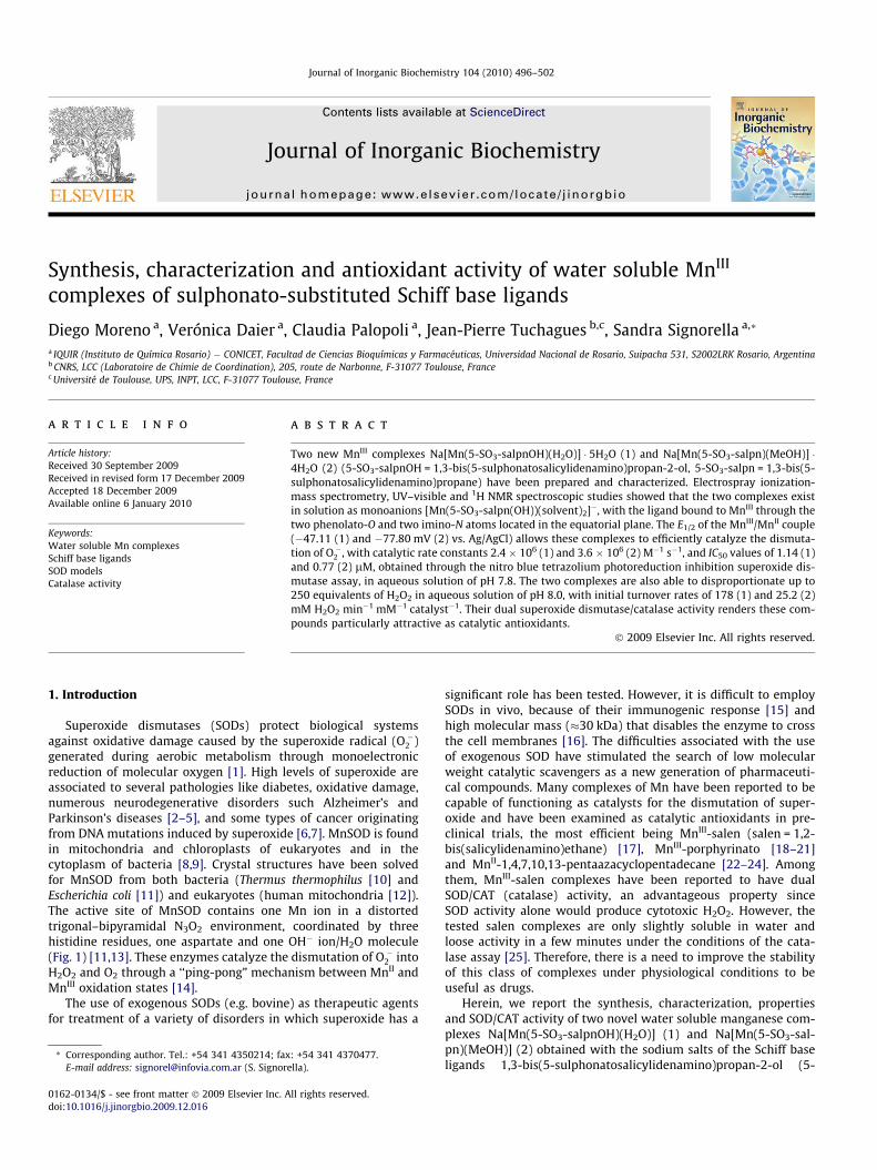
Superoxide dismutases (SODs) protect biological systemsagainst oxidative damage caused by the superoxide radical (O��2 )generated during aerobic metabolism through monoelectronicreduction of molecular oxygen [1]. High levels of superoxide areassociated to several pathologies like diabetes, oxidative damage,numerous neurodegenerative disorders such Alzheimer’s andParkinson’s diseases [2–5], and some types of cancer originatingfrom DNA mutations induced by superoxide [6,7]. MnSOD is foundin mitochondria and chloroplasts of eukaryotes and in thecytoplasm of bacteria [8,9]. Crystal structures have been solvedfor MnSOD from both bacteria (Thermus thermophilus [10] andEscherichia coli [11]) and eukaryotes (human mitochondria [12]).The active site of MnSOD contains one Mn ion in a distortedtrigonal–bipyramidal N3O2 environment, coordinated by threehistidine residues, one aspartate and one OH� ion/H2O molecule(Fig. 1) [11,13]. These enzymes catalyze the dismutation of O��2 intoH2O2 and O2 through a ‘‘ping-pong” mechanism between MnII andMnIII oxidation states [14].
The use of exogenous SODs (e.g. bovine) as therapeutic agentsfor treatment of a variety of disorders in which superoxide has a
ll rights reserved.
: +54 341 4370477.ella).
significant role has been tested. However, it is difficult to employSODs in vivo, because of their immunogenic response [15] andhigh molecular mass (�30 kDa) that disables the enzyme to crossthe cell membranes [16]. The difficulties associated with the useof exogenous SOD have stimulated the search of low molecularweight catalytic scavengers as a new generation of pharmaceuti-cal compounds. Many complexes of Mn have been reported to becapable of functioning as catalysts for the dismutation of super-oxide and have been examined as catalytic antioxidants in pre-clinical trials, the most efficient being MnIII-salen (salen = 1,2-bis(salicylidenamino)ethane) [17], MnIII-porphyrinato [18–21]and MnII-1,4,7,10,13-pentaazacyclopentadecane [22–24]. Amongthem, MnIII-salen complexes have been reported to have dualSOD/CAT (catalase) activity, an advantageous property sinceSOD activity alone would produce cytotoxic H2O2. However, thetested salen complexes are only slightly soluble in water andloose activity in a few minutes under the conditions of the cata-lase assay [25]. Therefore, there is a need to improve the stabilityof this class of complexes under physiological conditions to beuseful as drugs.
Herein, we report the synthesis, characterization, propertiesand SOD/CAT activity of two novel water soluble manganese com-plexes Na[Mn(5-SO3-salpnOH)(H2O)] (1) and Na[Mn(5-SO3-sal-pn)(MeOH)] (2) obtained with the sodium salts of the Schiff baseligands 1,3-bis(5-sulphonatosalicylidenamino)propan-2-ol (5-
N
OHNaO3S
N
HO SO3Na
Y
Scheme 1. Disodium salts of 5-SO3-salpnOH (Y = JY) and 5-SO3-salpn (Y = H) usedin the synthesis of the complexes.
Fig. 1. Active-site structure of human mitochondrial MnSOD based on coordinatesfrom the PDB file 1N0J [12].
D. Moreno et al. / Journal of Inorganic Biochemistry 104 (2010) 496–502 497
SO3-salpnOH) and 1,3-bis(5-sulphonatosalicylidenamino)propane(5-SO3-salpn) (see Scheme 1), and compare their SOD activity withthat of the complex obtained with 1,2-bis(5-sulphonatosalicylide-namino)ethane (5-SO3-salen).
2. Experimental section
2.1. Materials
All reagents or analytical grade chemicals were used as pur-chased. Solvents were purified by standard methods. The concen-tration of H2O2 stock solution was determined by iodometrictitration. Sodium salicylaldehyde-5-sulphonate was preparedaccording to a published method [26,27].
2.2. Physical measurements
Electronic spectra were recorded on a JASCO V550 spectropho-tometer with thermostated cell compartments. IR spectra wererecorded on a Perkin-Elmer Spectrum One FT-IR spectrophotome-ter. ESI-mass spectra were recorded on a Perkin-Elmer SCIEX 365LCMSMS mass spectrometer. The electrospray solutions were pre-pared from water solutions of the complexes and diluted withmethanol to a � 10�5 M concentration at a flow rate of5 lL min�1. 1H and 13C NMR spectra were recorded on a BrukerAC 200 NMR spectrometer at ambient probe temperature (ca26 �C), with nominal operating frequencies of 200.1 and50.3 MHz. Variable-temperature magnetic susceptibility datawere obtained with a Quantum Design MPMS SQUID susceptom-eter, under a magnetic field of 0.5 T in the temperature range 2–300 K. Diamagnetic corrections were applied by using Pascal’sconstants [28]. Least-squares fittings were accomplished with
an adapted version of the function-minimization program MINUIT[29]. Conductivity measurements were performed using a HoribaF-54 BW conductivity meter, on 1.0 mM solutions of the com-plexes in water. The electrochemical experiments were performedwith a computer-controlled Princeton Applied Research potentio-stat, model VERSASTAT II, with model 270/250 Research Electro-chemistry Software. Studies were carried out under Ar, in watersolutions using 0.1 M KNO3 as supporting electrolyte and�10�3 M of the complex. The working electrode was a Pt wireand the reference electrode was Ag/AgCl with Pt as the auxiliaryelectrode.
2.3. Synthesis of ligands
2.3.1. Disodium salt of 1,3-bis(5-sulphonatosalicylidenamino)propan-2-ol (5-SO3-salpnOH)
Na2[5-SO3-salpnOH] was prepared by Schiff-base condensationof sodium salicylaldehyde-5-sulphonate (200 mg, 0.825 mmol)with 1,3-diaminopropan-2-ol (37 mg, 0.413 mmol) in ethanol(40 mL), at reflux for 6 h. Na2[5-SO3-salpnOH] was isolated as apure yellow solid by precipitation from the reaction mixture atroom temperature. Yield: 171 mg (0.375 mmol, 91%). 1H NMR(D2O) d: 8.35 (singlet (s), 2H, N@CHA), 6.5–7.8 (multiplet (m),6H, Ar), 4.4–4.8 (m, 5H, NACH2AR and R2ACHAOH). 13C RMN(D2O) d: 175.52 (Ar), 168.62 (CH@N), 133.62, 132.48, 128.96,122.41, 114.18 (Ar), 67.9 (R2CHAOH), 54,90 (NACH2ACHOHA).Significant IR bands (KBr, m cm�1): mOH 3420 (broad), mC@N 1613,mSO3 1108/1034. UV–visible (UV–vis) kmax (nm) in H2O: 222, 234,255 (sh), 330 (sh), 375.
2.3.2. Disodium salt of 1,3-bis(5-sulphonatosalicylidenamino)propane(5-SO3-salpn)
Na2[5-SO3-salpn] was obtained from a mixture of sodium sali-cylaldehyde-5-sulphonate (500 mg, 2.57 mmol) and 1,3-diamino-propane (0.107 mL, 1.28 mmol) in 50 mL EtOH, stirred underreflux for 24 h. After cooling, the Schiff base precipitated fromthe reaction mixture as a pure yellow powder. Yield: 500 mg(1.14 mmol, 89%). 1H NMR (D2O) d: 8.40 (s, 2H, N@CHA), 6.6–7.9(m, 6H, Ar), 3.77 (triplet (t), 4H, NACH2AR), 2.11 (m, 2H,NACH2ACH2ACH2AN). 13C NMR (D2O, d) 176.45 (Ar), 167.92(CH@N), 133.53, 132.56, 128.46, 122.70, 113.86 (Ar), 49.00(NACH2AR), 29.00 (NACH2ACH2ACH2AN). Significant IR bands(KBr, m cm�1): mOH 3405 (broad), mC@N 1612, mSO3 1108/1035. UV–vis kmax (nm) in H2O: 222, 236, 254 (sh), 331 (sh), 373.
2.4. Synthesis of complexes
2.4.1. Na[Mn(5-SO3-salpnOH)(H2O)] � 5H2O (1 � 5H2O)Mn(OAc)2 � 4H2O (294 mg, 1.2 mmol) was added to a solution of
Na2[5-SO3-salpnOH] (300 mg, 0.6 mmol) in methanol (12 mL) andleft to stir for 1 h. The formed green precipitate was filtered off,washed with ether and dried under vacuum. Yield: 305 mg(0.48 mmol, 80%). Anal. calcd. for MnC17H16N2O10S2Na � 5H2O: C31.88, H 4.09, N 4.37, Mn 8.59, Na 3.58%; found: C 31.80, H 3.10,N 3.83, Mn 8.79, Na 4.18%. Although the analysis results for Hand Na are somewhat unsatisfactory, the chemical formula is con-sistent with the NMR, IR and thermogravimmetric data. SignificantIR bands (KBr, m cm�1): mOH 3478 (broad), mCH 2950, mC@N 1611, mSO3
1112/1029. UV–vis kmax nm (e M�1cm�1) in H2O: 277 (23970), 363(8980). Molar conductivity = 84 X�1 cm2 mol�1. The content of fivemolecules of non-coordinated water per complex molecule wasconfirmed by thermogravimetric analysis of the complex whichshowed 14% mass loss below 120 �C.
498 D. Moreno et al. / Journal of Inorganic Biochemistry 104 (2010) 496–502
2.4.2. Na[Mn(5-SO3-salpn)(MeOH)] � 4H2O (2 � 4H2O)Na2[5-SO3-salpn] (450 mg, 1.01 mmol) and Mn(OAc)2 � 4H2O
(373 mg, 1.5 mmol) were mixed in methanol (20 mL). A greenpowder formed immediately. The mixture was stirred for 1 h andthe green solid was collected by filtration, washed with etherand dried under vacuum. Yield: 444 mg (0.72 mmol, 71%). Anal.Calcd. for MnC18H18N2O9S2Na � 4H2O: C 34.84, H 4.22, N 4.51, Mn8.85, Na 3.70%; found: C 35.02, H 3.60, N 5.08, Mn 8.92, Na3.52%. Significant IR bands (KBr, m cm�1): mOH 3474(broad), mCH
2950, mC@N 1613, mSO3 1112/1029. UV–vis kmax nm (e M�1cm�1)H2O: 278 (19060), 362 (6870). Molar conductiv-ity = 111 X�1 cm2 mol�1. The content of four molecules of non-coordinated water per complex molecule was confirmed by ther-mogravimetric analysis of the complex, which showed 11.6% massloss below 115 �C.
2.5. Indirect SOD assay
The SOD activity of the complexes was assayed by measuringinhibition of the photoreduction of nitro blue tetrazolium (NBT),by a method slightly modified from that originally described byBeauchamps and Fridovich [30]. The solutions containing ribofla-vin (3.4 � 10�6 M), methionine (0.01 M), NBT (4.6 � 10�5 M) andcomplex of various concentrations were prepared with phosphatebuffer (pH 7.8). The mixtures were illuminated by a fluorescentlamp with a constant light intensity at 25 �C. The reduction ofNBT was monitored at 560 nm with various illumination periods(t). Rates in the absence and in the presence of different concentra-tions of complex were determined and plotted vs. complex concen-tration. Inhibition percentage was calculated according to: {(DAbs/t)without complex � (DAbs/t)with complex} � 100/(DAbs/t)without complex.The IC50 value represents the concentration of the SOD mimic thatinduces a 50% inhibition of the reduction of NBT. Control experi-ments were performed on mixtures of NBT + complex, ribofla-vin + complex, and NBT + methionine + complex, in phosphatebuffer, to ensure that the complex does not react independentlywith any of the components of the mixture. Based on these exper-iments, cross reactivity of the complex with NBT or riboflavin wasdisregarded. Under conditions used in this work, the catalytic rateconstant (kMcCF) for MnSOD was found to be 5.5 � 108 M�1 s�1.
2.6. Disproportionation of H2O2
The H2O2 disproportionation catalyzed by 1–2 was measured byvolumetric determination of the evolved O2 from reaction mixturesin water, as previously described [31].
Mn
NN
O O
S
MnNN
O O
O3S
S
O3S SO3Na
SO3Na
SO3Na
Mn
NN
O O
O3S
S
Scheme 2. One-dimensional {Na[MnIII-5-SO3-salpn(OH)]}n polymer in the solidstate. S@H2O (1) or MeOH (2).
3. Results and discussion
3.1. Synthesis of the complexes
Complexes 1 and 2 were prepared from 1:1 mixtures of the li-gand with Mn(OAc)2 in methanol. When Mn(ClO4)2 was used in-stead of acetate, no color change was observed until NaOH orEt3N was added to the solution of complex, such as observed forpreviously reported Mn complexes of tetradentate Schiff base li-gands [32–34]. Therefore, acetate facilitates the aerobic oxidationof MnII and deprotonation of the phenol for coordination to themetal. Although the disodium salts of the ligands were used, theanalytical results show that the complexes retain only one sodiumion per complex molecule in the solid state, and the thermo-gravimmetric analyses indicate that only one of the solvent mole-cules is coordinated to the metal centre of each complex. Theremaining coordination position of the Mn ion is probably occu-pied by the sulphonate group of an adjacent complex moleculeforming a one-dimensional polymer, as shown in Scheme 2. In thisscheme, a planar imino/phenolato N2O2 donor set around the Mnion is proposed for the two complexes, in accordance with the 1HNMR results (Section 3.3.3). In spite of our numerous efforts, wedid not succeed to grow single crystals of good enough qualityfor an XRD study.
3.2. Solid state studies
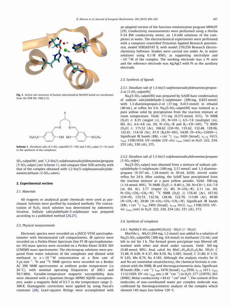
3.2.1. FT-IR spectroscopyComparison of the IR spectra of 1–2 evidences their similar
structures. The IR spectra of the two complexes (Fig. 2) exhibitstrong imine and phenolato absorptions at 1613/1611 and1543 cm�1, two strong bands at 1112 and 1029 cm�1 attributableto the asymmetric and symmetric stretching modes of the ASO�3group, and a broad OAH stretching band at �3480 cm�1. AlthoughMn(OAc)2 was used in the synthesis of the complexes, the lack oftypical acetate stretching bands and the analogous spectrumshown by the complex obtained by reaction of 5-SO3-salpn withMn(ClO4)2 in the presence of base, confirm the absence of acetatein these complexes.
3.2.2. Magnetic propertiesThe molar magnetic susceptibility (vM) of complex 1 in the solid
state is independent of the applied magnetic field strength from 0.1to 2.1 T. The temperature dependence of vM was measured in the2–300 K temperature range on cylindrical 3 mm diameter pelletspressed from powdered 1, under an applied magnetic field of0.5 T. The room temperature magnetic moment (leff) value of4.87 BM is in excellent accordance with the theoretical value forthe spin-only moment of one high-spin MnIII ion (lSO = 4.90 BMfor g = 2). Upon cooling, the leff values are practically constant
4000 3500 3000 2500 2000 1500 1000 500
ν (cm-1)
Fig. 2. FT-IR spectra of complexes 1 ( ) and 2 ( ).
D. Moreno et al. / Journal of Inorganic Biochemistry 104 (2010) 496–502 499
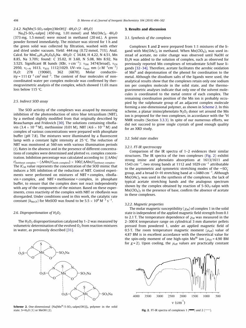
down to 100 K (4.81 BM), then decrease slightly from 100 to �20 K(4.51), and finally decrease to 3.29 BM at 2 K, indicating operationof either small antiferromagnetic interactions, or single MnIII ionzero field splitting, or both. Plots of the inverse corrected molarsusceptibility vs. temperature yielded straight lines, indicatingCurie–Weiss behavior, with C = 2.91 cm3 mol�1 K and h = �6.7 K.The Curie constant C value is close to the expected value of3.0 cm3 mol�1 K for an isolated MnIII ion (S = 2, g = 2), and the neg-ative h value further confirms the possible operation of weak anti-ferromagnetic interactions in this complex. Considering that allsolid state and solution studies point to the mononuclear natureof 1 and 2, the magnetic behavior of 1 may originate from antifer-romagnetic intermolecular spin–spin interactions and/or single ionzero field splitting (zfs) of MnIII. Consequently, we initially fittedthe magnetic data by considering the theoretical magnetic suscep-tibility of an S = 2 spin system taking into account an axial zfs term,D. The magnetic susceptibility has been computed by exact calcu-lation of the energy levels associated with the spin Hamiltonianthrough diagonalization of the full energy-matrix with a generalprogram for axial and rhombic symmetries [35]. Two possibilitieswere considered, either without or with additional operation of ex-tended intermolecular magnetic interactions (zj0) computed withinthe molecular field approximation [36]. When zj0 was set to zero,the fits were very poor; when zj0 was allowed to vary, similarlypoor fits were obtained with zj0 values in the 10�8 cm�1 range. Inboth cases, the resulting D values (10 cm�1 range) were physicallymeaningless (positive and unreasonably large). Considering that
Fig. 3. Temperature dependence of vM (h) and vMT (4) for complex 1. The solidlines show the best fit based on the Heisenberg chain model, see text.
400 420 440 460 480 500 520 540 560 580 600
(b)
(a)
m/z
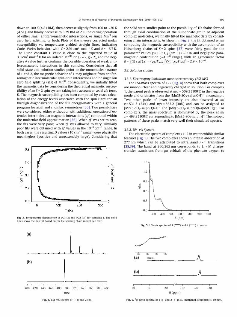
Fig. 4. ESI-MS spectra of 1 (a) and 2 (b).
the solid state studies point to the possibility of 1D chains formedthrough axial coordination of the sulphonate group of adjacentcomplex molecules, we finally fitted the magnetic data by consid-ering chain interactions. As shown in Fig. 3, the fit obtained whencomputing the magnetic susceptibility with the assumption of anHeisenberg chains of S = 2 spins [37] were fairly good for theparameter values g = 1.931, J (cm�1) = �0.16 and negligible para-magnetic contribution (�10�8 range), with an agreement factorR =
P[(vMT)obs � (vMT)calc]2/
P[(vMT)obs]2 = 2.9 � 10�4.
3.3. Solution studies
3.3.1. Electrospray ionization-mass spectrometry (ESI-MS)The ESI-mass spectra of 1–2 (Fig. 4) show that both complexes
are mononuclear and negatively charged in solution. For complex1, the parent peak is observed at m/z = 509.3 (100%) in the negativemode and originates from the [Mn(5-SO3-salpnOH)]� monoanion.Two other peaks of lower intensity are also observed at m/z = 531.5 (34%) and m/z = 563.2 (30%) and can be assigned to[Mn(5-SO3-salpnO)Na]� and [Mn(5-SO3-salpnO)Na(MeOH)]�. Forcomplex 2, the mass spectrum is dominated by the peak at m/z = 493.3 (100%) corresponding to [Mn(5-SO3-salpn)]�. The isotopicpatterns of these peaks match very well their simulated spectra.
3.3.2. UV–vis SpectraThe electronic spectra of complexes 1–2 in water exhibit similar
features (Fig. 5). The two complexes show an intense absorption at277 nm which can be attributed to intraligand p–p* transitions[38,39]. The band at 360/365 nm corresponds to L ? M charge-transfer transitions from pp orbitals of the phenoxo oxygen to
300 400 500 600 700 800 9000.0
0.3
0.6
0.9
1.2
1.5
Abs
λ (nm)
Fig. 5. UV–vis spectra of 1 ( ) and 2 ( ) in water.
40 30 20 -10 -20 -30
(b)
(a)
δ (ppm)
35 30 25 20δ (ppm)
Fig. 6. 1H NMR spectra of 1 (a) and 2 (b) in D4-methanol. [complex] � 10 mM.
500 D. Moreno et al. / Journal of Inorganic Biochemistry 104 (2010) 496–502
the partially filled dp orbitals of the MnIII ion, as also observed forother MnIII complexes with phenoxo ligands [33,40,41]. Absorptionat 545 nm (e = 397 (1) and 444 (2) M�1 cm�1), can be assigned to ad–d transition in agreement with the values reported for relatedMnIII complexes with the metal in a pseudotetragonal environment[42,43].
3.3.3. 1H NMR spectroscopyThe paramagnetic 1H NMR spectra of 1 and 2 in D4-methanol
(Fig. 6) revealed a simple pattern for the two complexes outsidethe diamagnetic region (ca. 0–10 ppm). In both cases, one reso-nance is observed up-field at d: �22 (1) and �23 ppm (2), whichcan be assigned to the H4 aromatic ring proton on the basis of com-parison with reported spectra for other phenolato based MnIII com-plexes [44,45]. Because of their closeness to the Mn centre, protonsadjacent to the donor groups of the Schiff base ligand (aromatic H3and H6, N@CHA and ACH2AN@C) are not observed, and this isconsistent with previous finding for related MnIII complexes [46].A very broad and weak resonance at +28 ppm (inset of Fig. 6a) isobserved in the spectrum of 1. This signal may arise from the car-binolic proton of [Mn(5-SO3-salpnOH)]�, in accordance with theresonance at +28.6 ppm reported for [Mn(salpnOH)(MeOH)2]+
[44], absent in the alkoxo-bridged dimer [Mn(salpnO)]2 [45,47].NMR has proven to be a useful probe of the ligand conformation
for Mn-Schiff-base complexes in solution [44]. 1H NMR spectra oftetragonal mononuclear Mn complexes with tetradentate Schiffbase ligands symmetrically arranged in the equatorial plane showtwo up-field resonances around �20 to �25 ppm assigned to H4/H40 and H5/H50 protons of the phenolato rings [44,48–50]. A morecomplex pattern is observed in the 1H NMR spectra of Mn com-plexes in which the ligand is not symmetrically arranged aroundthe Mn ion because the different extent of charge transfer for pro-tons of the phenolato trans to different groups results in the mag-netic nonequivalence of protons of the two phenolato rings [44,51].Therefore, observation of only one resonance for the aromatic ringprotons in the 1H NMR spectra of 1–2 indicates that these com-plexes possess trans-diaxial symmetry with the N2O2 donor setof the ligand located in the equatorial plane.
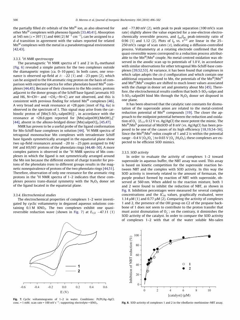
3.3.4. Electrochemical studiesThe electrochemical properties of complexes 1–2 were investi-
gated by cyclic voltammetry in degassed aqueous solutions con-taining 0.1 M KNO3. The two complexes exhibit one quasi-reversible reduction wave (shown in Fig. 7) at E1/2 �47.11 (1)
-0.6 -0.4 -0.2 0.0 0.2 0.4 0.6
12
E (V)
Fig. 7. Cyclic voltammograms of 1–2 in water. Conditions: Pt/Pt/Ag–AgCl;conc. = 1 mM; scan rate = 100 mV s�1; supporting electrolyte = KNO3.
and �77.80 mV (2), with peak to peak separation (100 mV/s scanrate) slightly above the value expected for a one-electron electro-chemically reversible process, and Ipa/Ipc peak-intensity ratio of0.96 (1) and 1.12 (2). Plots of Ip vs. v1/2 are linear in the 25–250 mV/s range of scan rates (v), indicating a diffusion-controlledprocess. Voltammetry at a rotating electrode confirmed that thequasi-reversible waves correspond to a reduction process attribut-able to the MnIII/MnII couple. No metal-centred oxidation was ob-served in the anodic scan up to potentials of 1.0 V, in accordancewith similar observations for other tetragonal Mn-Schiff-base com-plexes [39,52,53]. At variance, it has been found that complexes inwhich salpn adopts the cis-b configuration and which contain oneadditional oxyanion bound to Mn, the potentials of the MnIII/MnIV
and MnIII/MnII couples are shifted to much lower values associatedwith the change in donor set and geometry about Mn [45]. There-fore, the electrochemical results confirm that both 5-SO3-salpn and5-SO3-salpnOH bind Mn through the N2O2 donor set disposed inone plane.
It has been observed that the catalytic rate constants for dismu-tation of the superoxide anion are related to the metal-centredreduction potential of MnIII compounds [19]. The closer the ap-proach to the midpoint potential between the reduction and oxida-tion of O��2 (E1/2 0.12 V vs. Ag/AgCl) the more potent the mimic. TheMnIII/MnII potential of MnSOD of 8 mV (vs. Ag/AgCl) has been pro-posed to be one of the causes of its high efficiency [18,19,54–56].Since the MnIII/MnII redox couple of 1 and 2 is within the potentialrange�0.4 V (O2/O��2 ) to 0.65 V (O��2 /H2O2), these complexes are ex-pected to be efficient SOD mimics.
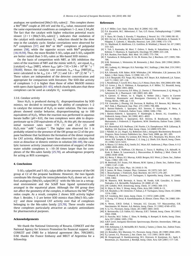
3.3.5. SOD activityIn order to evaluate the activity of complexes 1–2 toward
superoxide in aqueous buffer, the NBT assay was used. This assayis based on kinetic competition for the superoxide reaction be-tween NBT and the complex with SOD activity. In this way theSOD activity is inversely related to the amount of formazan, thepurple product formed by reaction of NBT with superoxide, ob-served at 560 nm. When added to the reaction mixture, both 1and 2 were found to inhibit the reduction of NBT, as shown inFig. 8. Inhibition percentages were measured for several complexconcentrations and the IC50 values, graphically evaluated, were1.14 lM (1) and 0.77 lM (2). Comparing the activity of complexes1 and 2, the presence of the OH group on C2 of the propane back-bone of 1 does not seem to contribute to the proton transfer thatmust assist dismutation of O��2 ; on the contrary, it diminishes theSOD activity of the catalyst. In order to compare the SOD activityof complexes 1–2 with that of the water soluble Mn-salen
0 2 4 6 8 10
0
20
40
60
80
100
12
Inhi
bitio
n of
NB
T r
educ
tion
(%)
[catalyst] (µM)
Fig. 8. SOD-activity of complexes 1 and 2 in the riboflavin–methionine–NBT assay.
D. Moreno et al. / Journal of Inorganic Biochemistry 104 (2010) 496–502 501
analogue, we synthesized [Mn(5-SO3-salen)]�. This complex showsa MnIII/MnII couple at 205 mV and the IC50 value, measured underthe same experimental conditions as complexes 1–2, was 2.34 lM.The fact that the catalyst with higher reduction potential reactsslower (2 > 1 > [Mn(5-SO3-salen)]�), indicates that oxidation ofthe catalyst with simultaneous O��2 reduction should be the slowstep in the catalytic cycle. Similar correlations were observed forFeII complexes [57] and MnII or MnIII complexes of polypodalamines [58], while the opposite occurs with MnIII-porphyrins[18,19,59]. Thus, the more flexible 5-SO3-salpn and 5-SO3-salpnOHare better suited than 5-SO3-salen as Mn-based SOD mimics.
On the basis of competition with NBT, at 50% inhibition therates of the reactions of NBT and the mimic with O��2 are equal, kcat
[catalyst] = kNBT [NBT], where kNBT (pH = 7.8) = 5.94 � 104 M�1 s�1
[58,60]. Hence, the catalytic rate constant, kcat = kNBT [NBT]/IC50,were calculated to be kcat 2.4 � 106 (1) and 3.6 � 106 (2) M�1 s�1.These values are independent of the detector concentration andappropriate for comparison with literature. The SOD-like activityof complexes 1–2 is higher than those of other MnSOD mimicswith open chain ligands [61–65], which clearly indicates that thesecomplexes can be used as catalytic O��2 scavengers.
3.3.6. Catalase activitySince H2O2 is produced during O��2 disproportionation by SOD
mimics, we decided to investigate the ability of complexes 1–2to catalyze the removal of H2O2 in water solution. The two com-plexes showed similar behavior, disproportionating up to 100equivalents of H2O2. When the reaction was performed in aqueousborate buffer (pH = 8.0), the two complexes were able to dispro-portionate up to 250 equivalents of H2O2. At this pH, the measuredinitial turnover rates were 178 (1) and 25.2 (2) mMH2O2 min�1 mM�1 catalyst�1. The higher rate observed for 1 isprobably related to the presence of the OH group on C2 of the pro-pane backbone that facilitates the formation of the dimer requiredfor CAT activity. Although lower than observed for catalysts thatexist as dinuclear or dimeric entities in solution [66–69], the cata-lytic turnover activity (maximal concentration of oxygen) of thesewater soluble complexes is �10–30 times larger than for com-plexes of the Mn-salen family [25,70], an advantageous propertyfor acting as ROS scavengers.
4. Conclusions
5-SO3-salpnOH and 5-SO3-salpn differ in the presence of the OHgroup at C2 of the propane backbone. However, the two ligandscoordinate Mn through the imino/phenolato N2O2 donor set, to af-ford analogous [Mn(SO3-salpn(OH))]� with the Mn ion in a tetrag-onal environment and the Schiff base ligand symmetricallyarranged in the equatorial plane. Although the OH group doesnot affect the geometry of the complex, it influences the MnIII/MnII
redox couple. As a result, complex 2 shows SOD activity higherthan 1. Besides, 1–2 are better SOD mimics than [Mn(5-SO3-sale-n)]� and show improved CAT activity over that of complexesbelonging to the Mn-salen family [25,70]. These results renderthese complexes particularly attractive as catalytic antioxidantsfor pharmaceutical purposes.
Acknowledgements
We thank the National University of Rosario, CONICET and theNational Agency for Sciences Promotion for financial support, andCONICET and CNRS for a bilateral agreement (Res. 709/2005).D.M. thanks the France Embassy and MECT of Argentina for afellowship.
References
[1] A.-F. Miller, Curr. Opin. Chem. Biol. 8 (2004) 162–168.[2] P.A. Kocatürk, M.C. Akbostanci, F. Tan, G.Ö. Kavas, Pathophysiology 7 (2000)
63–67.[3] Y. Ihara, M. Chuda, S. Kuroda, T. Hayabara, J. Neurol. Sci. 170 (1999) 90–95.[4] M.E. De Leo, S. Borrello, M. Passantino, B. Palazzotti, A. Mordente, A. Daniele, V.
Filippini, T. Galeotti, C. Masullo, Neurosci. Lett. 250 (1998) 173–176.[5] S.L. Marklund, R. Adolfsson, C.G. Gottfries, B. Winblad, J. Neurol. Sci. 67 (1985)
319–325.[6] Y. Toh, S. Kuninaka, M. Mori, T. Oshiro, Y. Ikeda, H. Nakashima, H. Baba, S.
Kohnoe, T. Okamura, K. Sugimachi, Oncology 59 (2000) 223–228.[7] R.H. Burdon, Free Radic. Biol. Med. 18 (1995) 775–794.[8] F. Yamakura, K. Kobayashi, H. Ue, M. Konno, Eur. J. Biochem. 227 (1995) 700–
706.[9] H.M. Steinman, L. Weinstein, M. Brenowitz, J. Biol. Chem. 269 (1994) 28629–
28634.[10] M.L. Ludwig, A.L. Metzger, K.A. Pattridge, W.C. Stallings, J. Mol. Biol. 219 (1991)
335–358.[11] R.A. Edwards, H.M. Baker, M.M. Whittaker, J.W. Whittaker, G.B. Jameson, E.N.
Baker, J. Biol. Inorg. Chem. 3 (1998) 161–171.[12] G.E.O. Borgstahl, H.E. Parge, M.J. Hickey, W.F. Beyer, R.A. Hallewell, J.A. Tainer,
Cell 71 (1992) 107–118.[13] L. Noodleman, T. Lovell, W.G. Han, J. Li, F. Himo, Chem. Rev. 104 (2004) 459–508.[14] L.E. Grove, J. Xie, E. Yikilmaz, A. Karapetyan, A.-F. Miller, T.C. Brunold, Inorg.
Chem. 47 (2008) 3993–4004.[15] C. Muscoli, S. Cuzzocrea, D.P. Riley, J.L. Zweier, C. Thiemermann, Z.-Q. Wang, D.
Salvemini, Br. J. Pharmacol. 140 (2003) 445–460.[16] A. Gärtner, U. Weser, in: E. Vögtle, E. Weber (Eds.), Topics in Current
Chemistry: Biomimetic and Bioorganic Chemistry II, vol. 132, Springer-Verlag, Berlin, 1986, pp. 1–61.
[17] P.K. Gonzalez, J. Zhuang, S.R. Doctrow, B. Malfroy, P.F. Benson, M.J. Menconi,M.P. Fink, J. Pharmacol. Exp. Ther. 275 (1995) 798–806.
[18] I. Batinic-Haberle, I. Spasojevi, R.D. Stevens, P. Hambright, P. Neta, A.Matsumoto, I. Fridovich, Dalton Trans. (2004) 1696–1702.
[19] I. Batinic-Haberle, I. Spasojevic, P. Hambright, L. Benov, A.L. Crumbliss, I.Fridovich, Inorg. Chem. 38 (1999) 4011–4022.
[20] I. Batinic-Haberle, I. Spasojevic, R.D. Stevens, B. Bondurant, A. Okado-Matsumoto, I. Fridovich, Z. Vujaskovic, M.W. Dewhirsta, Dalton Trans. (2005)617–624.
[21] R.A. Rosenthal, K.D. Huffman, L.W. Fisette, C.A. Damphousse, W.B. Callaway, B.Malfroy, S.R. Doctrow, J. Biol. Inorg. Chem. 14 (2009) 979–991.
[22] I. Pálinkó, in: J.G. Huges, A.J. Robinson (Eds.), Inorganic Biochemistry ResearchProgress, Nova Science Publishers Inc., New York, 2008, pp. 281–303.
[23] D.P. Riley, S.L. Henke, P.J. Lennon, R.H. Weiss, W.L. Neumann, W.J. Rivers, K.W.Aston, K.R. Sample, H. Rahman, C.-S. Ling, J.-J. Shieh, D.H. Busch, Inorg. Chem.35 (1996) 5213–5231.
[24] A. Maroz, G.F. Kelso, R.A.J. Smith, D.C. Ware, R.F. Anderson, J. Phys. Chem. A 112(2008) 4929–4935.
[25] S.R. Doctrow, K. Huffman, C.B. Marcus, C. Tocco, E. Malfroy, C.A. Adinolfi, H.Kruk, K. Baker, N. Lazarowych, J. Mascarenhas, B. Malfroy, J. Med. Chem. 45(2002) 4549–4558.
[26] K.J. Berry, F. Moya, K.S. Murray, A.M.B. Bergen, B.O. West, J. Chem. Soc., DaltonTrans. (1982) 109–116.
[27] M. Botsivali, D.F. Evans, P.H. Missen, M.W. Upton, J. Chem. Soc., Dalton Trans.(1985) 1147–1149.
[28] P. Pascal, Ann. Chim. Phys. 19 (1910) 5–70.[29] F. James, M. Roos, Comput. Phys. Commun. 10 (1975) 343–367.[30] C. Beauchamps, I. Fridovich, Anal. Biochem. 44 (1971) 276–287.[31] C. Palopoli, B. Chansou, J.-P. Tuchagues, S. Signorella, Inorg. Chem. 39 (2000)
1458–1462.[32] M. Maneiro, M.R. Bermejo, A. Sousa, M. Fondo, A.M. González, A. Sousa-
Pedrades, C.A. McAuliffe, Polyhedron 19 (2000) 47–54.[33] J.W. Gohdes, W.H. Armstrong, Inorg. Chem. 31 (1992) 368–373.[34] H. Shyu, H. Wei, Y. Wang, Inorg. Chim. Acta 290 (1999) 8–13.[35] A.K. Boudalis, J.-M. Clemente, F. Dahan, J.-P. Tuchagues, Inorg. Chem. 43 (2004)
1574–1586.[36] A.P. Ginsberg, M.E. Lines, Inorg. Chem. 11 (1972) 2289–2290.[37] K. Konig, V.P. Desai, B. Kanellakopulos, R. Klenze, Chem. Phys. 54 (1980) 109–
113.[38] A. Neves, S.M.D. Erthal, I. Vencato, A.S. Ceccato, Y.P. Mascarenhas, O.R.
Nascimento, M. Horner, A.A. Batista, Inorg. Chem. 31 (1992) 4749–4755.[39] X. Li, V.L. Pecoraro, Inorg. Chem. 28 (1989) 3403–3410.[40] K. Bertoncello, G.D. Fallon, K.S. Murray, E.R.T. Tiekink, Inorg. Chem. 30 (1991)
3562–3568.[41] D. Pursche, M.U. Triller, C. Slinn, N. Reddig, A. Rompel, B. Krebs, Inorg. Chim.
Acta 357 (2004) 1695–1702.[42] A.B.P. Lever, Inorganic Electronic Spectroscopy, Elsevier, Amsterdam, 1984. pp.
440–444.[43] F.M. Ashmawy, C.A. McAuliffe, R.V. Parish, J. Tames, J. Chem. Soc., Dalton Trans.
(1985) 1391–1397.[44] J.A. Bonadies, M.J. Maroney, V.L. Pecoraro, Inorg. Chem. 28 (1989) 2044–2051.[45] E.J. Larson, V.L. Pecoraro, J. Am. Chem. Soc. 113 (1991) 3810–3818.[46] M. Hoogenraad, K. Ramkisoensing, W.L. Driessen, H. Kooijman, A.L. Spek, E.
Bouwman, J.G. Haasnoot, J. Reedijk, Inorg. Chim. Acta 320 (2001) 117–126.
502 D. Moreno et al. / Journal of Inorganic Biochemistry 104 (2010) 496–502
[47] J.A. Bonadies, M.L. Kirk, M.S. Lah, D.P. Kessissoglou, W.E. Hatfield, V.L. Pecoraro,Inorg. Chem. 28 (1989) 2037–2044.
[48] M.T. Caudle, P. Riggs-Gelasco, A.K. Gelasco, J.E. Penner-Hahn, V.L. Pecoraro,Inorg. Chem. 35 (1996) 3577–3584.
[49] M.R. Bermejo, A.M. González, M. Fondo, A. García-Deibe, M. Maneiro, J.Sanmartín, O. Hoyos, M. Watkinson, New J. Chem. 24 (2000) 235–241.
[50] M.R. Bermejo, A.M. González-Noya, V. Abad, M.I. Fernández, M. Maneiro, R.Pedrido, M. Vásquez, Eur. J. Inorg. Chem. (2004) 3696–3705.
[51] D.P. Kessissoglou, W.M. Butler, V.L. Pecoraro, Inorg. Chem. 26 (1987) 495–503.[52] E. Larson, M.S. Lah, X. Li, J.A. Bonadies, V.L. Pecoraro, Inorg. Chem. 31 (1992)
373–378.[53] R.K. Boggess, J.W. Hughes, W.M. Coleman, L.T. Taylor, Inorg. Chim. Acta 38
(1980) 183–189.[54] W.C. Barrette, D.T. Sawyer, J.A. Fee, K. Asada, Biochemistry 22 (2002) 624–627.[55] W.H. Koppenol, F. Levine, T.L. Hatmaker, J. Epp, J.D. Rush, Arch. Biochem.
Biophys. 251 (1986) 594–599.[56] J. Stein, J.P.J. Fackler, G.J. McClune, J.A. Fee, L.T. Chan, Inorg. Chem. 18 (1979)
3511–3519.[57] P.J.F. Gauuan, M.P. Trova, L. Gregor-Boros, S.B. Bocckino, J.D. Crapo, B.J. Day,
Bioorg. Med. Chem. 10 (2002) 3013–3021.[58] S. Durot, C. Policar, F. Cisnetti, F. Lambert, J.-P. Renault, G. Pelosi, G. Blain, H.
Korri-Youssoufi, J.-P. Mahy, Eur. J. Inorg. Chem. (2005) 3513–3523.
[59] R. Kachadourian, I. Batinic-Haberle, I. Fridovich, Inorg. Chem. 38 (1999) 391–396.
[60] Z.R. Liao, X.F. Zheng, B.S. Luo, L.R. Shen, D.F. Li, H.L. Liu, W. Zhao, Polyhedron 20(2001) 2813–2821.
[61] D.P. Riley, Chem. Rev. 99 (1999) 2573–2588.[62] Q.X. Li, Q.H. Luo, Y.Z. Li, Z.Q. Pan, M.C. Shen, Eur. J. Inorg. Chem. (2004) 4447–
4456.[63] E.A. Lewis, H.H. Khodr, R.C. Hilder, J.R. Smith, P.H. Walton, Dalton Trans. (2004)
187–188.[64] J. Lin, C. Tu, H. Lin, P. Jiang, J. Ding, Z. Guo, Inorg. Chem. Comm. 6 (2003) 262–
265.[65] U.P. Singh, A.K. Sharma, P. Tyagi, S. Upreti, R.K. Singh, Polyhedron 25 (2006)
3628–3638.[66] A. Gelasco, S. Bensiek, V.L. Pecoraro, Inorg. Chem. 37 (1998) 3301–3309.[67] H. Biava, C. Palopoli, S. Shova, M. De Gaudio, V. Daier, M. González-Sierra, J.-P.
Tuchagues, S. Signorella, J. Inorg. Biochem. 100 (2006) 1660–1671.[68] D. Moreno, C. Palopoli, V. Daier, S. Shova, L. Vendier, M. González-Sierra, J.-P.
Tuchagues, S. Signorella, Dalton Trans. 10 (2006) 5156–5166.[69] S. Signorella, A. Rompel, K. Buldt-Karentzopoulos, B. Krebs, V.L. Pecoraro, J.-P.
Tuchagues, Inorg. Chem. 46 (2007) 10864–10868.[70] W. Park, D. Lim, Bioorg. Med. Chem. Lett. 19 (2009) 614–617.


2•H2O precursor 5-Other characterizations](https://static.fdocuments.us/doc/165x107/5f1f4ac97ba5686a2771fb18/polyoxometalate-based-mniii-schiff-base-composite-4-magnetic-properties.jpg)


![Magnetic Properties of a Family of [MnIII LnIII ] Wheel ...](https://static.fdocuments.us/doc/165x107/62a6a00d7a3cb7039422b877/magnetic-properties-of-a-family-of-mniii-lniii-wheel-.jpg)

