Synthesis and Structural Properties of P-Stereodefined ... fileElectronic Supplementary Information...
25
Electronic Supplementary Information Phosphorothioate Analogs of Glycol Nucleic Acids Synthesis and Structural Properties of P-Stereodefined Phosphorothioate Analogs of Glycol Nucleic Acids. Agnieszka Tomaszewska-Antczak, Anna Maciaszek, Katarzyna Jastrzębska, Barbara Mikołajczyk, Piotr Guga Centre of Molecular and Macromolecular Studies, Polish Academy of Sciences, Department of Bioorganic Chemistry, Sienkiewicza 112. 90-363 Łódź, Poland Electronic Supplementary Material (ESI) for RSC Advances. This journal is © The Royal Society of Chemistry 2018
Transcript of Synthesis and Structural Properties of P-Stereodefined ... fileElectronic Supplementary Information...

Electronic Supplementary Information Phosphorothioate Analogs of Glycol Nucleic Acids
Synthesis and Structural Properties of P-Stereodefined
Phosphorothioate Analogs of Glycol Nucleic Acids.
Agnieszka Tomaszewska-Antczak, Anna Maciaszek, Katarzyna Jastrzębska,
Barbara Mikołajczyk, Piotr Guga
Centre of Molecular and Macromolecular Studies, Polish Academy of Sciences, Department of
Bioorganic Chemistry, Sienkiewicza 112. 90-363 Łódź, Poland
Electronic Supplementary Material (ESI) for RSC Advances.This journal is © The Royal Society of Chemistry 2018
Electronic Supplementary Information Phosphorothioate Analogs of Glycol Nucleic Acids
Table 1S. HRMS data for 5a-d (unseparated P-diastereomers) and physico-chemical
characteristics of fast and slow-eluting pairs of enantiomers of 5a-d. A mixture of
EtOAc and hexane at given ratio (v/v) was used for TLC and HPLC analyses.
a) A Pursuit XRs column (10µ silica, 100 Å; 250 × 21.2 mm; flow rate 25 mL min−1);
b) In CDCl3;
In principle, the phosphitylation of eight DMT-GN’s (obtained from 2 enantiomeric
glycidols and 4 nucleobases) should provide 16 OTP-GN’s, but because 4d was
obtained only from (R)-(+)-glycidol we actually have obtained 14 diastereomerically
different OTP-GN’s. However, Table 1S does not contain 14 data sets but only 8 (5a
fast/slow to 5d fast/slow) because each of 5a-c consists of two pairs of enantiomers
(RPRC/SPSC and SPRC/RPSC) and within each pair the components cannot be
distinguished by the chromatographic and spectroscopic methods applied (no chiral
auxiliaries were used). From this perspective 5d consisted of two diastereomeric
RPSC and SPSC components.
5a
OTP-GT
5b
OTP-GABz
5c
OTP-GCBz
5d
OTP-GGiBu
HRMSc) (Da) calc.
found
707.2015
707.2015
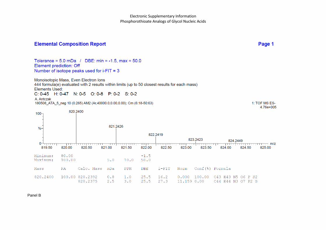
820.2392
820.2400
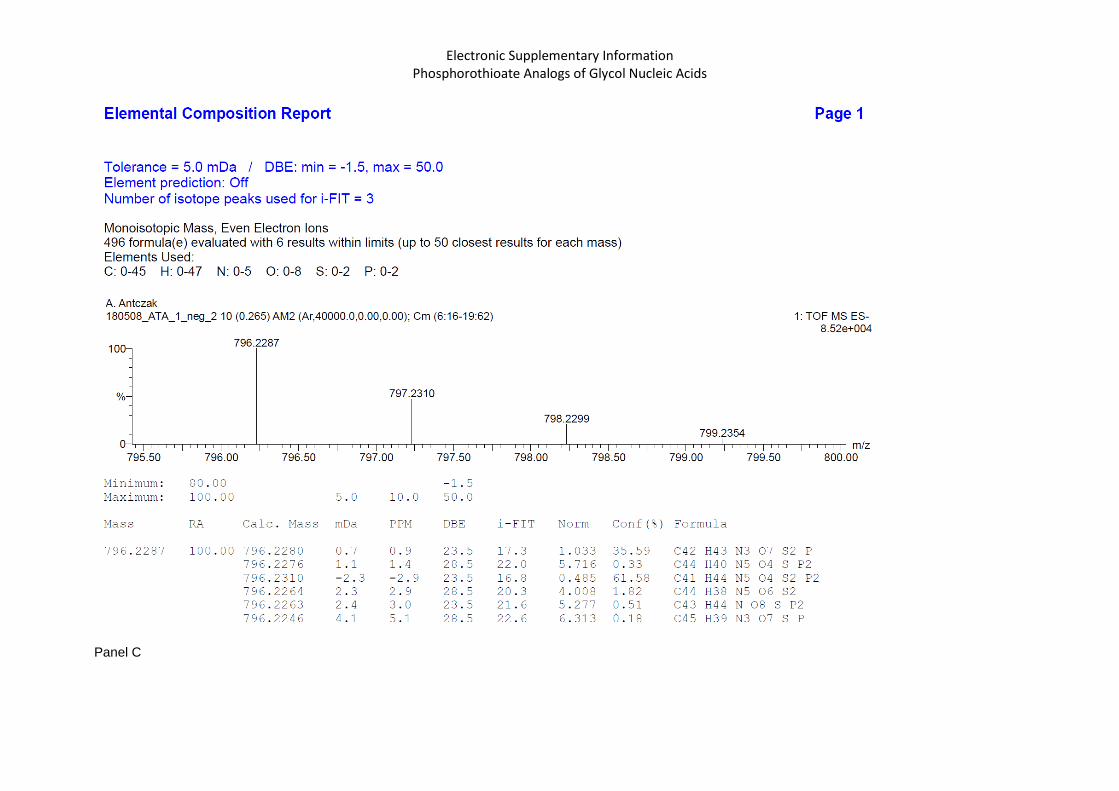
796.2280
796.2287
802.2498
802.2514
EtOAc : hexane 50 : 50 45 : 55 55 : 45 85 : 15
fast/ slow f s f s f s f s
TLC, Rf 0.68 0.60 0.72 0.58 0.70 0.58 0.65 0.50
HPLCa), Rt (min) 19.2 21.5 18.0 23.0 27.0 31.2 13.5 17.0
δ 31P NMRb) (ppm) 105.6 106.1 105.8 106.6 105.1 105.7 106.1 106.5

Electronic Supplementary Information Phosphorothioate Analogs of Glycol Nucleic Acids
Panel A – isomer fast
Panel B – isomer slow
Figure 1S. 31
P NMR spectra (CDCl3) for fast- and slow-eluting 5a, panel A and B, respectively; recorded with a Bruker AV-200 spectrometer (200 MHz).

Electronic Supplementary Information Phosphorothioate Analogs of Glycol Nucleic Acids
Panel A
Panel B
Figure 2S. 1H NMR spectra (CDCl3) for fast-eluting and slow-eluting 5a, panel A and B, respectively;
recorded with a Bruker AV-200 spectrometer (200 MHz).

Electronic Supplementary Information Phosphorothioate Analogs of Glycol Nucleic Acids
Panel A
Panel B
Figure 3S. 13
C NMR spectra (CDCl3) for fast-eluting and slow-eluting 5a, panel A and B, respectively; recorded with a Bruker AV-200 spectrometer (200 MHz).
Electronic Supplementary Information Phosphorothioate Analogs of Glycol Nucleic Acids
Panel A
Electronic Supplementary Information Phosphorothioate Analogs of Glycol Nucleic Acids
Panel B
Electronic Supplementary Information Phosphorothioate Analogs of Glycol Nucleic Acids
Panel C

Electronic Supplementary Information Phosphorothioate Analogs of Glycol Nucleic Acids
Panel D
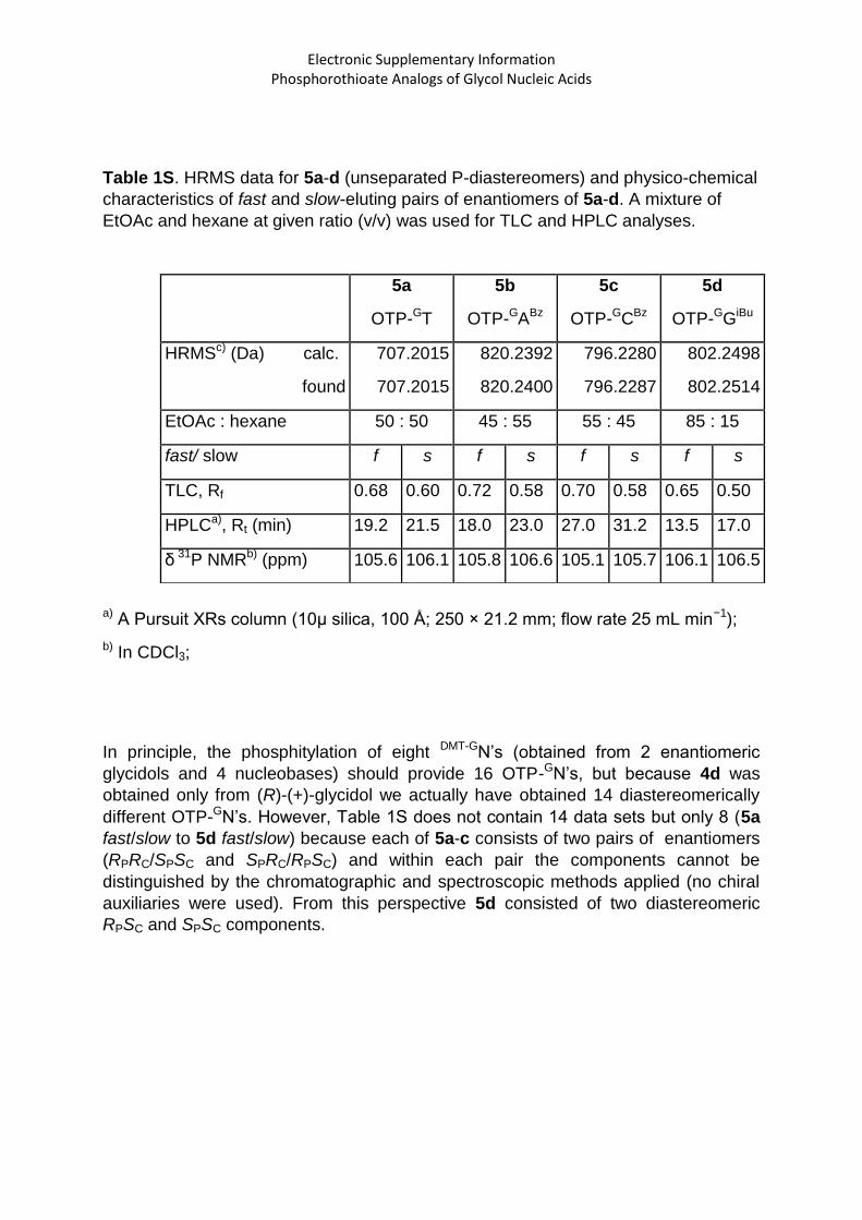
Figure 4S. HRMS spectra for 5a-d (panel A, B, C, and D, respectively).
Electronic Supplementary Information Phosphorothioate Analogs of Glycol Nucleic Acids
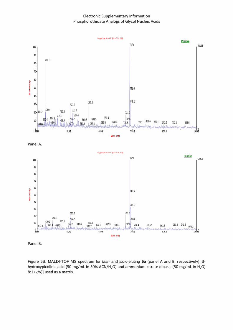
Panel A.
Panel B.
Figure 5S. MALDI-TOF MS spectrum for fast- and slow-eluting 5a (panel A and B, respectively). 3-hydroxypicolinic acid (50 mg/mL in 50% ACN/H2O) and ammonium citrate dibasic (50 mg/mL in H2O) 8:1 (v/v)] used as a matrix.
399.0 519.2 639.4 759.6 879.8 1000.0
Mass (m/z)
0
5212.6
0
10
20
30
40
50
60
70
80
90
100
% In
te
ns
ity
V oyager Spec #1=>NF0.7[BP = 747.6, 5213]
Positive747.6
429.5
748.6
749.6591.3523.5
430.4 530.3485.5401.2 731.7537.4475.3
651.4447.3 732.6524.5 604.5420.4 569.5486.4 808.6 839.1683.3 778.1 870.2449.4 639.5 955.6724.5 907.9517.5 598.5561.4404.4
399.0 519.2 639.4 759.6 879.8 1000.0
Mass (m/z)
0
5025.9
0
10
20
30
40
50
60
70
80
90
100
% In
te
ns
ity
V oyager Spec #1=>NF0.7[BP = 747.6, 5026]
Positive747.6
748.6
749.6
731.6523.5
456.3 750.6524.5485.5430.3 591.3517.4 732.6548.6 657.5 941.5911.4691.4 764.4622.0440.4 815.3468.5 863.6402.3 586.2 970.3

Electronic Supplementary Information Phosphorothioate Analogs of Glycol Nucleic Acids
Figure 6S. MS data for cGTMPS isolated from the mixture after detritylation of 5a.

Electronic Supplementary Information Phosphorothioate Analogs of Glycol Nucleic Acids
Panel A
Panel B
Figure 7S. 31
P NMR spectra (no deuterated solvent) for DMT-GC
BzPSTAc 10c obtained from fast-5c and
slow-5c, panel A and B, respectively. The monomer 5c was obtained from R-(+)-glycidol.
Electronic Supplementary Information Phosphorothioate Analogs of Glycol Nucleic Acids
Panel A
Panel B
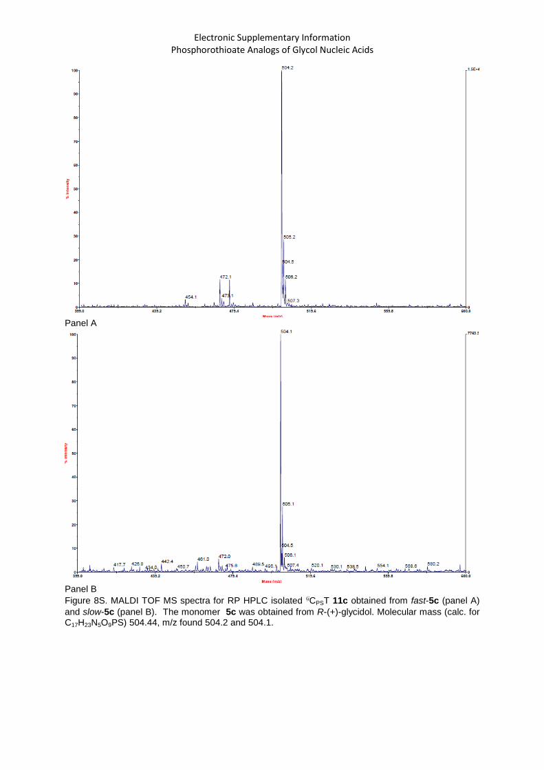
Figure 8S. MALDI TOF MS spectra for RP HPLC isolated GCPST 11c obtained from fast-5c (panel A)
and slow-5c (panel B). The monomer 5c was obtained from R-(+)-glycidol. Molecular mass (calc. for C17H23N5O9PS) 504.44, m/z found 504.2 and 504.1.
Electronic Supplementary Information Phosphorothioate Analogs of Glycol Nucleic Acids
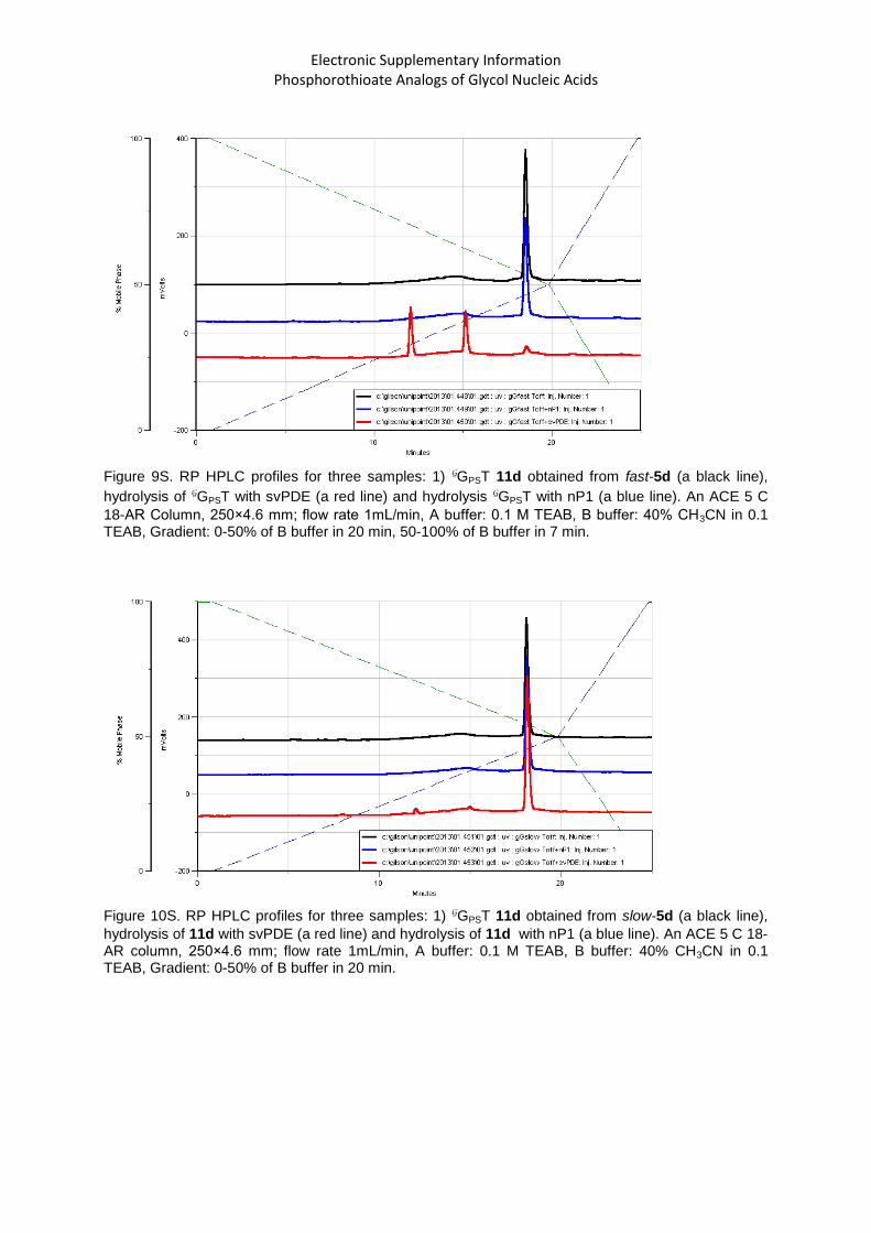
Figure 9S. RP HPLC profiles for three samples: 1) GGPST 11d obtained from fast-5d (a black line),
hydrolysis of GGPST with svPDE (a red line) and hydrolysis GGPST with nP1 (a blue line). An ACE 5 C
18-AR Column, 250×4.6 mm; flow rate 1mL/min, A buffer: 0.1 M TEAB, B buffer: 40% CH3CN in 0.1 TEAB, Gradient: 0-50% of B buffer in 20 min, 50-100% of B buffer in 7 min.
Figure 10S. RP HPLC profiles for three samples: 1) GGPST 11d obtained from slow-5d (a black line),
hydrolysis of 11d with svPDE (a red line) and hydrolysis of 11d with nP1 (a blue line). An ACE 5 C 18-AR column, 250×4.6 mm; flow rate 1mL/min, A buffer: 0.1 M TEAB, B buffer: 40% CH3CN in 0.1 TEAB, Gradient: 0-50% of B buffer in 20 min.

Electronic Supplementary Information Phosphorothioate Analogs of Glycol Nucleic Acids
Figure 11S. HPLC profiles recorded for four samples: 1) d(CPST), a mixture of both P-diastereomers – a black line; 2) d(CPST), a mixture of both P-diastereomers, treated with
svPDE – a blue line; 3) GCPST 10c (derived from fast-5c) treated with svPDE – a red line; 4) GCPST 10c (derived from fast-5c) – a pink line. A Kinetex 5μ C18 column, 250×4.6 mm; flow
rate 1mL/min, A buffer: 0.1 M TEAB, B buffer: 40% CH3CN in 0.1 TEAB, Gradient: 0-50% of B buffer in 20 min.
TMPS
Electronic Supplementary Information Phosphorothioate Analogs of Glycol Nucleic Acids
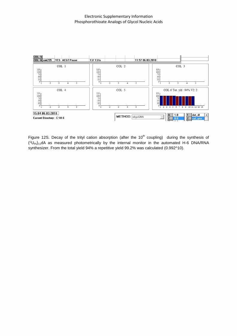
Figure 12S. Decay of the trityl cation absorption (after the 10
th coupling) during the synthesis of
(GUPS)11dA as measured photometrically by the internal monitor in the automated H-6 DNA/RNA
synthesizer. From the total yield 94% a repetitive yield 99.2% was calculated (0.992^10).
Electronic Supplementary Information Phosphorothioate Analogs of Glycol Nucleic Acids
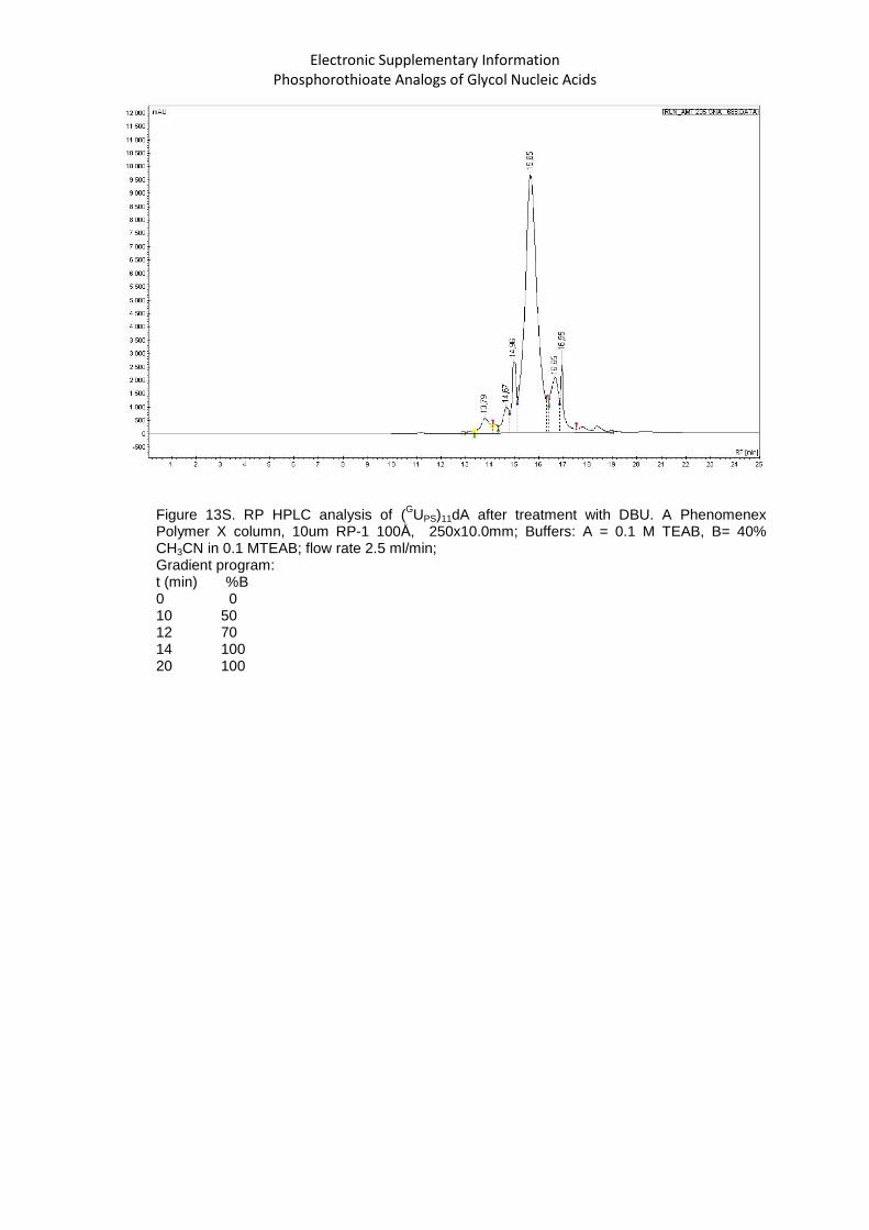
Figure 13S. RP HPLC analysis of (GUPS)11dA after treatment with DBU. A Phenomenex
Polymer X column, 10um RP-1 100Å, 250x10.0mm; Buffers: A = 0.1 M TEAB, B= 40% CH3CN in 0.1 MTEAB; flow rate 2.5 ml/min; Gradient program: t (min) %B 0 0 10 50 12 70 14 100 20 100
Electronic Supplementary Information Phosphorothioate Analogs of Glycol Nucleic Acids
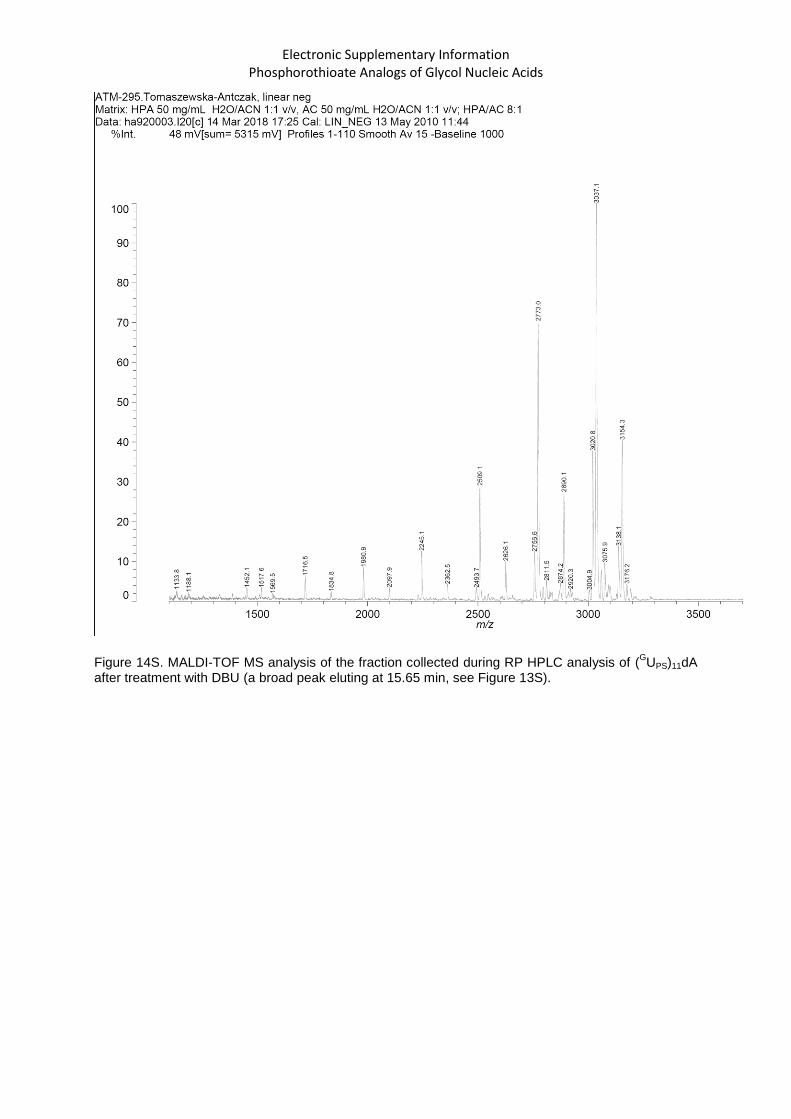
Figure 14S. MALDI-TOF MS analysis of the fraction collected during RP HPLC analysis of (
GUPS)11dA
after treatment with DBU (a broad peak eluting at 15.65 min, see Figure 13S).

Electronic Supplementary Information Phosphorothioate Analogs of Glycol Nucleic Acids
Figure 15S. Decay of the trityl cation absorption during the manual synthesis of SP-17
(AGTGGCGGCAT) measured photometrically.

Electronic Supplementary Information Phosphorothioate Analogs of Glycol Nucleic Acids
Figure 16S. RP HPLC profile for the detritylated SP-17 oligomer. A Kinetex 5μ C18 column, 250×4.6 mm; flow rate 1mL/min, A buffer: 0.1 M TEAB, B buffer: 40% CH3CN in 0.1 TEAB, Gradient: 0-100% of B buffer in 22 min.
Electronic Supplementary Information Phosphorothioate Analogs of Glycol Nucleic Acids
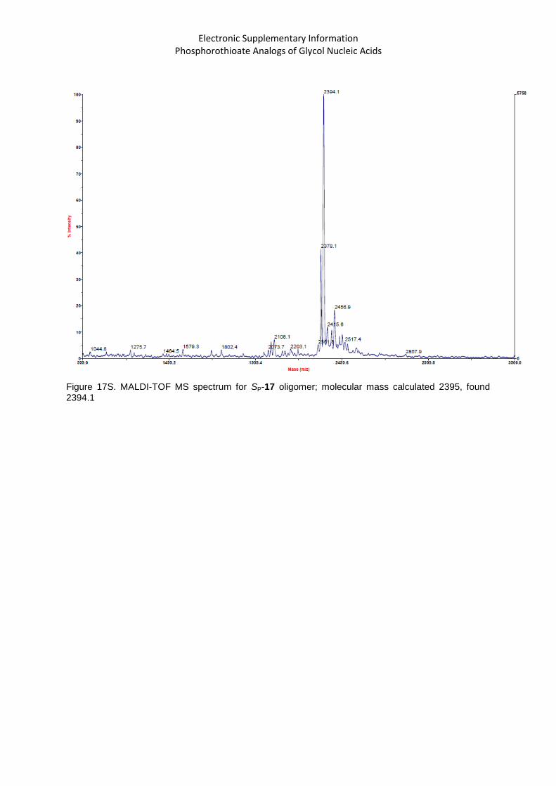
Figure 17S. MALDI-TOF MS spectrum for SP-17 oligomer; molecular mass calculated 2395, found 2394.1
Electronic Supplementary Information Phosphorothioate Analogs of Glycol Nucleic Acids
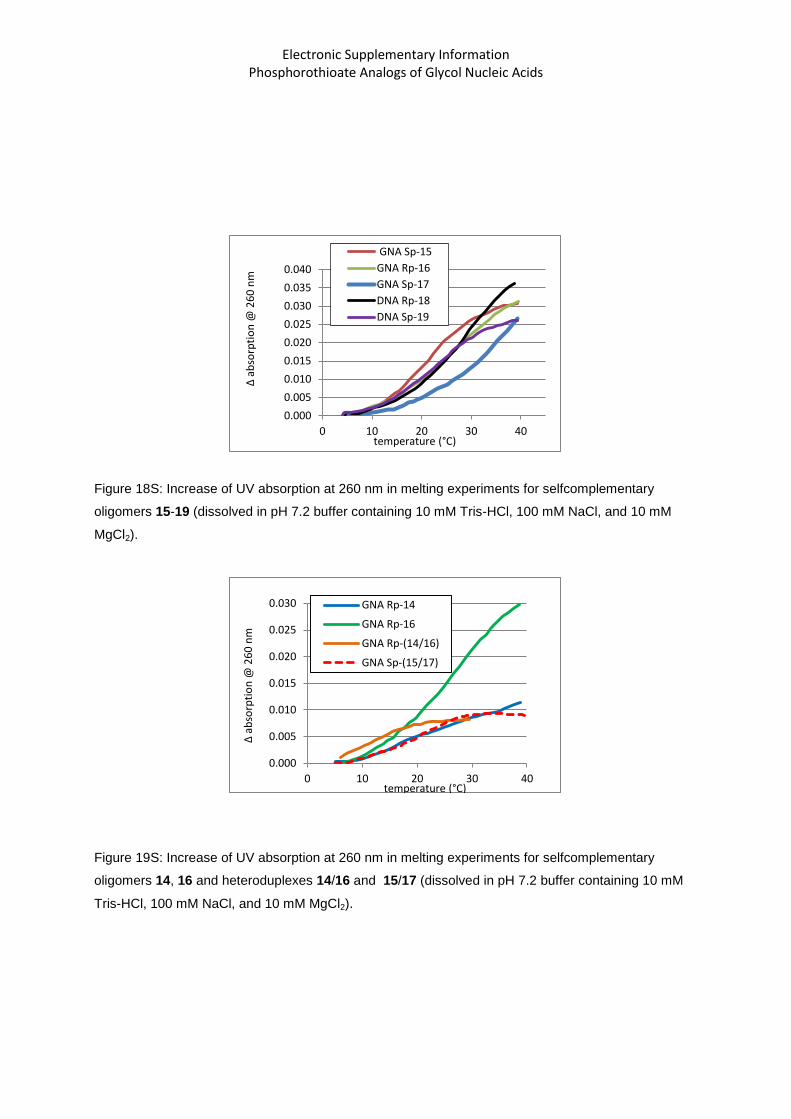
Figure 18S: Increase of UV absorption at 260 nm in melting experiments for selfcomplementary
oligomers 15-19 (dissolved in pH 7.2 buffer containing 10 mM Tris-HCl, 100 mM NaCl, and 10 mM
MgCl2).
Figure 19S: Increase of UV absorption at 260 nm in melting experiments for selfcomplementary
oligomers 14, 16 and heteroduplexes 14/16 and 15/17 (dissolved in pH 7.2 buffer containing 10 mM
Tris-HCl, 100 mM NaCl, and 10 mM MgCl2).
0.000
0.005
0.010
0.015
0.020
0.025
0.030
0.035
0.040
0 10 20 30 40
GNA Sp-15
GNA Rp-16
GNA Sp-17
DNA Rp-18
DNA Sp-19
temperature (°C)
Δ a
bso
rpti
on
@ 2
60
nm
0.000
0.005
0.010
0.015
0.020
0.025
0.030
0 10 20 30 40
GNA Rp-14
GNA Rp-16
GNA Rp-(14/16)
GNA Sp-(15/17)
Δ a
bso
rpti
on
@ 2
60
nm
temperature (°C)

Electronic Supplementary Information Phosphorothioate Analogs of Glycol Nucleic Acids
Table 2S. Melting temperatures for mixtures of RP-PS-(GNA/DNA) 14 or 16, and SP-PS-(GNA/DNA)
15 or 17 with DNA (d(ATGCGCAT)) or (m)RNA ((2’-OMe)-AUGCGCAU) templates. Melting
temperatures for homoduplexes DNA/DNA and (m)RNA/(m)RNA are given as the reference.
Temperature gradients of 1°C/min for annealing and 0.5°C/min for melting were applied.
Tm (°C)
template DNA (m)RNA
5’-(GA TGG CGG CGA T )-3’ 5’-(A GT G GC G GC A T)-3’
RP-14 SP-15 RP-16 SP-17
DNA
d(ATGCGCAT) 43 × 41 42 42 41
(m)RNA (2’-OMe)-
AUGCGCAU × 62 59 60 59 58
Electronic Supplementary Information Phosphorothioate Analogs of Glycol Nucleic Acids
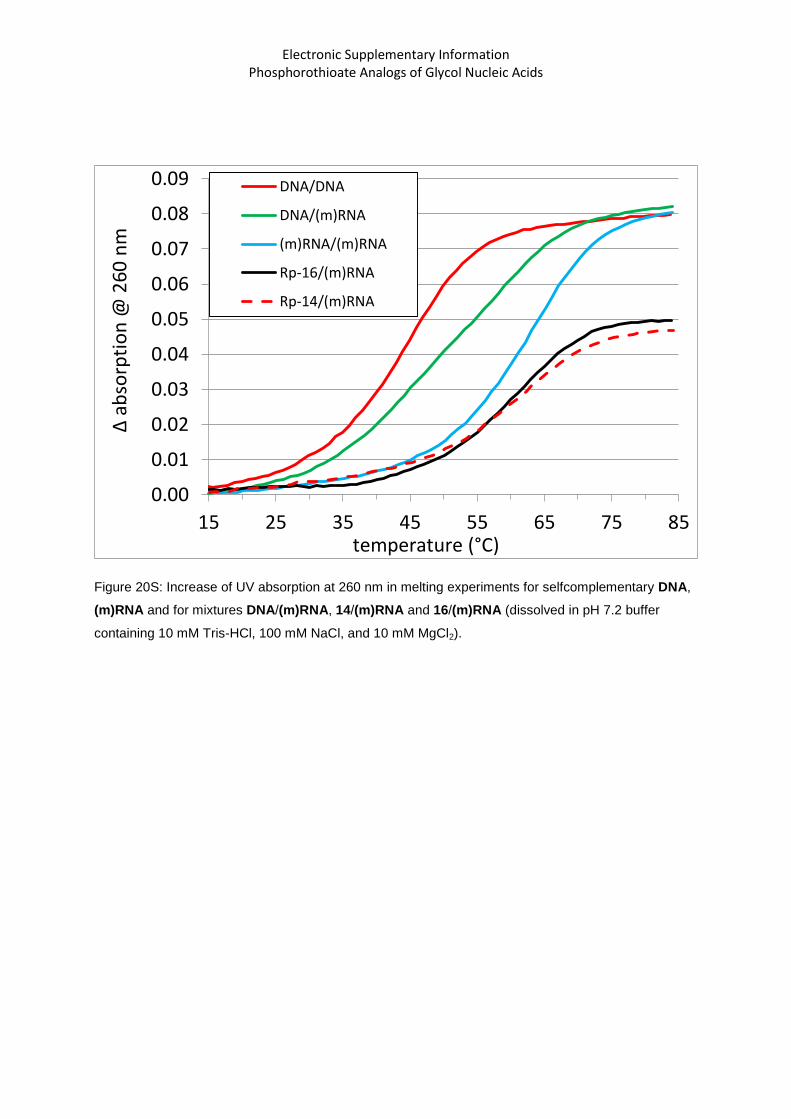
Figure 20S: Increase of UV absorption at 260 nm in melting experiments for selfcomplementary DNA,
(m)RNA and for mixtures DNA/(m)RNA, 14/(m)RNA and 16/(m)RNA (dissolved in pH 7.2 buffer
containing 10 mM Tris-HCl, 100 mM NaCl, and 10 mM MgCl2).
0.00
0.01
0.02
0.03
0.04
0.05
0.06
0.07
0.08
0.09
15 25 35 45 55 65 75 85
DNA/DNA
DNA/(m)RNA
(m)RNA/(m)RNA
Rp-16/(m)RNA
Rp-14/(m)RNA
Δ a
bso
rpti
on
@ 2
60
nm
temperature (°C)
Electronic Supplementary Information Phosphorothioate Analogs of Glycol Nucleic Acids
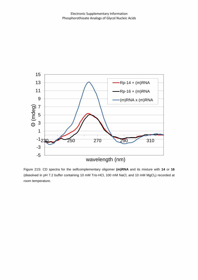
Figure 21S: CD spectra for the selfcomplementary oligomer (m)RNA and its mixture with 14 or 16
(dissolved in pH 7.2 buffer containing 10 mM Tris-HCl, 100 mM NaCl, and 10 mM MgCl2) recorded at
room temperature.
-5
-3
-1
1
3
5
7
9
11
13
15
230 250 270 290 310
Θ (
md
eg
)
wavelength (nm)
Rp-14 + (m)RNA
Rp-16 + (m)RNA
(m)RNA x (m)RNA