
Synthesis and Screening of a Combinatorial Peptide …/67531/metadc30440/m2/1/high...sequencing t he...
71
APPROVED: Robby A. Petros, Major Professor Guido F. Verbeck IV, Committee Member William E. Acree Jr., Chair of the Department of Chemistry James D. Meernik, Acting Dean of the Robert B. Toulouse School of Graduate Studies SYNTHESIS AND SCREENING OF A COMBINATORIAL PEPTIDE LIBRARY FOR LIGANDS TO TARGET TRANSFERRIN: MINIATURIZING THE LIBRARY Jennifer M. Brown, B. S. Thesis Prepared for the Degree of MASTER OF SCIENCE UNIVERSITY OF NORTH TEXAS August 2010
Transcript of Synthesis and Screening of a Combinatorial Peptide …/67531/metadc30440/m2/1/high...sequencing t he...

APPROVED: Robby A. Petros, Major Professor Guido F. Verbeck IV, Committee
Member William E. Acree Jr., Chair of the
Department of Chemistry James D. Meernik, Acting Dean of the
Robert B. Toulouse School of Graduate Studies
SYNTHESIS AND SCREENING OF A COMBINATORIAL PEPTIDE LIBRARY FOR
LIGANDS TO TARGET TRANSFERRIN: MINIATURIZING THE LIBRARY
Jennifer M. Brown, B. S.
Thesis Prepared for the Degree of
MASTER OF SCIENCE
UNIVERSITY OF NORTH TEXAS
August 2010

Brown, Jennifer M. Synthesis and Screening of a Combinatorial Peptide Library
for Ligands to Target Transferrin: Miniaturizing the Library. Master of Science
(Chemistry-Organic Chemistry), August 2010, 64 pp., 4 tables, 30 illustrations, works
cited, 30 titles.
Combinatorial libraries are used in the search for ligands that bind to target
proteins. Fmoc solid-phase peptide synthesis is routinely used to generate such
libraries. Microwave-assisted peptide synthesis was employed here to decrease
reaction times by 80-90%. Two One-Bead-One-Compound combinatorial libraries were
synthesized on 130μm beads (one containing 750 members and the other 16, 807). The
use of smaller solid supports would have many important practical advantages
including; increased library diversity per unit mass, smaller quantities of library needed
to generate hits, and screening could be conducted by using a standard flow cytometer.
To this end, a miniaturized peptide library was synthesized on 20 μm beads to
demonstrate proof of principle.
A small sample from the 16,807-member library was screened against
transferrin-AlexaFluro 647, a protein responsible for iron transport in vivo. A number of
hits were identified and sequenced using techniques coupling nanomanipulation with
nanoelectrospray mass spectrometry.

ii
Copyright 2010
by
Jennifer M. Brown

iii
TABLE OF CONTENTS
Page LIST OF TABLES ................................................................................................................iv LIST OF ILLUSTRATIONS ................................................................................................. v Chapters
I. INTRODUCTION ........................................................................................... 1 II. EXPERIMENTAL........................................................................................... 5
Peptide Synthesis
One Bead One Compound Library Synthesis
Gas-Phase Cleavage
Peptide Sequencing
Screening the Peptide Library for Ligands that Bind Transferrin
Synthesis and Comparison of Positive and Negative Hit Beads
Peptide Synthesis on Smaller Beads
Synthesis of a Peptide Library on 20 µm Beads III. RESULTS AND DISCUSSION ................................................................... 19
Introduction
Peptide Synthesis
One Beads One Compound Combinatorial Library
High Pressure Ammonia Gas Cleavage
Peptide Sequencing
Library Screening
Comparison of Positive and Negative Beads
Peptide Synthesis on Smaller Beads
20 µm Peptide Library
Conclusions WORKS CITED ................................................................................................................. 62
iv
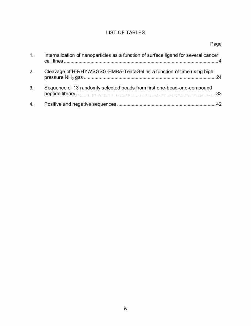
LIST OF TABLES
Page 1. Internalization of nanoparticles as a function of surface ligand for several cancer
cell lines .................................................................................................................... 4
2. Cleavage of H-RHYWSGSG-HMBA-TentaGel as a function of time using high pressure NH3 gas ................................................................................................... 24
3. Sequence of 13 randomly selected beads from first one-bead-one-compound peptide library ......................................................................................................... 33
4. Positive and negative sequences .......................................................................... 42

v
LIST OF FIGURES
Page
1. Mechanism of ammonia gas cleavage of the peptide........................................... 22
2. Schematic of nanomanipulation............................................................................. 25
3. Mass spectrum of H-GGGG-NH2........................................................................... 27
4. Injection and extraction onto a single bead ........................................................... 28
5. Mass spectrum of H-PWSG-NH2 ........................................................................... 30
6. Mass spectra of H-Y-NH2, H-HY-NH2, and H-WHY-NH2 ...................................... 32
7. Mass spectrum of H-GWGWGGGG-NH2 .............................................................. 34
8. Mass spectrum of H-SGWRGGGG-NH2 ............................................................... 35
9. Bead A and Bead B from screening 1 ................................................................... 38
10. Positive hit 1 and hit 2 fluorescence images ......................................................... 39
11. Positive hits 3, 4, 5, 6, and 7 fluorescence images ............................................... 40
12. Positive hits 8, 9, and 10 fluorescence images ..................................................... 41
13. Negative beads 1, 2, and 3 fluorescence images ................................................. 41
14. Comparison of positive and negative beads using 83 nM concentration ............. 56
15. Comparison of positive and negative beads using 500 nM concentration ........... 57
16. Mass spectra of positive hit 1 ................................................................................ 43
17. Mass spectra of positive hit 2 ................................................................................ 44
18. Mass spectra of positive hit 3 ................................................................................ 45
19. Mass spectra of positive hit 4 ................................................................................ 46
20. Mass spectra of positive hit 5 ................................................................................ 47
21. Mass spectra of positive hit 6 ................................................................................ 48
22. Mass spectra of positive hit 7 ................................................................................ 49
23. Mass spectra of positive hit 8 ................................................................................ 50

vi
24. Mass spectra of positive hit 9 ................................................................................ 51
25. Mass spectra of positive hit 10 .............................................................................. 52
26. Mass spectra of negative bead 1 ........................................................................... 53
27. Mass spectra of negative bead 2 ........................................................................... 54
28. Mass spectra of negative bead 3 ........................................................................... 55
29. Comparison of 130, 30, 20, and 10 µm resin beads ............................................. 58
30. Mass spectrum of H-GGGG-NH2 on 30 µm beads ............................................... 59

1
CHAPTER I
INTRODUCTION
Combinatorial l ibraries ar e r outinely em ployed i n t he di scovery of new lead
compounds for drugs [20] and i n the search for high-affinity l igands to target proteins.
[15; 21] Peptide-based libraries have proven invaluable in cancer research where efforts
focus primarily on f inding ligands for proteins that facilitate targeted binding to diseased
cells. [ 21] Such l ibraries al low f or t he gener ation of broad c hemical di versity t hrough
relatively s traight f orward t echniques a nd pr ocedures. [ 13] T heir i ncarnations r ange
from c hemically-inspired one -bead, one -compound (OBOC) l ibraries, [ 11] al so as
known as split-mix libraries,[11; 12; 13; 14] to biologically-inspired ones such as phage
display. [22; 23; 24]
Another w idely us ed m ethod f or i dentifying t argeting l igands that w arrants
mention is the use of aptamer libraries. This technique works in the same fashion as a
peptide combinatorial l ibrary but w ith each member of the l ibrary being a nucleic ac id
instead of a pept ide. E ach member of the pool has a di fferent sequence and, thus, a
different set of chemical groups that will fold into different structures that have differing
properties or ar e c apable of di fferent f unctions. I ndividual m embers of a pool ar e
separated from one another on the basis of their ability to perform an arbitrary task. For
example, nucleic ac ids t hat i nteract w ith pr otein t argets hav e been t rapped o n
nitrocellulose f ilters or separated f rom uncomplexed species on nat ive polyacrylamide
gels. Aptamers that bind small molecules such as cofactors or vitamins can be isolated
by affinity chromatography. Unbound nucleic acids are washed away from bound, and

2
the bound species ar e t hen el uted by addi ng an e xcess of s oluble l igand, c hanging
solvent conditions, or cleaving from the solid support. [25]
In the case of this OBOC library, a method for cleaving the peptide from the solid
support once a hi t had been i dentified were needed as well as ones that facilitate the
identification of the released peptides. In practice, a variety of approaches are available
for releasing the peptide f rom an individual library bead for analysis. Th ese schemes
utilize a c arefully chosen c leavage s ite that is installed between the pept ide and resin
bead, such as certain types of ester linkages that can be cleaved with ammonia gas,[3;
4; 5; 6] a CNBr-cleavable methionine residue, as well as a v ariety of other chemically
and phot olytically s ensitive on es. S everal t echniques hav e been em ployed f or
sequencing t he c leaved pept ides i ncluding E dman degr adation ( ED), par tial E D
followed b y m atrix-assisted l aser des orption i onization m ass s pectrometry [ 19] ( PED
MALDI MS), MALDI tandem mass spectrometry [3] (MALDI MS/MS), and electrospray
ionization tandem mass spectrometry [15] (ESI MS/MS). In order to avoid sample loss
and to reduce analysis time, the number of processing steps required specifically for hit
identification should be kept to a minimum and, for those that are absolutely necessary,
ones that are solvent-free are preferable. A n inherent aspect of utilizing bead libraries
is the need for manipulating relatively small amounts of both library compound and t he
solvents used for cleavage/extraction. Nanomanipulation, with spatial resolution as fine
as 3. 4 n m us ing c urrent t echnology, o ffers one par tial r emedy t o t his di lemma.
Nanomanipulation has already proven to be a useful technique for the analysis of trace
analyte pa rticles f ound on f ibers [ 26] and f or l ipid anal ysis [ 27] w ith l iquid-phase
microextractions. Due to <200 μL volumes of solvent and picomolar sensitivity,

3
nanomanipulation i s coupled t o nanoel ectrospray i onization m ass s pectrometry (NSI
MS) for sensitive analysis.
Using t he t echniques des cribed her e, anal ysis of c ombinatorial library
constituents on sub-micron-sized solid supports could theoretically be performed, which
would al low f or m iniaturization of l ibrary s ynthesis, s creening, and i dentification.
Current methods for automated screening of combinatorial l ibraries ut ilizing t raditional
solid supports (90-300 μm) based on fluorescence measurements require the use of a
non-standard f low c ytometer specifically engi neered t o al low s orting of large
particles.[28] This need could be obviated through the use of smaller solid supports, 30
µm and 20 µm resin beads . A nother adv antage of s ynthesizing l ibraries on s maller
supports i s t he s ignificant gai ns t hat c an be m ade i n l ibrary d iversity on a per gr am
basis. For instance, there are 6.55x10⁴ beads per gram of 300 μm TentaGel resin; if
the bead s ize w ere r educed t o 800 nm , t here w ould be 3. 45x1012 beads per gr am
(assuming s imilar c ompositions). Thi s would ena ble s creening of hi ghly di verse
libraries using only microgram quantities of beads.
Targeting cancer cells can be achieved through the attachment of ligands to the
surface of the nanoparticle known to bind to receptors over-expressed on the surface of
rapidly dividing cancer cells. Because of the high metabolic demands engendered by
rapid pr oliferation, m any c ancer c ells over-express t ransferrin, al bumin, and f olate
receptor, which makes conjugation of transferrin, albumin, folate, or antibodies to these
receptors a s uccessful t argeting appr oach. S ome of t he l igands dev eloped her e t o
target serum proteins for particle stealthing will have the added b enefit of targeting the
nanoparticle (NP) to cancer cells, i.e., NPs coated with a ligand to transferrin or albumin

4
should s how pr eferential upt ake i n c ancerous c ells. A nti-TfR-coated par ticles hav e
been shown [29] to be efficiently internalized by seven different cancer cell lines, Table
1, in cluding: B c ell l ymphoma ( Ramos, S up-B8), c ervical ( HeLa), ov arian ( SKOV3),
brain (MGR3), prostate (LNCaP), and l ung (H125). Conversely, nanopar ticles coated
with the isotype control, IgG1, were not. Furthermore, neither particle composition was
taken up by the negative control cell line, mouse embryonic fibroblasts (MEF).
Table 1 - Internalization of nan oparticles as a f unction of s urface l igand f or s everal cancer c ell l ines. P articles w ere c onjugated w ith ei ther ant i-TfR or I gG1, an i sotype control.
0
20
40
60
80
100
% U
ptak
e Anti-TfRIgG1

5
CHAPTER II
EXPERIMENTAL
Peptide Synthesis
A four-glycine peptide chain was synthesized on 130 µ m TentaGel S NH2 (0.29
mmol am ine/g) r esin beads . The K aiser t est w as used t o e nsure t he presence or
absence o f t he f ree pr imary am ine gr oup. The s olutions us ed f or t his t est w ere
ninhydrin, 5 g ni nhydrin in 100 ml ethanol, phenol , 80 g pheno l in 20 m l e thanol, and
pyridine/KCN, 2 m l of 0. 001 M KCN in 98 m l py ridine. Thi s t est consisted of add ing
three drops of each of the solutions to a s mall sample of the resin beads, 1-2 mg, in a
glass reaction vial. The sample was then heated to 120° C for two minutes in the CEM
Discover SPS M icrowave. A pos itive r esult s hows a dar k blue c olored s olution
indicating t he pr esence of a f ree pr imary amine. A negative r esult s hows a y ellow
colored solution indicating no presence of any free primary amines.
Initially the resin beads alone were tested to show the presence of a free primary
amine. A s mall s ample of b eads w as swollen i n di methylformamide ( DMF) f or 30
minutes. The be ads w ere t hen w ashed t hree t imes w ith d ichloromethane ( DCM) t o
remove al l D MF. T hey w ere t hen put i n t he microwave and a pos itive result w as
obtained.
Several methods for the synthesis of the peptide chain were tested. One 100 mg
sample, Sample A, of resin beads (1 equiv) was swollen for 30 minutes in DMF. During
this t ime a l inker, 4 -hydroxymethylbenzoic ac id ( HMBA, 3 equi v) and the r eagent
hydroxybenzotriazole (HOBt, 3 equiv) were dissolved in separate 1 ml solutions of DMF.
These were then added to the resin beads along with 1,3-diisopropylcarbodiimide (DIC,

6
6 equiv). The solution was left to stir overnight and then washed with DMF and DCM 3
times each. The Kaiser test was performed and gave a negative result.
The c hain w as c ontinued on S ample A by addi ng glycine ( Fmoc-Gly-OH, 5
equiv) to the beads and sufficient amount of DMF to make the beads mobile. DI C (6
equiv) and 10% 4-dimethylaminopyridine (DMAP) dissolved in DMF (3 equiv) were also
added to the resin beads. The solution was left to stir overnight and was followed with 3
washes of DMF. A 30% piperidine solution in DMF was used for the fmoc deprotection.
The amount of deprotection reagent used was just enough to cover the beads, 1-2 ml.
The solution was left to stir for f ive hours and then washed with DMF and DCM. The
Kaiser test was performed and a pos itive result was obtained. In a separate v ial t he
next amino acid, Fmoc-Gly-OH, (5 equiv), DIC (6 equiv), HOBt (3 equiv) are mixed in 1
ml 1 -methyl-2-pyrrolidone ( NMP). The m ixture i s ad ded t o t he r esin af ter 2 m inutes.
The solution was p laced in a m icrowave vessel and then put in the microwave for 10
minutes at 75° C (SPS mode, max power 10W, ∆T = 3° C). The beads were washed
with D MF and t he s ubjected t o the K aiser t est, w hich gav e a negative r esult. The
deprotection step was repeated but the Kaiser test gave another negative result. Thi s
sample was discarded.
The l inker, H MBA, w as added t he s ame way us ing t he s ame equi valents t o
Sample B as to sample A, but was only left to stir for one ho ur rather than overnight.
The Kaiser test gave a negative result. Fmoc-Gly-OH (5 equiv), 10% DMAP solution in
DMF (3 equiv), and DIC (6 equiv) were mixed in a separate vial then added to the resin
beads. The sample was stored overnight and then washed with DMF. Piperidine (30%
in DMF) was added and the beads were stirred for 15 m inutes, the results of a K aiser

7
test w ere positive. Fm oc-Gly-OH ( 5 equi v), H OBt ( 3 equi v), D IC ( 6 equi v) and N MP
were mixed in a separate vial and then added to the beads. The sample was placed in
the microwave for 10 minutes at 75° C (SPS mode, max power 10W, ∆T=3° C). The
beads were washed five times with DMF and 30% piperidine (1-2 ml per washing). The
sample was put in the microwave reactor for 2.5 minutes at 75° C, but gave a negat ive
result when the Kaiser test was performed. The s ample was de-protected again with a
30% pi peridine s olution, 1 -2 m l, and allowed t o s tir f or 15 m inutes. The Kaiser t est
again gave a negative result and Sample B was discarded.
One hundred milligrams of beads (1 equiv), Sample C, was swelled in DMF for
30 minutes. H MBA (3 equiv) and H OBt was dissolved in DMF in a s eparate v ial and
then added to the beads. D IC (6 equiv) was then added and the vessel was placed in
the m icrowave for 10 minutes at 75 ° C with the s tir rate set to medium and power at
10W. The beads were then washed with DMF and DCM 5 times each. The Kaiser test
gave a negative result, as expected. Ten percent DMAP in DMF (3 equiv), Fmoc-Gly-
OH (5 equiv), DIC (6 equiv) and DMF were mixed in a separate vial and then added to
the resin. It was t hen placed in the m icrowave for 10 m inutes a t 75 ° C. The beads
were w ashed f ive t imes w ith DMF and then de -protected us ing a 30% pi peridine
solution in DMF, 1-2 ml. After two minutes of stirring, a Kaiser test was performed and
a pos itive result was obtained as expected. Fm oc-Gly-OH (5 equiv), HOBt (3 equiv),
DIC (6 equiv) and NMP were m ixed i n a s eparate v ial and t hen added to t he beads.
The sample was then placed in the microwave for 10 minutes at 75° C (SPS mode, max
power 10W, ∆T=3° C). The Kaiser test gave a negative result. Thirty percent piperidine
solution in DMF, 1-2 ml, was added t o the beads followed by s tirring for two minutes.

8
The K aiser t est gav e a pos itive r esult i ndicating t he peptide c hain n ow c ontains t wo
glycines. The t hird and f ourth g lycine un its were add ed t o t he chain us ing the same
procedure for the second one.
Through many trials a method was obtained and us ed in peptide synthesis from
this point on. This method consisted of allowing the resin beads to swell in DMF for 30
minutes and then washing with 3 resin bead volumes of DMF pr ior to use. The r esin
beads ( 1 e quiv, 0. 29 mmol N H2) w ere add ed t o a 25 m L m icrowave r eaction v essel
(CEM C orporation) a nd s wollen as des cribed abov e. H MBA ( 3 equi v) a nd H OBt ( 6
equiv) were dissolved in DMF and then transferred to the vessel containing the beads.
The vessel was heated to 75° C for 10 minutes (SPS mode, max power 10W, ∆T=3° C )
using m icrowave irradiation. A s mall s ample of r esin be ads, 2 -5 m g, was r emoved,
washed with DMF then DCM three times each and t hen subjected to the Kaiser test to
confirm that the reaction had gone to completion. The resin was washed with DMF (10
x 2 min each) and then stirred with aqueous sodium hydroxide (1M NaOH:DMF, 1:1 v/v)
for 15 min. The resin was washed with DMF/H2O (1:1 v/v, 5 x 2 min each) followed by
DMF (10 x 2 min each).
Fmoc-Gly-OH (3 equiv) was dissolved in DMF, which was then added to HMBA
beads. DIC (4 equiv) was added followed by the drop-wise addition of DMAP dissolved
in DMF (0.1 equiv, 0.05 M stock solution). The reaction vessel was heated to 75° C for
10 minutes (SPS mode, max power 10W, ∆T=3° C) using microwave irradiation. T he
resin was washed with DMF (2 x 2 m in each). G lycine coupling was repeated us ing
identical amounts of each reagent to ensure complete coupling of the f irst amino acid.
After the second reaction, the resin was washed w ith DMF (5 x 2 m in each) and any

9
remaining hydroxyl groups were acetylated by the addition of DMF, Ac2O (6 equiv), and
DMAP (0.1 equiv, 0.05 M stock solution). After 1 h, the resin was washed with DMF (10
x 2 min each).
Fmoc deprotection was achieved by the addition of 30% piperidine in DMF (v/v)
followed b y gent le a gitation f or t wo m inutes. The solution w as r emoved and t he
deprotection steps were repeated two more times. The resin was washed with DMF (5
x 2 m in e ach). A s mall s ample of r esin beads ( 2-5 m g) w as r emoved, and t hen
subjected t o t he K aiser t est t o confirm depr otection. Fm oc-amino ac id-OH (5 equi v),
HOBt ( 5 e quiv), and DIC ( 5 eq uiv) w ere dissolved i n D MF an d s tirred f or 10 -15 min.
This solution was added to the resin and the reaction vessel was heated to 75° C for 10
minutes using m icrowave i rradiation. A s mall s ample of r esin beads ( 2-5 m g) w as
removed, washed with DMF and DCM three times and then subjected to the Kaiser test
to c onfirm c oupling. The r esin w as w ashed w ith D MF ( 10 x 2 m in e ach). Thi s
procedure was repeated for each additional amino acid.
Side c hain de -protection w as c onducted as f ollows. A ll t he am ino ac ids us ed
except gl ycine hav e a s ide c hain pr otection gr oup. I n or der t o use and a nalyze t he
peptides the protecting groups need to be removed. The f irst step in this process is to
remove t he f moc pr otecting gr oup f rom t he l ast am ino ac id o n t he c hain. Thi s w as
achieved by allowing the sample to stir for two minutes with 30% piperidine solution in
DMF. The solution was removed and t he deprotection s teps were repeated two more
times. Th e s ample was t hen washed s everal t imes w ith D CM. A c leavage c ocktail
consisting of t rifluoroacetic ac id ( TFA), t riisopropylsilane ( TIS), and w ater ( 95:2.5:2.5)
was used f or t he s ide chain deprotection. The solution was added to t he beads and

10
allowed to stir for 90 minutes. The sample was then washed with TFA, DCM, and ether
(3x 2min each).
One Bead One Compound Library Synthesis
A bu lk s ample of H-GGGG-HMBA-TentaGel ( 750 m g) w as pr epared us ing t he
procedures outlined above. The resin was then divided into six aliquots (125 mg each)
and one of six different amino acids (X1 = G, S, H, R, Y, or W) coupled to each aliquot
using t he c oupling p rocedures out lined abov e. The s ix a liquots w ere m ixed af ter
rinsing, dr ied under vacuum for 1 h, and t hen weighed (533 mg, 71% recovery). The
beads w ere agai n s plit i nto s ix aliquots ( 89 m g eac h) and one of s ix different am ino
acids (X2 = G , S , H , R , Y , or W) coupled t o each a liquot. D uring r insing, one of t he
samples was lost due to a malfunctioning frit. The remaining five aliquots containing X2
= G , S , H , R , or W were poo led af ter r insing, dr ied under vacuum and t hen weighed
(383 m g, 8 9% r ecovery). For X 3, t he resin w as s plit i nto f ive al iquots ( 76 mg eac h)
instead of six, due to the aforementioned material loss, and each coupled to a different
amino ac id, X 3 = G, S, H, R, or W. The five al iquots were pooled af ter r insing, dr ied
under vacuum and then weighed (322 mg, 84% recovery). For X4, the resin was again
split into five aliquots (64 mg each), and each coupled to a different amino acid, X4 = G,
S, H, R, or W. The five aliquots were pooled after rinsing, dried under vacuum and then
weighed (295 mg, 92% recovery). After accounting for the loss of one of the samples,
the r esulting l ibrary consisted of 750 unique p eptides of t he gener al f ormula H -
X4X3X2X1GGGG-HMBA-TentaGel, X1 = G, S, H, R, Y, or W; and X2 = X3 = X4 = G, S, H,
R, or W and a total yield of 39%.
A s econd library w as s ynthesized i n t he s ame f ashion as m entioned ab ove

11
starting with 3 g of resin beads. The sample was split into seven aliquots (485 mg) and
one of the seven amino acids (X1 = G, R, S, W, H, Y, or K) was coupled to each sample.
All samples were mixed (2.5 g) and split into seven aliquots (350 mg). The same amino
acids were added t o each of the seven samples. Thi s process was repeated until f ive
random amino acids were added to the chain giving a t otal of nine amino acids in the
peptide. The general formula of the peptides is H-X5X4X3X2X1GGGG-HMBA-TentaGel.
This gives 16,807 different peptide combinations.
Gas-Phase Cleavage
Peptide was released from the resin (after deprotecting the final amino acid and
side chains as outlined above) by exposure to ammonia/tetrahydrofuran (THF) vapor on
a S chlenk line. Briefly, one m anifold of t he S chlenk line ( app. 335 m L v olume) w as
purged w ith ammonia gas . The gas contained in the manifold was condensed into a
round bottom flask containing degassed THF (30 mL) cooled in liquid N2. Separately, a
flask containing resin beads was evacuated. The two flasks were isolated from the rest
of t he S chlenk line, f ollowed by open ing stopcocks between t he t wo f lasks. The
ammonia/THF s olution w as t hen al lowed t o w arm t o r oom t emperature and t he
apparatus was l eft u ndisturbed f or 20 ho urs. A fter t he r eaction, am monia gas w as
condensed ba ck into t he f lask containing THF by c ooling i n l iquid N2, an d t he f lask
containing resin beads was carefully removed from the Schlenk line.
Peptide was also released from the resin (after deprotecting the final amino acid
and side chains as outlined above) by exposure to high-pressure NH3 gas in a general-
purpose pressure vessel (Parr Instrument Company). The sample to be c leaved was
transferred t o a 3 5 mm gl ass bottom di sh t hat w as t hen p laced i nside t he pr essure

12
vessel. The top was secured and the vessel was pressurized with NH3 gas (anhydrous
ammonia, 99. 99%) and t hen l eft undi sturbed f or t he des ired r eaction t ime. S everal
different t imes at a pressure of 100 psi were tested and compared to cleavage on t he
Schlenk l ine. A t the end of the reaction, the vessel was vented, the top disengaged,
and the sample removed.
Sequencing the Peptide
Bulk samples and s ingle beads can be an alyzed and sequenced w ith a DECA
Ion Tr ap M S. For b ulk samples, pept ide-coated bea ds t hat have been c leaved w ere
placed in a 1 m l solution consisting of 50:50 methanol/water and 1% acetic acid. The
sample was vortexed and the beads were left to settle to the bottom of the eppendorf
tube. The s olution was e xtracted and t hen i njected i nto t he el ectrospray i onization
source. The par ent peak was identified a nd a t andem M S was per formed t o i dentify
each amino acid in the peptide chain.
Single beads were i solated on a glass bot tom dish and l ocated w ith a TE 2000
inverted m icroscope (Nikon). Fol lowing i dentification of a s ingle bea d, t he L200
nanomanipulator (Zyvex) was utilized to land a 1 μm nanoelectrospray tip (New
Objective) in close proximity to the bead. The solvent was injected onto the bead with a
PE2000b f our-channel pr essure i njector ( MicroData I nstruments). The s olvent w as
allowed t o ex tract on t he bead f or appr oximately 30 s econds b efore being f illed bac k
into the tip. Once the peptide-containing solution was retrieved, the tip was transferred
to t he nanoel ectrospray i onization s ource ( Proxeon B iosystems) f or anal ysis. Th e
single bead extractions were completed w ith an injection pressure of 30 ps i and a f ill
pressure of 40 psi.

13
During the analysis of a pept ide, the parent peak is identified after about one to
two minutes into the run time. This peak is the most dominant in the scan and is often
the singly protonated form of the peptide. The doubly protonated peak is also identified
and t he t andem m ass s pectra i s c onducted on t his s pecies t o provide m ore c harged
fragments that can be detected. The analysis is allowed to run for an additional two or
so minutes or until the doubly protonated peak is gone. From the data, the b and y ions
are identified. Fr om these ions each individual amino acid and its place in the peptide
chain c an be det ermined. [ 30] Thi s m ethod c an b e us ed f or a bul k s ample or a n
individual bead.
Screening the Peptide Library for Ligands that Bind Transferrin
Initial at tempts to screen for l igands that bind to t ransferrin (Tf) were performed
by using both Tf-AlexaFluor 647 (red fluorescent marker) and Tf-AlexaFluor 488 (green
fluorescent marker) in different combinations. A small sample of beads from the library,
25 mg, 8.87x10⁵ beads /gram with a 28 0-330 pmol capacity/bead, was f irst swelled in
DCM for 30 minutes. It was then washed with DCM, DMF, DMF/H2O (7:3, 5:5, 3:7) and
H2O (5 x 1 minute each). A blocking buffer was prepared which consisted of 10% skim
milk, 2% c hicken egg al bumin, and 0. 05% Tw een 8 0 i n p hosphate buf fered s aline
(PBS). B locking solution (0.5 mL) was added to the sample and left to incubate for 30
minutes. The beads w ere t hen w ashed 5 x 1 m inute w ith 0. 05% Tw een 80 i n P BS
(PBS-Tween). A s tock s olution of t he Tf marker w as m ade by addi ng 1. 2 m g of t he
protein to 1 ml ultra pure water. Fr om this an 83 nM concentration of the Tf marker in
blocker solution was made by adding 2.8 µL of the desired Tf marker stock solution to
500 µL of blocker s olution. Th is w as a dded t o t he sample an d al lowed to i ncubate

14
overnight i n t he r efrigerator. The beads w ere t hen w ashed w ith P BS-Tween ( 10 x 1
min), PBS (5 x 1 min), and water (3 x 1min). The beads were taken up into an aqueous
solution (0.5 mL), transferred to a 6-well cell culture plate, the excess water removed by
pipette, and the beads were left to dry.
This pr ocess w as per formed on five di fferent 25 m g s amples f rom t he pept ide
library. Each sample was incubated overnight in the refrigerator as follows: Sample D-
red f luorescent m arker i n t he bl ocker s olution; S ample E – red fluorescent m arker i n
PBS without the blocker solution; Sample F – red and gr een f luorescent marker in the
blocker solution; Sample G – green fluorescent marker in the blocker solution; Sample
H – no marker, only the blocker solution.
Hit beads were i dentified by m anual s creening of t he s ample f or i ncreased
fluorescence. I t was noted that the beads have an auto-fluorescence. Because of this
using b oth t he r ed a nd gr een marker t ogether w ould not gi ve a t rue pos itive hi t. T o
solve this problem only the red f luorescent marker was used for all future screenings.
Using t his m ethod, hi t beads were i dentified by m anually l ooking f or a n i ncreased
fluorescence emission in the far red (ex. 650 nm, em. 665 nm) with no similar increase
in the green channel (ex. 495, em. 519). Observing enhanced fluorescence only in the
far r ed m akes i t l ess likely t hat t he i ncreased f luorescence i s a r esult of t he i nherent
auto-fluorescence of the beads themselves.
Positive hi ts w ere r emoved m anually w ith a s yringe need le a nd pl aced into a
different well on t he 6-well cell culture plate. To denat ure the t ransferrin 2 µL of 8 M
guanidine hydrochloride w as ad ded t o eac h bead. T he s tock s olution w as m ade by
adding 3.8 g guanidine hydrochloride to 5 ml DI water. The denaturing solution was left

15
on the bead for 20 m inutes and t hen extracted with a p ipette. 3 µL of water was then
added t o eac h bead and ex tracted af ter 1-2 m inutes of s tanding. Thi s s tep w as
repeated once more and the beads were left to dry. They were then t ransferred to a
glass bottom dish using a syringe needle. The dish with the individual beads separated
out was then placed in the pressure-vessel to c leave the pept ide f rom the bead. Th e
cleavage process and sequencing of the individual beads was performed as mentioned
above.
Synthesis and Comparison of Positive and Negative Hit Beads
From the positive hits identified one w as chosen for further analysis. Thi s bead
sequence was W KRYRGGGG. Th is p eptide was s ynthesized us ing t he s ame
procedure out lined ab ove. The K aiser t ests s howed t hat t he H MBA l inker and eac h
amino ac id w ere a dded s uccessfully. A n egative b ead w ith t he s equence
HWHWHGGGG was also synthesized in the same fashion.
A 5 m g bead sample from both the positive and negative hits was washed with
DCM, DMF, DMF/H2O (7:3, 5:5, 3:7) and H 2O (3 x 1 m inute each). A blocking buf fer
was m ade w hich c onsisted of 10% s kim milk, 2% c hicken egg al bumin, and 0. 05%
Tween 80 in phosphate buffered saline (PBS). Blocking solution (0.2 mL) was added to
the sample and left t o i ncubate f or 30 m inutes. The beads were t hen washed 3 x 1
minute with 0.05% Tween 80 i n PBS (PBS-Tween). A 500 nM solution containing the
Tf-AlexaFluor 647 was made by add ing 6.7 µL of the s tock solution to 0.2 ml blocker
solution. Thi s was t hen added to each sample and l eft t o i ncubate overnight at 4 °C.
The beads were then washed with PBS-Tween (5 x 1 min), PBS (3 x 1 min), and water
(3 x 1min). The bead s were taken up into an aqueous solution (0.5 mL), transferred to

16
a 6-well cell culture plate, the excess water removed by pipette, and the beads were left
to dry. Both samples were placed in the same well to compare the fluorescence.
Peptide Synthesis on Smaller Beads
The original synthesis of peptides mentioned above was performed using 130 µm
beads. There are many benefits to miniaturizing the library as outlined above. The first
step in the synthesis of the miniature library was to add a f our-glycine peptide chain to
20 µm and 30 µm resin beads. The same procedure described in the section “Peptide
Synthesis” was used for the smaller beads. The s ame equivalents were used and t he
starting amount was 100 mg for each. The major difference in the procedure used here
compared to abov e was t hat s ince t he beads ar e much s maller a d ifferent r eaction
vessel had to be us ed. The r eaction vessel that had been us ed up to this point had a
frit with 30 m pore size, which would not retain theses smaller beads. The manufacturer
was contacted in an at tempt to have reaction vessels made with smaller pore size frits;
however, they were unable to accommodate. The new reaction was a 2 m l centrifugal
filter d evice, U ltrafree-CL, f rom M illipore C orporation. Thes e v essels c ould not b e
placed a lone i nto t he m icrowave and t he reagents would l eak out dur ing t he reaction
time. To s olve this problem parafilm was used to seal the bottom of the vessel. I t was
then placed in the original microwave vessel. The Kaiser tests were as expected for the
addition of the HMBA linker and four glycines to both the 30 µm and 20 µm beads.
The ana lysis of t hese pept ides w as at tempted us ing t he s ame pr ocedure
employed f or t he bi gger beads . D ata was obt ained f or a bu lk s ample of t he 30 µm
beads but anal yzing a single b ead is s till in t he pr ocess. B oth bul k and single be ad
analysis is also still under investigation for the 20 µm beads.
17
Synthesis of a Peptide Library on 20 µm Beads
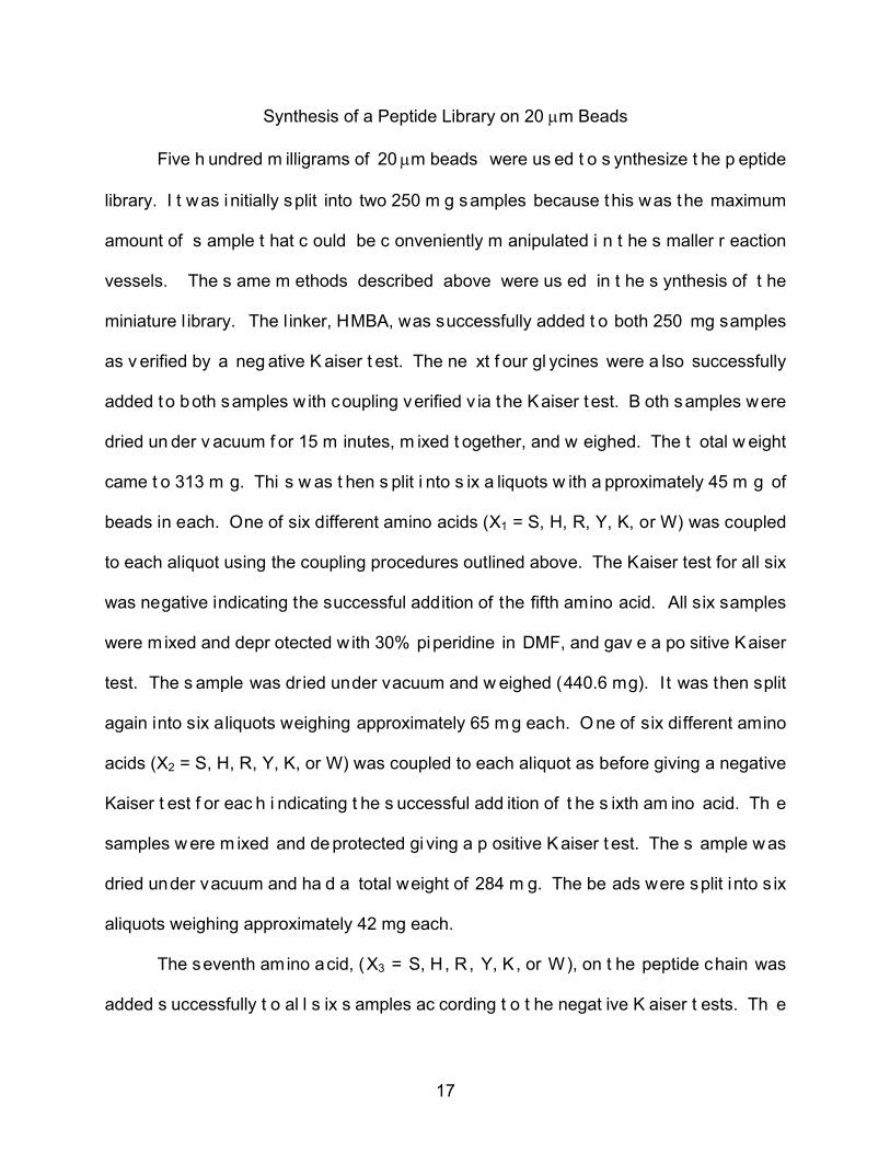
Five h undred m illigrams of 20 µm beads were us ed t o s ynthesize t he p eptide
library. I t was i nitially split into two 250 m g samples because this was the maximum
amount of s ample t hat c ould be c onveniently m anipulated i n t he s maller r eaction
vessels. The s ame m ethods described above were us ed in t he s ynthesis of t he
miniature l ibrary. The l inker, HMBA, was successfully added t o both 250 mg samples
as v erified by a neg ative K aiser t est. The ne xt f our gl ycines were a lso successfully
added to both samples w ith coupling verified v ia the Kaiser test. B oth samples were
dried un der v acuum f or 15 m inutes, m ixed t ogether, and w eighed. The t otal w eight
came t o 313 m g. Thi s w as t hen s plit i nto s ix a liquots w ith a pproximately 45 m g of
beads in each. One of six different amino acids (X1 = S, H, R, Y, K, or W) was coupled
to each aliquot using the coupling procedures outlined above. The Kaiser test for all six
was negative indicating the successful addition of the fifth amino acid. All six samples
were m ixed and depr otected w ith 30% piperidine in DMF, and gav e a po sitive Kaiser
test. The s ample was dried under vacuum and w eighed (440.6 mg). It was then split
again into six aliquots weighing approximately 65 m g each. O ne of six different amino
acids (X2 = S, H, R, Y, K, or W) was coupled to each aliquot as before giving a negative
Kaiser t est f or eac h i ndicating t he s uccessful add ition of t he s ixth am ino acid. Th e
samples w ere m ixed and de protected gi ving a p ositive K aiser t est. The s ample w as
dried under vacuum and ha d a total weight of 284 m g. The be ads were split into s ix
aliquots weighing approximately 42 mg each.
The seventh amino acid, (X3 = S, H, R, Y, K, or W), on t he peptide chain was
added s uccessfully t o al l s ix s amples ac cording t o t he negat ive K aiser t ests. Th e

18
samples w ere m ixed and depr otected gi ving a pos itive K aiser t est. T he beads w ere
dried und er v acuum and w eighed 271. 2 mg. The s ample w as s plit i nto s ix aliquots
weighing about 40 m g each. The ei ghth amino ac id, (X4 = S, H, R, Y, K, or W), was
added and each sample gave a negative Kaiser test. A ll six samples were mixed and
deprotected gi ving a positive Kaiser t est. The be ads w ere dr ied under v acuum and
weighed 184 mg. The beads were split into another six aliquots weighing about 30 mg
each. Th e ninth and last amino acid, (X5 = S, H, R, Y, K, or W), was added g iving a
negative Kaiser t est f or eac h of t he s ix. The s amples w ere m ixed an d d ried unde r
vacuum. The final weight came to 195.4 mg giving a 39% yield. The general formula of
the pept ides is H-X5X4X3X2X1GGGG-HMBA-TentaGel where X = R, S, W, H, Y, or K.
This gives 7,776 different peptide combinations in the library.

19
CHAPTER III
RESULTS AND DISCUSSION
Introduction
Various b iological s creenings c an be ut ilized us ing a c ombinatorial l ibrary. A
library of t he ge neral f ormula H-X6X5X4X3X2X1-GGGG-HMBA-TentaGel, w here X =
randomized am ino a cid is i deal f or t his pr oject. Fou r t o s ix r andomized amino ac ids
were c hosen bec ause t hose lengths hav e bee n r eported t o be s ufficient f or t he
discovery of hi gh-affinity l igands t o t arget a v ariety of pr oteins.[2] Furthermore, th e
addition of four glycine units before each chain was used to give some flexibility to the
binding p ortion of t he pept ide to al low f or pr oductive i nteractions w ith t he t arget of
interest; an appr oach that has also been reported by others.[3] An HMBA resin l inker
was s elected bec ause t he c leavage r eaction c an b e ac hieved under s olvent-free
conditions v ia exposure to NH3. Most other c leavage s trategies require long reaction
times or hav e ot her significant detractors such as t he r equirement of a m ultistep
synthesis t o pr epare t he l inker, or ex tensive pr ocessing s teps pr e- or pos t-cleavage.
The use of HMBA has been r eported in the l iterature;[3; 4; 5; 6; 7; 8; 9] however, the
experimental pr ocedures t hat hav e be en em ployed t o dat e i nvolving N H3 are
unnecessarily l ong and c umbersome as w ill be h ighlighted be low. The goal w as t o
demonstrate t he abi lity t o ( 1) s ynthesize a n O BOC pept ide l ibrary us ing m icrowave-
assisted, s olid-phase pe ptide s ynthesis, ( 2) r apidly a nd ef ficiently

20
cleave t he peptide f rom t he s olid s upport, ( 3) s equence t he pept ide f rom i ndividual
members of the library with the aid of mass spectrometry, (4) identify a l igand to target
transferrin, ( 5) m easure t he av idity of p ositive hi t l igands, ( 6) gener alize pept ide
synthesis for smaller supports such as 30 µm and 20 µm resin beads, (7) synthesize a
miniaturized version of the library on 20 µm beads, and (8) sequence peptide on the 30
µm and 20 µm individual beads.
Peptide Synthesis
HMBA was coupled to TentaGel S NH2 resin using a s lightly modified version of
published procedures des cribed by M ellor, et al .[7] The pr ocedure w as modified t o
reduce reaction time by conducting the coupling reaction at 75° C for 10 minutes under
microwave heating. This is the first report of microwave-assisted HMBA coupling, which
reduced r eaction t ime f rom 2 -24 hour s t o 10 m inutes. A mino ac id c oupling w as
conducted using an Fmoc protection scheme and ei ther 4-(N,Ndimethylamino)pyridine
(DMAP)/1,3-diisopropylcarbodiimide ( DIC) or 1 -hydroxybenzotriazole (H OBt)/DIC i n
DMF for activation of the C-terminal or all other amino acids, respectively. Coupling of
the C -terminal amino ac id was r epeated once t o e nsure t hat t he r eaction w ent t o
completion. C oupling reactions for both the C-terminal and al l remaining amino ac ids
were conducted at 75° C for 10 m inutes under m icrowave hea ting t o reduce reaction
times.[10] A Kaiser test was also performed after the linker coupling, each amino acid
coupling, and deprotection to ensure the reaction took place. The Kaiser test is used to
trace t he presence of f ree pr imary am ines. A fter each c oupling no primary am ines
should b e present w hich gi ves t he s olution a y ellow color, a ne gative r esult. A fter
removal of t he f moc protecting group, a f ree pr imary am ine i s present w hich g ives a

21
dark blue solution, a positive result.
One Bead One Compound Combinatorial Library
The f irst OBOC l ibrary was synthesized us ing the split-mix method.[11; 12; 13;
14] A bulk sample of H-GGGG-HMBA-TentaGel was prepared and then divided into six
equal aliquots. A different amino acid (X1 = G, S, H, R, Y, or W) was coupled to each
aliquot, an d af ter r epeated w ashings t he samples w ere c ombined. The beads were
again split into six aliquots; however, during coupling of the second randomized amino
acid, (X2), a sample was lost due to equipment malfunction. The remaining five aliquots
containing X 2 = G , S , H , R , or W w ere pool ed. For X 3, t he r esin w as s plit i nto f ive
aliquots instead of six, due to the aforementioned material loss, and each was coupled
to a different amino acid, X3 = G, S, H, R, or W. The split mix procedure was repeated
one final time where X4 = G, S, H, R, or W. A fter accounting for the loss of one of the
samples, the resulting library consisted of 750 unique peptides of the general formula H-
X4X3X2X1GGGG-HMBA-TentaGel, X1 = G, S, H, R, Y, or W; and X2 = X3 = X4 = G, S, H,
R, or W. Although the microwave-assisted synthesis of β-peptide [15; 16; 17; 18] and
peptoid [ 19] O BOC l ibraries have bee n reported, t his i s t he f irst ac count of t he
microwave as sisted synthesis of an O BOC pept ide l ibrary c omposed ent irely of
standard amino acids.
The second OBOC peptide library was synthesized using the same method as
mentioned above. To expand the number of possible peptide combinations, the peptide
chain w as l engthened and t wo m ore r andom am ino ac ids w ere us ed. The r andom
amino acids chosen were G, R, S, W, H, Y, and K. These were chosen because of their
high r ate of oc currence i n ot her pept ide bas ed t argeting ligands [ 2]. The g eneral

22
formula of the pept ide l ibrary is H-X5X4X3X2X1GGGG-HMBA-TentaGel, X1-5 = G, R , S,
W, H, Y, and K. This gives a total of 16, 807 possible peptide combinations.
High Pressure Ammonia Gas Cleavage
Exposure of a r esin-bound, HMBA-coupled pept ide to NH3 leads to c leavage of
the es ter bond anc horing t he pept ide t o t he r esin bead a nd f ormation of t he
corresponding peptide amide. T he mechanism for this reaction i s shown in Figure 1 .
The combination of HMBA/NH3 as a c leavage s trategy benef its t remendously from i ts
conceptual simplicity. B ecause i t can be per formed under solvent-free conditions, i t is
inherently green, w hich m eans l ess w aste gener ated, and no energy i s needed f or
product separation. Reaction work-up is accomplished in one easy step by just venting
excess NH3. A nother benef it i s t hat t he peptide i s l eft i n place on t he resin bead f or
future r etrieval a nd analysis w ithout any unnecessary di lution that c ould j eopardize
detection and/or s equencing ( no at tempt w as m ade t o det ermine i f pe ptide w as
transferred t o t he w alls of t he Parr r eactor; how ever, i n al l c ases s ufficient pept ide
remained on the bead to facilitate MS analysis).
Figure 1 – Mechanism of ammonia gas cleavage of the peptide.

23
While the technique is conceptually very simple, the initial cleavage experiments
conducted us ed a complicated pr ocedure w hereby a S chlenk l ine w as us ed t o
manipulate N H3 gas. N H3 gas was f irst c ondensed i nto a f lask c ontaining degas sed
THF cooled in liquid N2. B eads containing pept ide to be c leaved were then placed in
another vessel and p ut onto a s eparate port on the Schlenk l ine. The f lask containing
beads was ev acuated f ollowed by e xposure t o t he T HF/NH3 solution overnight. A ll
reports detailing the use of HMBA/NH3 involve similar steps that use dry ice or liquid N2
to m anipulate N H3. We hy pothesized t hat t he s ame r esults c ould b e obt ained i f t he
beads were exposed to NH3 gas in a h igh-pressure reactor. W hile such a reactor has
been described,[7] NH3 was condensed into the reactor in much the same manner as
described above, and that the reaction was run overnight. S everal tests were done t o
observe w hether or not pept ide c leavage c ould be ac complished i n s horter r eaction
times by s imply put ting the beads inside an appropriate reactor and then pressurizing
with NH3 gas.
In order to examine the effect of time on the cleavage of HMBA-coupled peptides
from TentaGel r esin, t he c leavage of t wo p eptides w as i nvestigated: H -RHYWSGSG-
HMBA-TentaGel ( 1a) and H -PWSG-HMBA-TentaGel ( 2a). P eptide pr ecursor 1a was
chosen because it represents the target length of constituents in the peptide library and
2a was t o be us ed as an internal s tandard f or quant ifying t he r elative amounts o f
cleaved peptide, 1b, obtained under a v ariety of c leavage conditions. S amples to be
cleaved were pl aced i nside a g eneral pur pose pr essure v essel and, w ithout pur ge or
evacuation, the vessel was brought to 117 ps i using NH3 gas. A series of 10 m g bulk
bead samples were analyzed as a function of time Table 2.

24
Table 2 - Cleavage of H−RHYWSGSG−HMBA−TentaGel as a function of time using high pressure NH3 gas
time (min) normalized peak areaa
Percent completionb
high pressure
0 10.8 2.4 5 315.5 72.7 15 344.7 79.5 30 387.0 89.2
120 384.7 88.7 240 384.9 88.7
1440 433.8 100.0 Schlenk line
1440 358.9 82.7
a peak areas were normalized using an internal standard, b cleavage under high pressure for 24 h was designated as 100% completion.
There was a s light dependence of the amount of cleaved pept ide on i ncubation
time; however, the reaction was >70% complete in just 5 minutes and was almost 90%
complete within 30 minutes (the data was normalized to 24 h o f incubation at 117psi).
The amount of 1b obtained af ter 24 hour s using the complicated Schlenk l ine method
was on ly approximately 1 3% hi gher t han that obt ained i n 5 m inutes us ing t he ne w,
much s impler, hi gh-pressure t echnique. Fur thermore, t he m aximum am ount of 1b
obtained via this new technique was >20% higher than that obtained using the Schlenk
line m ethod. N o s ignificant de composition w as obs erved i n s amples c leaved at hi gh
NH3 pressure. C leavage and M S analysis of pept ides f rom 1-8 amino ac ids in length
were c arried out t o demonstrate t he t echnique’s br oad ap plicability, and though o nly
one ot her ex ample h as been s tudied t hus f ar, t his technique i s not l imited t o hav ing
glycine as t he anc horing r esidue. Thi s t echnique has also been expanded t o s ingle

25
bead analysis, and u nless otherwise not ed, t he s equencing dat a r eported bel ow was
collected from bulk or single bead samples cleaved at 117 psi for 30 minutes.
Peptide Sequencing
A schematic representation of the nanomanipulation device is shown in Figure 2.
The base, which has four stage controller units, sits atop an inverted microscope. Each
stage c ontroller c an be f itted w ith one of t he f ollowing: a nanoel ectrospray t ip,
microgrippers, a t ungsten end effector pr obe, or a m icrocapillary. R esolution of
movement is roughly 3.4 nm in the x,y-plane and 100 nm in the z-direction. A s ingle
stage controller unit f itted with a nanoelectrospray t ip was used in each of the studies
outlined below.
Figure 2 – Schematic of nanomanipulator.
The spacer peptide (H-GGGG-NH2, 3b) was first synthesized to test the ability to
extract pe ptide f rom i ndividual r esin b eads. A bul k s ample of 3b was c ollected f rom
beads, po st am monia c leavage, by el ution i nto a 5 0:50 M eOH:H2O 1% ac etic ac id
solution an d t hen an alyzed by electrospray i onization ma ss s pectrometry (E SI MS )
Figure 3. Analysis of the MS data confirmed the presence of 3b in high abundance, but
also indicated the presence of a small amount of a f ive-glycine peptide. From this data

26
it i s bel ieved t hat doubl e c oupling m ost l ikely oc curred dur ing t he addi tion of t he f irst
amino ac id w here t he hy pernucleophile 4 -(N,N-dimethylamino) p yridine ( DMAP) w as
used to catalyze ester bond formation. DMAP could also act as a base resulting in some
fmoc deprotection in addition to its role as catalyst. DMAP’s role in double coupling has
been an area of concern,[6; 9] and is magnified here because the reaction conditions
include microwave irradiation.

27
Figure 3 – A) ESI MS of 3b. B) NSI MS of 3b. [1]

28
Next, 3b was collected from individual resin beads by positioning the probe tip of
the nanomanipulator within a few microns of the bead, dispensing a 50: 50 MeOH:H2O
1% ac etic ac id s olution ont o t he bead, w aiting 30 s econds f or el ution of 3b from the
bead, and then retrieving the solution back into the probe t ip, Figure 4. The pr obe t ip
was t ransferred di rectly t o t he mass s pectrometer and t he s olution ana lyzed. D ata
obtained for 3b from the single bead extraction was virtually identical to that of the bulk
sample.
Figure 4 – Injection of solvent onto a single bead (A). Extraction of solvent and peptide from bead (B).
Next, t o i nvestigate t he pos sible or igin of t he doubl e c oupling pr oduct and t o
demonstrate t he abi lity t o sequence peptide on i ndividual r esin beads, H -PWSG-NH2
(2b) was synthesized. Four di fferent amino ac ids were chosen to al low unambiguous
identification of any p roducts due t o doubl e c oupling r eactions while k eeping t he C -
terminal amino acid constant.
Again, a s ample of 2b was an alyzed by di ssolving t he cleaved pept ide i n a
solution of 50: 50 M eOH:H2O 1% ac etic ac id. S amples of 2b were ana lyzed by bot h

29
conventional E SI M S ( bulk pept ide) and by N SI M S ( individual beads ). B oth t he
protonated and s odiated i ons of 2b were observed along w ith a s mall a mount of a
peptide resulting from double coupling during the addition of glycine. Peptides resulting
from o ther pos sible double c oupling r eactions w ere not obs erved s upporting t he
assertion t hat doubl e c oupling was l ikely due t o f moc c leavage by DMAP dur ing
coupling of t he f irst a mino ac id. Fi nally, M S/MS anal ysis of 2b was c onducted o n
peptide t aken f rom bul k s amples and ones c ollected f rom i ndividual beads v ia
nanomanipulation, Figure 5 . Th e M S/MS data of bot h s amples of 2b were v irtually
identical w ith t he ex pected f ragmentation pat tern bei ng c learly observed. Fr om the
fragmentation data, a-, b- and y-ions were identifiable allowing the sequence of 2b on
individual beads t o be r eadily elucidated v ia s tandard m ethods of M S/MS anal ysis.
Nanomanipulation/NSI MS analysis of 2b was repeated a total of eight times to ensure
method reproducibility and each time the virtual identical MS/MS data was obtained.

30
Figure 5 - A) ESI MS/MS spectrum of 2b. H-PWSG-NH2 + H+: 445.33 m/z, H2O: 427.27 m/z, b3: 371 .27 m/z, y 3: 348. 27 m/z, y3*: 330. 20 m/z, b2: 284 .20 m/z, a2: 256.20 m/z; B) N SI MS /MS s pectrum o f 2b. H -PWSG-NH2 + H +: 445. 33 m/z, -H2O: 427.33 m/z, b3: 371 .27 m/z, y 3: 348. 27 m/z, y3*: 330. 20 m/z, b2: 284 .20 m/z, a2: 256.27 m/z. [1]

31
Hit screenings based on MALDI MS often necessitate the addition of extra non-
random amino acid residues to library constituents to increase peptide molecular weight
so t hat ov erlap of pept ide-fragment peak s w ith t hose of t he matrix m aterial c an b e
avoided. Therefore H-Y- NH2, H-HY-NH2, and H-WHY-NH2 were synthesized and bulk
samples were ana lyzed by E SI M S t o es tablish i f t here w ere any r estrictions on t he
minimum number of amino acid residues required for these analytical techniques. In all
three instances, the expected peptide was present at high abundance Figure 6.

32
Figure 6 – ESI MS of H-Y-NH2, H-HY-NH2, and H-WHY-NH2

33
Individual beads f rom the 750 member OBOC peptide l ibrary were analyzed to
ascertain whether or not peptides 8-amino acids in length could be sequenced and what
percentage of selected beads could be unambiguously sequenced f rom a given pool.
Thirteen b eads w ere c hosen at r andom, t he pept ide c leaved, and an alyzed as
described above. T he pept ide sequences for al l 13 beads analyzed were determined
and listed in Table 3. The MS data is shown in Figures 7-8.
Table 3 - Sequences of 13 randomly selected beads from first OBOC peptide library Bead no. Peptide sequence
1 H –GGGSGGGG-NH2 2 H-WGWRGGGG-NH2 3 H-WRGHGGGG-NH2 4 H-SGHYGGGG-NH2 5 H-WGSSGGGG-NH2 6 H-HWWRGGGG-NH2 7 H-HSHWGGGG-NH2 8 H-RHSWGGGG-NH2 9 H-GGGYGGGG-NH2 10 H-WWGSGGGG-NH2 11 12 13
H-GWGWGGGG-NH2 H-GHWHGGGG-NH2 H-SGWRGGGG-NH2

34
Figure 7 - A) N SI MS s pectrum o f H -GWGWGGGG-NH2, bead number 11. H -GWGWGGGG-NH2 + H +: 732. 25 m/z; B) N SI MS /MS s pectrum o f H-GWGWGGGG-NH2. H -GWGWGGGG-NH2 + H+: 732. 25 m/z, b7: 658. 07 m/z, b6: 601 .07 m/z, b 5: 544.00 m/z, y6: 489.07 m/z, b4: 487.00 m/z, y5: 432.07 m/z, b3: 301.00 m/z, y4: 246.01 m/z, b2: 243.93 m/z. [1]

35
Figure 8 - A) N SI MS s pectrum o f H -SGWRGGGG-NH2, bead number 13. H -SGWRGGGG-NH2 + H+: 732.33 m/z, H-SGWRGGGG-NH2 + 2H+: 366.83 m/z; B) NSI MS/MS spectrum of H-SGWRGGGG-NH2. H-SGWRGGGG-NH2 + H+: 732.33 m/z, b7: 658.04 m/z, y7: 645.2 m/z, b6: 601.02 m/z, y6: 588.11 m/z, b5: 544.07 m/z, b4: 487.06 m/z, y 5: 4 02.04 m/z, H -SGWRGGGG-NH2 + 2H +: 366. 83 m/z, b3: 331. 14 m/z, y 4: 245.90 m/z, b2: 144.84 m/z, y2: 131.87 m/z. [1]
36
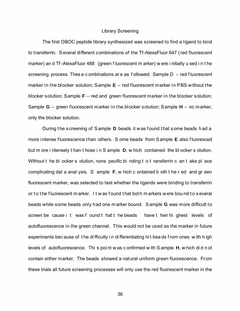
Library Screening
The first OBOC peptide library synthesized was screened to find a l igand to bind
to transferrin. S everal different combinations of the Tf-AlexaFluor 647 ( red fluorescent
marker) an d Tf -AlexaFluor 488 (green f luorescent m arker) w ere i nitially u sed i n t he
screening process. Thes e c ombinations ar e as f ollowed: Sample D - red f luorescent
marker in the blocker solution; Sample E – red f luorescent marker in PBS without the
blocker solution; Sample F – red and green f luorescent marker in the blocker solution;
Sample G – green f luorescent marker in the blocker solution; Sample H – no marker,
only the blocker solution.
During t he screening of Sample D beads it was f ound t hat some beads h ad a
more i ntense f luorescence t han others. S ome beads from Sample E also f luoresced
but m ore i ntensely t han t hose i n S ample D, w hich contained the bl ocker s olution.
Without t he bl ocker s olution, nons pecific bi nding t o t ransferrin c an t ake pl ace
complicating dat a anal ysis. S ample F, w hich c ontained b oth t he r ed and gr een
fluorescent marker, was selected to test whether the ligands were binding to transferrin
or t o t he f luorescent m arker. I t w as f ound t hat bot h m arkers w ere bou nd t o s everal
beads while some beads only had one m arker bound. S ample G was more difficult to
screen be cause i t was f ound t hat t he beads have t heir hi ghest levels of
autofluorescence in the green channel. This would not be used as the marker in future
experiments bec ause of t he di fficulty i n di fferentiating hi t bea ds f rom ones w ith h igh
levels of autofluorescence. Thi s poi nt w as c onfirmed w ith S ample H, w hich di d n ot
contain either marker. The beads showed a natural uniform green fluorescence. From
these trials all future screening processes will only use the red fluorescent marker in the

37
blocker solution.
The f irst s creening, Screening 1 , of the s econd pept ide l ibrary, w hich c ontains
16, 807 m embers, was per formed us ing two 25 m g samples. O ne sample contained
the red fluorescent marker and the other no marker, the negative control. The samples
were prepared as described in the experimental section using an 83 nM concentration
of the red Tf marker. Four beads were found that fluoresced brightly in the red channel
and not in t he gr een. Thes e were pi cked out and separated. The c ontrol s ample
showed beads that fluoresced brightly in the red channel however they also fluoresced
in the green channel indicating the natural fluorescence of the beads, Figure 9 ( A and
B).
The four positive hi t beads, one being broken, were t reated with 8 M guanidine
HCl to denature the transferrin. These hits were washed with DCM but were lost in the
cell culture plate due to the incompatibility of the plate materials with DCM. The s ame
sample w as s creened agai n an d f ive hi ts were obt ained. Thes e w ere d enatured as
mentioned above but it was noticed that a thick white precipitate formed over the beads
from a wash with DMF. Because of this only two of the hits, Figure 9, could be cleaved
and analyzed. The peptide sequence for positive hits C and D could not be determined.
A1 A2

38
Figure 9 – Images A 1, A 2, B 1and B 2 ar e beads f rom t he c ontrol s ample. A rrow i s pointing to positive hit bead from Screening 1. Another s creening, Screening 2 , w as per formed us ing one 25 m g s ample of
beads. Eleven h its were i dentified an d r etrieved, b ut onl y s ix w ere d enatured and
cleaved. Of t hese s ix, o ne w as l ost dur ing ana lysis and t he r emaining f ive w ere
analyzed b y M S. Figure 10 shows t he images of both H it 1 and Hit 2 f rom t he
screening. The m ass s pectrum f or bot h H it 1 an d H it 2 ar e i n Figure 16 and 17,
respectively.
Hit 1a Hit 1b
B2 B1
D2
D1
C2 C1
H-WKRYRGGGG-NH2

39
Figure 10 – Arrow is pointing to positive hit beads from Screening 2. Peptide sequence is shown.
Screening 3 i nvolved two samples f rom 16 ,807-member bead l ibrary. Thi rteen
positive h its w ere i dentified, r etrieved ( Figure 11) , denatured and c leaved. W hen
looking at the images before the analysis process, one hi t was not analyzed because it
did not f luoresce as brightly as desired. A ll twelve beads were analyzed but only five
positive beads were able to be sequenced. The m ass spectrum data are shown below
for each hit 3, 4, 5, 6, and 7 in Figure 18, 19, 20, 21, and 22, respectively.
Hit 2a Hit 2b
H-WYHYRGGGG-NH2
Hit 3b
Hit 5a Hit 5b
Hit 4b Hit 4a
Hit 3a
H-WKWYSGGGG-NH2
H-GRWWWGGGG-NH2
H-WYKKWGGGG-NH2

40
Figure 11 – Arrow i s pointing to pos itive hi ts f rom Screening 3 . Peptide sequence is shown.
Twelve positive hi ts were i dentified and r etrieved f rom t wo s amples du ring
Screening 4. M S dat a c ould only be obt ained on t hree of t hese hi ts, Fi gure 7. The
mass spectrum data for these three hits, Hit 8, Hit 9, and Hit 10, are shown in Figure 23,
24, and 25 . The ot hers were either lost during the washing process or analysis or did
not fluoresce as brightly when the images were reviewed.
Hit 8a
Hit 9b Hit 9a
Hit 8b
H-WRYYHGGGG-NH2
H-WRWYRGGGG-NH2
Hit 6b Hit 6a
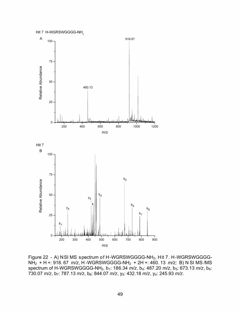
Hit 7a Hit 7b
H-WGRSWGGGG-NH2
H-RYYGWGGGG-NH2

41
Figure 12 – Arrow i s pointing to pos itive hi ts f rom Screening 4 . Peptide sequence is
shown.
The last screening, Screening 5, was performed to obtain negat ive beads to be
used in comparison with positive ones. The beads that f luoresced in both the red and
green c hannel were identified a s negat ive beads . S even be ads w ere i dentified an d
retrieved f rom t his s creening bu t t andem MS dat a w as at tained f or onl y t hree of t he
beads, Figure 13. The mass spectrum data is shown in Figure 26, 27, and 28.
Figure 13 - Arrow is pointing to the negative bead from Screening 5. Peptide sequence is shown.
Neg 1b Neg 1a
Neg 2b Neg 2a
Neg 3b
Neg 3a
Hit 10b Hit 10a
H-WRSSWGGGG-NH2
H-HWHWHGGGG-NH2
H-HWKWHGGGG-NH2
H-HWSHHGGGG-NH2

42
The nano manipulator w as us ed t o ex tract t he pept ide f rom ea ch of t he beads
mentioned above. The peptide was analyzed by use of the nanospray. Table 4 lists the
sequences for all ten positive hit beads and three negative beads.
Table 4 - Sequences of 10 positive hit beads and 3 negative beads. Bead Peptide sequence
Hit 1 H –WKRYRGGGG-NH2
Hit 2 H-WYHYRGGGG-NH2
Hit 3 H-WKWYSGGGG-NH2
Hit 4 H-GRWWWGGGG-NH2
Hit 5 H-WYKKWGGGG-NH2
Hit 6 H-RYYGWGGGG-NH2
Hit 7 H-WGRSWGGGG-NH2
Hit 8 H-WRYYHGGGG-NH2
Hit 9 H-WRWYRGGGG-NH2
Hit 10 H-RYYGWGGGG-NH2
Neg 1
Neg 2
Neg 3
H-HWHWHGGGG-NH2
H-HWSHHGGGG-NH2
H-HWKWHGGGG-NH2

43
Figure 16 - A) NSI MS spectrum of H-WKRYRGGGG-NH2, Hit 1. H-WKRYRGGGG-NH2 + H+: 1070.60 m/z, H-WKRYRGGGG-NH2 + 2H+: 535.98 m/z; B) NSI MS/MS spectrum of H-WKRYRGGGG-NH2. b2: 350.04 m/z, b3: 506.25 m/z, y6: 565.26 m/z.

44
Figure 17 - A) NSI MS spectrum of H-WYHYRGGGG-NH2, Hit 2. H-WYHYRGGGG-NH2 + H+: 1051.87 m/z, H-WYHYRGGGG-NH2 + 2H+: 527.07 m/z; B) NSI MS/MS spectrum of H -WYHYRGGGG-NH2. b 3: 4 87.15 m/z, b 4: 65 0.08 m/z, b 5: 806. 22 m/z, y 7: 702. 32 m/z, y6: 565.26 m/z, y5: 402.26 m/z.

45
Figure 18 - A) N SI MS s pectrum o f H-WKWYSGGGG-NH2, Hit 3 . H -WKWYSGGGG-NH2 + H+: 996. 67 m/z, H -WKWYSGGGG-NH2 + 2 H+: 499. 13 m/z; B) N SI MS /MS spectrum of H-WKWYSGGGG-NH2. b2: 314.96 m/z, b3: 499.11 m/z, b4: 663.54 m/z, b5: 750.57 m/z, b6: 807.59 m/z, b8: 921.63 m/z, y7: 682.27 m/z, y5: 332.99 m/z, y4: 245.77 m/z.

46
Figure 19 - A) NSI MS spectrum of H-GRWWWGGGG-NH2, Hit 4. H-GRWWWGGGG-NH2 + H +: 1017. 56 m/z, H -GRWWWGGGG-NH2 + 2H +: 509. 88 m/z; B) NSI M S/MS spectrum of H-GRWWWGGGG-NH2. b2: 243.2 m/z, b3: 399.3 m/z, b4: 585.38 m/z, b5: 771.46 m/z, b6: 828.48 m/z, b7: 885.5 m/z, y7: 804.34 m/z, y6: 618.26 m/z.

47
Figure 20 - A) NSI MS spectrum of H-WYKKWGGG-NH2, Hit 5. H-WYKKWGGGG-NH2 + H+: 1037.40 m/z, H-WYKKWGGGG-NH2 + 2H+: 519.53 m/z; B) NSI MS/MS spectrum of H-WYKKWGGGG-NH2. b2: 349.04 m/z, y8: 851.13 m/z, y7: 688.20 m/z, y5: 432.07.

48
Figure 21 - A) NSI MS spectrum of H-RYYGWGGGG-NH2, Hit 6. H-RYYGWGGGG-NH2 + H+: 971.73 m/z, H-RYYGWGGGG-NH2 + 2H+: 486.53 m/z; B) NSI MS/MS spectrum of H -RYYGWGGGG-NH2. b 2: 320. 07 m/z, b 4: 540. 07 m/z, b 5: 726. 13 m/z, b 6: 783. 13 m/z, b7: 840.13 m/z, b8: 897.20 m/z, y7: 652.07 m/z, y4: 245.93 m/z.
49
Figure 22 - A) NSI MS spectrum of H-WGRSWGGGG-NH2, H it 7 . H-WGRSWGGGG-NH2 + H +: 918. 67 m/z, H -WGRSWGGGG-NH2 + 2H +: 460. 13 m/z; B) N SI MS /MS spectrum of H-WGRSWGGGG-NH2. b1: 186.34 m/z, b4: 487.20 m/z, b5: 673.13 m/z, b6: 730.07 m/z, b7: 787.13 m/z, b8: 844.07 m/z, y5: 432.18 m/z, y4: 245.93 m/z.

50
Figure 23 - A) NSI MS spectrum of H-WRYYHGGGG-NH2, Hit 8. H-WRYYHGGGG-NH2 + H+: 1051.60 m/z, H-WRYYHGGGG-NH2 + 2H+: 526.47 m/z; B) NSI MS/MS spectrum of H -WRYYHGGGG-NH2. b 4: 6 69.13 m/z, b 5: 80 6.07 m/z, b 6: 863. 20 m/z, y 5: 383. 07 m/z.

51
Figure 24 - A) NSI MS spectrum o f H-WRWYRGGGG-NH2, H it 9 . H-WRWYRGGGG-NH2 + H +: 1093. 40 m/z, H -WRWYRGGGG-NH2 + 2H +: 547. 53 m/z; B) NSI M S/MS spectrum of H-WRWYRGGGG-NH2. b2: 343.07 m/z, b5: 847.3 m/z, y7: 751.20 m/z, y6: 565.13 m/z.

52
Figure 25 - A) NSI MS spectrum of H-WRSSWGGGG-NH2, Hit 10. H-WRSSWGGGG-NH2 + H+: 948. 67 m/z, H -WRSSWGGGG-NH2 + 2H+: 475. 13 m/z; B) NSI MS /MS spectrum of H-WRSSWGGGG-NH2. b2: 343.07 m/z, b3: 430.07 m/z, b4: 514.07 m/z, b5: 703.13 m/z, b6: 760.13 m/z, b7: 817.17 m/z, b8: 874.20 m/z, y7: 606.07 m/z, y6: 519.07 m/z, y4: 245.93 m/z.

53
Figure 26 - A) N SI MS s pectrum o f H-HWHWHGGGG-NH2, N egative bead 1. H -HWHWHGGGG-NH2 + H+: 1029.47 m/z, H-HWHWHGGGG-NH2 + 2H+: 515.81 m/z; B) NSI MS /MS s pectrum o f H -HWHWHGGGG-NH2. b 2: 324. 00 m/z, b 4: 64 7.07 m/z, b 5: 784.00 m/z, y8: 892.13 m/z, y7: 706.06 m/z, y6: 569.07 m/z, y5: 383.07 m/z.

54
Figure 27 - A) NSI M S s pectrum o f H -HWSHHGGGG-NH2, N egative bead 2. H -HWSHHGGGG-NH2 + H+: 930.47 m/z, H -HWSHHGGGG-NH2 + 2H+: 465.93 m/z; B) NSI MS /MS s pectrum o f H -HWSHHGGGG-NH2. b 2: 324. 07 m/z, b 4: 5 48.00 m/z, b 5: 685.13 m/z, y8: 793.13 m/z, y7: 607.07 m/z, y5: 383.07 m/z.

55
Figure 28 - A) N SI MS s pectrum o f H -HWKWHGGGG-NH2, N egative bead 3. H -HWKWHGGGG-NH2 + H+: 1020.47 m/z, H-HWKWHGGGG-NH2 + 2H+: 511.00 m/z; B) NSI MS /MS s pectrum o f H -HWKWHGGGG-NH2. b 2: 324. 07 m/z, b 3: 45 1.23 m/z, b 4: 638.07 m/z, b5: 774.37 m/z, y8: 883.07 m/z, y7: 697.07 m/z, y6: 569.00 m/z, y5: 383.00 m/z, y4: 245.87 m/z.

56
Comparison of Positive and Negative Beads
The pos itive hi t us ed f or c omparison w as f rom S creening 2, hi t 1, w ith t he
sequence H-WKRYRGGGG-NH2. The negative bead was from Screening 5, negative
bead 1, with the sequence H-HWHWHGGGG-NH2. Both of these were synthesized as
described in the experimental. B ulk samples of pos itive and ne gative hi t beads were
incubated with a n 83 nM c oncentration of t he r ed fluorescent marker. The pos itive
beads s howed i ncreased f luorescence i n t he r ed c hannel; how ever, i t w as n ot
significantly di fferent t han t hat of t he negat ive bea ds, Fi gure 14. W hen a 500 nM
concentration of the red fluorescent marker was used a greater increase of fluorescence
was observed on t he positive beads compared to the negative ones, Figure 15. Mo re
tests to compare the avidity of transferrin for the hit sequence will be performed.
Figure 14 – Images A1, A2, B1, B2, C1, C2, D1 and D2 show transferrin bound t o the target l igand. I mages E1, E2, F1 and F2 s how the negative control. Thi s was done using an 83 nM transferrin concentration.
A1
D2 D1 C2
C1
B2 B1
A2
F2 F1
E2 E1

57
Figure 15 – Images A1-D2 show transferrin bound to the target ligand. Images E1, E2, F1 and F2 show the negative control. Images G1, G2, H1 and H2 show a s ide by side comparison of the positive bead (on left) and negative bead ( on right). Thi s was done using a 500 nM transferrin concentration.
Peptide Synthesis on Smaller Beads
Many ben efits c an b e obt ained f rom us ing s maller beads in a pept ide l ibrary.
First, t he us e of s maller beads leads t o a more di verse l ibrary on a per g ram bas is.
Conducting s creenings with s maller be ads w ill t herefore gene rate t he des ired “ hits”
using much smaller quantities of the library. This will be faster and more cost effective
in terms of library synthesis and screening. Second, smaller beads can be screened by
using a s tandard flow cytometer. This will again decrease the screening time and a lso
A1 B1 A2 B2
C1
F2 F1 E2 E1
G1 G2 H1 H2
D2 D1
C2

58
prevent the need for a non-standard flow cytometer needed for screening larger beads.
A comparison of the bead size is shown in Figure 29.
Figure 29 – 130, 30, 20, and 10 µm beads, respectively at 100x magnification.
In order to prove that peptide synthesis is possible on 30 µm and 20 µm beads, a
four glycine chain was synthesized. D uring the synthesis of the peptide on t he 30 µm
beads, a ll Kaiser t ests w ere as expected f or eac h of t he f our g lycines i ndicating t hat
they w ere added s uccessfully. A bul k s ample of t he pe ptide anal yzed by t he
electrospray showed the presence of four glycine units, Figure 30. Single bead peptide
analysis is currently underway.
The Kaiser tests for the synthesis of the four glycine peptide on the 20 um beads
were also as expected; however, no usable data has been collected to date. Bulk and
single bead analysis are currently underway.

59
Figure 30 – ESI MS spectrum of H-GGGG-NH2 on 30 µm resin beads.
20 µm Peptide Library
The s ynthesis of t he m iniaturized pept ide l ibrary w as m ore t edious t han t he
original library on 1 30 µm beads. The m ain di fficulty is the use of the smaller vessel
containing t he s maller por e-size f rit. A dequate s tirring i s c hallenging ev en w ith t he
smallest stir bars available. Manual stirring was often employed to ensure all reagents
accessed the entire bead sample. The l ibrary was synthesized starting with 500 mg of
20 µm beads. The reagents and procedures were similar to those used previously. The

60
general formula for the library was H-X5X4X3X2X1GGGG-HMBA-TentaGel where X = R,
S, W , H , Y, or K . This gi ves 7, 776 di fferent pept ide c ombinations i n t he l ibrary.
According to the Kaiser tests each amino acid was added and deprotected successfully
during ea ch s plit and m ix step. S ince t hese beads ar e m uch smaller a
micromanipulator ep pendorf Tr ansferMan N K 2at tachment on t he N ikon E clipse T i
microscope w as us ed t o s eparate out i ndividual beads f or s ingle bead anal ysis.
Although m any i ndividual beads h ave be en ana lyzed via
nanomanipulation/nanoelectrospray M S, n o i nterpretable data has b een obt ained.
Efforts are underway to sequence individual members of this library.
Conclusions
The majority of the goals set out at the beginning of this project have been met.
First an ef ficient pr otocol f or gener al pe ptide s ynthesis w as put i n pl ace ut ilizing
microwave irradiation to reduce reaction times. Synthesis times which once took up to
24 hour s w ere dec reased t o 10 m inutes. W ith this new m ethod an O BOC pept ide
library was generated straightforwardly in little time meeting two of the eight goals.
A new t echnique f or c leavage o f pept ides from resin b eads w as dev eloped by
which e xposure of an H MBA-linked pept ide t o hi gh pr essure ammonia gas l ed t o
efficient cleavage in as little as 5 minutes. Cleavage could be achieved in a single step
without the use of any solvents, which is not only faster than all other reported methods
employing HMBA as a r esin l inker, but i s al so s ignificantly f aster t han m ost ot her
techniques currently in use. The successful extraction of peptides from individual resin
beads was achieved. The ab ility to sequence peptides sequestered on individual resin
beads of a pr ototypical c ombinatorial library w ith t he a id of nanomanipulation/NSI

61
MS/MS was also demonstrated. The data generated from individual beads matched that
of authentic bulk samples showing this method to be successful. It was also shown
that peptides of varying length are amenable to the process and that it is not dependent
on hav ing gl ycine a s t he anc horing r esidue. The se new t echniques a re at tractive
because they reduce the amount of pos t-screening p rocessing needed to identify h its
(the only solvent needed is one for collection of the hit peptide from the resin bead for
analysis). Thi s new c ombination o f t echniques al lows f or r apid, r eliable,
environmentally responsible sequencing of hit beads from combinatorial libraries.
Our s creening pr ocess w as al so s uccessful in identifying s everal ligands that
bind transferrin. The av idity of one of those hits was demonstrated qualitatively. More
experiments w ill be c onducted w ith t he pos itive hi t s equences gener ated t o quant ify
relative binding avidity to transferrin.
Initial s teps i n t he s ynthesis of a m iniaturized pept ide hav e b een c onducted.
Synthesis of a f our glycine spacer pept ide was successful on bo th 30 µm and 20 µm
beads according to Kaiser tests. H owever, MS data confirming the synthesis has only
been collected thus far for bulk samples of the 30 µm beads. The synthesis of the 20
µm peptide library also appears to have been successful according to the Kaiser tests,
but no M S dat a h as been c ollected. Fu ture w ork i s ai med at s equencing pept ide
contained on individual 30 µm beads, and bulk and individual bead analysis of peptide
contained on 20 µm beads. Random individual beads from the 20 µm library will also be
analyzed and sequenced.

62
WORKS CITED
[1] J.M. Brown, W.D. Hoffmann, C.M. Alvey, A.R. Wood, G.F. Verbeck, R.A. Petros, OBOC Peptide Library Sequencing via High-Pressure Ammonia Cleavage Coupled with Nanomanipulation/Nanoelectrospray Mass Spectrometry, Analytical Biochemistry (2009), doi: 10.1016/j.ab.2009.10.044 [2] O.H. Aina, R.W. Liu, J.L. Sutcliffe, J. Marik, C.X. Pan, and K.S. Lam, From combinatorial chemistry to cancer-targeting peptides. Mol. Pharmaceutics 4 (2007) 631-651. [3] M.M. Marani, E. Oliveira, S. Côté, S.A. Camperi, F. Albericio, and O. Cascone, Identification of protein-binding peptides by direct matrix-assisted laser desorption ionization time-of-flight mass spectrometry analysis of peptide beads selected from the screening of one bead-one peptide combinatorial libraries. Anal. Biochem. 370 (2007) 215-222. [4] S.A. Camperi, M.M. Marani, N.B. Iannucci, S. Côté, F. Albericio, and O. Cascone, An efficient strategy for the preparation of one-bead-one-peptide libraries on a new biocompatible solid support. Tetrahedron Lett. 46 (2005) 1561-1564. [5] A.M. Bray, R.M. Valerio, and N.J. Maeji, Cleavage of resin-bound peptide esters with ammonia vapour. Simultaneous multiple synthesis of peptide amides. Tetrahedron Lett. 34 (1993) 4411- 4414. [6] E. Atherton, M.J. Gait, R.C. Sheppard, and B.J. Williams, The polyamide method of solid phase peptide and oligonucleotide synthesis. Bioorg. Chem. 8 (1979) 351-370. [7] S.L. Mellor, D.A. Wellings, J.-A. Fehrentz, M. Paris, J. Martinez, N.J. Ede, A.M. Bray, D.J. Evans, and G.B. Bloomberg, Synthesis of Modified Peptides. in: W.C. Chan, and P.D. White, (Eds.), Fmoc Solid Phase Peptide Synthesis, Oxford University Press, USA, New York, 2000, pp. 137-181. [8] A.M. Bray, N.J. Maeji, A.G. Jhingran, and R.M. Valerio, Gas-phase cleavage of peptides from a solid support with ammonia vapor. Application in simultaneous multple peptide synthesis. Tetrahedron Lett. 32 (1991) 6163-6. [9] E. Atherton, C.J. Logan, and R.C. Sheppard, Peptide synthesis. Part 2. Procedures for solid-phase synthesis using N-alpha-fluorenylmethoxycarbonyl amino acids on polyamide supports. Synthesis of substance P and of acyl carrier protein 65-74 decapeptide. J. Chem. Soc., Perkin Trans. 1 (1981) 538-46. [10] B. Bacsa, K. Horváti, S. Bõsze, F. Andreae, and C.O. Kappe, Solid-Phase Synthesis of Difficult Peptide Sequences at Elevated Temperatures: A Critical Comparison of Microwave and Conventional Heating Technologies. J. Org. Chem. 73 (2008) 7532-7542.

63
[11] K.S. Lam, S.E. Salmon, E.M. Hersh, V.J. Hruby, W.M. Kazmierski, and R.J. Knapp, A new type of synthetic peptide library for identifying ligand-binding activity. Nature 354 (1991) 82-4. [12] R.A. Houghten, C. Pinilla, S.E. Blondelle, J.R. Appel, C.T. Dooley, and J.H. Cuervo, Generation and use of synthetic peptide combinatorial libraries for basic research and drug discovery. Nature 354 (1991) 84-6. [13] A. Furka, F. Sebestyen, M. Asgedom, and G. Dibo, General method for rapid synthesis of multicomponent peptide mixtures. Int. J. Pept. Protein Res. 37 (1991) 487-93. [14] K.S. Lam, M. Lebl, and V. Krchnak, The ''one-bead-one-compound'' combinatorial library method. Chem. Rev. 97 (1997) 411-448. [15] J.A. Kritzer, N.W. Luedtke, E.A. Harker, and A. Schepartz, A rapid library screen for tailoring betapeptide structure and function. J. Am. Chem. Soc. 127 (2005) 14584-14585. [16] J.K. Murray, J.D. Sadowsky, M. Scalf, L.M. Smith, Y. Tomita, and S.H. Gellman, Exploration of structure - Activity relationships among foldamer ligands for a specific protein binding site via parallel and split-and-mix library synthesis. J. Comb. Chem. 10 (2008) 204-215. [17] J.K. Murray, B. Farooqi, J.D. Sadowsky, M. Scalf, W.A. Freund, L.M. Smith, J.D. Chen, and S.H. Gellman, Efficient synthesis of a beta-peptide combinatorial library with microwave irradiation. J. Am. Chem. Soc. 127 (2005) 13271-13280. [18] J.K. Murray, and S.H. Gellman, Application of microwave irradiation to the synthesis of 14-helical beta-peptides. Org Lett 7 (2005) 1517-1520. [19] A. Thakkar, A.S. Cohen, M.D. Connolly, R.N. Zuckermann, and D. Pei, High-Throughput Sequencing of Peptoids and Peptide−Peptoid Hybrids by Partial Edman Degradation and Mass Spectrometry. J. Comb. Chem. 11 (2009) 294-302. [20] R.E. Dolle, B. Le Bourdonnec, A.J. Goodman, G.A. Morales, C.J. Thomas, and W. Zhang, Comprehensive Survey of Chemical Libraries for Drug Discovery and Chemical Biology: 2007. J. Comb. Chem. 10 (2008) 753-802. [21] O.H. Aina, R.W. Liu, J.L. Sutcliffe, J. Marik, C.X. Pan, and K.S. Lam, From combinatorial chemistry to cancer-targeting peptides. Mol. Pharmaceutics 4 (2007) 631-651. [22] J.K. Scott, and G.P. Smith, Searching for Peptide Ligands with an Epitope Library. Science 249 (1990) 386-390.

64
[23] S.E. Cwirla, E.A. Peters, R.W. Barrett, and W.J. Dower, Peptides on Phage: A Vast Library of Peptides for Identifying Ligands. Proc. Natl. Acad. Sci. U.S.A. 87 (1990) 6378-6382. [24] J.J. Devlin, L.C. Panganiban, and P.E. Devlin, Random Peptide Libraries: A Source of Specific Protein Binding Molecules. Science 249 (1990) 404-406. [25] S. E. Osborne and Andrew D. Ellington, Nucleic Acid Selection and the Challenge of Combinatorial Chemistry. Chem. Rev. 97 (2) (1997) 349–370. [26] N.L. Ledbetter, B.L. Walton, P. Davila, W.D. Hoffmann, R.N. Ernest, and G.F. Verbeck, Nanomanipulation Coupled to Nanospray Mass Spectrometry Applied to the Extraction and Analysis of Trace Analytes Found on Fibers. J. For. Sci. in press (2009). [27] P.J. Horn, A.K. Behrendt, K.D. Chapman, and G.F. Verbeck, Liquid-Phase Microextraction and Controlled Emitter Tip Chemistry Coupled to Nanospray Mass Spectrometry for Direct Lipid Analysis. unpublished results. [28] M.M. Marani, M.C.M. Ceron, S.L. Giudicessi, E. de Oliveira, S. Côté, R. Erra-Balsells, F. Albericio, O. Cascone, and S.A. Camperi, Screening of One-Bead-One-Peptide Combinatorial Library Using Red Fluorescent Dyes. Presence of Positive and False Positive Beads. J. Comb. Chem. 11 (2009) 146-150. [29] J. Wang, S. Tian, R. A. Petros, M. E. Napier, J. M. DeSimone, The Complex Role of Multivalency in Nanoparticles Targeting the Transferrin Receptor for Cancer Therapies J. Am. Chem. Soc., submitted. [30] M. Kinter, and N.E. Sherman, Protein Sequencing and Identification Using Tandem Mass Spectrometry, John Wiley, New York, 2000.


![[G. C. Barrett, D. T. Elmore] Amino Acids and Pept(BookFi.org)](https://static.fdocuments.us/doc/165x107/55cf8c775503462b138cb813/g-c-barrett-d-t-elmore-amino-acids-and-peptbookfiorg.jpg)