Syncope - OSU Center for Continuing Medical Education - PDF of Slides.pdf · Syncope • The only...
15
1 Syncope David T. Hart MBBS, FACC Division of Cardiovascular Medicine The Ohio State University • “Syn-kope” means to “cut short” Syncope: A Symptom…Not a Diagnosis • Self-limited loss of consciousness and postural tone • Relatively rapid onset • Variable warning symptoms • Spontaneous complete recovery The Significance of Syncope • The only difference between syncope and sudden death is that in one you wake up. 1 1 Engel GL. Psychologic stress, vasodepressor syncope, and sudden death. Ann Intern Med 1978; 89: 403-412.
Transcript of Syncope - OSU Center for Continuing Medical Education - PDF of Slides.pdf · Syncope • The only...

1
Syncope David T. Hart MBBS, FACC
Division of Cardiovascular MedicineThe Ohio State University
• “Syn-kope” means to“cut short”
Syncope:A Symptom…Not a Diagnosis
• Self-limited loss of consciousness and postural tone
• Relatively rapid onset• Variable warning symptoms• Spontaneous complete recovery
The Significance of Syncope
• The only difference between syncope and sudden death is that in one you wake up.1
1 Engel GL. Psychologic stress, vasodepressor syncope, and sudden death. Ann Intern Med 1978; 89: 403-412.
2
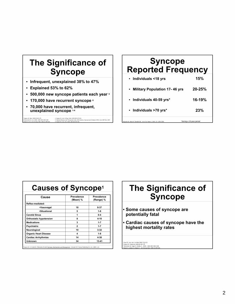
• Infrequent, unexplained 38% to 47%• Explained 53% to 62%• 500,000 new syncope patients each year 5
• 170,000 have recurrent syncope 6
• 70,000 have recurrent, infrequent, unexplained syncope 1-4
1 Kapoor W, Med. 1990;69:160-175.2 Silverstein M, et al. JAMA. 1982;248:1185-1189.3 Martin G, et al. Ann Emerg. Med. 1984;12:499-504.
4 Kapoor W, et al. N Eng J Med. 1983;309:197-204.5 National Disease and Therapeutic Index, IMS America, Syncope and Collapse #780.2; Jan 1997-Dec 1997.6 Kapoor W, et al. Am J Med. 1987;83:700-708.
The Significance of Syncope
Cause Prevalence (Mean) %
Prevalence (Range) %
Reflex-mediated:Vasovagal 18 8-37Situational 5 1-8
Carotid Sinus 1 0-4Orthostatic hypotension 8 4-10Medications 3 1-7Psychiatric 2 1-7Neurological 10 3-32Organic Heart Disease 4 1-8Cardiac Arrhythmias 14 4-38Unknown 34 13-41
Causes of Syncope1
1Kapoor W. In Grubb B, Olshansky B (eds) Syncope: Mechanisms and Management. Armonk NY; Futura Publishing Co, Inc: 1998; 1-13.
• Individuals <18 yrs
• Military Population 17- 46 yrs
• Individuals 40-59 yrs*
• Individuals >70 yrs*
15%
20-25%
16-19%
23%
Syncope Reported Frequency
*during a 10-year periodBrignole M, Alboni P, Benditt DG, et al. Eur Heart J, 2001; 22: 1256-1306.
1 Day SC, et al. Am J of Med 1982;73:15-23.2 Kapoor W. Medicine 1990;69:160-175.3 Silverstein M, Sager D, Mulley A. JAMA. 1982;248:1185-1189.4 Martin G, Adams S, Martin H. Ann Emerg Med. 1984;13:499-504.
• Some causes of syncope are potentially fatal
• Cardiac causes of syncope have the highest mortality rates
The Significance of Syncope

3
Causes of Syncope-like States
• Migraine*• Acute hypoxemia*• Hyperventilation*• Somatization disorder (psychogenic syncope)• Acute Intoxication (e.g., alcohol)• Seizures• Hypoglycemia• Sleep disorders
* may cause ‘true’ syncope
Syncope: EtiologyOrthostatic Cardiac
Arrhythmia
StructuralCardio-
Pulmonary
*
1• Vasovagal• Carotid
Sinus• Situational
CoughPost-micturition
2• Drug
Induced• ANS
FailurePrimarySecondary
3• Brady
Sick sinusAV block
• TachyVTSVT
• Long QT Syndrome
4 • Aortic
Stenosis• HOCM• PulmonaryHypertension
5• Psychogenic• Metabolic
e.g. hyper-ventilation
• Neurological
Non-Cardio-vascular
Neurally-Mediated
Unknown Cause = 34%
24% 11% 14% 4% 12%
DG Benditt, UM Cardiac Arrhythmia Center
• Distinguish ‘True’ Syncope from other ‘Loss of Consciousness’ spells:
SeizuresPsychiatric disturbances
• Establish the cause of syncope with sufficient certainty to:
Assess prognosis confidentlyInitiate effective preventive treatment
Syncope Diagnostic Objectives
Initial Evaluation(Clinic/Emergency Dept.)
• Detailed history• Physical examination• 12-lead ECG• Echocardiogram (as available)

4
Syncope Evaluation and
Differential Diagnosis
• Complete DescriptionFrom patient and observers
• Type of Onset• Duration of Attacks• Posture• Associated Symptoms• Sequelae
History – What to Look for
Syncope Basic Diagnostic Steps
• Detailed History & PhysicalDocument details of eventsAssess frequency, severityObtain careful family history
• Heart disease present?Physical examECG: long QT, WPW, conduction system diseaseEcho: LV function, valve status, HOCM
• Follow a diagnostic plan...
Unexplained Syncope Diagnosis
Adapted from: W.Kapoor.An overview of the evaluation and management of syncope. From Grubb B, Olshansky B (eds) Syncope: Mechanisms and Management. Armonk, NY: Futura Publishing Co., Inc.1998.
History and Physical Exam Surface ECG
Neurological Testing
• Head CT Scan• Carotid Doppler• MRI• Skull Films• Brain Scan• EEG
CV Syncope Workup
• Holter• ELR or ILR• Tilt Table• Echo• EPS
Other CV Testing
• Angiogram• Exercise Test• SAECG
Psychological Evaluation
ENT Evaluation Endocrine Evaluation
Conventional Diagnostic Methods/Yield
Test/Procedure Yield(based on mean time to diagnosis of 5.1 months7
History and Physical (including carotid sinus massage)
49-85% 1, 2
ECG 2-11% 2
Electrophysiology Study without SHD* 11% 3
Electrophysiology Study with SHD 49% 3
Tilt Table Test (without SHD) 11-87% 4, 5
Ambulatory ECG Monitors:Holter 2% 7
External Loop Recorder(2-3 weeks duration)
20% 7
Insertable Loop Recorder(up to 14 months duration)
65-88% 6, 7
Neurological †
(Head CT Scan, Carotid Doppler) 0-4% 4,5,8,9,10
* Structural Heart Disease† MRI not studied
1 Kapoor, et al N Eng J Med, 1983.2 Kapoor, Am J Med, 1991.3 Linzer, et al. Ann Int. Med, 1997.4 Kapoor Medicine 1990
5 Kapoor, JAMA, 19926 Krahn, Circulation, 19957 Krahn, Cardiology Clinics, 1997.8 Eagle K et al The Yale J Biol and Medicine 1983; 56: 1-8
9 Day S, et al. Am J Med. 1982; 73: 15-23.10 Stetson P, et al. PACE. 1999; 22 (part II): 782.
5
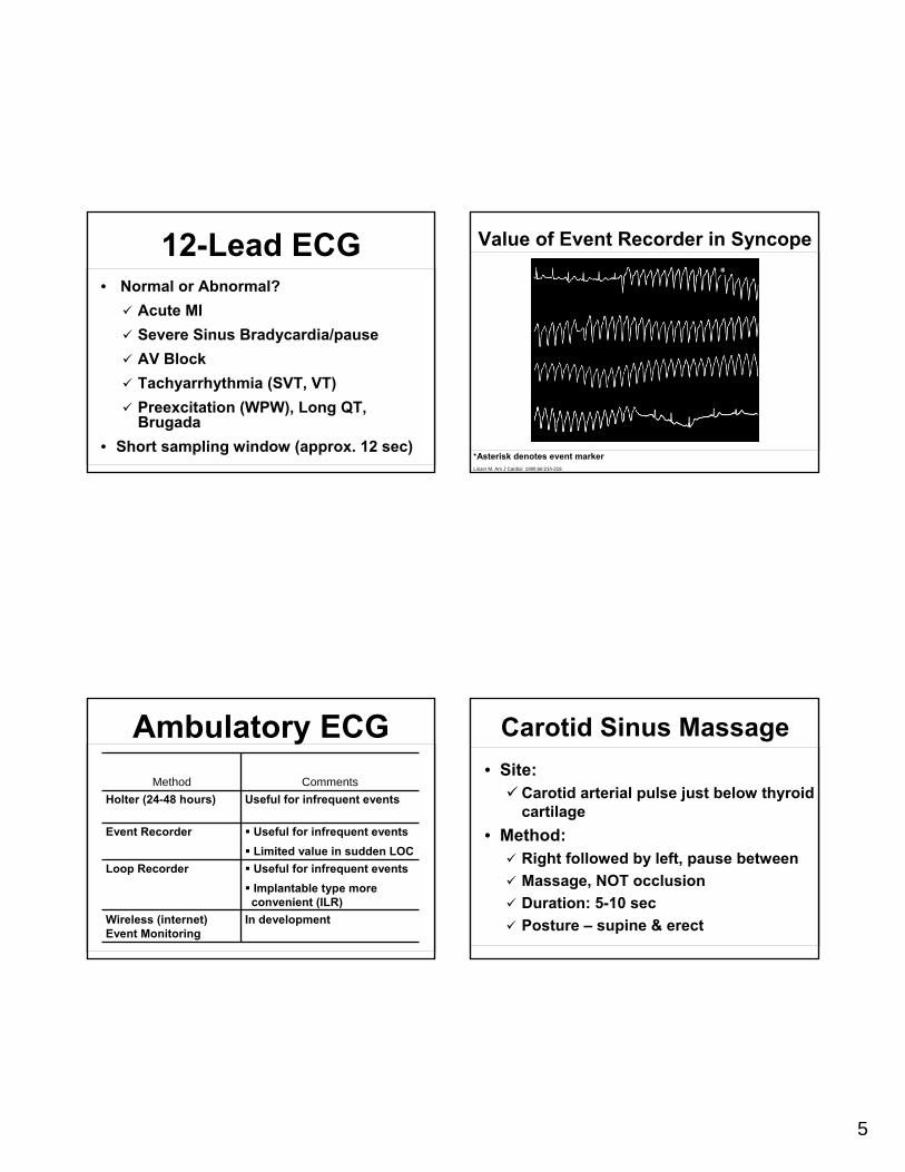
12-Lead ECG• Normal or Abnormal?
Acute MISevere Sinus Bradycardia/pauseAV BlockTachyarrhythmia (SVT, VT)Preexcitation (WPW), Long QT, Brugada
• Short sampling window (approx. 12 sec)
Method CommentsHolter (24-48 hours) Useful for infrequent events
Event Recorder Useful for infrequent eventsLimited value in sudden LOC
Loop Recorder Useful for infrequent eventsImplantable type moreconvenient (ILR)
Wireless (internet) Event Monitoring
In development
Ambulatory ECG
Value of Event Recorder in Syncope
Linzer M. Am J Cardiol. 1990;66:214-219.
*Asterisk denotes event marker
Carotid Sinus Massage• Site:
Carotid arterial pulse just below thyroid cartilage
• Method:Right followed by left, pause betweenMassage, NOT occlusionDuration: 5-10 secPosture – supine & erect

6
• Outcome:3 sec asystole and/or 50 mmHg fall in systolic bloodpressure with reproduction of symptoms =
Carotid Sinus Syndrome (CSS)• Contraindications
Carotid bruit, known significant carotid arterial disease, previous CVA, MI last 3 months
• Risks1 in 5000 massages complicated by TIA
Carotid Sinus Massage
Head-up Tilt Test (HUT)
• Unmasks VVS susceptibility
• Reproduces symptoms• Patient learns VVS
warning symptoms • Physician is better able
to give prognostic / treatment advice
Head-Up Tilt Test (HUT)
DG Benditt, UM Cardiac Arrhythmia Center
Electroencephalogram• Not a first line of testing
• Syncope from Seizures
• Abnormal in the interval between two attacks – Epilepsy
• Normal – Syncope

7
• Limited utility in syncope evaluation• Most useful in patients with structural
heart diseaseHeart disease……..50-80%No Heart disease…18-50%
• Relatively ineffective for assessing bradyarrhythmias
Brignole M, Alboni P, Benditt DG, et al. Eur Heart Journal 2001; 22: 1256-1306.
Conventional EP Testing in Syncope
EP Testing in Syncope:Useful Diagnostic
Observations• Inducible monomorphic VT• SNRT > 3000 ms or CSRT > 600 ms• Inducible SVT with hypotension• HV interval ≥ 100 ms (especially in
absence of inducible VT)• Pacing induced infra-nodal block
Diagnostic Limitations
• Difficult to correlate spontaneous events and laboratory findings
• Often must settle for an attributable cause
• Unknowns remain 20-30% 1
1Kapoor W. In Grubb B, Olshansky B (eds) Syncope: Mechanisms and Management. Armonk NY; Futura Publishing Co, Inc: 1998; 1-13.
Section IV:Specific Conditions

8
Neurally-Mediated Reflex Syncope (NMS)
• Vasovagal syncope (VVS)• Carotid sinus syndrome (CSS)• Situational syncope
post-micturitioncoughswallow defecationblood drawingetc.
NM Reflex Syncope: Pathophysiology
• Multiple triggers
• Variable contribution of vasodilatation and bradycardia
NMS – Basic Pathophysiology
CerebralCortex
VascularBed Bradycardia/
Hypotension
Baro-receptors
Heart
Feedback viaCarotid Baroreceptors
Other Mechanoreceptors
Parasympathetic (+)
sympathetic (+) ¯ Heart Rate¯ AV Conduction
- Vasodilatation
Benditt DG, Lurie KG, Adler SW, et al. Pathophysiology of vasovagal syncope. In: Neurally mediated syncope: Pathophysiology, investigations and treatment. Blanc JJ, Benditt D, Sutton R. Bakken Research Center Series, v. 10. Armonk, NY: Futura, 1996
• Neurally Mediated Physiologic Reflex Mechanism with two Components:
Cardioinhibitory (HR)Vasodepressor (BP)
• Both components are usually present
Vasovagal Syncope (VVS):Clinical Pathophysiology

9
Prevalence of VVS• Prevalence is poorly known
Various studies report 8% to 37% (mean 18%) of cases of syncope (Linzer 1997)
• In general:VVS patients younger than CSS patientsAges range from adolescence to elderly (median 43 years)Pallor, nausea, sweating, palpitations are commonAmnesia for warning symptoms in older patients
Management Strategies for VVS• Optimal management strategies for VVS are a
source of debatePatient education, reassurance, instructionFluids, salt, dietTilt TrainingSupport hose
• Drug therapies• Pacing
Class II indication for VVS patients with positive HUT and cardioinhibitory or mixed reflex
Carotid Sinus Syndrome (CSS)
• Syncope clearly associated with carotid sinus stimulation is rare (≤1% of syncope)
• CSS may be an important cause of unexplained syncope / falls in older individuals
Etiology of CSS• Sensory nerve endings in the
carotid sinus walls respond to deformation
• “Deafferentation” of neck muscles may contribute
• Increased afferent signals to brain stem
• Reflex increase in efferent vagalactivity and diminution of sympathetic tone results in bradycardia and vasodilation
Carotid Sinus
10
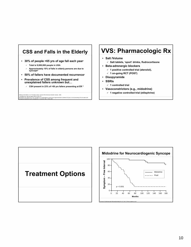
CSS and Falls in the Elderly
• 30% of people >65 yrs of age fall each year1
Total is 9,000,000 people in USAApproximately 10% of falls in elderly persons are due to syncope2
• 50% of fallers have documented recurrence3
• Prevalence of CSS among frequent and unexplained fallers unknown but…
CSH present in 23% of >50 yrs fallers presenting at ER 3
1Falling in the Elderly: U.S. Prevalence Data. Journal of the American Geriatric Society, 1995.2 Campbell et al: Age and Aging 1981;10:264-270.3Richardson DA, Bexton RS, et al. Prevalence of cardioinhibitory carotid sinus hypersensitivity in patients 50 years or over presenting to the Accident and Emergency Department with “unexplained” or “recurrent” falls. PACE 1997
Treatment Options
VVS: Pharmacologic Rx• Salt /Volume
Salt tablets, ‘sport’ drinks, fludrocortisone• Beta-adrenergic blockers
1 positive controlled trial (atenolol), 1 on-going RCT (POST)
• Disopyramide• SSRIs
1 controlled trial• Vasoconstrictors (e.g., midodrine)
1 negative controlled trial (etilephrine)
Midodrine for Neurocardiogenic Syncope
Journal of Cardiovascular Electrophysiology Vol. 12, No. 8, Perez-Lugones, et al.
Months
p < 0.001Sym
ptom
–Fr
ee In
terv
al
180160140120100806040200
100
80
60
40
20
0
Fluid
Midodrine
11
VVS: Tilt-Training• Objectives
Enhance Orthostatic ToleranceDiminish Excessive Autonomic Reflex ActivityReduce Syncope Susceptibility / Recurrences
• TechniquePrescribed Periods of Upright PostureProgressive Increased Duration
Status of Pacing in VVS• Perception of pacing for VVS changing:
VVS with +HUT and cardioinhibitory response a Class IIbindication1
• Recent clinical studies demonstrated benefits of pacing in select VVS patients:
VPS IVASIS SYDITVPS II –Phase IROME VVS Trial
1Gregoratos G, et al. ACC/AHA Guidelines for Implantation of Cardiac Pacemakers and Antiarrhythmic Devices. Circulation. 1998; 97: 1325-1335.
VVS Pacing Trials Conclusions
• DDD pacing reduces the risk of syncope in patients with recurrent, refractory, highly-symptomatic, cardioinhibitoryvasovagal syncope.
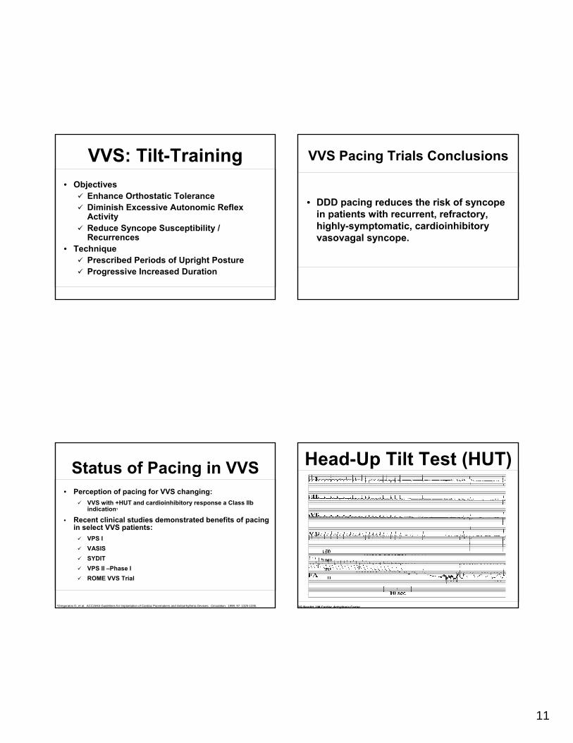
Head-Up Tilt Test (HUT)
DG Benditt, UM Cardiac Arrhythmia Center

12
• Drug-induced (very common)diureticsvasodilators
• Primary autonomic failuremultiple system atrophyParkinsonism
• Secondary autonomic failurediabetes alcohol amyloid
• Alcohol orthostatic intolerance apart from neuropathy
Principal Causes of Orthostatic Syncope
Syncope Due to Arrhythmia orStructural CV Disease: General Rules
• Often life-threatening and/or exposes patient to high risk of injury
• May be warning of critical CV diseaseAortic stenosis, Myocardial ischemia, Pulmonary hypertension
• Assess culprit arrhythmia / structural abnormality aggressively
• Initiate treatment promptly
Principal Causes of Syncope due to Structural
Cardiovascular Disease• Acute MI / Ischemia
Acquired coronary artery disease Congenital coronary artery anomalies
• HOCM• Acute aortic dissection• Pericardial disease / tamponade• Pulmonary embolus / pulmonary hypertension• Valvular abnormalities
Aortic stenosis, Atrial myxoma
• BradyarrhythmiasSinus arrest, exit blockHigh grade or acute complete AV block
• TachyarrhythmiasAtrial fibrillation / flutter with rapid ventricular rate (e.g. WPW syndrome)Paroxysmal SVT or VTTorsades de pointes
Syncope Due to Cardiac Arrhythmias

13
AECG: 74 yr Male, Syncope
From the files of DG Benditt, UM Cardiac Arrhythmia Center
Syncope: Torsades
From the files of DG Benditt, UM Cardiac Arrhythmia Center
83 yo womanBradycardia: Pacemaker implanted
28 yo man in the ER multiple times after falls resulting in traumaVT: ablated and medicated
Reveal ® ILR recordings; Medtronic data on file.
Infra-His Block
From the files of DG Benditt, UM Cardiac Arrhythmia Center

14
Drug-Induced QT Prolongation• Antiarrhythmics
Class IA ...Quinidine, Procainamide, DisopyramideClass III…Sotalol, Ibutilide, Dofetilide, Amiodarone, (NAPA)
• Antianginal Agents(Bepridil)
• Psychoactive AgentsPhenothiazines, Amitriptyline, Imipramine, Ziprasidone
• AntibioticsErythromycin, Pentamidine, Fluconazole
• Nonsedating antihistamines(Terfenadine), Astemizole
• Others(Cisapride), Droperidol
Treatment of Syncope Due to Bradyarrhythmia
• Class I indication for pacing using dual-chamber system wherever adequate atrialrhythm is available
• Ventricular pacing in atrial fibrillation with slow ventricular response
• Atrial Tachyarrhythmias;AVRT due to accessory pathway – ablate pathwayAVNRT – ablate AV nodal slow pathwayAtrial fib – Pacing, linear / focal ablation, ICD selected ptsAtrial flutter – Ablation of reentrant circuit
• Ventricular Tachyarrhythmias;Ventricular tachycardia – ICD or ablation where appropriateTorsades de Pointes – withdraw offending Rx or ICD (long-QT/Brugada)
• Drug therapy may be an alternative in many cases
Treatment of Syncope Due to Tachyarrhythmia
Typical Cardiovascular Diagnostic Pathway
History and Physical, ECG
Syncope
KnownSHD No
SHD
Echo
EPS
+
Treat
> 30 days; > 2 Events
Tilt ILR
Tilt Holter/ ELR
ILR
Tilt/ILR
< 30 days
-
Adapted from:Linzer M, et al. Annals of Int Med, 1997. 127:76-86.Syncope: Mechanisms and Management. Grubb B, Olshansky B (eds) Futura Publishing 1999Zimetbaum P, Josephson M. Annals of Int Med, 1999. 130:848-856.Krahn A et al. ACC Current Journal Review,1999. Jan/Feb:80-84.

15
• “I want to die like my father, peacefully in his sleep; not screaming like the passengers in his car”
George Burns
The End