Synaptic organisation of the basal ganglia
16
J. Anat. (2000) 196, pp. 527–542, with 4 figures Printed in the United Kingdom 527 Review Synaptic organisation of the basal ganglia J. P. BOLAM, J. J. HANLEY, P. A. C. BOOTH AND M.D. BEVAN MRC Anatomical Neuropharmacology Unit, Department of Pharmacology, Oxford, UK (Accepted 5 November 1999) The basal ganglia are a group of subcortical nuclei involved in a variety of processes including motor, cognitive and mnemonic functions. One of their major roles is to integrate sensorimotor, associative and limbic information in the production of context-dependent behaviours. These roles are exemplified by the clinical manifestations of neurological disorders of the basal ganglia. Recent advances in many fields, including pharmacology, anatomy, physiology and pathophysiology have provided converging data that have led to unifying hypotheses concerning the functional organisation of the basal ganglia in health and disease. The major input to the basal ganglia is derived from the cerebral cortex. Virtually the whole of the cortical mantle projects in a topographic manner onto the striatum, this cortical information is ‘processed’ within the striatum and passed via the so-called direct and indirect pathways to the output nuclei of the basal ganglia, the internal segment of the globus pallidus and the substantia nigra pars reticulata. The basal ganglia influence behaviour by the projections of these output nuclei to the thalamus and thence back to the cortex, or to subcortical ‘premotor’ regions. Recent studies have demonstrated that the organisation of these pathways is more complex than previously suggested. Thus the cortical input to the basal ganglia, in addition to innervating the spiny projection neurons, also innervates GABA interneurons, which in turn provide a feed-forward inhibition of the spiny output neurons. Individual neurons of the globus pallidus innervate basal ganglia output nuclei as well as the subthalamic nucleus and substantia nigra pars compacta. About one quarter of them also innervate the striatum and are in a position to control the output of the striatum powerfully as they preferentially contact GABA interneurons. Neurons of the pallidal complex also provide an anatomical substrate, within the basal ganglia, for the synaptic integration of functionally diverse information derived from the cortex. It is concluded that the essential concept of the direct and indirect pathways of information flow through the basal ganglia remains intact but that the role of the indirect pathway is more complex than previously suggested and that neurons of the globus pallidus are in a position to control the activity of virtually the whole of the basal ganglia. Key words : Striatum ; globus pallidus ; corticostriatal ; pallidostriatal ; GABA interneurons ; substantia nigra ; synaptic convergence. The basal ganglia are a group of subcortical nuclei involved in a variety of processes including motor, associative, cognitive and mnemonic functions. The dorsal division of the basal ganglia consists of the striatum (or caudate-putamen), the globus pallidus (GP, external segment of the globus pallidus in primates), entopeduncular nucleus (EP, internal seg- ment of globus pallidus in primates, GPi), the subthalamic nucleus (STN) and the substantia nigra Correspondence to Dr J. P. Bolam, MRC Anatomical Neuropharmacology Unit, Department of Pharmacology, Mansfield Road, Oxford OX1 3TH, UK. Tel: ›44 (0) 1865 271 869; fax: ›44 (0) 1865 271 647 ; e-mail : paul.bolam!pharmacology.ox.ac.uk (SN). The latter structure is divided into 2 main parts, the dorsal pars compacta (SNc) in which the dopa- minergic nigrostriatal neurons are located and the more ventral pars reticulata (SNr). In addition to these structures which are associated with motor and associative functions there is a ventral division of the basal ganglia (ventral striatum or nucleus accumbens ; ventral pallidum and ventral tegemental area) that is associated with limbic functions. The major input to the basal ganglia is derived from the cortex ; virtually the whole of the cortical mantle
Transcript of Synaptic organisation of the basal ganglia

J. Anat. (2000) 196, pp. 527–542, with 4 figures Printed in the United Kingdom 527
Review
Synaptic organisation of the basal ganglia
J. P. BOLAM, J. J. HANLEY, P. A. C. BOOTH AND M. D. BEVAN
MRC Anatomical Neuropharmacology Unit, Department of Pharmacology, Oxford, UK
(Accepted 5 November 1999)
The basal ganglia are a group of subcortical nuclei involved in a variety of processes including motor,
cognitive and mnemonic functions. One of their major roles is to integrate sensorimotor, associative and
limbic information in the production of context-dependent behaviours. These roles are exemplified by the
clinical manifestations of neurological disorders of the basal ganglia. Recent advances in many fields,
including pharmacology, anatomy, physiology and pathophysiology have provided converging data that
have led to unifying hypotheses concerning the functional organisation of the basal ganglia in health and
disease. The major input to the basal ganglia is derived from the cerebral cortex. Virtually the whole of the
cortical mantle projects in a topographic manner onto the striatum, this cortical information is ‘processed’
within the striatum and passed via the so-called direct and indirect pathways to the output nuclei of the
basal ganglia, the internal segment of the globus pallidus and the substantia nigra pars reticulata. The basal
ganglia influence behaviour by the projections of these output nuclei to the thalamus and thence back to the
cortex, or to subcortical ‘premotor’ regions. Recent studies have demonstrated that the organisation of these
pathways is more complex than previously suggested. Thus the cortical input to the basal ganglia, in
addition to innervating the spiny projection neurons, also innervates GABA interneurons, which in turn
provide a feed-forward inhibition of the spiny output neurons. Individual neurons of the globus pallidus
innervate basal ganglia output nuclei as well as the subthalamic nucleus and substantia nigra pars compacta.
About one quarter of them also innervate the striatum and are in a position to control the output of the
striatum powerfully as they preferentially contact GABA interneurons. Neurons of the pallidal complex also
provide an anatomical substrate, within the basal ganglia, for the synaptic integration of functionally diverse
information derived from the cortex. It is concluded that the essential concept of the direct and indirect
pathways of information flow through the basal ganglia remains intact but that the role of the indirect
pathway is more complex than previously suggested and that neurons of the globus pallidus are in a
position to control the activity of virtually the whole of the basal ganglia.
Key words : Striatum; globus pallidus ; corticostriatal ; pallidostriatal ; GABA interneurons; substantia nigra; synaptic convergence.
The basal ganglia are a group of subcortical nuclei
involved in a variety of processes including motor,
associative, cognitive and mnemonic functions. The
dorsal division of the basal ganglia consists of the
striatum (or caudate-putamen), the globus pallidus
(GP, external segment of the globus pallidus in
primates), entopeduncular nucleus (EP, internal seg-
ment of globus pallidus in primates, GPi), the
subthalamic nucleus (STN) and the substantia nigra
Correspondence to Dr J. P. Bolam, MRC Anatomical Neuropharmacology Unit, Department of Pharmacology, Mansfield Road, Oxford
OX1 3TH, UK. Tel : 44 (0) 1865 271 869; fax: 44 (0) 1865 271 647; e-mail : paul.bolam!pharmacology.ox.ac.uk
(SN). The latter structure is divided into 2 main parts,
the dorsal pars compacta (SNc) in which the dopa-
minergic nigrostriatal neurons are located and the
more ventral pars reticulata (SNr). In addition to
these structures which are associated with motor and
associative functions there is a ventral division of the
basal ganglia (ventral striatum or nucleus accumbens;
ventral pallidum and ventral tegemental area) that is
associated with limbic functions.
The major input to the basal ganglia is derived from
the cortex; virtually the whole of the cortical mantle

Fig. 1. Simplified block diagram of the circuitry of the basal
ganglia. Inhibitory projections are shown by mottled lines,
excitatory projections by dotted lines. Cortical information that
reaches the striatum is conveyed to the basal ganglia output
structures (SNr}EP, substantia nigra pars reticulata}entopedun-
cular nucleus) via 2 pathways, a direct inhibitory projection from
the striatum to SNr}EP and an indirect pathway, which involves an
inhibitory projection from the striatum to globus pallidus (GP), an
inhibitory projection from the GP to the subthalamic nucleus
(STN) and to the output nuclei and an excitatory projection from
the STN to SNr}EP. The information is then transmitted back
to the cerebral cortex via the thalamus or conveyed to various
brainstem structures including the superior colliculus (SC) and the
parvicellular reticular formation (RF). Dopaminergic neurons of
the SNc provide a massive feedback projection to the striatum
(hatched line) and modulate the flow of cortical information. A
proportion of GP neurons also feedback to the striatum where they
innervate interneurons which also receive cortical input. Cortical
information can also reach the basal ganglia via the cortico-
subthalamic projection.
projects onto the basal ganglia in a highly topo-
graphical manner. The main point of entry of this
cortical information to the basal ganglia is the
striatum although there are significant cortical pro-
jections to the STN. The corticostriatal projection
imparts functionality on to the striatum and conse-
quently other divisions of the basal ganglia. In what is
now considered the classic view of basal ganglia
circuitry (Albin et al. 1989; DeLong, 1990; Smith et
al. 1998), the functional organisation is such that
cortical information carried by the corticostriatal
projection is processed within the striatum, integrated
with the many other inputs to the basal ganglia (e.g.
intralaminar thalamic nuclei, amygdala, hippocam-
pus, dorsal raphe) which primarily innervate the
striatum, and then the ‘processed information’ is
transmitted to the output nuclei of the basal ganglia,
the EP and the SNR. The basal ganglia influence
behaviour by these output nuclei projecting to the
ventral thalamus and then back to the cortex or by
projecting to subcortical ‘premotor’ regions including
the superior colliculus, the pedunculopontine nucleus
or the reticular formation (Fig. 1) (see Albin et al.
1989; DeLong, 1990; Bolam & Bennett, 1995; Gerfen
& Wilson, 1996; Smith et al. 1998 for recent reviews).
The transmission of cortical information through
the basal ganglia occurs through 2 routes, the ‘direct’
and ‘indirect’ pathways (Albin et al. 1989; DeLong,
1990). In the direct pathway corticostriatal infor-
mation is transmitted directly from the striatum to the
output nuclei. In the indirect pathway corticostriatal
information is transmitted indirectly to the output
nuclei via the complex network interconnecting the
GP and STN (Shink et al. 1996; Fig. 1). Data from a
variety of disciplines, but particularly the pioneering
work of Deniau and colleagues (Chevalier & Deniau,
1990), Albin and colleagues (Albin et al. 1989) and
DeLong (DeLong, 1990), has shown that the output
signal of the basal ganglia under resting conditions is
one of inhibition, and that there is a loss of inhibition
during a basal ganglia associated behaviour. This is
brought about by the neurochemical nature of
neurons in the pathways and their basal activity.
Striatal projection neurons are GABAergic and
quiescent under resting conditions ; basal ganglia
output neurons are also GABAergic but have a high
discharge rate, tonically inhibiting the targets of the
basal ganglia, i.e. neurons in the ventral thalamus or
subcortical premotor regions. When the system is
activated by the firing of corticostriatal glutamatergic
neurons, striatal neurons discharge, which in turn
causes inhibition of basal ganglia output neurons in
the SNR and EP. This reduction in firing of basal
ganglia output neurons leads to release from in-
hibition, or disinhibition, of neurons in the targets of
the basal ganglia and is associated with ‘basal ganglia
behaviour’. In contrast to this, activation of the
indirect pathway or network leads to the opposite
physiological effect, i.e. increased firing of output
neurons and increased inhibition of basal ganglia
targets. It has been suggested that tonic activity of
STN neurons is the driving force for the resting
activity in basal ganglia output nuclei (Bevan &
Wilson, 1999; Nakanishi et al. 1987). Under normal
conditions during basal ganglia associated behaviour,
the output of the basal ganglia is a complex
528 J. P. Bolam and others

spatiotemporal pattern of increased and decreased
firing, i.e. inhibition and disinhibition. It has been
suggested that the indirect pathway acts to attenuate
or terminate a basal ganglia associated movement or
to suppress unwanted sequences of movement (Mink
& Thach, 1993).
Overlying this ‘feed-forward’ organisation of the
basal ganglia are many feedback pathways. The major
one of these is the dopaminergic projection from the
SNC to the striatum. This projection modulates the
flow of cortical information through the basal ganglia.
Loss of these neurons in Parkinson’s disease leads to
an imbalance of the flow of cortical information
through the basal ganglia in favour of the indirect
pathway and hence the akinetic behaviour associated
with this disorder (Albin et al. 1989; DeLong, 1990).
The subject of this communication is first, to briefly
review the synaptology underlying the pathways of
information flow through the basal ganglia, secondly,
to demonstrate how new knowledge of the connec-
tions of individual neurons in the basal is leading to
modifications or an elaboration of the classical models
of the circuits and thirdly, to demonstrate one of the
sites of integration of functionally diverse information
in the basal ganglia.
The striatum contains both projection neurons and
several populations of interneurons (Bolam & Ben-
nett, 1995; Kawaguchi et al. 1995; Kawaguchi, 1997).
The major type of projection neuron is the medium
size densely spiny neuron (spiny neuron; Fig. 2A).
They account for 90–95% of the total population of
striatal neurons (Kemp & Powell, 1971a), utilise
GABA as their major neurotransmitter and are
subdivided into 2 major populations on the basis of
their projection region, pattern of axonal collateral-
isation and their neurochemical content (for reviews
see Smith & Bolam, 1990; Bolam & Bennett, 1995;
Gerfen & Wilson, 1996; Kawaguchi, 1997; Smith et
al. 1998). One subpopulation projects preferentially to
the output nuclei of the basal ganglia and expresses, in
addition to GABA, the neuropeptides substance P
and dynorphin and the D1 subtype of dopamine
receptors. The second subpopulation projects almost
exclusively to the GP and expresses enkephalin and
the D2 subtype of dopamine receptors. Evidence from
morphological studies, including intracellular filling
of neurons (see Chang & Wilson, 1990) or Golgi
impregnation (see Pasik et al. 1979) have demo-
nstrated that spiny neurons also give rise to extensive
local axon collaterals, one of the major synaptic
targets of which are other spiny neurons (Wilson &
Groves, 1980; Somogyi et al. 1981). This is supported
by the findings that terminals that display immuno-
reactivity for the neuropeptides expressed by spiny
neurons, i.e. enkephalin or substance P, and possess
the morphological features of spiny neuron terminals,
make symmetric synaptic contact with the dendrites,
spines and perikarya of spiny neurons (Pickel et al.
1980, 1992; Aronin et al. 1981; DiFiglia et al. 1982;
Somogyi et al. 1982; Bolam et al. 1983; Bouyer et al.
1984b ; Bolam & Izzo, 1988).
The early anterograde degeneration studies demo-
nstrated that corticostriatal terminals form asym-
metric synapses primarily with dendritic spines (Kemp
& Powell, 1971b, c). The fact that spiny projection
neurons are the major spine-bearing neurons in the
striatum indicates that these cells are likely to be the
major targets of the corticostriatal projection (Kemp
& Powell, 1971b, c). Indeed, the result of direct
analysis has demonstrated that corticostriatal ter-
minals make synaptic contact with the heads of spines
of spiny projection neurons which give rise both to the
direct and indirect pathways (Frotscher et al. 1981;
Somogyi et al. 1981; Dube et al. 1988; Hersch et al.
1995; Fig. 2B). Wilson and colleagues (Kincaid et al.
1998) have proposed that an individual cortical
neuron makes very few synaptic contacts with an
individual striatal neuron, that there is a high degree
of convergence of corticostriatal neurons onto indi-
vidual striatal neurons but close neighbours do not
share common cortical inputs. The activation of
corticostriatal neurons leads to the release of gluta-
mate, activation of both AMPA and NMDA receptors
that are localised almost exclusively within the synapse
(Ber-nard et al. 1997; Bernard & Bolam, 1998; Fig.
2C) which leads to depolarisation of the neuron
(Wilson, 1993; Kita, 1996) ; a volley of action
potentials follows if there is sufficient convergent
excitatory input to an individual spiny neuron
(Wilson, 1993; Stern et al. 1997, 1998).
The excitatory cortical input to spiny neurons is
modulated by the many other inputs to spiny neurons,
including those from extrinsic sources and from local
interneurons (see Smith & Bolam, 1990; Bolam &
Bennett, 1995; Kawaguchi, 1997 for reviews of the
synaptology of spiny neurons). Of particular im-
portance is the input from the dopaminergic terminals
derived from the SNC which degenerate in Parkin-
son’s disease. These terminals form symmetric syn-
aptic contacts mainly with the necks of dendritic
spines of spiny projection neurons (Bouyer et al. 1984;
Freund et al. 1984; Smith et al. 1994; Hanley &
Synaptic organisation of the basal ganglia 529
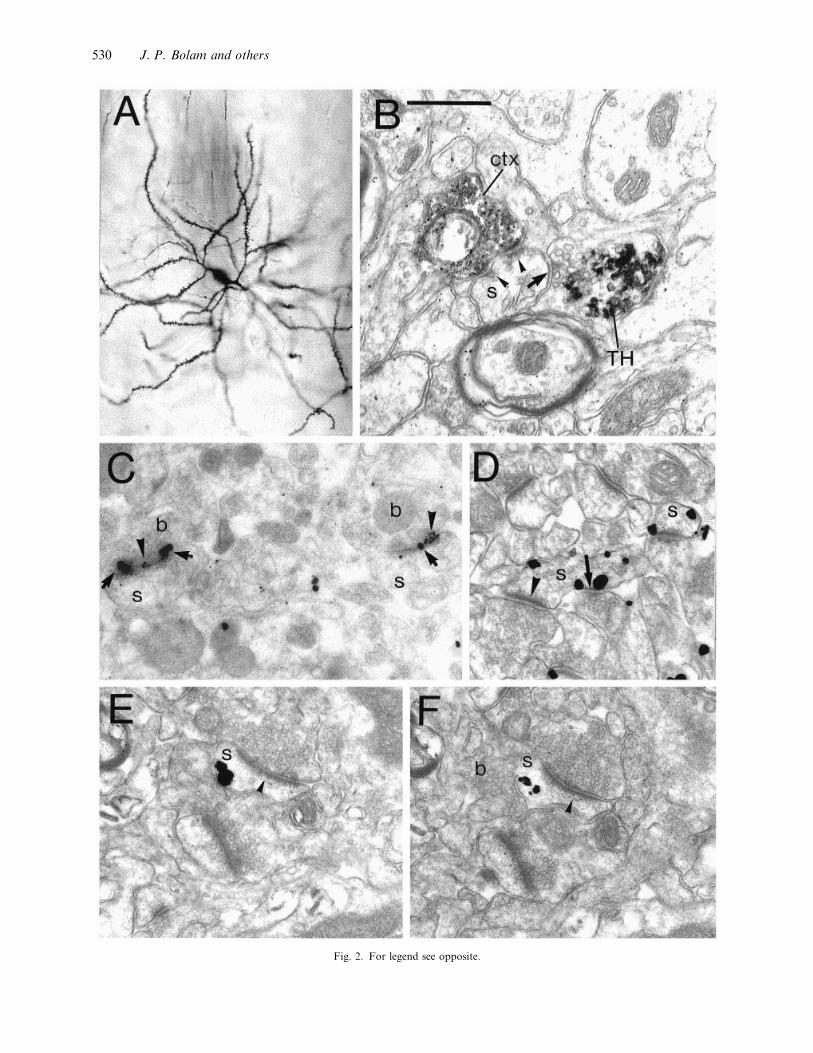
Fig. 2. For legend see opposite.
530 J. P. Bolam and others

Bolam, 1997). The head of spines that receive the
dopaminergic input invariably receive input from
terminals forming asymmetric synapses (Freund et al.
1984) which are generally derived from the cortex
(Fig. 2B ; Bouyer et al. 1984; Smith et al. 1994). This
anatomical arrangement is ideally suited for the
dopamine released from the nigrostriatal terminal,
which is likely to act on dopamine receptors localised
both within the synapse and at extrasynaptic sites
(Fig. 2D–F ; Yung et al. 1995), to very selectively
modulate the response to the excitatory input at the
head of the spine. Other inputs to spiny neurons, e.g.
cholinergic input, exhibit a similar anatomical or-
ganisation (Izzo & Bolam, 1988; Pickel & Chan, 1990)
and GABAergic terminals are also observed in contact
with the necks of spines (Bolam et al. 1985).
The synaptology of the direct and indirect pathways
downstream of the striatum is essentially as indicated
in Figure 1 (see Smith et al. 1998 for a detailed
bibliography). Thus spiny neurons giving rise to the
direct pathway make direct synaptic contact with
neurons of the EP (or GPi; Moriizumi et al. 1987;
Bolam & Smith, 1992; Bevan et al. 1994; Shink &
Smith, 1995), the majority of which are output
neurons (Carter & Fibiger, 1978), and output neurons
of the substantia nigra reticulata (Somogyi et al. 1979;
Fig. 2. (A) Light micrograph of a Golgi impregnated medium size spiny neuron in the striatum of a rat. Note the medium sized perikaryon
(approximately 15 µm in diameter), the spine-free proximal dendrites and the densely spiny secondary and higher order dendrites. (B)
Convergence of cortical and dopaminergic terminals at the level of an individual spine in the striatum. Electron micrograph of a dendritic
spine (s) in the putamen of a squirrel monkey. The spine is postsynaptic to a terminal that forms an asymmetric specialisation (arrowheads)
and is anterogradely labelled from the motor cortex (ctx) (anterograde tracer : biocytin, revealed using the peroxidase method and
diaminobenzidine (DAB) as the chromogen). The terminal contains the characteristic electron dense and amorphous DAB reaction product.
The spine is also postsynaptic to a terminal (TH) that forms symmetric specialisations (arrow) and is immunoreactive for tyrosine hydroxylase
thus identifying it as a nigrostriatal dopaminergic terminal. The tyrosine hydroxylase immunoreactive sites were identified by an immuno-
peroxidase method using benzidine dihydrochloride as the chromogen which produces an irregular and more electron dense reaction product.
(Data from Smith et al. 1994). (C) Colocalisation of the NR1 subunit of the NMDA receptor and the GluR2}3 subunit of the AMPA
receptor at synapses in the striatum. Immunoreactive sites were revealed by a postembedding immunogold method with silver intensification
on freeze-substituted Lowicryl-embedded sections. The NR1 subunit was detected using a monoclonal antibody NR1 and secondary antibody
coupled to 10 nm diameter colloidal gold (large immunoparticles ; arrows). The GluR2}3 subunits were identified with a rabbit antibody and
a secondary antibody coupled to 1±4 nm diameter colloidal gold (small immunoparticles ; arrowheads). The 2 asymmetric axospinous synapses
illustrated (which are probably derived from the cortex) are positive for both the AMPA and NMDA receptor subunits. b, boutons; s, spine.
(Data from Bernard & Bolam, 1998). (D) Localisation of the D1 subtype of the dopamine receptor in the striatum. Immunoreactive sites
were identified by the pre-embedding immunogold method (with silver enhancement). Two D1-immunolabelled spines (s) both of which
receive asymmetric synaptic input at the head (indicated by an arrowhead in the one on the left) from a terminal that is probably derived
from the cortex. One of the spines also receives input (arrow) from a bouton that forms a symmetric synapse that is associated with
immunogold labelling denoting D1 receptor. This bouton has the morphological characteristics of a dopaminergic nigrostriatal terminal.
(Data from Yung et al. 1995). (E, F) Localisation of the D2 subtype of the dopamine receptor in the striatum. Immunoreactive sites were
identified by the pre-embedding immunogold method (with silver enhancement). Serial sections of a spine (s) that receives synaptic input
from a terminal forming an asymmetric synaptic specialisation (arrowheads) (probably derived from the cortex). A second bouton that
similar in morphological characteristics to a dopaminergic nigrostriatal bouton (labelled b in F) closely apposes the spine but a synaptic
specialisation was not observed. Immunogold particles are associated with the membrane apposed to the bouton indicating the presence of
D2 receptor. Note that in F the silver-intensified immunogold particles are at their periphery and no longer touching the membrane. (Data
from Yung et al. 1995). Bar in B applies to all electron micrographs: 0±5 µm.
Williams & Faull, 1985; Smith & Bolam, 1991; Bolam
et al. 1993). The release of GABA following their
stimulation will therefore lead to an inhibition of the
output neurons of the basal ganglia and thus a
disinhibition of the targets of the basal ganglia.
Similarly, spiny neurons giving rise to the indirect
pathway make direct synaptic contact with neurons of
the GP (Chang et al. 1981; Totterdell et al. 1984),
these neurons make synaptic contact with the neurons
of the STN (Smith et al. 1990; Shink et al. 1996) which
in turn innervate neurons of the output nuclei (Bevan
et al. 1994; Bevan & Bolam, 1995; Shink & Smith,
1995). Neurons of the GP also directly contact the
output neurons of the basal ganglia (Smith & Bolam,
1989; Kincaid et al. 1991). Thus inhibition of neurons
of the GP, which themselves are also GABAergic, by
the increased activity of striatal spiny neurons will
lead to increased firing of the output neurons by 2
mechanisms. First, the loss of inhibitory input to the
excitatory neurons of the STN will lead to increased
activity and hence increased excitation of the output
neurons. Secondly, the inhibition of GP neurons will
have a direct disinhibitory effect on the output
neurons. The consequence of the increased firing of
the output neurons will be increased inhibition of
neurons in the targets of the basal ganglia. Thus, in
this greatly simplified scheme of the functional
organisation of the basal ganglia, the synaptic
organisation of the cortico–striato–fugal pathways
are such that it offers simple possible explanations for
Synaptic organisation of the basal ganglia 531
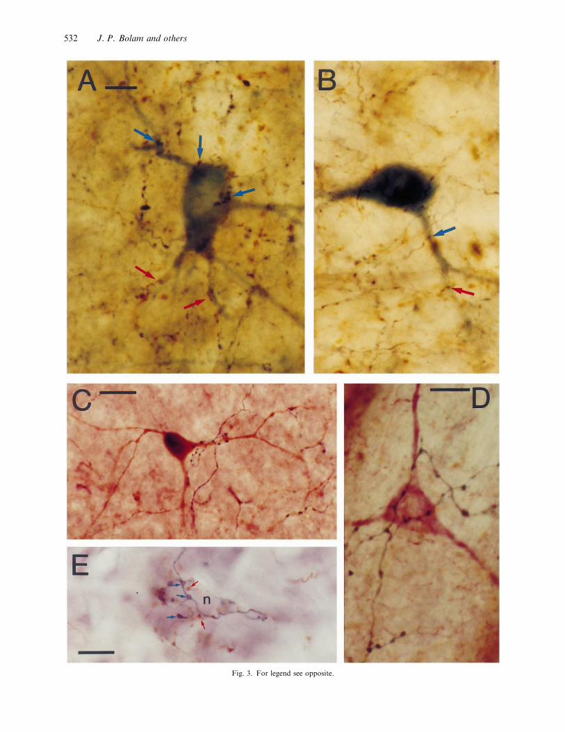
Fig. 3. For legend see opposite.
532 J. P. Bolam and others

the increased and}or decreased firing of the output
neurons of the basal ganglia and the neurons in the
targets of basal ganglia. (For detailed discussions of
the anatomical and functional organisation of the
basal ganglia see reviews by Gerfen & Wilson, 1996
and Smith et al. 1998).
In the following sections, recently discovered ana-
tomical features of the organisation of the basal
ganglia that are likely to have a bearing on our
understanding of the transmission of cortical in-
formation will be discussed.
Striatal GABAergic interneurons were originally
identified on the basis of the selective uptake of
exogenous [$H]GABA (Iversen & Schon, 1973; Bolam
et al. 1983) and glutamate decarboxylase immuno-
cytochemistry (Ribak et al. 1979; Bolam et al. 1985;
Kita & Kitai, 1988). There are several subtypes of
GABA interneurons (Kubota et al. 1993; Kawaguchi,
1997) but the largest population is characterised by
the presence of calcium binding protein, parvalbumin
(PV) (Fig. 3A–C ; Cowan et al. 1990; Kita et al. 1990;
Kita, 1993). These neurons account for only a small
proportion of the total population of striatal neurons,
possess smooth spine-free dendrites, have the ability
to fire at high rates (Kawaguchi, 1993) and are
interconnected by gap junctions (Kita, 1993; Koos &
Tepper, 1999). Electron microscopic analysis has
revealed that these cells receive synaptic input from
many terminals that form asymmetric synaptic special-
isations (Kita et al. 1990; Bolam et al. 1983, 1985), i.e.
the type of synapses associated with the corticostriatal
projection (see above). Indeed, anterograde tracing
studies have demonstrated that one of the major
Fig. 3. (A, B) Convergence of motor and sensory corticostriatal fibres on GABA interneurons in the rat striatum. Animals received injections
of Phaseolus vulgaris-leucoagglutinin in the motor cortex (M1) and biotinylated dextran amine in the sensory cortex (S1). The striatum was
then processed to reveal the anterogradely corticostriatal fibres with different reaction products (blue}black nickel enhanced DAB for motor
cortical fibres (blue arrows) and brown DAB for sensory cortical fibres (red arrows)) and PV immunoreactive neurons (using Vector SG as
the chromogen). Parvalbumin is a marker for the major population of GABA interneurons in the striatum. The GABA neurons are located
in a region of striatum where there is overlap between the motor cortical and sensory cortical fibres, and are closely apposed by terminals
derived from both regions of cortex (arrows). (Unpublished data, J. J. Hanley, J.-M. Deniau and J. P. Bolam). Bar, 20 µm. (C) Selective
innervation of GABA interneurons by pallidostriatal neurons. A parvalbumin-positive GABA interneuron in the striatum revealed by the
brown reaction product formed by using DAB as the chromogen for the peroxidase reaction. A pallidostriatal axon arising from the neuron
in Fig. 4B revealed by blue}black reaction product (nickel-enhanced DAB) forms many contacts with the proximal dendrites of the
interneuron. Bar, 20 µm. (Data from Bevan et al. 1998). (D) Selective innervation of nitric oxide-positive interneurons by pallidostriatal
neurons. A nitric oxide-positive interneuron in the striatum revealed by the purple reaction product formed by using Vector VIP as the
chromogen for the peroxidase reaction. A pallidostriatal axon arising from the neuron in Fig. 4B revealed by blue}black reaction product
(nickel-enhanced DAB) forms many contacts with the perikaryon of the interneuron. Bar, 10 µm. (Data from Bevan et al. 1998). (E)
Convergence of afferents from the globus pallidus and ventral pallidum on basal ganglia output neurons. An unlabelled neuron in the
substantia nigra pars reticulata apposed by boutons labelled with PHA-L which was anterogradely transported from the ventral pallidum
and revealed using nickel-enhanced DAB as the chromogen for the peroxidase reaction (blue-black boutons, indicated by blue arrows) and
boutons labelled with BDA which was anterogradely transported from the globus pallidus and visualised using DAB as the chromogen
(brown boutons indicated by red arrows). Bar, 10 µm. (Data from Bevan et al. 1996).
inputs to PV-positive, GABA interneurons is from the
cortex (Lapper et al. 1992; Bennett & Bolam, 1994).
As with the cortical input to spiny neurons the input
to GABA interneurons is associated with AMPA
receptors localised within the synaptic specialisation
(Bernard et al. 1997). It is interesting to note that
following cortical stimulation these neurons increase
expression of Fos over a larger area of striatum than
do spiny neurons (Parthasarathy & Graybiel, 1997).
Furthermore, these neurons may subserve some role
in the integration of cortical afferents from different
functional territories as preliminary light microscopic
analyses in double anterograde tracing studies suggest
that an individual neuron may receive input from
both motor and sensory regions of cortex (Fig. 3A, B ;
J. J. Hanley, J.-M. Deniau and J. P. Bolam, un-
published observations). Indeed, in individual sections
up to 25% of all PV-positive interneurons were found
to be apposed by boutons derived from both motor
and sensory regions of cortex. Furthermore, up to
50% of those interneurons that were identified as
receiving cortical input were apposed by terminals
from both the motor and sensory regions of cortex.
The major synaptic target of the GABA inter-
neurons are spiny output neurons (Kita et al. 1990;
Bennett & Bolam, 1994). Individual spiny neurons
receive a basket-like innervation around their peri-
karya from PV-positive terminals, the majority of
which are likely to be derived from the PV-positive
GABA interneurons. On the basis of this anatomical
organisation and the failure to detect inhibitory
signals mediated by the collaterals of spiny neurons
(Jaeger et al. 1994) despite the presence of synapses
(Wilson & Groves, 1980; Somogyi et al. 1981a ; Yung
et al. 1996), it has been proposed that the GABA
interneurons are the principal mediators of lateral
Synaptic organisation of the basal ganglia 533

inhibition in the striatum and provide a feed-forward
inhibition of cortical information to spiny neurons
(Pennartz & Kitai, 1991; Jaeger et al. 1994; Plenz &
Kitai, 1998). Indeed, paired recordings in vitro have
shown that GABA interneurons produce large unitary
IPSPs in spiny neurons (Koos & Tepper, 1999).
Although the precise role of this inhibition is
unknown, there are several possibilities. First, the
effect may simply be to shunt coincident cortical
excitation and limit the duration of excitation.
Secondly, depending on their pattern of connectivity,
the interneurons may underlie surround inhibition
thereby focusing cortical excitation (Parthasarathy &
Graybiel, 1997). Thirdly, by their dense local axonal
arbors they might synchronise sub- and supra-
threshold activity of groups of neighbouring spiny
output neurons (Koos & Tepper, 1999).
It is thus evident that the PV-positive GABA
interneurons receive cortical input and, despite their
relatively low numbers, are in a position to powerfully
control the activity of neurons giving rise to the direct
and indirect pathways and thus the output of the
striatum.
The main synaptic targets of spiny neurons that give
rise to the indirect pathway are the GABAergic
neurons of the GP; they are thus key structures of the
circuitry underlying the indirect pathway. The GP in
turn projects to the STN, the output nuclei of the
basal ganglia and the SNC. The results of tracing
studies at the electron microscope level combined with
postembedding immunolabelling for glutamate and
GABA, suggest that individual neurons of the GP
innervate the STN and output structures of the basal
ganglia (see Bolam et al. 1993; Smith et al. 1998).
Tracing and physiological studies have also indicated
that the GP in addition, provides a feedback to the
striatum (for references see Bevan et al. 1998). Single
cell filling studies have confirmed these suggestions
(Fig. 4; Kita & Kitai, 1994; Bevan et al. 1998). All
pallidal neurons give rise to local axon collaterals
within the GP and collalteral projections to the STN,
EP and SN. About a quarter of pallidal neurons also
give rise to collateral projections to the striatum (Fig.
4; Kita & Kitai, 1994; Bevan et al. 1998). On average,
pallidostriatal neurons give rise to 790.6 boutons
within the striatum. From the known number of
neurons in the striatum and GP (2±79¬10' and
4±6¬10% respectively ; Oorschot, 1996) and the pro-
portion of pallidal neurons giving rise to striatal
collaterals, it can be calculated that on average a
striatal neuron will receive input from 3±3 pallidal
boutons. It is unlikely that such a small number of
synapses in a projection can impart significant
information on, or significantly affect, the function of
the striatum. However, combination of single-cell
filling with immunolabelling for subpopulations of
striatal neurons has revealed that pallidostriatal axons
selectively innervate striatal interneurons (Bevan et al.
1998). Up to 60% (mean³.. : 43±7³17±8) of the
striatal terminals of an individual pallidostriatal
neuron make contact with PV-positive GABA inter-
neurons (Fig. 3C). An individual PV-positive neuron
receives on average 6.7 boutons from an individual
pallidal axon and these make contact primarily in the
proximal regions of the neurons. In addition, 3–32%
of terminals of a single pallidal neuron make contact
with nitric oxide synthase (NOS)-positive inter-
neurons (Fig. 3D). The synaptic target of the
remainder of the pallidal boutons is at present
unknown. A quantitative model of the connectivity
between pallidal neurons and GABA interneurons,
assuming similar patterns of connectivity, suggests
that each GABA interneuron receives input on
average from 7±1 pallidal neurons that give rise to a
total of 47±6 synaptic boutons (Table).
Thus despite the relatively small number of neurons
and boutons that comprise the pallidostriatal path-
way, pallidostriatal neurons are in a position to
powerfully control the activity of the striatum by
selective innervation of PV-positive GABA inter-
neurons which in turn control the activity of the
output neurons of the striatum. Since GP neurons
receive monosynaptic and}or rapid disynaptic ac-
tivation (via the STN; Tremblay & Filion, 1989; Ryan
& Clark, 1991; Kita, 1992; Naito & Kita, 1994; Plenz
& Kitai, 1998), following cortical activation they are
well placed to modulate the cortical activation of PV
interneurons (Pennartz & Kitai, 1991; Plenz & Kitai,
1998) through shunting of coincidental cortical ex-
citatory postsynaptic potentials and}or phase-lock
action potential generation (see Pennartz & Kitai,
1991; Cobb et al. 1995). The total number and
placement of GP terminals on PV-positive GABA
interneurons when compared with studies of similar
unitary inhibitory connections suggest that they might
powerfully shunt excitatory inputs, phase-lock or
prevent action potential generation (Cobb et al. 1995).
The same pallidostriatal neurons that innervate PV
interneurons also provide a major input to NOS
interneurons which themselves are likely to regulate
striatal activity through the release of GABA (Kubota
et al. 1993), nitric oxide (Hanbauer et al. 1992;
534 J. P. Bolam and others

Table 1. Quantitative model of the connectivity between pallidostriatal neurons and GABA interneurons in the striatum
1. Number of striatal boutons arising from pallidostriatal neurons 790.6³404.2
2. Average proportion of pallidal boutons in contact with GABA interneurons 43.7³17.8%
3. Number of boutons of 1 pallidal neuron in contact with GABA interneurons (43.7% of 790.6) 345.5
4. Number of boutons of 1 pallidal neuron contacting 1 GABA interneuron 6.7
5. Number of GABA interneurons innervated by 1 pallidal neuron (3 divided by 4) 51.6
6. Total number GP neurons projecting to striatum (25% of neurons in GP) 11500
7. Theoretical total number of GABA interneurons innervated by pallidostriatal neurons (5 multiplied by 6) 593400
8. Number of GABA interneurons in striatum (3% of total number of neurons in striatum*) 83700
9. Number of pallidostriatal neurons innervating one GABA interneuron (7 divided 8) 7.1
10. Number of pallidostriatal boutons in contact with one GABA interneuron (8 multiplied by 4) 47.6
The values in 1, 2 and 3 are from Bevan et al. (1998). The total number of neurons in the GP (4.6¬10%) and total number in the striatum
(2.79¬10') are from Oorschot (1996).
* Estimate from Kita et al. (1990).
Guevara-Guzman et al. 1994; Lonart & Johnson,
1994; Stewart et al. 1996) and neuropeptides (Radke
et al. 1993).
The classic view of the organisation of the basal
ganglia is that the functionally diverse information
arising from the cerebral cortex is processed in the
striatum and subsequent divisions of the basal ganglia
by parallel and segregated cortical-basal ganglia-
thalamocortical loops (Alexander et al. 1986; Alex-
ander & Crutcher, 1990; Hoover & Strick, 1993;
Groenewegen & Berendse, 1994; Joel & Weiner, 1994,
1997). However, it is clear that the basal ganglia
integrate functionally diverse information derived
from different cortical regions to generate context
dependent, goal-directed patterns of behaviour (Wurtz
& Hikosaka, 1986; Graybiel et al. 1994; Aosaki et al.
1995; Schultz et al. 1995, 1997). Anatomical analyses
have identified several neuronal elements or systems
which could provide the morphological basis of such
integration within the basal ganglia. These include the
local circuit neurons of the striatum (Gerfen, 1984;
Chesselet & Graybiel, 1986; Kubota & Kawaguchi,
1993; Bolam & Bennett, 1995; Kawaguchi et al.
1995), the ascending projections of midbrain dopa-
mine neurons (Somogyi et al. 1981b ; Nauta & Dome-
sick, 1984; Gerfen et al. 1987; Jimenez-Castellanos
& Graybiel, 1987), the GPi output to the pedunculo-
pontine nucleus (Shink et al. 1997) and open-
interconnected cortico-basal ganglia-thalamocortical
loops (Joel & Weiner, 1994, 1997). It has recently been
demonstrated that the circuitry of the indirect path-
way may underlie this type of integration at the
synaptic level and in particular the synaptic or-
ganisation of the descending projections of neurons of
the GP and its ventral equivalent the ventral pallidum
(VP; Bevan et al. 1996, 1997). In the output nuclei,
pallidal neurons give rise to large synaptic boutons
that selectively innervate the proximal regions of basal
ganglia output neurons, often in a basket-like manner
(see Smith et al. 1998). This is also the case in the
subthalamic nucleus although the terminals are more
distributed across the somatodendritic trees of STN
neurons. The descending projections of the VP, which
largely receive limbic cortical information via the
nucleus accumbens (Alexander et al. 1986; Alexander
& Crutcher, 1990; Groenewegen & Berendse, 1994)
and the GP, which receives mostly sensorimotor and
associative cortical information via the striatum
(Alexander et al. 1986; Alexander & Crutcher, 1990;
Hoover & Strick, 1993; Groenewegen & Berendse,
1994; Joel & Weiner, 1994, 1997) give rise to
topographically segregated fields of anterogradely
labelled terminals in the output nuclei and the STN.
However, double anterograde tracing from the 2
divisions of the pallidal complex in individual animals
has revealed, in addition to topographically segre-
gated projections, zones of overlap of the 2 projections
(Bevan et al. 1996, 1997). Electron microscopy
demonstrated that in the regions of overlap in each
nucleus the proximal parts of many neurons, including
tyrosine hydroxylase-immunopositive neurons (i.e.
dopaminergic) in the SNc, receive convergent synaptic
input from both the VP and GP (Bevan et al. 1996,
1997; Fig. 3E). Thus individual output neurons of the
basal ganglia as well as neurons of the STN and
dopaminergic neurons receive convergent input from
pallidal afferents carrying motor}associative infor-
mation and limbic information. Another way by
which basal ganglia output neurons and STN neurons
may integrate functionally diverse information from
the pallidal complex is via their dendrites as they also
Synaptic organisation of the basal ganglia 535

(a)
Fig. 4A. For legend see page 538.
536 J. P. Bolam and others

(b)
Fig. 4B. For legend see page 538.
Synaptic organisation of the basal ganglia 537

receive pallidal inputs (Smith & Bolam, 1989; Bolam
& Smith, 1992; Bevan et al. 1996, 1997) and are often
oriented to cross the functional boundaries defined by
inputs from different functional divisions of the
pallidal complex (Grofova et al. 1982; Kita et al.
1983; Nakanishi et al. 1991; Bevan et al. 1997).
1. Cortical input to the basal ganglia, carried by the
corticostriatal pathway, imparts functionality on the
basal ganglia.
2. The cortical information is received primarily by
the spiny projection neurons whose function is to
transmit the information via 2 pathways through the
basal ganglia to the output nuclei.
3. The basal ganglia influence behaviour by the
output nuclei projecting to the ventral thalamus and
thence motor and premotor regions of cortex or by
projecting to subcortical premotor regions.
4. Cortical input is also received by GABA
interneurons which provide feed-forward inhibition of
spiny neurons. These neurons may ‘limit’ cortical
excitation of spiny neurons, may ‘focus’ the cortical
input to the striatum and}or may synchronise the
activity of spiny neurons.
5. A subpopulation of neurons of the GP, in
addition to being key components of the indirect
pathway and innervating the STN, EP and SN, also
provide a powerful inhibitory feedback to the striatum
that is in a position to modify the flow of cortical
information through the basal ganglia.
6. The ‘power’ of the pallidostriatal pathway lies in
the fact that pallidostriatal neurons selectively in-
nervate GABA interneurons which in turn, innervate
spiny neurons. Thus by inhibiting or synchronising
the activity of GABA interneurons which in turn
inhibit or synchronise the activity of spiny neurons,
Fig. 4. (A) Drawing tube reconstructions of 2 neurons in the GP of a rat (animal no. 9665) that were labelled in vivo with neurobiotin by
the juxtacellular method and revealed by the peroxidase method using DAB as the chromogen (Bevan et al. 1998). The cells have been
reconstructed along the rostrocaudal axis but the drawings have been bisected approximately at the level of the internal capsule (a connects
to b). The cell bodies and dendrites are shown in red, the axon and boutons in black and regions of the axonal arbors in the STN (484
boutons) and SNr (50 boutons) that could not be unequivocally ascribed to one or other of the neurons are shown in grey. The upper of
the 2 neurons did not give rise to a projection to the striatum but gave rise to local terminations in the GP (92 boutons) and projected to
all caudal basal ganglia nuclei : EP (108 boutons), STN (184 boutons), SNr (209 boutons) and SNc (16 boutons). The lower of the 2 neurons
gave rise to a projection to the striatum (478 boutons) as well as local collaterals in the GP (233 boutons), the STN (274 boutons), the SNc
(11 boutons ) and probably also the SNr. (B) Two pallidal cells from another animal (no. 9671) reconstructed and represented as above. The
area of overlap of the arbors are shown in grey and contain 407 boutons in the STN and 157 in the EP. The upper cell gave rise to a projection
to the STR (851 boutons; see Figs 3C, D), the other nulcei of the basal ganglia, EP (130 boutons), STN (41 boutons), SNr (36 boutons),
SNc (28 boutons) as well as local collaterals in the GP (140 boutons). The lower cell did not innervate the striatum but gave rise to local
collaterals in the GP (294 boutons) and projected to caudal basal ganglia nuclei (STN; 60 boutons; SNr; 159 boutons), except the SNc. It
showed no clear projection to the EP, this however cannot be ruled out, as the undertermined projection (grey axon) to the EP could be from
this cell. (Unpublished data, M. D. Bevan, P. A. C. Booth and J. P. Bolam and from Bevan et al. 1998). Bars, 300 µm.
they are in the position to modulate the flow of
cortical information through the basal ganglia.
7. Neurons of different functional territories of the
pallidal complex may subserve an integrative role by
making convergent synaptic contacts with individual
neurons to the output nuclei of the basal ganglia and
the STN and SNC.
8. Finally, although the essential concept of the
direct and indirect pathways remains intact, the
findings summarised here suggest that the indirect
pathway is much more complex than previously
described and is likely to profoundly influence the
flow of cortical information through the basal ganglia.
The author’s work described in this review was
supported by the Medical Research Council UK; the
Wellcome Trust (MDB; Wellcome Trust Advanced
Training Fellowship 046613}Z}96}Z) and The Euro-
pean Community (JPB and JJH; BIOMED 2 Project :
BMH4-CT-97-2215). The authors thank Caroline
Francis, Liz Norman and Paul Jays for technical
assistance.
ALBIN RL, YOUNG AB, PENNEY JB (1989) The functional
anatomy of basal ganglia disorders. Trends in Neurosciences 12,
366–375.
ALEXANDER GE, DELONG MR, STRICK PL (1986) Parallel
organization of functionally segregated circuits linking basal
ganglia and cortex. Annual Review of Neuroscience 9, 357–381.
ALEXANDER GE, CRUTCHER ME (1990) Functional archi-
tecture of basal ganglia circuits : neural substrates of parallel
processing. Trends in Neurosciences 13, 266–271.
AOSAKI T, KIMURA M, GRAYBIEL AM (1995) Temporal and
spatial characteristics of tonically active neurons of the primate’s
striatum. Journal of Neurophysiology 73, 1234–1252.
ARONIN N, DIFIGLIA M, LIOTTA AS, MARTIN JB (1981)
538 J. P. Bolam and others

Ultrastructural localization and biochemical features of immuno-
reactive leu-enkephalin in monkey dorsal horn. Journal of
Neuroscience 1, 561–577.
BENNETT BD, BOLAM JP (1994) Synaptic input and output of
parvalbumin-immunoreactive neurones in the neostriatum of the
rat. Neuroscience 62, 707–719.
BERNARD V, SOMOGYI P, BOLAM JP (1997) Cellular,
subcellular, and subsynaptic distribution of AMPA-type gluta-
mate receptor subunits in the neostriatum of the rat. Journal of
Neuroscience 17, 819–833.
BERNARD V, BOLAM JP (1998) Subcellular and subsynaptic
distribution of the NR1 subunit of the NMDA receptor in the
neostriatum and globus pallidus of the rat : co-localization at
synapses with the GluR2}3 subunit of the AMPA receptor.
European Journal of Neuroscience 10, 3721–3736.
BEVAN MD, BOLAM JP (1995) Cholinergic, GABAergic and
glutamate-enriched inputs from the mesopontine tegmentum to
the subthalamic nucleus in monkeys. Journal of Neuroscience 15,
7105–7120.
BEVAN MD, CROSSMAN AR, BOLAM JP (1994) Neurons
projecting from the entopeduncular nucleus to the thalamus
receive convergent synaptic inputs from the subthalamic nucleus
and the neostriatum. Brain Research 659, 99–109.
BEVAN MD, SMITH AD, BOLAM JP (1996) The substantia
nigra as a site of synaptic integration of functionally diverse
information arising from the ventral pallidum and the globus
pallidus in the rat. Neuroscience 75, 5–12.
BEVAN MD, CLARKE NP, BOLAM JP (1997) Synaptic
integration of functionally diverse pallidal information in the
entopeduncular nucleus and subthalamic nucleus in the rat.
Journal of Neuroscience 17, 308–324.
BEVAN MD, BOOTH PAC, EATON SA, BOLAM JP (1998)
Selective innervation of neostriatal interneurons by a subclass of
neuron in the globus pallidus of the rat. Journal of Neuroscience
18, 9438–9452.
BEVAN MD, WILSON CJ (1999) Mechanisms underlying spon-
taneous oscillation and rhythmic firing in rat subthalamic
neurons. Journal of Neuroscience 19, 7617–7628.
BOLAM JP, CLARKE DJ, SMITH AD, SOMOGYI P (1983) A
type of aspiny neuron in the rat neostriatum accumulates ($H) γ
aminobutyric acid: combination of Golgi-staining, autoradio-
graphy and electron microscopy. Journal of Comparative Neur-
ology 213, 121–134.
BOLAM JP, SOMOGYI P, TAKAGI H, FODOR I, SMITH AD
(1983) Localization of substance P-like immunoreactivity in
neurons and nerve terminals in the neostriatum of the rat : a
correlated light and electron microscopic study. Journal of
Neurocytology 12, 325–344.
BOLAM JP, POWELL JF, WU J-Y, SMITH AD (1985) Glutamate
decarboxylase-immunoreactive structures in the rat neostriatum.
A correlated light and electron microscopic study including a
combination of Golgi-impregnation with immunocytochemistry.
Journal of Comparative Neurology 237, 1–20.
BOLAM JP, IZZO PN (1988) The postsynaptic targets of substance
P-immunoreactive terminals in the rat neostriatum with par-
ticular reference to identified spiny striatonigral neurons. Experi-
mental Brain Research 70, 361–377.
BOLAM JP, SMITH Y (1992) The striatum and the globus pallidus
send convergent synaptic inputs onto single cells in the
entopeduncular nucleus of the rat : a double anterograde labeling
study combined with post-embedding immunocytochemistry for
GABA. Journal of Comparative Neurology 321, 456–476.
BOLAM JP, SMITH Y, INGHAM CA, VON-KROSIGK M &
SMITH AD (1993) Convergence of synaptic terminals from the
striatum and the globus pallidus onto single neurones in the
substantia nigra and the entopeduncular nucleus. In Chemical
Signalling in the Basal Ganglia (ed. Arbuthnott GW, Emson PC).
Progress in Brain Research 99, 73–88. Oxford: Elsevier.
BOLAM JP, BENNETT B (1995) The microcircuitry of the
neostriatum. In Molecular and Cellular Mechanisms of Neostriatal
Functions (ed. Ariano M, Surmeier DJ), pp. 1–19. Austin, TX:
R. G. Landes.
BOUYER JJ, MILLER RJ, PICKEL VM (1984a) Ultrastructural
relation between cortical efferents and terminals containing
enkephalin-like innunoreactivity in rat neostriatum. Regulatory
Peptides 8, 105–115.
BOUYER JJ, PARK DH, JOH TH, PICKEL VM (1984b)
Chemical and structural analysis of the relation between cortical
inputs and tyrosine hydroxylase-containing terminals in rat
neostriatum. Brain Research 302, 267–275.
CARTER DA, FIBIGER HC (1978) The projections of the
entopeduncular nucleus and globus pallidus in rat as demo-
nstrated by autoradiography and horseradish peroxidase histo-
chemistry. Journal of Comparative Neurology 177, 113–121.
CHANG HT, WILSON CJ, KITAI ST (1981) Single neostriatal
efferent axons in the globus pallidus : a light and electron
microscopic study. Science 213, 915–918.
CHANG HT, WILSON CJ (1990) Anatomical analysis of
electrophysiologically characterized in the rat strio-pallidal
system. In Analysis of Neuronal Microcircuits and Synaptic
Interactions (ed. Bjo$ rklund A, Ho$ kfelt T, Wouterlood F, van
den Pol A). Handbook of Chemical Neuroanatomy 8, 351–402.
Amsterdam: Elsevier Biomedical
CHESSELET MF, GRAYBIEL AM (1986) Striatal neurons
expressing somatostatin-like immunoreactivity : evidence for a
peptidergic interneuronal system in the cat. Neuroscience 17,
547–571.
CHEVALIER G, DENIAU J-M (1990) Disinhibition as a basic
process in the expression of striatal functions. Trends in
Neuroscience 13, 277–280.
COBB SR, BUHL EH, HALASY K, PAULSEN O, SOMOGYI P
(1995) Synchronization of neuronal activity in hippocampus by
individual GABAergic interneurons. Nature 378, 75–78.
COWAN RL, WILSON CJ, EMSON PC, HEIZMANN CW
(1990) Parvalbumin-containing GABAergic interneurons in the
rat neostriatum. Journal of Comparative Neurology 302, 197–205.
DELONG MR (1990) Primate models of movement disorders of
basal ganglia origin. Trends in Neuroscience 13, 281–285.
DIFIGLIA M, ARONIN N, MARTIN JB (1982) Light and
electron microscopic localization of immunoreactive leu-enkeph-
alin in the monkey basal ganglia. Journal of Neuroscience 2,
303–320.
DUBE! L, SMITH AD, BOLAM JP (1988) Identification of
synaptic terminals of thalamic or cortical origin in contact with
distinct medium size spiny neurons in the rat neostriatum.
Journal of Comparative Neurology 267, 455–471.
FREUND TF, POWELL J, SMITH AD (1984) Tyrosine hydro-
xylase-immunoreactive boutons in synaptic contact with identi-
fied striatonigral neurons, with particular reference to dendritic
spines. Neuroscience 13, 1189–1215.
FROTSCHER M, RINNE U, HASSLER R, WAGNER A (1981)
Termination of cortical afferents on identified neurons in the
caudate nucleus of the cat. A combined Golgi-EM degeneration
study. Experimental Brain Research 41, 329–337.
GERFEN CR (1984) The neostriatal mosaic : compartmental-
ization of corticostriatal input and striatonigral output systems.
Nature 311, 461–464.
GERFEN CR, HERKENHAM M, THIBAULT J (1987) The
neostriatal mosaic : II. patch- and matrix-directed mesostriatal
dopaminergic and non-dopaminergic systems. Journal of Neuro-
science 7, 3915–3934.
GERFEN CR, WILSON CJ (1996). In The Basal Ganglia (ed.
Bjo$ rklund A, Ho$ kfelt T, Swanson L). Handbook of Chemical
Neuroanatomy, Integrated Systems of the CNS, Part III, pp.
369–466. Amsterdam: Elsevier Science.
GRAYBIEL AM, AOSAKI T, FLAHERTY AW, KIMURA M
Synaptic organisation of the basal ganglia 539

(1994) The basal ganglia and adaptive motor control. Science
265, 1826–1831.
GROENEWEGEN HJ, BERENDSE HW (1994) Anatomical
relationships between the prefrontal cortex and the basal ganglia
in the rat. In Motor and Cognitive Functions of the Prefrontal
Cortex (ed. Thierry A-M, Glowinski J, Goldman-Rakic PS,
Christen Y), pp. 52–76. Berlin: Springer.
GROFOVA I, DENIAU JM, KITAI ST (1982) Morphology of the
substantia nigra pars reticulata projection neurons intracellularly
labeled with HRP. Journal of Comparative Neurology 208,
352–368.
GUEVARA-GUZMAN R, EMSON PC, KENDRICK KM (1994)
Modulation of in vivo striatal transmitter release by nitric oxide
and cyclic GMP. Journal of Neurochemistry 62, 807–810.
HANBAUER I, WINK D, OSAWA Y, EDELMAN GM, GALLY
JA (1992) Role of nitric oxide in NMDA-evoked release of [$H]-
dopamine from striatal slices. NeuroReport 3, 409–412.
HANLEY JJ, BOLAM JP (1997) Synaptology of the nigrostriatal
projection in relation to the compartmental organization of the
neostriatum in the rat. Neuroscience 81, 353–370.
HERSCH SM, CILIAX BJ, GUTEKUNST CA, REES HD,
HEILMAN CJ, YUNG KKL et al. (1995) Electron microscopic
analysis of D1 and D2 dopamine receptor proteins in the dorsal
striatum and their synaptic relationships with motor corti-
costriatal afferents. Journal of Neuroscience 15, 5222–5237.
HOOVER JE, STRICK PL (1993) Multiple output channels in the
basal ganglia. Science 259, 819–821.
IVERSEN LL, SCHON FE (1973) The use of autoradiographic
techniques for the identification and mapping of transmitter-
specific neurones in CNS. In New Concepts in Neurotransmitter
Regulation (ed. Mandell AJ), pp. 153–193. New York: Plenum
Press.
IZZO PN, BOLAM JP (1988) Cholinergic synaptic input to
different parts of spiny striatonigral neurons in the rat. Journal of
Comparative Neurology 269, 219–234.
JAEGER D, KITA H, WILSON C J (1994) Surround inhibition
among projection neurons is weak or nonexistent in the rat
neostriatum. Journal of Neurophysiology 72, 2555–2558.
JIMENEZ-CASTELLANOS J, GRAYBIEL AM (1987) Sub-
divisions of the dopamine-containing A8-A9-A10 complex
identified by their differential mesostriatal innervation of stri-
osomes and extrastriosomal matrix. Neuroscience 23, 223–242.
JOEL D, WEINER I (1994) The organization of the basal ganglia-
thalamocortical circuits : open interconnected rather than closed
segregated. Neuroscience 63, 363–379.
JOEL D, WEINER I (1997) The connections of the primate
subthalamic nucleus : indirect pathways and the open-inter-
connected scheme of basal ganglia-thalamocortical circuitry.
Brain Research Reviews 23, 62–78.
KAWAGUCHI Y (1993) Physiological, morphological, and histo-
chemical characterization of three classes of interneurons in rat
neostriatum. Journal of Neuroscience 13, 4908–4923.
KAWAGUCHI Y (1997) Neostriatal cell subtypes and their
functional roles. Neuroscience Research 27, 1–8.
KAWAGUCHI Y, WILSON CJ, AUGOOD SJ, EMSON PC
(1995) Striatal interneurones: chemical, physiological and mor-
phological characterization. Trends in Neurosciences 18, 527–535.
KEMP JM, POWELL TPS (1971a) The structure of the caudate
nucleus of the cat : light and electron microscopy. Philosophical
Transactions of the Royal Society of London B 262, 383–401.
KEMP JM, POWELL TPS (1971b) The site of termination of
afferent fibres in the caudate nucleus. Philosophical Transactions
of the Royal Society of London B 262, 413–427.
KEMP JM, POWELL TPS (1971c) The termination of fibres from
the cerebral cortex and thalamus upon dendritic spines in the
caudate nucleus : a study with the Golgi mehod. Philosophical
Transactions of the Royal Society of London B 262, 429–439.
KINCAID AE, PENNEY JB, YOUNG AB, NEWMAN SW
(1991) Evidence for a projection from the globus pallidus to the
entopeduncular nucleus in the rat. Neuroscience Letters 128,
121–125.
KINCAID AE, ZHENG T, WILSON CJ (1998) Connectivity and
convergence of single corticostriatal axons. Journal of Neuro-
science 18, 4722–4731.
KITA H (1992) Responses of globus pallidus neurons to cortical
stimulation: intracellular study in the rat. Brain Research 589,
84–90.
KITA H (1993) GABAergic circuits of the striatum. In Chemical
Signalling in the Basal Ganglia (ed. Arbuthnott GW, Emson PC).
Progress in Brain Research 99, 51–72. Oxford: Elsevier.
KITA H (1996) Glutamatergic and GABAergic postsynaptic
responses of striatal spiny neurons to intrastriatal and cortical
stimulation recorded in slice preparations. Neuroscience 70,
925–940.
KITA H, CHANG HT, KITAI ST (1983) Pallidal inputs to
subthalamus: intracellular analysis. Brain Research 264, 255–265.
KITA H, KITAI ST (1988) Glutamate decarboxylase immuno-
reactive neurons in rat neostriatum: their morphological types
and populations. Brain Research 447, 346–352.
KITA H, KOSAKA T, HEIZMANN CW (1990) Parvalbumin-
immunoreactive neurons in the rat neostriatum: a light and
electron microscopic study. Brain Research 536, 1–15.
KITA H, KITAI ST (1994) The morphology of globus pallidus
projection neurons in the rat : an intracellular staining study.
Brain Research 636, 308–319.
KOOS T, TEPPER JM (1999) Inhibitory control of neostriatal
projection neurons by GABAergic interneurons. Nature Neuro-
science 2, 467–472.
KUBOTA Y, KAWAGUCHI Y (1993) Spatial distribution of
chemically identified intrinsic neurons in relation to path and
matrix compartments of rat neostriatum. Journal of Comparative
Neurology 332, 499–513.
KUBOTA Y, MIKAWA S, KAWAGUCHI Y (1993) Neostriatal
GABAergic interneurones contain NOS, calretinin or parv-
albumin. NeuroReport 5, 205–208.
LAPPER SR, SMITH Y, SADIKOT AF, PARENT A, BOLAM
JP (1992) Cortical input to parvalbumin-immunoreactive neur-
ones in the putamen of the squirrel monkey. Brain Research 580,
215–224.
LONART G, JOHNSON KM (1994) Inhibitory effects of nitric
oxide on the uptake of [$H]dopamine and [$H]glutamate by
striatal synaptosomes. Journal of Neurochemistry 63, 2108–2117.
MINK JW, THACH WT (1993) Basal ganglia intrinsic circuits and
their role in behavior. Current Opinion in Neurobiology 3,
950–957.
MORIIZUMI T, NAKAMURA Y, OKOYAMA S, KITAO Y
(1987) Synaptic organization of the cat entopeduncular nucleus
with special reference to the relationship between the afferents to
entopedunculothalamic projection neurons: an electron micro-
scope study by a combined degeneration and horseradish
peroxidase tracing technique. Neuroscience 20, 797–816.
NAITO A, KITA H (1994) The cortico-pallidal projection in the
rat : an anterograde tracing study with biotinylated dextran
amine. Brain Research 653, 251–257.
NAKANISHI H, KITA H, KITAI ST (1987) Intracellular study of
rat substantia nigra pars reticulata neurons in an in vitro slice
preparation: electrical membrane properties and response chara-
cteristics to subthalamic stimulation. Brain Research 437, 45–55.
NAKANISHI H, KITA H, KITAI ST (1991) Intracellular study of
rat entopeduncular nucleus neurons in an in vitro slice prep-
aration: response to subthalamic stimulation. Brain Research
549, 285–291.
NAUTA WJH, DOMESICK VB (1984) Afferent and efferent
relationships of the basal ganglia. In Functions of the Basal
Ganglia (ed. Evered D, O’Connor M). Ciba Foundation Sym-
posium 107, 3–23. London: Pitman.
540 J. P. Bolam and others

OORSCHOT DE (1996) Total number of neurons in the neo-
striatal, pallidal, subthalamic, and substantia nigral nuclei of the
rat basal ganglia : a stereological study using the cavalieri and
optical dissector methods. Journal of Comparative Neurology 366,
580–599.
PARTHASARATHY HB, GRAYBIEL AM (1997) Cortically
driven immediate-early gene expression reflects modular influence
of sensorimotor cortex on identified striatal neurons in the
squirrel monkey. Journal of Neuroscience 17, 2477–2491.
PASIK P, PASIK T, DIFIGLIA M (1979) The internal organi-
zation of the neostriatum in mammals. In The Neostriatum (ed.
Divac I, Oberg FGE), pp. 5–36. Oxford: Pergamon Press.
PENNARTZ CM, KITAI ST (1991) Hippocampal inputs to
identified neurons in an in vitro slice preparation of the rat
nucleus accumbens: evidence for feed-forward inhibition. Journal
of Neuroscience 11, 2838–2847.
PICKEL VM, SUMAL KK, BECKLEY SC, MILLER RJ, REIS
DJ (1980) Immunocytochemical localization of enkephalin in the
neostriatum of rat brain: a light and electron microscopic study.
Journal of Comparative Neurology 189, 721–740.
PICKEL VM, CHAN J (1990) Spiny neurons lacking choline
acetyltransferase immunoreactivity are major targets of cholin-
ergic and catecholaminergic terminals in rat striatum. Journal of
Neuroscience Research 25, 263–280.
PICKEL VM, CHAN J, SESACK SR (1992) Cellular basis for
interactions between catecholaminergic afferents and neurons
containing leu-enkephalin-like immunoreactivity in rat caudate-
putamen nuclei. Journal of Neuroscience Research 31, 212–230.
PLENZ D, KITAI ST (1998) Up and down states in striatal
medium spiny neurons simultaneously recorded with spon-
taneous activity in fast-spiking interneurons studied in cortex-
sriatum-substantia nigra organotypic cultures. Journal of Neuro-
science 18, 266–283.
RADKE JM, SPYRAKI C, THERMOS K (1993) Neuronal
release of somatostatin in the rat striatum – an in vivo
microdialysis study. Neuroscience 54, 493–498.
RIBAK CE, VAUGHN JE, ROBERTS E (1979) The GABA
neurons and their axon terminals in rat corpus striatum as
demonstrated by GAD immunocytochemistry. Journal of Com-
parative Neurology 187, 261–284.
RYAN LJ, CLARK KB (1991) The role of the subthalamic nucleus
in the response of globus pallidus neurons to stimulation of the
prelimbic and agranular frontal cortices in rats. Experimental
Brain Research 86, 641–651.
SCHULTZ W, APICELLA P, ROMO R, SCARNATI E (1995)
Context-dependent activity in primate striatum reflecting past
and future behavioral events. In Models of Information Processing
in the Basal Ganglia (ed. Houk JC, Davis JL, Beiser DG),
pp. 11–28. Massachusetts : MIT Press.
SCHULTZ W, DAYAN P, MONTAGUE PR (1997) A neural
substrate of prediction and reward. Science 275, 1593–1599.
SHINK E, SMITH Y (1995) Differential synaptic innervation of
neurons in the internal and external segments of the globus
pallidus by the GABA- and glutamate-containing terminals in
the squirrel monkey. Journal of Comparative Neurology 358,
119–141.
SHINK E, BEVAN MD, BOLAM JP, SMITH Y (1996) The
subthalamic nucleus and the external pallidum: two tightly
interconnected structures that control the output of the basal
ganglia in the monkey. Neuroscience 73, 335–357.
SHINK E, SIDIBE M, SMITH Y (1997) Efferent connections of
the internal globus pallidus in the squirrel monkey. II. Top-
ography and synaptic organization of pallidal efferents to the
pedunculopontine nucleus. Journal of Comparative Neurology
382, 348–363.
SMITH AD, BOLAM JP (1990) The neural network of the basal
ganglia as revealed by the study of synaptic connections of
identified neurones. Trends in Neurosciences 13, 259–265.
SMITH Y, BENNETT BD, BOLAM JP, PARENT A, SADIKOT
AF (1994) Synaptic relationships between dopaminergic afferents
and cortical or thalamic input in the sensorimotor territory of the
striatum in monkey. Journal of Comparative Neurology 344, 1–19.
SMITH Y, BOLAM JP (1989) Neurons of the substantia nigra
reticulata receive a dense GABA-containing input from the
globus pallidus in the rat. Brain Research 493, 160–167.
SMITH Y, BOLAM JP (1990) The output neurons and the
dopaminergic neurones of the substantia nigra receive a GABA-
containing input from the globus pallidus in the rat. Journal of
Comparative Neurology 296, 47–64.
SMITH Y, BOLAM JP, VON KROSIGK M (1990) Topographical
and synaptic organization of the GABA-containing pallido-
subthalamic projection in the rat. European Journal of Neuro-
science 2, 500–511.
SMITH Y, BOLAM JP (1991) Convergence of synaptic inputs
from the striatum and the globus pallidus onto identified
nigrocollicular cells in the rat : a double anterograde labelling
study. Neuroscience 44, 45–73.
SMITH Y, BEVAN MD, SHINK E, BOLAM JP (1998) Micro-
circuitry of the direct and indirect pathways of the basal ganglia.
Neuroscience 86, 353–387.
SOMOGYI P, HODGSON AJ, SMITH AD (1979) An approach
to tracing neuron networks in the cerebral cortex and basal
ganglia. Combination of Golgi staining, rerograde transport of
horseradish peroxidase and anterograde degeneration of synaptic
boutons in the same material. Neuroscience 4, 1805–1852.
SOMOGYI P, BOLAM JP, SMITH AD (1981a) Monosynaptic
cortical input and local axon collaterals of identified striatonigral
neurons. A light and electron microscopic study using the Golgi-
peroxidase transport-degeneration procedure. Journal of Com-
parative Neurology 195, 567–584.
SOMOGYI P, BOLAM JP, TOTTERDELL S, SMITH AD
(1981b) Monosynaptic input from the nucleus accumbens-ventral
striatum region to retrogradely labelled nigrostriatal neurones.
Brain Research 217, 245–263.
SOMOGYI P, PRIESTLEY JV, CUELLO AC, SMITH AD,
TAKAGI H (1982) Synaptic connections of enkephalin-immuno-
reactive nerve terminals in the neostriatum: a correlated light
and electron microscopic study. Journal of Neurocytology 11,
779–807.
STERN EA, KINCAID AE, WILSON CJ (1997) Spontaneous
subthreshold membrane potential fluctuations and action po-
tential variability of rat corticostriatal and striatal neurons in
vivo. Journal of Neurophysiology 77, 1697–1715.
STERN EA, JAEGER D, WILSON CJ (1998) Membrane potential
synchrony of simultaneously recorded striatal spiny neurons in
vivo. Nature 394, 475–478.
STEWART TL, MICHEL AD, BLACK MD, HUMPHREY PPA
(1996) Evidence that nitric oxide causes calcium-independent
release of [$H]dopamine from rat striatum in vitro. Journal of
Neurochemistry 66, 131–137.
TOTTERDELL S, BOLAM JP, SMITH AD (1984) Character-
ization of pallidonigral neurons in the rat by a combination of
Golgi-impregnation and retrograde transport of horseradish
peroxidase : their monosynaptic input from the neostriatum.
Journal of Neurocytology 13, 593–616.
TREMBLAY L, FILION M (1989) Responses of pallidal neurons
to striatal stimulation in intact waking monkeys. Brain Research
498, 1–16.
WILLIAMS MN, FAULL RLM (1985) The striatonigral pro-
jection and nigrotectal neurons in the rat. A correlated light and
electron microscopic study demonstrating a monosynaptic
striatal input to identified nigrotectal neurons using a combined
degeneration and horseradish peroxidase procedure. Neuro-
science 14, 991–1010.
WILSON CJ (1993) The generation of natural firing patterns in
Synaptic organisation of the basal ganglia 541

neostriatal neurons. In Chemical Signalling in the Basal Ganglia
(ed. Arbuthnott GW, Emson PC). Progress in Brain Research 99,
277–297. Oxford: Elsevier.
WILSON CJ, GROVES PM (1980) Fine structure and synaptic
connections of the common spiny neuron of the rat neostriatum:
a study employing intracellular injection of horseradish per-
oxidase. Journal of Comparative Neurology 194, 599–615.
WURTZ RH, HIKOSAKA O (1986) Role of the basal ganglia in
the initiation of saccadic eye movements. Progress in Brain
Research 64, 175–190.
YUNG KKL, BOLAM JP, SMITH AD, HERSCH SM, CILIAX
BJ, LEVEY AI (1995) Ultrastructural localisation of D1 and D2
dopamine receptors in the basal ganglia of the rat by light and
electron microscopic immunocytochemistry. Neuroscience 65,
709–730.
YUNG KKL, SMITH AD, LEVEY AI, BOLAM JP (1996)
Synaptic connections between spiny neurons of the direct and
indirect pathways in the neostriatum of the rat : evidence from
dopamine receptor and neuropeptide immunostaining. European
Journal of Neuroscience 8, 861–869.
542 J. P. Bolam and others