Symposium: Emergencies in GI and liver disease Upper ...
39
Symposium: Emergencies in GI and liver disease Upper gastrointestinal bleeding Piyapan P. M.D. M.Sc. Division of Gastroenterology, Department of Medicine, Chulalongkorn University Thursday 26 th April 2018 34 th Annual Meeting of the Royal College of Physicians of Thailand
Transcript of Symposium: Emergencies in GI and liver disease Upper ...

Symposium:Emergencies in GI and liver disease
Upper gastrointestinalbleeding
Piyapan P. M.D. M.Sc.Division of Gastroenterology, Department of Medicine,
Chulalongkorn University
Thursday 26th April 201834th Annual Meeting of the Royal College of Physicians of Thailand

GI bleeding:a clinical challenge
•To anticipate the anatomy and etiology of bleeding
•To assess the patients’ risk (how urgent?)
Choose the right procedure
At the right time
Prevent re-bleeding and reduce mortality

GI hemorrhageor not?
•Per mouth:
•Vomiting blood (hematemesis), coffee ground emesis
•Definitely an upper GI hemorrhage
•Differential diagnosis:
•Coughing up blood (hemoptysis), oral bleeding, epistaxis
and vomiting swallowed blood

GI hemorrhageor not?
•Rectal bleeding:
•Hematochezia, maroon stool, melena
•The destination of blood from any part of GI tract
•Differential diagnosis:
•Hemorrhoid, anal fissure
•Mucous bloody diarrhea?!

Anatomy of GI hemorrhage
•Upper GIB
•Variceal or Non-variceal
•Obscure GIB
•Overt or Occult
•Active or Inactive
•Lower GIB
Ligament of Treitz
IC valve

How to anticipate the anatomy of bleeding?
Patient’s risk factor(s) andthe presenting symptoms
Vomiting blood = upper GIH (but not vice versa)
Rectal bleeding
Character of the rectal content
Color and consistency- Melena vs maroon color vs hematochezia- Vital signs?
Mixing up with fecal material?

Evaluation
•Lab: Based on History & Physical examination (Optional:
Coagulogram, CXR, EKG, M/G, Chemistry, UPT)
•Risk stratification
•Age > 60 years
•Multiple co-morbidities
• Large amount of blood. Fresh blood. Continuous bleeding.
•Unstable vital signs. Sign and symptom of inadequate tissue
perfusion.
• Severe anemia

How urgent should endoscopy be performed?Condition Urgency
Upper GIH: high risk
Portal hypertensive patients or suspected variceal bleeding
Unstable vital signs despite of an adequate resuscitation
Vomiting fresh blood or fresh blood from NG aspiration
Anticoagulant which can not be discontinued
Multiple co-morbidities
Very early EGD
Within 12 hours
Upper GIH without high risk featuresEarly EGD
Within 24 hours
Lower GIH: high risk and continuous bleedingWithin 24 hours
After a rapid bowel purge
Lower GIH without high risk features
Within 24-48 hours
After adequate bowel
preparation
Gralnek Ian M et al. ESGE guideline Endoscopy 2015

Risk score
•Glasgow-Blatchford score for patients with UGIH
•Score 0-1 “safely D/C without early endoscopy”
•Allow early discharge in 16-25% of patients
•Sn 99-100% for a severe bleed
(<1% chance requiring intervention)
•Sp 4-44%
•Score 2 or above “therapeutic endoscopy needed”. Refer if
necessary.
Gralnek Ian M et al. ESGE guideline Endoscopy 2015

Alan N. Barkun et al. Ann Intern Med.2010Non-variceal UGIB GIE 2012
Admission risk markers Scores
BUN (mg/dl) 6.5-7.9 2
8.0-9.9 3
10.0-24.9 4
≥ 25.0 6
Hemoglobin (g/L) 12-12.9(M), 10-11.9(F) 1
10-11.9g/L(M) 3
<10 (M and F) 6
Systolic BP (mmHg) 100-109 1
90-99 2
<90 3
Other markers
Pulse ≥ 100/min 1
Presentation with melena 1
Presentation with syncope 2
Hepatic disease 2
Cardiac failure 2

NG tube
•2 purposes
Distinguish between upper and lower GIH?
Determine whether or not a patient has a massive
upper GIH?
•Good for “rule in” Bad for “rule out”
•Not necessary in cases with clinically obvious upper GIH or
massive upper GIH
•No therapeutic role!
•Example: a patient with rectal bleeding
!

Blood transfusion
•Red blood cells transfusion:
•Hemoglobin <7 g/dL, unstable vital signs, continuous or recurrent bleeding
•Patients without significant co-morbidities: 7-9 g/dL•Patients with CAD, COPD: >10 g/dL
•Platelets transfusion:
•In general: 50,000 -75,000 x 106/L
•Patients with platelets dysfunction: 100,000 x 106/L
•INR:•Less than 1.5 or the lowest limit of therapeutic range
Gralnek Ian M et al. ESGE guideline Endoscopy 2015
!

Management of bleeding patients treated with NOACs
•The time of last intake of NOAC.
•The half-life by measurement of serum creatinine.
•Minor bleeding:
•Temporary drug withdrawal
•Time is the most important antidote
Veitch AM, Vanbiervliet G, Gershlick AH, et al. Gut 2016;65: 374–389.
Ruben D. Acosta et al. ASGE.GIE 2016

•Antidotes for more severe bleeding:
•Dabigatran - adequate diuresis and hemodialysis, Idarucizumab
•Others - Andexanet alfa (Acts as a decoy factor Xa receptor),
Aripazine (A universal reversal agent )
•Prothrombin complex concentrate, Recombinant factor VIIa (NovoSeven)
•Tranexamic acid (anti-fibrinolytics), Desmopressin (DDAVP)
•Activated charcoal for major ingestion (within 1-2 hours)
•No benefit: Protamine sulfate, vitamin K, FFP
Veitch AM, Vanbiervliet G, Gershlick AH, et al. Gut 2016;65: 374–389.
Ruben D. Acosta et al. ASGE.GIE 2016
Management of bleeding patients treated with NOACs

Endotracheal tube
•To prevent aspiration in patients with upper GIH
•Risk factors:
•Massive upper GIH, impaired consciousness, other
risks for aspiration

Proton pump inhibitor
•Pre-endoscopic IV PPI:
•Reducing rate of high risk ulcer (OR, 0.67) and need for
endoscopic intervention (OR, 0.68)
•Does not reduce rebleeding rate, surgery and mortality
•Additional treatment while waiting for endoscopy
Can not take over the role of endoscopy!
•Prokinetic: “not routinely recommended”
Gralnek Ian M et al. ESGE guideline Endoscopy 2015

When would variceal bleeding be suspected?
•Presentation
•Most common: upper GIH from esophageal and gastric
varices, vomiting large amount of fresh blood
•Caution: ectopic varices can be anywhere
•Evidences suspecting a presence of portal
hypertension

Evidences suspecting a presence
of portal hypertension•History:
•Chronic liver disease, cirrhosis, chronic alcoholism,
splenic vein thrombosis (isolated gastric varices)
•Physical examination:
•Chronic liver stigmata: spider nevi, palmar erythema
•Signs of portal hypertension: ascites, splenomegaly
•Lab:
•Bilirubin, albumin, platelets, INR

Suspected variceal bleeding:5 to survive
1.) Admit at critical care unit if possible
• Not only to survive GI hemorrhage but also liver
deterioration
2.) Overly transfused red blood cells may increase rebleeding
rate
3.) Prophylactic antibiotic reduce bacterial infection rate, LOS,
and mortality rate:
• Ceftriaxone 1 gm IV OD for a max. duration of 7 days
GARCIA-TSAO ET AL. Hepatology 2017

4.) Vasoactive agent reduces the mortality rate
• Start ASAP in combination with prophylactic antibiotic
5.) Endotracheal intubation
• Massive upper GIH in patients with hepatic
encephalopathy. Ascites may be present.
Suspected variceal bleeding:5 to survive
GARCIA-TSAO ET AL. Hepatology 2017
!

Vasoactive agents
•Octreotide (SMT analogue)
• Initial IV bolus of 50 micrograms (can be repeated in first hour if
ongoing bleeding) Continuous IV infusion of 50mg/hr
•Somatostatin
• Initial IV bolus 250mcg (can be repeated in the first hour if ongoing
bleeding) Continuous IV infusion of 250-500mcg/h
•Terlipressin (VP analogue)
• Initial 48 hours: 2 mg IV every 4 hours until control of bleeding
•Maintenance: 1 mg IV every 4 hours to prevent rebleeding
GARCIA-TSAO ET AL. Hepatology 2017

Imaging is required when..
• GI hemorrhage with severe abdominal pain
• Hemobilia, hemosuccus pancreaticus, ischemic bowel
disease
• Patients with a disease of abdominal aorta
• Aorto-enteric fistula
• Mass / Cancer / Gut obstruction
!

Remember to look at a whole clinical picture…
• Patients with
• Septic shock and a small amount of coffee ground per NG
• Acute anemia with stool occult blood positive without
any overt GI bleeding
•Anemia ≠ GI bleeding
• Patients with
• Secondary myocardial ischemia from acute GI
hemorrhage

NG tube lavage Massive bleeding
Negative aspiratePositive aspirate or risk factors for UGI lesion
EGD Colonoscopy
Surgical consult
Angiography
Refractory bleeding
Successful embolization
Surgery Observe
negative
Initial management
CT angiography
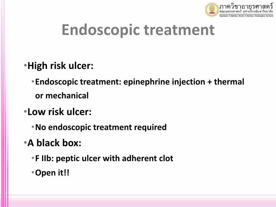
Endoscopic treatment
•High risk ulcer:
•Endoscopic treatment: epinephrine injection + thermal
or mechanical
•Low risk ulcer:
•No endoscopic treatment required
•A black box:
•F IIb: peptic ulcer with adherent clot
•Open it!!

Hemostatic powders
•TC-325 (Hemospray, Cook Medical)
•Action: binds to actively bleeding sites, and the granules
absorb all of the water from blood or secretions, and then
swell and adhere to the bleeding sites.
•Advantages: Easy. Non-contact. Spray onto the bleeding
site.
•Disadvantages: Limited data. Safety. Price.
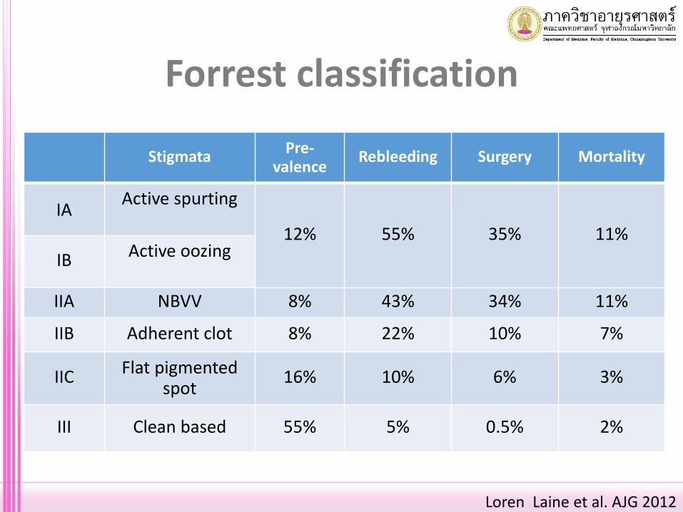
Forrest classification
StigmataPre-
valenceRebleeding Surgery Mortality
IAActive spurting
12% 55% 35% 11%
IB Active oozing
IIA NBVV 8% 43% 34% 11%
IIB Adherent clot 8% 22% 10% 7%
IIC Flat pigmented spot
16% 10% 6% 3%
III Clean based 55% 5% 0.5% 2%
Loren Laine et al. AJG 2012

Endoscopic treatment for a variceal hemorrhage
• Varices with active bleeding or with a sign of
recent bleeding “white nipple sign”
• Varices with absence of those but..
• Blood in stomach without other more possible etiology
• Delayed > 24 hours after bleeding
GARCIA-TSAO ET AL. Hepatology 2017
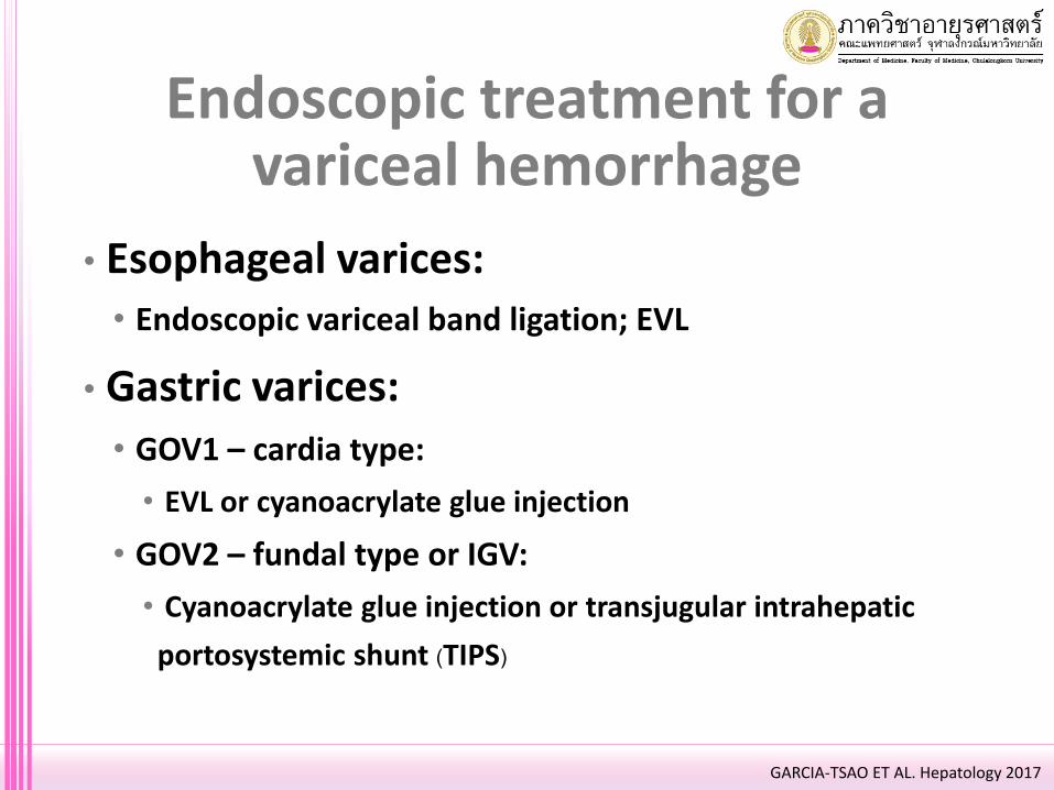
• Esophageal varices:• Endoscopic variceal band ligation; EVL
• Gastric varices:
• GOV1 – cardia type:
• EVL or cyanoacrylate glue injection
• GOV2 – fundal type or IGV:
• Cyanoacrylate glue injection or transjugular intrahepatic
portosystemic shunt (TIPS)
Endoscopic treatment for a variceal hemorrhage
GARCIA-TSAO ET AL. Hepatology 2017

Post-endoscopic treatment
• High risk ulcer:• Continue IV PPI and NPO for 72 hours (reduce rebleeding 57%,
surgery 40%, mortality 43%), discharge with a single daily oral
PPI
• Recurrent bleeding: 2/3 of patients with rebleeding did so
within the first 72 hours
• Rescue treatments: repeated endoscopy. Intervention “TAE”.
Surgery.
• Routine second look: not recommended
Gralnek Ian M et al. ESGE guideline Endoscopy 2015

Post-endoscopic treatment
• Low risk ulcer:
• Discharge with oral PPI
• Also consider: serious co-morbidities (CHF, recent CV
event, chronic alcoholism or active cancer), patient
location /transportation, social support
Gralnek Ian M et al. ESGE guideline Endoscopy 2015

Post-endoscopic treatment
• H. pylori infection:
• Test and treat with confirmation of eradication
• More effective than PPI alone in preventing rebleeding PUD
• Biopsy-based test: false negative 25-55% during acute bleeding. UBT
or repeat at OPD.
• NSAIDs:
• Discontinuation. Choose other options.
• COX-2 plus prophylactic oral PPI
!

Post-endoscopic treatment
• ASA:
• Primary prevention: risks and benefits. Discontinue.
• Secondary prevention: restart within 1-3 days, at most not more than 7
days. Long term prophylactic oral PPI.
• Dual antiplatelets: ASA as above. Risks and benefits reconsideration of
second antiplatelets.
• Anticoagulant:
• Restart within 7 days
• When CV risk outweighs GI bleeding risk
Gralnek Ian M et al. ESGE guideline Endoscopy 2015
!
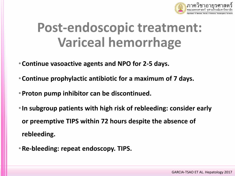
•Continue vasoactive agents and NPO for 2-5 days.
•Continue prophylactic antibiotic for a maximum of 7 days.
•Proton pump inhibitor can be discontinued.
• In subgroup patients with high risk of rebleeding: consider early
or preemptive TIPS within 72 hours despite the absence of
rebleeding.
•Re-bleeding: repeat endoscopy. TIPS.
GARCIA-TSAO ET AL. Hepatology 2017
Post-endoscopic treatment:Variceal hemorrhage

Secondary Prevention
Therapy Recommended Dose Therapy Goals Maintenance, F/U
Propranolol
- 20-40 mg po BID- Adjust every 2-3 days until treatment goal is achieved- Maximal daily dose:
- 320 mg/day in patients without ascites
- 160 mg/day in patients with ascites
- Resting HR 55-60 bpm
- SBP should not decrease below 90
mmHg
- At every outpatient visit make sure that heart rate is on target- Continue indefinitely
EVLEvery 1-4 weeks until the
eradication of varices
Variceal eradication (no further ligation
possible)
First EGD performed 3-6 months after
eradication and every 6-12 months
thereafter
GARCIA-TSAO ET AL. Hepatology 2017
AND

• Buying time before a definite treatment
• Indication
• Before endoscopy: unstable vital signs or during transfer
• After endoscopy: failure to stop bleeding or recurrent
bleeding
• Maximum duration of 24 hours
• Complications: aspiration, esophageal rupture
Sengstaken-Blakemore tube

Take home message!!
•Remember to look at a whole clinical picture.
•We need a good doctor and also a good team.
•The best way to stop the variceal bleeding is to
prevent people from a liver disease.

“Do I not destroy my enemies when I make them my friends?”
― Abraham Lincoln

Thank you