Renal Sympathetic Afferent Nerves: Kidney as Origin of Central Sympathetic Drive
SYMPATHETIC AND PARASYMPATHETIC NERVES INTHE ORBIT OF THE CAT
BY KERMIT CHRISTENSEN
Department of Microanatomy, St Louis University School of Medicine
INTRODUCTION
CRANIAL autonomic ganglia in mammalian species commonly are describedas having three roots, viz. a motor, a sensory and a sympathetic root. Themotor root carries pre-ganglionic fibres which arise in the brain stem andterminate in the ganglion in synaptic relationship with ganglion cells. Thesympathetic root consists of the post-ganglionic sympathetic fibres whicharise in the superior cervical ganglion and extend cephalad via the internalcarotid plexus. The sensory root consists of afferent cranial nerve components.Both the sympathetic and the sensory fibres pass through the ganglion withoutinterruption and become incorporated in the nerves which arise from it. Themotor and sympathetic roots of the spheno-palatine ganglion, for example,are derived from the nerve of the pterygoid canal. This nerve is formed bythe union of the great superficial petrosal nerve, which conveys pre-ganglionicfibres, and the deep petrosal nerve, which conveys sympathetic fibres. Thesensory root is composed of the spheno-palatine nerves, which convey most ofthe sensory fibres which pass through the spheno-palatine ganglion. In viewof these anatomical relationships, it would be impossible, by surgical inter-vention, to eliminate the parasympathetic fibres to the organs innervated bythe nerves from the spheno-palatine ganglion and still retain the sympatheticsupply to these organs, because removal of the spheno-palatine ganglion orsection of the nerves arising from it would interrupt not only the para-sympathetic fibres which arise in the ganglion but also sympathetic fibreswhich pass through it.
The anatomical relationships of the ciliary ganglion, according to mostaccounts, are comparable to those of the spheno-palatine. The inferior ramusof the oculomotor nerve is designated as the short or motor root; fibresderived from the cavernous plexus compose the sympathetic root; and fibresderived from the nasociliary nerve constitute the long or sensory root. Thesympathetic root usually fuses with the sensory root before entering theganglion; consequently, branches of the nasociliary nerve contain both sensoryand sympathetic fibres. Removal of the ciliary ganglion in such cases resultsin degeneration not only of the5ympathetic fibres but also of all sympatheticfibres passing through the ganglion. It is obvious that whenever most of the

226 Kermit Christensen
sympathetic fibres reach the eye by way of the ciliary ganglion,' the para-sympathetic fibres cannot be destroyed without complete destruction of thesympathetic fibres associated with them. In certain species the relationshipsof the nerves to the ciliary ganglion undoubtedly are as summarised above.A study of the ciliary ganglion in the cat, however, shows that in this speciesthe relationships indicated above do not obtain except with respect to themotor root. In this species sympathetic fibres usually do not traverse theciliary ganglion. Jegerow (1887) failed to detect a root to the ciliary ganglionin cats. Anderson (1905) also failed to find one. Dupas (1924) states that the
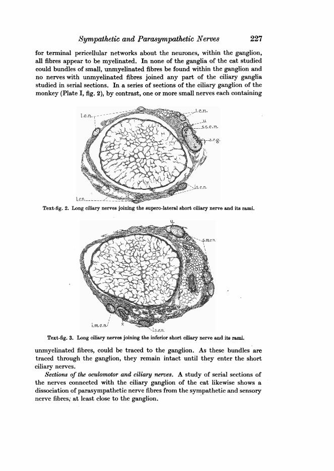
Text-figs. 1-3. A series of sketches of sections of orbital nerves, made by bleaching retouchedphotomicrographs. These sections were selected from the serial sections of one orbit.
Text-fig. 1. The ciliary ganglion is shown with fibres of the third nerve entering and the fibresof the short ciliary nerves leaving the ganglion. Branches of the nasociliary nerve are foundabove and to one side of the optic nerve.
ciliary ganglion receives directly only a single root which is the motor one.He found macroscopically no sympathetic root, although he thought thatsympathetic fibres might reach the ganglion through the motor root.
OBSERVATIONS
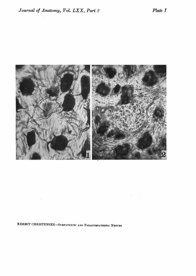
Sections of the ciliary ganglion. Serial sections of the ciliary ganglion ofthe cat prepared by the pyridine silver method reveal no bundles of sympatheticor sensory nerve fibres passing through the ganglion. Since the neurons arewidely separated from one another in the ciliary ganglion (Plate I, fig. 1),the types of fibres and their distribution within the ganglion can be readilystudied. No fibres of very large or very small diameter are present and except
1 In serial sections of the one orbit of a monkey practically all of th e sympathetic fibres tothe eye did join the ciliary ganglion.
Sympathetic and Parasympathetic Nerves
for terminal pericellular networks about the neurones, within the ganglion,all fibres appear to be myelinated. In none of the ganglia of the cat studiedcould bundles of small, unmyelinated fibres be found within the ganglion andno nerves with unmyelinated fibres joined any part of the ciliary gangliastudied in serial sections. In a series of sections of the ciliary ganglion of themonkey (Plate I, fig. 2), by contrast, one or more small nerves each containing
Text-fig. 2. Long ciliary nerves joining the supero-lateral short ciliary nerve and its rami.
I-.. ..-- .
Text-fig. 3. Long ciliary nerves joining the inferior short ciliary nerve and its rami.
unmyelinated fibres, could be traced to the ganglion. As these bundles aretraced through the ganglion, they remain intact until they enter the shortciliary nerves.
Sections of the oculomotor and ciliary nerves. A study of serial sections ofthe nerves connected with the ciliary ganglion of the cat likewise shows adissociation of parasympathetic nerve fibres from the sympathetic and sensorynerve fibres; at least close to the ganglion.
227

Kermit ChristensenThe ramus of the oculomotor nerve (Plate II, fig. 4) which carries the
pre-ganglionic fibres to the ciliary ganglion consists chiefly of large and smallmyelinated fibres. Pyridine silver preparations of this ramus (Plate II, fig. 3)reveal few unmyelinated fibres or none at all. The smaller myelinated fibres,which become aggregated at one side of the nerve, are those which enter theganglion as pre-ganglionic fibres from the oculomotor nerve.
The sections of the short ciliary nerves taken near the ganglion are composedentirely of small myelinated fibres of nearly uniform diameter. This observationhas been confirmed in both osmic acid (Plate II, fig. 6) and silver preparations(Plate II, fig. 5). Myelination of post-ganglionic fibres is uncommon exceptin the short ciliary nerves. The histological appearance of the short ciliarynerve, therefore, is particularly favourable for detecting sympathetic fibreswherever any are present, since the latter are mainly unmyelinated.
Sections of the short ciliary nerves taken farther distally, however(text-figs. 2 and 3), in almost all cats, show that branches of long ciliary nervesunite with them before they reach the eyeball.
The long ciliary nerves are composed of large myelinated, small myelinated(Plate II, fig. 8) and unmyelinated fibres (Plate II, fig. 7). The unmyelinatedfibres probably are in part sympathetic, and the larger myelinated ones sensory.
Whenever a union of long and short ciliary nerves occurs, the largemyelinated fibres (text-fig. 1) and the unmyelinated fibres from the nasociliarynerves can always be detected in the resulting mixed nerve, since they appearin sharp contrast to the smaller, myelinated parasympathetic fibres of theshort ciliary nerves. At the places where the first long ciliary nerves superiorlyjoin the short ciliary nerves, it is not uncommon to find scattered ganglioncells in the short ciliary nerves (text-fig. 2). These are designated as accessoryciliary ganglia. The functional connections of these cells undoubtedly areaffected through pre-ganglionic fibres from the third nerve which traversethe ciliary ganglion and the short ciliary nerves until the cells are reached.
The ciliary ganglion and the ciliary nerves-gross dissectionThe microscopic findings as described above were confirmed in a series
of dissections made with the aid of a dissecting microscope (text-figs. 4-7).The demonstration of the ciliary nerves is facilitated by treating the orbital
contents, fixed in a solution of 10 per cent. formalin, washed in water, andthen partially dissected with a 0*5 per cent. solution of silver nitrate for about12 hours. In such material the nerves appear dark brown while other tissueshave a very light brownish colour. The darkness of the other tissues is greaterwhen stronger silver solutions are used; when the treatment with the silvernitrate is prolonged; or when the silvered material has been exposed to thelight for long periods of time. If dissections are made under water with theuse of a dissecting microscope and a projection lamp the differentiation,however, remains good for a long time providing the material, when not in use,is kept in distilled water in a dark place. The ganglia in the preparations after
228

Sympathetic and Parasympathetic Nerves 229
being in the silver nitrate are usually much lighter than the nerves; conse-quently with the use of the microscope even the position of accessory ciliaryganglia of any size can be readily determined.
s. e.n fi. A.'' ;-i ..
In ~~~~~~~~~~~~ ~~op.n.V~~~~Text-fig. 4. Text-fig. 5.
nen opi-i~Kis- n
Text fig. 41. ~~~~~~Te-xt-fic.7Text-figs. 4-7. A series ofsketches of dissections of orbital nerves made from photographs. The
relations of ciliary ganglion, short ciliary nerves, and long ciliary nerves are shown.
The main ciliary ganglion is a small, flattened, ovoid or triangular bodylocated in the deep part of the orbit, lateral to the optic nerve and superiorto the inferior ramus of the oculomotor nerve. From the inferior ramus of theoculomotor nerve, pre-ganglionic fibres enter the ganglion where they formsynapses with ganglion cells. Besides the inferior ramus, no other nerves joiningthe ganglion have been found. The nerves which arise from the ganglion usuallyconsist of two short ciliary nerves which extend forwards from the anteriorborder of the ganglion. The superior one, which is the larger, lies on the lateral

Kermit Christensen
surface of the optic nerve in the first part of its course and then on the lateraland supero-lateral surface, where it divides into numerous small branches. Theinferior and smaller one usually bifurcates as it leaves the ciliary ganglion.The two branches then pass lateral to the optic nerve and beneath it wherethey also divide into a number of small nerves. These, however, are never quiteas numerous as those of the superior group.
The ciliary nerves and their branches course along the optic nerve to theeyeball together with the long ciliary nerves. Although in the cat no longciliary nerves have been found to join the ganglion, it is not uncommon to findboth short ciliary nerves or their branches joined by long ciliary nerves betweenthe ganglion and the eyeball. The number of long ciliary nerves is variable.Two branches or a bifurcated branch or a single nerve may join the proximalthird of the superior short ciliary nerve, and it is at these points of union thataccessory ciliary ganglia usually occur. Beyond this region superiorly a varietyof conditions may appear from a complete absence of further union betweenlong and short ciliary nerve branches to a union of all remaining long ciliarybranches with short ciliary branches. The latter usually takes place close to theeyeball. In the case of the ciliary nerve branches beneath the optic nervewhere they are not so numerous, only single or double long ciliary nervesappear, and they may not join the short ciliary nerve branches until closeto the eyeball or they may form with the short ciliary nerve branches asimple network of fibres along the distal two-thirds of their length.
Whenever union of long ciliary branches with short ciliary branches occurs,mixed nerves result which contain sympathetic and sensory fibres from theformer and parasympathetic fibres from the latter (a section is shown in Plate II,fig. 9).
In the preceding description a summary of the general details only hasbeen attempted. Specific individual details have not been included. It is to bepointed out, however, that variations in the relationships between long andshort ciliary nerves differ from individual to individual, and also between thenerves of the orbit of the one side and the nerves of the orbit of the oppositeside of the same individual.
SUMMARY
The results of the anatomical study may be summarised as follows:1. The so-called sympathetic and the sensory roots of the ciliary ganglion
are not found in the cat.2. The short ciliary nerves when they first arise from the ciliary ganglion
contain only post-ganglionic parasympathetic fibres.3. Sympathetic fibres become incorporated in the short ciliary nerves only
after those nerves have been joined by long ciliary nerves somewhere betweenthe ganglion and the eyeball.
4. In the orbit of the cat there is at least a partial separation of post-ganglionic sympathetic and parasympathetic fibres which innervate the eye:
230
Sympathetic and Parasympathetic Nerves 231
(a) since the ctliary ganglion has no sympathetic fibres passing through it;(b) since short ciliary nerves are separate for some distance.
It follows from these.conclusions that, by careful operative procedure, theeye can be deprived of its post-ganglionic parasympathetic supply by removalof the ciliary ganglion without great disturbance to its sympathetic supply.
REFERENCES
ANDERSON, H. K. (1905). " Paralysis ofinvoluntary muscle. Part II. On paralysis ofthe sphincterof the pupil with special reference to paradoxical constriction and functions of the ciliaryganglion." J. Physiol. vol. xxxm, p. 156.
BEAUVIEUX, J. and DUPAS, J. H. L. (1926). "etude anatomo-topographique et histologique duganglion ophtalmique chez l'homme et divers animaux." Arch. Ophta., Paris, t. xun, p. 641.
CARDozA, E. L. (1933). "Investigations into the distribution of the sympathetic fibres of the first,second and third thoracic roots, over the long ciliary nerves." Arch. nierl. Physiol. t. xvrln,p. 193.
DUPAS, J. H. L. (1924). "Contribution a l'6tude anatomique et histologique du ganglion ophtal-mique chez l'homme et divers animaux." These, Facult6 de M6decine et de PharmacieUniversity de Bordeaux.
GASKELL, W. H. (1899). " On the relation between the structure, function, distribution, and originof the cranial nerves, together with a theory of the origin of the nervous system of theVertebrate." J. Physiol. vol. x, p. 153.
JEGEROW, J. (1887). "Recherches anatomo-physiologiques sur le ganglion ophtalmique." Arch.8lav. Biol. t. 2.
WHITNALL, S. E. (1921). The Anatomy of the Human Orbit and Accessory Organs of Vision.London: Henry Frowde, and Hodder and Stoughton.
WINDLE, W. F. (1924). "The distribution and probable significance of unmyelinated fibres in thetrigeminal nerve of the cat." J. comp. Neurol. vol. xu, p. 453.
EXPLANATION OF PLATES
PLATE IFig. 1. Photomicrograph of a typical section through the ciliary ganglion of a cat.Fig. 2. Photomicrograph of a section through the ciliary ganglion of a monkey which shows
a long ciliary nerve in section that passes through the ganglion.
PLATE IIFig. 3. Photomicrograph of a section of the inferior ramus of the oculomotor nerve. Pyridine
silver method.Fig. 4. Photomicrograph of a section of the inferior ramus of the oculomotor nerve. Osmic acid
method.Fig. 5. Photomicrograph of a section of one of the short ciliary nerves, taken near the ciliary
ganglion. This nerve was photographed at a magnification higher than that used for othernerves on this plate. Pyridine silver method.
Fig. 6. Photomicrograph of a section of one of the short ciliary nerves taken near the ganglion.Osmic acid method.
Fig. 7. Photomicrograph of a section of a long ciliary nerve. Pyridine silver method.Fig. 8. Photomicrograph of a section of a long ciliary nerve. Osmic acid method.Fig. 9. Photomicrograph ofa section ofa mixed ciliary nerve. A short ciliary nerve has been joined
by long ciliary nerves, whose large myelinated sheaths show clearly.
232 Kermit ChristensenABBREVIATIONS
Text-figs. 1-3a.c.g. Accessory ciliary ganglion.c.g. Ciliary ganglion.i.m.c.n. Inferior mixed ciliary nerves.i.n.III. Fibres of inferior ramus, oculomotor nerve entering the ciliary ganglion.i.s.c.n. Inferior short ciliary nerves.l.e.n. Long ciliary nerves.n.c.n. Branches of nasociliary nerve.8.C.n. Fibres of short ciliary nerves leaving the ciliary ganglion.s.m.c.n. Supero-lateral mixed ciliary nerves.s.sc.n. Supero-lateral short ciliary nerves.U. Points of union between supero-lateral short ciliary nerves and long ciliary nerves.x. Points of union between inferior short ciliary nerves and long ciliary nerves.
Text-figs. 4-7
a.c.g. Accessory ciliary ganglion.c.g. Ciliary ganglion.i.n.III. Inferior ramus, oculomotor nerve to ciliary ganglion.i.8.c.n. Inferior short ciliary nerves.I.c.n. Long ciliary nerves.n.c.n. Nasociliary nerves.op.n. Optic nerve.s.c.n. Short ciliary nerves.
Journal of Anatomy, Vol. LXX, Part 2
KERMIT CHRISTENSEN-SYMPATHETIC AND PARASYMPATHETIC NERVES
Plate I

Plate IIJournal of Anatomy, Vol. LXX, Part 2
.4' *2' s..
1. *,.
-a*. o :.Je* / +
40~ 3
-via%IC~~~~~~~
;w^... _ . 5.
"e W -~ i
Nme
,1~~~~~~~~e .l4
Lo Am~An 81
KERMIT CHRISTENSEN-SYMPATHETIC AND PARASYMPATHETIC NERVES