SUSAN MARIE PARLATO
63
ANTIMICROBIAL SENSITIVITY AND RESISTANCE DEVELOPMENT CAUSED BY NUTRACEUTICALS by SUSAN MARIE PARLATO A thesis submitted to the Graduate School-New Brunswick Rutgers, The State University of New Jersey And The Graduate School of Biomedical Sciences University of Medicine and Dentistry of New Jersey In partial fulfillment of the requirements For the degree of Master of Science Graduate Program in Microbiology and Molecular Genetics Written under the direction of Dr. Stanley E. Katz And approved by ________________________ ________________________ ________________________ New Brunswick, New Jersey January, 2011
Transcript of SUSAN MARIE PARLATO

ANTIMICROBIAL SENSITIVITY AND RESISTANCE DEVELOPMENT CAUSED
BY NUTRACEUTICALS
by
SUSAN MARIE PARLATO
A thesis submitted to the
Graduate School-New Brunswick
Rutgers, The State University of New Jersey
And
The Graduate School of Biomedical Sciences
University of Medicine and Dentistry of New Jersey
In partial fulfillment of the requirements
For the degree of
Master of Science
Graduate Program in Microbiology and Molecular Genetics
Written under the direction of
Dr. Stanley E. Katz
And approved by
________________________
________________________
________________________
New Brunswick, New Jersey
January, 2011

ABSTRACT OF THE THESIS
ANTIMICROBIAL SENSITIVITY AND RESISTANCE DEVELOPMENT CAUSED
BY NUTRACEUTICALS
By SUSAN MARIE PARLATO
Thesis Director:
Dr. Stanley E. Katz
For centuries, people have used herbal supplements to treat a host of medical
ailments. Their use had declined with the discovery of potent pharmaceuticals, however,
in recent years, the use of nutraceutical products has seen a huge increase and the
industry has grown exponentially. With this increasing use of nutraceutical products,
there still remains little knowledge concerning the effects of herbal products on
commonly used antibiotics and antimicrobials. These studies were conducted to
examine the effects of a small sample of herbal products on antibiotic resistance and
sensitivity in bacteria. The herbal products studied were Bee Pollen, Black Walnut,
Calendula, Copaiba, Clove, Eucalyptus and Prickly Ash in the form of tinctures, essential
oils and 1:1 dilutions of essential oils. Two test strains, Staphylococcus aureus ATCC
29213 and Escherichia coli ATCC 25922 were used as representatives of Gram-positive
and Gram-negative organisms. These studies showed a thirty-fold increase in the
ampicillin MIC values for the Bee Pollen and Prickly Ash exposed Staphylococcus
aureus ATCC 29213 as well as a four-fold increase in the Bee Pollen and 1:1 diluted
Eucalyptus oil exposed Escherichia coli ATCC 25922. Additionally, a four-fold
decrease in tetracycline and norfloxacin MIC values was observed for the Bee Pollen and
ii

Prickly Ash exposed Staphylococcus aureus ATCC 29213 and a four-fold decrease in the
sulfamethazine MIC values was observed in the Prickly Ash exposed Staphylococcus
aureus ATCC 29213. There was neither a substantive increase nor decrease in MIC
values for the other products in this study.
iii

Acknowledgements
I would first like to extend my sincere thanks to Dr. Stanley Katz for giving me
the opportunity to work in his lab and to pursue my graduate studies. I would also like to
thank him for his incredible patience during these long years that I have worked to
complete my degree. The kindness and understanding he has showed me throughout one
of the most difficult times of my life is something that I will eternally be grateful for and
will never forget. Additionally, I would like to thank Drs. Alan Antoine and Douglas
Eveleigh for the advice and support over the years and for serving on my thesis
committee.
I would also like to thank my two previous managers, Dr. Lei Tang, from
Epigenesis Pharmaceuticals Inc. and Dr. James Beasley, of Pharmacopeia, Inc. Without
their understanding and flexibility, I would never have been able to attend classes and
work at the same time.
My parents have always been my biggest fans and have given me love and
support over the years. I will always be grateful for all their words of encouragement that
helped me get through the tough times. They were always there to keep me motivated,
even when I felt like giving up.
Last, but certainly not least, I would like to extend a heartfelt thanks to my
boyfriend, John Tallon, without whom I may never have finished my thesis this year. He
has always told me how proud he is of me for working hard on my studies and I believe
iv

that he gave me a reason to complete my degree. Looking forward to a future together
and of good things to come has helped me to persevere and to finally close this chapter of
my life.
v

Table of Contents
Page
Abstract ii
Acknowledgements iv
Table of Contents vi
List of Tables viii
Introduction 1
Survey of Literature of Herbal Products and Antimicrobial Properties 7
A - General Characteristics of Herbals 7
Alkaloids 10
Phenols and Polyphenols 10
Essential oils and Terpenoids 13
Glycosides and Saponins 14
Resins and Gums 14
Tannins 15
Sterols 16
B - Experimental Herbs in these Studies 17
Calendula (Marigold) 17
Clove 20
Copaiba 21
Eucalyptus 22
Bee Pollen 24
Prickly Ash 25
vi

Page
Black Walnut 26
Hypothesis 29
Materials and Methods 30
Nutraceutical Products 30
Indicator Organisms 30
Culture Preparation 31
Preparation of Nutraceuticals for Experimentation 32
Agar Diffusion Assay 33
Antibiotic Preparation and MIC Determination 34
Results and Discussion 36
Bee Pollen 42
Black Walnut 43
Calendula 44
Clove Oil 44
Clove Tincture 46
Copaiba 46
Eucalyptus Oil 47
Eucalyptus Tincture 49
Prickly Ash 49
Conclusions 52
References 53
vii

List of Tables
Page
Table 1 - Categories of Herbal Chemicals 9
Table 2 - Nutraceuticals Studied and their Formulations 18
Table 3 – Resistance development in test strains over 39 the course of three passages caused by nutraceutical alcohol extracts
Table 4 – MICs (µg/mL) resulting from the nutraceutical extract 40 and antimicrobial using Staphylococcus aureus ATCC 29213 as the indicator organism
Table 5 – MICs (µg/mL) resulting from the nutraceutical extract 41 and antimicrobial using Escherichia coli ATCC 29522 as the indicator organism
viii

1
Introduction
For centuries people have used plants for healing. Plant products, as parts of
foods or botanical potions and powders, have been used with varying success to cure and
prevent diseases throughout history [1]. Written records regarding medicinal plants date
back at least 5000 years to the Sumerians, and archeological records suggest even earlier
use of medicinal plants [1]. However, the strong bond between plants and humans began
to unwind and the twentieth century became a triumph for the synthetic-chemistry-
dominated pharmaceutical industry, particularly in the United States [2].
The discovery of new drugs resulted in treatments for conditions that were once
considered fatal [2]. The development of antimicrobials, such as sulfonamide in the
mid-1930s, made it possible to treat some common and equally life-threatening infections
contracted by injury, surgery, or epidemic [2, 3]. This introduction of new, and often
extremely useful, synthetic drugs replaced the old botanical products. By the mid-1900s,
almost all botanical remedies disappeared from the shelves of pharmacies [3]. By the
1960s, the medicines available in the United States, unlike those utilized by other
countries, were primarily synthetic [3].
In the 70s and 80s, however, scientific and clinical reports began to come out of
some European countries, especially Germany, indicating that the herbal remedies, which
had never been discarded, had many substantial therapeutic and economic benefits for the
consumer [3, 4]. With this new knowledge and the rising widespread recognition of
recurring problems in treating disease with manufactured drugs, Americans began to

2
demand herbal products [2, 3]. Answering the call, companies began to supply these
products to the public and by the late 1990s annual sales in the USA had reached almost
$4 billion [3]. Currently, it is reported that annual sales have increased remarkably
creating a $30 billion industry which continues to grow at 5% annum [5].
In the past, food was only thought of as something having taste, aroma or general
nutrition. Today, consumers recognize additional categories of foods [6]. Within the
last decade, consumers have made increasing reference to the terms “nutraceuticals” and
“functional foods”, recognizing the relationship between nutrition and health and the
potential of forgoing the use of pharmaceutical drugs [7]. The term “nutraceutical”, a
hybrid term between nutrients and pharmaceuticals, was coined in 1989 by the
Foundation for Innovation in Medicine, to provide a name for this rapidly growing area
of biomedical research [8, 5]. A nutraceutical is defined as “any substance that may be
considered a food or part of a food that provides medical or health benefits, including the
prevention and treatment of disease” [5]. They are generally sold in medicinal forms not
usually associated with food; usually in pill, capsular or ampoule form [9]. Alternatively,
functional foods are foods that, by virtue of the presence of physiologically-active
components, provide a health benefit beyond basic nutrition [10].
While there has been a growing popularity in nutraceutical products and
functional foods, it is noteworthy to mention that these markets are not well-regulated by
the U.S. Food and Drug Administration (FDA) [7]. The FDA had long insisted that to
obtain approval as a drug in the United States, herbs had to be supported by the same

3
amount of costly evidence of efficacy required for synthetic drugs [3]. However, the
natural products industries faced diverse challenges.
The composition and contents of active constituents in natural plants vary
depending on season, climate, temperature, humidity, soil and several other factors [7].
Therefore, the collection, identification and maintenance of uniform quality,
quantification and standardization methods are of critical importance [7]. Also, the
manufacturing processes, use of solvents and/or additives, purification and drying
techniques, and storage conditions can play a major role on the occurrence of significant
amounts of contaminants, chemical, physical and biological, in the products [7]. These
issues made it very difficult for companies to create a standardized product, which
deterred them from investing in the costly tests and research to prove their product’s
efficacy. Consequently, since the standards were too high, only a handful of herbs have
ever received drug approval in the U.S. [3].
The laws that have eventually shaped the dietary supplement industry as we know
it today began in 1906, when Congress passed the Pure Food and Drugs Act and the Meat
Inspection Act, both defining food as “articles used for food, drink, confectionery, or
condiment by man or other animals, whether simple, mixed or compounded” [11]. They
primarily focused on adulteration, misrepresentation or sale of otherwise unfit products
[12]. In the following years, the laws governing food and drugs were periodically
tweaked and modified.

4
The landmark 1958 Food Additive Amendment (FAA) to the Federal Food, Drug
and Cosmetic Act (FFD&C) addressed concerns for safety and accomplished several
things [11]. Firstly, it changed the meaning of the term “food additive” from a
technological term to one of legal status by defining the term as a substance not generally
recognized as safe and created an entirely new class of substances; those that are
generally recognized as safe (GRAS) [12]. The amendment also defines who can
determine what GRAS is and the process by which these experts may determine
something is GRAS [12].
In 1994, the passage of the Dietary Supplement Health and Education Act
(DSHEA) to the FFD&C delineated the role of the FDA in regulating nutraceutical
products and dietary supplements and has essentially deregulated the industry [13]. This
act does not permit the FDA to consider a new product a “drug” or “food additive” if it
falls under the definition of a “dietary supplement” [6]. Dietary supplements are defined
in the DSHEA as a product, other than tobacco, intended to supplement the diet that bears
or contains one or more of the following ingredients: a vitamin, mineral, herb or other
botanical or amino acid [13]. The DSHEA makes manufacturers responsible for the
safety of products marketed without pre-marketing safety determination by the FDA and
allows certain nutrition support statements on labels also without FDA approval [13].
With this new relaxed law, the industry has grown essentially exponentially [14].
As part of the passage of DSHEA, a new law requiring pre-market safety
notification for new dietary ingredients (NDI) became part of the new regulatory
5
landscape [14]. According to the law, every dietary ingredient in the market before
October 15, 1994 is considered an old dietary ingredient and presumed to be safe [14].
Any dietary ingredient marketed after this date is considered new and requires FDA pre-
market review for safety [14]. Although the concept may seem easy to understand, it has
not been as easy to follow and many marketers are much less aware of the requirements
than they are concerned about their implications [14].
Though herbal remedies have been used for centuries, modern chemical
pharmaceuticals began to overtake the market, making the use of folk medicine almost
nonexistent, particularly in the United States. However, nutraceutical products have
gained tremendous popularity over the past few decades and the response of Congress to
a public anxious to preserve its access to dietary supplements resulted in a more flexible
approach to the law governing food [12]. Where the 1958 Amendment changed the role
of the FDA in GRAS matters to gatekeeper, aggressive enforcement of dietary
supplements on the part of the Agency prompted Congress to enact amendments to the
FD&C Act, specifically for dietary supplements, reducing the burden of proof of safety
[12, 14]. Now, the FDA must prove that a dietary supplement is unsafe before
appropriate action could be taken [12].
Further curtailing the FDA’s authority are the DSHEA amendments [12]. Though
it did establish standards for good manufacturing practices for dietary supplements,
DSHEA did not establish standards of quality for individual products and this lack of
quality assurance is the biggest single problem in the entire field today [3]. Repeated

6
studies have shown that the quality of products, even those purported to be standardized
on the basis of active or marker compounds varies enormously [3]. There is absolutely
no way that consumers can be assured that what is on the label is actually in the package,
other than the reputation of the producer [3]. This leaves even knowledgeable consumers
bewildered. Until new, stricter laws are created and enforced however, the decision
making power will still lie in the hands of the consumers and it is the consumer who will
ultimately have to decide what is right for them.

7
Survey of Literature of Herbal Products and Antimicrobial Properties
A- General Characteristics of Herbals
Centuries of human experience with plants demonstrated that some plants have
strong physiological effects that can relieve or cure disease [15]. Many herbalists claimed
that all plants have medicinal value, but scientifically, that has not been shown. Plants
used for food, for example, are not strictly medicinal though it is clear that they may
contain factors such as vitamins and minerals that prevent disease [15].
Plants are continuously in contact with microorganisms, including viruses,
bacteria and fungi. Some of these interactions are beneficial to plants, for example the
symbiotic relationship between the nitrogen-fixing bacteria rhizobia with leguminous
plants [16]. However, many plant-associated microbes are pathogens that affect plant
development, reproduction and ultimately yield production [16].
It has been observed that plants produce materials such as starches, fibers, latexes,
vitamins and minerals that are necessary for life as well as several distinctive secondary
products, or metabolites [15]. And in many cases, it is these secondary metabolites that
can serve as plant defense mechanisms against predation by microorganisms and also
insects, herbivores and environmental conditions [17, 16]. They may contribute to a
plant’s medicinal value [15, 17].
There are numerous secondary metabolites; about 12,000 have been isolated, a
number estimated to be less than 10% of the total [17]. A select few however, often

8
serve as therapeutic chemicals [18]. These include alkaloids, bioflavonoids, essential
oils, glycosides, resins, saponins, sterols, tannins, terpenes and other phytochemicals
(Table 1). These secondary metabolites are found in specific groups of plants [18]. Of
the 310 or so families of seed plants, medicinal plants occur in perhaps 200 to 250 [15].
The daisy family (Compositae), the mint family (Labiatae), the bean family
(Leguminosae), the lily family (Liliaceae), the buttercup family (Ranunculaceae), the
rose family (Rosaceae) and the carrot family (Umbelliferae) are especially rich in
medicinally useful species [15].

9
Table 1 - Categories of Herbal Chemicals [18]
Class Definition Properties Alkaloids Basic amines (names end in “-ine”). Includes potent drugs and
narcotics. Over 12,000 known; over 13 classes.
Bioflavonoids Plant pigments; vitamin-like. Over 4000 known; over 14 classes.
Essential Oils Isoprene derivatives; oxidized terpenes and phenylpropanoids.
Used in perfumes and in aromatherapy. Over 9 classes. Also known as volatile oils, ethereal oils, essences.
Glycosides Sugar derivatives attached to aglycones Over 10 classes. Over 3000 known.
Resins Oxidation products of terpenes; resins are insoluble in water.
Includes oleoresins, gum resins and balsams.
Saponins Soap-like glycosides; cause hemolysis if directly introduced into the blood serum.
Various groups of chemicals. Some are involved in steroid metabolism.
Sterols Steroid and vitamin D precursors. Found in soy and other plants; also produced by microorganisms (e.g., sitosterol, stigmasterol).
Tannins Polyphenolics, mostly based on gallic acid. Astringent compounds, bind to protein (tanning); reduce diarrhea, act as hemostatics.
Terpenes (Terpenoids)
Derived from 5-carbon isoprene units (10, 15, 20, 30, 40, >40)
Over 20,000 known; 6 classes. Most structurally varied phytochemicals.

10
Alkaloids
Alkaloids have some of the most potent effects on animals and humans, and they
demonstrate both therapeutic and toxic properties [18]. They are basic amines,
heterocyclic nitrogen-containing substances, which are clear, crystalline and non-volatile
[18]. They lack odor, but are bitter in taste and insoluble in water, although their salts are
soluble [18]. Included in this class of over 12,000 known agents are purines,
pyrrolidines, piperidines, pyridines and quinolines [18]. Some of the best known herbal
drugs are alkaloids, including atropine, capsaicin, morphine, quinine, methylxanthines
(such as caffeine and theophylline) and nicotine [17, 18].
Many alkaloids have been shown to possess some antimicrobial effects.
Diterpinoid alkaloids, commonly isolated from the plants of the Ranunculaceae, or
buttercup family are commonly found to have antimicrobial properties [17]. One
chemical compound, berberine, is an especially important representative of the alkaloid
group [17]. It is potentially effective against trypanosomes and plasmodia [17]. The
mechanism of action of highly aromatic planar quaternary alkaloids such as berberine is
attributed to their ability to intercalate with DNA [17].
Phenols and Polyphenols
There are about 8000 known plant phenolics [18]. The simplest phenol is phenol
(hydroxybenzene or carbolic acid), which was the first surgical antiseptic used in modern
medicine [20]. Other phenols include eugenol (used as a dental analgesic and

11
disinfectant), carvacrol and thymol, all antiseptics [20]. They are found in essential oils
of clove, cinnamon, thyme, oregano and savory and are bactericidal and antifungal [20].
Other classes of phenols include coumarins and bioflavonoids. Coumarins are
phenolic substances made of fused benzene and α-pyrone rings and are responsible for
the characteristic odor of hay [17]. Their fame has come mainly from their
antithrombotic, anti-inflammatory and vasodilatory activities, as demonstrated by the
well-known coumarin and warfarin; however there are several other coumarins that have
antimicrobial properties [17]. General antimicrobial activity was documented in
woodruff (Galium odoratum) extracts and coumarin was also found in vitro to inhibit
Candida albicans [17].
Bioflavonoids or simply flavonoids are therapeutically the most important group
of polyphenols that can be broken down into 12 classes: flavans, flavones, flavanones,
flavonols, flavanolols, isoflavones, leukoanthocyanins, chalcones, dihydrochalcones,
aurones, anthocyanidins and catechins [20]. Several thousand bioflavonoid compounds
are known, occurring freely or as glycosides [18]. In general, they are phenolic structures
containing one carbonyl group, lack nitrogen, and usually contain two 6-carbon rings
joined by three carbon atoms, but 5-membered rings (aurone) and open-chain (chalcone)
compounds are included in this group [17, 18]. The flavonoids are often pigmented,
appearing yellow or other colors of petals, fruits, and berries in higher plants [18]. They
are found in high concentrations in many flowers, and in foods such as citrus fruits,
tomatoes, red wine, onions and tea [18]

12
Since they are known to be synthesized by plants in response to microbial
infection, it should not be surprising that they have been found in vitro to be effective
antimicrobial substances against an array of microorganisms [17]. Their activity is
probably due to their ability to complex with extracellular and soluble proteins and to
complex with bacterial cell walls [17]. More lipophilic flavonoids may also disrupt
microbial membranes [17].
Catechins, the most reduced form of the C3 unit in flavonoid compounds, deserve
special mention [17]. These flavonoids have been extensively researched due to their
occurrence in oolong green teas [17]. It was noted that teas contain a mixture of catechin
compounds, which have shown in in vitro studies to inhibit Vibrio cholera O1,
Streptococcus mutans, Shigella spp., and other bacteria and microorganisms [17]. The
catechins inactivated cholera toxin in Vibrio cholera and inhibited isolated bacterial
glucosyltransferases in S. mutans, possibly due to complexing activities [17].
There are two possible explanations for the mechanism of action of flavonoids.
Some research has found that flavonoids lacking hydroxyl groups on their β-rings are
more active against microorganisms than those with the hydroxyl groups, which supports
the proposed idea that their microbial target is the membrane [17, 18]. Lipophilic
compounds would be more disruptive of this structure [17, 18]. However, there has been
research that has found the opposite effect, which proves that there is no clear
predictability for the degree of hydroxylation and toxicity to microorganisms [17, 18].

13
Essential Oils and Terpenoids
The antimicrobial effects of essential oils have been reported and used in herbal
medicine in many countries [21]. Essential oils, also known as volatile oils or essences,
constitute the attractant odors of flowers and the attractant or defensive components of
the other parts of the plant [18, 21]. These oils are secondary metabolites that are highly
enriched in compounds based on an isoprene structure (C5H8) [17, 18]. Some are
terpenes (C10H16) and can occur as diterpenes, triterpenes, tetraterpenes, hemiterpenes
and sesquiterpenes [17]. When the compounds contain additional elements, usually
oxygen, they are termed terpenoids [17].
Terpenoids have been shown to be active against many bacteria, fungi, viruses
and protozoa [17, 18]. In 1977, it was reported that 60% of essential oil derivatives
examined at the time were inhibitory to fungi while 30% inhibited bacteria [17, 18]. The
mechanism of action of terpenes remains not fully understood; however, it was
speculated that terpenes disrupt cell membranes [17, 18].
Food scientists found the terpenoids present in essential oils of plants to be useful
in the control of Listeria monocytogenes [17]. Also, the ethanol-soluble fraction of
purple prairie clover yielded a terpenoid called petalostemumol, which showed excellent
activity against Bacillus subtilis and Staphylococcus aureus and lesser activity against
gram-negative bacteria as well as Candida albicans [17].

14
Glycosides and Saponins
Numerous plant chemicals contain a carbohydrate residue, or glycone, attached to
a noncarbohydrate residue, or aglycone, to form a glycoside [18]. If hydrolysis yields
glucose, the originating glycoside is termed a glucoside, in contrast to non-glucosides that
yield other sugars [18]. Glycosides usually have a characteristic odor of bitter almonds
and are usually bitter tasting; however, some are sweet [18, 20].
Saponins are glycosides with terpenoid aglycone components [18]. They possess
two major characteristics: they have soap-like surfactant effects, and they cause
hemolysis when directly introduced into the blood stream [18]. There are three major
classes of saponins, steroidal, terpenoid and glycoalkaloid saponins, and a number of
them have potential medicinal effects, including anti-inflammatory, immune-boosting
and expectorant properties [18, 20].
Resins and Gums
When a plant is injured, it may exude a hard-setting material to cover the wound
[18]. This material, known as a resin, is a semi-solid, amorphous substance that, in
general, is soluble in alcohol and ether [20]. Resins are composed of resin alcohols,
resenes, esters and other compounds; the main components are oxidation products of
terpenes [18]. There are three main types of resins: oleoresins, gum resins and balsamic
resins [20].

15
Oleoresins are a mixture of resin components and volatile oils [18]. Turpentine is
a primary example of an oleoresin. Gum resins are complex oleoresins that are soluble in
alcohol and form emulsions with water [18, 20]. Myrrh and frankincense are both gum
resins. Balsam of Peru and benzoin are examples of balsamic resins. Balsamic resins are
resins combined with cinnamic or benzoic acids or their esters [18]. They dissolve in
alcohol, but not water [20].
Resins are used as astringents and antiseptics, and seem to have immune-
stimulating properties along with antiseptic activity [20]. They are traditionally used for
wounds, both to seal and to disinfect [20]. Balsam of Peru has been shown to directly kill
bacteria, fungi and parasites.
Tannins
Tannins are polymeric phenolic substances, which have an astringent effect and
can precipitate proteins in the tanning process that produces leather [17, 18]. They can be
divided into two main groups, hydrolyzable and condensed tannins [17, 18].
Hydrolyzable tannins are based on gallic acid, usually as multiple esters with D-glucose,
while the more numerous condensed tannins called proanthocyanidins are derived from
flavonoid monomers [17].
Tannins have been shown to have antimicrobial action. They form complexes
with proteins through so-called nonspecific forces such as hydrogen bonding and
hydrophobic effects, as well as by covalent bond formation [17]. Thus, their mode of

16
antimicrobial action may be related to their ability to inactivate microbial adhesions,
enzymes and cell envelope transport protein [17]. They also form complexes with
polysaccharides [17].
According to studies, tannins can be toxic to filamentous fungi, yeasts and
bacteria [17]. Condensed tannins have been determined to bind to cell walls of ruminal
bacteria, preventing growth and protease activity [17]. Also, though still speculative,
tannins are considered at least partially responsible for the antibiotic activity of
methanolic extracts of the bark of Terminalia alata found in Nepal [17].
Sterols
These compounds are precursors of steroids, including ergosterol,
dihydrotachysterol and ergocalciferol, which are part of the Vitamin D complex [18].
The important phytosterols, β-sitosterol, stigmasterol and campesterol, are found in
popular sources such as olive oil and soy and have been shown to possibly reduce LDL
cholesterol and lower serum cholesterol [18]. Similar sterol combinations in ginseng
are credited with adaptogenic properties [18]. Other phytosterol benefits that are claimed
include anti-inflammatory properties, immune enhancement, anti-cancer activity and
antioxidant effects [17].

17
B- Experimental Herbs in these Studies
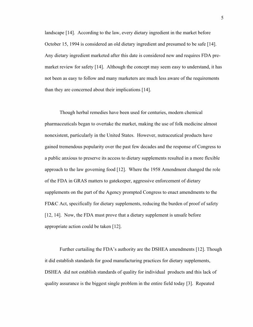
In this study, seven nutraceuticals were chosen based on some indication of
antimicrobial activity. Many of the families which contain a high number of medicinally
active species were represented, such as the daisy, mint and bean families. The herbs
also represent a wide range of activity against gram-negative and gram-positive
organisms as well as activity against certain types of parasites and fungi. These
nutraceuticals studied are individually derived from almost every part of the plant and are
used in a variety of forms (Table 2).
Calendula (Marigold)
Calendula (Calendula officinalis) is commonly known as pot marigold or golden
marigold and is a member of the Asteraceae family. The name refers to its tendency to
bear flowers by the calendar, once a month, in warm climates during the new moon [2]
“Marigold” also refers to the Virgin Mary and is traditionally used in Catholic
celebrations concerning the Virgin [2].
It is an annual aromatic plant native to the Mediterranean countries, but is found
throughout central and southern Europe, western Asia, and the United States [2, 22, 23].
The plant grows between 30 and 50 cm high and on the tip of each stem, there is a many-
petaled orange or yellow flowering head 4 to 7 cm in diameter [2, 23]. Though widely
grown as an ornamental garden plant, it is also cultivated and the flowers harvested for
use in herbal medicine throughout Eastern Europe and Latin America [2].
18
Table 2 – Nutraceuticals Studied and their Formulations
Name Latin Name Common Name Part Used Antimicrobial Activity
Available Form
Calendula Calendula officinalis (Asteraceae [composite] family)
Pot calendula, pot marigold
Flowers This herb has strong bactericidal effect and may counteract infection with H. pylori and S. aureus.
Tinctures, cream and aerosol sprays
Clove Syzygium aromaticum (Myrtaceae [clove] family)
None Oil extracted from leaves and flowers and fresh dried flower buds
Kills some types of bacteria associated with food poisoning, including P. aeruginosa, Shigella (many species), S. aureus, and S. pneumoniae
Tincture and essential oil
Copaiba Capifera species (Fabaceae [legume] family)
Balsam copaiba, copal, Jesuit’s balsam, mal-dos-sete-dias
Resin is collected, and the sap is distilled to concentrate its oils
Volatile oil acts as an antimicrobial and prevents secondary infections in eczema, herpes, and psoriasis
Oil, capsules, ointments, powders, and tinctures
Eucalyptus Eucalyptus globules (Myrtaceae[clove] family)
Blue gum, red gum Leaves are distilled for the oil
Contains the chemical eucalyptol, which kills several types of bacteria, including B. subtilis and several strains of Streptococcus.
Tincture and essential oil
Bee Pollen None None Microspores (male reproductive elements)
Acts against bacteria, including Colibacilli and certain strains of Salmonella
Raw and micronized form
Prickly Ash Xanthoxylum americanum and Xanthoxylum bungeanum (Rutaceae [citrus] family)
Angelica tree, xanthoxylum
Fruits and bark The chemical constituents that give prickly ash its heat kills food borne bacteria
Tincture, tea, capsules
Walnut leaf Juglans nigra, Juglans regia (Juglandaceae [walnut] family)
Black walnut and white walnut
Leaves Contain two antibacterial agents, walnut essential oil and juglone, which act directly on infectious microorganisms
Teas

19
Many studies of calendula flowers have been carried out, especially in Europe,
without revealing anything unique in their physiological properties [22]. However, a
volatile oil, bitter principles, carotenoids, mucilage, resin, polysaccharides, plant acids,
various triterpene alcohols, saponins and other glycosides and sterols were found to be
present [22, 23, 25]. Many of the individual constituents in these general groups have
been identified.
Calendula has been used as an antibacterial, anti-inflammatory and pain killer
[22]. Numerous studies demonstrated that the flowers had antimicrobial activity due to
the terpene alkaloids, lactone and flavones contained in the essential oil [23, 24].
Flavonoids isolated from calendula flowers showed positive antimicrobial activity against
Staphylococcus aureus at a concentration of 1mg/mL [2, 23]. Other studies have
demonstrated the flavones to be effective against Klebsiella pneumoniae, Micrococcus
lutea and Candida monosa [23].
Calendula extract is available as an aerosol spray which is applied to cuts and
scrapes to prevent infection and to help stop bleeding [2]. It is also used to treat
conjunctivitis infections in the form of eye drops and taken internally as a tea [2]. It was
shown to have a strong bactericidal effect on Helicobacter pylori, the bacterium
associated with gastritis and peptic ulcers, and aids in healing duodenal and peptic ulcers
[2].

20
Clove
One of the most popular spices throughout history, clove (Syzygium aromaticum)
is a product of an evergreen tree native to the Spice Islands [2, 19]. The clove tree thrives
only in a tropical climate near the sea where it grows to a height of about thirty feet [19].
The leaves resemble bay leaves and the flower buds are pinkish-red in color [19, 23].
The oil of cloves is extracted from the leaves and flower buds and is the principal form of
clove used medicinally [2, 23].
The use of clove in medicine dates back to ancient China and was widely used
throughout the ages [2]. In China, the members of the upper classes would suck on the
spice to cure halitosis [2]. Early physicians prescribed the spice as an aid to digestion,
believing that it strengthened the stomach, liver and heart [19].
Today, clove has been shown to be antiseptic, antibacterial, antifungal, antiviral,
spasmolytic and a local anesthetic [2, 23]. There are a number of compounds discovered
in the oil including flavonoids, tannins, triterpenes, steroids and volatile oil that
contribute to its medicinal properties [23]. In one study, conducted by Nascimento, et al.
[25], the phytochemical eugenol, which was extracted from clove and is a constituent of
the essential oil, was found to have the highest antimicrobial activity against a number of
microorganisms including Klebsiella pneumoniae and Proteus spp. The clove oil kills
other types of bacteria, including Pseudomonas aeruginosa, Shigella (many species),
Staphylococcus aureus, and Streptococcus pneumonia, all of which can be involved in
food poisoning [2]. Also, most importantly, another study reported clove oil to have a

21
high antimicrobial activity against methicillin-resistant Staphylococcus aureus (MRSA)
and Bacillus subtilis [21].
Clove is also taken internally for relieving the sensation of gas that frequently
troubles patients with peptic ulcers [2, 23]. It is thought that the chief component of the
volatile oil, eugenol, depresses nerve impulses that convey the feeling of bloating [2]. It
has also been used around the world for generations to relieve pain from toothache and
dental treatment [2, 19, 23].
Copaiba
Copaiba is a member of the Fabaceae (legume) family. Other common names for
this medicinal plant are balsam copaiba, copal and Jesuit’s balsam [2]. It is a giant
tropical legume found in the rainforests of Brazil, Colombia and Venezuela and can grow
in height from sixty to one hundred feet [2]. The part used medicinally is the resin oil
that accumulates in cavities within the tree trunk [2, 23, 26]. The tree is tapped, much
like a rubber tree, to harvest the oil [2]. Once collected, it is distilled to concentrate the
essential oils [2]. The resin oil consists of diterpinoid oleoresins and essential oil made
up of alpha-and beta-caryophyllene, beta-bisabolene and L-cadinene [23]. The resin oil
(oleoresin) ranges in viscosity from very liquid to a resin-like substance, and in color
from a pale yellow to a red or fluorescent tint [23].
The medicinal properties of copaiba oils were known among Native American,
who observed that animals rubbed themselves on copaiba tree trunks to heal their wounds

22
[27]. The effects attributed to copaiba oils in folk medicine include anti-inflammatory,
anti-tetanus, anti-tumor, anti-blenorrhagea, and urinary antiseptic activities [23, 27].
Laboratory research showed that the resin acted by reducing the permeability of
capillary walls to histamine, the chemical responsible for painful swelling [2]. The
oleoresin also showed a bacteriostatic effect on the urinary tract and has antimicrobial
properties that are used to prevent secondary infections in eczema, herpes and psoriasis
[2, 23]. Studies indicated that the main constituents of the copaiba oil, sesquiterpenes
and diterpenes, have bactericidal activity against a wide spectrum of gram-positive
organisms, including methicillin-resistant Staphylococcus aureus (MRSA), for which
there are very few therapeutic alternatives [26, 27].
Eucalyptus
The plant family Myrtaceae is comprised of 3800 species distributed in 140
genera occurring along tropical and subtropical regions of the world [28]. This family
represents an important source of essential oils with biological activities such as being
bacteriostatic, fungistatic and having anti-inflammatory properties [28]. Within this
family, the Eucalyptus genus has been cultivated and exploited on a large scale for many
years [28].
Eucalyptus (Eucalyptus globulus) is one of the fastest growing trees in existence
[19]. Also known as red gum or blue gum, the Eucalyptus tree is a native of Australia
and Tasmania, but is now currently cultivated in warm climates throughout the world [2,

23
19, 23]. The leaves are the medicinal part of the plant and have been used extensively in
traditional aboriginal herbal remedies [2]. Though native to Australia, its therapeutic
uses have been introduced and integrated into traditional medicine systems, including
Chinese, Indian, Ayurvedic, and Greco-European [29].
The young leaves of the tree are heart-shaped, bluish-green and sticky whereas
the mature leaves are lance-shaped, green and smooth in appearance [19]. The leaves of
the tree can be harvested at any time of the year and undergo steam distillation, followed
by rectification to obtain the essential oil [2, 23, 29]. The essential oil consists of a
volatile oil rich in 1,8-cineole, specifically p-cymene, alpha-pinenes, limonene, geraniol
and camphene [23]. This volatile oil, eucalyptol, is the active ingredient of the
eucalyptus oil and is responsible for its various pharmacological actions [29].
Eucalyptus is used to treat many different ailments. Taken as a tea, it is used to
loosen phlegm in the chest and helps open clogged nasal passages [2]. It is also used in
ointments to ease muscle soreness due to the oil’s ability to increase blood flow to muscle
tissue [2, 23]. Eucalyptus oil has antibacterial and fungicidal activity; in particular, it has
been shown to kill Bacillus subtilis and several strains of Streptococcus [2]. Studies
conducted by Trivedi et al. [29], demonstrated that eucalyptus oil had antimicrobial
activity against many antibiotic-resistant species including Klebsiella spp., E. coli,
Proteus spp., and Pseudomonas spp. Because of its antiseptic properties, eucalyptus can
be used as a topical antiseptic for minor cuts and scrapes [2].

24
Bee Pollen
Bee pollen consists of the dustlike, air- or insect-borne male reproductive cells of
flowering plants [2]. Often, the marketed product is designated bee pollen, which implies
that a mixture of pollens from various plants was collected by bees; however this is not
always the case [22]. A meshlike pollen trap has been developed where upon reentry to
the hive; some of the pollen is removed from the bee’s back legs [22]. However, since it
is impossible to determine if a particular pollen grain was originally collected by a bee or
not, the marketed product is usually referred to simply as pollen [2].
While pollen is a plant product, it is not technically an herb, but it has been called
the miracle food [2]. The chemical constituents of pollen have been extensively
investigated and although the different components vary greatly in quantity among
pollens of different species, some general ranges have been determined [2, 22].
Polysaccharides particularly starch and cell-wall constituents constitute up to 50% of
typical pollen [22]. Low molecular weight carbohydrates make up another 4 to 10
percent and lipids, such as waxes, fats and oils, are extremely variable ranging from 1 to
20% [22]. Protein exists to the extent of 5.9 to 28.3% and approximately 6% of free
amino acids are present [22]. Other constituents include about 0.2% of carotenoid and
flavonoid pigments plus small amounts of terpenes and sterols [22]. In addition, pollen
had also been found to contain vitamins including the B Vitamins and Vitamins A, C, E
and K and minerals, such as calcium, copper, iron, magnesium, phosphorous, potassium,
silicon and zinc [2].

25
Pollen, as well as other apicultural products, has gained increased attention for its
therapeutic properties. In studies, pollen was reported to both protect the prostate gland
and stimulate the production of testosterone [2]. For patients undergoing radiation
therapy, pollen helps to protect the liver from depletion of antioxidant stores, which fight
free radicals that can harm healthy tissues during the treatments [2]. It has been
suggested that pollen has positive effects in treating rheumatoid arthritis and disorders of
the stomach, intestines, gallbladder and liver [2, 22].
Many studies have also demonstrated that pollen contains antimicrobial
substances that act against bacteria, including Colibacilli and certain strains of
Salmonella [2]. The main antimicrobial action has been attributed to several phenolic
compounds with antioxidant activity [30]. Studies conducted by Carpes et al. [30],
showed that pollen has strong antimicrobial effects against B. subtilis, P. aeruginosa,
Klebsiella spp., B. cereus and S. aureus. An additional study conducted by Knazovicka
et al. [32], demonstrated antimicrobial activity against P. aeruginosa, S. aureus, and E.
coli, as well as Listeria monocytogenes and Salmonella enterica.
Prickly Ash
Prickly ash, Xanthoxylum americanum, also referred to as toothache tree, yellow
wood and angelica tree, is a member of the Rutaceae (citrus) family [2, 15, 23]]. It grows
throughout China, especially in the Szechuan province and in North America, including
New Jersey [2, 15].

26
The plant is an aromatic shrub or small tree that grows to about 3 meters in height
[23]. The branches are prickly, with leaves divided into 5 to 11 toothed leaflets that are
lemon-scented when crushed [15]. Flowers, which bloom in April shortly before the
leaves appear, are small and green in color and grow in auxiliary clusters [15]. The
shiny red berries, which first appear in July, and the root bark are the parts harvested and
used for their medicinal qualities [2, 15, 23]. The medicinal parts have shown to contain
active compounds including pyranocoumarins, isoquinoline alkaloids, volatile oils and
resins [23].
Prickly ash has many medicinal applications. Poultices and infusions of the tree
are used to treat colds, coughs and pulmonary problems [15]. A combination of the bark
and berries was used to make expectorants and cough syrups [15]. Along with the
prickly ash’s products pain-relieving and anti-inflammatory properties, the products also
showed to have strong antimicrobial effects [2, 15]. The chemical constituents that give
prickly ash its “heat” also kill many foodborne bacteria and parasites [2]. Also, studies
by Borchardt et al. [32], have found that prickly ash has potent antimicrobial activity
against Staphylococcus aureus.
Black Walnut
Walnut leaf, commonly referred to as black walnut, Juglans nigra, is a member of
the Juglandaceae (walnut) family [2, 15]. Walnut trees are native to the dry temperate
zones of western Asia, China, India and the United States [23]. The tree can grow to
about 120 feet in height. It has pinnate leaves, with 12 to 23 almost alternate, toothed

27
leaflets and produces spherical fruit [15]. The leaves of the black walnut tree are the
parts that are harvested in the spring and summer and dried for medicinal purposes [2].
Walnut leaves have been used in herbal medicine for thousands of years. The
Roman naturalist, Pliny the Elder, reported the cultivation of walnut trees in the first
century; the trees having reached Rome from the Middle East [2]. The Latin name of the
tree is derived from a reference to the god Jupiter [2]. Juglans is derived from combining
the name Jupiter with glans (acorn), literally interpreted as “Jupiter’s nuts” [2]. Native
Americans made poultices, infusions, and decoctions of the bark and leaves of the walnut
tree to treat many different ailments for generations [15]. It was noted that the famed
seventeenth-century English herbalist Nicholas Culpepper combined walnut leaf with
honey, onion and salt to draw out venom from the bites of snakes and spiders [2].
A number of chemical compounds including fatty oil, tannins and juglone
contribute to the medicinal properties of the black walnut [23, 33]. Black walnut is used
to treat athlete’s foot and a variety of parasitic infections. Studies have shown that the oil
has strong antifungal activity against a variety of yeast species, including Candida
albicans, Saccharomyces cerevisiae, and others [33]. It has also shown to have potent
antimicrobial properties.
Walnut leaves contain astringent tannins, which cross-link skin cells making them
impermeable to allergens and infectious microorganisms [2]. In addition, the leaves
contain two antimicrobial agents, walnut essential oil and juglone, which act directly on

28
infectious microorganisms [2]. Juglone is a naphthioquinone, which is found in all plant
organs in most members of the family Juglandaceae [33]. Studies by Borchardt et al.
[33], showed through the disk diffusion technique that walnut oil has antimicrobial
activity against Escherichia coli, Pseudomonas aeruginosa, and Staphylococcus aureus.
The leaves also contain relatively large concentrations of Vitamin C, which helps fight
infection [2].

29
Hypothesis
When chemical compounds exhibit antimicrobial properties against a
microorganism, the microorganism evolves over time, to develop resistance towards the
inhibitory substances. In this research, seven different herbal products were tested, as
tinctures, essential oils and dilutions of oils, against two different bacterial species.
These indicator organisms represented Gram-positive and Gram-negative bacteria. Two
different methods, the agar diffusion assay and the MIC determination, were used to
determine if the hypothesis is proven or disproven.

30
Materials and Methods
Nutraceutical Products
The following lists the nutraceutical tinctures, essential oils and other products
used in this research along with the manufactures: Bee Pollen capsules, Nature’s Herbs,
Wild Countryside Herbal Supplement (Twinlabs, American Fork, Utah); Juglans nigra,
Black Walnut Liquid Herbal Extract, Fresh green hull (Herb Pharm, Williams, OR);
Calendula officinalis, Calendula flower herbal tincture (Nature’s Answer, Hauppauge,
N.Y.); Eugenia caryophyllata, Clove Buds, Aromatherapy Essential Oil, Aroma Vera
(American Brand Labs); Syzygium aromaticum (flower), Organic Clove herbal tincture,
Energique Herbal (Energique, Inc., Woodbine, IA); Copaifera officinalis, Amazon
Copaiba Oil (Raintree Nutrition, Inc, Austin, TX); Eucalyptus globules (leaf), Eucalyptus
herbal tincture, Energique Herbal (Energique, Inc., Woodbine, IA); Eucalyptus globules,
Eucalyptus Aromatherapy Essential Oil, Aroma Vera (American Brand Labs);
Zanthoxylum americanus (bark), Prickly Ash herbal tincture, Energique Herbal
(Energique, Inc., Woodbine, IA).
Indicator Organisms
The organisms used represented both gram-negative and gram-positive species.
They were Escherichia coli ATCC 25922 and Staphylococcus aureus ATCC 29213.
These ATCC strains were chosen because they demonstrated an acceptable range of
sensitivity to a wide range of antimicrobials and antibiotics.

31
The panel of antibiotics chosen as markers of resistance was as follows:
ampicillin (Ampicillin Sodium Salt, Sigma-Aldrich Inc., St. Louis, MO); erythromycin
(Sigma-Aldrich Inc., St. Louis MO); kanamycin (Kanamycin Monosulfate, Sigma-
Aldrich Inc., St. Louis, MO); norfloxacin (Sigma-Aldrich Inc., St. Louis, MO);
vancomycin (Vancomycin Hydrochloride, Sigma-Aldrich Inc., St. Louis, MO);
sulfamethazine (Sigma-Aldrich Inc., St. Louis, MO); and tetracycline (Tetracycline
Hydrochloride, Sigma-Aldrich Inc., St. Louis, MO). These antibiotics represent many of
the common classes of antibiotics used, in both human and veterinary medicine.
The minimum inhibitory concentrations (MICs) for S. aureus ATCC 29213 for
this marker panel in µg/mL were as follows: ampicillin, 0.4; erythromycin, 0.4;
kanamycin, 1.6; norfloxacin, 0.8; vancomycin, 0.8; sulfamethazine, 200; tetracycline, 0.2.
For E. coli ATCC 25922, the MICs in µg/mL were as follows: ampicillin, 3.1;
erythromycin, 0.2; kanamycin, 3.1; norfloxacin, 0.2; vancomycin, 0.8; sulfamethazine,
25; tetracycline, 0.8.
Culture Preparation
To grow and maintain the cultures, tryptic soy broth (TSB) and tryptic soy agar
(TSA) (Difco Laboratories, Detroit, MI) were used. Tryptic Soy Broth (TSB) was used
for the MIC determinations and TSA was used for the agar diffusion assays. Escherichia
coli ATCC 25922 and Staphylococcus aureus ATCC 29213 were purchased from the
American Type Culture Collection as a lyophilized form. The organisms were prepared
by reconstituting the lyophilized cultures and then growing the organisms in TSB, at

32
37°C overnight with gentle shaking. E. coli cultures were streaked onto eosin methylene
blue (EMB) agar for single colony isolation and purity confirmation. Likewise, S. aureus
cultures were streaked onto TSA containing 5% NaCl for single colony isolation and
purity confirmation. Single colonies were picked and inoculated into tubes containing
TSB and grown as previously described. Stock cultures were streaked on TSA slants and
stored at 4°C. Fresh stocks were prepared and stored every two weeks; however, on
experiment days, overnight cultures were used.
Preparation of Nutraceuticals for Experimentation
The three forms of nutraceutical products used were commercially available
tinctures, tinctures prepared in laboratory from capsule or tablet form and commercially
produced essential oils. All commercially produced tinctures were used directly from the
bottles with no alterations or dilutions. To make tinctures from a capsule or tablet form, a
daily dose (according to the manufacture’s specifications) of the nutraceutical was
extracted in 50mL of 50% sterile ethanol at room temperature with shaking for a 24 hour
period. The essential oils were used at both 100% concentration and at 50%
concentration (diluted 1:1 with sterile 50% ethanol). Before experimentation, all herbal
products were sterilized, using a syringe and a 0.22 µm filter disk attachment. Sterile
50% ethanol, prepared by filtering through a 0.22 µm filter, was used as the control
throughout all exposures.

33
Agar Diffusion Assay
The agar diffusion technique is a routine, economical and easy screening method
used to determine the susceptibility or resistance of a bacterial strain to antibacterial
agents. In the assay, the antimicrobial agents were placed into cylinders on top of seeded
agar. Following an overnight incubation at 37°C, the agar is examined. If bacterial growth
is continuous to the reservoir, then the bacterial strain is deemed to be either insensitive to or
resistant to that substance. However, if there is a circular clearing around the well or cylinder,
the bacteria were inhibited by the agent. The size of the inhibition zone can be measured.
In this study, the cylinder-plate diffusion assay was used to determine the response
of E. coli ATCC 25922 and S. aureus ATCC 29213 to eleven nutraceutical tinctures and
essential oils. For this assay, 20mL of seeded TSA (cooled to 50°C before the inoculum)
was added to Petri dishes. When the seeded agar solidified, four 8mm OD stainless steel
cylinders were placed evenly on the surface of all the seeded plates at 90° intervals.
Three of these wells were charged with 100uL of the nutraceutical being studied; one was
a control well charged with 100uL of 50% sterile ethanol. The plates were incubated
overnight at 37°C, and were examined the next day. Clear zones of inhibition and zones
displaying regrowth were measured using a millimeter ruler and calipers.
In order to study the development of bacterial resistance after the exposure to the
nutraceuticals, bacteria found in any of the clear zones of inhibition or the regrowth zones
were aseptically removed and transferred to sterile TSB tubes. These isolated organisms
were used as the inoculum for the second exposure, again using the agar diffusion
cylinder technique. Each strain of the isolated bacteria was exposed to the nutraceutical

34
tinctures a total of three times, as previously mentioned for a view of developing
resistance, if any.
Antibiotic Preparation and MIC Determination
E. coli ATCC 25922 and S. aureus ATCC 29213 cultures were grown in TSB
overnight at 37°C with shaking. Based on turbidity measurements, correlated with plate
counts, the cultures were diluted to approximately 105 CFU/mL in sterile saline. From
the 105 CFU/mL dilution, 200 µL were added to 19.8mL of tryptic soy broth (TSB) to
yield approximately 103 CFU/mL of organism. Using a multichannel pipettor, 125 uL of
seeded TSB was added to each well of a 96-well microtiter plate. To the first well of
each of the next seven rows in the 96-well microtiter plate, 125 uL of the antibiotic stocks
were added to the 125 uL of seeded TSB. The first well of the last row in the 96-well
microtiter plate will contain 125 uL of 50% ethanol as a control. The wells were mixed
well using the multichannel pipettor. After mixing, 125 uL of nutraceutical/seeded TSB
from column 1 was transferred to the 125 uL of seeded broth in column 2. These wells
were mixed and the process continued in the same manner to the column 11, changing
pipettor tips for each dilution. For column 12, 125 uL were transferred from column 11
to column 12, the contents of these wells were mixed, and then 125 uL were discarded
from column 12, which assured that every well in the 96-well microtiter plate had only
125 uL.
After incubating overnight at 37°C, the absorbance of the medium in the wells of
the microtiter plates was measured using an ELISA reader (SLT Lab Instruments,

35
Research Triangle Park, N.C.). The absorbance of each well was measured at 620nm
[34]. The MIC was defined as the concentration of the last well where no growth
occurred within 24 hrs [34]. A substantive increase in the absorbance was at least twice
the baseline absorbance [34].

36
Results and Discussion
All of the products used in these studies were chosen based on the availability of
the products commercially and the purported antimicrobial activity found in the literature.
In this research, a total of 7 products were studied for their antimicrobial activity and the
effect they had on the MICs of the test organisms, Staphylococcus aureus ATCC 29213
and Escherichia coli ATCC 25922. The products were Calendula, Clove, Copaiba,
Eucalyptus, Bee Pollen, Prickly Ash, and Black Walnut. Calendula was reported to have
antibacterial activity [22]; Clove was reported to have antiseptic properties along with
antibacterial, antifungal and antiviral properties [23]; Copaiba is reported to have
antiseptic properties [26]; Eucalyptus has been reported to have antiseptic properties and
strong antibacterial activity, including activity against some antibiotic resistant organisms
[2, 29]; Bee Pollen was reported to possess antibacterial properties against some species
that cause food borne illnesses [2]; Prickly Ash was used for its anti-inflammatory and
antimicrobial properties [32]; and, Black Walnut was purported to have antifungal, anti-
parasitic and potent antimicrobial activity [33].
The products were randomly chosen based on their commercial availability. No
brand was specifically chosen over another. All nutraceuticals were purchased in either
tincture, essential oil or in capsule form and both tinctures and essential oils were used in
experimentation. Products in capsule form were made into a tincture by diluting a daily
dose, based on the directions of the specific manufacturer, into 50mL of 50% sterile
alcohol. Black Walnut, Calendula, and Prickly Ash were purchased in tincture form
only; Clove and Eucalyptus were purchased as a tincture, an essential oil, and an

37
additional tincture was made from the oil by diluting the product 1:1 in 50% sterile
alcohol; Copaiba was purchased as an essential oil and was made into a tincture by
diluting the oil 1:1 in 50% sterile alcohol; and, Bee Pollen was purchased in capsule form
and, for this particular brand, Nature’s Promise (Twinlabs, American Fork, Utah), the
daily dose of nine capsules was dissolved in 50% sterile alcohol to make a tincture.
One main concern with these products is the lack of standardization, particularly
with dosages and alcohol content in the commercially available tinctures. Only one
brand was studied for each herb in these experiments. Research into other brands and
their daily dosages and alcohol content could demonstrate that huge variability exists
between company products. The same herb may be grown, harvested, and processed
differently, and therefore, daily dosages will differ immensely between various
manufacturers. Some products may be inherently more or less potent depending upon the
alcohol content of the tincture. Since there is no standardization in the nutraceutical
market it is very difficult to form generalized conclusions for any of the experimental
herbs. The data presented, therefore represents results for these specific brands of herbs
only.
To establish whether these specific herbal products possessed antibacterial
activity, two analytical procedures were used; the agar diffusion assay and the MIC
determination. The agar diffusion assay was used to determine whether the tinctures, oil
or dilutions expressed any antibacterial activity. The second analytical system, the MIC

38
determination, was used to determine the presence of antibacterial activity in the extracts
and to titrate this activity after the third exposure of the test culture [34].
Table 3 shows the average results from the three exposures (P1, P2, and P3) in the
agar diffusion assays for all products assayed against the S. aureus ATCC 29213 and E.
coli ATCC 25922. The numbers represent the diameter of the zones of inhibition on the
agar plate in millimeters. Tables 4 and 5 show the MICs (µg/mL of antimicrobial
activity) resulting from the nutraceutical extract against S. aureus ATCC 29213 and E.
coli ATCC 25922 respectively.

39
Table 3 – Resistance development in test strains over the course of three passages caused by nutraceutical alcohol extracts E. coli ATCC
25922 Resistance
Development S. aureus ATCC
29213 Resistance
Development Product P1 P2 P3 P1 P2 P3 Bee Pollen 23.0 36.7 38.3 Decrease 23.0 24.7 26.0 Decrease Black Walnut 40.3 41.7 41.7 No Change 43.0 43.7 42.3 No Change Calendula 45.0 44.7 40.3 Increase 37.7 34.3 40.7 No Change Clove Oil - - - NA 13.7 18.0 24.0 Decrease Clove Oil 1:1 *
- - - NA 12.0 15.7 16.3 Decrease
Clove Tincture
31.3 36.0 38.7 Decrease 27.7 30.3 34.0 Decrease
Copaiba Oil 1:1 *
NZ NZ NZ NA 25.0 22.3 22.7 Increase
Eucalyptus Oil
29.0 26.3 12.7 Increase 22.7 19.3 21.3 No Change
Eucalyptus Oil 1:1 *
21.0 15.3 12.3 Increase 17.8 16.3 20.3 Decrease
Eucalyptus Tincture
NZ NZ NZ NA 26.7 24.3 27.3 No Change
Prickly Ash NZ NZ NZ NA 9.3 13.7 19.7 Decrease P indicates passage * Essential oils were diluted 1:1 using 50% sterile ethanol - complete sensitivity; no zones of inhibition could be measured NZ no zones NA not applicable

40
Table 4 – MICs (µg/mL) resulting from the nutraceutical extract and antimicrobial using Staphylococcus aureus ATCC 29213 as the indicator organisma
AMP ERY KAN NOR VAN SUL TET Bee Pollen 12.5 0.2 1.6 0.2 0.8 100 0.8 Black Walnut
0.4 0.2 1.6 0.8 0.8 100 0.1
Calendula 0.4 0.4 1.6 0.8 0.8 200 0.2 Clove Oil 0.8 0.2 3.1 0.8 0.8 200 0.1 Clove Oil 1:1
0.8 0.2 3.1 0.4 0.8 100 0.1
Clove Tincture
0.8 0.4 3.1 0.8 0.8 100 0.1
Copaiba Oil 1:1
0.8 0.4 1.6 1.6 0.8 200 0.1
Eucalyptus Oil
0.8 0.2 1.6 1.6 0.8 100 0.1
Eucalyptus Oil 1:1
0.8 0.2 1.6 0.8 0.8 100 0.1
Eucalyptus Tincture
0.4 0.4 1.6 0.8 0.8 200 0.2
Prickly Ash
12.5 0.2 1.6 0.2 0.8 50 0.8
b Control 0.4 0.4 1.6 0.8 0.8 200 0.2 a AMP, ampicillin; ERY, erythromycin; KAN, kanamycin; NOR, norfloxacin; VAN, vancomycin; SUL, sulfamethazine; TET, tetracycline b Control virgin culture of Staphylococcus aureus ATCC 29213 that has not been exposed to herbal products

41
Table 5 – MICs (µg/mL) resulting from the nutraceutical extract and antimicrobial using Escherichia coli ATCC 29522 as the indicator organisma
AMP ERY KAN NOR VAN SUL TET Bee Pollen 12.5 0.2 1.6 0.2 0.8 50 0.8 Black Walnut
6.3 0.1 1.6 0.2 0.8 50 0.8
Calendula 3.1 0.2 1.6 0.2 0.8 25 0.8 Clove Tincture
3.1 0.2 1.6 0.2 0.8 12.5 0.8
Eucalyptus Oil
6.3 0.2 1.6 0.2 0.8 50 0.8
Eucalyptus Oil 1:1
12.5 0.2 1.6 0.4 0.8 50 0.8
b Control 3.1 0.2 3.1 0.2 0.8 25 0.8 a AMP, ampicillin; ERY, erythromycin; KAN, kanamycin; NOR, norfloxacin; VAN, vancomycin; SUL, sulfamethazine; TET, tetracycline b Control virgin culture of Escherichia coli ATCC 29522 that has not been exposed to herbal products

42
Bee Pollen
In both the gram negative and gram positive test organisms, gradually increasing
sensitivity was observed over three passages using the agar diffusion assay. E. coli
ATCC 25922, when first exposed to the bee pollen tincture exhibited a zone of inhibition
measuring 23.0 millimeters (mm), (Table 3). Surprisingly, increased sensitivity was
quickly observed after the second exposure, where the zone of inhibition increased to
36.7 mm and then increased to 38.0 mm upon the third exposure, (Table 3). The greater
the zone of inhibition, the more sensitive the organism is to the compound. S. aureus
ATCC 29213 exhibited a small increase of sensitivity over the three exposures to the bee
pollen tincture, with a zone of inhibition first measured at 23.0 mm and finally at 26.0
mm upon the third exposure, (Table 3).
There were also some increases in both antibiotic resistance and sensitivity in
both test cultures exposed to bee pollen tincture. When E. coli ATCC 25922 was
exposed to bee pollen tincture, an increase in the MIC values for ampicillin and
sulfamethazine were observed. The MIC against ampicillin increased dramatically from
the control observation of 3.1µg/mL to 12.5µg/mL, (Table 5). An increase from
25µg/mL to 50µg/mL was observed in sulfamethazine, (Table 5). A minor increase in
sensitivity from 3.1µg/mL in the control to 1.6µg/mL was observed in the MIC values for
kanamycin, (Table 5). Similar to the results in E. coli ATCC 25922, S. aureus ATCC
29213 also showed a marked increase in ampicillin MICs from 0.4µg/mL to 12.5µg/mL,
(Table 4). In addition, the MIC values for tetracycline increased from 0.2µg/mL, in the
control, to 0.8µg/mL in the exposed culture, (Table 4). A small decrease in the

43
erythromycin MICs was observed from 0.4µg/mL to 0.2µg/mL in the bee pollen tincture
exposed culture, (Table 4). It should be reiterated that substantive increases or decreases
in resistance or sensitivity is seen as a four-fold shift in MIC values.
Black Walnut
In the agar diffusion assay, both test strains of bacteria demonstrated about equal
sensitivity to black walnut tincture over the three passages. E. coli ATCC 25922 showed
a 40.3-41.7 mm zone of inhibition and S. aureus ATCC 29213, a 42.3-43.7 mm zone of
inhibition, (Table 3). No pattern of sensitivity or resistance however can be inferred from
the measurements of the zones of inhibition.
In the MIC determination, increases in the MIC values to ampicillin, and
sulfamethazine as compared to the control results were observed in E. coli ATCC 25922
exposed to the black walnut tincture. MIC values of 6.3 µg/mL for ampicillin and 50
µg/mL of, sulfamethazine compared to 3.1 µg/mL and 25 µg/mL in the control
respectively, were observed (Table 5). A decrease in MICs from the control values to the
tincture exposed culture was observed in the values for erythromycin (0.2 µg/mL to 0.1
µg/mL) and kanamycin (3.1 µg/mL to 1.6 µg/mL), (Table 5). No increased rise in the
MIC was observed in S. aureus ATCC 29213 exposed to the black walnut tincture,
(Table 4). However, a slight increase in sensitivity, lower MIC values, was observed to
erythromycin, sulfamethazine and tetracycline as compared to the control culture, (Table
4).

44
Calendula
E. coli ATCC 25922 did not demonstrate any increase in sensitivity or resistance
to the tincture over three exposures in the agar diffusion assay. Exposures one and two
showed zones of inhibition measuring 45.0 mm and 44.7 mm, respectively, however,
with exposure three, there was some modest increase in resistance to the calendula
tincture with a zone of inhibition measuring 40.3 mm, (Table 3). S. aureus ATCC 29213
was exposed to the calendula tincture over three passages, but no pattern of sensitivity or
resistance can be inferred from the measurements of the zones of inhibition, (Table 3).
The MIC determination did not show any major resistance or sensitivity increases
to the marker antimicrobials. Sensitivity to kanamycin in the calendula-exposed E. coli
culture was 1.6 µg/mL as compared to the control at 3.1 µg/mL, (Table 5). No changes
were observed in the S. aureus MICs.
Clove Oil
Clove oil was assayed in the agar diffusion assay as 100% essential oil and as a
1:1 dilution with 50% sterile alcohol. Initial testing using E. coli ATCC 25922
demonstrated that the oil and its 1:1 dilution were extremely potent. No organism growth
on the assay plates was detected after the initial exposure and therefore no further testing
was conducted. It appears that the volatile materials in the clove oil and its 1:1 dilution
inhibited all growth.

45
Clove oil and its 1:1 dilution had observable zones of inhibition against S. aureus
ATCC 29213 upon first exposure. For the first exposure, the zones of inhibition were
measured at 13.7 mm and 12.0 mm for the oil and its 1:1 dilution in 50% sterile alcohol,
respectively, (Table 3). A decrease in resistance was observed over the three exposures
for the both the oil and its dilution. Zones of inhibition were measured at 18.0 mm and
24.0 mm for exposures two and three to the clove oil, respectively, (Table 3). The diluted
oil also exhibited a similar pattern of a modest decrease for exposures two and three, with
zones measuring 15.7 mm and 16.3 mm, respectively, (Table 3).
The MIC assay showed increases in both resistance and sensitivity in almost all of
the antimicrobial markers. For both the 100% clove oil and its dilution, a non-substantive
increase in resistance was shown to ampicillin with values recorded at 0.8 µg/mL,
compared to the test sample at 0.4 µg/mL, (Table 4). This modest pattern of resistance
was also observed in the kanamycin antimicrobial marker, with an increase from 1.6
µg/mL to 3.1 µg/mL, (Table 4). A similar increase in resistance to tetracycline was
observed in both, with the oil-exposed culture showing MIC values of 0.2 µg/mL,
compared to the test culture at 0.1 µg/mL, (Table 4). Additional modest increases in
sensitivities were observed to norfloxacin. MIC values shifted from 0.8 µg/mL to 0.4
µg/mL for norfloxacin, (Table 4). The MIC values decreased for sulfamethazine from
200 µg/mL to 100 µg/mL, which could be considered substantive because of the large
numerical change in concentration.

46
Clove Tincture
Both E. coli ATCC 25922 and S. aureus ATCC 29213 exhibited sensitivities to
the clove tincture in the agar diffusion assay. Over three exposures, E. coli showed an
increase in sensitivity to the tincture with zones of inhibition measuring 31.3 mm, 36.0
and 38.7 mm, respectively, (Table 3). Similar increases in sensitivity were found with S.
aureus over three exposures. Zones of inhibition measuring 27.7 mm, 30.3 mm, and 34.0
were recorded for exposures one, two and three, (Table 3).
For S. aureus, the MIC values for ampicillin increased slightly from 0.4 µg/mL in
the control culture to 0.8 µg/mL, (Table 4). A modest increase in resistance was also
noted to kanamycin. The values increased from 1.6 µg/mL in the control to 3.1 µg/mL
in the clove tincture exposed S. aureus, (Table 4). The MIC determination assay showed
an increase in sensitivity to norfloxacin and sulfamethazine in E. coli ATCC 25922
exposed to the clove tincture. The MIC for norfloxacin decreased from 3.1 µg/mL to 1.6
µg/mL as well as a decrease to sulfamethazine from 25 µg/mL, in the control culture, to
12.5 µg/mL in the exposed E. coli, (Table 5).
Copaiba
Copaiba oil was diluted 1:1 with 50% sterile ethanol and the resulting tincture
was used in the agar diffusion assay, where no zones of inhibition were observed in the E.
coli ATCC 25922 plates. No further assays were performed. In contrast, measurable
zones of inhibition were seen on the plates using S. aureus ATCC 29213. Slight
resistance developed after the first exposure and the zone of inhibition shrunk from 25

47
mm at exposure one to 22.3 mm at exposure two, (Table 3). There was no further
resistance that developed at exposure three, showing a zone measuring 22.7 mm, (Table
3).
The MIC determination assay showed an increase in resistance in two of the
antibiotic markers. The MIC value for ampicillin increased slightly from 0.4 µg/mL in
the control strain to 0.8 µg/mL in the exposed S. aureus culture, (Table 4). An increase
from 0.8 µg/mL to 1.6 µg/mL was also shown in the norfloxacin MIC value. The MIC
value of tetracycline decreased from 0.2 µg/mL to 0.1µg/mL, (Table 4). At the very least
these increases in resistance are interesting trends, but are not substantive in nature.
Eucalyptus Oil
Eucalyptus oil was studied in the agar diffusion assay as the 100% essential oil
and as a 1:1 dilution with 50% sterile alcohol. Both the oil and its dilution showed
antimicrobial activity in both assay strains. E. coli ATCC 25922 showed a development
of resistance over the three exposures to both the concentrated oil and the diluted oil.
Zones of inhibition at exposure one and two to the oil measured 29.0 mm and 26.3mm,
respectively, but decreased in the third exposure to 12.7 mm, (Table 3). The diluted oil
exhibited a similar pattern over three exposures, with zones measuring 21.0 mm, 15.3
mm and 12.3 mm, respectively, (Table 3).
S. aureus ATCC 29213 did not show an increase in resistance or sensitivity over
the threes exposures to the essential oil. Zones of inhibition measured 22.7 mm, 19.3 mm

48
and 21.3 mm for exposures one, two and three respectively, (Table 3). A slight increase
in sensitivity was observed over the three exposures to the diluted oil with zones
measuring 17.8 mm and 16.3 mm for exposures one and two and 20.3 mm for exposure
three, (Table 3).
Both the oil and diluted oil-exposed S. aureus ATCC 29213 cultures showed an
increase of resistance to ampicillin. An increase from 0.4 µg/mL in the test strain to 0.8
µg/mL was observed in the MIC assay, (Table 4). Slight increases in sensitivities in both
cultures were observed in the MIC values for tetracycline, which shifted from 0.2 µg/mL
to 0.1 µg/mL, (Table 4). Against the sulfamethazine marker, there was a decrease from
200 µg/mL to 100 µg/mL, which is probably substantive because of the large
concentration change, (Table 4). An increase in sensitivity was also observed in the MIC
values for erythromycin. Sensitivity was halved from 0.4 µg/mL in the test strain to 0.2
µg/mL in both exposed cultures of S. aureus ATCC 29213, (Table 4). An increase of
resistance was seen in the MIC values for norfloxacin, which increased from 0.8 µg/mL
in the test strain to 1.6 µg/mL in the culture exposed to the pure essential eucalyptus oil,
(Table 4). This increase in resistance was not observed in the S. aureus culture exposed
to the diluted oil.
The MIC determination showed interesting results in E. coli ATCC 25922.
Resistance development was evident in the MIC values for ampicillin and sulfamethazine
in both the oil and diluted oil exposed tinctures. MIC values for ampicillin increased
from 3.1 µg/mL, in the test strain to 6.3 µg/mL and 12.5 µg/mL in the E. coli ATCC

49
25922 exposed to the oil and the dilution, respectively, (Table 5). In both exposed
cultures, the MIC values for sulfamethazine increased from 25 µg/mL to 50 µg/mL,
possible a substantive increase, (Table 5). Additionally, sensitivity to norfloxacin
decreased for the oil dilution exposed strain, with a shift observed from 0.2 µg/mL to 0.4
µg/mL, (Table 5).
Eucalyptus Tincture
Commercially available eucalyptus tincture was assayed using both cultures in the
agar diffusion assay. Antimicrobial activity was only detected in S. aureus ATCC 29213.
In the assay, no increases in sensitivity or resistance developed over the three exposures.
Zones of inhibition were measured at 26.7 mm, 24.3 mm and 27.3 mm in exposures one,
two and three, respectively, (Table 3). The MIC determination assay also did not show
any development of sensitivity or resistance to any of the antimicrobial markers.
Prickly Ash
Prickly ash showed no antimicrobial effect against the E. coli assay strain in the
agar diffusion assay. Zones of inhibition and resistance development were observed in
the S. aureus ATCC 29213 plates. Prickly ash was not very potent against S. aureus at
the first exposure yielding a zone of inhibition measuring 9.3 mm, (Table 3). Sensitivity
developed in the second exposure and increased in the third exposure with zones of
inhibition measuring at 13.7 mm and 19.7 mm, respectively, (Table 3).

50
A significant increase in the ampicillin MIC values, from 0.4 µg/mL in the control
culture to12.5 µg/mL in the exposed, S. aureus strain, was observed, (Table 4). MICs
also increased for tetracycline, which increased from 0.2 µg/mL to 0.8 µg/mL, (Table 4).
Notable sensitivities were observed for norfloxacin and sulfamethazine and a slight
increase in sensitivity was seen in erythromycin. MICs for norfloxacin fell from 0.8
µg/mL in the control to 0.2 µg/mL in the exposed culture, 200 µg/mL to 50 µg/mL for
sulfamethazine, and 0.4 µg/mL to 0.2 µg/mL for erythromycin, (Table 4).
Closer examination of the data showed some interesting trends. In the agar
diffusion assay, the test organisms E. coli ATCC 25922 and S. aureus ATCC 29213
showed trends both towards resistance and sensitivity to the nutraceutical tinctures.
However, a trend towards increased sensitivity was observed more frequently for both
test organisms to the nutraceutical extracts. There appeared to be no correlation between
the results in the agar diffusion assay and the MIC assay, and results in one were not
indicative of the other.
In the MIC assay, there appeared to be a development of trends and substantive
increases and decreases in MIC values. Bee Pollen and Prickly Ash exposed cultures
showed both substantive increases and decreases in MIC values. A substantive increase
in the MIC values for ampicillin and tetracycline was observed in the Bee Pollen and
Prickly Ash tincture exposed cultures. Ampicillin MIC values showed a thirty-fold
increase from 0.4 µg/mL in the test strain to 12.5 µg/mL in the tincture exposed cultures
and tetracycline MIC values also displayed a four-fold shift from 0.2 µg/mL to 0.8
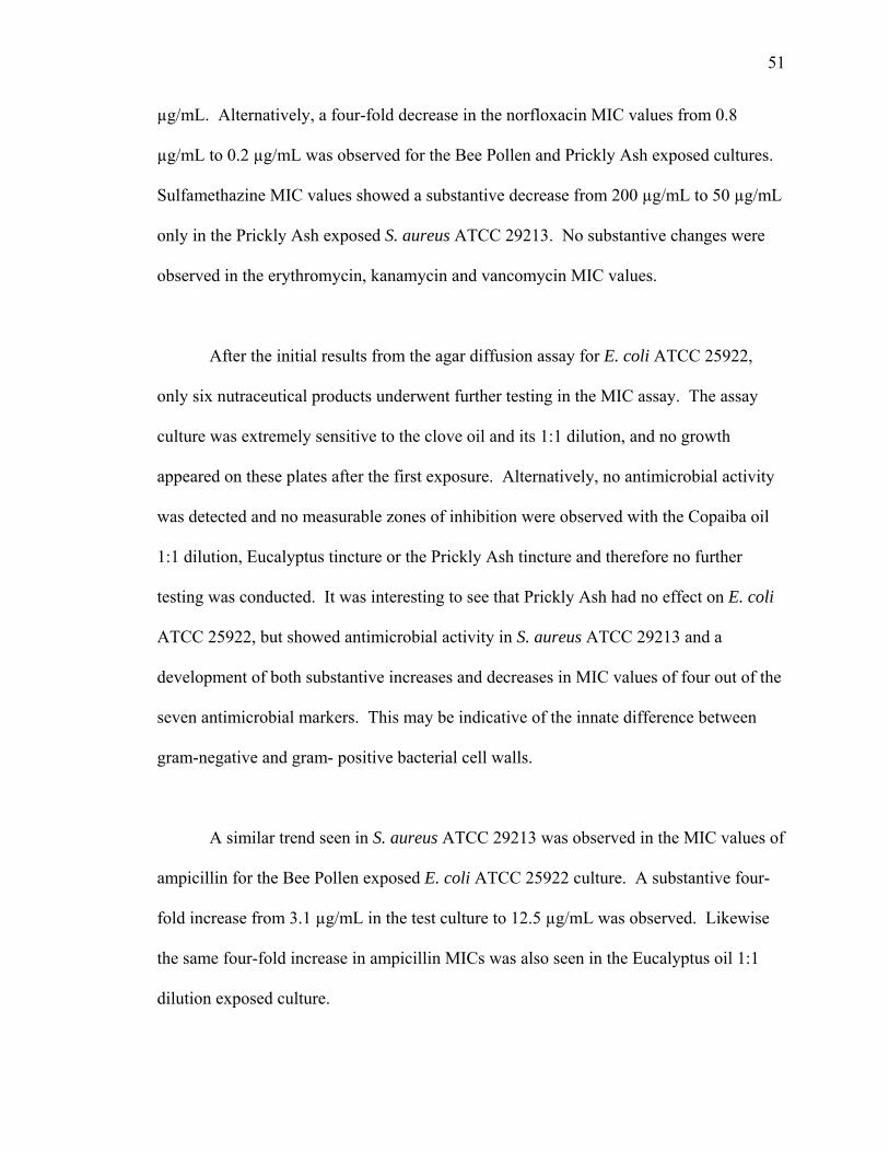
51
µg/mL. Alternatively, a four-fold decrease in the norfloxacin MIC values from 0.8
µg/mL to 0.2 µg/mL was observed for the Bee Pollen and Prickly Ash exposed cultures.
Sulfamethazine MIC values showed a substantive decrease from 200 µg/mL to 50 µg/mL
only in the Prickly Ash exposed S. aureus ATCC 29213. No substantive changes were
observed in the erythromycin, kanamycin and vancomycin MIC values.
After the initial results from the agar diffusion assay for E. coli ATCC 25922,
only six nutraceutical products underwent further testing in the MIC assay. The assay
culture was extremely sensitive to the clove oil and its 1:1 dilution, and no growth
appeared on these plates after the first exposure. Alternatively, no antimicrobial activity
was detected and no measurable zones of inhibition were observed with the Copaiba oil
1:1 dilution, Eucalyptus tincture or the Prickly Ash tincture and therefore no further
testing was conducted. It was interesting to see that Prickly Ash had no effect on E. coli
ATCC 25922, but showed antimicrobial activity in S. aureus ATCC 29213 and a
development of both substantive increases and decreases in MIC values of four out of the
seven antimicrobial markers. This may be indicative of the innate difference between
gram-negative and gram- positive bacterial cell walls.
A similar trend seen in S. aureus ATCC 29213 was observed in the MIC values of
ampicillin for the Bee Pollen exposed E. coli ATCC 25922 culture. A substantive four-
fold increase from 3.1 µg/mL in the test culture to 12.5 µg/mL was observed. Likewise
the same four-fold increase in ampicillin MICs was also seen in the Eucalyptus oil 1:1
dilution exposed culture.

52
Conclusions
The results of this study are indicative of the fact that exposure to nutraceuticals
can have a direct impact on the effectiveness of antimicrobials and/or antibiotics. This is
important because an increase in resistance can lead to problems in terms of antibiotic
efficacy and an increase in resistant organisms. A trend showing increases in sensitivity,
which was observed in both test strains more frequently in this study, may be the key to
discovering ways to enhance antibiotics used in therapies.
The chemical composition of the nutraceutical tinctures is very complex and it is
unsure if it is one particular active ingredient or a combination that is increasing the
sensitivity or resistance of the test organisms to the antibiotic markers. Further
experiments on the chemical components of the active tinctures would need to be
conducted. Chemically separating the active ingredients contained in each extract,
particularly the Bee Pollen and Prickly Ash tinctures and Eucalyptus essential oil might
yield interesting results. Once separated, each individual active ingredient could be
assayed against E. coli ATCC 25922 and S. aureus ATCC 29213 in both the agar
diffusion assay and the MIC assay. It should be noted however, that the MIC assay
proved to be a much better indicator of sensitivity and resistance than the agar diffusion
assay. Consistently, it was shown in this study that there were no real parallels between
the agar diffusion assay and MIC assay.

53
References
1. Raskin, I., Ribnicky, D.M., Komarnytsky, S., Ilic, N., & Poulev, A. (2002). Plants and human health in the twenty-first century. TRENDS in Biotechnology, 20(12), 522-531.
2. Balch, P.A. (2002). Prescription for herbal healing. New York: Penguin
Putnam, Inc., p. 4, 40, 49, 52, 64, 65, 109, 110, 141. 3. Tyler, V.E. (2000). Herbal medicine: from the past to the future. Public Health
Nutrition, 3(4A), 447-452.
4. Mowrey, D.B. (1990). Next Generation herbal medicine, 2nd Ed. New Canaan: Keats Publishing, Inc., (5)
5. Andlauer, W, & Furst, P. (2002). Nutraceuticals: a piece of history, present
status and outlook. Food Research International, 35, 171-176. 6. Burdock, G.A., Carabin, I.G., & Griffiths, J.C. (2006). The Importance of GRAS
to the functional food and nutraceutical industries. Toxicology, 221(1), 17-27.
7. Bagchi, D. (2006). Nutraceuticals and functional foods regulations in the United States and around the world. Toxicology, 221, 1-3.
8. Espín, J.C., García-Conesa, M.T., & Tomás-Barberán, F.A. (2007).
Nutraceuticals: facts and fiction. Phytochemistry, 68(22-24), 2986-3008. 9. Jones, P.J. (2002). Clinical nutrition: 7. Functional foods - more than just
nutrition. CMAJ, 166(12), 1555-1563.
10. Hasler, C.M. (2000). The Changing face of functional foods. Journal of the American College of Nutrition, 19(5), 499S-506S.
11. Burdock, G.A., & Carabin, I.G. (2004). Generally recognized as safe (GRAS):
history and description. Toxicology Letters, 150(1), 3-18.
12. Burdock, G.A. (2000). Dietary supplements and lessons to be learned from GRAS. Regulatory Toxicology and Pharmacology, 31(1), 68-76.
13. Nesheim, M.C. (1999). What is the Research base for the use of dietary
supplements?. Public Health Nutrition, 2(1), 35-38.
14. Noonan, C., & Noonan, W.P. (2006). Marketing dietary supplements in the United States: a review of the requirements for new dietary ingredients. Toxicology, 221(1), 4-8.
54
15. Still, C.C. (1998). Botany and healing medicinal plants of New Jersey and the region. New Brunswick: Rutgers University Press, p. 11, 12, 114, 115, 185, 186.
16. González-Lamothe, R., Mitchell, G., Gattuso, M., Diarra, M.S., Malouin, F., &
Bouarab, K. (2009). Plant antimicrobial agents and their effects on plant and human pathogens. International Journal of Molecular Sciences, 10(8), 3400-3419.
17. Cowan, M.M. (1999). Plant products as antimicrobial agents. Clinical
Microbiology Reviews, 12(4), 564-582.
18. Hoffman, F., & Manning, M. (2002). Herbal medicine and botanical medical fads. New York: Haworth Press, p. 29-43.
19. Rotblatt, M., & Ziment, I. (2002). Evidence based herbal medicine.
Philadelphia: Haley & Belfus, Inc., p. 50, 51, 65, 66.
20. Kenner, D., & Requena, Y. (2001). Botanical medicine: a European professional perspective. Brookline: Paradigm Publications, p. 8-14.
21. Mahboobi, M., Shahcheraghi, F., & Feizabadi, M.M. (2006). Bactericidal effects
of essential oils from clove, lavender and geranium on multi-drug resistant isolates of Pseudomonas aeruginosa. Iranian Journal of Biotechnology, 4(2), 137-140.
22. Foster, S, & Tyler, V.E. (1999). Tyler's honest herbal: a sensible guide to the
use of herbs and related remedies, 4th Ed. New York: The Haworth Press, p. 85, 86, 299-301.
23. Healthcare, T. (2000). PDR for herbal medicines, 2nd Ed. Montvale: Medical
Economics Company, Inc., p. 195, 196, 220, 283-285, 497-499, 595, 793, 794.
24. Gomes, H.E., Vieira, M.C., & Heredia, Z.N.A. (2007). Density and plant arrangement on Calendula officinalis L. yield. Rev. Bras. PL. Med., 9(3), 117-123.
25. Nascimento, G.G.F., Locatelli, J., Freitas, P.C., & Silva, G.L. (2000).
Antibacterial activity of plant extracts and phytochemicals on antibiotic-resistant bacteria. Brazilian Journal of Microbiology, 31(1), 247-256.
26. Pacheco, T.A.R.C., Barata, L.E.S., & Duarte, M.C.T. (2006). Antimicrobial
activity of copaiba (Copaifera spp) balsams. Rev. Bras. Pl. Med., 8, 123-124.
27. Oliveira dos Santos, A., Ueda-Nakamura, T., Prado Dias Filho, B., Veiga, V.F., Nakamura, C.V. & Pinto, A.C. (2008). Antimicrobial activity of Brazilian

55
copaiba oils obtained from different species of the Copaifera genus. Mem. Inst. Oswaldo Cruz, 103(3), 277-281.
28. Sartorelli, P., Maquioreto, A.D., Amaral-Baroli, A., Lima, M.E.L., & Moreno,
P.R.H. (2007). Chemical composition and antimicrobial activity of the essential oils from two species of Eucalyptus. Phytotherapy Research, 21, 231-233.
29. Trivedi, N.A., & Hotchandani, S.C. (2004). A study of the antimicrobial activity
of oil of Eucalyptus. Indian J. Pharmacol. 36, 93-94. 30. Carpes, S.T., Begnini, R., Matias de Alencar, S., & Masson, M.L. (2007). Study
of preparations of bee pollen extracts, antioxidant and antibacterial activity. Ciênc. Agrotec. 31(6), 1818-1825.
31. Kňazovická, V., Melich, M., Kačániová, M., Fikselová, M., Haščík, P. &
Chlebo, R. (2009). Antimicrobial activity of selected bee products. Acta fytotechnica et zootechnica, 12, 280-285.
32. Borchardt, J.R., Wyse, D.L., Sheaffer, C.C., Kauppi, K.L., & Fulcher, R.G.
(2008). Antioxidant and antimicrobial activity of seeds from plants of the Mississippi river basin. Journal of Medicinal Plants Research, 2(4), 81-93.
33. Borchardt, J.R., Wyse, D.L., Sheaffer, C.C., Kauppi, K.L., & Fulcher, R.G.
(2008). Antimicrobial activity of native and naturalized plants of Minnesota and Wisconsin. Journal of Medicinal Plants Research, 2(5), 98-110.
34. Ward, PML, Fasitsas, S, & Katz, SE. (2001). Inhibition, resistance development,
and increased antibiotic and antimicrobial resistance caused by nutraceuticals. Journal of Food Protection, 65(3), 528-533.