Surviving Sepsis Campaign: International guidelines for ...€¦ · Erasme University Hospital,...
44
Intensive Care Med (2008) 34:17–60 DOI 10.1007/s00134-007-0934-2 SPECIAL ARTICLE R. Phillip Dellinger Mitchell M. Levy Jean M. Carlet Julian Bion Margaret M. Parker Roman Jaeschke Konrad Reinhart Derek C. Angus Christian Brun-Buisson Richard Beale Thierry Calandra Jean-Francois Dhainaut Herwig Gerlach Maurene Harvey John J. Marini John Marshall Marco Ranieri Graham Ramsay Jonathan Sevransky B. Taylor Thompson Sean Townsend Jeffrey S. Vender Janice L. Zimmerman Jean-Louis Vincent Surviving Sepsis Campaign: International guidelines for management of severe sepsis and septic shock: 2008 Received: 3 August 2007 Accepted: 25 October 2007 Published online: 4 December 2007 © Society of Critical Care Medicine 2007 The article will also be published in Critical Care Medicine. * Sponsor of 2004 guidelines; ** Sponsor of 2008 guidelines but did not participate formally in revision process; *** Members of the 2007 SSC Guidelines Committee are listed in Appendix I.; **** Please see Ap- pendix J for author disclosure information. for the International Surviving Sepsis Campaign Guidelines Committee***, **** Sponsoring Organizations: American Asso- ciation of Critical-Care Nurses*, American College of Chest Physicians*, American College of Emergency Physicians*, Cana- dian Critical Care Society, European Soci- ety of Clinical Microbiology and Infectious Diseases*, European Society of Intensive Care Medicine*, European Respiratory Society*, International Sepsis Forum*, Japanese Association for Acute Medicine, Japanese Society of Intensive Care Medi- cine, Society of Critical Care Medicine*, Society of Hospital Medicine**, Surgical Infection Society*, World Federation of Societies of Intensive and Critical Care Medicine**. Participation and endorsement by the German Sepsis Society and the Latin American Sepsis Institute. R. P. Dellinger (✉) Cooper University Hospital, One Cooper Plaza, 393 Dorrance, Camden 08103, NJ, USA e-mail: [email protected] M. M. Levy · S. Townsend Rhode Island Hospital, Providence RI, USA J. M. Carlet Hospital Saint-Joseph, Paris, France J. Bion Birmingham University, Birmingham, UK M. M. Parker SUNY at Stony Brook, Stony Brook NY, USA R. Jaeschke McMaster University, Hamilton, Ontario, Canada K. Reinhart Friedrich-Schiller-University of Jena, Jena, Germany D. C. Angus University of Pittsburgh, Pittsburgh PA, USA C. Brun-Buisson Hopital Henri Mondor, Créteil, France R. Beale Guy’s and St Thomas’ Hospital Trust, London, UK T. Calandra Centre Hospitalier Universitaire Vaudois, Lausanne, Switzerland J.-F. Dhainaut French Agency for Evaluation of Research and Higher Education, Paris, France H. Gerlach Vivantes-Klinikum Neukoelln, Berlin, Germany M. Harvey Consultants in Critical Care, Inc., Glenbrook NV, USA J. J. Marini University of Minnesota, St. Paul MN, USA
Transcript of Surviving Sepsis Campaign: International guidelines for ...€¦ · Erasme University Hospital,...
Intensive Care Med (2008) 34:17–60DOI 10.1007/s00134-007-0934-2 SPECIAL ARTICLE
R. Phillip DellingerMitchell M. LevyJean M. CarletJulian BionMargaret M. ParkerRoman JaeschkeKonrad ReinhartDerek C. AngusChristian Brun-BuissonRichard BealeThierry CalandraJean-Francois DhainautHerwig GerlachMaurene HarveyJohn J. MariniJohn MarshallMarco RanieriGraham RamsayJonathan SevranskyB. Taylor ThompsonSean TownsendJeffrey S. VenderJanice L. ZimmermanJean-Louis Vincent
Surviving Sepsis Campaign:International guidelines for managementof severe sepsis and septic shock: 2008
Received: 3 August 2007Accepted: 25 October 2007Published online: 4 December 2007© Society of Critical Care Medicine 2007
The article will also be published in CriticalCare Medicine.
* Sponsor of 2004 guidelines; ** Sponsorof 2008 guidelines but did not participateformally in revision process; *** Membersof the 2007 SSC Guidelines Committee arelisted in Appendix I.; **** Please see Ap-pendix J for author disclosure information.
for the International Surviving SepsisCampaign Guidelines Committee***, ****
Sponsoring Organizations: American Asso-ciation of Critical-Care Nurses*, AmericanCollege of Chest Physicians*, AmericanCollege of Emergency Physicians*, Cana-dian Critical Care Society, European Soci-ety of Clinical Microbiology and InfectiousDiseases*, European Society of IntensiveCare Medicine*, European RespiratorySociety*, International Sepsis Forum*,Japanese Association for Acute Medicine,Japanese Society of Intensive Care Medi-cine, Society of Critical Care Medicine*,Society of Hospital Medicine**, SurgicalInfection Society*, World Federation ofSocieties of Intensive and Critical Care
Medicine**. Participation and endorsementby the German Sepsis Society and the LatinAmerican Sepsis Institute.
R. P. Dellinger (�)Cooper University Hospital,One Cooper Plaza, 393 Dorrance,Camden 08103, NJ, USAe-mail: [email protected]
M. M. Levy · S. TownsendRhode Island Hospital,Providence RI, USA
J. M. CarletHospital Saint-Joseph,Paris, France
J. BionBirmingham University,Birmingham, UK
M. M. ParkerSUNY at Stony Brook,Stony Brook NY, USA
R. JaeschkeMcMaster University,Hamilton, Ontario, Canada
K. ReinhartFriedrich-Schiller-University of Jena,Jena, Germany
D. C. AngusUniversity of Pittsburgh,Pittsburgh PA, USA
C. Brun-BuissonHopital Henri Mondor,Créteil, France
R. BealeGuy’s and St Thomas’ Hospital Trust,London, UK
T. CalandraCentre Hospitalier Universitaire Vaudois,Lausanne, Switzerland
J.-F. DhainautFrench Agency for Evaluation of Researchand Higher Education,Paris, France
H. GerlachVivantes-Klinikum Neukoelln,Berlin, Germany
M. HarveyConsultants in Critical Care, Inc.,Glenbrook NV, USA
J. J. MariniUniversity of Minnesota,St. Paul MN, USA
18
J. MarshallSt. Michael’s Hospital,Toronto, Ontario, Canada
M. RanieriUniversità di Torino,Torino, Italy
G. RamsayWest Hertfordshire Health Trust,Hemel Hempstead, UK
J. SevranskyThe Johns Hopkins University Schoolof Medicine,Baltimore MD, USA
B. T. ThompsonMassachusetts General Hospital,Boston MA, USA
J. S. VenderEvanston Northwestern Healthcare,Evanston IL, USA
J. L. ZimmermanThe Methodist Hospital,Houston TX, USA
J.-L. VincentErasme University Hospital,Brussels, Belgium
Abstract Objective: To providean update to the original SurvivingSepsis Campaign clinical manage-ment guidelines, “Surviving SepsisCampaign guidelines for managementof severe sepsis and septic shock,”published in 2004. Design: ModifiedDelphi method with a consensusconference of 55 international ex-perts, several subsequent meetingsof subgroups and key individuals,teleconferences, and electronic-baseddiscussion among subgroups andamong the entire committee. Thisprocess was conducted independentlyof any industry funding. Methods:We used the GRADE system toguide assessment of quality of evi-dence from high (A) to very low (D)and to determine the strength ofrecommendations. A strong rec-ommendation [1] indicates that anintervention’s desirable effects clearlyoutweigh its undesirable effects (risk,burden, cost), or clearly do not. Weakrecommendations [2] indicate thatthe tradeoff between desirable andundesirable effects is less clear. Thegrade of strong or weak is consideredof greater clinical importance thana difference in letter level of quality
of evidence. In areas without completeagreement, a formal process of re-solution was developed and applied.Recommendations are grouped intothose directly targeting severe sepsis,recommendations targeting generalcare of the critically ill patient thatare considered high priority in severesepsis, and pediatric considerations.Results: Key recommendations,listed by category, include: earlygoal-directed resuscitation of theseptic patient during the first 6 hrsafter recognition (1C); blood culturesprior to antibiotic therapy (1C); imag-ing studies performed promptly toconfirm potential source of infection(1C); administration of broad-spectrum antibiotic therapy within1 hr of diagnosis of septic shock (1B)and severe sepsis without septic shock(1D); reassessment of antibiotic ther-apy with microbiology and clinicaldata to narrow coverage, when ap-propriate (1C); a usual 7–10 days ofantibiotic therapy guided by clinicalresponse (1D); source control withattention to the balance of risks andbenefits of the chosen method (1C);administration of either crystalloid orcolloid fluid resuscitation (1B); fluidchallenge to restore mean circulatingfilling pressure (1C); reduction in rateof fluid administration with risingfiling pressures and no improvementin tissue perfusion (1D); vasopressorpreference for norepinephrine ordopamine to maintain an initial targetof mean arterial pressure ≥ 65 mm Hg(1C); dobutamine inotropic therapywhen cardiac output remains lowdespite fluid resuscitation and com-bined inotropic/vasopressor therapy(1C); stress-dose steroid therapygiven only in septic shock after bloodpressure is identified to be poorlyresponsive to fluid and vasopressortherapy (2C); recombinant activatedprotein C in patients with severesepsis and clinical assessment of highrisk for death (2B except 2C for post-operative patients). In the absence oftissue hypoperfusion, coronary arterydisease, or acute hemorrhage, targeta hemoglobin of 7–9 g/dL (1B); a lowtidal volume (1B) and limitation ofinspiratory plateau pressure strategy(1C) for acute lung injury (ALI)/
acute respiratory distress syndrome(ARDS); application of at leasta minimal amount of positive end-expiratory pressure in acute lunginjury (1C); head of bed elevationin mechanically ventilated patientsunless contraindicated (1B); avoid-ing routine use of pulmonary arterycatheters in ALI/ARDS (1A); to de-crease days of mechanical ventilationand ICU length of stay, a conserva-tive fluid strategy for patients withestablished ALI/ARDS who are notin shock (1C); protocols for weaningand sedation/analgesia (1B); usingeither intermittent bolus sedation orcontinuous infusion sedation withdaily interruptions or lightening (1B);avoidance of neuromuscular blockers,if at all possible (1B); institutionof glycemic control (1B) targetinga blood glucose < 150 mg/dL afterinitial stabilization ( 2C ); equivalencyof continuous veno-veno hemofiltra-tion or intermittent hemodialysis(2B); prophylaxis for deep veinthrombosis (1A); use of stress ulcerprophylaxis to prevent upper GIbleeding using H2 blockers (1A) orproton pump inhibitors (1B); andconsideration of limitation of supportwhere appropriate (1D). Recommen-dations specific to pediatric severesepsis include: greater use of physicalexamination therapeutic end points(2C); dopamine as the first drug ofchoice for hypotension (2C); steroidsonly in children with suspected orproven adrenal insufficiency (2C);a recommendation against the use ofrecombinant activated protein C inchildren (1B). Conclusion: Therewas strong agreement among a largecohort of international experts regard-ing many level 1 recommendationsfor the best current care of patientswith severe sepsis. Evidenced-basedrecommendations regarding the acutemanagement of sepsis and septicshock are the first step toward im-proved outcomes for this importantgroup of critically ill patients.
Keywords Sepsis · Severe sepsis ·Septic shock · Sepsis syndrome ·Infection · GRADE · Guidelines ·Evidence-based medicine · SurvivingSepsis Campaign · Sepsis bundles

19
Introduction
Severe sepsis (acute organ dysfunction secondary to in-fection) and septic shock (severe sepsis plus hypotensionnot reversed with fluid resuscitation) are major healthcareproblems, affecting millions of individuals around theworld each year, killing one in four (and often more),and increasing in incidence [1–5]. Similar to polytrauma,acute myocardial infarction, or stroke, the speed andappropriateness of therapy administered in the initialhours after severe sepsis develops are likely to influenceoutcome. In 2004, an international group of experts in thediagnosis and management of infection and sepsis, repre-senting 11 organizations, published the first internationallyaccepted guidelines that the bedside clinician could use toimprove outcomes in severe sepsis and septic shock [6, 7].These guidelines represented Phase II of the SurvivingSepsis Campaign (SSC), an international effort to increaseawareness and improve outcomes in severe sepsis. Joinedby additional organizations, the group met again in 2006and 2007 to update the guidelines document using a newevidence-based methodology system for assessing qualityof evidence and strength of recommendations [8–11].
These recommendations are intended to provide guid-ance for the clinician caring for a patient with severe sepsisor septic shock. Recommendations from these guidelinescannot replace the clinician’s decision-making capability
• Underlying methodologyA RCTB Downgraded RCT or upgraded observational studiesC Well-done observational studiesD Case series or expert opinion• Factors that may decrease the strength of evidence1. Poor quality of planning and implementation of available RCTs suggesting high likelihood of bias2. Inconsistency of results (including problems with subgroup analyses)3. Indirectness of evidence (differing population, intervention, control, outcomes, comparison)4. Imprecision of results5. High likelihood of reporting bias• Main factors that may increase the strength of evidence1. Large magnitude of effect (direct evidence, relative risk (RR) > 2 with no plausible confounders)2. Very large magnitude of effect with RR > 5 and no threats to validity (by two levels)3. Dose response gradient
RCT, randomized controlled trial; RR, relative risk
Table 1 Determinationof the Quality of Evidence
Table 2 Factors Determining Strong vs. Weak Recommendation
What should be considered Recommended Process
Quality of evidence The lower the quality of evidence the less likely a strong recommendationRelative importance of the outcomes If values and preferences vary widely, a strong recommendation becomes less likelyBaseline risks of outcomes The higher the risk, the greater the magnitude of benefitMagnitude of relative risk including Larger relative risk reductions or larger increases in relative risk of harm make a strongbenefits, harms, and burden recommendation more or less likely respectivelyAbsolute magnitude of the effect The larger the absolute benefits and harms, the greater or
lesser likelihood respectively of a strong recommendationPrecision of the estimates of the effects The greater the precision the more likely is a strong recommendationCosts The higher the cost of treatment, the less likely a strong recommendation
when he or she is provided with a patient’s unique set ofclinical variables. Most of these recommendations are ap-propriate for the severe sepsis patient in both the intensivecare unit (ICU) and non-ICU settings. In fact the commit-tee believes that, currently, the greatest outcome improve-ment can be made through education and process changefor those caring for severe sepsis patients in the non-ICUsetting and across the spectrum of acute care. It should alsobe noted that resource limitations in some institutions andcountries may prevent physicians from accomplishing par-ticular recommendations.
Methods
Sepsis is defined as infection plus systemic manifestationsof infection (Table 1) [12]. Severe sepsis is defined assepsis plus sepsis-induced organ dysfunction or tissuehypoperfusion. The threshold for this dysfunction hasvaried somewhat from one severe sepsis research study toanother. An example of typical thresholds identificationof severe sepsis is shown in Table 2 [13]. Sepsis inducedhypotension is defined as a systolic blood pressure(SBP)of < 90 mm Hg or mean arterial pressure < 70 mm Hg ora SBP decrease > 40 mm Hg or < 2 SD below normalfor age in the absence of other causes of hypotension.Septic shock is defined as sepsis induced hypotension
20
persisting despite adequate fluid resuscitation. Sepsisinduced tissue hypoperfusion is defined as either septicshock, an elevated lactate or oliguria.
The current clinical practice guidelines build onthe first and second editions from 2001 (see below)and 2004 [6, 7, 14]. The 2001 publication incorporateda MEDLINE search for clinical trials in the preceding10 years, supplemented by a manual search of other rele-vant journals [14]. The 2004 publication incorporated theevidence available through the end of 2003. The currentpublication is based on an updated search into 2007 (seemethods and rules below).
The 2001 guidelines were coordinated by the Inter-national Sepsis Forum (ISF); the 2004 guidelines werefunded by unrestricted educational grants from industryand administered through the Society of Critical CareMedicine (SCCM), the European Society of IntensiveCare Medicine (ESICM), and ISF. Two of the SSCadministering organizations receive unrestricted industryfunding to support SSC activities (ESICM and SCCM), butnone of this funding was used to support the 2006–2007committee meetings.
It is important to distinguish between the process ofguidelines revision and the Surviving Sepsis Campaign.The Surviving Sepsis Campaign (SSC) is partially fundedby unrestricted educational industry grants, includingthose from Edwards LifeSciences, Eli Lilly and Com-pany, and Philips Medical Systems. SSC also receivedfunding from the Coalition for Critical Care Excellenceof the Society of Critical Care Medicine. The greatmajority of industry funding has come from Eli Lilly andCompany.
Current industry funding for the Surviving Sepsis Cam-paign is directed to the performance improvement initia-tive. No industry funding was used in the guidelines revi-sion process.
For both the 2004 and the 2006/2007 efforts there wereno members of the committee from industry, no industryinput into guidelines development, and no industry pres-ence at any of the meetings. Industry awareness or com-ment on the recommendations was not allowed. No mem-ber of the guideline committee received any honoraria forany role in the 2004 or 2006/2007 guidelines process. Thecommittee considered the issue of recusement of individ-ual committee members during deliberation and decisionmaking in areas where committee members had either fi-nancial or academic competing interests; however, consen-sus as to threshold for exclusion could not be reached. Al-ternatively, the committee agreed to ensure full disclosureand transparency of all committee members’ potential con-flicts at time of publication (see disclosures at the end ofthis document).
The guidelines process included a modified Delphimethod, a consensus conference, several subsequent meet-ings of subgroups and key individuals, teleconferencesand electronically based discussions among subgroups
and members of the entire committee and two follow-upnominal group meetings in 2007.
Subgroups were formed, each charged with updatingrecommendations in specific areas, including corti-costeroids, blood products, activated protein C, renalreplacement therapy, antibiotics, source control, andglucose control, etc. Each subgroup was responsible forupdating the evidence (into 2007, with major additionalelements of information incorporated into the evolvingmanuscript throughout 2006 and 2007). A separate searchwas performed for each clearly defined question. Thecommittee chair worked with subgroup heads to identifypertinent search terms that always included, at a minimum,sepsis, severe sepsis, septic shock and sepsis syndromecrossed against the general topic area of the subgroup aswell as pertinent key words of the specific question posed.All questions of the previous guidelines publications weresearched, as were pertinent new questions generated bygeneral topic related search or recent trials. Quality ofevidence was judged by pre-defined Grades of Recom-mendation, Assessment, Development and Evaluation(GRADE) criteria (see below). Significant educationof committee members on the GRADE approach wasperformed via email prior to the first committee meetingand at the first meeting. Rules were distributed concerningassessing the body of evidence and GRADE experts wereavailable for questions throughout the process. Subgroupsagreed electronically on draft proposals that were pre-sented to committee meetings for general discussion. InJanuary 2006, the entire group met during the 35th SCCMCritical Care Congress in San Francisco, California, USA.The results of that discussion were incorporated into thenext version of recommendations and again discussedusing electronic mail. Recommendations were finalizedduring nominal group meetings (composed of a subset ofthe committee members) at the 2007 SCCM (Orlando)and 2007 International Symposium on Intensive Care andEmergency Medicine (Brussels) meetings with recircu-lation of deliberations and decisions to the entire groupfor comment or approval. At the discretion of the chairand following adequate discussion, competing proposalsfor wording of recommendations or assigning strength ofevidence were resolved by formal voting. On occasions,voting was performed to give the committee a sense ofdistribution of opinions to facilitate additional discussion.The manuscript was edited for style and form by thewriting committee with final approval by section leads fortheir respective group assignment and then by the entirecommittee.
The development of guidelines and grading of recom-mendations for the 2004 guideline development processwere based on a system proposed by Sackett in 1989,during one of the first American College of Chest Physi-cians (ACCP) conferences on the use of antithrombotictherapies [15]. The revised guidelines recommendationsare based on the Grades of Recommendation, Assessment,
21
Development and Evaluation (GRADE) system – a struc-tured system for rating quality of evidence and gradingstrength of recommendation in clinical practice [8–11].The SSC Steering Committee and individual authorscollaborated with GRADE representatives to apply theGRADE system to the SSC guidelines revision process.The members of GRADE group were directly involved,either in person or via e-mail, in all discussions anddeliberations amongst the guidelines committee membersas to grading decisions. Subsequently, the SSC authorsused written material prepared by the GRADE groupand conferred with GRADE group members who wereavailable at the first committee meeting and subsequentnominal group meetings. GRADE representatives werealso used as a resource throughout subgroup delibera-tion.
The GRADE system is based on a sequential assess-ment of the quality of evidence, followed by assessmentof the balance between benefits versus risks, burden, andcost and, based on the above, development and grading ofa management recommendations [9–11]. Keeping the rat-ing of quality of evidence and strength of recommendationexplicitly separate constitutes a crucial and defining fea-ture of the GRADE approach. This system classifies qual-ity of evidence as high (Grade A), moderate (Grade B), low(Grade C), or very low (Grade D). Randomized trials be-gin as high quality evidence, but may be downgraded dueto limitations in implementation, inconsistency or impreci-sion of the results, indirectness of the evidence, and possi-ble reporting bias (see Table 1). Examples of indirectnessof the evidence include: population studied, interventionsused, outcomes measured, and how these relate to the ques-tion of interest. Observational (non-randomized) studiesbegin as low-quality evidence, but the quality level maybe upgraded on the basis of large magnitude of effect. Anexample of this is the quality of evidence for early admin-istration of antibiotics.
The GRADE system classifies recommendations asstrong (Grade 1) or weak (Grade 2). The grade of strongor weak is considered of greater clinical importance thana difference in letter level of quality of evidence. The com-mittee assessed whether the desirable effects of adherencewill outweigh the undesirable effects, and the strengthof a recommendation reflects the group’s degree of con-fidence in that assessment. A strong recommendation infavor of an intervention reflects that the desirable effectsof adherence to a recommendation (beneficial health out-comes, less burden on staff and patients, and cost savings)will clearly outweigh the undesirable effects (harms, moreburden and greater costs). A weak recommendation infavor of an intervention indicates that the desirable effectsof adherence to a recommendation probably will outweighthe undesirable effects, but the panel is not confident aboutthese tradeoffs – either because some of the evidence islow-quality (and thus there remains uncertainty regardingthe benefits and risks) or the benefits and downsides are
closely balanced. While the degree of confidence is a con-tinuum and there is a lack of a precise threshold betweena strong and a weak recommendation, the presence ofimportant concerns about one or more of the above factorsmakes a weak recommendation more likely. A “strong”recommendation is worded as “we recommend” anda weak recommendation as “we suggest.”
The implications of calling a recommendation “strong”are that most well-informed patients would accept thatintervention, and that most clinicians should use it inmost situations. There may be circumstances in whicha “strong” recommendation cannot or should not befollowed for an individual patient because of that patient’spreferences or clinical characteristics which make therecommendation less applicable. It should be noted thatbeing a “strong” recommendation does not automaticallyimply standard of care. For example, the strong recom-mendation for administering antibiotics within one hourof the diagnosis of severe sepsis, although desirable,is not currently standard of care as verified by currentpractice (personal communication, Mitchell Levy fromfirst 8,000 patients entered internationally into the SSCperformance improvement data base). The implication ofa “weak” recommendation is that although a majority ofwell-informed patients would accept it (but a substantialproportion would not), clinicians should consider its useaccording to particular circumstance.
Differences of opinion among committee membersabout interpretation of evidence, wording of proposals,or strength of recommendations were resolved usinga specifically developed set of rules. We will describe thisprocess in detail in a separate publication. In summary,the main approach for converting diverse opinions intoa recommendation was: 1. to give a recommendationa direction (for or against the given action). a majorityof votes were to be in favor of that direction, with nomore than 20% preferring the opposite direction (therewas a neutral vote allowed as well); 2. to call a givenrecommendation “strong” rather than “weak” at least 70%“strong” votes were required; 3. if fewer than 70% of votesindicated “strong” preference, the recommendation wasassigned a “weak” category of strength. We used a combi-nation of modified Delphi Process and Nominal (Expert)Group techniques to ensure both depth and breadth ofreview. The entire review group (together with theirparent organizations as required) participated in the larger,iterative, modified Delphi process. The smaller workinggroup meetings which took place in person functioned asthe Nominal Groups. If a clear consensus could not beobtained by polling within the Nominal Group meetings,the larger group was specifically asked to use the pollingprocess. This was only required for corticosteroids andglycemic control. The larger group had the opportunity toreview all outputs. In this way the entire review combinedintense focused discussion (Nominal Group) with broaderreview and monitoring using the Delphi process.
22
Note: Refer to Tables 3, 4, and 5 for condensed adultrecommentations.
I. Management of Severe Sepsis
A. Initial Resuscitation
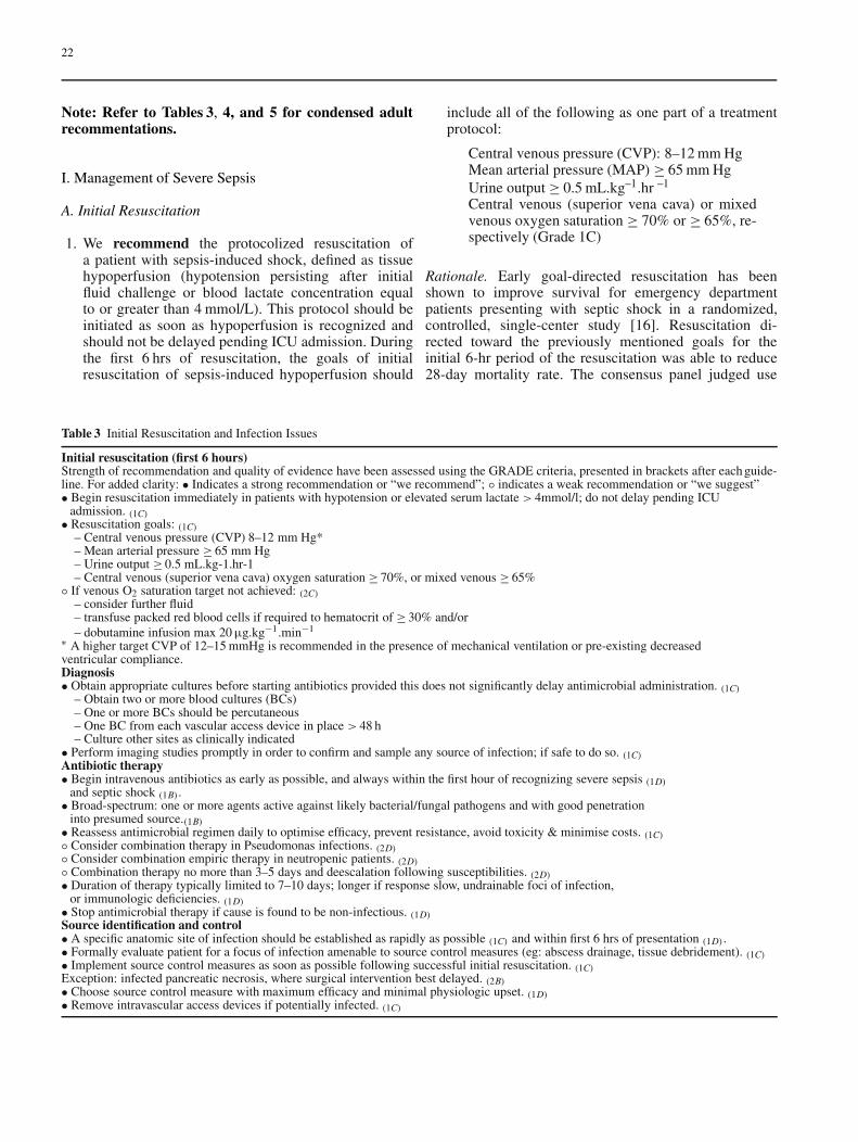
1. We recommend the protocolized resuscitation ofa patient with sepsis-induced shock, defined as tissuehypoperfusion (hypotension persisting after initialfluid challenge or blood lactate concentration equalto or greater than 4 mmol/L). This protocol should beinitiated as soon as hypoperfusion is recognized andshould not be delayed pending ICU admission. Duringthe first 6 hrs of resuscitation, the goals of initialresuscitation of sepsis-induced hypoperfusion should
Table 3 Initial Resuscitation and Infection Issues
Initial resuscitation (first 6 hours)Strength of recommendation and quality of evidence have been assessed using the GRADE criteria, presented in brackets after each guide-line. For added clarity: • Indicates a strong recommendation or “we recommend”; ◦ indicates a weak recommendation or “we suggest”• Begin resuscitation immediately in patients with hypotension or elevated serum lactate > 4mmol/l; do not delay pending ICU
admission. (1C)• Resuscitation goals: (1C)
– Central venous pressure (CVP) 8–12 mm Hg*– Mean arterial pressure ≥ 65 mm Hg– Urine output ≥ 0.5 mL.kg-1.hr-1– Central venous (superior vena cava) oxygen saturation ≥ 70%, or mixed venous ≥ 65%
◦ If venous O2 saturation target not achieved: (2C)
– consider further fluid– transfuse packed red blood cells if required to hematocrit of ≥ 30% and/or– dobutamine infusion max 20 µg.kg−1.min−1
∗ A higher target CVP of 12–15 mmHg is recommended in the presence of mechanical ventilation or pre-existing decreasedventricular compliance.Diagnosis• Obtain appropriate cultures before starting antibiotics provided this does not significantly delay antimicrobial administration. (1C)
– Obtain two or more blood cultures (BCs)– One or more BCs should be percutaneous– One BC from each vascular access device in place > 48 h– Culture other sites as clinically indicated
• Perform imaging studies promptly in order to confirm and sample any source of infection; if safe to do so. (1C)
Antibiotic therapy• Begin intravenous antibiotics as early as possible, and always within the first hour of recognizing severe sepsis (1D)
and septic shock (1B).• Broad-spectrum: one or more agents active against likely bacterial/fungal pathogens and with good penetrationinto presumed source.(1B)• Reassess antimicrobial regimen daily to optimise efficacy, prevent resistance, avoid toxicity & minimise costs. (1C)◦ Consider combination therapy in Pseudomonas infections. (2D)◦ Consider combination empiric therapy in neutropenic patients. (2D)◦ Combination therapy no more than 3–5 days and deescalation following susceptibilities. (2D)• Duration of therapy typically limited to 7–10 days; longer if response slow, undrainable foci of infection,or immunologic deficiencies. (1D)• Stop antimicrobial therapy if cause is found to be non-infectious. (1D)
Source identification and control• A specific anatomic site of infection should be established as rapidly as possible (1C) and within first 6 hrs of presentation (1D).• Formally evaluate patient for a focus of infection amenable to source control measures (eg: abscess drainage, tissue debridement). (1C)• Implement source control measures as soon as possible following successful initial resuscitation. (1C)
Exception: infected pancreatic necrosis, where surgical intervention best delayed. (2B)• Choose source control measure with maximum efficacy and minimal physiologic upset. (1D)• Remove intravascular access devices if potentially infected. (1C)
include all of the following as one part of a treatmentprotocol:
Central venous pressure (CVP): 8–12 mm HgMean arterial pressure (MAP) ≥ 65 mm HgUrine output ≥ 0.5 mL.kg–1.hr –1
Central venous (superior vena cava) or mixedvenous oxygen saturation ≥ 70% or ≥ 65%, re-spectively (Grade 1C)
Rationale. Early goal-directed resuscitation has beenshown to improve survival for emergency departmentpatients presenting with septic shock in a randomized,controlled, single-center study [16]. Resuscitation di-rected toward the previously mentioned goals for theinitial 6-hr period of the resuscitation was able to reduce28-day mortality rate. The consensus panel judged use
23
Table 4 Hemodynamic Support and Adjunctive Therapy
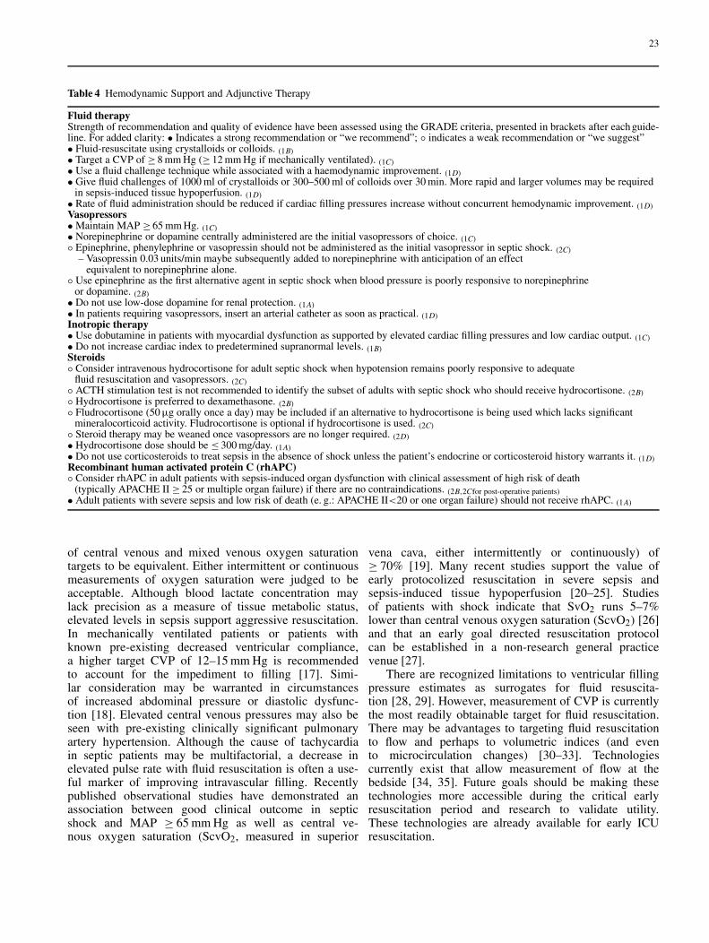
Fluid therapyStrength of recommendation and quality of evidence have been assessed using the GRADE criteria, presented in brackets after each guide-line. For added clarity: • Indicates a strong recommendation or “we recommend”; ◦ indicates a weak recommendation or “we suggest”• Fluid-resuscitate using crystalloids or colloids. (1B)• Target a CVP of ≥ 8 mm Hg (≥ 12 mm Hg if mechanically ventilated). (1C)• Use a fluid challenge technique while associated with a haemodynamic improvement. (1D)• Give fluid challenges of 1000 ml of crystalloids or 300–500 ml of colloids over 30 min. More rapid and larger volumes may be required
in sepsis-induced tissue hypoperfusion. (1D)• Rate of fluid administration should be reduced if cardiac filling pressures increase without concurrent hemodynamic improvement. (1D)
Vasopressors• Maintain MAP ≥ 65 mm Hg. (1C)• Norepinephrine or dopamine centrally administered are the initial vasopressors of choice. (1C)◦ Epinephrine, phenylephrine or vasopressin should not be administered as the initial vasopressor in septic shock. (2C)
– Vasopressin 0.03 units/min maybe subsequently added to norepinephrine with anticipation of an effectequivalent to norepinephrine alone.
◦ Use epinephrine as the first alternative agent in septic shock when blood pressure is poorly responsive to norepinephrineor dopamine. (2B)• Do not use low-dose dopamine for renal protection. (1A)• In patients requiring vasopressors, insert an arterial catheter as soon as practical. (1D)
Inotropic therapy• Use dobutamine in patients with myocardial dysfunction as supported by elevated cardiac filling pressures and low cardiac output. (1C)• Do not increase cardiac index to predetermined supranormal levels. (1B)
Steroids◦ Consider intravenous hydrocortisone for adult septic shock when hypotension remains poorly responsive to adequate
fluid resuscitation and vasopressors. (2C)◦ ACTH stimulation test is not recommended to identify the subset of adults with septic shock who should receive hydrocortisone. (2B)◦ Hydrocortisone is preferred to dexamethasone. (2B)◦ Fludrocortisone (50 µg orally once a day) may be included if an alternative to hydrocortisone is being used which lacks significantmineralocorticoid activity. Fludrocortisone is optional if hydrocortisone is used. (2C)◦ Steroid therapy may be weaned once vasopressors are no longer required. (2D)• Hydrocortisone dose should be ≤ 300 mg/day. (1A)• Do not use corticosteroids to treat sepsis in the absence of shock unless the patient’s endocrine or corticosteroid history warrants it. (1D)
Recombinant human activated protein C (rhAPC)◦ Consider rhAPC in adult patients with sepsis-induced organ dysfunction with clinical assessment of high risk of death
(typically APACHE II ≥ 25 or multiple organ failure) if there are no contraindications. (2B,2Cfor post-operative patients)• Adult patients with severe sepsis and low risk of death (e. g.: APACHE II<20 or one organ failure) should not receive rhAPC. (1A)
of central venous and mixed venous oxygen saturationtargets to be equivalent. Either intermittent or continuousmeasurements of oxygen saturation were judged to beacceptable. Although blood lactate concentration maylack precision as a measure of tissue metabolic status,elevated levels in sepsis support aggressive resuscitation.In mechanically ventilated patients or patients withknown pre-existing decreased ventricular compliance,a higher target CVP of 12–15 mm Hg is recommendedto account for the impediment to filling [17]. Simi-lar consideration may be warranted in circumstancesof increased abdominal pressure or diastolic dysfunc-tion [18]. Elevated central venous pressures may also beseen with pre-existing clinically significant pulmonaryartery hypertension. Although the cause of tachycardiain septic patients may be multifactorial, a decrease inelevated pulse rate with fluid resuscitation is often a use-ful marker of improving intravascular filling. Recentlypublished observational studies have demonstrated anassociation between good clinical outcome in septicshock and MAP ≥ 65 mm Hg as well as central ve-nous oxygen saturation (ScvO2, measured in superior
vena cava, either intermittently or continuously) of≥ 70% [19]. Many recent studies support the value ofearly protocolized resuscitation in severe sepsis andsepsis-induced tissue hypoperfusion [20–25]. Studiesof patients with shock indicate that SvO2 runs 5–7%lower than central venous oxygen saturation (ScvO2) [26]and that an early goal directed resuscitation protocolcan be established in a non-research general practicevenue [27].
There are recognized limitations to ventricular fillingpressure estimates as surrogates for fluid resuscita-tion [28, 29]. However, measurement of CVP is currentlythe most readily obtainable target for fluid resuscitation.There may be advantages to targeting fluid resuscitationto flow and perhaps to volumetric indices (and evento microcirculation changes) [30–33]. Technologiescurrently exist that allow measurement of flow at thebedside [34, 35]. Future goals should be making thesetechnologies more accessible during the critical earlyresuscitation period and research to validate utility.These technologies are already available for early ICUresuscitation.
24
Table 5 Other Supportive Therapy of Severe Sepsis
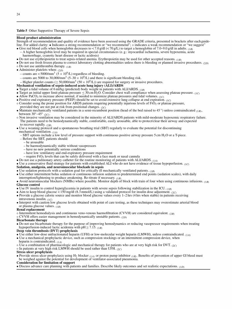
Blood product administrationStrength of recommendation and quality of evidence have been assessed using the GRADE criteria, presented in brackets after each guide-line. For added clarity: • Indicates a strong recommendation or “we recommend”; ◦ indicates a weak recommendation or “we suggest”• Give red blood cells when hemoglobin decreases to < 7.0 g/dl (< 70 g/L) to target a hemoglobin of 7.0–9.0 g/dl in adults. (1B)
– A higher hemoglobin level may be required in special circumstances (e. g.: myocardial ischaemia, severe hypoxemia, acutehaemorrhage, cyanotic heart disease or lactic acidosis)
• Do not use erythropoietin to treat sepsis-related anemia. Erythropoietin may be used for other accepted reasons. (1B)• Do not use fresh frozen plasma to correct laboratory clotting abnormalities unless there is bleeding or planned invasive procedures. (2D)◦ Do not use antithrombin therapy. (1B)• Administer platelets when: (2D)
– counts are < 5000/mm3 (5 × 10⁹/L) regardless of bleeding.– counts are 5000 to 30,000/mm3 (5–30 × 109/L) and there is significant bleeding risk.– Higher platelet counts (≥ 50,000/mm3 (50 × 109/L)) are required for surgery or invasive procedures.
Mechanical ventilation of sepsis-induced acute lung injury (ALI)/ARDS• Target a tidal volume of 6 ml/kg (predicted) body weight in patients with ALI/ARDS. (1B)• Target an initial upper limit plateau pressure ≤ 30 cm H2O. Consider chest wall compliance when assessing plateau pressure. (1C)• Allow PaCO2 to increase above normal, if needed to minimize plateau pressures and tidal volumes. (1C)• Positive end expiratory pressure (PEEP) should be set to avoid extensive lung collapse at end expiration. (1C)◦ Consider using the prone position for ARDS patients requiring potentially injurious levels of FiO2 or plateau pressure,
provided they are not put at risk from positional changes. (2C)• Maintain mechanically ventilated patients in a semi-recumbent position (head of the bed raised to 45 ◦) unless contraindicated (1B),between 30◦–45◦
(2C).◦ Non invasive ventilation may be considered in the minority of ALI/ARDS patients with mild-moderate hypoxemic respiratory failure.The patients need to be hemodynamically stable, comfortable, easily arousable, able to protect/clear their airway and expectedto recover rapidly. (2B)• Use a weaning protocol and a spontaneous breathing trial (SBT) regularly to evaluate the potential for discontinuing
mechanical ventilation. (1A)
– SBT options include a low level of pressure support with continuous positive airway pressure 5 cm H2O or a T-piece.– Before the SBT, patients should:
– be arousable– be haemodynamically stable without vasopressors– have no new potentially serious conditions– have low ventilatory and end-expiratory pressure requirement– require FiO2 levels that can be safely delivered with a face mask or nasal cannula
• Do not use a pulmonary artery catheter for the routine monitoring of patients with ALI/ARDS. (1A)• Use a conservative fluid strategy for patients with established ALI who do not have evidence of tissue hypoperfusion. (1C)
Sedation, analgesia, and neuromuscular blockade in sepsis• Use sedation protocols with a sedation goal for critically ill mechanically ventilated patients. (1B)• Use either intermittent bolus sedation or continuous infusion sedation to predetermined end points (sedation scales), with daily
interruption/lightening to produce awakening. Re-titrate if necessary. (1B)• Avoid neuromuscular blockers (NMBs) where possible. Monitor depth of block with train of four when using continuous infusions. (1B)
Glucose control• Use IV insulin to control hyperglycemia in patients with severe sepsis following stabilization in the ICU. (1B)• Aim to keep blood glucose < 150 mg/dl (8.3 mmol/L) using a validated protocol for insulin dose adjustment. (2C)• Provide a glucose calorie source and monitor blood glucose values every 1–2 hrs (4 hrs when stable) in patients receiving
intravenous insulin. (1C)• Interpret with caution low glucose levels obtained with point of care testing, as these techniques may overestimate arterial bloodor plasma glucose values. (1B)
Renal replacement◦ Intermittent hemodialysis and continuous veno-venous haemofiltration (CVVH) are considered equivalent. (2B)◦ CVVH offers easier management in hemodynamically unstable patients. (2D)
Bicarbonate therapy• Do not use bicarbonate therapy for the purpose of improving hemodynamics or reducing vasopressor requirements when treating
hypoperfusion-induced lactic acidemia with pH ≥ 7.15. (1B)
Deep vein thrombosis (DVT) prophylaxis• Use either low-dose unfractionated heparin (UFH) or low-molecular weight heparin (LMWH), unless contraindicated. (1A)• Use a mechanical prophylactic device, such as compression stockings or an intermittent compression device, when
heparin is contraindicated. (1A)◦ Use a combination of pharmacologic and mechanical therapy for patients who are at very high risk for DVT. (2C)◦ In patients at very high risk LMWH should be used rather than UFH. (2C)
Stress ulcer prophylaxis• Provide stress ulcer prophylaxis using H2 blocker (1A) or proton pump inhibitor (1B). Benefits of prevention of upper GI bleed must
be weighed against the potential for development of ventilator-associated pneumonia.Consideration for limitation of support• Discuss advance care planning with patients and families. Describe likely outcomes and set realistic expectations. (1D)
25
2. We suggest that during the first 6 hrs of resuscitation ofsevere sepsis or septic shock, if SCVO2 or SvO2 of 70%or 65% respectively is not achieved with fluid resusci-tation to the CVP target, then transfusion of packed redblood cells to achieve a hematocrit of ≥ 30% and/oradministration of a dobutamine infusion (up to a max-imum of 20 µg.kg–1.min–1) be utilized to achieve thisgoal (Grade 2C).
Rationale. The protocol used in the study cited previouslytargeted an increase in SCVO2 to ≥ 70% [16]. This wasachieved by sequential institution of initial fluid resusci-tation, then packed red blood cells, and then dobutamine.This protocol was associated with an improvementin survival. Based on bedside clinical assessment andpersonal preference, a clinician may deem either bloodtransfusion (if Hct is less than 30%) or dobutamine thebest initial choice to increase oxygen delivery and therebyelevate SCVO2. When fluid resuscitation is believed tobe already adequate. The design of the afore mentionedtrial did not allow assessment of the relative contributionof these two components (i. e. increasing O2 content orincreasing cardiac output) of the protocol on achievementof improved outcome.
B. Diagnosis
1. We recommend obtaining appropriate cultures beforeantimicrobial therapy is initiated if such cultures do notcause significant delay in antibiotic administration. Tooptimize identification of causative organisms, we rec-ommend at least two blood cultures be obtained prior toantibiotics with at least one drawn percutaneously andone drawn through each vascular access device, unlessthe device was recently (< 48 h) inserted. Cultures ofother sites (preferably quantitative where appropriate)such as urine, cerebrospinal fluid, wounds, respiratorysecretions, or other body fluids that may be the sourceof infection should also be obtained before antibiotictherapy if not associated with significant delay in anti-biotic administration (Grade 1C).
Rationale. Although sampling should not delay timelyadministration of antibiotics in patients with severe sepsis(example: lumbar puncture in suspected meningitis),obtaining appropriate cultures prior to their administrationis essential to confirm infection and the responsiblepathogen(s), and to allow de-escalation of antibiotictherapy after receipt of the susceptibility profile. Samplescan be kept in the refrigerator or frozen if processingcannot be performed immediately. Immediate transport toa microbiological lab is necessary. Because rapid steriliza-tion of blood cultures can occur within a few hours afterthe first antibiotic dose, obtaining those cultures beforestarting therapy is essential if the causative organism is
to be identified. Two or more blood cultures are recom-mended [36]. In patients with indwelling catheters (for> 48 h) at least one blood culture should be drawn througheach lumen of each vascular access device. Obtainingblood cultures peripherally and through a vascular accessdevice is an important strategy. If the same organismis recovered from both cultures, the likelihood that theorganism is causing the severe sepsis is enhanced. Inaddition, if the culture drawn through the vascular ac-cess device is positive much earlier than the peripheralblood culture (i. e., > 2 hrs earlier), the data support theconcept that the vascular access device is the source ofthe infection [37]. Quantitative cultures of catheter andperipheral blood are also useful for determining whetherthe catheter is the source of infection. Volume of blooddrawn with the culture tube should be at least 10 mL [38].Quantitative (or semi-quantitative) cultures of respiratorytract secretions are recommended for the diagnosis ofventilator-associated pneumonia [39]. Gram stain canbe useful, in particular for respiratory tract specimens,to help decide the micro-organisms to be targeted. Thepotential role of biomarkers for diagnosis of infection inpatients presenting with severe sepsis remains at presentundefined. The procalcitonin level, although often useful,is problematic in patients with an acute inflammatorypattern from other causes (e. g. post-operative, shock) [40]In the near future, rapid diagnostic methods (polymerasechain reaction, micro-arrays) might prove extremelyhelpful for a quicker identification of pathogens and majorantimicrobial resistance determinants [41].
2. We recommend that imaging studies be performedpromptly in attempts to confirm a potential source ofinfection. Sampling of potential sources of infectionshould occur as they are identified; however, somepatients may be too unstable to warrant certain in-vasive procedures or transport outside of the ICU.Bedside studies, such as ultrasound, are useful in thesecircumstances (Grade 1C).
Rationale. Diagnostic studies may identify a source ofinfection that requires removal of a foreign body or drain-age to maximize the likelihood of a satisfactory responseto therapy. However, even in the most organized andwell-staffed healthcare facilities, transport of patientscan be dangerous, as can placing patients in outside-unitimaging devices that are difficult to access and monitor.Balancing risk and benefit is therefore mandatory in thosesettings.
C. Antibiotic Therapy
1. We recommend that intravenous antibiotic therapybe started as early as possible and within the firsthour of recognition of septic shock (1B) and severesepsis without septic shock (1D). Appropriate cultures
26
should be obtained before initiating antibiotic therapy,but should not prevent prompt administration ofantimicrobial therapy (Grade 1D).
Rationale. Establishing vascular access and initiatingaggressive fluid resuscitation is the first priority whenmanaging patients with severe sepsis or septic shock.However, prompt infusion of antimicrobial agents shouldalso be a priority and may require additional vascularaccess ports [42, 43]. In the presence of septic shockeach hour delay in achieving administration of effectiveantibiotics is associated with a measurable increase inmortality [42]. If antimicrobial agents cannot be mixedand delivered promptly from the pharmacy, establishinga supply of premixed antibiotics for such urgent situationsis an appropriate strategy for ensuring prompt adminis-tration. In choosing the antimicrobial regimen, cliniciansshould be aware that some antimicrobial agents have theadvantage of bolus administration, while others requirea lengthy infusion. Thus, if vascular access is limited andmany different agents must be infused, bolus drugs mayoffer an advantage.
2a. We recommend that initial empirical anti-infectivetherapy include one or more drugs that have activityagainst all likely pathogens (bacterial and/or fungal)and that penetrate in adequate concentrations into thepresumed source of sepsis (Grade 1B).
Rationale. The choice of empirical antibiotics dependson complex issues related to the patient’s history includ-ing drug intolerances, underlying disease, the clinicalsyndrome, and susceptibility patterns of pathogens inthe community, in the hospital, and that previously havebeen documented to colonize or infect the patient. Thereis an especially wide range of potential pathogens forneutropenic patients.
Recently used antibiotics should generally be avoided.Clinicians should be cognizant of the virulence andgrowing prevalence of oxacillin (methicillin) resistantStaphylococcus aureus (ORSA or MRSA) in some com-munities and healthcare associated settings (especially inthe United States) when they choose empiric therapy. Ifthe prevalence is significant, and in consideration of thevirulence of this organism, empiric therapy adequate forthis pathogen would be warranted. Clinicians should alsoconsider whether Candidemia is a likely pathogen whenchoosing initial therapy. When deemed warranted, theselection of empiric antifungal therapy (e. g., fluconazole,amphotericin B, or echinocandin) will be tailored to thelocal pattern of the most prevalent Candida species, andany prior administration of azoles drugs [44]. Risk factorsfor candidemia should also be considered when choosinginitial therapy.
Because patients with severe sepsis or septic shockhave little margin for error in the choice of therapy,
the initial selection of antimicrobial therapy should bebroad enough to cover all likely pathogens. There isample evidence that failure to initiate appropriate therapy(i. e. therapy with activity against the pathogen that issubsequently identified as the causative agent) correlateswith increased morbidity and mortality [45–48].
Patients with severe sepsis or septic shock warrantbroad-spectrum therapy until the causative organismand its antibiotic susceptibilities are defined. Restrictionof antibiotics as a strategy to reduce the developmentof antimicrobial resistance or to reduce cost is not anappropriate initial strategy in this patient population.
All patients should receive a full loading dose of eachantimicrobial. However, patients with sepsis or septicshock often have abnormal renal or hepatic functionand may have abnormal volumes of distribution due toaggressive fluid resuscitation. Drug serum concentrationmonitoring can be useful in an ICU setting for those drugsthat can be measured promptly. An experienced physicianor clinical pharmacist should be consulted to ensure thatserum concentrations are attained that maximize efficacyand minimize toxicity [49–52].
2b. We recommend that the antimicrobial regimen be re-assessed daily to optimize activity, to prevent the de-velopment of resistance, to reduce toxicity, and to re-duce costs (Grade 1C).
Rationale. Although restriction of antibiotics as a strategyto reduce the development of antimicrobial resistanceor to reduce cost is not an appropriate initial strategyin this patient population, once the causative pathogenhas been identified, it may become apparent that noneof the empiric drugs offers optimal therapy; i. e., theremay be another drug proven to produce superior clin-ical outcome which should therefore replace empiricagents.
Narrowing the spectrum of antibiotic coverage and re-ducing the duration of antibiotic therapy will reduce thelikelihood that the patient will develop superinfection withpathogenic or resistant organisms such as Candida species,Clostridium difficile, or vancomycin-resistant Enterococ-cus faecium. However, the desire to minimize superinfec-tions and other complications should not take precedenceover the need to give the patient an adequate course of ther-apy to cure the infection that caused the severe sepsis orseptic shock.
2c. We suggest combination therapy for patients withknown or suspected Pseudomonas infections asa cause of severe sepsis (Grade 2D).
2d. We suggest combination empiric therapy for neu-tropenic patients with severe sepsis (Grade 2D).
2e. When used empirically in patients with severe sepsis,we suggest that combination therapy should not be ad-ministered for more than 3 to 5 days. De-escalation
27
to the most appropriate single therapy should be per-formed as soon as the susceptibility profile is known.(Grade 2D).
Rationale. Although no study or meta-analysis has con-vincingly demonstrated that combination therapy producesa superior clinical outcome for individual pathogens ina particular patient group, combination therapies doproduce in vitro synergy against pathogens in somemodels (although such synergy is difficult to define andpredict). In some clinical scenarios, such as the two above,combination therapies are biologically plausible and arelikely clinically useful even if evidence has not demon-strated improved clinical outcome [53–56]. Combinationtherapy for suspected known Pseudomonas pendingsensitivities increases the likelihood that at least one drugis effective against that strain and positively affects out-come [57].
3. We recommend that the duration of therapy typicallybe 7–10 days; longer courses may be appropriate in pa-tients who have a slow clinical response, undrainablefoci of infection, or who have immunologic deficien-cies including neutropenia (Grade 1D).
4. If the presenting clinical syndrome is determined to bedue to a noninfectious cause, we recommend antimi-crobial therapy be stopped promptly to minimize thelikelihood that the patient will become infected withan antibiotic resistant pathogen or will develop a drugrelated adverse effect (Grade 1D).
Rationale. Clinicians should be cognizant that blood cul-tures will be negative in more than 50% of cases of se-vere sepsis or septic shock, yet many of these cases arevery likely caused by bacteria or fungi. Thus, the decisionsto continue, narrow, or stop antimicrobial therapy must bemade on the basis of clinician judgment and clinical infor-mation.
D. Source Control
1a. We recommend that a specific anatomic diagnosisof infection requiring consideration for emergentsource control- for example necrotizing fasciitis,diffuse peritonitis, cholangitis, intestinal infarction– be sought and diagnosed or excluded as rapidlyas possible (Grade 1C) and within the first 6 hoursfollowing presentation (Grade 1D).
1b. We further recommend that all patients presentingwith severe sepsis be evaluated for the presenceof a focus of infection amenable to source controlmeasures, specifically the drainage of an abscessor local focus of infection, the debridement of in-fected necrotic tissue, the removal of a potentiallyinfected device, or the definitive control of a source
of ongoing microbial contamination (Grade 1C) (seeAppendix A for examples of potential sites needingsource control).
2. We suggest that when infected peripancreatic necrosisis identified as a potential source of infection, defini-tive intervention is best delayed until adequate demar-cation of viable and non-viable tissues has occurred(Grade 2B).
3. We recommend that when source control is required,the effective intervention associated with the leastphysiologic insult be employed e. g., percutaneousrather than surgical drainage of an abscess (Grade1D).
4. We recommend that when intravascular accessdevices are a possible source of severe sepsis or septicshock, they be promptly removed after establishingother vascular access (Grade 1C).
Rationale. The principles of source control in the man-agement of sepsis include a rapid diagnosis of thespecific site of infection, and identification of a focus ofinfection amenable to source control measures (specif-ically the drainage of an abscess, the debridement ofinfected necrotic tissue, the removal of a potentiallyinfected device, and the definitive control of a sourceof ongoing microbial contamination) [58]. Foci of in-fection readily amenable to source control measuresinclude an intra-abdominal abscess or gastrointestinalperforation, cholangitis or pyelonephritis, intestinal is-chemia or necrotizing soft tissue infection, and otherdeep space infection such as an empyema or septicarthritis. Such infectious foci should be controlled assoon as possible following successful initial resuscita-tion [59], accomplishing the source control objectivewith the least physiologic upset possible (e. g., percuta-neous rather than surgical drainage of an abscess [60],endoscopic rather than surgical drainage of biliarytree), and removing intravascular access devices thatare potentially the source of severe sepsis or septicshock promptly after establishing other vascular ac-cess [61, 62]. A randomized, controlled trial comparingearly vs. delayed surgical intervention for peripancre-atic necrosis showed better outcomes with a delayedapproach [63]. However, areas of uncertainty, such asdefinitive documentation of infection and appropriatelength of delay exist. The selection of optimal sourcecontrol methods must weigh benefits and risks of thespecific intervention as well as risks of transfer [64].Source control interventions may cause further compli-cations such as bleeding, fistulas, or inadvertent organinjury. Surgical intervention should be considered whenlesser interventional approaches are inadequate, or whendiagnostic uncertainty persists despite radiological eval-uation. Specific clinical situations require considerationof available choices, patient’s preferences, and clinician’sexpertise.
28
E. Fluid Therapy
1. We recommend fluid resuscitation with either nat-ural/artificial colloids or crystalloids. There is noevidence-based support for one type of fluid overanother (Grade 1B).
Rationale. The SAFE study indicated albumin adminis-tration was safe and equally effective as crystalloid [65].There was an insignificant decrease in mortality rates withthe use of colloid in a subset analysis of septic patients(p = 0.09). Previous meta-analyses of small studies of ICUpatients had demonstrated no difference between crystal-loid and colloid fluid resuscitation [66–68]. Although ad-ministration of hydroxyethyl starch may increase the riskof acute renal failure in patients with sepsis variable find-ings preclude definitive recommendations [69, 70]. As thevolume of distribution is much larger for crystalloids thanfor colloids, resuscitation with crystalloids requires morefluid to achieve the same end points and results in moreedema. Crystalloids are less expensive.
2. We recommend fluid resuscitation initially targeta CVP of at least 8 mm Hg (12 mm Hg in mechani-cally ventilated patients). Further fluid therapy is oftenrequired (Grade 1C).
3a. We recommend that a fluid challenge technique beapplied, wherein fluid administration is continuedas long as the hemodynamic improvement (e. g.,arterial pressure, heart rate, urine output) continues(Grade 1D).
3b. We recommend fluid challenge in patients withsuspected hypovolemia be started with at least1000 mL of crystalloids or 300–500 mL of colloidsover 30 min. More rapid administration and greateramounts of fluid may be needed in patients with sepsisinduced tissue hypoperfusion (see initial resuscitationrecommendations) (Grade 1D).
3c. We recommend the rate of fluid administration bereduced substantially when cardiac filling pressures(CVP or pulmonary artery balloon-occluded pres-sure) increase without concurrent hemodynamicimprovement (Grade 1D).
Rationale. Fluid challenge must be clearly separated fromsimple fluid administration; it is a technique in which largeamounts of fluids are administered over a limited periodof time under close monitoring to evaluate the patient’s re-sponse and avoid the development of pulmonary edema.The degree of intravascular volume deficit in patients withsevere sepsis varies. With venodilation and ongoing capil-lary leak, most patients require continuing aggressive fluidresuscitation during the first 24 hours of management. In-put is typically much greater than output, and input/outputratio is of no utility to judge fluid resuscitation needs dur-ing this time period.
F. Vasopressors
1. We recommend mean arterial pressure (MAP) bemaintained ≥ 65 mm Hg (Grade 1C).
Rationale. Vasopressor therapy is required to sustain lifeand maintain perfusion in the face of life-threateninghypotension, even when hypovolemia has not yet beenresolved. Below a certain mean arterial pressure, autoreg-ulation in various vascular beds can be lost, and perfusioncan become linearly dependent on pressure. Thus, somepatients may require vasopressor therapy to achievea minimal perfusion pressure and maintain adequateflow [71, 72]. The titration of norepinephrine to as lowas MAP 65 mm Hg has been shown to preserve tissueperfusion [72]. In addition, pre-existing comorbiditiesshould be considered as to most appropriate MAP target.For example, a MAP of 65 mm Hg might be too low ina patient with severe uncontrolled hypertension, and ina young previously normotensive, a lower MAP mightbe adequate. Supplementing end points such as bloodpressure with assessment of regional and global perfusion,such as blood lactate concentrations and urine output, isimportant. Adequate fluid resuscitation is a fundamentalaspect of the hemodynamic management of patients withseptic shock, and should ideally be achieved before vaso-pressors and inotropes are used, but using vasopressorsearly as an emergency measure in patients with severeshock is frequently necessary. When that occurs greateffort should be directed to weaning vasopressors withcontinuing fluid resuscitation.
2. We recommend either norepinephrine or dopamine asthe first choice vasopressor agent to correct hypoten-sion in septic shock (administered through a centralcatheter as soon as one is available) (Grade 1C).
3a. We suggest that epinephrine, phenylephrine, orvasopressin should not be administered as the initialvasopressor in septic shock (Grade 2C). Vasopressin.03 units/min may be subsequently added to nore-pinephrine with anticipation of an effect equivalent tonorepinephrine alone.
3b. We suggest that epinephrine be the first chosen alter-native agent in septic shock that is poorly responsiveto norepinephrine or dopamine (Grade 2B).
Rationale. There is no high-quality primary evidence torecommend one catecholamine over another. Much litera-ture exists that contrasts the physiologic effects of choiceof vasopressor and combined inotrope/vasopressors inseptic shock [73–85]. Human and animal studies suggestsome advantages of norepinephrine and dopamine overepinephrine (the latter with the potential for tachycardia aswell as disadvantageous effects on splanchnic circulationand hyperlactemia) and phenylephrine (decrease in strokevolume). There is, however, no clinical evidence that
29
epinephrine results in worse outcomes, and it should bethe first chosen alternative to dopamine or norepinephrine.Phenylephrine is the adrenergic agent least likely toproduce tachycardia, but as a pure vasopressor would beexpected to decrease stroke volume. Dopamine increasesmean arterial pressure and cardiac output, primarilydue to an increase in stroke volume and heart rate.Norepinephrine increases mean arterial pressure due toits vasoconstrictive effects, with little change in heartrate and less increase in stroke volume compared withdopamine. Either may be used as a first-line agent to cor-rect hypotension in sepsis. Norepinephrine is more potentthan dopamine and may be more effective at reversinghypotension in patients with septic shock. Dopaminemay be particularly useful in patients with compromisedsystolic function but causes more tachycardia and maybe more arrhythmogenic [86]. It may also influence theendocrine response via the hypothalamic-pituitary axisand have immunosuppressive effects.
Vasopressin levels in septic shock have been reportedto be lower than anticipated for a shock state [87]. Lowdoses of vasopressin may be effective in raising bloodpressure in patients refractory to other vasopressors, andmay have other potential physiologic benefits [88–93].Terlipressin has similar effects but is long lasting [94].Studies show that vasopressin concentrations are elevatedin early septic shock, but with continued shock, con-centration decreases to normal range in the majority ofpatients between 24 and 48 hrs [95]. This has been called“relative vasopressin deficiency” because in the presenceof hypotension, vasopressin would be expected to beelevated. The significance of this finding is unknown.The recent VASST trial, a randomized, controlled trialcomparing norepinephrine alone to norepinephrine plusvasopressin at .03 units per minute showed no differencein outcome in the intent to treat population. An a prioridefined subgroup analysis showed that the survival ofpatients receiving less than 15 µg/min norepinephrine atthe time of randomization was better with vasopressin. Itshould be noted however that the pre-trial rationale forthis stratification was based on exploring potential benefitin the 15 µg or greater norepinephrine requirement popu-lation. Higher doses of vasopressin have been associatedwith cardiac, digital, and splanchnic ischemia and shouldbe reserved for situations where alternative vasopressorshave failed [96]. Cardiac output measurement to allowmaintenance of a normal or elevated flow is desirablewhen these pure vasopressors are instituted.
5. We recommend that low dose dopamine not be usedfor renal protection (Grade 1A).
Rationale. A large randomized trial and meta-analysiscomparing low-dose dopamine to placebo found no differ-ence in either primary outcomes (peak serum creatinine,need for renal replacement, urine output, time to recovery
of normal renal function), or secondary outcomes (survivalto either ICU or hospital discharge, ICU stay, hospitalstay, arrhythmias) [97, 98]. Thus the available data do notsupport administration of low doses of dopamine solely tomaintain renal function.
6. We recommend that all patients requiring vasopressorshave an arterial line placed as soon as practical if re-sources are available (Grade 1D).
Rationale. In shock states, estimation of blood pressureusing a cuff is commonly inaccurate; use of an arterial can-nula provides a more appropriate and reproducible meas-urement of arterial pressure. These catheters also allowcontinuous analysis so that decisions regarding therapy canbe based on immediate and reproducible blood pressure in-formation.
G. Inotropic Therapy
1. We recommend a dobutamine infusion be adminis-tered in the presence of myocardial dysfunction assuggested by elevated cardiac filling pressures and lowcardiac output (Grade 1C).
2. We recommend against the use of a strategy toincrease cardiac index to predetermined supranormallevels (Grade 1B).
Rationale. Dobutamine is the first-choice inotrope forpatients with measured or suspected low cardiac output inthe presence of adequate left ventricular filling pressure (orclinical assessment of adequate fluid resuscitation) and ad-equate mean arterial pressure. Septic patients who remainhypotensive after fluid resuscitation may have low, normal,or increased cardiac outputs. Therefore, treatment witha combined inotrope/vasopressor such as norepinephrineor dopamine is recommended if cardiac output is not mea-sured. When the capability exists for monitoring cardiacoutput in addition to blood pressure, a vasopressor such asnorepinephrine may be used separately to target specificlevels of mean arterial pressure and cardiac output. Twolarge prospective clinical trials that included critically illICU patients who had severe sepsis failed to demonstratebenefit from increasing oxygen delivery to supranormaltargets by use of dobutamine [99, 100]. These studiesdid not target specifically patients with severe sepsis anddid not target the first 6 hours of resuscitation. The first6 hours of resuscitation of sepsis induced hypoperfusionneed to be treated separately from the later stages of severesepsis (see initial resuscitation recommendations).
H. Corticosteroids
1. We suggest intravenous hydrocortisone be given onlyto adult septic shock patients after blood pressure is
30
identified to be poorly responsive to fluid resuscitationand vasopressor therapy (Grade 2C).
Rationale. One french multi-center, randomized, con-trolled trial (RCT) of patients in vasopressor-unresponsiveseptic shock (hypotension despite fluid resuscitationand vasopressors) showed a significant shock reversaland reduction of mortality rate in patients with relativeadrenal insufficiency (defined as post-adrenocorticotropichormone (ACTH) cortisol increase 9 µg/dL or less) [101].Two additional smaller RCTs also showed significanteffects on shock reversal with steroid therapy [102, 103].However, a recent large, European multicenter trial (COR-TICUS), which has been presented in abstract form butnot yet published, failed to show a mortality benefit withsteroid therapy of septic shock [104]. CORTICUS didshow a faster resolution of septic shock in patients whoreceived steroids. The use of the ACTH test (respondersand nonresponders) did not predict the faster resolutionof shock. Importantly, unlike the French trial, whichonly enrolled shock patients with blood pressure unre-sponsive to vasopressor therapy, the CORTICUS studyincluded patients with septic shock, regardless of howthe blood pressure responded to vasopressors. Althoughcorticosteroids do appear to promote shock reversal, thelack of a clear improvement in mortality-coupled withknown side effects of steroids such as increased risk ofinfection and myopathy-generally tempered enthusiasmfor their broad use. Thus, there was broad agreementthat the recommendation should be downgraded from theprevious guidelines (Appendix B). There was considerablediscussion and consideration by the committee on theoption of encouraging use in those patients whose bloodpressure was unresponsive to fluids and vasopressors,while strongly discouraging use in subjects whose shockresponded well to fluids and pressors. However, this morecomplex set of recommendations was rejected in favor ofthe above single recommendation (see Appendix B).
2. We suggest the ACTH stimulation test not be usedto identify the subset of adults with septic shock whoshould receive hydrocortisone (Grade 2B).
Rationale. Although one study suggested those who didnot respond to ACTH with a brisk surge in cortisol (failureto achieve or > 9 µg/dL increase in cortisol 30–60 minspost-ACTH administration) were more likely to benefitfrom steroids than those who did respond, the overalltrial population appeared to benefit regardless of ACTHresult, and the observation of a potential interactionbetween steroid use and ACTH test was not statisticallysignificant [101]. Furthermore, there was no evidence ofthis distinction between responders and nonresponders ina recent multicenter trial [104]. Commonly used cortisolimmunoassays measure total cortisol (protein-bound andfree) while free cortisol is the pertinent measurement.
The relationship between free and total cortisol varieswith serum protein concentration. When compared toa reference method (mass spectrometry), cortisol im-munoassays may over- or underestimate the actual cortisollevel, affecting the assignment of patients to respondersor nonresponders [105]. Although the clinical significanceis not clear, it is now recognized that etomidate, whenused for induction for intubation, will suppress the HPAaxis [106].
3. We suggest that patients with septic shock should notreceive dexamethasone if hydrocortisone is available(Grade 2B).
Rationale. Although often proposed for use until an ACTHstimulation test can be administered, we no longer sug-gest an ACTH test in this clinical situation (see #3 above).Furthermore, dexamethasone can lead to immediate andprolonged suppression of the HPA axis after administra-tion [107].
4. We suggest the daily addition of oral fludrocortisone(50 µg) if hydrocortisone is not available and thesteroid that is substituted has no significant minera-locorticoid activity. Fludrocortisone is consideredoptional if hydrocortisone is used (Grade 2C).
Rationale. One study added 50 µg of fludrocortisoneorally [101]. Since hydrocortisone has intrinsic miner-alcorticoid activity, there is controversy as to whetherfludrocortisone should be added.
5. We suggest clinicians wean the patient from steroidtherapy when vasopressors are no longer required(Grade 2D).
Rationale. There has been no comparative study betweena fixed duration and clinically guided regimen, or betweentapering and abrupt cessation of steroids. Three RCTs useda fixed duration protocol for treatment [101, 103, 104],and in two RCTs, therapy was decreased after shock reso-lution [102, 108]. In four RCTs steroids were tapered overseveral days [102–104, 108], and in two RCTs [101, 109]steroids were withdrawn abruptly. One cross-over studyshowed hemodynamic and immunologic rebound effectsafter abrupt cessation of corticosteroids [110]. It remainsuncertain whether outcome is affected by tapering ofsteroids or not.
6. We recommend doses of corticosteroids comparableto > 300 mg hydrocortisone daily not be used in severesepsis or septic shock for the purpose of treating septicshock (Grade 1A).
Rationale. Two randomized prospective clinical trials anda meta-analyses concluded that for therapy of severe sepsis
31
or septic shock, high-dose corticosteroid therapy is inef-fective or harmful [111–113]. Reasons to maintain higherdoses of corticosteroid for medical conditions other thanseptic shock may exist.
7. We recommend corticosteroids not be administeredfor the treatment of sepsis in the absence of shock.There is, however, no contraindication to continuingmaintenance steroid therapy or to using stress doessteroids if the patient’s endocrine or corticosteroidadministration history warrants (Grade 1D).
Rationale. No studies exist that specifically target severesepsis in the absence of shock that offer support for useof stress doses of steroids in this patient population.Steroids may be indicated in the presence of a priorhistory of steroid therapy or adrenal dysfunction. A re-cent preliminary study of stress dose level steroids incommunity- acquired pneumonia is encouraging but needsconfirmation [114].
I. Recombinant Human Activated Protein C (rhAPC)
1. We suggest that adult patients with sepsis inducedorgan dysfunction associated with a clinical assess-ment of high risk of death, most of whom will haveAPACHE II ≥ 25 or multiple organ failure, receiverhAPC if there are no contraindications (Grade 2Bexcept for patients within 30 days of surgery where itis Grade 2C). Relative contraindications should alsobe considered in decision making.
2. We recommend that adult patients with severe sep-sis and low risk of death, most of whom will haveAPACHE II < 20 or one organ failure, do not receiverhAPC (Grade 1A).
Rationale. The evidence concerning use of rhAPC inadults is primarily based on two randomized controlledtrials (RCTs): PROWESS (1,690 adult patients, stoppedearly for efficacy) [115] and ADDRESS (stopped early forfutility) [116]. Additional safety information comes froman open-label observational study ENHANCE [117]. TheENHANCE trial also suggested early administration ofrhAPC was associated with better outcomes.
PROWESS involved 1,690 patients and documented6.1% in absolute total mortality reduction with a relativerisk reduction (RRR) of 19.4%, 95% CI 6.6–30.5%,number needed to treat (NNT):16 [115]. Controversyassociated with the results focused on a number of sub-group analyses. Subgroup analyses have the potential tomislead due to the absence of an intent to treat, samplingbias, and selection error [118]. The analyses suggestedincreasing absolute and relative risk reduction with greaterrisk of death using both higher APACHE II scores andgreater number of organ failures [119]. This led to drugapproval for patients with high risk of death (such as
APACHE II ≥ 25) and more than one organ failure inEurope.
The ADDRESS trial involved 2,613 patients judged tohave a low risk of death at the time of enrollment. 28 daymortality from all causes was 17% on placebo vs. 18.5%on APC, relative risk (RR) 1.08, 95% CI 0.92–1.28 [116].Again, debate focused on subgroup analyses; analyses re-stricted to small subgroups of patients with APACHE IIscore over 25, or more than one organ failures which failedto show benefit; however these patient groups also hada lower mortality than in PROWESS.
Relative risk reduction of death was numerically lowerin the subgroup of patients with recent surgery (n = 502) inthe PROWESS trial (30.7% placebo vs. 27.8% APC) [119]when compared to the overall study population (30.8%placebo vs. 24.7% APC) [115]. In the ADDRESS trial,patients with recent surgery and single organ dysfunc-tion who received APC had significantly higher 28 daymortality rates (20.7% vs. 14.1%, p = 0.03, n = 635) [116].
Serious adverse events did not differ in thestudies [115–117] with the exception of serious bleeding,which occurred more often in the patients treated withAPC: 2% vs. 3.5% (PROWESS; p = 0.06) [115]; 2.2% vs.3.9% (ADDRESS; p < 0.01) [116]; 6.5% (ENHANCE,open label) [117]. The pediatric trial and implications arediscussed in the pediatric consideration section of thismanuscript (see Appendix C for absolute contraindicationsto use of rhAPC and prescribing information for relativecontraindications).
Intracranial hemorrhage (ICH) occurred in thePROWESS trial in 0.1% (placebo) and 0.2% (APC)(n. s.) [106], in the ADDRESS trial 0.4% (placebo) vs.0.5% (APC) (n. s.) [116]; in ENHANCE 1.5% [108].Registry studies of rhAPC report higher bleeding ratesthan randomized controlled trials, suggesting that the riskof bleeding in actual practice may be greater than reportedin PROWESS and ADDRESS [120, 121].
The two RCTs in adult patients were methodologicallystrong, precise, and provide direct evidence regardingdeath rates. The conclusions are limited, however, byinconsistency that is not adequately resolved by subgroupanalyses (thus the designation of moderate quality evi-dence). Results, however, consistently fail to show benefitfor the subgroup of patients at lower risk of death, andconsistently show increases in serious bleeding. The RCTin pediatric severe sepsis failed to show benefit and hasno important limitations. Thus, for low risk and pediatricpatients, we rate the evidence as high quality.
For adult use there is probable mortality reduction inpatients with clinical assessment of high risk of death, mostof whom will have APACHE II ≥ 25 or multiple organ fail-ure. There is likely no benefit in patients with low risk ofdeath, most of whom will have APACHE II < 20 or singleorgan dysfunction. The effects in patients with more thanone organ failure but APACHE II < 25 are unclear and inthat circumstance one may use clinical assessment of the
32
risk of death and number of organ failures to support de-cision. There is a certain increased risk of bleeding withadministration of rhAPC which may be higher in surgicalpatients and in the context of invasive procedures. Deci-sion on utilization depends upon assessing likelihood ofmortality reduction versus increases in bleeding and cost(see appendix D for nominal committee vote on recom-mendation for rhAPC). A European Regulatory mandatedrandomized controlled trial of rhAPC vs. placebo in pa-tients with septic shock is now ongoing [122].
J. Blood Product Administration
1. Once tissue hypoperfusion has resolved and inthe absence of extenuating circumstances, such asmyocardial ischemia, severe hypoxemia, acute hem-orrhage, cyanotic heart disease, or lactic acidosis(see recommendations for initial resuscitation), werecommend that red blood cell transfusion occurwhen hemoglobin decreases to < 7.0 g/dL (< 70 g/L)to target a hemoglobin of 7.0–9.0 g/dL (70–90 g/L) inadults (Grade 1B).
Rationale. Although the optimum hemoglobin for patientswith severe sepsis has not been specifically investigated,the Transfusion Requirements in Critical Care trialsuggested that a hemoglobin of 7–9 g/dL (70–90 g/L)when compared to 10–12 g/dL (100–200 g/L) was notassociated with increased mortality rate in adults [123].Red blood cell transfusion in septic patients increasesoxygen delivery but does not usually increase oxygenconsumption [124–126]. This transfusion threshold of7 g/dL (70 g/L) contrasts with the early goal-directedresuscitation protocol that uses a target hematocrit of 30%in patients with low SCVO2 (measured in superior venacava) during the first 6 hrs of resuscitation of septic shock.
2. We recommend that erythropoietin not be used asa specific treatment of anemia associated with severesepsis, but may be used when septic patients have otheraccepted reasons for administration of erythropoietinsuch as renal failure-induced compromise of red bloodcell production (Grade 1B).
Rationale. No specific information regarding erythro-poietin use in septic patients is available, but clinicaltrials in critically ill patients show some decrease in redcell transfusion requirement with no effect on clinicaloutcome [127, 128]. The effect of erythropoietin in severesepsis and septic shock would not be expected to bemore beneficial than in other critical conditions. Patientswith severe sepsis and septic shock may have coexistingconditions that do warrant use of erythropoietin.
3. We suggest that fresh frozen plasma not be used to cor-rect laboratory clotting abnormalities in the absence ofbleeding or planned invasive procedures (Grade 2D).
Rationale. Although clinical studies have not assessed theimpact of transfusion of fresh frozen plasma on outcomesin critically ill patients, professional organizations haverecommended fresh frozen plasma for coagulopathy whenthere is a documented deficiency of coagulation factors(increased prothrombin time, international normalizedratio, or partial thromboplastin time) and the presenceof active bleeding or before surgical or invasive proce-dures [129–131]. In addition, transfusion of fresh frozenplasma in nonbleeding patients with mild abnormalities ofprothrombin time usually fails to correct the prothrombintime [132]. There are no studies to suggest that correctionof more severe coagulation abnormalities benefits patientswho are not bleeding.
4. We recommend against antithrombin administrationfor the treatment of severe sepsis and septic shock(Grade 1B).
Rationale. A phase III clinical trial of high-dose an-tithrombin did not demonstrate any beneficial effect on28-day all-cause mortality in adults with severe sepsisand septic shock. High-dose antithrombin was associatedwith an increased risk of bleeding when administered withheparin [133]. Although a post hoc subgroup analysis ofpatients with severe sepsis and high risk of death showedbetter survival in patients receiving antithrombin, an-tithrombin cannot be recommended at this time untilfurther clinical trials are performed [134].
5. In patients with severe sepsis, we suggest that plateletsshould be administered when counts are < 5000/mm3
(5 × 10⁹/L) regardless of apparent bleeding. Platelettransfusion may be considered when counts are5,000–30,000/mm3 (5–30 × 109/L) and there isa significant risk of bleeding. Higher platelet counts(≥ 50,000/mm3 (50 × 109/L)) are typically requiredfor surgery or invasive procedures (Grade 2D).
Rationale. Guidelines for transfusion of platelets are de-rived from consensus opinion and experience in patientsundergoing chemotherapy. Recommendations take into ac-count the etiology of thrombocytopenia, platelet dysfunc-tion, risk of bleeding, and presence of concomitant disor-ders [129, 131].
II. Supportive Therapy of Severe Sepsis
A. Mechanical Ventilation of Sepsis-Induced Acute LungInjury (ALI)/Acute Respiratory Distress Syndrome(ARDS).
1. We recommend that clinicians target a tidal volumeof 6 ml/kg (predicted) body weight in patients withALI/ARDS (Grade 1B).
33
2. We recommend that plateau pressures be measuredin patients with ALI/ARDS and that the initial upperlimit goal for plateau pressures in a passively inflatedpatient be ≤ 30 cm H2O. Chest wall compliance shouldbe considered in the assessment of plateau pressure(Grade 1C).
Rationale. Over the past 10 yrs, several multi-centerrandomized trials have been performed to evaluate theeffects of limiting inspiratory pressure through moder-ation of tidal volume [135–139]. These studies showeddiffering results that may have been caused by differ-ences between airway pressures in the treatment andcontrol groups [135, 140]. The largest trial of a volume-and pressure-limited strategy showed a 9% decreaseof all-cause mortality in patients with ALI or ARDSventilated with tidal volumes of 6 mL/kg of predictedbody weight (PBW), as opposed to 12 mL/kg, and aimingfor a plateau pressure ≤ 30 cm H2O [135]. The use of lungprotective strategies for patients with ALI is supportedby clinical trials and has been widely accepted, but theprecise choice of tidal volume for an individual patientwith ALI may require adjustment for such factors as theplateau pressure achieved, the level of PEEP chosen,the compliance of the thoracoabdominal compartmentand the vigor of the patient’s breathing effort. Someclinicians believe it may be safe to ventilate with tidalvolumes higher than 6 ml/kg PBW as long as the plateaupressure can be maintained ≤ 30 cm H2O [141, 142]. Thevalidity of this ceiling value will depend on breathingeffort, as those who are actively inspiring generate highertrans-alveolar pressures for a given plateau pressure thanthose who are passively inflated. Conversely, patientswith very stiff chest walls may require plateau pressureshigher than 30 cm H2O to meet vital clinical objectives.One retrospective study suggested that tidal volumesshould be lowered even with plateau pressures that are≤ 30 cm H2O [143]. An additional observational studysuggested that knowledge of the plateau pressures wasassociated with lower plateau pressures; however in thistrial, plateau pressure was not independently associatedwith mortality rates across a wide range of plateaupressures that bracketed 30 cm H2O [144]. The largestclinical trial employing a lung protective strategy coupledlimited pressure with limited tidal volumes to demonstratea mortality benefit [135].
High tidal volumes that are coupled with high plateaupressures should be avoided in ALI/ARDS. Cliniciansshould use as a starting point the objective of reducingtidal volumes over 1–2 hrs from its initial value towardthe goal of a “low” tidal volume (≈ 6 mL per kilogram ofpredicted body weight) achieved in conjunction with anend-inspiratory plateau pressure ≤ 30 cm H2O. If plateaupressure remains > 30 after reduction of tidal volume to6 ml/kg/PBW, tidal volume should be reduced further toas low as 4 ml/kg/PBW (see Appendix E for ARDSnet
ventilator management and formula to calculate predictedbody weight).
No single mode of ventilation (pressure control, vol-ume control, airway pressure release ventilation, high fre-quency ventilation, etc.) has been consistently shown ad-vantageous when compared with any other that respectsthe same principles of lung protection.
3. We recommend that hypercapnia (allowing PaCO2to increase above its pre-morbid baseline, so-calledpermissive hypercapnia) be allowed in patients withALI/ARDS if needed to minimize plateau pressuresand tidal volumes (Grade 1C).
Rationale. An acutely elevated PaCO2 may have phys-iologic consequences that include vasodilation as wellas an increased heart rate, blood pressure, and cardiacoutput. Allowing modest hypercapnia in conjunctionwith limiting tidal volume and minute ventilation hasbeen demonstrated to be safe in small, nonrandomizedseries [145, 146]. Patients treated in larger trials that havethe goal of limiting tidal volumes and airway pressureshave demonstrated improved outcomes, but permissivehypercapnia was not a primary treatment goal in thesestudies [135]. The use of hypercapnia is limited in patientswith preexisting metabolic acidosis and is contraindicatedin patients with increased intracranial pressure. Sodiumbicarbonate or tromethamine (THAM®) infusion maybe considered in selected patients to facilitate use ofpermissive hypercarbia [147, 148].
4. We recommend that positive end-expiratory pressure(PEEP) be set so as to avoid extensive lung collapse atend-expiration (Grade 1C).
Rationale. Raising PEEP in ALI/ARDS keeps lungunits open to participate in gas exchange. This willincrease PaO2 when PEEP is applied through either anendotracheal tube or a face mask [149–151]. In ani-mal experiments, avoidance of end-expiratory alveolarcollapse helps minimize ventilator induced lung injury(VILI) when relatively high plateau pressures are in use.One large multi-center trial of the protocol-driven use ofhigher PEEP in conjunction with low tidal volumes didnot show benefit or harm when compared to lower PEEPlevels [152]. Neither the control nor experimental groupin that study, however, was clearly exposed to hazardousplateau pressures. A recent multi-center Spanish trial com-pared a high PEEP, low-moderate tidal volume approachto one that used conventional tidal volumes and the leastPEEP achieving adequate oxygenation. A marked survivaladvantage favored the former approach in high acuitypatients with ARDS [153]. Two options are recommendedfor PEEP titration. One option is to titrate PEEP (and tidalvolume) according to bedside measurements of thora-copulmonary compliance with the objective of obtaining
34
the best compliance, reflecting a favorable balance of lungrecruitment and overdistention [154]. The second optionis to titrate PEEP based on severity of oxygenation deficitand guided by the FIO2 required to maintain adequateoxygenation [135] (see Appendix D.). Whichever theindicator-compliance or oxygenation-recruiting maneu-vers are reasonable to employ in the process of PEEPselection. Blood pressure and oxygenation should bemonitored and recruitment discontinued if deteriorationin these parameters is observed. A PEEP > 5 cm H2O isusually required to avoid lung collapse [155].
5. We suggest prone positioning in ARDS patients requir-ing potentially injurious levels of FIO2 or plateau pres-sure who are not at high risk for adverse consequencesof positional changes in those facilities who have expe-rience with such practices (Grade 2C).
Rationale. Several smaller studies and one larger studyhave shown that a majority of patients with ALI/ARDSrespond to the prone position with improved oxygena-tion [156–159]. One large multi-center trial of pronepositioning for approximately 7 hrs/day did not show im-provement in mortality rates in patients with ALI/ARDS;however, a post hoc analysis suggested improvementin those patients with the most severe hypoxemia byPaO2/FIO2 ratio, in those exposed to high tidal volumes,and those who improved CO2 exchange as a result ofproning [159]. A second large trial of prone positioning,conducted for an average of approximately 8 hours perday for 4 days in adults with hypoxemic respiratoryfailure of low-moderate acuity, confirmed improvementin oxygenation but also failed to show a survival advan-tage [160]. However, a randomized study that extended thelength of time for proning each day to a mean of 17 hoursfor a mean of 10 days supported benefit of proning,with randomization to supine position an independentrisk factor for mortality by multivariate analysis [161].Prone positioning may be associated with potentiallylife-threatening complications, including accidental dis-lodgment of the endotracheal tube and central venouscatheters, but these complications can usually be avoidedwith proper precautions.
6. A) Unless contraindicated, we recommend mechani-cally ventilated patients be maintained with the headof the bed elevated to limit aspiration risk and to pre-vent the development of ventilator-associated pneumo-nia (Grade 1B).B) We suggest that the head of bed is elevated approx-imately 30–45 degrees (Grade 2C).
Rationale. The semirecumbent position has been demon-strated to decrease the incidence of ventilator-associatedpneumonia (VAP) [164]. Enteral feeding increased the riskof developing VAP; 50% of the patients who were fed en-
terally in the supine position developing VAP [162]. How-ever, the bed position was only monitored once a day, andpatients who did not achieve the desired bed elevation werenot included in the analysis [162]. A recent study did notshow a difference in in incidence of VAP between patientsmaintained in supine and semirecumbent positions [163].In this study, patients in the semirecumbent position didnot consistently achieve the desired head of the bed eleva-tion, and the head of bed elevation in the supine group ap-proached that of the semirecumbent group by day 7 [163].When necessary, patients may be laid flat for procedures,hemodynamic measurements, and during episodes of hy-potension. Patients should not be fed enterally with thehead of the bed at 0°.
7. We suggest that noninvasive mask ventilation (NIV)only be considered in that minority of ALI/ARDS pa-tients with mild-moderate hypoxemic respiratory fail-ure (responsive to relatively low levels of pressure sup-port and PEEP) with stable hemodynamics who can bemade comfortable and easily arousable, who are ableto protect the airway, spontaneously clear the airway ofsecretions, and are anticipated to recover rapidly fromthe precipitating insult. A low threshold for airway in-tubation should be maintained (Grade 2B).
Rationale. Obviating the need for airway intubation con-fers multiple advantages: better communication, lower in-cidence of infection, reduced requirements for sedation.Two RCTs demonstrate improved outcome with the useof NIV when it can be employed successfully [164, 165].Unfortunately, only a small percentage of patients with lifethreatening hypoxemia can be managed in this way.
8. We recommend that a weaning protocol be in place,and mechanically ventilated patients with severe sep-sis undergo spontaneous breathing trials on a regularbasis to evaluate the ability to discontinue mechani-cal ventilation when they satisfy the following criteria:a) arousable; b) hemodynamically stable (without va-sopressor agents); c) no new potentially serious con-ditions; d) low ventilatory and end-expiratory pressurerequirements; and e) FIO2 requirements that could besafely delivered with a face mask or nasal cannula. Ifthe spontaneous breathing trial is successful, consider-ation should be given for extubation (see Appendix E).Spontaneous breathing trial options include a low levelof pressure support, continuous positive airway pres-sure (≈ 5 cm H2O) or a T-piece (Grade 1A).
Rationale. Recent studies demonstrate that daily spont-aneous breathing trials in appropriately selected patientsreduce the duration of mechanical ventilation [166–169].Successful completion of spontaneous breathing trialsleads to a high likelihood of successful discontinuation ofmechanical ventilation.
35
9. We recommend against the routine use of the pul-monary artery catheter for patients with ALI/ARDS(Grade 1A).
Rationale. While insertion of a pulmonary artery cathetermay provide useful information on a patient’s volumestatus and cardiac function, potential benefits of such infor-mation may be confounded by differences in interpretationof results [170–172], lack of correlation of pulmonaryartery occlusion pressures with clinical response [173],and absence of a proven strategy to use catheter resultsto improve patient outcomes [174]. Two multi-centerrandomized trials: one in patients with shock or acutelung injury [175], and one in patients with acute lunginjury [176] failed to show benefit with the routine useof pulmonary artery catheters in patients with acute lunginjury. In addition, other studies in different types of criti-cally ill patients have failed to show definitive benefit withroutine use of the pulmonary artery catheter [177–179].Well-selected patients remain appropriate candidates forpulmonary artery catheter insertion when the answers toimportant management decisions depend on informationonly obtainable from direct measurements made withinthe pulmonary artery.
10. To decrease days of mechanical ventilation and ICUlength of stay we recommend a conservative fluidstrategy for patients with established acute lung injurywho do not have evidence of tissue hypoperfusion(Grade 1C).
Rationale. Mechanisms for the development of pulmonaryedema in patients with acute lung injury include increasedcapillary permeability, increased hydrostatic pressure anddecreased oncotic pressure [180, 181]. Small prospectivestudies in patients with critical illness and acute lung injuryhave suggested that less weight gain is associated with im-proved oxygenation [182] and fewer days of mechanicalventilation [183, 184]. Use of a fluid conservative strat-egy directed at minimizing fluid infusion and weight gainin patients with acute lung injury based on either a cen-tral venous catheter or a pulmonary artery catheter alongwith clinical parameters to guide treatment strategies ledto fewer days of mechanical ventilation and reduced lengthof ICU stay without altering the incidence of renal failureor mortality rates [185]. Of note, this strategy was onlyused in patients with established acute lung injury, some ofwhom had shock present. Active attempts to reduce fluidvolume were conducted only during periods free of shock.
B. Sedation, Analgesia, and Neuromuscular Blockadein Sepsis
1. We recommend sedation protocols with a sedationgoal when sedation of critically ill mechanicallyventilated patients with sepsis is required (Grade 1B).
Rationale. A growing body of evidence indicates that theuse of protocols for sedation of critically ill ventilatedpatients can reduce the duration of mechanical venti-lation and ICU and hospital length of stay [186–188].A randomized, controlled clinical trial found that protocoluse resulted in reduced duration of mechanical ventila-tion, reduced lengths of stay, and reduced tracheostomyrates [186].
A report describing the implementation of protocols,including sedation and analgesia, using a short-cycleimprovement methodology in the management of criti-cally ill patients demonstrated a decrease in the cost perpatient day and a decrease of ICU length of stay [187].Furthermore, a prospective before-and-after study onthe implementation of a sedation protocol demonstratedenhanced quality of sedation with reduced drug costs. Al-though this protocol also may have contributed to a longerduration of mechanical ventilation, ICU discharge wasnot delayed [188]. Despite the lack of evidence regardingthe use of subjective methods of evaluation of sedation inseptic patients, the use of a sedation goal has been shownto decrease the duration of mechanical ventilation in criti-cally ill patients [186]. Several subjective sedation scaleshave been described in the medical literature. Currently,however, there is not a clearly superior sedation evaluationmethodology against which these sedation scales can beevaluated [189]. The benefits of sedation protocols appearto outweigh the risks.
2. We recommend intermittent bolus sedation orcontinuous infusion sedation to predetermined endpoints (e. g., sedation scales) with daily interrup-tion/lightening of continuous infusion sedation withawakening and retitration if necessary for sedation ad-ministration to septic mechanically ventilated patients(Grade 1B).
Rationale. Although not specifically studied in patientswith sepsis, the administration of intermittent sedation,daily interruption, and retitration or systemic titration toa predefined end point have been demonstrated to decreasethe duration of mechanical ventilation [186, 189, 190]. Pa-tients receiving neuromuscular blocking agents (NMBAs)must be individually assessed regarding discontinuationof sedative drugs because neuromuscular blocking drugsmust also be discontinued in that situation. The use ofintermittent vs. continuous methods for the delivery ofsedation in critically ill patients has been examined. Anobservational study of mechanically-ventilated patientsshowed that patients receiving continuous sedation hadsignificantly longer durations of mechanical ventilationand ICU and hospital length of stay [191].
Similarly, a prospective, controlled study in 128mechanically-ventilated adults receiving continuous intra-venous sedation demonstrated that a daily interruption inthe “continuous” sedative infusion until the patient was
36
awake decreased the duration of mechanical ventilationand ICU length of stay [192]. Although the patients didreceive continuous sedative infusions in this study, thedaily interruption and awakening allowed for titrationof sedation, in effect, making the dosing intermittent.Systematic (protocolized) titration to a predefined endpoint has also been shown to alter outcome [186]. Addi-tionally, a randomized prospective blinded observationalstudy demonstrated that although myocardial ischemia iscommon in critically ill ventilated patients, daily sedativeinterruption is not associated with an increased occurrenceof myocardial ischemia [193]. Thus, the benefits of dailyinterruption of sedation appear to outweigh the risks.These benefits include potentially shorter duration ofmechanical ventilation and ICU stay, better assessment ofneurologic function, and reduced costs.
3. We recommend that NMBAs be avoided if possible inthe septic patient due to the risk of prolonged neuro-muscular blockade following discontinuation. If NM-BAs must be maintained, either intermittent bolus asrequired or continuous infusion with monitoring thedepth of blockade with train-of-four monitoring shouldbe used (Grade 1B).
Rationale. Although NMBAs are often administeredto critically ill patients, their role in the ICU setting isnot well defined. No evidence exists that maintainingneuromuscular blockade in this patient population reducesmortality or major morbidity. In addition, no studies havebeen published that specifically address the use of NMBAsin septic patients.
The most common indication for NMBA use inthe ICU is to facilitate mechanical ventilation [194].When appropriately utilized, NMBAs may improvechest wall compliance, prevent respiratory dyssyn-chrony, and reduce peak airway pressures [195]. Mus-cle paralysis may also reduce oxygen consumptionby decreasing the work of breathing and respiratorymuscle blood flow [196]. However, a randomized,placebo-controlled clinical trial in patients with se-vere sepsis demonstrated that oxygen delivery, oxy-gen consumption, and gastric intramucosal pH werenot improved during profound neuromuscular block-ade [197].
An association between NMBA use and myopathiesand neuropathies has been suggested by case studiesand prospective observational studies in the criticalcare population [195, 198–201]. The mechanisms bywhich NMBA’s produced or contribute to myopathiesand neuropathies in critically ill patients are presentlyunknown. There appears to be an added association withthe concurrent use o NMBA’s and steroids. Althoughno specific studies exist specific to the septic patientpopulation, it seems clinically prudent based on existentknowledge that NMBA’s not be administered unless there
is a clear indication for neuromuscular blockade thatcan not be safely achieved with appropriate sedation andanalgesia” [195].
Only one prospective, randomized clinical trial hasevaluated peripheral nerve stimulation vs. standard clinicalassessment in ICU patients. Rudis et al. [202] randomized77 critically ill patients requiring neuromuscular blockadein the ICU to receive dosing of vecuronium based ontrain-of-four stimulation or clinical assessment (control).The peripheral nerve stimulation group received less drugand recovered neuromuscular function and spontaneousventilation faster than the control group. Nonrandomizedobservational studies have suggested that peripheral nervemonitoring reduces or has no effect on clinical recoveryfrom NMBAs in the ICU setting [203, 204].
Benefits to neuromuscular monitoring, including fasterrecovery of neuromuscular function and, shorter intubationtimes, appear to exist. A potential for cost savings (reducedtotal dose of NMBAs and shorter intubation times) alsomay exist, although this has not been studied formally.
C. Glucose Control
1. We recommend that, following initial stabilization, pa-tients with severe sepsis and hyperglycemia who areadmitted to the ICU receive IV insulin therapy to re-duce blood glucose levels (Grade 1B).
2. We suggest use of a validated protocol for insulindose adjustments and targeting glucose levels to the< 150 mg/dl range (Grade 2C).
3. We recommend that all patients receiving intravenousinsulin receive a glucose calorie source and that bloodglucose values be monitored every 1–2 hours until glu-cose values and insulin infusion rates are stable andthen every 4 hours thereafter (Grade 1C).
4. We recommend that low glucose levels obtained withpoint-of-care testing of capillary blood be interpretedwith caution, as such measurements may overestimatearterial blood or plasma glucose values (Grade 1B).
Rationale. The consensus on glucose control in severesepsis was achieved at the first committee meeting andsubsequently approved by the entire committee (seeAppendix G for committee vote). One large randomizedsingle center trial in a predominantly cardiac surgicalICU demonstrated a reduction in ICU mortality withintensive IV insulin (Leuven Protocol) targeting bloodglucose to 80–110 mg/dl (for all patients relative 43%,and absolute 3.4% mortality reduction, and for those with> 5 day ICU length of stays (LOS) a 48% relative and9.6% absolute mortality reduction) [205]. A reduction inorgan dysfunction and ICU LOS (from a median of 15to12 days) was also observed in the subset with ICU LOS> 5 days. A second randomized trial of intensive insulintherapy using the Leuven Protocol enrolled medical ICU
37
patients with an anticipated ICU LOS of > 3 days inthree MICUs [206]. Overall, mortality was not reducedbut ICU and hospital LOS were reduced associated withearlier weaning from mechanical ventilation and lessacute kidney injury. In patients with a medical ICU LOS> 3 days, hospital mortality was reduced with intensiveinsulin therapy (43% versus 52.5%; p = 0.009). However,investigators were unsuccessful in predicting ICU LOSand 433 patients (36%) had an ICU LOS of < 3 days.Furthermore, use of the Leuven Protocol in the medicalICU resulted in a nearly three-fold higher rate of hypo-glycemia than in the original experience (18% versus6.2% of patients) [205, 206].
One large before-and-after observational trial showeda 29% relative and 6.1% absolute reduction in mortalityand a 10.8% reduction in median ICU LOS [207]. In a sub-group of 53 patients with septic shock there was an abso-lute mortality reduction of 27% and a relative reduction of45% (p = 0.02). Two additional observational studies re-port an association of mean glucose levels with reductionsin mortality, polyneuropathy, acute renal failure, nosoco-mial bacteremia, and number of transfusions, and suggesta glucose threshold for improved mortality lies somewherebetween 145 and 180 mg/dl [208, 209]. However, a largeobservational study (n = 7,049) suggested that both a lowermean glucose and less variation of blood glucose may beimportant [210]. A meta-analysis of 35 trials on insulintherapy in critically ill patients, including 12 randomizedtrials, demonstrated a 15% reduction in short term mor-tality (RR 0.85, 95% confidence interval 0.75–0.97) butdid not include any studies of insulin therapy in medicalICUs [211].
Two additional multicenter RCTs of intensive insulintherapy, one focusing on patients with severe sepsis(VISEP) and the second on medical and surgical ICUpatients, failed to demonstrate improvement in mortality,but are not yet published [212, 213]. Both stopped earlierthan planned because of high rates of hypoglycemia andadverse events in the intensive insulin groups. A largeRCT that is planned to compare targeting 80–110 mg/dl(4.5–6.0 mmol/L) versus 140–180 mg/dl (8–10 mmol/L)and recruit more than 6,000 patients (Normoglycemia inIntensive Care Evaluation and Survival Using Glucose Al-gorithm Regulation, or NICE-SUGAR) is ongoing [214].
Several factors may affect the accuracy and repro-ducibility of point-of-care testing of blood capillary bloodglucose, including the type and model of the device used,user expertise, and patient factors including hematocrit(false elevation with anemia), PaO2, and drugs [215]. Onereport showed overestimation of arterial plasma glucosevalues by capillary point-of-care testing sufficient to resultin different protocol-specified insulin dose titration. Thedisagreement between protocol-recommended insulindoses was largest when glucose values were low [216].A recent review of 12 published insulin infusion protocolsfor critically ill patients showed wide variability in insulin
dose recommendations and variable glucose control dur-ing simulation [217]. This lack of consensus about optimaldosing of IV insulin may reflect variability in patientfactors (severity of illness, surgical vs. medical settings,etc) or practice patterns (e. g., approaches to feeding,IV dextrose) in the environments in which these protocolswere developed and tested. Alternatively, some protocolsmay be more effective than other protocols. This conclu-sion is supported by the wide variability in hypoglycemiarates reported with protocols [205–207, 212, 213]. Thus,the use of a validated and safe intensive insulin protocolis important not only for clinical care but also for theconduct of clinical trials to avoid hypoglycemia, adverseevents, and premature termination of these trials beforethe efficacy signal, if any, can be determined.
The finding of reduced morbidity and mortality withinthe longer ICU length of stay subsets along with accept-able cost weighed heavily on our recommendation to at-tempt glucose control after initial stabilization of the pa-tient with hyperglycemia and severe sepsis. However, themortality benefit and safety of intensive insulin therapy(goal to normalize blood glucose) has been questioned by2 recent trials and we recommend maintaining glucose lev-els < 150 mg/dl until recent and ongoing trials are pub-lished or completed. Further study of protocols that havebeen validated to be safe and effective for controlling bloodglucose concentrations and blood glucose variation in thesevere sepsis population are needed.
D. Renal Replacement
1. We suggest that continuous renal replacement ther-apies and intermittent hemodialysis are equivalentin patients with severe sepsis and acute renal failure(Grade 2B).
2. We suggest the use of continuous therapies to facili-tate management of fluid balance in hemodynamicallyunstable septic patients (Grade 2D).
Rationale. Although numerous nonrandomized studieshave reported a nonsignificant trend toward improvedsurvival using continuous methods [218–225], 2 meta-analyses [226, 227] report the absence of significant dif-ference in hospital mortality between patients who receivecontinuous and intermittent renal replacement therapies.This absence of apparent benefit of one modality over theother persists even when the analysis is restricted to onlyrandomized studies [227]. To date, 5 prospective random-ized studies have been published [228–232]. Four of themfound no significant difference in mortality [229–232].One study found significantly higher mortality in thecontinuous treatment group [228], but imbalanced ran-domization had led to a higher baseline severity of illnessin this group. When a multivariable model was used toadjust for severity of illness, no difference in mortality was
38
apparent between the groups [228]. It is important to notethat most studies comparing modes of renal replacementin the critically ill have included a small number of pa-tients and some major weaknesses (randomization failure,modifications of therapeutic protocol during the studyperiod, combination of different types of continuous renalreplacement therapies, small number of heterogenousgroups of patients enrolled). The most recent and largestrandomized study [232] enrolled 360 patients and foundno significant difference in survival between the 2 groups.Moreover, there is no current evidence to support the useof continuous therapies in sepsis independent of renalreplacement needs.
Concerning the hemodynamic tolerance of eachmethod, no current evidence exists to support a bettertolerance with continuous treatments. Only 2 prospectivestudies [230, 233] have reported a better hemodynamictolerance with continuous treatment, with no improvementin regional perfusion [233] and no survival benefit [230].Four other prospective studies did not find any significantdifference in mean arterial pressure or drop in systolicpressure between the 2 methods [229, 231, 232, 234].Concerning fluid balance management, 2 studies reporta significant improvement in goal achievement with con-tinuous methods [228, 230]. In summary, current evidenceis insufficient to draw strong conclusions regarding themode of replacement therapy for acute renal failure inseptic patients.
Four randomized, controlled trials have addressedwhether the dose of continuous renal replacement affectsoutcomes in patients with acute renal failure [235–238].Three found improved mortality in patients receivinghigher doses of renal replacement [235, 237, 238], whileone [236] did not. None of these trials was conductedspecifically in patients with sepsis. Although the weightof current evidence suggests that higher doses of renalreplacement may be associated with improved outcomes,these results may not be easily generalizable. The resultsof 2 very large multicenter randomized trials comparingthe dose of renal replacement (ATN in the United Statesand RENAL in Australia and New Zealand) will beavailable in 2008 and will greatly inform practice.
E. Bicarbonate Therapy
1. We recommend against the use of sodium bicarbonatetherapy for the purpose of improving hemodynamicsor reducing vasopressor requirements in patients withhypoperfusion-induced lactic acidemia with pH ≥ 7.15(Grade 1B).
Rationale. No evidence supports the use of bicarbonatetherapy in the treatment of hypoperfusion-induced lac-tic acidemia associated with sepsis. Two randomized,blinded, crossover studies that compared equimolar saline
and bicarbonate in patients with lactic acidosis failed toreveal any difference in hemodynamic variables or vaso-pressor requirements. [239, 240] The number of patientswith pH < 7.15 in these studies was small. Bicarbonateadministration has been associated with sodium and fluidoverload, an increase in lactate and pCO2, and a decreasein serum ionized calcium; but the relevance of theseparameters to outcome is uncertain. The effect of bicar-bonate administration on hemodynamics and vasopressorrequirements at lower pH as well as the effect on clinicaloutcomes at any pH is unknown. No studies have examinedthe effect of bicarbonate administration on outcomes.
F. Deep Vein Thrombosis Prophylaxis
1. We recommend that severe sepsis patients receivedeep vein thrombosis (DVT) prophylaxis with either(a) low-dose unfractionated heparin (UFH) adminis-tered b.i.d. or t.i.d. or (b) daily low-molecular weightheparin (LMWH) unless there are contraindications(i. e., thrombocytopenia, severe coagulopathy, activebleeding, recent intracerebral hemorrhage) (Grade 1A).
2. We recommend that septic patients who have a con-traindication for heparin use receive mechanical pro-phylactic device such as graduated compression stock-ings (GCS) or intermittent compression devices (ICD)unless contraindicated (Grade 1A).
3. We suggest that in very high-risk patients such as thosewho have severe sepsis and history of DVT, trauma,or orthopedic surgery, a combination of pharmacologicand mechanical therapy be used unless contraindicatedor not practical (Grade 2C).
4. We suggest that in patients at very high risk, LMWHbe used rather than UFH as LMWH is proven superiorin other high-risk patients (Grade 2C).
Rationale. ICU patients are at risk for DVT [241]. Signifi-cant evidence exists for benefit of DVT prophylaxis in ICUpatients in general. No reasons suggest that severe sepsispatients would be different from the general patient popu-lation.
Nine randomized placebo controlled clinical trials ofDVT prophylaxis in general populations of acutely ill pa-tients exist [242–250]. All 9 trials showed reduction inDVT or PE. The prevalence of infection/sepsis was 17%in all studies in which this was ascertainable, with a 52%prevalence of infection/sepsis patients in the study that in-cluded ICU patients only. Benefit of DVT prophylaxis isalso supported by meta-analyses [251, 252]. With that inmind, DVT prophylaxis would appear to have a high gradefor quality of evidence (A). As the risk of administrationto the patient is small, the gravity of the potential result ofnot administering is great, and the cost is low, the gradingof the strength of the recommendation is strong. The evi-dence supports equivalency of LMWH and UFH in gen-

39
eral medical populations. A recent meta-analysis compar-ing b.i.d. and t.i.d. UFH demonstrated that t.i.d. UFH pro-duced better efficacy and b.i.d. less bleeding [253]. Practi-tioners should use underlying risk for VTE and bleeding toindividualize choice of b.i.d. versus t.i.d.
The cost of LMWH is greater and the frequency of in-jection is less. UFH is preferred over LMWH in patientswith moderate to severe renal dysfunction.
Mechanical methods (ICD and GCS) are recom-mended when anticoagulation is contraindicated or asan adjunct to anticoagulation in the very high-risk pa-tients [254–256]. In very high-risk patients, LMWH ispreferred over UFH [257–259]. Patients receiving heparinshould be monitored for development of heparin-inducedthrombocytopenia (HIT).
G. Stress Ulcer Prophylaxis (SUP)
We recommend that stress ulcer prophylaxis using H2blocker (Grade 1A) or proton pump inhibitor PPI (Grade1B) be given to patients with severe sepsis to prevent upperGI bleed. Benefit of prevention of upper GI bleed must beweighed against potential effect of an increased stomachpH on development of ventilator-associated pneumonia.
Rationale. Although no study has been performedspecifically in patients with severe sepsis, trials confirmingthe benefit of stress ulcer prophylaxis reducing upper GIbleeds in general ICU populations would suggest that20–25% of patients enrolled in these types of trials havesepsis [260–263]. This benefit should be applicable to pa-tients with severe sepsis and septic shock. In addition, theconditions shown to benefit from stress ulcer prophylaxis(coagulopathy, mechanical ventilation, hypotension) arefrequently present in patients with severe sepsis and septicshock [264, 265].
Although there are individual trials that have notshown benefit from SUP, numerous trials and a meta-analysis show reduction in clinically significant upperGI bleeding, which we consider significant even in theabsence of proven mortality benefit [266–269]. The benefitof prevention of upper GI bleed must be weighed againstthe potential effect of increased stomach pH on greaterincidence of ventilator-associated pneumonia [270]. Thosesevere sepsis patients with the greatest risk of upper GIbleeding are likely to benefit most from stress ulcer pro-phylaxis. The rationale for the preference for suppressionof acid production over sulcrafate was based on the studyof 1200 patients by Cook et al comparing H2 blockersand sucralfate and a meta-analysis [271, 272]. 2 studiessupport equivalency between H2 blockers and PPIs. Onewas in very ill ICU patients. The second study is larger anddemonstrates non-inferiority of omeprazole suspensionfor clinically significant stress ulcer bleeding [273, 274].No data relating to utility of enteral feeding in stressulcer prophylaxis exist. Patients should be periodicallyevaluated for continued need for prophylaxis.
H. Selective Digestive Tract Decontamination (SDD)
The guidelines group was evenly split on the issue of SDD,with equal numbers weakly in favor and against recom-mending the use of SDD (see appendix H). The committeetherefore chose not to make a recommendation for the useof SDD specifically in severe sepsis at this time. The finalconsensus on use of SDD in severe sepsis was achieved atthe last nominal committee meeting and subsequently ap-proved by the entire committee (see Appendix H for com-mittee vote).
Rationale. The cumulative conclusion from the liter-ature demonstrates that prophylactic use of SDD (enteralnon-absorbable antimicrobials and short-course intra-venous antibiotics) reduces infections, mainly pneumonia,and mortality in the general population of criticallyill and trauma patients [275–286] without promotingemergence of resistant Gram negative bacteria. Post hocsubgroup analyses [287, 288] of two prospective blindedstudies [289, 290] suggest that SDD reduces nosocomial(secondary) infections in ICU patients admitted withprimary infections [268] and may reduce mortality [288].No studies of SDD specifically focused on patients withsevere sepsis or septic shock. The use of SDD in severesepsis patients would be targeted toward preventingsecondary infection. As the main effect of SDD is in pre-venting ventilator-associated pneumonia (VAP), studiescomparing SDD with non-antimicrobial interventionssuch as ventilator bundles for reducing VAP are needed.Further investigation is required to determine the com-parative efficacy of these two interventions, separately orin combination. Although studies incorporating enteralvancomycin in the regimen appear to be safe [291, 292,293] concerns persist about the potential for emergence ofresistant Gram positive infections.
I. Consideration for Limitation of Support
We recommend that advance care planning, including thecommunication of likely outcomes and realistic goals oftreatment, be discussed with patients and families (Grade1D).
Rationale. Decisions for less aggressive supportor withdrawal of support may be in the patient’s bestinterest. [294–296] Too frequently, inadequate physi-cian/family communication characterizes end-of-life carein the ICU. The level of life support given to ICU patientsmay not be consistent with their wishes. Early and frequentcaregiver discussions with patients who face death in theICU and with their loved ones may facilitate appropriateapplication and withdrawal of life-sustaining therapies.A recent RCT demonstrated reduction of anxiety anddepression in family members when end-of-life meetingswere carefully planned, conducted, included advancecare planning, and provided relevant information aboutdiagnosis, prognosis, and treatment [297].
40
III. Pediatric Considerations in Severe Sepsis
While sepsis in children is a major cause of mortality, theoverall mortality from severe sepsis in children is muchlower that that in adults, estimated at about 10% [298].The definitions for severe sepsis and septic shock inchildren are similar but not identical to the definitions inadults [299]. In addition to age-appropriate differencesin vital signs, the definition of systemic inflammatoryresponse syndrome requires the presence of either tem-perature or leukocyte abnormalities. The presence ofsevere sepsis requires sepsis plus cardiovascular dys-function or ARDS or 2 or more other organ dysfunc-tions [299].
A. Antibiotics
1. We recommend antibiotics be administered within onehour of the identification of severe sepsis, after appro-priate cultures have been obtained (Grade 1D).
Early antibiotic therapy is as critical for children with se-vere sepsis as it is for adults.
B. Mechanical Ventilation
No graded recommendations.Due to low functional residual capacity, young infants
and neonates with severe sepsis may require early intuba-tion [300]. Drugs used for intubation have important sideeffects in these patients, for example, concerns have beenraised about the safety of using etomidate in children withmeningococcal sepsis because of adrenal suppression ef-fect [301]. The principles of lung-protective strategies areapplied to children as they are to adults.
C. Fluid Resuscitation
1. We suggest initial resuscitation begin with in-fusion of crystalloids with boluses of 20 mL/kgover 5–10 minutes, titrated to clinical monitorsof cardiac output, including heart rate, urine out-put, capillary refill, and level of consciousness(Grade 2C).
Intravenous access for fluid resuscitation and in-otrope/vasopressor infusion is more difficult to attainin children than in adults. The American Heart Associ-ation along with the American Academy of Pediatricshas developed pediatric advanced life support guidelinesfor emergency establishment of intravascular supportencouraging early intraosseous access [302]. On the basisof a number of studies, it is accepted that aggressive
fluid resuscitation with crystalloids or colloids is offundamental importance to survival of septic shock inchildren [303–308]. Three randomized, controlled trialscompare the use of colloid to crystalloid resuscitation inchildren with dengue shock [303, 307, 308]. No differencein mortality between colloid or crystalloid resuscitationwas shown.
Children normally have a lower blood pressure thanadults, and fall in blood pressure can be prevented byvasoconstriction and increasing heart rate. Therefore,blood pressure by itself is not a reliable end point forassessing the adequacy of resuscitation. However, oncehypotension occurs, cardiovascular collapse may soonfollow. Hepatomegaly occurs in children who are fluidoverloaded and can be a helpful sign of adequacy of fluidresuscitation. Large fluid deficits typically exist and initialvolume resuscitation usually requires 40–60 mL/kg butcan be much higher [304–308]. However, the rate of fluidadministration should be reduced substantially when thereare (clinical) signs of adequate cardiac filling withouthemodynamic improvement.
D. Vasopressors/Inotropes (should be used in volumeloaded patients with fluid refractory shock)
1. We suggest dopamine as the first choice of supportfor the pediatric patient with hypotension refractory tofluid resuscitation (Grade 2C).
In the initial resuscitation phase, vasopressor therapy maybe required to sustain perfusion pressure, even when hy-povolemia has not yet been resolved. Children with severesepsis can present with low cardiac output and high sys-temic vascular resistance, high cardiac output and low sys-temic vascular resistance, or low cardiac output and lowsystemic vascular resistance shock. At various stages ofsepsis or the treatment thereof, a child may move fromone hemodynamic state to another. Vasopressor or inotropetherapy should be used according to the clinical state of thechild.
Dopamine-refractory shock may reverse withepinephrine or norepinephrine infusion [309].
2. We suggest that patients with low cardiac outputand elevated systemic vascular resistance states (coolextremities, prolonged capillary refill, decreased urineoutput but normal blood pressure following fluidresuscitation) be given dobutamine (Grade 2C).
The choice of vasoactive agent is determined by the clin-ical examination. For the child with a persistent low car-diac output state with high systemic vascular resistancedespite fluid resuscitation and inotropic support, vasodila-tor therapy may reverse shock [310]. When pediatric pa-tients remain in a normotensive low cardiac output and
41
high vascular resistance state despite epinephrine and va-sodilator therapy, the use of a phosphodiesterase inhibitormay be considered [311–313]. In the case of extremelylow systemic vascular resistance despite the use of nore-pinephrine, vasopressin use has been described in a num-ber of case-reports. Thus far there is no clear evidence forthe use of vasopressin in pediatric sepsis [314, 315].
E. Therapeutic End Points
1. We suggest that the therapeutic end points of re-suscitation of septic shock be normalization ofthe heart rate, capillary refill of < 2 secs, nor-mal pulses with no differential between peripheraland central pulses, warm extremities, urine output> 1mL.kg –1.hr–1, and normal mental status [290](Grade 2C).
Capillary refill may be less reliable in a cold environment.Other end points that have been widely used in adults andmay logically apply to children include decreased lactateand improved base deficit, ScvO2 ≥ 70% or SvO2 ≥ 65%,CVP of 8–12 mm Hg or other methods to analyze cardiacfilling. Optimizing preload optimizes cardiac index. Whenusing measurements to assist in identifying acceptable car-diac output in children with systemic arterial hypoxemiasuch as cyanotic congenital heart disease or severe pul-monary disease, arterial-venous oxygen content differenceis a better marker than mixed venous hemoglobin satura-tion with oxygen. As noted previously, blood pressure byitself is not a reliable end point for resuscitation. If a ther-modilution catheter is used, therapeutic end points are car-diac index > 3.3 and < 6.0 L.min–1.m–2 with normal coro-nary perfusion pressure (mean arterial pressure – centralvenous pressure) for age. [290] Using clinical endpointssuch as reversal of hypotension and restoration of capil-lary refill for initial resuscitation at the community hospi-tal level before transfer to a tertiary center was associatedwith significantly improved survival rates in children withseptic shock [305]. Development of a transport system in-cluding publicizing to local hospitals and transport withmobile intensive care services significantly decreased thecase fatality rate from meningococcal disease in the UnitedKingdom [316].
F. Approach to Pediatric Septic Shock
Figure 1 shows a flow diagram summarizing an approachto pediatric septic shock [317].
G. Steroids
1. We suggest that hydrocortisone therapy be reserved foruse in children with catecholamine resistance and sus-pected or proven adrenal insufficiency (Grade 2C).
Patients at risk for adrenal insufficiency include childrenwith severe septic shock and purpura [318, 319], childrenwho have previously received steroid therapies for chronicillness, and children with pituitary or adrenal abnormali-ties. Children who have clear risk factors for adrenal insuf-ficiency should be treated with stress dose steroids (hydro-cortisone 50 mg/m2/24hr).
Adrenal insufficiency in pediatric severe sepsis isassociated with a poor prognosis [320]. No strict def-initions exist, but absolute adrenal insufficiency in thecase of catecholamine-resistant septic shock is assumedat a random total cortisol concentration < 18 µg/dL(496 nmol/L). A post 30- or 60-min ACTH stimulationtest increase in cortisol of ≤ 9 µg/dL (248 mmol/L) hasbeen used to define relative adrenal insufficiency. Thetreatment of relative adrenal insufficiency in childrenwith septic shock is controversial. A retrospective studyfrom a large administrative database recently reportedthat the use of any corticosteroids in children with severesepsis was associated with increased mortality (OR 1.995% CI 1.7–2.2) [321]. While steroids may have beengiven preferentially to more severely ill children, theuse of steroids was an independent predictor of mor-tality in multivariable analysis [321]. Given the lackof data in children and potential risk, steroids shouldnot be used in those children who do not meet minimalcriteria for adrenal insufficiency. A randomized, con-trolled trial in children with septic shock is very muchneeded.
H. Protein C and Activated Protein C
1. We recommend against the use rhAPC in children(Grade 1B).
Protein C concentrations in children reach adult valuesat the age of 3 yrs. This might indicate that the impor-tance of protein C supplementation either as proteinC concentrate or as rhAPC is even greater in youngchildren than in adults [322]. There has been one dosefinding, randomized, placebo-controlled study performedusing protein C concentrate. This study was not pow-ered to show an effect on mortality rate, but did showa positive effect on sepsis-induced coagulation distur-bances [323]. An RCT of rhAPC in pediatric severesepsis patients was stopped by recommendation of theData Monitoring Committee for futility after enrol-lment of 399 patients. 28-day all cause mortality: 18%placebo group vs. 17% APC group. Major amputa-tions occurred in 3% of the placebo group vs. 2% inthe APC group [324]. Due to the increased risk ofbleeding (7% vs. 6% in the pediatric trial) and lack ofproof of efficacy, rhAPC is not recommended for use inchildren.
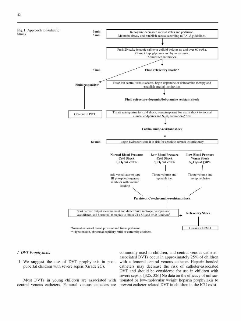
42
Fig. 1 Approach to PediatricShock
I. DVT Prophylaxis
1. We suggest the use of DVT prophylaxis in post-pubertal children with severe sepsis (Grade 2C).
Most DVTs in young children are associated withcentral venous catheters. Femoral venous catheters are
commonly used in children, and central venous catheter-associated DVTs occur in approximately 25% of childrenwith a femoral central venous catheter. Heparin-bondedcatheters may decrease the risk of catheter-associatedDVT and should be considered for use in children withsevere sepsis. [325, 326] No data on the efficacy of unfrac-tionated or low-molecular weight heparin prophylaxis toprevent catheter-related DVT in children in the ICU exist.
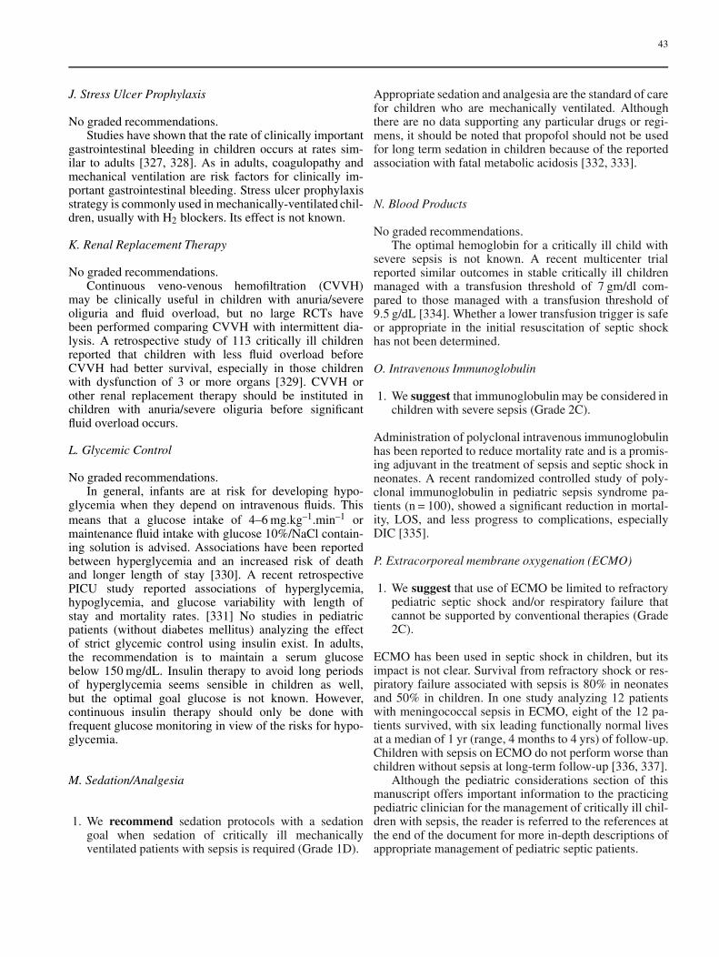
43
J. Stress Ulcer Prophylaxis
No graded recommendations.Studies have shown that the rate of clinically important
gastrointestinal bleeding in children occurs at rates sim-ilar to adults [327, 328]. As in adults, coagulopathy andmechanical ventilation are risk factors for clinically im-portant gastrointestinal bleeding. Stress ulcer prophylaxisstrategy is commonly used in mechanically-ventilated chil-dren, usually with H2 blockers. Its effect is not known.
K. Renal Replacement Therapy
No graded recommendations.Continuous veno-venous hemofiltration (CVVH)
may be clinically useful in children with anuria/severeoliguria and fluid overload, but no large RCTs havebeen performed comparing CVVH with intermittent dia-lysis. A retrospective study of 113 critically ill childrenreported that children with less fluid overload beforeCVVH had better survival, especially in those childrenwith dysfunction of 3 or more organs [329]. CVVH orother renal replacement therapy should be instituted inchildren with anuria/severe oliguria before significantfluid overload occurs.
L. Glycemic Control
No graded recommendations.In general, infants are at risk for developing hypo-
glycemia when they depend on intravenous fluids. Thismeans that a glucose intake of 4–6 mg.kg–1.min–1 ormaintenance fluid intake with glucose 10%/NaCl contain-ing solution is advised. Associations have been reportedbetween hyperglycemia and an increased risk of deathand longer length of stay [330]. A recent retrospectivePICU study reported associations of hyperglycemia,hypoglycemia, and glucose variability with length ofstay and mortality rates. [331] No studies in pediatricpatients (without diabetes mellitus) analyzing the effectof strict glycemic control using insulin exist. In adults,the recommendation is to maintain a serum glucosebelow 150 mg/dL. Insulin therapy to avoid long periodsof hyperglycemia seems sensible in children as well,but the optimal goal glucose is not known. However,continuous insulin therapy should only be done withfrequent glucose monitoring in view of the risks for hypo-glycemia.
M. Sedation/Analgesia
1. We recommend sedation protocols with a sedationgoal when sedation of critically ill mechanicallyventilated patients with sepsis is required (Grade 1D).
Appropriate sedation and analgesia are the standard of carefor children who are mechanically ventilated. Althoughthere are no data supporting any particular drugs or regi-mens, it should be noted that propofol should not be usedfor long term sedation in children because of the reportedassociation with fatal metabolic acidosis [332, 333].
N. Blood Products
No graded recommendations.The optimal hemoglobin for a critically ill child with
severe sepsis is not known. A recent multicenter trialreported similar outcomes in stable critically ill childrenmanaged with a transfusion threshold of 7 gm/dl com-pared to those managed with a transfusion threshold of9.5 g/dL [334]. Whether a lower transfusion trigger is safeor appropriate in the initial resuscitation of septic shockhas not been determined.
O. Intravenous Immunoglobulin
1. We suggest that immunoglobulin may be considered inchildren with severe sepsis (Grade 2C).
Administration of polyclonal intravenous immunoglobulinhas been reported to reduce mortality rate and is a promis-ing adjuvant in the treatment of sepsis and septic shock inneonates. A recent randomized controlled study of poly-clonal immunoglobulin in pediatric sepsis syndrome pa-tients (n = 100), showed a significant reduction in mortal-ity, LOS, and less progress to complications, especiallyDIC [335].
P. Extracorporeal membrane oxygenation (ECMO)
1. We suggest that use of ECMO be limited to refractorypediatric septic shock and/or respiratory failure thatcannot be supported by conventional therapies (Grade2C).
ECMO has been used in septic shock in children, but itsimpact is not clear. Survival from refractory shock or res-piratory failure associated with sepsis is 80% in neonatesand 50% in children. In one study analyzing 12 patientswith meningococcal sepsis in ECMO, eight of the 12 pa-tients survived, with six leading functionally normal livesat a median of 1 yr (range, 4 months to 4 yrs) of follow-up.Children with sepsis on ECMO do not perform worse thanchildren without sepsis at long-term follow-up [336, 337].
Although the pediatric considerations section of thismanuscript offers important information to the practicingpediatric clinician for the management of critically ill chil-dren with sepsis, the reader is referred to the references atthe end of the document for more in-depth descriptions ofappropriate management of pediatric septic patients.
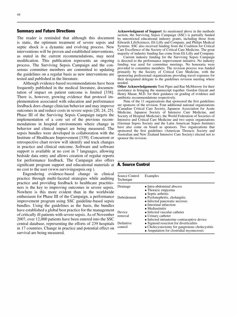
44
Summary and Future DirectionsThe reader is reminded that although this documentis static, the optimum treatment of severe sepsis andseptic shock is a dynamic and evolving process. Newinterventions will be proven and established interventions,as stated in the current recommendations, may needmodification. This publication represents an ongoingprocess. The Surviving Sepsis Campaign and the con-sensus committee members are committed to updatingthe guidelines on a regular basis as new interventions aretested and published in the literature.
Although evidence-based recommendations have beenfrequently published in the medical literature, documen-tation of impact on patient outcome is limited [338].There is, however, growing evidence that protocol im-plementation associated with education and performancefeedback does change clinician behavior and may improveoutcomes in and reduce costs in severe sepsis [20, 24, 25].Phase III of the Surviving Sepsis Campaign targets theimplementation of a core set of the previous recom-mendations in hospital environments where change inbehavior and clinical impact are being measured. Thesepsis bundles were developed in collaboration with theInstitute of Healthcare Improvement [339]. Concurrent orretrospective chart review will identify and track changesin practice and clinical outcome. Software and softwaresupport is available at no cost in 7 languages, allowingbedside data entry and allows creation of regular reportsfor performance feedback. The Campaign also offerssignificant program support and educational materials atno cost to the user (www.survivingsepsis.org ).
Engendering evidence-based change in clinicalpractice through multi-faceted strategies while auditingpractice and providing feedback to healthcare practitio-ners is the key to improving outcomes in severe sepsis.Nowhere is this more evident than in the worldwideenthusiasm for Phase III of the Campaign, a performanceimprovement program using SSC guideline-based sepsisbundles. Using the guidelines as the basis, the bundleshave established a global best practice for the managementof critically ill patients with severe sepsis. As of November2007, over 12,000 patients have been entered into the SSCcentral database, representing the efforts of 239 hospitalsin 17 countries. Change in practice and potential effect onsurvival are being measured.
Acknowledgment of Support As mentioned above in the methodssection, the Surviving Sepsis Campaign (SSC) is partially fundedby unrestricted educational industry grants, including those fromEdwards LifeSciences, Eli Lilly and Company, and Philips MedicalSystems. SSC also received funding from the Coalition for CriticalCare Excellence of the Society of Critical Care Medicine. The greatmajority of industry funding has come from Eli Lilly and Company.
Current industry funding for the Surviving Sepsis Campaignis directed to the performance improvement initiative. No industryfunding was used for committee meetings. No honoraria wereprovided to committee members. The revision process was fundedprimarily by the Society of Critical Care Medicine, with thesponsoring professional organizations providing travel expenses fortheir designated delegate to the guidelines revision meeting whereneeded.Other Acknowledgements Toni Piper and Rae McMorrow for theirassistance in bringing the manuscript together. Gordon Guyatt andHenry Masur, M.D. for their guidance on grading of evidence andantibiotic recommendations respectively.
Nine of the 11 organizations that sponsored the first guidelinesare sponsors of the revision. Four additional national organizations(Canadian Critical Care Society, Japanese Association for AcuteMedicine, Japanese Society of Intensive Care Medicine, andSociety of Hospital Medicine), the World Federation of Societies ofIntensive and Critical Care Medicine and two sepsis organizations(German Sepsis Society and the Latin American Sepsis Institute)have also come on board as sponsors. Two organizations thatsponsored the first guidelines (American Thoracic Society andAustralian and New Zealand Intensive Care Society) elected not tosponsor the revision.
A. Source Control
Source Control ExamplesTechnique
Drainage • Intra-abdominal abscess• Thoracic empyema• Septic arthritis
Debridement • Pyelonephritis, cholangitis• Infected pancreatic necrosis• Intestinal infarction• Mediastinitis
Device • Infected vascular catheterremoval • Urinary catheter
• Infected intrauterine contraceptive deviceDefinitive • Sigmoid resection for diverticulitiscontrol • Cholecystectomy for gangrenous cholecystitis
• Amputation for clostridial myonecrosis
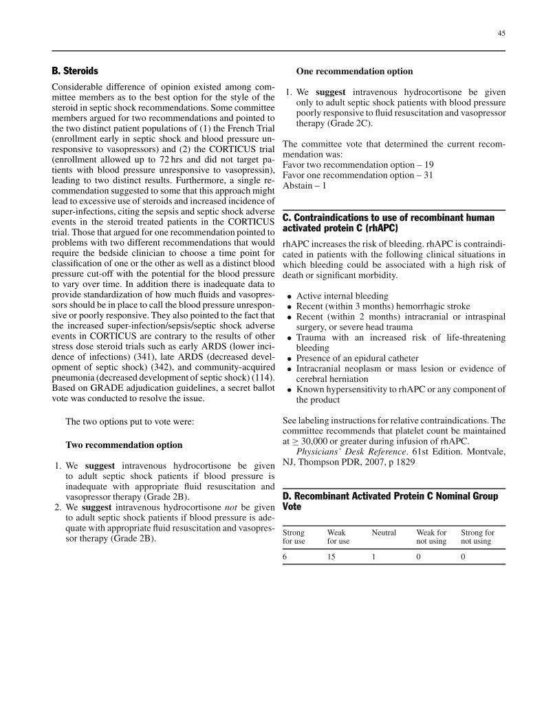
45
B. SteroidsConsiderable difference of opinion existed among com-mittee members as to the best option for the style of thesteroid in septic shock recommendations. Some committeemembers argued for two recommendations and pointed tothe two distinct patient populations of (1) the French Trial(enrollment early in septic shock and blood pressure un-responsive to vasopressors) and (2) the CORTICUS trial(enrollment allowed up to 72 hrs and did not target pa-tients with blood pressure unresponsive to vasopressin),leading to two distinct results. Furthermore, a single re-commendation suggested to some that this approach mightlead to excessive use of steroids and increased incidence ofsuper-infections, citing the sepsis and septic shock adverseevents in the steroid treated patients in the CORTICUStrial. Those that argued for one recommendation pointed toproblems with two different recommendations that wouldrequire the bedside clinician to choose a time point forclassification of one or the other as well as a distinct bloodpressure cut-off with the potential for the blood pressureto vary over time. In addition there is inadequate data toprovide standardization of how much fluids and vasopres-sors should be in place to call the blood pressure unrespon-sive or poorly responsive. They also pointed to the fact thatthe increased super-infection/sepsis/septic shock adverseevents in CORTICUS are contrary to the results of otherstress dose steroid trials such as early ARDS (lower inci-dence of infections) (341), late ARDS (decreased devel-opment of septic shock) (342), and community-acquiredpneumonia (decreased development of septic shock) (114).Based on GRADE adjudication guidelines, a secret ballotvote was conducted to resolve the issue.
The two options put to vote were:
Two recommendation option
1. We suggest intravenous hydrocortisone be givento adult septic shock patients if blood pressure isinadequate with appropriate fluid resuscitation andvasopressor therapy (Grade 2B).
2. We suggest intravenous hydrocortisone not be givento adult septic shock patients if blood pressure is ade-quate with appropriate fluid resuscitation and vasopres-sor therapy (Grade 2B).
One recommendation option
1. We suggest intravenous hydrocortisone be givenonly to adult septic shock patients with blood pressurepoorly responsive to fluid resuscitation and vasopressortherapy (Grade 2C).
The committee vote that determined the current recom-mendation was:Favor two recommendation option – 19Favor one recommendation option – 31Abstain – 1
C. Contraindications to use of recombinant humanactivated protein C (rhAPC)rhAPC increases the risk of bleeding. rhAPC is contraindi-cated in patients with the following clinical situations inwhich bleeding could be associated with a high risk ofdeath or significant morbidity.
• Active internal bleeding• Recent (within 3 months) hemorrhagic stroke• Recent (within 2 months) intracranial or intraspinal
surgery, or severe head trauma• Trauma with an increased risk of life-threatening
bleeding• Presence of an epidural catheter• Intracranial neoplasm or mass lesion or evidence of
cerebral herniation• Known hypersensitivity to rhAPC or any component of
the product
See labeling instructions for relative contraindications. Thecommittee recommends that platelet count be maintainedat ≥ 30,000 or greater during infusion of rhAPC.
Physicians’ Desk Reference. 61st Edition. Montvale,NJ, Thompson PDR, 2007, p 1829
D. Recombinant Activated Protein C Nominal GroupVote
Strong Weak Neutral Weak for Strong forfor use for use not using not using
6 15 1 0 0
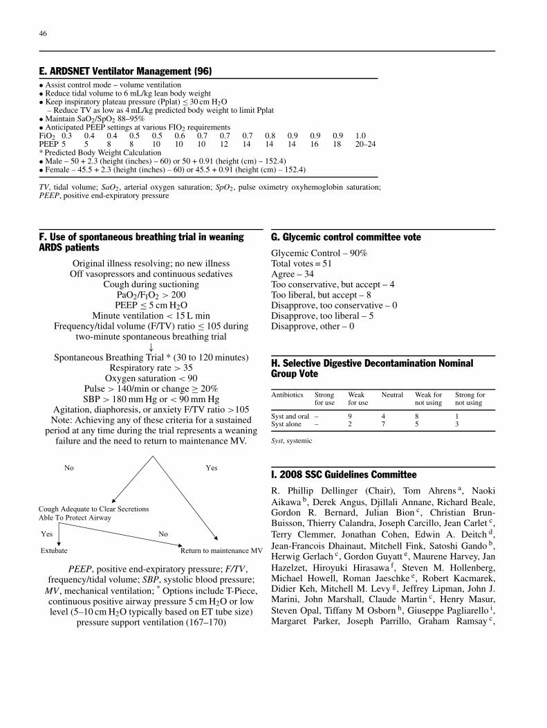
46
E. ARDSNET Ventilator Management (96)• Assist control mode – volume ventilation• Reduce tidal volume to 6 mL/kg lean body weight• Keep inspiratory plateau pressure (Pplat) ≤ 30 cm H2O
– Reduce TV as low as 4 mL/kg predicted body weight to limit Pplat• Maintain SaO2/SpO2 88–95%• Anticipated PEEP settings at various FIO2 requirementsFiO2 0.3 0.4 0.4 0.5 0.5 0.6 0.7 0.7 0.7 0.8 0.9 0.9 0.9 1.0PEEP 5 5 8 8 10 10 10 12 14 14 14 16 18 20–24* Predicted Body Weight Calculation• Male – 50 + 2.3 (height (inches) – 60) or 50 + 0.91 (height (cm) – 152.4)• Female – 45.5 + 2.3 (height (inches) – 60) or 45.5 + 0.91 (height (cm) – 152.4)
TV, tidal volume; SaO2, arterial oxygen saturation; SpO2, pulse oximetry oxyhemoglobin saturation;PEEP, positive end-expiratory pressure
F. Use of spontaneous breathing trial in weaningARDS patients
Original illness resolving; no new illnessOff vasopressors and continuous sedatives
Cough during suctioningPaO2/FIO2 > 200PEEP ≤ 5 cm H2O
Minute ventilation < 15 L minFrequency/tidal volume (F/TV) ratio ≤ 105 during
two-minute spontaneous breathing trial↓
Spontaneous Breathing Trial * (30 to 120 minutes)Respiratory rate > 35
Oxygen saturation < 90Pulse > 140/min or change ≥ 20%SBP > 180 mm Hg or < 90 mm Hg
Agitation, diaphoresis, or anxiety F/TV ratio >105Note: Achieving any of these criteria for a sustained
period at any time during the trial represents a weaningfailure and the need to return to maintenance MV.
PEEP, positive end-expiratory pressure; F/TV ,frequency/tidal volume; SBP, systolic blood pressure;
MV , mechanical ventilation; * Options include T-Piece,continuous positive airway pressure 5 cm H2O or lowlevel (5–10 cm H2O typically based on ET tube size)
pressure support ventilation (167–170)
G. Glycemic control committee voteGlycemic Control – 90%Total votes = 51Agree – 34Too conservative, but accept – 4Too liberal, but accept – 8Disapprove, too conservative – 0Disapprove, too liberal – 5Disapprove, other – 0
H. Selective Digestive Decontamination NominalGroup Vote
Antibiotics Strong Weak Neutral Weak for Strong forfor use for use not using not using
Syst and oral – 9 4 8 1Syst alone – 2 7 5 3
Syst, systemic
I. 2008 SSC Guidelines CommitteeR. Phillip Dellinger (Chair), Tom Ahrens a, NaokiAikawa b, Derek Angus, Djillali Annane, Richard Beale,Gordon R. Bernard, Julian Bion c, Christian Brun-Buisson, Thierry Calandra, Joseph Carcillo, Jean Carlet c,Terry Clemmer, Jonathan Cohen, Edwin A. Deitch d,Jean-Francois Dhainaut, Mitchell Fink, Satoshi Gando b,Herwig Gerlach c, Gordon Guyatt e, Maurene Harvey, JanHazelzet, Hiroyuki Hirasawa f, Steven M. Hollenberg,Michael Howell, Roman Jaeschke e, Robert Kacmarek,Didier Keh, Mitchell M. Levy g, Jeffrey Lipman, John J.Marini, John Marshall, Claude Martin c, Henry Masur,Steven Opal, Tiffany M Osborn h, Giuseppe Pagliarello i,Margaret Parker, Joseph Parrillo, Graham Ramsay c,

47
Adrienne Randolph, Marco Ranieri c, Robert C. Read j,Konrad Reinhart k, Andrew Rhodes c, Emmanuel Rivers h,Gordon Rubenfeld, Jonathan Sevransky, Eliezer Silva l,Charles L. Sprung c, B. Taylor Thompson, Sean R.Townsend, Jeffery Vender m, Jean-Louis Vincent n, TobiasWelte o, Janice Zimmerman
a – American Association of Critical-Care Nursesb – Japanese Association for Acute Medicinec – European Society of Intensive Care Medicined – Surgical Infection Societye – Grades of Recommendation, Assessment, Develop-ment and Evaluation (GRADE) Groupf – Japanese Society of Intensive Care Medicineg – Society of Critical Care Medicineh – American College of Emergency Physiciansi – Canadian Critical Care Societyj – European Society of Clinical Microbiology and Infec-tious Diseasesk – German Sepsis Societyl – Latin American Sepsis Institutem – American College of Chest Physiciansn – International Sepsis Forumo – European Respiratory Society
J. Author Disclosure Information 2006–2007Dr. Dellinger has consulted for AstraZeneca, Talecris,and B Braun. He has received honoraria from Eli Lilly(2), Brahms (2), INO Therapeutics (1), Pulsion (1), andbioMerieux (1). He has also received grant support fromAstraZeneca and Artisan.
Dr. Levy has received honoraria from Eli Lilly andEdwards Lifesciences. He has also received grant supportfrom Phillips Medical Systems, Edwards Lifesciences,Phillips Medical Systems, Novartis, Biosite, and Eisai.
Dr. Carlet has consulted for Forrest, Wyeth, Chiron,bioMerieux, and GlaxoSmithKline. He has also receivedhonoraria from Eli Lilly, Becton Dickinson, Jansen, Cook,AstraZeneca, Hutchinson, Bayer, Gilead, MSD, and Tar-ganta.
Dr. Bion has not disclosed any potential conflicts of in-terest.
Dr. Parker has consulted for Johnson & Johnson.Dr. Jaeschke has received honoraria from AstraZeneca,
Boehringer, Eli Lilly, GlaxoSmithKline, and MSD.Dr. Reinhart has consulted for Eli Lilly and Edwards
Lifesciences. He has also received honoraria from B Braunand royalties from Edwards Lifesciences.
Dr. Angus has consulted for or received speakingfees from AstraZeneca, Brahms Diagnostica, Eisai, EliLilly, GlaxoSmithKline, OrthoBiotech, Takeda, andWyeth-Ayerst. He has also received grant support fromGlaxoSmithKline, OrthoBiotech, and Amgen.
Dr. Brun-Buisson has not disclosed any potential con-flicts of interest.
Dr. Beale has received honoraria from Eisai and speak-ing fees (paid to university) from Lilly UK, Philips, Lidco,and Chiron.
Dr. Calandra has consulted for Baxter, received hono-raria from Roche Diagnostics, and grant support from Bax-ter and Roche Diagnostics. He also served on the advisoryboard for Biosite.
Dr. Dhainaut has consulted for Eli Lilly and Novartis.He has also received honoraria from Eli Lilly.
Dr. Gerlach has not disclosed any potential conflicts ofinterest.
Ms. Harvey has not disclosed any potential conflicts ofinterest.
Dr. Marini has consulted for KCI and received hono-raria from Maquet.
Dr. Marshall has consulted for Becton-Dickinson,Takeda, Pfizer, Spectral Diagnostics, Eisai, and Leo-Pharma. He has also received honoraria from SpectralDiagnostics.
Dr. Ranieri has served on the advisory board forMaquet and received support for a sponsored trial fromEli Lilly. He has also received grant support from Tyco,Draeger, and Hamilton.
Dr. Ramsay has consulted for Edwards Lifesciencesand Respironics.
Dr. Sevransky has not disclosed any potential conflictsof interest.
Dr. Thompson has consulted for Eli Lilly, Abbott, andAstraZeneca. He has also received grant support from theNIH for a study on computerized glucose control.
Dr. Townsend has not disclosed any potential conflictsof interest.
Dr. Vender has consulted and received honoraria fromEli Lilly.
Dr. Zimmerman has not disclosed any potential con-flicts of interest.
Dr. Vincent has consulted for AstraZeneca, Biosite,bioMerieux, Edwards Lifesciences, Eli Lilly Eisai,Ferring, GlaxoSmithKline, Intercell, Merck, Novartis,NovoNordisk, Organon, Pfizer, Phillips Medical Systems,Roche Diagnostics, Spectral Diagnostics, Takeda, andWyethLederle. He has also received honoraria from EliLilly, Edwards Lifesciences, Eisai, GlaxoSmithKline,Novartis, NovoNordisk, and Pfizer.
48
References
Introduction and Methods
1. Angus DC, Linde-Zwirble WT,Lidicker J, Clermont G, Carcillo J,Pinsky MR (2001) Epidemiol-ogy of severe sepsis in the UnitedStates: analysis of incidence, out-come, and associated costs of care.Crit Care Med 29:1303–1310
2. Dellinger RP (2003) Cardiovascu-lar management of septic shock.Crit Care Med 31:946–955
3. Martin GS, Mannino DM, Eaton S,Moss M (2003) The Epidemi-ology of Sepsis in the UnitedStates from 1979 through 2000.N Engl J Med 348:1546–1554
4. Linde-Zwirble WT, Angus DC(2004) Severe sepsis epidemiology:sampling, selection, and society.Crit Care 8:222–226
5. Dombrovskiy VY, Martin AA, Sunder-ram J, Paz HL (2007) Rapid increasein hospitalization and mortality ratesfor severe sepsis in the United States:a trend analysis from 1993 to 2003.Crit Care Med 35:1414–1415
6. Dellinger RP, Carlet JM, Masur H,Gerlach H, Calandra T, Cohen J, Gea-Banacloche J, Keh D, Marshall JC,Parker MM, Ramsay G, Zimmer-man JL, Vincent JL, Levy MM,Surviving Sepsis Campaign Man-agement Guidelines Committee(2004) Surviving Sepsis Cam-paign guidelines for managementof severe sepsis and septic shock.Crit Care Med 32:858–873
7. Dellinger RP, Carlet JM, Masur H,Gerlach H, Calandra T, Cohen J, Gea-Banacloche J, Keh D, Marshall JC,Parker MM, Ramsay G, Zimmer-man JL, Vincent JL, Levy MM,Surviving Sepsis Campaign Man-agement Guidelines Committee(2004) Surviving Sepsis Campaignguidelines for management of severesepsis and septic shock. IntensiveCare Med 30:536–555
8. Guyatt G, Schünemann H, Cook D,Jaeschke R, Pauker S (2004) Applyingthe grades of recommendations forantithrombotic and thrombolytic ther-apy: The seventh ACCP conferenceof antithrombotic and thrombolytictherapy. Chest 126:179S–187S
9. GRADE working group (2004)Grading quality of evidence andstrength of recommendations.BMJ 328:1490–1498
10. Guyatt G, Gutterman D, Bau-mann MH, Addrizzo-Harris D,Hylek EM, Phillips B (2006) Grad-ing strength of recommendationsand quality of evidence in clinicalguidelines: report from an AmericanCollege of Chest Physicians task force.Chest 129:174–181
11. Schünemann HJ, Jaeschke R, Cook DJ,Bria WF, El-Solh AA, Ernst A,Fahy BF, Gould MK, Horan KL,Krishnan JA, Manthous CA, Mau-rer JR, McNicholas WT, Oxman AD,Rubenfeld G, Turino GM, Guyatt G,ATS Documents Development andImplementation Committee (2006) AnOfficial ATS Statement: Grading theQuality of Evidence and Strength ofRecommendations in ATS Guidelinesand Recommendations. Am J RespirCrit Care Med 174:605–614
12. Levy MM, Fink MP, Marshall JC,Abraham E, Angus D, Cook D,Cohen J, Opal SM, Vincent JL, Ram-say G, SCCM/ESICM/ACCP/ATS/SIS(2003) 2001 SCCM/ESICM/ACCP/ATS/SIS International Sep-sis Definitions Conference.Crit Care Med 31:1250–1256
13. Bone RC, Balk RA, Cerra FB,Dellinger RP, Fein AM, Knaus WA,Schein RMH, Sibbald WJ, Membersof the ACCP/SCCM Consensus Con-ference (1992) Definitions for Sepsisand Organ Failure and Guidelinesfor the Use of Innovative Therapiesin Sepsis. Chest 101:1644–1655and Crit Care Med 20:864–874
14. Sprung CL, Bernard GR, Dellinger RP(2001) Guidelines for the Manage-ment of Severe Sepsis and SepticShock. Intensive Care Med 27(Suppl 1):S1–S134
15. Sackett DL (1989) Rules of evidenceand clinical recommendations onthe use of antithrombotic agents.Chest 95:2S–4S
Initial Resuscitation
16. Rivers E, Nguyen B, Havstad S,Ressler J, Muzzin A, Knoblich B,Peterson E, Tomlanovich M, EarlyGoal-Directed Therapy Collabo-rative Group (2001) Early goal-directed therapy in the treatmentof severe sepsis and septic shock.N Engl J Med 345:1368–1377
17. Bendjelid K, Romand JA (2003)Fluid responsiveness in mechanicallyventilated patients: A review of indicesused in intensive care. IntensiveCare Med 29:352–360
18. Malbrain ML, Deeren D, De Pot-ter TJ (2005) Intra-abdominalhypertension in the critically ill: Itis time to pay attention. Curr OpinCrit Care 11:156–171
19. Varpula M, Tallgren M, Saukkonen K,Voipio-Pulkki LM, Pettilä V (2005)Hemodynamic variables related tooutcome in septic shock. IntensiveCare Med 31:1066–1071
20. Kortgen A, Niederprum P, Bauer M(2006) Implementation of an evidence-based “standard operating proce-dure” and outcome in septic shock.Crit Care Med 34:943–949
21. Sebat F, Johnson D, Musthafa AA,Watnik M, Moore S, Henry K, Saari M(2005) A multidisciplinary communityhospital program for early and rapidresuscitation of shock in nontraumapatients. Chest 127:1729–1743
22. Shapiro NI, Howell MD, Tal-mor D, Lahey D, Ngo L, Buras J,Wolfe RE, Weiss JW, Lisbon A (2006)Implementation and outcomesof the Multiple Urgent Sep-sis Therapies (MUST) protocol.Crit Care Med 34:1025–1032
23. Micek ST, Roubinian N, Heuring T,Bode M, Williams J, Harrison C,Murphy T, Prentice D, Ruoff BE,Kollef MH (2006) Before-after studyof a standardized hospital order setfor the management of septic shock.Crit Care Med 34:2707–2713
24. Nguyen HB, Corbett SW, Steele R,Banta J, Clark RT, Hayes SR, Ed-wards J, Cho TW, Wittlake WA (2007)Implementation of a bundle of qualityindicators for the early managementof severe sepsis and septic shock isassociated with decreased mortality.Crit Care Med 35:1105–1112
25. Shorr AF, Micek ST, Jackson WL Jr,Kollef MH (2007) Economic im-plications of an evidence-basedsepsis protocol: can we im-prove outcomes and lower costs?Crit Care Med 35:1257–1262
26. Reinhart K, Kuhn HJ, Hartog C,Bredle DL (2004) Continuous centralvenous and pulmonary artery oxygensaturation monitoring in the criticallyill. Intensive Care Med 30:1572–1578
27. Trzeciak S, Dellinger RP, Abate NL,Cowan RM, Stauss M, Kilgannon JH,Zanotti S, Parrillo JE (2006) Trans-lating research to clinical practice:a 1-year experience with implementingearly goal-directed therapy for septicshock in the emergency department.Chest 129:225–232
28. Magder S (2006) Central ve-nous pressure: A useful butnot so simple measurement.Crit Care Med 34:2224–2227
49
29. Bendjelid K (2005) Right arte-rial pressure: Determinant or re-sult of change in venous return?Chest 128:3639–3640
30. Vincent JL, Weil MH (2006)Fluid challenge revisited.Crit Care Med 34:1333–1337
31. Trzeciak S, Dellinger RP, Parrillo JE,Guglielmi M, Bajaj J, Abate NL,Arnold RC, Colilla S, Zanotti S,Hollenberg SM, MicrocirculatoryAlterations in Resuscitation and ShockInvestigators (2007) Early microcir-culatory perfusion derangements inpatients with severe sepsis and septicshock: Relationship to hemodynam-ics, oxygen transport, and survival.Ann Emerg Med 49:88–98
32. De Backer D, Creteur J, Dubois MJ,Sakr Y, Koch M, Verdant C, Vin-cent JL (2006) The effects ofdobutamine on microcirculatory al-terations in patients with septic shockare independent of its systemic effects.Crit Care Med 34:403–408
33. Buwalda M, Ince C (2002) Open-ing the microcirculation: Canvasodilators be useful in sepsis?Intensive Care Med 28:1208–1217
34. Boldt J (2002) Clinical review; hemo-dynamic monitoring in the intensivecare unit. Crit Care 6:52–59
35. Pinsky MR, Payen D (2005) Func-tional hemodynamic monitoring.Crit Care 9:566–572
Diagnosis
36. Weinstein MP, Reller LP, Murphy JR,Lichtenstein KA (1983) The clin-ical significance of positive bloodcultures: A comprehensive analysisof 500 episodes of bacteremia andfungemia in adults. I. Laboratoryand epidemiologic observations.Rev Infect Dis 5:35–53
37. Blot F, Schmidt E, Nitenberg G, Tan-crède C, Laplanche A, Andremont A(1998) Earlier positivity of central ve-nous versus peripheral blood culturesis highly predictive of catheter-relatedsepsis. J Clin Microbiol 36:105–109
38. Mermel LA, Maki DG (1993) De-tection of bacteremia in adults:consequences of culturing an inade-quate volume of blood. Ann InternMed 119:270–272
39. American Thoracic Society, Infec-tious Diseases Society of America(2005) Guidelines for the managementof adults with hospital-acquired,ventilator-associated, and healthcare-associated pneumonia. Am J RespirCrit Care Med 171:388–416
40. Giamarellos-Bourboulis EJ, Gi-annopoulou P, Grecka P, Voros D,Mandragos K, Giamarellou H (2004)Should procalcitonin be introduced inthe diagnostic criteria for the systemicinflammatory response syndrome andsepsis? J Crit Care 19:152–157
41. Tenover FC (2007) Rapid detec-tion and identification of bacterialpathogens using novel moleculartechnologies: infection control andbeyond. Clin Infect Dis 44:418–423
Antibiotic Therapy
42. Kumar A, Roberts D, Wood KE,Light B, Parrillo JE, Sharma S,Suppes R, Feinstein D, Zanotti S,Taiberg L, Gurka D, Kumar A,Cheang M (2006) Duration of hy-potension prior to initiation of effectiveantimicrobial therapy is the critical de-terminant of survival in human septicshock. Crit Care Med 34:1589–1596
43. Morrell M, Fraser VJ, Kollef MH(2005) Delaying the empiric treatmentof candida bloodstream infectionuntil positive blood culture results areobtained: a potential risk factor forhospital mortality. Antimicrob AgentsChemother 49:3640–3645
44. Pappas PG, Rex JH, Sobel JD,Filler SG, Dismukes WE, Walsh TJ,Edwards JE, Infectious DiseasesSociety of America (2004) Guide-lines for treatment of candidiasis.Clin Infect Dis 38:161–189
45. McCabe WR, Jackson GG (1962)Gram negative bacteremia. ArchIntern Med 110:92–100
46. Kreger BE, Craven DE, McCabe WR(1980) Gram negative bacteremia.IV. Re-evaluation of clinical fea-tures and treatment in 612 patients.Am J Med 68:344–355
47. Leibovici L, Shraga I, Drucker M,Konigsberger H, Samra Z, Pitlik SD(1998) The benefit of appropriateempirical antibiotic treatment inpatients with bloodstream infection.J Intern Med 244:379–386
48. Ibrahim EH, Sherman G, Ward S,Fraser VJ, Kollef MH (2000) Theinfluence of inadequate antimicrobialtreatment of bloodstream infections onpatient outcomes in the ICU setting.Chest 118:146–155
49. Hatala R, Dinh T, Cook DJ(1996) Once-daily aminogly-coside dosing in immunocom-petent adults: A meta-analysis.Ann Intern Med 124:717–725
50. Ali MZ, Goetz MB (1997) A meta-analysis of the relative efficacy andtoxicity of single daily dosing versusmultiple daily dosing of aminoglyco-sides. Clin Infec Dis 24:796–809
51. Amsden GW, Ballow CH, Bertino JS(2000) Pharmacokinetics and Phar-macodynamics of Anti-infectiveAgents. In: Mandell GL, Bennett JE,Dolin R (Eds) Principles and Practiceof Infectious Diseases. Fifth Edition.Philadelphia, Churchill Livingstone,pp 253–261
52. Hyatt JM, McKinnon PS, Zimmer GS,Schentag JJ (1995) The importanceof pharmacokinetic/pharmacodynamicsurrogate markers to outcomes.Focus on antibacterial agents.Clin Pharmacokinet 28:143–160
53. Hughes WT, Armstrong D, Bodey GP,Bow EJ, Brown AE, Calandra T,Feld R, Pizzo PA, Rolston KVI,Shenep JL, Young LS. 2002 Guide-lines for the Use of AntimicrobialAgents in Neutropenic Patients withCancer. www.idsociety.org. AccessedJuly 10, 2007.
54. Klastersky J (2004) Management offever in neutropenic patients withdifferent risks of complications.Clin Infect Dis 39(Suppl 1):S32–37
55. Safdar N, Handelsman J, Maki DG(2004) Does combination antimi-crobial therapy reduce mortalityin Gram-negative bacteraemia?A meta-analysis. Lancet Infect Dis4(8):519–527
56. Paul M, Silbiger I, Grozinsky S,Soares-Weiser K, Leibovici L (2006)Beta lactam antibiotic monotherapyversus beta lactam-aminoglycosideantibiotic combination therapy forsepsis. Cochrane Database Syst RevJan 25:CD003344
57. Garnacho-Montero J, Sa-Borges M,Sole-Violan J, Barcenilla F, Escoresca-Ortega A, Ochoa M, Cayuela A,Rello J (2007) Optimal manage-ment therapy for Pseudomonasaeruginosa ventilator-associatedpneumonia: An observational, multi-center study comparing monotherapywith combination antibiotic therapy.Crit Care Med 25:1888–1895
Source Control
58. Jimenez MF, Marshall JC(2001) Source control inthe management of sepsis.Intensive Care Med 27:S49–S62
59. Moss RL, Musemeche CA,Kosloske AM (1996) Necrotizingfascitis in children: prompt recognitionand aggressive therapy improvesurvival. J Pediatr Surg 31:1142–1146
50
60. Bufalari A, Giustozzi G, Moggi L(1996) Postoperative intraab-dominal abscesses: Percuta-neous versus surgical treatment.Acta Chir Belg 96:197–200
61. O’Grady NP, Alexander M,Dellinger EP, Gerberding JL,Heard SO, Maki DG, Masur H,McCormick RD, Mermel LA, Pear-son ML, Raad II, Randolph A,Weinstein RA (2002) Guidelinesfor the prevention of intravascularcatheter-related infections. Centersfor Disease Control and Prevention.MMWR 51(RR-10):1–29
62. O’Grady NP, Alexander M,Dellinger EP, Gerberding JL,Heard SO, Maki DG, Masur H,McCormick RD, Mermel LA, Pear-son ML, Raad II, Randolph A,Weinstein RA, Healthcare InfectionControl Practices Advisory Committee(2002) Guidelines for the preventionof intravascular catheter-related infec-tions. Clin Infect Dis 35:1281–1307
63. Mier J, Leon EL, Castillo A, Rob-ledo F, Blanco R (1997) Earlyversus late necrosectomy in se-vere necrotizing pancreatitis.Am J Surg 173:71–75
64. Evans A, Winslow BH (1995) Oxygensaturation and hemodynamic responsein critically ill mechanically ventilatedadults during intrahospital transport.Am J Crit Care 4:106–111
Fluid Therapy
65. Finfer S, Bellomo R, Boyce N,French J, Myburgh J, Norton R; SAFEStudy Investigators (2004) A compar-ison of albumin and saline for fluidresuscitation in the intensive care unit.N Engl J Med 350:2247–2256
66. Choi PTL, Yip G, Quinonez LG,Cook DJ (1999) Crystalloidsvs. colloids in fluid resusci-tation: A systematic review.Crit Care Med 27:200–210
67. Cook D, Guyatt G (2001) Colloid usefor fluid resuscitation: Evidence andspin. Ann Intern Med 135:205–208
68. Schierhout G, Roberts I (1998) Fluidresuscitation with colloid or crystal-loid solutions in critically ill patients:Asystematic review of randomizedtrials. BMJ 316:961–964
69. Schortgen F, Lacherade JC, Bruneel F,Cattaneo I, Hemery F, Lemaire F,Brochard L (2001) Effects of hy-droxyethyl starch and gelatin onrenal function in severe sepsis:a multicentre randomised study.Lancet 357:911–916
70. Sakr Y, Payen D, Reinhart K, Sip-mann FS, Zavala E, Bewley J, Marx G,Vincent JL (2007) Effects of hy-droxyethyl starch administration onrenal function in critically ill patients.Br J Anaesth 98:216–224
Vasopressors
71. Hollenberg SM, Ahrens TS, An-nane D, Astiz ME, Chalfin DB,Dasta JF, Heard SO, Martin C,Napolitano LM, Susla GM, Totaro R,Vincent JL, Zanotti-Cavazzoni S(2004) Practice parameters forhemodynamic support of sepsisin adult patients: 2004 update.Crit Care Med 32:1928–1948
72. LeDoux D, Astiz ME, Carpati CM,Rackow EC (2000) Effects ofperfusion pressure on tissueperfusion in septic shock.Crit Care Med 28:2729–32
73. Martin C, Papazian L, Perrin G,Saux P, Gouin F (1993) Nore-pinephrine or dopamine for thetreatment of hyperdynamic septicshock? Chest 103:1826–1831
74. Martin C, Viviand X, Leone M,Thirion X (2000) Effect of nore-pinephrine on the outcome of septicshock. Crit Care Med 28:2758–2765
75. De Backer D, Creteur J, Silva E,Vincent JL (2003) Effects ofdopamine, norepinephrine, andepinephrine on the splanchnic circu-lation in septic shock: Which is best?Crit Care Med 31:1659–1667
76. Day NP, Phu NH, Bethell DP,Mai NT, Chau TT, Hien TT, White NJ(1996) The effects of dopamine andadrenaline infusions on acid-base bal-ance and systemic haemodynamics insevere infection. Lancet 348:219–223
77. Le Tulzo Y, Seguin P, Gacouin A, Ca-mus C, Suprin E, Jouannic I, Thomas R(1997) Effects of epinephrine on rightventricular function in patients withsevere septic shock and right ventric-ular failure: a preliminary descriptivestudy. Intensive Care Med 23:664–670
78. Bollaert PE, Bauer P, Audibert G,Lambert H, Larcan A (1990) Effectsof epinephrine on hemodynam-ics and oxygen metabolism indopamine-resistant septic shock.Chest 98:949–953
79. Zhou SX, Qiu HB, Huang YZ,Yang Y, Zheng RQ (2002) Effectsof norepinephrine, epinephrine, andnorepinephrine-dobutamine on sys-temic and gastric mucosal oxygenationin septic shock. Acta PharmacologicaSinica 23:654–658
80. Levy B, Bollaert PE, Charpentier C,Nace L, Audibert G, Bauer P, Nabet P,Larcan A (1997) Comparison ofnorepinephrine and dobutamine toepinephrine for hemodynamics, lactatemetabolism, and gastric tonometricvariables in septic shock: a prospec-tive, randomized study. Intensive CareMed 23:282–287
81. Mackenzie SJ, Kapadia F, Nimmo GR,Armstrong IR, Grant IS (1991)Adrenaline in treatment of sep-tic shock: Effects on haemo-dynamics and oxygen transport.Intensive Care Med 17:36–39
82. Moran JL, O’Fathartaigh MS,Peisach AR, Chapman MJ, Leppard P(1993) Epinephrine as an inotropicagent in septic shock: a dose-profileanalysis. Crit Care Med 21:70–77
83. Yamazaki T, Shimada Y, Tae-naka N, Oshumi H, Takezawa J,Yoshiya I (1982) Circulatory re-sponses to afterloading with phenyle-phrine in hyperdynamic sepsis.Crit Care Med 10:432–435
84. Gregory JS, Bonfiglio MF, Dasta JF,Reilley TE, Townsend MC,Flancbaum L (1991) Experiencewith phenylephrine as a component ofthe pharmacologic support of septicshock. Crit Care Med 19:1395–1400
85. Djillali A, Vigno P, Renault A,Blooaert PE, Chrpentier C, Martin C,Troché G, Ricard JD, Nitenberg G,Papazian L, Azouly E, Bellissant E,for the CATS STUDY Group (2007)Norepinephrine plus donutamine ver-sus epinephrine alone for managementof septic shock: A randomized trial.Lancet 370:676–684
86. Regnier B, Rapin M, Gory G,Lemaire F, Teisseire B, Harari A(1977) Haemodynamic effects ofdopamine in septic shock. IntensiveCare Med 3:47–53
87. Landry DW, Levin HR, Gallant EM,Ashton RC Jr, Seo S, D’Alessandro D,Oz MC, Oliver JA (1997) Vaso-pressin deficiency contributes tothe vasodilation of septic shock.Circulation 95:1122–1125
88. Patel BM, Chittock DR, Russell JA,Walley KR (2002) Beneficial ef-fects of short-term vasopressininfusion during severe septic shock.Anesthesiology 96:576–582
89. Dunser MW, Mayr AJ, Ulmer H,Knotzer H, Sumann G, Pajk W,Friesenecker B, Hasibeder WR (2003)Arginine vasopressin in advancedvasodilatory shock: a prospec-tive, randomized, controlled study.Circulation 107:2313–2319
90. Holmes CL, Patel BM, Russell JA,Walley KR (2001) Physiology ofvasopressin relevant to management ofseptic shock. Chest 120:989–1002
51
91. Malay MB, Ashton RC, Landry DW,Townsend RN (1999) Low-dose vaso-pressin in the treatment of vasodilatoryseptic shock. J Trauma 47:699–705
92. Holmes CL, Walley KR, Chittock DR,Lehman T, Russell JA (2001) The ef-fects of vasopressin on hemodynamicsand renal function in severe septicshock: a case series. Intensive CareMed 27:1416–1421
93. Lauzier F, Levy B, Lamarre P, Lesur O(2006) Vasopressin or norepinephrinein early hyperdynamic septic shock:a randomized clinical trial. IntensiveCare Med 32:1782–1789
94. O’Brien A, Calpp L, Singer M(2002) Terlipressin fornorepinephrine-resistant septic shock.Lancet 359:1209–1210
95. Sharshar T, Blanchard A, Pail-lard M, Raphael JC, Gajdos P,Annane D (2003) Circulating va-sopressin levels in septic shock.Crit Care Med 31:1752–1758
96. Dunser MW, Mayr AJ, Tur A, Pajk W,Barbara F, Knotzer H, Ulmer H,Hasibeder WR (2003) Ischemicskin lesions as a complication ofcontinuous vasopressin infusion incatecholamine-resistant vasodilatoryshock: incidence and risk factors.Crit Care Med 31:1394–1398
97. Bellomo R, Chapman M, Finfer S,Hickling K, Myburgh J (2000)Low-dose dopamine in patients withearly renal dysfunction: a placebo-controlled randomised trial. Australianand New Zealand Intensive CareSociety (ANZICS) Clinical TrialsGroup. Lancet 356:2139–2143
98. Kellum J, Decker J (2001)Use of dopamine in acuterenal failure: a meta-analysis.Crit Care Med 29:1526–1531
Inotropic Therapy
99. Gattinoni L, Brazzi L, Pelosi P,Latini R, Tognoni G, Pesenti A, Fuma-galli R (1995) A trial of goal-orientedhemodynamic therapy in critically illpatients. N Engl J Med 333:1025–1032
100. Hayes MA, Timmins AC, Yau EH,Palazzo M, Hinds CJ, Watson D(1994) Elevation of systemic oxygendelivery in the treatment of critically illpatients. N Engl J Med 330:1717–1722
Steroids
101. Annane D, Sebille V, Charpentier C,Bollaert PE, François B, Korach JM,Capellier G, Cohen Y, Azoulay E,Troché G, Chaumet-Riffaut P, Bel-lissant E (2002) Effect of treatmentwith low doses of hydrocortisone andfludrocortisone on mortality in patientswith septic shock. JAMA 288:862–871
102. Briegel J, Forst H, Haller M,Schelling G, Kilger E, Kuprat G,Hemmer B, Hummel T, Lenhart A,Heyduck M, Stoll C, Peter K(1999) Stress doses of hydrocorti-sone reverse hyperdynamic septicshock: a prospective, randomized,double-blind, single-center study.Crit Care Med 27:723–732
103. Bollaert PE, Charpentier C, Levy B,Debouverie M, Audibert G, Lar-can A (1998) Reversal of lateseptic shock with supraphysio-logic doses of hydrocortisone.Crit Care Med 26:645–650
104. Sprung CL, Annane D, Briegel J,Keh D, Moreno R, Singer M, Weiss Y,Sorenson F (2007) Corticosteroidtherapy of septic shock (CORTI-CUS) (abstract). Am Rev RespCrit Care Med 175:A507
105. Briegel J, Vogeser M, Annane D,Keh D, Moreno R, Singer M, Weiss Y,Sprung CL (2007) Measurement ofcortisol in septic shock: Interlabora-tory harmonization. Am Rev Resp CritCare Med 175:A436
106. Allolio B, Dorr H, Stuttmann R,Knorr D, Engelhardt D, Winkel-mann W (1985) Effect of a single bolusof etomidate upon eight major corti-costeroid hormone and plasma ACTH.Clin Ednocrinol (oxf) 22:281–286
107. Reincke M, Allolio B, Würth G,Winkelmann W (1993) Thehypothalamic-pituitary-adrenalaxis in critical illness: Response todexamethasone and corticotropin-releasing hormone. J Clin EndocrinolMetab 77:151–156
108. Oppert M, Schindler R, Husung C,Offermann K, Graf KJ, Boenisch O,Barckow D, Frei U, Eckardt KU(2005) Low dose hydrocorti-sone improves shock reversaland reduces cytokine levels inearly hyperdynamic septic shock.Crit Care Med 33:2457–2464
109. Yildiz O, Doganay M, Aygen B, Gu-ven M, Keleutimur F, Tutuu A (2002)Physiologic-dose steroid therapy insepsis. Crit Care 6:251–259
110. Keh D, Boehnke T, Weber-Cartens S,Schulz C, Ahlers O, Bercker S,Volk HD, Doecke WD, Falke KJ,Gerlach H (2003) Immunologic andhemodynamic effects of “low-dose”hydrocortisone in septic shock:a double-blind, randomized, placebo-controlled, crossover study. Am JRespir Crit Care Med 167:512–520
111. Bone RC, Fisher CJ, Clemmer TP(1987) A controlled clinical trial ofhigh-dose methylprednisolone in thetreatment of severe sepsis and septicshock. N Engl J Med 317:653–658
112. Cronin L, Cook DJ, Carlet J, Hey-land DK, King D, Lansang MA, FisherCJ Jr (1995) Corticosteroid treatmentfor sepsis: a critical appraisal andmeta-analysis of the literature. CritCare Med 23:1430–1439
113. The Veterans Administration SystemicSepsis Cooperative Study Group(1987) Effect on high-dose gluco-corticoid therapy on mortality inpatients with clinical signs of sepsis.N Engl J Med 317:659–665
114. Confalonieri M, Urbino R, Potena A,Piattella M, Parigi P, Puccio G,Della Porta R, Giorgio C, Blasi F,Umberger R, Meduri GU (2005)Hydrocortisone infusion for severecommunity-acquired pneumonia.A preliminary randomized study. AmJ Respir Crit Care Med 171:242–248
Recombinant Human ActivatedProtein C
115. Bernard GR, Vincent JL, Lat-erre PF, LaRosa SP, Dhainaut JF,Lopez-Rodriguez A, Steingrub JS,Garber GE, Helterbrand JD, Ely EW,Fisher CJ Jr; Recombinant humanprotein C Worldwide Evaluationin Severe Sepsis (PROWESS)study group (2001) Efficacy andsafety of recombinant human ac-tivated protein c for severe sepsis.N Engl J Med 344:699–709
116. Abraham E, Laterre PF, Garg R,Levy H, Talwar D, Trzaskoma BL,Francois B, Guy JS, Bruckmann M,Rea-Neto A, Rossaint R, Perrotin D,Sablotzki A, Arkins N, Utterback BG,Macias WL, Administration ofDrotrecogin Alfa (Activated) in EarlyStage Severe Sepsis (ADDRESS)Study Group (2005) Drotrecoginalfa (activated) for adults with se-vere sepsis and a low risk of death.N Engl J Med 353:1332–1341
52
117. Vincent JL, Bernard GR, Beale R,Doig C, Putensen C, Dhainaut JF,Artigas A, Fumagalli R, Macias W,Wright T, Wong K, Sundin DP,Turlo MA, Janes J (2005) Drotreco-gin alfa (activated) treatment insevere sepsis from the global open-label trial ENHANCE: Furtherevidence for survival and safety andimplications for early treatment.Crit Care Med 33:2266–2277
118. Oxman AD, Guyatt GH (1992) A con-sumer’s guide to subgroup analyses.Ann Intern Med 116:78–84
119. Ely EW, Laterre PF, Angus DC,Helterbrand JD, Levy H, Dhainaut JF,Vincent JL, Macias WL, Bernard GR(2003) Drotrecogin alfa (activated)administration across clinically impor-tant subgroups of patients with severesepsis. Crit Care Med 31:12–19
120. Kanji S, Perreault MM, Chant C,Williamson D, Burry L (2007)Evaluating the use of Drotrecoginalfa activated in adult severe sepsis:a Canadian multicenter observationalstudy. Intensive Care Med 33:517–523
121. Bertolini G, Rossi C, Anghileri A,Livigni S, Addis A, Poole D (2007)Use of Drotrecogin alfa (activated)in Italian intensive care units: Theresults of a nationwide survey.Intensive Care Med 33:426–434
122. European Medicines Agency. Evalua-tion of Medicines for Human Use.www.emea.europa.eu/pdfs/human/press/pr/8509607en.pdf.Accessed October 22, 2007.
Blood Product Administration
123. Hebert PC, Wells G, Blajchman MA,Marshall J, Martin C, Pagliarello G,Tweeddale M, Schweitzer I, Yetisir E(1999) A multicenter, random-ized, controlled clinical trialof transfusion in critical care.N Engl J Med 340:409–417
124. Marik PE, Sibbald WJ (1993) Effectof stored-blood transfusion on oxy-gen delivery in patients with sepsis.JAMA 269:3024–3029
125. Lorente JA, Landín L, dePablo R,Renes E, Rodriguez-Diaz R, Liste D(1993) Effects of blood transfusion onoxygen transport variables in severesepsis. Crit Care Med 21:1312–1318
126. Fernandes CJ Jr, Akamine N, DeMarco FV, De Souza JA, Lagudis S,Knobel E (2001) Red blood celltransfusion does not increase oxygenconsumption in critically ill septicpatients. Crit Care 5:362–567
127. Corwin HL, Gettinger A, Ro-driguez RM, Pearl RG, Gubler KD,Enny C, Colton T, Corwin MJ (1999)Efficacy of recombinant humanerythropoietin in the critically illpatient: a randomized double-blind,placebo-controlled trial. Crit CareMed 27:2346–2350
128. Corwin HL, Gettinger A, Pearl RG,Fink MP, Levy MM, Shapiro MJ,Corwin MJ, Colton T, EPO CriticalCare Trials Group (2002) Efficacyof recombinant human erythro-poietin in critically ill patients.JAMA 28:2827–2835
129. College of American Patholo-gists (1994) Practice parameter forthe use of fresh-frozen plasma,cryoprecipitate, and platelets.JAMA 271:777–781
130. Canadian Medical Association ExpertWorking Group (1997) Guide-lines for red blood cell and plasmatransfusion for adults and children.Can Med Assoc J 156:S1-S24
131. American Society of Anaesthesiolo-gists Task Force on Blood ComponentTherapy (1996) Practice guide-lines for blood component therapy.Anesthesiology 84:732–47
132. Abdel-Wahab OI, Healy B, Dzik WH(2006) Effect of fresh-frozen plasmatransfusion on prothrombin timeand bleeding in patients withmild coagulation abnormalities.Transfusion 46:1279–85
133. Warren BL, Eid A, Singer P, Pillay SS,Carl P, Novak I, Chalupa P, Ather-stone A, Penzes I, Kubler A, Knaub S,Keinecke HO, Heinrichs H, Schin-del F, Juers M, Bone RC, Opal SM,KyberSept Trial Study Group (2001)High-dose antithrombin III in severesepsis. A randomized controlled trial.JAMA 286:1869–1878
134. Wiedermann CJ, Hoffmann JN,Juers M, Ostermann H, Kienast J,Briegel J, Strauss R, Keinecke HO,Warren BL, Opal SM, KyberSeptInvestigators (2006) High-dose an-tithrombin III in the treatment ofsevere sepsis in patients with a highrisk of death: Efficacy and safety. CritCare Med 34:285–292
Mechanical Ventilation
135. The Acute Respiratory DistressSyndrome Network (2000) Venti-lation with lower tidal volumes ascompared with traditional tidal vol-umes for acute lung injury and theacute respiratory distress syndrome.N Engl J Med 342:1301–1308
136. Amato MB, Barbas CS, Medeiros DM,Magaldi RB, Schettino GP, Lorenzi-Filho G, Kairalla RA, Deheinzelin D,Munoz C, Oliveira R, Takagaki TY,Carvalho CR (1998) Effect ofa protective-ventilation strat-egy on mortality in the acuterespiratory distress syndrome.N Engl J Med 338:347–354
137. Brochard L, Roudot-Thoraval F,Roupie E, Delclaux C, Chastre J,Fernandez-Mondejar E, Clementi E,Mancebo J, Factor P, Matamis D,Ranieri M, Blanch L, Rodi G,Mentec H, Dreyfuss D, Ferrer M,Brun-Buisson C, Tobin M, Lemaire F(1998) Tidal volume reduction forprevention of ventilator-inducedlung injury in acute respiratorydistress syndrome. Am J RespirCrit Care Med 158:1831–1838
138. Brower RG, Shanholtz CB,Fessler HE, Shade DM, White P Jr,Wiener CM, Teeter JG, Dodd-o JM,Almog Y, Piantadosi S (1999)Prospective, randomized, controlledclinical trial comparing traditional ver-sus reduced tidal volume ventilationin acute respiratory distress syndromepatients. Crit Care Med 27:1492–1498
139. Stewart TE, Meade MO, Cook DJ,Granton JT, Hodder RV, Lapinsky SE,Mazer CD, McLean RF, Rogov-ein TS, Schouten BD, Todd TR,Slutsky AS (1998) Evaluation ofa ventilation strategy to preventbarotrauma in patients at high risk foracute respiratory distress syndrome.N Engl J Med 338:355–361
140. Eichacker PQ, Gerstenberger EP,Banks SM, Cui X, Natanson C (2002)Meta-analysis of acute lung injury andacute respiratory distress syndrometrials testing low tidal volumes. AmJ Respir Crit Care Med166:1510–1514
141. Tobin MJ (2000) Culmination ofan era in research on the acuterespiratory distress syndrome.N Engl J Med 342:1360–1361
142. Marini JJ, Gattinoni L (2004) Ventila-tory management of acute respiratorydistress syndrome: A consensus oftwo. Crit Care Med 32:250–255
143. Hager DN, Krishnan JA, Hayden DL,Brower RG; ARDS Clinical Tri-als Network (2005) Tidal volumereduction in patients with acutelung injury when plateau pres-sures are not high. Am J RespirCrit Care Med 172:1241–1245
53
144. Ferguson ND, Frutos-Vivar F, Este-ban A, Anzueto A, Alia I, Brower RG,Stewart TE, Apezteguia C, Gonza-lez M, Soto L, Abroug F, Brochard L,Mechanical Ventilation InternationalStudy Group (2005) Airway pressures,tidal volumes, and mortality in pa-tients with acute respiratory distresssyndrome Crit Care Med 33:21–30
145. Hickling KG, Henderson S, Jack-son R (1994) Low mortality rate inadult respiratory distress syndromeusing low-volume, pressure-limitedventilation with permissive hy-percapnia: a prospective study.Crit Care Med 22:1568–1578
146. Bidani A, Tzouanakis AE, CardenasVJ Jr, Zwischenberger JB (1994)Permissive hypercapnia in acute respi-ratory failure. JAMA 272:957–962
147. Kallet RH, Jasmer RM, Luce JM,Lin LH, Marks JD (2000) Thetreatment of acidosis in acute lunginjury with tris-hydroxymethylaminomethane (THAM). Am J RespirCrit Care Med 161(4 Pt 1):1149–1153
148. Weber T, Tschernich H, Sitzwohl C,Ullrich R, Germann P, Zimpfer M,Sladen RN, Huemer G (2000)Tromethamine buffer modifies thedepressant effect of permissive hy-percapnia on myocardial contractilityin patients with acute respiratorydistress syndrome. Am J RespirCrit Care Med 162:1361–1365
149. Marini JJ, Ravenscraft SA (1992)Mean airway pressure: physio-logic determinants and clinicalimportance–Part 1: Physiologicdeterminants and measurements.Crit Care Med 20:1461–1472
150. Gattinoni L, Marcolin R, Caspani ML,Fumagalli R, Mascheroni D, Pesenti A(1985) Constant mean airway pressurewith different patterns of positivepressure breathing during the adultrespiratory distress syndrome. BullEur Physiopathol Respir 21:275–279
151. Pesenti A, Marcolin R, Prato P,Borelli M, Riboni A, Gattinoni L(1985) Mean airway pressure vs.positive end-expiratory pressureduring mechanical ventilation.Crit Care Med 13:34–37
152. The National Heart, Lung, andBlood Institute ARDS Clinical Tri-als Network (2004) Higher versuslower positive end-expiratory pres-sures in patients with the acuterespiratory distress syndrome.N Engl J Med 351:327–336
153. Villar J, Kacmarek RM, Pérez-MéndezL, Aguirre-Jaime A, for the ARIESNetwork (2006) A high PEEP-lowtidal volume ventilatory strategyimproves outcome in persistentARDS. A randomized controlled trial.Crit Care Med 34:1311–1318
154. Amato MB, Barbas CS, Medeiros DM,Schettino Gde P, Lorenzi Filho G,Kairalla RA, Deheinzelin D, Morais C,Fernandes Ede O, Takagaki TY, et al.(1995) Beneficial effects of the “openlung approach” with low distendingpressures in acute respiratory distresssyndrome. A prospective randomizedstudy on mechanical ventilation. AmJ Respir Crit Care Med152:1835–1846
155. Gattinoni L, Caironi P, Cressoni M,Chiumello D, Ranieri VM, Quin-tel M, Russo S, Patroniti N,Cornejo R, Bugedo G (2006)Lung recruitment in patients withacute respiratory distress syndrome.N Engl J Med 354:1775–1786
156. Stocker R, Neff T, Stein S, Eck-nauer E, Trentz O, Russi E (1997)Prone positioning and low-volumepressure-limited ventilation improvesurvival in patients with severe ARDS.Chest 111:1008–1017
157. Lamm WJ, Graham MM, Albert RK(1994) Mechanism by which proneposition improves oxygenation inacute lung injury. Am J Respir CritCare Med 150:184–193
158. Jolliet P, Bulpa P, Chevrolet JC (1998)Effects of the prone position on gasexchange and hemodynamics in severeacute respiratory distress syndrome.Crit Care Med 26:1977–1985
159. Gattinoni L, Tognoni G, Pesenti A,Taccone P, Mascheroni D, Labarta V,Malacrida R, Di Giulio P, Fumagalli R,Pelosi P, Brazzi L, Latini R, Prone-Supine Study Group (2001) Effect ofprone positioning on the survival ofpatients with acute respiratory failure.N Engl J Med 345:568–573
160. Guerin C, Gaillard S, Lemas-son S, Ayzac L, Girard R, Beuret P,Palmier B, Le QV, Sirodot M,Rosselli S, Cadiergue V, Sainty JM,Barbe P, Combourieu E, Debatty D,Rouffineau J, Ezingeard E, Millet O,Guelon D, Rodriguez L, Martin O,Renault A, Sibille JP, Kaidomar M(2004) Effects of systematic pronepositioning in hypoxemic acute respi-ratory failure: a randomized controlledtrial. JAMA 292:2379–2387
161. Mancebo J, Fernández R, Blanch L,Rialp G, Gordo F, Ferrer M, Ro-dríguez F, Garro P, Ricart P, VallverdúI, Gich I, Castaño J, Saura P,Domínguez G, Bonet A, Albert RK(2006) A multicenter trial of prolongedprone ventilation in severe acute respi-ratory distress syndrome. Am J RespirCrit Care Med 173:1233–1239
162. Drakulovic MB, Torres A, Bauer TT,Nicolas JM, Nogue S, Ferrer M (1999)Supine body position as a risk factorfor nosocomial pneumonia in mechan-ically ventilated patients: a randomisedtrial. Lancet 354:1851–1858
163. van Nieuwenhoven CA,Vandenbroucke-Grauls C,van Tiel FH, Joore HC, van Schijn-del RJ, van der Tweel I, Ramsay G,Bonten MJ (2006) Feasibility andeffects of the semirecumbent positionto prevent ventilator-associatedpneumonia: a randomized study. CritCare Med 34:396–402
164. Antonelli M, Conti G, Rocco M,Bufi M, De Blasi RA, Vivino G,Gasparetto A, Meduri GU (1998)A comparison of noninvasivepositive-pressure ventilation andconventional mechanical ventilation inpatients with acute respiratory failure.N Engl J Med 339:429–435
165. Ferrer M, Esquinas A, Leon M,Gonzalez G, Alarcon A, Torres A(2003) Noninvasive ventilation insevere hypoxemic respiratory failure:a randomized clinical trial. Am J RespCrit Care Med 168:1438–1444
166. Ely EW, Baker AM, Dunagan DP,Burke HL, Smith AC, Kelly PT,Johnson MM, Browder RW, Bow-ton DL, Haponik EF (1996) Effecton the duration of mechanicalventilation of identifying patientscapable of breathing spontaneously.N Engl J Med 335:1864–1869
167. Esteban A, Alía I, Tobin MJ, Gil A,Gordo F, Vallverdú I, Blanch L,Bonet A, Vázquez A, de Pablo R,Torres A, de La Cal MA, MacíasS (1999) Effect of spontaneousbreathing trial duration on out-come of attempts to discontinuemechanical ventilation. Am J RespirCrit Care Med 159:512–518
168. Esteban A, Alia I, Gordo F, Fer-nandez R, Solsona JF, Vallverdu I,Macias S, Allegue JM, Blanco J,Carriedo D, Leon M, de la Cal MA,Taboada F, Gonzalez de Velasco J,Palazon E, Carrizosa F, Tomas R,Suarez J, Goldwasser RS (1997)Extubation outcome after spontaneousbreathing trials with T-tube or pressuresupport ventilation. Am J Respir CritCare Med 156:459–465
169. Brochard L, Rauss A, Benito S,Conti G, Mancebo J, Rekik N,Gasparetto A, Lemaire F (1994) Com-parison of three methods of gradualwithdrawal from ventilatory supportduring weaning from mechanicalventilation. Am J Respir Crit CareMed 150:896–903
54
170. Connors AF Jr, McCaffree DR,Gray BA (1983) Evaluation of right-heart catheterization in the criticallyill patient without acute myocardialinfarction. N Engl J Med 308:263–267
171. Iberti TJ, Fischer EP, Leibowitz AB,Panacek EA, Silverstein JH, Al-bertson TE (1990) A multicenterstudy of physicians’ knowledge ofthe pulmonary artery catheter. Pul-monary artery catheter study group.JAMA 264:2928–2932
172. Al-Kharrat T, Zarich S, Amoateng-Adjepong Y, Manthous CA (1999)Analysis of observer variability inmeasurement of pulmonary arteryocclusion pressures. Am J Respir CritCare Med 160:415–420
173. Osman D, Ridel C, Ray P, Monnet X,Anguel N, Richard C, Teboul JL(2007) Cardiac filling pressures are notappropriate to predict hemodynamicresponse to volume challenge. CritCare Med 35:64–68
174. Gattinoni L, Brazzi L, Pelosi P,Latini R, Tognoni G, Pesenti A, Fuma-galli R (1995) A trial of goal-orientedhemodynamic therapy in critically illpatients: SvO2 collaborative group.N Engl J Med 333:1025–1032
175. Richard C, Warszawski J, Anguel N,Deye N, Combes A, Barnoud D,Boulain T, Lefort Y, Fartoukh M,Baud F, Boyer A, Brochard L,Teboul JL, French Pulmonary ArteryCatheter Study Group (2003) Earlyuse of the pulmonary artery catheterand outcomes in patients with shockand acute respiratory distress syn-drome: A randomized controlled trial.JAMA 290:2713–2720
176. National Heart, Lung and BloodInstitute Acute Respiratory DistressSyndrome (ARDS) Clinical TrialsNetwork, Wheeler AP, Bernard GR,Thompson BT, Schoenfeld D,Wiedemann HP, deBoisblanc B,Connors AF Jr, Hite RD, Hara-bin AL (2006) Pulmonary-arteryversus central venous catheter toguide treatment of acute lung injury.N Engl J Med 354:2213–2224
177. Sandham JD, Hull RD, Brant RF,Knox L, Pineo GF, Doig CJ, La-porta DP, Viner S, Passerini L,Devitt H, Kirby A, Jacka M, CanadianCritical Care Clinical Trials Group(2003) A randomized, controlledtrial of the use of pulmonary-arterycatheters in high-risk surgical patients.N Engl J Med 348:5–14
178. Shah MR, Hasselblad V, Steven-son LW, Binanay C, O’Connor CM,Sopko G, Califf RM (2005) Impactof the pulmonary artery catheterin critically ill patients: Meta-analysis of randomized clinical trials.JAMA 294:1664–1670
179. Harvey S, Harrison DA, Singer M,Ashcroft J, Jones CM, Elbourne D,Brampton W, Williams D, Young D,Rowan K, PAC-Man study collab-oration (2005) Assessment of theclinical effectiveness of pulmonaryartery catheters in management ofpatients in intensive care (PAC-man): A randomised controlled trial.Lancet 366:472–477
180. Ware LB, Matthay MA (2000) Theacute respiratory distress syndrome.N Engl J Med 342:1334–1349
181. Sibbald WJ, Short AK, War-shawski FJ, Cunningham DG,Cheung H (1985) Thermal dye meas-urements of extravascular lung waterin critically ill patients: intravascularstarling forces and extravascular lungwater in the adult respiratory distresssyndrome. Chest 87:585–592
182. Martin GS, Mangialardi RJ,Wheeler AP, Dupont WD, Mor-ris JA, Bernard GR (2002) Albuminand furosemide therapy in hypopro-teinemic patients with acute lunginjury. Crit Care Med 30:2175–2182
183. Schuller D, Mitchell JP, Calan-drino FS, Schuster DP (1991) Fluidbalance during pulmonary edema: Isfluid again a marker or a cause of pooroutcome? Chest 100:1068–1075
184. Mitchell JP, Schuller D, Calan-drino FS, Schuster DP (1992)Improved outcome based onfluid management in criticallyill patients requiring pulmonaryartery catheterization. Am RevRespir Dis 145:990–998
185. National Heart, Lung and BloodInstitute Acute Respiratory DistressSyndrome (ARDS) Clinical Trials Net-work, Wiedemann HP, Wheeler AP,Bernard GR, Thompson BT, Hay-den D, de Boisblanc B, ConnorsAF Jr, Hite RD, Harabin AL (2006)Comparison of two fluid-managementstrategies in acute lung injury. N EnglJ Med 354:2564–2575
Sedation, Analgesia andNeuromuscular Blockade
186. Brook AD, Ahrens TS, Schaiff R,Prentice D, Sherman G, Shannon W,Kollef MH (1999) Effect of a nursing-implemented sedation protocol on theduration of mechanical ventilation.Crit Care Med 27:2609–2615
187. Marx WH, DeMaintenon NL,Mooney KF, Mascia ML, Medicis J,Franklin PD, Sivak E, Rotello L(1999) Cost reduction and outcomeimprovement in the intensive care unit.J Trauma 46:625–629
188. MacLaren R, Plamondon JM, Ram-say KB, Rocker GM, Patrick WD,Hall RI (2000) A prospective eval-uation of empiric versus protocol-based sedation and analgesia.Pharmacotherapy 20:662–672
189. De Jonghe B, Cook D, Appere-De-Vecchi C, Guyatt G, Meade M,Outin H (2000) Using and under-standing sedation scoring systems:a systematic review. Intensive CareMed 20:662–672
190. Devlin JW, Boleski G, Mlynarek M,Nerenz DR, Peterson E, Jankowski M,Horst HM, Zarowitz BJ (1999) Motoractivity assessment scale: A valid andreliable sedation scale for use withmechanically ventilated patients in anadult surgical intensive care unit. CritCare Med 27:1271–1275
191. Kollef MH, Levy NT, Ahrens TS,Schaiff R, Prentice D, Sherman G(1998) The use of continuous IVsedation is associated with prolon-gation of mechanical ventilation.Chest 114:541–548
192. Kress JP, Pohlman AS, O’Connor MF,Hall JB (2000) Daily interruptionof sedative infusions in critically illpatients undergoing mechanical venti-lation. N Engl J Med 342:1471–1477
193. Kress JP, Vinayak AG, Levitt J,Schweickert WD, Gehlbach BK,Zimmerman F, Pohlman AS, Hall JB(2007) Daily sedative interruptionin mechanically ventilated pa-tients at risk for coronory disease.Crit Care Med 35:365–371
194. Klessig HT, Geiger HJ, Murray MJ,Coursin DB (1992) A nationalsurvey on the practice patternsof anesthesiologist intensivistsin the use of muscle relaxants.Crit Care Med 20:1341–1345
195. Murray MJ, Cowen J, DeBlock H,Erstad B, Gray AW Jr, Tescher AN,McGee WT, Prielipp RC, Susla G,Jacobi J, Nasraway SA Jr, Lumb PD,Task Force of the American Col-lege of Critical Care Medicine(ACCM) of the Society of CriticalCare Medicine (SCCM), AmericanSociety of Health-System Pharma-cists, American College of ChestPhysicians (2002) Clinical practiceguidelines for sustained neuromuscu-lar blockade in the critically ill adult.Crit Care Med 30:142–156
196. Hansen-Flaschen JH, Brazinsky S,Basile C, Lanken PN (1991) The useof sedating drugs and neuromuscularblocking agents in patients requiringmechanical ventilation for respiratoryfailure. JAMA 266:2870–2875
55
197. Freebairn RC, Derrick J, Gomer-sall CD, Young RJ, Joynt GM (1997)Oxygen delivery, oxygen consump-tion, and gastric intramucosal pHare not improved by a computer-controlled, closed-loop, vecuroniuminfusion in severe sepsis and septicshock. Crit Care Medicine 25:72–77
198. Shapiro BA, Warren J, Egol AB,Greenbaum DM, Jacobi J, Nasr-away SA, Schein RM, Spevetz A,Stone JR (1995) Practice param-eters for sustained neuromuscularblockade in the adult critically illpatient: An executive summary.Crit Care Med 23:1601–1605
199. Meyer KC, Prielipp RC, Grossman JE,Coursin DB (1994) Prolonged weak-ness after infusion of atracuriumin tow intensive care unit patients.Anesth Analg 78:772–774
200. Lacomis D, Petrella JT, Giuliani MJ(1998) Causes of neuromuscularweakness in the intensive care unit:A study of ninety-two patients. MuscleNerve 21:610–617
201. Gooch JL, Suchyta MR, Balbierz JM,Petajan JH, Clemmer TP (1991)Prolonged paralysis after treatmentwith neuromuscular blocking agents.Crit Care Med 19:1125–1131
202. Rudis MI, Sikora CA, Angus E,Peterson E, Popovich J Jr, Hyzy R,Zarowitz BJ (1997) A prospectiverandomized controlled evaluation ofperipheral nerve stimulation versusstandard clinical dosing of neuromus-cular blocking agents in critically illpatients. Crit Care Med 25:575–583
203. Frankel H, Jeng J, Tilly E, St Andre A,Champion H (1996) The impactof implementation of neuromuscu-lar blockade monitoring standardsin a surgical intensive care unit.Am Surg 62:503–506
204. Strange C, Vaughan L, Franklin C,Johnson J (1997) comparison of train-of-four and best clinical assessmentduring continuous paralysis. AmJ Respir Crit Care Med156:1556–1561
Glucose Control
205. van den Berghe G, Wouters P,Weekers F, Verwaest C, Bruyn-inckx F, Schetz M, Vlasselaers D,Ferdinande P, Lauwers P, Bouil-lon R (2001) Intensive InsulinTherapy in Critically Ill Patients.N Engl J Med 345:1359–1367
206. Van den Berghe G, Wilmer A, Her-mans G, Meersseman W, Wouters PJ,Milants I, Van Wijngaerden E, Bob-baers H, Bouillon R (2006) IntensiveInsulin Therapy in the Medical ICU.N Engl J Med 354:449–461
207. Krinsley JS (2004) Effect of an Inten-sive Glucose Management Protocolon the Mortality of Critically Ill AdultPatients. Mayo Clin Proc 79:992–1000
208. Finney SJ, Zekveld C, Elia A,Evans TW (2003) Glucose Controland Mortality in Critically Ill Patients.JAMA 290:2041–2047
209. Krinsley JS (2003) Association Be-tween Hyperglycemia and IncreasedHospital Mortality in a HeterogeneousPopulation of Critically Ill Patients.Mayo Clin Proc 78:1471–1478
210. Egi M, Bellomo R, Stachowski E,French C (2006) Variability in bloodglucose concentrations and short-termmortality in critically ill patients.Anesthesiology 105:233–234
211. Pittas AG, Siegel RD, Lau J(2004) Insulin therapy for crit-ically ill hospitalized patients.Arch Int Med 164:2005–2011
212. Brunkhorst FM, Kuhnt E, Engel C,Meier-Hellmann A, Ragaller M,Quintel M, Weiler N, Grundling M,Oppert M, Deufel T, et al. (2005)Intensive insulin therapy in patientwith severe sepsis and septic shock isassociated with an increased rate ofhypoglycemia – results from a ran-domized multicenter study (VISEP)[abstract]. Infection 33:19–20
213. Preiser JC (2007) Intensive glycemiccontrol in med-surg patients (Euro-pean Glucontrol trial). Program andabstracts of the Society of CriticalCare Medicine 36th Critical CareCongress; February 17–21, 2007;Orlando, Florida
214. Current Controlled Trials. A multi-centre, open label, randomisedcontrolled trial of two target rangesfor glycaemic control in intensive careunit (ICU) patients. Available fromhttp://controlled-trials.com/isrctn/trial/ISRCTN04968275/0/04968275.html[accessed June 10, 2007].
215. Nichols JH (2002) Bedside testing,glucose monitoring, and diabetesmanagement. In: Kost GJ, Principlesof Point of Care testing. LippincottWilliams & Wilkins, Philadelphia
216. Kanji S, Buffie J, Hutton B,Bunting PS, Singh A, McDon-ald K, Fergusson D, McIntyre LA,Hebert PC (2005) Reliability ofpoint-of-care testing for glucosemeasurement in critically ill adults.Crit Care Med 33:2778–2785
217. Wilson M, Weinreb J, Soo Hoo GW(2007) Intensive insulin therapy incritical care: A review of a dozen pro-tocols. Diabetes Care 30:1005–1011
Renal Replacement
218. Mauritz W, Sporn P, Schindler I,Zadrobilek E, Roth E, Appel W (1986)Acute renal failure in abdominalinfection. Comparison of hemodialysisand continuous arteriovenous andcontinuous hemofiltration. AnasthIntensivther Nortfallmed 21:212–217
219. Bartlett RH, Mault JR, Dechert RE,Palmer J, Swartz RD, Port FK(1986) Continuous arteriovenoushemofiltration: improved survivalin surgical acute renal failure.Surgery 100:400–408
220. Kierdorf H (1991) Continuousversus intermittent treatment: clin-ical results in acute renal failure.Contrib Nephrol 93:1–12
221. Bellomo R, Mansfield D, Rumble S,Shapiro J, Parkin G, Boyce N (1992)Acute renal failure in critical illness:conventional dialysis versus continu-ous hemodiafiltration. Am Soc ArtifIntern Organs J 38:M654–M657
222. Bellomo R, Farmer M, Parkin G,Wright C, Boyce N (1995) Severeacute renal failure: a comparison ofacute continuous hemodilafiltrationand conventional dialytic therapy.Nephron 71:59–64
223. Kruczinski K, Irvine-Bird K, Tof-felmire EB, Morton AR (1993)A comparison of continuous arteriove-nous hemofiltration and intermittenthemodialysis in acute renal failurepatients in intensive care unit. Am SocArtif Intern Organs J 38:M778–M781
224. van Bommel EF, Bouvy ND, So KL,Vincent HH, Zietse R, Bruining HA,Weimar W (1995) Acute dialyticsupport for the critically ill : intermit-tent hemodialysis versus continuousarteriovenous hemodialfiltration.Am J Nephrol 15:192–200
225. Guerin C, Girard R, Selli JM, Ayzac L(2002) Intermittent versus continuousrenal replacement therapy for acuterenal failure in intensive care units:results from a multicenter prospectiveepidemiological survey. Intensive CareMed 28:1411–1418
226. Kellum JA, Angus DC, Johnson JP,Leblanc M, Griffin M, Ramakrish-nan N, Linde-Zwirble WT (2002)Continuous versus intermittent renalreplacement therapy: a meta-analysis.Intensive Care Med 28:29–37
56
227. Tonelli M, Manns B, Feller-Kopman D(2002) Acute renal failure in the in-tensive care unit : a systematic reviewof the impact of dialytic modalityon mortality and renal recovery.Am J Kidney Dis 40:875–885
228. Mehta RL, McDonald B, Gabbai FB,Pahl M, Pascual MT, Farkas A,Kaplan RM, Collaborative Groupfor Treatment of ARF in the ICU(2001) A randomized clinical trialof continuous versus intermittentdialysis for acute renal failure.Kidney Int 60:1154–1163
229. Gasparovic V, Filipovic-Greie I,Merkler M, Pisl Z (2003) Con-tinuous renal replacement therapy(CRRT) or intermittent hemodialy-sis (IHD) – What is the procedureof choice in critically ill patients?Ren Fail 25:855–862
230. Augustine JJ, Sandy D, Seifert TH,Paganini EP (2004) A random-ized controlled trial comparingintermittent with continuousdialysis in patients with ARF.Am J Kidney Dis 44:1000–1007
231. Uehlinger DE, Jakob SM, Ferrari P,Eichelberger M, Huynh-Do U,Marti HP, Mohaupt MG, Vogt B,Rothen HU, Regli B, Takala J, Frey FJ(2005) Comparison of continuous andintermittent renal replacement therapyfor acute renal failure. Nephrol DialTransplant 20:1630–1637
232. Vinsonneau C, Camus C, Combes A,Costa de Beauregard MA, Klouche K,Boulain T, Pallot JL, Chiche JD,Taupin P, Landais P, Dhainaut JF,Hemodiafe Study Group (2006) Con-tinuous venovenous haemodiafiltrationversus intermittent haemodialysisfor acute renal failure in patientswith multiple-organ dysfunction syn-drome: a multicentre randomised trial.Lancet 368:379–385
233. John S, Griesbach D, Baumgär-tel M, Weihprecht H, Schmieder RE,Geiger H (2001) Effects of con-tinuous haemofiltration vs inter-mittent heamodialysis on systemicheamodynamics and splanchnicregional perfusion in septic shockpatients: a prospective, random-ized clinical trial. Nephrol DialTransplant 16:320–327
234. Misset B, Timsit JF, Chevret S, Re-naud B, Tamion F, Carlet J (1996)A randomized cross-over compari-son of the hemodynamic responseto intermittent hemodialysis andcontinuous hemofiltration in ICUpatients with acute renal failure.Intensive Care Med 22:742–746
235. Ronco C, Bellomo R, Homel P,Brendolan A, Dan M, Piccinni P, LaGreca G (2000) Effects of differentdoses in continuous veno-venoushaemofiltration on outcomes ofacute renal failure: A prospectiverandomised trial. Lancet 356:26–30
236. Bouman CS, Oudemans-VanStraaten HM, Tijssen JG, Zand-stra DF, Kesecioglu J (2002) Effects ofearly high-volume continuous venove-nous hemofiltration on survival andrecovery of renal function in intensivecare patients with acute renal failure:A prospective, randomized trial. CritCare Med 30:2205–2211
237. Schiffl H, Lang SM, Fischer R(2002) Daily hemodialysis andthe outcome of acute renal failure.N Engl J Med 346:305–310
238. Saudan P, Niederberger M, DeSeigneux S, Romand J, Pugin J,Perneger T, Martin PY (2006)Adding a dialysis dose to continuoushemofiltration increases survival inpatients with acute renal failure.Kidney Int 70:1312–1317
Bicarbonate Therapy
239. Cooper DJ, Walley KR, Wiggs BR,Russell JA (1990) Bicarbonatedoes not improve hemodynam-ics in critically ill patients whohave lactic acidosis: a prospec-tive, controlled clinical study.Ann Intern Med 112:492–498
240. Mathieu D, Neviere R, Billard V,Fleyfel M, Wattel F (1991) Effects ofbicarbonate therapy on hemodynamicsand tissue oxygenation in patientswith lactic acidosis: a prospective,controlled clinical study. Crit CareMed 19:1352–1356
DVT Prophylaxis
241. Cade JF (1982) High risk of the criti-cally ill for venous thromboembolism.Crit Care Med 10:448–450
242. Pingleton SK, Bone RC, Pingle-ton WW, Ruth WE (1981) Preventionof pulmonary emboli in a respiratoryintensive care unit. Chest 79:647–650
243. Halkin H, Goldberg J, Modan M,Modan B (1982) Reduction of mor-tality in general medical in-patientsby low-dose heparin prophylaxis.Ann Intern Med 96:561–565
244. Belch JJ, Lowe GD, Ward AG,Forbes CD, Prentice CR (1981) Pre-vention of deep vein thrombosis inmedical patients by low-dose heparin.Scott Med J 26:115–117
245. Gardlund B (1996) Randomized,controlled trial of low-dose heparinfor prevention of fatal pulmonaryembolism in patients with infectiousdiseases: The Heparin ProphylaxisStudy Group. Lancet 347:1357–1361
246. Samama MM, Cohen AT, Darmon JY,Desjardins L, Eldor A, Janbon C,Leizorovicz A, Nguyen H, Ols-son CG, Turpie AG, Weisslinger N(1999) A comparison of enoxa-parin with placebo for the pre-vention of venous thromboem-bolism in acutely ill medical patient.N Engl J Med 341:793–800
247. Dahan R, Houlbert D, Caulin C,Cuzin E, Viltart C, Woler M, Seg-restaa JM (1986) Prevention of deepvein thrombosis in elderly medicalin-patients by a low molecular weightheparin: A randomized double-blindtrial. Haemostasis 16:159–164
248. Hirsch DR, Ingenito EP, Goldhaber SZ(1995) Prevalence of deep venousthrombosis among patients in medicalintensive care. JAMA 274:335–337
249. Fraisse F, Holzapfel L, Couland JM,Simonneau G, Bedock B, Feissel M,Herbecq P, Pordes R, Poussel JF,Roux L (2000) Nadroparin in theprevention of deep vein thrombosisin acute decompensated COPD:The Association of Non-UniversityAffiliated Intensive Care SpecialistPhysicians of France. Am J RespirCrit Care Med 161:1109–1114
250. Kupfer Y, Anwar J, Seneviratne C,et al. (1999) Prophylaxis with subcu-taneous heparin significantly reducesthe incidence of deep venous throm-bophlebitis in the critically ill. Abstr.Am J Crit Care Med (Suppl) 159:A519
251. Geerts W, Cook D, Shelby R,Etchells E (2002) Venous thromboem-bolism and its prevention in criticalcare. J Crit Care 17:95–104
252. Attia J, Ray JG, Cook DJ, Douketis J,Ginsberg JS, Geerts WH (2001)Deep vein thrombosis and its pre-vention in critically ill adults.Arch Intern Med 161:1268–1279
253. King CS, Holley AB, Jackson JL,Shorr AF, Moores LK (2007) Twicevs three times daily heparin dosingfor thromboembolism prophylaxisin the general medical population:A metaanalysis. Chest 131:507–516
254. Turpie AG, Hirsh J, Gent M, Julian D,Johnson J (1989) Prevention of deepvein thrombosis in potential neuro-surgical patients: a randomized trialcomparing graduated compressionstockings alone or graduated com-pression stockings plus intermittentpneumatic compression with control.Arch Intern Med 149:679–681
57
255. Vanek VW (1998) Meta-analysis ofeffectiveness of intermittent pneumaticcompression devices with a compari-son of thigh-high to knee-high sleeves.Am Surg 64:1050–1058
256. Agu O, Hamilton G, Baker D (1999)Graduated compression stocking inthe prevention of venous thromboem-bolism. Br J Surg 86:992–1004
257. German Hip Arthroplasty TrialGroup (GHAT) (1992) Preventionof deep vein thrombosis with lowmolecular-weight heparin in patientsundergoing total hip replacement:a randomized trial. Arch OrthopTrauma Surg 111:110–120
258. Colwell CW Jr, Spiro TE, Trow-bridge AA, Morris BA, Kwaan HC,Blaha JD, Comerota AJ, Skoutakis VA(1994) Use of enoxaparin, a low-molecular-weight-heparin, andunfractionated heparin for the preven-tion of deep venous thrombosis afterelective hip replacement: a clinicaltrial comparing efficacy and safety.J Bone Joint Surg Am 76:3–14
259. Geerts WH, Jay RM, Code KI, Chen E,Szalai JP, Saibil EA, Hamilton PA(1996) A comparison of low-doseheparin with low-molecular-weightheparin as prophylaxis against venousthromboembolism after major trauma.N Engl J Med 335:701–707
Stress Ulcer Prophylaxis
260. Basso N, Bagarani M, Materia A,Fiorani S, Lunardi P, Speranza V(1981) Cimetidine and antacidprophylaxis of acute upper gastroin-testinal bleeding in high risk patients.Am J Surg 141:339–342
261. Bresalier RS, Grendell JH, Cello JP,Meyer AA (1987) Sucralfate versustitrated antacid for the prevention ofacute stress-related gastrointestinalhemorrhage in critically ill patients.Am J Med 83:110–116
262. Poleski MH, Spanier AH (1986)Cimetidine versus antacids inthe prevention of stress ero-sions in critically ill patients.Am J Gastroenterol 81:107–111
263. Stothert JC Jr, Simonowitz DA,Dellinger EP, Farley M, Edwards WA,Blair AD, Cutler R, Carrico CJ (1980)Randomized prospective evaluationof cimetidine and antacid controlof gastric pH in the critically ill.Ann Surg 192:169–174
264. Cook DJ, Fuller HD, Guyatt GH, Mar-shall JC, Leasa D, Hall R, Winton TL,Rutledge F, Todd TJ, Roy P, et al.(1994) Risk factors for gastrointestinalbleeding in critically ill patients.N Engl J Med 330:377–381
265. Schuster DP, Rowley H, Feinstein S,McGue MK, Zuckerman GR (1984)Prospective evaluation of the risk ofupper gastrointestinal bleeding afteradmission to a medical intensive careunit. Am J Med 76:623–629
266. Misra UK, Kalita J, Pandey S,Mandal SK, Srivastava M (2005)A randomized placebo controlledtrial of ranitidine versus sucral-fate in patients with spontaneousintracerebral hemorrhage for pre-vention of gastric hemorrhage.J Neurological Sciences 239:5–10
267. Friedman CJ, Oblinger MJ, Suratt PM,Bowers J, Goldberg SK, Sperling MH,Blitzer AH (1982) Prophylaxis ofupper gastrointestinal hemorrhagein patients requiring mechanicalventilation. Crit Care Med 10:316–319
268. Hastings PR, Skillman JJ, Bush-nell LS, Silen W (1978) Antacidtitration in the prevention of acutegastrointestinal bleeding: A controlled,randomized trial in 100 critically illpatients. N Engl J Med 298:1041–1045
269. Cook DJ, Witt LG, Cook RJ, Guy-att GH (1991) Stress ulcer prophylaxisin the critically ill: A meta-analysis.Am J Med 91:519–257
270. Kahn JM, Doctor JN, Rubenfeld GD(2006) Stress ulcer prophylaxis inmechanically ventilated patients:integrating evidence and judgmentusing a decision analysis. IntensiveCare Med 32:1151–1158
271. Cook D, Guyatt G, Marshall J,Leasa D, Fuller H, Hall R, Peters S,Rutledge F, Griffith L, McLellan A,Wood G, Kirby A (1998) A com-parison of sucralfate and ranitidinefor the prevention of upper gas-trointestinal bleeding in patientsrequiring mechanical ventilation.N Engl J Med 338:791–797
272. Cook DJ, Reeve BK, Guyatt GH,Heyland DK, Griffith LE, Bucking-ham L, Tryba M (1996) Stress ulcerprophylaxis in critically ill patients:resolving discordant meta-analyses.JAMA 275:308–314
273. Levy MJ, Seelig CB, Robin-son NJ, Ranney JE (1997) Com-parison of omeprazole and raniti-dine for stress ulcer prophylaxis.Dig Dis Sci 42:1255–1299
274. Quartin A, Hata JS, Frank WO,Bagin RG, Rock JA, Hepburn B,Laine L (2005) Randomized, double-blind comparison of immediate-releaseomeprazole oral suspension versusintravenous cimetidine for the pre-vention of upper gastrointestinalbleeding in critically ill patients.Crit Care Med 33:760–765
SDD References
275. Vandenbroucke-Grauls CMJ, Vanden-broucke JP (1991) Effect of selectivedecontamination of the digestive tracton respiratory tract infections andmortality in the intensive care unit.Lancet 338:859–862
276. Selective Decontamination of the Di-gestive Tract Trialists’ CollaborativeGroup (1993) Meta-analysis of ran-domised controlled trials of selectivedecontamination of the digestive tract.BMJ 307:525–532
277. Kollef M (1994) The role of se-lective digestive tract decontami-nation on mortality and respiratorytract infections. A meta-analysis.Chest 105:1101–1108
278. Heyland DK, Cook DJ, Jaeschke R,Griffith L, Lee HN, Guyatt GH(1994) Selective decontaminationof the digestive tract: an overview.Chest 105:1221–1229
279. Hurley JC (1995) Prophylaxis with en-teral antibiotics in ventilated patients:selective decontamination or selectivecross-infection? Antimicrob AgentsChemother 39:941–947
280. D’Amico R, Pifferi S, Leonetti C,Torri V, Tinazzi A, Liberati A (1998)Effectiveness of antibiotic prophylaxisin critically ill adult patients: system-atic review of randomised controlledtrials. BMJ 316:1275–1285
281. Nathens AB, Marshall JC (1999)Selective decontamination of thedigestive tract in surgical patients.A systematic review of the evidence.Arch Surg 134:170–176
282. Redman R, Ludington E, Crocker M,et al (2001) Analysis of respiratoryand non-respiratory infections inpublished trials of selective digestivedecontamination (abstract). IntensiveCare Med 27(Suppl 1):S128
283. Safdar N, Said A, Lucey MR(2004) The role of selective diges-tive decontamination for reducinginfection in patients undergoingliver transplantation: a system-atic review and meta-analysis.Liver Transpl 10:817–827
284. Liberati A, D’Amico R, Pifferi S,Torri V, Brazzi L (2004) Antibioticprophylaxis to reduce respiratory tractinfections and mortality in adultsreceiving intensive care (CochraneReview). In: The Cochrane Li-brary, Issue 1. Chichester, UK:Wiley & Sons, Ltd.
58
285. Silvestri L, van Saene HKF, Mi-lanese M, Gregori D (2005) Impactof selective decontamination of thedigestive tract on fungal carriageand infection: systematic review ofrandomised controlled trials. IntensiveCare Med 31:898–910
286. Silvestri L, Milanese M, Durì D, et al.(2005) Impact of SDD on bloodstreaminfections: a systematic review ofrandomized trials (abstract). IntensiveCare Med 31(Suppl 1):S87
287. Hammond JMJ, Potgieter PD (1995)Is there a role for selective decon-tamination of the digestive tract inprimarily infected patients in the ICU?Anaesth Intens Care 23:168–174
288. De Jonge E, Schultz M, Span-jaard L, Bossuvt P, Kesecioglu J(2003) Selective decontaminationof digestive tract in intensive care.Lancet 362:2119–2120
289. De Jonge E, Schultz MJ, Spanjaard L,Bossuyt PM, Vroom MB, Dankert J,Kesecioglu J (2003) Effects of selec-tive decontamination of the digestivetract on mortality and acquisitionof resistance bacteria on intensivecare: a randomised controlled trial.Lancet 362:1011–1016
290. Hammond JM, Potgieter PD, Saun-ders GL, Forder AA (1992) Doubleblind study of selective decontamina-tion of the digestive tract in intensivecare. Lancet 340:5–9
291. de la Cal MA, Cerda E, vanSaene HKF, Garca-Hierro P, Negro E,Parra ML, Arias S, Ballesteros D(2004) Effectiveness and safetyof enteral vancomycin to controlendemicity of methicillin-resistantStaphylococcus aureus in a med-ical/surgical intensive care unit.J Hospital Infect 56:175–183
292. Silvestri L, van Saene HKF, Mi-lanese M, Fontana F, Gregori D,Oblach L, Piacente N, Blazic M(2004) Prevention of MRSA pneu-monia by oral vancomycin de-contamination: a randomised trial.Eur Respir J 23:921–926
293. Cerda E, Abella A, de la Cal MA,Lorente JA, Garca-Hierro P, vanSaene HKF, Ala I, Aranguren A(2007) Enteral Vancomycin ControlsMethicillin-resistant StaphylococcusAureus Endemicity in an IntensiveCare Burn Unit. A 9-Year ProspectiveStudy. Ann Surg 245:397–407
Consideration for Limitation ofSupport
294. Curtis JR (2005) Interventions toimprove care during withdrawal oflife-sustaining treatments. J PalliativeMedicine (Suppl 1) 8:S116–31
295. Thompson BT, Cox PN, Antonelli M,Carlet JM, Cassell J, Hill NS,Hinds CJ, Pimentel JM, Reinhart K,Thijs LG, American Thoracic Soci-ety, European Respiratory Society,European Society of Intensive CareMedicine, Society of Critical CareMedicine, Sociètède Rèanimationde Langue Française (2004) Chal-lenges in end-of-life care in theICU: statement of the 5th Inter-national Consensus Conference inCritical Care: Brussels, Belgium,April 2003: executive summary.Crit Care Med 32:1781–1784
296. Heyland DK, Tranmer J,O’Callaghan CJ, Gafni A (2003) TheSeriously Ill Patient: Preferred Role inEnd of Life Decision Making. J CritCare 18:3–10
297. Curtis JR, Engelberg RA, Wen-rich MD, Shannon SE, Treece PD,Rubenfeld GD (2005) Missedopportunities during family con-ferences about end-of-life care inthe intensive care unit. Am J RespCrit Care Med 171:844–849
Pediatric Considerations
298. Watson RS, Carcillo JA, Linde-Zwirble WT, Clermont G, Lidicker J,Angus DC (2003) The epidemiologyof severe sepsis in children in theUnited States. Am J Respir Crit CareMed 167:695–701
299. Goldstein B, Giroir B, Ran-dolph A (2005) Internationalpediatric sepsis consensus confer-ence: Definitions for sepsis andorgan dysfunction in pediatrics.Pediatr Crit Care Med 6:2–8
300. Pollard AJ, Britto J, Nadel S,DeMunter C, Habibi P, Levin M(1999) Emergency manage-ment of meningococcal disease.Arch Dis Child 80:290–296
301. den Brinker M, Joosten KF, Liem O, deJong FH, Hop WC, Hazelzet JA, vanDijk M, Hokken-Koelega AC (2005)Adrenal insufficiency in meningo-coccal sepsis: Bioavailable cortisollevels and impact of interleukin-6levels and intubation with etomidateon adrenal function and mortality.Clin Endocrinol Metab 90:5110–5117
302. Kanter RK, Zimmerman JJ,Strauss RH, Stoeckel KA (1986)Pediatric emergency intravenousaccess. Evaluation of a protocol.Am J Dis Child 140:132–134
303. Ngo NT, Cao XT, Kneen R, Wills B,Nguyen VM, Nguyen TQ, Chu VT,Nguyen TT, Simpson JA, Solomon T,White NJ, Farrar J (2001) Acutemanagement of dengue shock syn-drome: a randomized double-blindcomparison of 4 intravenous fluidregimens in the first hour. Clin InfectDis 32:204–213
304. Carcillo JA, Davis AL, Zaritsky A(1991) Role of early fluid resus-citation in pediatric septic shock.JAMA 266:1242–1245
305. Han YY, Carcillo JA, Dragotta MA,Bills DM, Watson RS, Wester-man ME, Orr RA (2003) Earlyreversal of pediatric-neonatal septicshock by community physicians isassociated with improved outcome.Pediatrics 112:793–799
306. Ranjit S, Kissoon N, Jayakumar I(2005) Aggressive management ofdengue shock syndrome may decreasemortality rate: a suggested protocol.Pediatr Crit Care Med 6:412–419
307. Wills BA, Nguyen MD, Ha TL,Dong TH, Tran TN, Le TT, Tran VD,Nguyen TH, Nguyen VC, Step-niewska K, White NJ, Farrar JJ (2005)Comparison of three fluid solutionsfor resuscitation in dengue shocksyndrome. N Engl J Med 353:877–889
308. Dung NM, Day NP, Tam DT,Loan HT, Chau HT, Minh LN,Diet TV, Bethell DB, Kneen R,Hien TT, White NJ, Farrar JJ (1999)Fluid replacement in dengue shocksyndrome: a randomized, double-blindcomparison of four intravenous-fluidregimens. Clin Infect Dis 29:787–794
309. Ceneviva G, Paschall JA, Maffei F,Carcillo JA (1998) Hemodynamicsupport in fluid-refractory pediatricseptic shock. Pediatrics 102:e19
310. Keeley SR, Bohn DJ (1988) Theuse of inotropic and afterload-reducing agents in neonates.Clin Perinatol 15:467–489
311. Barton P, Garcia J, Kouatli A,Kitchen L, Zorka A, Lindsay C,Lawless S, Giroir B (1996) Hemo-dynamic effects of i.v. milrinonelactate in pediatric patients with septicshock. A prospective, double-blinded,randomized, placebo-controlled, inter-ventional study. Chest 109:1302–1312
312. Lindsay CA, Barton P, Lawless S,Kitchen L, Zorka A, Garcia J,Kouatli A, Giroir B (1998) Phar-macokinetics and pharmacody-namics of milrinone lactate inpediatric patients with septic shock.J Pediatr 132:329–334

59
313. Irazuzta JE, Pretzlaff RK, Rowin ME(2001) Amrinone in pediatric re-fractory septic shock: An open-labelpharmacodynamic study. Pediatr CritCare Med 2:24–28
314. Powell KR, Sugarman LI, Eske-nazi AE, Woodin KA, Kays MA, Mc-Cormick KL, Miller ME, Sladek CD(1991) Normalization of plasmaarginine vasopressin concentrationswhen children with meningitis aregiven maintenance plus replacementfluid therapy. J Pediatr 117:515–522
315. Masutani S, Senzaki H, Ishido H,Taketazu M, Matsunaga T,Kobayashi T, Sasaki N, Asano H,Kyo S, Yokote Y (2005) Vasopressinin the treatment of vasodilatory shockin children. Pediatr Int 47:132–136
316. Booy R, Habibi P, Nadel S, deMunter C, Britto J, Morrison A,Levin M, Meningococcal Re-search Group (2001) Reductionin case fatality rate from meningo-coccal disease associated withimproved healthcare delivery.Arch Dis Child85:386–390
317. Carcillo JA, Fields AI, AmericanCollege of Critical Care MedicineTask Force Committee Members(2002) Clinical practice parameters forhemodynamic support of pediatric andneonatal patients in septic shock. CritCare Med 30:1365–1378
318. Pizarro CF, Troster EJ, Dami-ani D, Carcillo JA (2005) Absoluteand relative adrenal insufficiencyin children with septic shock.Crit Care Med 33:855–859
319. Riordan FA, Thomson AP, Rat-cliffe JM, Sills JA, Diver MJ, Hart CA(1999) Admission cortisol and adreno-corticotrophic hormone levels inchildren with meningococcal disease:evidence of adrenal insufficiency?Crit Care Med 27:2257–2261
320. De Kleijn ED, Joosten KF, Van Rijn B,Westerterp M, De Groot R, Hokken-Koelega AC, Hazelzet JA (2002) Lowserum cortisol in combination withhigh adrenocorticotrophic hormoneconcentrations are associated withpoor outcome in children with severemeningococcal disease. Pediatr InfectDis J 21:330–336
321. Markovitz BP, Goodman DM, Wat-son S, Bertoch D, Zimmerman J(2005) A retrospective cohort studyof prognostic factors associated withoutcome in pediatric severe sepsis:What is the role of steroids? PediatrCrit Care Med 6:270–274
322. Hazelzet JA, de Kleijn ED, de Groot R(2001) Endothelial protein C acti-vation in meningococcal sepsis.N Engl J Med 345:1776–1777
323. de Kleijn ED, de Groot R, Hack CE,Mulder PG, Engl W, Moritz B,Joosten KF, Hazelzet JA (2003)Activation of protein C followinginfusion of protein C concentrate inchildren with severe meningococcalsepsis and purpura fulminans: A ran-domized, double-blinded, placebo-controlled, dose-finding study.Crit Care Med 31:1839–1847
324. Nadel S, Goldstein B, Williams MD,Dalton H, Peters M, Macias WL, Abd-Allah SA, Levy H, Angle R, Wang D,Sundin DP, Giroir B, Researchingsevere Sepsis and Organ dysfunctionin children: a gLobal perspective(RESOLVE) study group (2007)Drotrecogin alfa (activated) in childrenwith severe sepsis: A multicentrephase III randomized controlled trial.Lancet 369:836–843
325. Krafte-Jacobs B, Sivit CJ, Mejia R,Pollack MM (1995) Catheter-related thrombosis in critically illchildren: comparison of catheterswith and without heparin bonding.J Pediatr 126:50–54
326. Pierce CM, Wade A, Mok Q (2000)Heparin-bonded central venous linesreduce thrombotic and infectivecomplications in critically ill children.Intensive Care Med 26:967–972
327. Chaïbou M, Tucci M, Dugas MA,Farrell CA, Proulx F, Lacroix J(1998) Clinically significant up-per gastrointestinal bleeding ac-quired in a pediatric intensivecare unit: A prospective study.Pediatrics 102:933–938
328. Gauvin F, Dugas MA, Chaïbou M,Morneau S, Lebel D, Lacroix J (2001)The impact of clinically significantupper gastrointestinal bleeding ina pediatric intensive care unit. PediatrCrit Care Med 2:294–298
329. Foland JA, Fortenberry JD, War-shaw BL, Pettignano R, Merritt RK,Heard ML, Rogers K, Reid C, Tan-ner AJ, Easley KA (2004) Fluidoverload before continuous hemofil-tration and survival in critically illchildren: A retrospective analysis. CritCare Med 32:1771–1776
330. Branco RG, Garcia PC, Piva JP,Casartelli CH, Seibel V, Tasker RC(2005) Glucose level and risk ofmortality in pediatric septic shock.Pediatr Crit Care Med 6:470–472
331. Faustino EV, Apkon M (2005) Per-sistent hyperglycemia in critically illchildren. J Pediatr 146:30–34
332. Cam PC, Cardone D (2007)Propofol infusion syndrome.Anaesthesia 62:690–701
333. Parke TJ, Stevens JE, Rice AS,Greenaway CL, Bray RJ, Smith PJ,Waldmann CS, Verghese C (1992)Metabolic acidosis and fatal myo-cardial failure after propofol infu-sion in children: five case reports.BMJ 305:613–616
334. Lacroix J, Hébert PC, Hutchison JS,Hume HA, Tucci M, Ducruet T,Gauvin F, Collet JP, Toledano BJ, Ro-billard P, Joffe A, Biarent D, Meert K,Peters MJ, TRIPICU Investigators,Canadian Critical Care Trials Group,Pediatric Acute Lung Injury andSepsis Investigators Network (2007)Transfusion strategies for patients inpediatric intensive care units. NewEngl J Med 256:1609–1619
335. El-Nawawy A, El-Kinany H, HamdyEl-Sayed M, Boshra N (2005) Intra-venous polyclonal immunoglobulinadministration to sepsis syndromepatients: a prospective study ina pediatric intensive care unit.J Trop Pediatr 51:271–278
336. Meyer DM, Jessen ME (1997) Resultsof extracorporeal membrane oxygena-tion in children with sepsis. The Extra-corporeal Life Support Organization.Ann Thorac Surg 63:756–761
337. Goldman AP, Kerr SJ, Butt W,Marsh MJ, Murdoch IA, Paul T,Firmin RK, Tasker RC, Macrae DJ(1997) Extracorporeal support forintractable cardiorespiratory fail-ure due to meningococcal disease.Lancet 349:466–469
Summary and Future Directions
338. Cinel I, Dellinger RP (2006)Guidelines for severe infec-tions: are they useful. Curr OpinCrit Care 12:483–488

60
339. Levy MM, Pronovost PJ, Dellinger RP,Townsend S, Resar RK, Clemmer TP,Ramsay G (2004) Sepsis changebundles: converting guidelines intomeaningful change in behavior andclinical outcome. Crit Care Med[Suppl] 32:S595–S597
Appendicies
340. Meduri GU, Golden E, Freire AX,Taylor E, Zaman M, Carson SJ,Gibson M, Umberger R (2007)Methylprednisolone infusion in earlysevere ARDS: Results of a randomizedcontrolled trial. Chest 131:1954–1963
341. Steinberg KP, Hudson LD, Good-man RB, Hough CL, Lanken PN,Hyzy R, Thompson BT, An-cukiewicz M, National Heart, Lungand Blood Institute Acute RespiratoryDistress Syndrome (ARDS) ClinicalTrials Network (2006) Efficacy andsafety of corticosteroids for persistentacute respiratory distress syndrome.N Engl J Med 354:1671–1684