
Rev. Dra. Ofelia Ortega “Biblical – Theological Answers to Globalization”
Oral Pathology
Braz Oral Res., (São Paulo) 2013 Jul-Aug;27(4):349-55 349
Janet Ofelia Guevara-Canales(a)
Rafael Morales-Vadillo(b)
Carlos Enrique Cava-Vergiú(c)
Fabiola Pessoa Pereira Leite(d)
Henrique Duque de Miranda Chaves Netto(e)
Fernando Augusto Soares(f)
Maria das Graças Afonso Miranda Chaves(g)
(a) Departamento de Medicina y Patología Oral y Maxilofacial, Facultad de Odontología, Univ de San Martín de Porres - USMP, Lima, Peru.
(b) Departamento de Investigación Clínica, Facultad de Odontología, Univ de San Martín de Porres - USMP, Lima, Peru.
(c) Departamento de Cirugía Oral y Maxilofacial, Facultad de Odontología, Univ de San Martín de Porres - USMP, Lima, Peru.
(d) Departamento de Reabilitação, Faculdade de Odontologia, Univ Federal de Juiz de Fora - UFJF, Juiz de Fora, MG, Brazil.
(e) Departamento de Cirurgia Oral e Maxilofacial, Faculdade de Odontologia, Univ Federal de Juiz de Fora - UFJF, Juiz de Fora, MG, Brazil.
(f) Departamento de Anatomia Patológica, Hospital AC Camargo, São Paulo, SP, Brazil.
(g) Departamento de Pós-graduação, Faculdade de Odontologia, Univ Federal de Juiz de Fora - UFJF, Juiz de Fora, MG, Brazil.
Corresponding Author: Janet Ofelia Guevara-Canales E-mail: [email protected]
Survival in patients with oral and maxillofacial diffuse large B-cell lymphoma
Abstract: The purpose of this study was to determine the survival and prognostic factors of patients with diffuse large B-cell lymphoma (DL-BCL) of the oral cavity and maxillofacial region. Retrospectively, the clinical records of patients with a primary diagnosis of DLBCL of the oral cavity and maxillofacial region treated at the A.C. Camargo Hospi-tal for Cancer, São Paulo, Brazil, between January 1980 and December 2005 were evaluated to determine (A) overall survival (OS) at 2 and 5 years and the individual survival percentage for each possible prognos-tic factor by means of the actuarial technique (also known as mortality tables), and the Kaplan Meier product limit method (which provided the survival value curves for each possible prognostic factor); (B) prognos-tic factors subject to univariate evaluation with the log-rank test (also known as Mantel-Cox), and multivariate analysis with Cox’s regression model (all the variables together). The data were considered significant at p ≤ 0.05. From 1980 to 2005, 3513 new cases of lymphomas were treated, of which 151 (4.3%) occurred in the oral cavity and maxillofa-cial region. Of these 151 lesions, 48 were diffuse large B-cell lymphoma, with 64% for OS at 2 years and 45% for OS at 5 years. Of the variables studied as possible prognostic factors, multivariate analysis found the following variables have statistically significant values: age (p = 0.042), clinical stage (p = 0.007) and performance status (p = 0.031). These data suggest that patients have a higher risk of mortality if they are older, at a later clinical stage, and have a higher performance status.
Descriptors: Lymphoma, Large B-Cell, Diffuse; Survival; Mouth Neoplasms; Prognosis.
IntroductionLymphomas represent a heterogeneous group of malignant clonal
diseases. The characteristic they share is that they arise as a result of somatic mutation of a lymphocyte progenitor.1 Although lymphomas rep-resent less than 5% of all oral cancers,2 they are the most frequent non-epithelial malignant tumors in the oral cavity and maxillofacial region (OC-MR).3 Further, lymphoma is a general term for a complex group of malignant neoplasms of the lymphoreticular system,4 traditionally defined as either Hodgkin lymphoma (HL) or non-Hodgkin lymphoma (NHL).5 The current classification of lymphoma subtypes was proposed by the World Health Organization in 2008.6
Declaration of Interests: The authors certify that they have no commercial or associative interest that represents a conflict of interest in connection with the manuscript.
Submitted: Nov 22, 2012 Accepted for publication: Mar 29, 2013 Last revision: Apr 15, 2013
Survival in patients with oral and maxillofacial diffuse large B-cell lymphoma
350 Braz Oral Res., (São Paulo) 2013 Jul-Aug;27(4):349-55
NHL occurs mainly in the lymph nodes, though approximately 24% of cases affect extra-nodal loca-tions,7,8 such as stomach, skin, lung, central nervous system, orbit, salivary glands and oral cavity.9 The type of NHL most frequently diagnosed is DLBCL, which is in turn the most frequent type of primary lymphoma of the oral cavity.10,11 Factors which have been shown to have significant influence on the sur-vival of patients with NHL include: • age, • presence or absence of constitutional symptoms, • performance status, • serum lactate dehydrogenase (LDH), • Ann Arbor stage, • tumor size, • number of nodal and extranodal disease sites, and • bone marrow involvement.12,13
It is important to identify, measure, and inter-pret the characteristics of alterations which have prognostic implications and influence in a DLBCL patient’s survival. This is important to predict pa-tient survival, and to understand the natural history of the disease in order to provide an appropriate treatment plan according to the response to therapy. Thus, the purpose of the study was to determine the survival and prognostic factors of patients with DL-BCL of the oral cavity and maxillofacial region.
MethodologyAn observational, descriptive, cross-sectional,
retrospective research design was followed. The patients included in this study were treated at A.C. Camargo Hospital for Cancer, São Paulo, Brazil, be-tween January 1980 and December 2005 and their clinical histories contained a primary diagnosis of DLBCL of the OC-MR. Clinical records with in-complete data were excluded.
Overall survival was defined as the percentage of patients remaining alive during the period from the beginning of treatment to the last visit or date of death (in years). The following variables were con-sidered for the analysis of prognostic factors: • age, • gender, • location,
• size of lesion, • increased volume, • pain, • local symptoms, • general symptoms, • histologic malignancy grade (according to The
International Working Formulation for Clinical Usage),14
• clinical stage (based on the Ann Arbor staging system),15-17
• International Prognostic Index (IPI),18 • performance status evaluated according to the
Eastern Cooperative Oncology Group (ECOG),19 • serum concentration of LDH, • extranodal involvement, • treatment, • follow-up state and • follow-up time.
The collected data were transferred to the Mi-crosoft Excel program (Microsoft, Inc., Redmond, USA), and the analysis was conducted with the Sta-tistical Package for Social Sciences - SPSS (version 18.0 for Windows, IBM Inc., Chicago, USA).
Survival analysis was calculated using two statis-tical tests: 1. the actuarial technique (also known as mortal-
ity tables) to determine the percentage of OS at 2 and 5 years and the percentage of individual survival for each possible prognostic factor and
2. the Kaplan Meier product limit method, a test that provides the curves or lines for the survival value for each possible prognostic factor.
Prognostic factors were evaluated in two ways:1. univariate analysis, with the log-rank test (also
known as Mantel-Cox) which provides the sta-tistical significance of the differences between the survival curves or lines of the Kaplan Meier product limit individually for each variable and
2. multivariate analysis, using the Cox regression model, considering all variables and possible prognostic factors together.
For all cases, significance was considered as p ≤ 0.05.
Guevara-Canales JO, Morales-Vadillo R, Cava-Vergiú CE, Leite FPP, Chaves Netto HDM, Soares FA, Chaves MGAM
351Braz Oral Res., (São Paulo) 2013 Jul-Aug;27(4):349-55
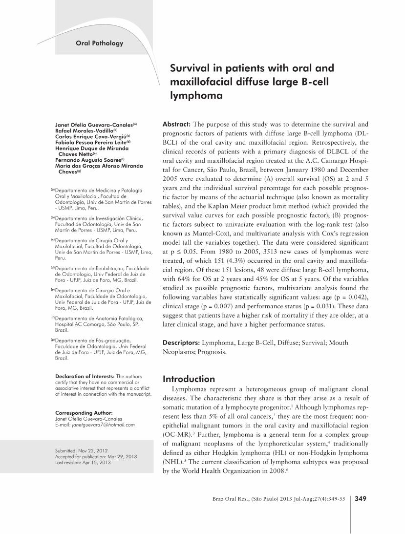
64% at 2 years and 45% at 5 years (Figure 1). Table 1 shows the survival percentage and signif-
icance value for each variable analyzed as a possible prognostic factor for survival.
Multivariate analysis of different variables stud-ied as possible prognostic factors showed three sta-
ResultsFrom January 1980 to December 2005, 3513
new cases of lymphoma were treated at the A.C. Camargo Hospital, of which 151 (4.3%) occurred in the OC-MR. Of these 151 lymphomas, 48 (31.79%) were DLBCL. Of the 48 patients evaluated, OS was
Follow-up Time (years)109876543210
Cum
ulat
ive
Surv
ival
1.0
0.9
0.8
0.7
0.6
0.5
0.4
0.3
2nd year survival: 64%
5th year survival: 45%
Follow-up Time (years)109876543210
Cum
ulat
ive
Surv
ival
1.0
0.8
0.6
0.4
0.2
> 60 years old≤ 60 years old
Age
Follow-up Time (years)109876543210
Cum
ulat
ive
Surv
ival
1.0
0.8
0.6
0.4
0.2
0.0IVIII
III
Clinical Stage
Follow-up Time (years)109876543210
Cum
ulat
ive
Surv
ival
1.0
0.8
0.6
0.4
0.2
0.0IVIII
III
Clinical Stage
Follow-up Time (years)109876543210
Cum
ulat
ive
Surv
ival
1.0
0.8
0.6
0.4
0.2
0.0IVIII
III
Clinical Stage
Follow-up Time (years)109876543210
Cum
ulat
ive
Surv
ival
1.0
0.8
0.6
0.4
0.2
≥ 2≤ 1
Performance Status
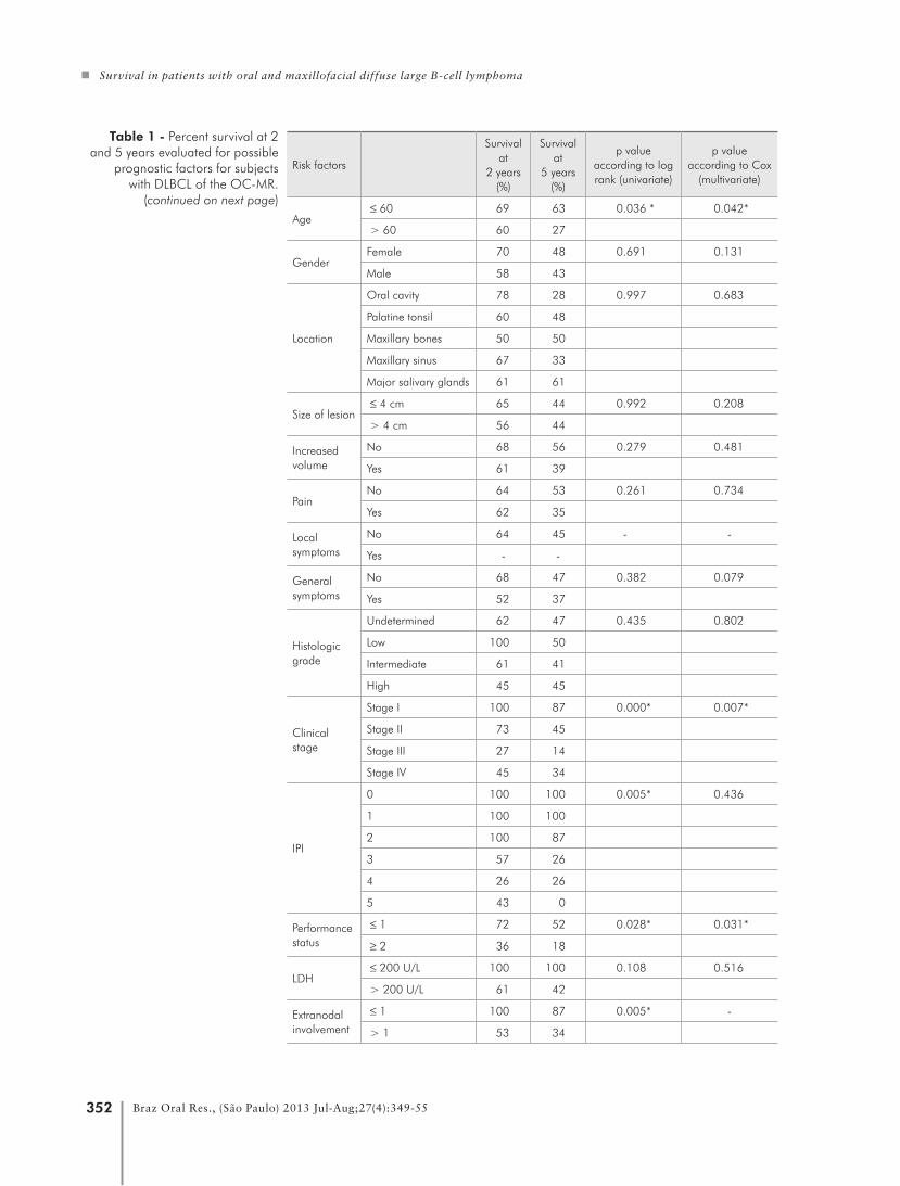
Figure 4 - Mortality and Kaplan-Meier curves with uni-variate significance determined by the log-rank test, per-formance status predictor, for subjects with DLBCL of the OC-MR, seen at the A.C. Camargo Hospital for Cancer, between January 1980 and December 2005.
Figure 1 - Overall survival curve for patients with DLBCL of the OC-MR, seen at the A.C. Camargo Hospital for Cancer, between January 1980 and December 2005.
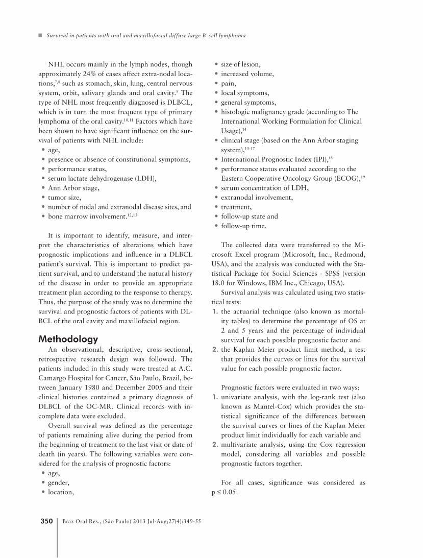
Figure 2 - Mortality and Kaplan-Meier curves with univari-ate significance determined by the log-rank test, age predic-tor, for subjects with DLBCL of the OC-MR, seen at the A.C. Camargo Hospital for Cancer, between January 1980 and December 2005.
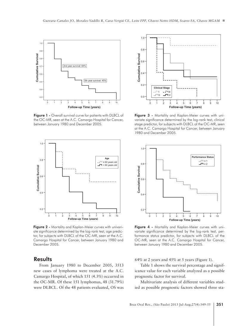
Figure 3 - Mortality and Kaplan-Meier curves with uni-variate significance determined by the log-rank test, clinical stage predictor, for subjects with DLBCL of the OC-MR, seen at the A.C. Camargo Hospital for Cancer, between January 1980 and December 2005.
Survival in patients with oral and maxillofacial diffuse large B-cell lymphoma
352 Braz Oral Res., (São Paulo) 2013 Jul-Aug;27(4):349-55
Risk factors
Survival at
2 years (%)
Survival at
5 years (%)
p value according to log rank (univariate)
p value according to Cox
(multivariate)
Age ≤ 60 69 63 0.036 * 0.042*
> 60 60 27
GenderFemale 70 48 0.691 0.131
Male 58 43
Location
Oral cavity 78 28 0.997 0.683
Palatine tonsil 60 48
Maxillary bones 50 50
Maxillary sinus 67 33
Major salivary glands 61 61
Size of lesion ≤ 4 cm 65 44 0.992 0.208
> 4 cm 56 44
Increased volume
No 68 56 0.279 0.481
Yes 61 39
PainNo 64 53 0.261 0.734
Yes 62 35
Local symptoms
No 64 45 - -
Yes - -
General symptoms
No 68 47 0.382 0.079
Yes 52 37
Histologicgrade
Undetermined 62 47 0.435 0.802
Low 100 50
Intermediate 61 41
High 45 45
Clinical stage
Stage I 100 87 0.000* 0.007*
Stage II 73 45
Stage III 27 14
Stage IV 45 34
IPI
0 100 100 0.005* 0.436
1 100 100
2 100 87
3 57 26
4 26 26
5 43 0
Performance status
≤ 1 72 52 0.028* 0.031*
≥ 2 36 18
LDH ≤ 200 U/L 100 100 0.108 0.516
> 200 U/L 61 42
Extranodal involvement
≤ 1 100 87 0.005* -
> 1 53 34
Table 1 - Percent survival at 2 and 5 years evaluated for possible
prognostic factors for subjects with DLBCL of the OC-MR.
(continued on next page)
Guevara-Canales JO, Morales-Vadillo R, Cava-Vergiú CE, Leite FPP, Chaves Netto HDM, Soares FA, Chaves MGAM
353Braz Oral Res., (São Paulo) 2013 Jul-Aug;27(4):349-55
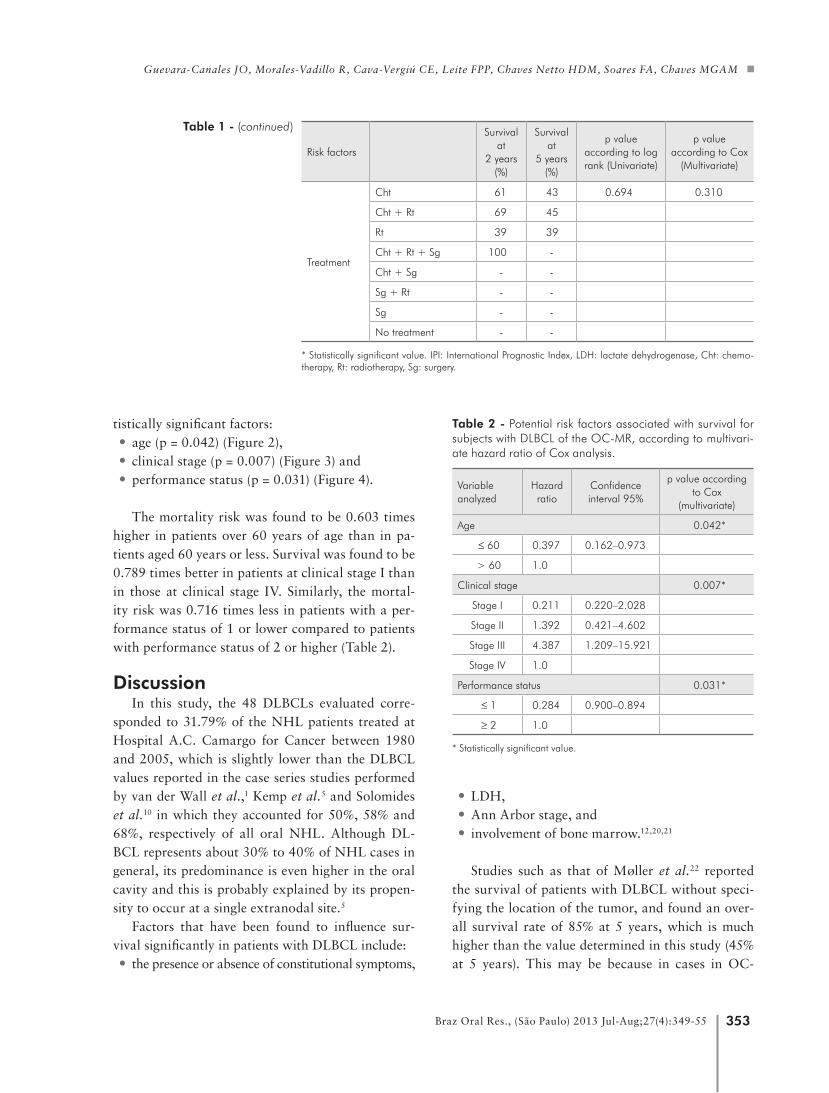
tistically significant factors: • age (p = 0.042) (Figure 2), • clinical stage (p = 0.007) (Figure 3) and • performance status (p = 0.031) (Figure 4).
The mortality risk was found to be 0.603 times higher in patients over 60 years of age than in pa-tients aged 60 years or less. Survival was found to be 0.789 times better in patients at clinical stage I than in those at clinical stage IV. Similarly, the mortal-ity risk was 0.716 times less in patients with a per-formance status of 1 or lower compared to patients with performance status of 2 or higher (Table 2).
DiscussionIn this study, the 48 DLBCLs evaluated corre-
sponded to 31.79% of the NHL patients treated at Hospital A.C. Camargo for Cancer between 1980 and 2005, which is slightly lower than the DLBCL values reported in the case series studies performed by van der Wall et al.,1 Kemp et al.5 and Solomides et al.10 in which they accounted for 50%, 58% and 68%, respectively of all oral NHL. Although DL-BCL represents about 30% to 40% of NHL cases in general, its predominance is even higher in the oral cavity and this is probably explained by its propen-sity to occur at a single extranodal site.5
Factors that have been found to influence sur-vival significantly in patients with DLBCL include:• the presence or absence of constitutional symptoms,
• LDH, • Ann Arbor stage, and • involvement of bone marrow.12,20,21
Studies such as that of Møller et al.22 reported the survival of patients with DLBCL without speci-fying the location of the tumor, and found an over-all survival rate of 85% at 5 years, which is much higher than the value determined in this study (45% at 5 years). This may be because in cases in OC-
Risk factors
Survival at
2 years (%)
Survival at
5 years (%)
p value according to log rank (Univariate)
p value according to Cox
(Multivariate)
Table 1 - (continued)
Treatment
Cht 61 43 0.694 0.310
Cht + Rt 69 45
Rt 39 39
Cht + Rt + Sg 100 -
Cht + Sg - -
Sg + Rt - -
Sg - -
No treatment - -
* Statistically significant value. IPI: International Prognostic Index, LDH: lactate dehydrogenase, Cht: chemo-therapy, Rt: radiotherapy, Sg: surgery.
Table 2 - Potential risk factors associated with survival for subjects with DLBCL of the OC-MR, according to multivari-ate hazard ratio of Cox analysis.
Variable analyzed
Hazard ratio
Confidence interval 95%
p value according to Cox
(multivariate)
Age 0.042*
≤ 60 0.397 0.162–0.973
> 60 1.0
Clinical stage 0.007*
Stage I 0.211 0.220–2.028
Stage II 1.392 0.421–4.602
Stage III 4.387 1.209–15.921
Stage IV 1.0
Performance status 0.031*
≤ 1 0.284 0.900–0.894
≥ 2 1.0
* Statistically significant value.
Survival in patients with oral and maxillofacial diffuse large B-cell lymphoma
354 Braz Oral Res., (São Paulo) 2013 Jul-Aug;27(4):349-55
MR, patients often take too long to seek medical attention, which results in more difficult treatment and poorer prognosis. Vose23 reported a survival rate of about 50% at 5 years, which is similar to the result found in our study.
Multivariate analysis of prognostic factors showed that factors having significant influence are age, clinical stage and performance status. This cor-responds with the results reported by Møller et al.,22 who also found that age and clinical stage influence the survival of patients with DLBCL. Ho et al.24 also report that the prognosis is influenced by clini-cal stage and histologic grade because large cell lym-phomas are considered aggressive and have a poor prognosis.
Many variables have been studied with regards to the survival prognosis of patients with DLBCL, such as hematological and biochemical profiles. Even though these profiles are often normal, patients may have a reduction in the number of peripheral blood lymphocytes, reductions in serum albumin levels, and increases in LDH, which has been shown to cor-
relate with a poor prognosis.25 Some authors have not found the expression of Bcl-2 protein, though it occurs in 30% to 60% of cases, and it has been found to have prognostic value.26 Nevertheless, other studies suggest that the expression of Bcl-2 is related to a significantly poorer survival rate.27 It has also been reported that multiple myeloma oncogene 1 (MUM1) expression is significantly associated to a lower survival rate.28 The study by Bhattacharyya et al.29 considered the type of DLBCL in the oral cav-ity as a prognostic factor. Similarly, Tibiletti et al.30 evaluated fluorescence in situ hybridization to detect the heterogeneity of DLBCL and identify alterations with prognostic implications.
ConclusionAccording to the results of this study, it may be
concluded that the mortality risk is significantly higher in patients with OC-MR DLBCL who are older, at a higher clinical stage, and have higher per-formance status suggesting these are survival prog-nosis factors.
References 1. van der Waal RI, Huijgens PC, van der Valk P, van der Waal
I. Characteristics of 40 primary extranodal non-Hodgkin
lymphomas of the oral cavity in perspective of the new WHO
classification and the International Prognostic Index. Int J
Oral Maxillofac Surg. 2005 Jun;34(4):391-5.
2. Eisenbud L, Sciubba J, Mir R, Sachs SA. Oral presentations
in non-Hodgkin’s lymphoma: a review of thirty-one cases.
Part I. Data analysis. Oral Surg Oral Med Oral Pathol. 1983
Aug;56(2):151-6.
3. Shindoh M, Takami T, Arisue M, Yamashita T, Saito T, Ko-
hgo T, et al. Comparison between submucosal (extra-nodal)
and nodal non-Hodgkin’s lymphoma (NHL) in the oral and
maxillofacial region. J Oral Pathol Med. 1997 Jul;26(6):283-9.
4. Epstein JB, Epstein JD, Le ND, Gorsky M. Characteristics of
oral and paraoral malignant lymphoma: a population-based
review of 361 cases. Oral Surg Oral Med Oral Pathol Oral
Radiol Endod. 2001 Nov;92(5):519-25.
5. Kemp S, Gallagher G, Kabani S, Noonan V, O’Hara C. Oral
non-Hodgkin’s lymphoma: review of the literature and World
Health Organization classification with reference to 40 cases.
Oral Surg Oral Med Oral Pathol Oral Radiol Endod. 2008
Feb;105(2):194-201.
6. Jaffe ES. The 2008 WHO classification of lymphomas: impli-
cations for clinical practice and translational research. Hema-
tology Am Soc Hematol Educ Program. 2009;2009(1):523-31.
7. Clark RM, Fitzpatrick PJ, Gospodarowicz MK. Extranodal
malignant lymphomas of the head and neck. J Otolaryngol.
1983 Aug;12(4):239-45.
8. Spatafore CM, Keyes G, Skidmore AE. Lymphoma: an unusual
oral presentation. J Endod. 1989 Sep;15(9):438-41.
9. Otter R, Gerrits WB, vd Sandt MM, Hermans J, Willemze
R. Primary extranodal and nodal non-Hodgkin’s lymphoma.
A survey of a population-based registry. Eur J Cancer Clin
Oncol. 1989 Aug;25(8):1203-10.
10. Solomides CC, Miller AS, Christman RA, Talwar J, Simpkins
H. Lymphomas of the oral cavity: histology, immunologic
type, and incidence of Epstein-Barr virus infection. Hum
Pathol. 2002 Feb;33(2):153-7.
11. Kolokotronis A, Konstantinou N, Christakis I, Papadimitriou
P, Matiakis A, Zaraboukas T, et al. Localized B-cell non-
Hodgkin’s lymphoma of oral cavity and maxillofacial region:
a clinical study. Oral Surg Oral Med Oral Pathol Oral Radiol
Endod. 2005 Mar;99(3):303-10.
Guevara-Canales JO, Morales-Vadillo R, Cava-Vergiú CE, Leite FPP, Chaves Netto HDM, Soares FA, Chaves MGAM
355Braz Oral Res., (São Paulo) 2013 Jul-Aug;27(4):349-55
12. Coiffier B, Gisselbrecht C, Vose JM, Tilly H, Herbrecht R,
Bosly A, et al. Prognostic factors in aggressive malignant lym-
phomas: description and validation of a prognostic index that
could identify patients requiring a more intensive therapy. The
Groupe d’Etudes des Lymphomes Agressifs. J Clin Oncol.
1991 Feb;9(2):211-9.
13. Velasquez WS, Fuller LM, Jagannath S, Tucker SL, North
LB, Hagemeister FB, et al. Stages I and II diffuse large cell
lymphomas: prognostic factors and long-term results with
CHOP-bleo and radiotherapy. Blood. 1991 Mar 1;77(5):942-7.
14. National Cancer Institute sponsored study of classifications
of non-Hodgkin’s lymphomas: summary and description of a
working formulation for clinical usage. The Non-Hodgkin’s
Lymphoma Pathologic Classification Project. Cancer. 1982
May 15;49(10):2112-35.
15. Carbone PP, Kaplan HS, Musshoff K, Smithers DW, Tubiana
M. Report of the Committee on Hodgkin’s disease staging
classification. Cancer Res. 1971 Nov;31(11):1860-1.
16. Rosenberg SA. Validity of the Ann Arbor staging classification
for the non-Hodgkin’s lymphomas. Cancer Treat Rep. 1977
Sep;61(6):1023-7.
17 Moormeier JA, Williams SF, Golomb HM. The staging of non-
Hodgkin’s lymphomas. Semin Oncol. 1990 Feb;17(1):43-50.
18. A predictive model for aggressive non-Hodgkin’s lymphoma.
The International Non-Hodgkin’s Lymphoma Prognostic Fac-
tors Project. N Engl J Med. 1993 Sep 30;329(14):987-94.
19. Mounier N, Morel P, Haioun C, Coiffier B, Tilly H, Chat-
elain C, et al. A multivariate analysis of the survival of pa-
tients with aggressive lymphoma: variations in the predictive
value of prognostic factors during the course of the disease.
Groupe d’Etudes des lymphomes de l’Adulte. Cancer. 1998
May 15;82(10):1952-62.
20. Shipp MA, Harrington DP, Klatt MM, Jochelson MS, Pinkus
GS, Marshall JL, et al. Identification of major prognostic
subgroups of patients with large-cell lymphoma treated
with m-BACOD or M-BACOD. Ann Intern Med. 1986
Jun;104(6):757-65.
21. Litam P, Swan F, Cabanillas F, Tucker SL, McLaughlin P,
Hagemeister FB, et al. Prognostic value of serum beta-2 mi-
croglobulin in low-grade lymphoma. Ann Intern Med. 1991
May 15;114(10):855-60.
22. Møller MB, Pedersen NT, Christensen BE. Factors predicting
long-term survival in low-risk diffuse large B-cell lymphoma.
Am J Hematol. 2003 Oct;74(2):94-8.
23. Vose JM. Current approaches to the management of non-
Hodgkin’s lymphoma. Semin Oncol. 1998 Aug;25(4):483-91.
24. Ho FC, Todd D, Loke SL, Ng RP, Khoo RK. Clinico-patho-
logical features of malignant lymphomas in 294 Hong Kong
Chinese patients, retrospective study covering an eight-year
period. Int J Cancer. 1984 Aug 15;34(2):143-8.
25. de Leval L, Braaten KM, Ancukiewicz M, Kiggundu E, Del-
aney T, Mankin HJ, et al. Diffuse large B-cell lymphoma of
bone: an analysis of differentiation-associated antigens with
clinical correlation. Am J Surg Pathol. 2003 Sep;27(9):1269-
77.
26. Pezzella F, Tse AG, Cordell JL, Pulford KA, Gatter KC, Mason
DY. Expression of the bcl-2 oncogene protein is not specific
for the 14;18 chromosomal translocation. Am J Pathol. 1990
Aug;137(2):225-32.
27. Rantanen S, Imonni O, Joensuu H, Franssila K, Knuutila
S. Causes and consequences of BCL2 overexpression in dif-
fuse large B-cell lymphoma. Leuk Lymphoma. 2001 Sep-
Oct;42(5):1089-98.
28. Hans CP, Weisenburger DD, Greiner TC, Gascoyne RD, Dela-
bie J, Ott G, et al. Confirmation of the molecular classification
of diffuse large B-cell lymphoma by immunohistochemistry
using a tissue microarray. Blood. 2004 Jan 1;103(1):275-82.
29. Bhattacharyya I, Chehal HK, Cohen DM, Al-Quran SZ.
Primary diffuse large B-cell lymphoma of the oral cav-
ity: germinal center classification. Head Neck Pathol. 2010
Sep;4(3):181-91.
30. Tibiletti MG, Martin V, Bernasconi B, Del Curto B, Pecciarini
L, Uccella S, et al. BCL2, BCL6, MYC, MALT 1, and BCL10
rearrangements in nodal diffuse large B-cell lymphomas: a
multicenter evaluation of a new set of fluorescent in situ hy-
bridization probes and correlation with clinical outcome. Hum
Pathol. 2009 May;40(5):645-52.