Surface-Modified Multifunctional Thymol-Loaded ...
23
pharmaceutics Article Surface-Modified Multifunctional Thymol-Loaded Biodegradable Nanoparticles for Topical Acne Treatment Camila Folle 1 , Natalia Díaz-Garrido 2,3,4 , Elena Sánchez-López 1,5, * , Ana Maria Marqués 6 , Josefa Badia 2,3,4 , Laura Baldomà 2,3,4 , Marta Espina 1,5 , Ana Cristina Calpena 1,5 and María Luisa García 1,5 Citation: Folle, C.; Díaz-Garrido, N.; Sánchez-López, E.; Marqués, A.M.; Badia, J.; Baldomà, L.; Espina, M.; Calpena, A.C.; García, M.L. Surface-Modified Multifunctional Thymol-Loaded Biodegradable Nanoparticles for Topical Acne Treatment. Pharmaceutics 2021, 13, 1501. https://doi.org/10.3390/ pharmaceutics13091501 Academic Editor: Bozena B. Michniak-Kohn Received: 15 August 2021 Accepted: 10 September 2021 Published: 18 September 2021 Publisher’s Note: MDPI stays neutral with regard to jurisdictional claims in published maps and institutional affil- iations. Copyright: © 2021 by the authors. Licensee MDPI, Basel, Switzerland. This article is an open access article distributed under the terms and conditions of the Creative Commons Attribution (CC BY) license (https:// creativecommons.org/licenses/by/ 4.0/). 1 Department of Pharmacy and Pharmaceutical Technology and Physical Chemistry, Faculty of Pharmacy and Food Sciences, University of Barcelona, 08028 Barcelona, Spain; [email protected] (C.F.); [email protected] (M.E.); [email protected] (A.C.C.); [email protected] (M.L.G.) 2 Department of Biochemistry and Physiology, Faculty of Pharmacy and Food Sciences, University of Barcelona, 08028 Barcelona, Spain; [email protected] (N.D.-G.); [email protected] (J.B.); [email protected] (L.B.) 3 Institute of Biomedicine, University of Barcelona (IBUB), 08028 Barcelona, Spain 4 Sant Joan de Déu Research Institute (IR-SJD), 08950 Barcelona, Spain 5 Institute of Nanoscience and Nanotechnology (IN2UB), University of Barcelona, 08028 Barcelona, Spain 6 Department of Biology, Healthcare and Environment, Faculty of Pharmacy and Food Sciences, University of Barcelona, 08028 Barcelona, Spain; [email protected] * Correspondence: [email protected] Abstract: The present work is focused on the development of novel surface-functionalized poly(lactic- co-glycolic acid) nanoparticles loaded with thymol (TH-NPs) for topical administration enhancing thymol anti-inflammatory, antioxidant and wound healing activities against acne. TH-NPs were prepared by solvent evaporation method using different surface functionalization strategies and obtaining suitable physicochemical parameters and a good short-term stability at 4 ◦ C. Moreover, TH-NPs skin penetration and antioxidant activity were assessed in ex vivo pig skin models. Skin penetration of TH-NPs followed the follicular route, independently of the surface charge and they were able to enhance antioxidant capacity. Furthermore, antimicrobial activity against Cutibacterium acnes was evaluated in vitro by the suspension test showing improved antibacterial performance. Using human keratinocyte cells (HaCat), cytotoxicity, cellular uptake, antioxidant, anti-inflammatory and wound healing activities were studied. TH-NPs were non-toxic and efficiently internalized inside the cells. In addition, TH-NPs displayed significant anti-inflammatory, antioxidant and wound healing activities, which were highly influenced by TH-NPs surface modifications. Moreover, a synergic activity between TH-NPs and their surface functionalization was demonstrated. To conclude, surface-modified TH-NPs had proven to be suitable to be used as anti-inflammatory, antioxidant and wound healing agents, constituting a promising therapy for treating acne infection and associated inflammation. Keywords: PLGA; thymol; nanoparticles; skin; HaCaT cells; anti-inflammatory; chitosan; phos- phatidylcholine; poloxamer 1. Introduction Acne vulgaris is one of the most prevalent skin inflammatory disorders affecting 9.4% of the population worldwide [1,2]. It is a multifactorial disease. Their pathophysiology is complex, with both internal and external triggers [3]. This disease is induced by several factors such as irregular keratinocyte proliferation and differentiation, increased sebum production by active sebaceous glands and imbalances in the skin microbiota. This imbal- ance is particularly related to certain Cutibacterium strains, among them Cutibacterium acnes, a normal skin commensal previously known as Propionibacterium acnes [4]. In addition, exogenous factors such as hormones, drugs, nutrition, stress or smoke habits, can also trigger acne development [5,6]. This combination of factors leads to skin lesions such as whiteheads, blackheads, pustules and cysts developing into swelling and inflammation [7]. Pharmaceutics 2021, 13, 1501. https://doi.org/10.3390/pharmaceutics13091501 https://www.mdpi.com/journal/pharmaceutics
Transcript of Surface-Modified Multifunctional Thymol-Loaded ...
pharmaceutics
Article
Surface-Modified Multifunctional Thymol-LoadedBiodegradable Nanoparticles for Topical Acne Treatment
Camila Folle 1, Natalia Díaz-Garrido 2,3,4 , Elena Sánchez-López 1,5,* , Ana Maria Marqués 6 , Josefa Badia 2,3,4,Laura Baldomà 2,3,4 , Marta Espina 1,5 , Ana Cristina Calpena 1,5 and María Luisa García 1,5
�����������������
Citation: Folle, C.; Díaz-Garrido, N.;
Sánchez-López, E.; Marqués, A.M.;
Badia, J.; Baldomà, L.; Espina, M.;
Calpena, A.C.; García, M.L.
Surface-Modified Multifunctional
Thymol-Loaded Biodegradable
Nanoparticles for Topical Acne
Treatment. Pharmaceutics 2021, 13,
1501. https://doi.org/10.3390/
pharmaceutics13091501
Academic Editor: Bozena
B. Michniak-Kohn
Received: 15 August 2021
Accepted: 10 September 2021
Published: 18 September 2021
Publisher’s Note: MDPI stays neutral
with regard to jurisdictional claims in
published maps and institutional affil-
iations.
Copyright: © 2021 by the authors.
Licensee MDPI, Basel, Switzerland.
This article is an open access article
distributed under the terms and
conditions of the Creative Commons
Attribution (CC BY) license (https://
creativecommons.org/licenses/by/
4.0/).
1 Department of Pharmacy and Pharmaceutical Technology and Physical Chemistry, Faculty of Pharmacy andFood Sciences, University of Barcelona, 08028 Barcelona, Spain; [email protected] (C.F.);[email protected] (M.E.); [email protected] (A.C.C.); [email protected] (M.L.G.)
2 Department of Biochemistry and Physiology, Faculty of Pharmacy and Food Sciences, University of Barcelona,08028 Barcelona, Spain; [email protected] (N.D.-G.); [email protected] (J.B.); [email protected] (L.B.)
3 Institute of Biomedicine, University of Barcelona (IBUB), 08028 Barcelona, Spain4 Sant Joan de Déu Research Institute (IR-SJD), 08950 Barcelona, Spain5 Institute of Nanoscience and Nanotechnology (IN2UB), University of Barcelona, 08028 Barcelona, Spain6 Department of Biology, Healthcare and Environment, Faculty of Pharmacy and Food Sciences, University of
Barcelona, 08028 Barcelona, Spain; [email protected]* Correspondence: [email protected]
Abstract: The present work is focused on the development of novel surface-functionalized poly(lactic-co-glycolic acid) nanoparticles loaded with thymol (TH-NPs) for topical administration enhancingthymol anti-inflammatory, antioxidant and wound healing activities against acne. TH-NPs wereprepared by solvent evaporation method using different surface functionalization strategies andobtaining suitable physicochemical parameters and a good short-term stability at 4 ◦C. Moreover,TH-NPs skin penetration and antioxidant activity were assessed in ex vivo pig skin models. Skinpenetration of TH-NPs followed the follicular route, independently of the surface charge and theywere able to enhance antioxidant capacity. Furthermore, antimicrobial activity against Cutibacteriumacnes was evaluated in vitro by the suspension test showing improved antibacterial performance.Using human keratinocyte cells (HaCat), cytotoxicity, cellular uptake, antioxidant, anti-inflammatoryand wound healing activities were studied. TH-NPs were non-toxic and efficiently internalizedinside the cells. In addition, TH-NPs displayed significant anti-inflammatory, antioxidant and woundhealing activities, which were highly influenced by TH-NPs surface modifications. Moreover, asynergic activity between TH-NPs and their surface functionalization was demonstrated. To conclude,surface-modified TH-NPs had proven to be suitable to be used as anti-inflammatory, antioxidant andwound healing agents, constituting a promising therapy for treating acne infection and associatedinflammation.
Keywords: PLGA; thymol; nanoparticles; skin; HaCaT cells; anti-inflammatory; chitosan; phos-phatidylcholine; poloxamer
1. Introduction
Acne vulgaris is one of the most prevalent skin inflammatory disorders affecting 9.4%of the population worldwide [1,2]. It is a multifactorial disease. Their pathophysiology iscomplex, with both internal and external triggers [3]. This disease is induced by severalfactors such as irregular keratinocyte proliferation and differentiation, increased sebumproduction by active sebaceous glands and imbalances in the skin microbiota. This imbal-ance is particularly related to certain Cutibacterium strains, among them Cutibacterium acnes,a normal skin commensal previously known as Propionibacterium acnes [4]. In addition,exogenous factors such as hormones, drugs, nutrition, stress or smoke habits, can alsotrigger acne development [5,6]. This combination of factors leads to skin lesions such aswhiteheads, blackheads, pustules and cysts developing into swelling and inflammation [7].
Pharmaceutics 2021, 13, 1501. https://doi.org/10.3390/pharmaceutics13091501 https://www.mdpi.com/journal/pharmaceutics

Pharmaceutics 2021, 13, 1501 2 of 23
C. acnes is a normal resident of healthy skin, mainly located surrounding the hairfollicle, which is likely to proliferate under unbalanced function of the sebaceous glands,contributing to inflammation and acne development [8]. Hence, C. acnes has a dual activity.This skin commensal strain is essential for sebum control and maintenance of the acidicpH of the pilosebaceous follicle by hydrolysing sebum triglycerides and via propionic acidsecretion [4,9]. However, it can act as a pathogen under dysbiosis conditions that help itsovergrowth in active sebaceous glands [10].
Imbalanced skin microbiome, including C. acnes overgrowth, trigger innate immunesystem activation, leading to cutaneous inflammation. Consequently, the great array ofinduced immune-regulatory and pro-inflammatory mediators amplify direct damagingeffects on molecules and cells, including DNA, proteins and lipids, causing immunosup-pression [11].
Skin inflammation is an innate and non-specific skin immunological response towardsexternal and internal environment modifications or aggressions. The skin immunolog-ical activity includes microbial ligands for toll-like receptors (TLR). Host responses togram-positive bacteria peptidoglycan and gram-negative bacterial lipopolysaccharide aremediated by TLR-2 and TLR-4, respectively [12]. The skin anti-inflammatory mechanismof action consists in modulating the expression of certain genes in order to produce anincrease in anti-inflammatory proteins and inhibit pro-inflammatory cytokines release. Cy-tokines are small proteins which modulate immune responses, regulate cell activation andproliferation. They are produced by epithelial cells, macrophages, CD4 and CD8 T cells. Onkeratinocytes, C. acnes activates TLR-2 and TLR-4 leading to the activation of MAPK andNF-kB pathways. Moreover, they produce interleukins such as (IL)-1, IL-6, IL-8 and TNF-α.Then, ROS production is stimulated, when CD-36 recognizes C. acnes, clearing away thebacteria and inducing the inflammation [13]. In acute inflammation, these mediators arepresent for short periods of time, whereas in chronic inflammation, there is an imbalancedproduction. Chemokines are cytokine subtypes that take on monocytes/macrophages,neutrophils and T cells from the circulation to the areas of infection. Moreover, neutrophilspromote wound healing due to secretion of cytokines, chemokines and growth factors toabolish bacteria and adjust wound microenvironment through oxygen metabolism [14].
Furthermore, sebum composition is severely altered in acne, the ROS produced byneutrophils are involved in the irritation and disruption of the follicular wall, leadingto a progressive inflammatory response. ROS overflow might be associated to the lipidspresent in the sebum. A mediated production of monounsaturated oleic acid is a crucialstep in virulence factor of biofilm formation, playing a critical role for bacterial adherence,since it has been found to be an essential nutrient for resident C. acnes microbiota [15]. Itis also known to enhance epidermal calcium influx in keratinocytes, inducing abnormalkeratinization and barrier function associated with increased release of IL-1α in come-dones [16]. Furthermore, uric acid increases IL-1β expression through a TLR4-mediatedpathway. Thus, increased sebum in acne may activate uric acid–mediated inflammasome.Hyper-keratinization might be initiated via IL-1, suggesting keratinocyte activation cy-cles, and hence, hyper-proliferation [17]. Hyper-keratinization leads to sebaceous glandobstruction clogging the follicle in function of lipids, bacteria and induced cytokines. Inaddition to C. acnes being a gram-positive bacterium, it possesses a featured cell walland outer envelope that synthesizes phosphatidylinositol. Its surrounding peptidoglycancontaining a cross-linkage peptide chain may allow recognition of receptors contributingto inflammation, secreting TNF-α, IL-1α and IL-8 [12]. In particular, IL-8 together withother factors, may attract neutrophils to the pilosebaceous unit.
Superoxide or hydroxyl ions, and hydrogen peroxide are highly reactive moleculesgenerated in normal cell metabolism. Moreover, acne-related strains generate ROS andraise inflammation in keratinocytes. Then, it can cause oxidative damage to proteins,lipids, enzymes and DNA [18,19]. These factors reflect on impaired skin function, thus aninflammatory response and cell death. Therefore, the skin is constantly exposed to inducedoxidative stress. However, intrinsic antioxidant defence mechanisms contribute to barrier

Pharmaceutics 2021, 13, 1501 3 of 23
integrity, which is essential for a healthy skin. The cellular redox environment plays a keyrole in skin homeostasis, preventing oxidative damage of lipids and proteins, and avoidingan imbalanced pro-oxidant/antioxidant stimulus. The oxidation of phenolic compoundsnormally takes place in the cytosol, in contact with peroxidase/H2O2. This would producephenoxyl radical and may co-oxidize glutathione [20]. Consecutive inflammation mayaffect the skin internal components and their functions. Moreover, cicatrisation processesrely on cell regeneration while healing the infection and the inflammatory responses.
Natural anti-inflammatory, antioxidants and antimicrobial agents could be the key forpreventing or ameliorating acne associated symptoms [21]. In this area, natural compoundsare gaining increased importance. Among them, Thymol (TH) is a monoterpene with aphenolic structure associated with several activities such as antioxidant, antimicrobial,antifungal, antiseptic as well as anti-inflammatory [22–24]. Despite its multifunctionalproperties, TH present a low penetration trough skin which could decrease its potentialeffects against acne. Moreover, TH commercial use is still underexplored, probably becauseits low water solubility, high volatility and high light sensibility and its low solubility inwater [25,26].
In order to overcome TH physicochemical drawbacks, several authors have attemptedto encapsulate it into several delivery platforms such as cyclodextrins [26,27], and lipidnanoparticles [25] among others [28]. Moreover, in order to improve active compoundsbioavailability after topical administration, polymeric nanoparticles (NPs) constitute excel-lent potential candidates. Specially for acne treatment due to their small particle diameter,able to penetrate the skin inside the follicle. Additionally, NPs enable the encapsulatedactive compounds for long-term release inside the lesions. Among the most widely usedpolymers, poly(lactic-co-glycolic acid) (PLGA) has been approved by the Food and DrugAdministration and is one of the most successful biodegradable polymers [29]. Despitetheir great potential, to our knowledge, to date no other group have developed TH loadedPLGA nanoparticles. In addition, PLGA negative surface is highly versatile and can bemodified using several compounds in order to improve NPs performance after topicalskin delivery. In this area, Chitosan is a natural polysaccharide, positively charged, thathas known anti-inflammatory and wound healing activities [30]. Chitosan can be usedeither as polymer carrier or be adhered on the surface of other types of negatively chargedNPs [31]. In addition, some types of natural phospholipids such as phosphatidylcholinethat are normally used to produce liposomes, also have demonstrated to possess antioxi-dant or anti-inflammatory activities enhancing the efficacy of some encapsulated drugs [32].Moreover, some synthetic types of surfactants such as Poloxamers, have antioxidant andanti-inflammatory activities [33,34].
Therefore, this study was developed to designed thymol-loaded polymeric NPs (TH-NPs) assessing several surface functionalization compounds and comparing their activityin vitro and ex vivo for the treatment of acne. TH-NPs were functionalized either withphosphatidylcholine, Poloxamer 188 or Poloxamer 407. Additionally, combination ofchitosan and Poloxamer surface functionalization was also carried out. Bacterial infectioninflammation was induced by C. acnes inoculation in keratinocyte cells (HaCaT). Moreover,the antioxidant properties, cell regeneration and wound healing activities were also assayedcomparing the different formulations developed.
2. Materials and Methods2.1. Materials
PLGA Resomer® RG 504H (consisting of a carboxylic terminal group, molecularweight 38,000–54,000 Da and molar ratio lactide:glycolide 50:50) was purchased fromBoehringer Ingelheim (Ingelheim am Rhein, Germany). Thymol (TH), Poloxamer 188 (P)and Poloxamer 407 (PP) were purchased from Sigma Aldrich (Madrid, Spain). Chitosan(C) was supplied by HMC+ (GmbH, Saale, Germany), and phosphatidylcholine (L) wasacquired from Lipoid® (GmbH, Ludwigshafen am Rhein, Germany). Double distilled

Pharmaceutics 2021, 13, 1501 4 of 23
water was used after filtration in a Millipore system. All other chemicals and reagents usedin the study were of analytical grade.
2.2. Methods2.2.1. Preparation of Thymol Loaded Nanoparticles
Thymol-loaded PLGA NPs (TH-NPs) containing a matrix structure (nanospheres)were obtained by solvent displacement evaporation, as described by Fessi et al. [35]. In thecurrent study, a previously optimized formulation based on TH-PLGA-NPs was modifiedby functionalizing TH-NPs surface using several compounds. In order to prepare the NPs,the organic phase composed by PLGA and 2.5 mg/mL of TH was dissolved in acetone andthe aqueous phase consisted on either phosphatidylcholine (TH-NP-L-) or Poloxamer 188(TH-NP-P-) or Poloxamer 407 (TH-NP-PP-), for negatively charged particles. Additionally,positively charged particles were also produced containing chitosan (TH-NP-P-C+, TH-NP-PP-C+), where the aqueous solution contained 1% acetic acid. The organic phase wasadded dropwise into the aqueous phase, under continuous stirring. A rotatory evaporator(Buchi, Flawil, Switzerland) under constant pressure was used to evaporate the organicphase, obtaining the nanoparticles. Empty NPs (B-NPs) were prepared using the sameprocedure but without the addition of TH.
2.2.2. Nanoparticles Physicochemical Characterization
The average particle size (Zav) and polydispersity index (PI) were determined by pho-ton correlation spectroscopy, using a ZetaSizer Nano ZS (Malvern Instruments; Malvern,UK). The surface charge, measured as zeta-potential (ZP), was determined by electrophoreticmobility using the same instrument.
Encapsulation of TH was measured indirectly by quantification of unloaded amount.Samples were diluted 1:10 in Milli-Q water:ethanol (90:10) and centrifuged (Centrifuge5415C, Geratebau Eppendorf GmbH, Engelsdorf, Germany) for 10 min at 14,000 rpm, usingMillipore filter device (Amicon® Ultra, 0.5 mL 100 K, Merck Millipore Ltd., CarrigtwohillCo. Cork IRL, Darmstadt, Germany). The filtered fractions were quantified by HPLC, andthe EE was determined by the Equation (1):
EE = (Ci − Cs)/Ci·100 (1)
where Ci is the initial concentration of the active and Cs is the concentration of the unloadedamount found in the filtered fraction.
HPLC quantitative analysis was performed by reverse-phase high-performance liquidchromatography (HPLC) by a modification of the method described previously [36]. Stud-ies were carried out in Acquity Waters System with UV detector, using a Kromasil® column(C18, 5 µm, 150 mm × 4.6 mm), (Teknokroma, Barcelona, Spain). The mobile phase con-sisted of acetonitrile:water under gradient conditions of 30:70/58:42/30:70 during 20 min.TH was determined at wavelength of 275 nm.
2.2.3. Stability of Thymol Loaded Nanoparticles
Short-term stability of TH NPs was evaluated after one month of storage at 4, 25, 30and 40 ◦C by measuring Zav, PI, ZP and pH. Additional long-term studies were carried outduring 6 months by measuring the physicochemical parameters monthly at 4 ◦C. Moreover,backscattering profiles of TH-NPs stored at 4 ◦C, were also recorded using TurbiscanLab®
(Formulation, Toulousse, France) [37].
2.2.4. Ex Vivo Skin Penetration Route of Thymol Loaded Nanoparticles
Ex vivo pig skin was obtained from the animal house (Bellvitge, University ofBarcelona), used in accordance with the protocol approved by the Ethics Committeeof the University of Barcelona.
Prior to the experiment, rhodamine-labelled PLGA (R-PLGA) was synthesized asdescribed by Gonzalez-Pizarro et al. [38] and used at 0.01% obtaining rhodamine labelled

Pharmaceutics 2021, 13, 1501 5 of 23
nanoparticles (R-TH-NPs). Skin penetration assay was executed using vertical diffusionFranz cells (FDC-400, Vidra-Foc, Barcelona, Spain) with a thermal bath set to 32 ◦C to mimicskin in vivo conditions, under constant stirring. R-TH-NPs were applied onto ex vivo pigskin 0.64 cm2 (donor compartment) and penetration was allowed for 24 h. Skin sampleswere washed, fixed in PBS containing 4% paraformaldehyde (PFA) for 4 h, followed bycryoprotection into PBS with 30% sucrose for 24 h, snap-frozen in isopentane at−50 ◦C andthen kept overnight at−80 ◦C. Samples were mounted in O.C.T. ® Compound (Tissue-Tek®,Sakura Finetek, Torrance, CA, USA) and sliced with cryostat microtome (LEICA CM3050S) at −20 ◦C onto glass-slides Superfrost® Plus (Menzel-Glaser, Thermo Scientific, USA),covered with Fluoromount G® (Invitrogen, Thermo Fisher Scientific, Rockford, IL, USA).Samples were visualized by confocal laser scanning microscopy (Zeiss LSM 880). Imageswere acquired using Zen Black 2.3 software performing z-stack sections and thus processedwith ImageJ software v1.53k.
2.2.5. Ex Vivo Skin Antioxidant Activity by Methylene Blue Reduction
A colorimetric assay was performed using methylene blue dye to test the ex vivoantioxidant activity of TH and surface functionalized TH-NPs. Methylene blue, in combi-nation with an antioxidant molecule, reduces into a colorless lecomethylene blue [39].
Pig skin samples were cut into 2 cm2 and placed into a 6-well plate containing 0.5 mLof PBS, with the stratum corneum (SC) facing up. Then, a methylene blue solution at0.01% was applied on the surface of each skin sample and incubated for 4 h at 32 ◦C, in thepresence of humidity. Skin fragments were washed with PBS and the SC was dried withfilter paper. TH-NPs were applied onto the skin (30 µL) and further incubated for 1 h. Thecontrol sample was treated with distilled water. Images were recorded at initial and 1 hpost-treatment to assess differences in methylene blue reduction.
2.2.6. Free-Radical Scavenging by DPPH
The scavenging capacity of TH, surface functionalized TH-NPs and surface function-alization components was evaluated using DPPH (2,2-diphenyl-1-picrylhydrazyl) assay,based on other authors with some modifications [40]. Samples were dissolved and dilutedin methanol at concentrations ranging from 0.1 to 10 mg/mL, and DPPH, a free radicalcompound, was prepared in 80% methanol at 0.1 mM. Sample dilutions were transferredinto a 96-well plate (200 µL/well) and 20 µL of the DPPH stock solution was added intoeach well. BHT (butyl-hydroxytoluene), a known antioxidant compound was used asendogenous control.
Samples without DPPH were used as blank. Samples were incubated in the dark for45 min on a mechanical shaker. The UV-VIS absorbance was measured at 517 nm and datawere calculated using the Equation (2):
Free Radical Scavening(%) =Ac− (As− Ab)
Ac·100 (2)
where Ac, As and Ab are the absorbances of the control, sample and blank, respectively.
2.2.7. Antimicrobial Efficacy of Thymol Loaded Nanoparticles
A fresh inoculum of C. acnes was prepared in PBS adjusted to an optical density of0.72 at 550 nm, using a UV-visible spectrophotometer (Shimadzu Corp., Kyoto, Japan). Theassay was carried out in a total volume of 1 mL containing TH or TH-NPs (900 µL) at afinal concentration of 250 µg/mL and fresh bacterial inoculum (100 µL). Samples were keptat 37 ◦C in a shaker incubator (Innova 4080, New Brunswick Scientific, Edison, NJ, USA)for 30 min. Then, 100 µL of each test tube was neutralized in 900 µL of Berens cosmeticdiluent (Scharlab, Barcelona, Spain) for 15 min [41]. Ten-fold dilutions in PBS (10 µL) wereadded to clostridium reinforced medium agar dishes for enumerating bacteria by the dropcount method. Microbial count was performed after incubation for 48 h under anaerobicconditions at 37 ◦C. Bacterial viability was expressed as CFU/mL against time (h).

Pharmaceutics 2021, 13, 1501 6 of 23
2.2.8. Cytotoxicity and Cellular Uptake of Thymol Loaded Nanoparticles
Human keratinocytes (HaCaT) cells were cultured in high glucose Dulbecco’s Modi-fied Eagle’s Medium (DMEM) from Thermofisher, supplemented with 10% fetal bovineserum (FBS), 2 mM L-glutamine, 100 units/mL penicillin G and 100 µg/mL streptomycin.Cells were incubated at 37 ◦C and 5% CO2 and experiments were performed when cellsreached 80–90% of confluence.
Cytotoxicity of TH-NPs was evaluated by the MTT (3-(4,5-Dimethylthiazol-2-yl)-2,5-diphenyl tetrazolium bromide) assay that is based on the mitochondrial reduction oftetrazolium salt by intracellular dehydrogenases of viable cells. Samples were tested atconcentrations up to 2, 10 and 20 µg/mL. Briefly, HaCaT cells were seeded in 96-well plates(100 µL) at a density of 2 × 10⁵ cells/well, adjusted in an automated cell counter (Countess,Invitrogen, Thermofisher) and grown for 24 h at 37 ◦C. Then, TH-NPs were added at theindicated concentrations and cells were further incubated for 24 h. Finally, the mediumwas removed and MTT (Sigma-Aldrich Chemical Co, St. Louis, MO, USA) was addedat 0.25% in PBS. After 2 h incubation, the medium was replaced by 100 µL DMSO (99%dimethyl sulfoxide, Sigma-Aldrich, Madrid, Spain) [42]. Cell viability was then measuredat 570 nm in a Modulus® Microplate Photometer (Turner BioSystems Inc., Sunnyvale, CA,USA). Results were expressed as percentage of cell survival relative to untreated cells.
HaCaT cells were seeded in an 8-well µ-slide (Ibidi®). Cells were incubated inFBS/phenol red-free medium in the presence or absence of TH-NPs for 2 h at the indicatedconcentration. Cell membrane was stained with wheat germ agglutinin (WGA) Alexa-488(Molecular Probes) at 1 µg/mL for 15 min followed by fixation with 3% paraformaldehydefor 25 min. Cell nuclei were stained with 4′,6-diamidino-2-phenylindole (DAPI, SigmaAldrich, Madrid, Spain) at 0.5 µg/mL for 15 min. Internalization of NPs in HaCaT cells wasassessed by confocal microscopy (Leica TCS SPII), using the 63X oil immersion objectivelens [38]. Images were processed using Fiji image software [38].
2.2.9. Anti-Inflammatory Activity in TNF-α-Induced Inflammation Model
HaCaT cells were seeded in 12-well plates at a density of 2 × 10⁵ cells/well andgrown until 80–90% confluence. Cells were then treated with TH-NPs for 2 h, followedby stimulation with 50 µM TNF-α for 2 h to induce inflammation [43]. The medium wasreplaced by fresh FSB-free medium and cells were incubated overnight. Supernatantswere collected, and quantification of secreted interleukin-6 (IL-6) was carried out by usingELISA Human kit (BD OptEIA® Set Human IL-6, BD Biosciences, Franklin Lakes, NJ, USA)following manufacturer instructions. Absorbance was measured at 450 and 560 nm using aplate reader (Varioskan, Thermo Fisher Scientific, Rockford, IL, USA). Data were processedand analyzed using version 5 GraphPad® Prism software.
2.2.10. Anti-Inflammatory Activity in C. acnes-Induced Inflammation Model
This experiment was performed as described in the previous section, but C. acnesfresh inoculum was added instead of TNF-α. C. acnes was grown until the stationaryphase (5 days incubation under anaerobiosis in BHI culture medium). Then bacterial cellswere harvested and diluted in FSB-free DMEM medium, adjusted to OD 1.2 at 550 nm.Different dilutions of this inoculum were added to HaCaT cells and incubated overnight.Quantification of IL-6 in cell culture supernatants was assayed by using ELISA Humankit (BD OptEIA® Set Human IL-6, BD Biosciences, Franklin Lakes, NJ, USA) followingmanufacturer instructions [44].
2.2.11. Real-Time Quantitative Polymerase Chain Reaction (RT-qPCR)
HaCaT cells were adjusted to a density of 2 × 10⁵ cells/well and seeded in 12-wellplates. After 48 h, cells were treated with TH or TH-NPs or blank NPs (B-NPs) for 2 h. Next,cells were stimulated for 4 h with C. acnes prepared in FBS-free DMEM medium, adjustedto OD 1.2 at 550 nm. HaCaT cells without any treatment were used as a negative controland cells incubated only with C. acnes as a positive control. Total RNA was isolated from

Pharmaceutics 2021, 13, 1501 7 of 23
cells using an RNA extraction kit (Qiagen RNeasy, Germantown, MD, USA) following themanufacturer’s guide (Qiagen, Crawley, UK) and quantified by the ratio of absorbancevalues at 260 and 280 nm using a NanoDrop TM-2000 spectrophotometer (Thermo FisherScientific, Waltham, MA, USA). cDNA was synthesized from RNA (1 µg) by using the High-Capacity cDNA Reverse Transcription kit (Applied Biosystems, Foster City, CA, USA) in afinal volume of 20 µL. Quantitative PCR reactions were performed in a StepOne Plus PCRcycler (Applied Biosystems, Foster City, CA, USA) by using SYBR® Green PCR Master Mix(Applied Biosystems, Foster City, CA, USA) and specific human oligonucleotide primersfor IL-1α, IL-1β, IL-6, IL-8, TNF-α and β-actin (endogenous control, primers specified inTable S1 of Supplementary Materials). Control reactions were performed in the absence ofRNA. The standard PCR program was conducted by one denaturation cycle for 10 min at95 ◦C followed by 40 cycles of 15 s at 95 ◦C and 1 min at 60 ◦C. Relative gene expression wascalculated as fold change compared to sample control by means of 2−∆∆Ct formula [42].
2.2.12. Antioxidant Activity Assessed by ROS Quantification
HaCaT cells were seeded in 96-well plates at 2 × 10⁵ cells/well (100 µL) for 72 h.Cells were loaded with the fluorogenic dye H2DCFDA (2′,7′-dichlorodihydrofluoresceindiacetate) at 25 µM diluted in phenol red/FBS-free DMEM medium, for 45 min in thedark. This fluorogenic dye passively diffuses into the cells, is deacetylated by intracellularesterase and emits fluorescence upon oxidation by reactive oxygen species (ROS) [45].Then, cells were washed with PBS and incubated for 2 h with TH, surface-functionalizationcompounds, functionalized TH-NPs or B-NPs. At this time, 10 µL of 20 mM H2O2 wereadded to the wells. Fluorescence was measured at excitation and emission wavelengthsof 485 and 530 nm, respectively. Data were acquired at 0, 30, 60 and 120 min. The dataof the positive control (H2O2) after 2 h was used to normalize values (%). Backgroundfluorescence of control cells were subtracted from all measurements.
2.2.13. Wound Healing Activity in HaCaT Cells by the Scratch Assay
In order to study wound healing activity of the developed TH-NPs, prevention andtreatment were assessed. In order to study wound healing prevention, HaCaT cells wereseeded in 12-well plates at a density of 5 × 10⁴ cells/well and grown for 24 h until 70–80%confluence. Cells were treated for 2 h with free TH, surface-functionalized TH-NPs orfunctionalization compounds, washed with PBS and further incubated for 24 h. Then,scratches were performed in the middle of each well using a 200 µL pipette tip, washedwith PBS, refilled with FBS-free culture medium and incubated for 24 h [46,47]. Contrastphase images of the scratches were obtained at the beginning of the experiment (T0) andafter 24 h using a fluorescent microscope, and the wound area was measured using ImageJsoftware.
In the previous assay, the capacity to prevent wound healing was assessed, whereas ina second assay, wound healing treatment was examined by applying the formulations afterthe lesion was caused. For this second experiment, HaCaT cells were seeded and grown for24 h. After creating the scratches as previously mentioned, cells were washed with PBS andrefilled with DMEM containing 1% FBS. Images at this timepoint were recorded by usingfluorescent microscope at 10X (LEICA DFC300FX). Cells were then treated with either TH,surface functionalized TH-NP or surface functionalization compounds for 2 h and furtherincubated for 24 h in 1% FBS—culture medium [30]. Images at 24 h were recorded andprocessed using ImageJ software.
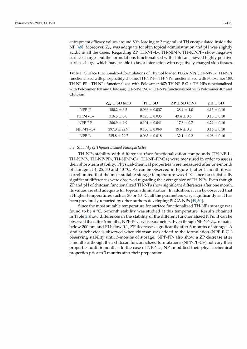
3. Results and Discussion3.1. Thymol Loaded Nanoparticles Physicochemical Characterization
TH-NPs were prepared using the solvent displacement method and surface func-tionalized with different compounds (TH-NP-L-, TH-NP-P-; TH-NP-PP-, TH-NP-P-C+,TH-NP-PP-C+). Their physical-chemistry characterization is shown in Table 1. All formula-tions presented good homogeneity below 0.2 indicating monodisperse systems and high
Pharmaceutics 2021, 13, 1501 8 of 23
entrapment efficacy values around 80% leading to 2 mg/mL of TH encapsulated inside theNP [48]. Moreover, Zav was adequate for skin topical administration and pH was slightlyacidic in all the cases. Regarding ZP, TH-NP-L-, TH-NP-P-; TH-NP-PP- show negativesurface charges but the formulations functionalized with chitosan showed highly positivesurface charge which may be able to favor interaction with negatively charged skin tissues.
Table 1. Surface functionalized formulations of Thymol loaded PLGA NPs (TH-NP-L-: TH-NPsfunctionalized with phosphatidylcholine; TH-NP-P-: TH-NPs functionalized with Poloxamer 188;TH-NP-PP-: TH-NPs functionalized with Poloxamer 407; TH-NP-P-C+: TH-NPs functionalizedwith Poloxamer 188 and Chitosan; TH-NP-PP-C+: TH-NPs functionalized with Poloxamer 407 andChitosan).
Zav ± SD (nm) PI ± SD ZP ± SD (mV) pH ± SD
NPP-P- 180.2 ± 6.5 0.066 ± 0.037 −28.9 ± 1.0 4.15 ± 0.10
NPP-P-C+ 316.5 ± 3.8 0.123 ± 0.035 43.4 ± 0.6 3.15 ± 0.10
NPP-PP- 206.9 ± 9.9 0.101 ± 0.041 −17.8 ± 0.7 4.29 ± 0.10
NPP-PP-C+ 297.3 ± 22.9 0.150 ± 0.068 19.6 ± 0.8 3.16 ± 0.10
NPP-L- 235.8 ± 29.7 0.063 ± 0.018 −32.1 ± 0.2 4.08 ± 0.10
3.2. Stability of Thymol Loaded Nanoparticles
TH-NPs stability with different surface functionalization compounds (TH-NP-L-,TH-NP-P-; TH-NP-PP-, TH-NP-P-C+, TH-NP-PP-C+) were measured in order to assesstheir short-term stability. Physical-chemical properties were measured after one-monthof storage at 4, 25, 30 and 40 ◦C. As can be observed in Figure 1, after 1 month it wascorroborated that the most suitable storage temperature was 4 ◦C since no statisticallysignificant differences were observed regarding the average size of TH-NPs. Even thoughZP and pH of chitosan functionalized TH-NPs show significant differences after one month,its values are still adequate for topical administration. In addition, it can be observed thatat higher temperatures such as 30 or 40 ◦C, all the parameters vary significantly as it hasbeen previously reported by other authors developing PLGA NPs [49,50].
Since the most suitable temperature for surface functionalized TH-NPs storage wasfound to be 4 ◦C, 6-month stability was studied at this temperature. Results obtainedin Table 2 show differences in the stability of the different functionalized NPs. It can beobserved that after 6 months, NPP-P- vary its parameters. Even though NPP-P- Zav remainsbelow 200 nm and PI below 0.1, ZP decreases significantly after 6 months of storage. Asimilar behavior is observed when chitosan was added to the formulation (NPP-P-C+)observing stability until 3-months of storage. NPP-PP- also show a ZP decrease after3 months although their chitosan functionalized formulations (NPP-PP-C+) not vary theirproperties until 6 months. In the case of NPP-L-, NPs modified their physicochemicalproperties prior to 3 months after their preparation.

Pharmaceutics 2021, 13, 1501 9 of 23
Figure 1. One-month stability of TH NPs with different surface functionalization strategies at 4,25, 30 and 40 ◦C. (A) Average size, (B) Zeta potential (ZP), (C) pH values. Statistical significancewas analyzed against freshly prepared formulations (one month), * p < 0.5; ** p < 0.01; *** p < 0.001;**** p < 0.0001.

Pharmaceutics 2021, 13, 1501 10 of 23
Table 2. Physicochemical values of TH-NPs with different surface functionalization stored at 4 ◦C.
Month Zav (nm) ± SD PI ± SD ZP ± SD (mV)
NPP-P-
0 172.9 ± 1.9 0.066 ± 0.037 −24.5 ± 0.9
1 177.7 ± 2.2 0.071 ± 0.015 −20.9 ± 0.5
3 183.8 ± 3.6 0.082 ± 0.005 −18.3 ± 0.7
6 191.5 ± 1.5 0.091 ± 0.015 −14.3 ± 0.6
NPP-P-C+
0 337.3 ± 7.4 0.123 ± 0.035 23.6 ± 0.3
1 365.4 ± 3.2 0.141 ± 0.011 23.2 ± 0.9
3 392.9 ± 8.0 0.158 ± 0.037 21.0 ± 0.3
6 419.6 ± 11.5 0.197 ± 0.066 17.2 ± 0.2
NPP-PP-
0 184.0 ± 0.9 0.101 ± 0.041 −22.2 ± 0.6
1 191.2 ± 0.7 0.098 ± 0.022 −18.2 ± 0.6
3 189.1 ± 10.1 0.099 ± 0.015 −12.1 ± 0.4
6 220.0 ± 10.8 0.123 ± 0.33 −8.4 ± 0.7
NPP-PP-C+
0 221.1 ± 3.3 0.149 ± 0.036 10.5 ± 0.6
1 224.1 ± 5.3 0.150 ± 0.068 9.8 ± 0.6
3 256.1 ± 5.3 0.152 ± 0.077 7.3 ± 0.7
6 348.5 ± 17.2 0.189 ± 0.023 6.1 ± 0.5
NPP-L-
0 178.5 ± 0.6 0.063 ± 0.018 −41.2 ± 1.8
1 180.9 ± 1.4 0.076 ± 0.019 −39.7 ± 0.5
3 214.7 ± 2.7 0.114 ± 0.032 −29.7 ± 0.5
6 201.9 ± 3.4 0.132 ± 0.027 −23.9 ± 0.8
In addition, backscattering profile of surface-functionalized TH-NPs was analyzed byTurbiscan® and results are shown in Figure 2. All optimized TH-NPs underwent a slightsedimentation that was reversible by agitation. This sedimentation might be the causeof the physicochemical modifications previously observed (Table 2). Moreover, since thedifference between the obtained profiles was below 10%, this indicates a suitable stabilityof all the functionalized TH-NPs. However, in order to ensure long-term stability, TH-NPslyophilization and incorporation into semi-solid formulations would be contemplated infurther studies.

Pharmaceutics 2021, 13, 1501 11 of 23
Figure 2. Backscattering profile of NPs measured monthly for 6 months after storage at 4 ◦C. (A)TH-NP-L-, (B) TH-NP-P-, (C) TH-NP-P-C+, (D) TH-NP-PP- and (E) TH-NP-PP-C+.

Pharmaceutics 2021, 13, 1501 12 of 23
3.3. Ex Vivo Skin Penetration of Thymol Loaded Nanoparticles
As can be observed in Figure 3, R-TH-NPs successfully penetrated into the skinhair follicle within 24 h. This is of extreme relevance since it is the main site where acneassociated infection and inflammation occurs. Moreover, it can be observed that penetrationwas not influenced by the surface charge carried out since both negatively and positivelycharge TH NPs were able to penetrate through the skin follicle ex vivo.
Figure 3. Pig skin hair follicle penetration of R-TH-NPs in 24 h by confocal microscopy. (A) untreated(control), (B) R-TH-NP-L-, (C) R-TH-NP-P- and (D) R-TH-NP-P+. Scale bar: 200 µm.
3.4. Ex Vivo Methylene Blue Reduction
The antioxidant efficiency of TH and surface functionalized TH-NPs was evaluatedin the ex vivo pig skin model by measuring methylene blue reduction, which results ina colorless compound (Figure 4). Results showed that all the assessed TH-NPs showedantioxidant activity, greater than that of free TH. No qualitative differences between TH-NPs were observed.
Figure 4. Ex vivo antioxidant activity by methylene blue reduction in pig skin. Images recorded at time 0 and 1 h of thestudied compounds (control, free TH, TH-NP-L-, TH-NP-P-, TH-NP-P-C+, TH-NP-PP- and TH-NP-PP-C+).
3.5. In Vitro Antioxidant Activity
The in vitro antioxidant activity of the different compounds used to prepare theformulations was evaluated individually by the DPPH assay (Figure 5). Results expressedas free radical scavenging capacity (%) showed that TH has similar antioxidant activity asthe control BHT, although slightly higher. When tested separately, the surface compoundsP, PP and L displayed slightly in vitro free radical scavenging activity, although it waslower than the activity of TH and BHT in all dosages tested.

Pharmaceutics 2021, 13, 1501 13 of 23
Figure 5. Antioxidant activity of TH, BHT and surface compounds alone (P, PP and L) assessed by the DPPH free-radicalscavenging assay. The 100% ROS was obtained by the value of the control (H2O2) in 2 h.
3.6. In Vitro Antimicrobial Efficacy
The in vitro antimicrobial activity of optimized surface functionalized TH-NPs againstC. acnes was similar to that of TH and in all the cases, statistical signifcant differences wereobtained against the positive control (Figure 6). The higher antimicrobial activity wasobtained with TH-NP-P-, although no statistically significant differences were observedbetween the different formulations.
Figure 6. Antimicrobial activity of NPs against C. acnes measured by the suspension test. Valuesrepresent microbial counts in CFU/mL after 30 min incubation and are expressed as Mean ± SD(n = 3). Statistical analysis was carried out via one-way ANOVA, with Tukey’s Multiple ComparisonTest: **, p < 0.001 against control (C. acnes without any treatment).
3.7. Cytotoxicity and Cellular Uptake of Thymol Loaded NPs in HaCaT Cells
Cytotoxicity of TH-NPs was evaluated on HaCaT cells using the MTT assay. Cellswere incubated for 24 h with TH or each TH-NP at concentrations of 2, 10 or 20 µg/mL.The surface compounds alone (P, PP, L) were tested at concentrations equivalent to thosepresent in each formulation. Results showed that none of the samples were cytotoxic ascell viability was kept close to the untreated control cells, above 90% (data not shown).
Cellular uptake was evaluated for R-TH-NP-L-, R-TH-NP-P- and R-TH-NP-P-C+(20 µg/mL) in HaCaT cells in order to evaluate composition and surface charge influenceon cellular uptake. After 2 h incubation, TH-NPs-associated fluorescence was detected byconfocal microscopy in cells treated with any of the R-TH-NPs tested (Figure 7). The cell

Pharmaceutics 2021, 13, 1501 14 of 23
membrane was stained with WGA (green) and the nucleus (blue) with DAPI. In the mergedimages the internalized TH-NPs were mainly localized in the cytoplasm in all the cases.
Figure 7. Cellular uptake by confocal microscopy analysis of HaCaT cells incubated with theindicated rhodamine-labelled NPs. (A) membrane staining with WGA, (B) nuclei staining with DAPI;(C) fluorescence of internalized R-TH-NPs, (D) 3D-plot of C, (E) merged A and C, (F) merged B andC, (G) merged A, B and C, (H) 3D-plot of G. Figure scale bar corresponds to 10 µm.

Pharmaceutics 2021, 13, 1501 15 of 23
3.8. Anti-Inflammatory Activity of Thymol Loaded NPs in HaCaT Cells Treated with TNF-α
The anti-inflammatory activity of the formulated NPs was evaluated in the TNF-α-induced inflammation model using HaCaT cells. Secretion of IL-6 was quantified byELISA in cell supernatants of untreated control HaCaT cells (basal IL-6 expression), TNFα-treated HaCaT cells in the absence (positive control of inflammation) or in the presenceof the different NPs. Free TH and surface compounds (C, L, P, PP) were also testedin parallel (Figure 8). Results showed that TH significantly reduced TNF-α-inducedsecretion of IL-6. All surface compounds analyzed individually, except PP, have similaranti-inflammatory activity as TH. Additionally, all TH-NPs, except TH-NP-PP-, presentedhigher anti-inflammatory activity than free compounds. The most effective NPs were thepositively charged formulations, containing C. However, only TH-NP-P-C+ displayed asignificant difference compared to TH.
Figure 8. Quantification of secreted IL-6 by ELISA in TNF-α-stimulated HaCaT cells pre-incubatedwith formulated NPs and free compounds. Values of IL-6 (pg/mL) are the Mean ± SD, n = 3.Negative control: HaCaT cells without any treatment; Positive control: HaCaT cells treated only withTNF-α. Statistical analysis was performed by via one-way ANOVA, followed by Tukey’s MultipleComparison Test. ** p < 0.001 compared to positive control, and # p < 0.01 compared to TH.
3.9. Anti-Inflammatory Activity of Thymol Loaded NPs in HaCaT Cells Treated with C. acnes
The inflammatory activity of C. acnes was assessed in HaCaT cells treated with dif-ferent dilutions of a C. acnes stock inoculum prepared in DMEM medium (OD 1.2 at550 nm). Inflammation was evaluated by quantification of secreted IL-8 by ELISA. Resultsshowed that C. acnes triggered IL-8 secretion in a dose- dependent manner (Figure S1 ofSupplementary Materials).
From these results, the undiluted C. acnes stock inoculum prepared in DMEM medium(OD 1.2 at 550 nm) was optimized to be added directly to HaCaT cells to induce inflam-mation in further experiments aimed at evaluating the anti-inflammatory potential ofTH-NPs. In this context, expression of genes encoding the inflammatory cytokines TNF-α,IL-1α, IL-1β, IL-6 and IL-8 was analyzed by RT-qPCR after C. acnes infection in HaCaTcells pre-treated with TH, TH-NPs or B-NPs (NPs without TH). Cells challenged only withC. acnes were used as a positive control of the inflammatory response. Results are illustratedin Figure 9. Infection by C. acnes strongly induced the expression of all pro-inflammatorycytokines tested. According to its anti-inflammatory properties, TH significantly decreasedthe expression of all of them.

Pharmaceutics 2021, 13, 1501 16 of 23
Figure 9. Gene expression levels of inflammatory cytokines in C. acnes-infected HaCaT cells. Beforethe addition of undiluted C. acnes inoculum (adjusted to OD 1.2 at 550 nm), HaCaT cells werepre-incubated with TH or the indicated NPs. Relative mRNA levels of (A) IL-6, (B) IL-8, (C) IL-1α,(D) IL-1β and (E) TNF-α were measured by RT-qPCR, using β-actin as the reference gene. Values(Mean ± SEM, n = 3) are expressed as fold-change compared to untreated HaCaT cells (control-).Statistical analysis was performed via one-way ANOVA, followed by Tukey’s Multiple ComparisonTest (p < 0.05* or p < 0.001**): versus positive control (control+); (p < 0.05$ or p < 0.001$$): versus THand (p < 0.05# or p < 0.001##): versus the respective B-NP.
Infection by C. acnes strongly induced the expression of all pro-inflammatory cytokinestested (positive control). According to its anti-inflammatory properties, TH significantlydecreased the expression of all of them. In general, all TH-NPs have anti-inflammatoryactivity, being able to reduce the mRNA levels of the different cytokines to a greater or lesserdegree depending on the type of TH-NPs and the cytokine analyzed. Some TH-NPs have ananti-inflammatory activity greater than that of free TH. Statistically significant differenceswith respect the reduction caused by TH were apparent when analyzing the expressionof IL-1α and IL-6. In the case of the TH-NP-P-C + formulation, statistical differences withrespect to TH were also significant for TNF-α and IL-8. The results showed that B-NPScan also reduce significantly the expression of all cytokines except IL-1α. However, theanti-inflammatory activity was less than that exhibited by the equivalent TH-NP. The onlyTH-NP that did not show significant differences with respect the equivalent B-NP is theTH-NP-L formulation.
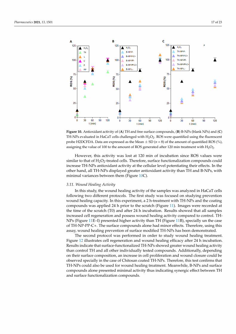
3.10. Antioxidant Activity via ROS Quantification in H2O2-Induce H2DCFDALabelled HaCaT
The antioxidant activity of TH-NPs was evaluated by ROS quantification in HaCaTcells stressed with hydrogen peroxide. Treatment with any of the surface funtionalizedTH-NPs significantly reduced intracellular ROS to a greater extent than TH (Figure 10).TH displayed antioxidant activity at all assay times (Figure 10A). Moreover, the surfacecompounds tested individually did not show significant antioxidant activity (Figure 10A),indicating their inability to act as free radical scavengers at the cellular level. In the caseof the B-NPs (Figure 10B), they showed a ROS scavenging activity similar to TH up to60 min after H2O2 challenge. Even though no significant differences were found, TH-NP-L-showed an increased antioxidant activity that may be due to phosphaditilcholine coatingpotentiation of TH antioxidant effects [51].
Pharmaceutics 2021, 13, 1501 17 of 23
Figure 10. Antioxidant activity of (A) TH and free surface compounds, (B) B-NPs (blank NPs) and (C)TH-NPs evaluated in HaCaT cells challenged with H2O2. ROS were quantified using the fluorescentprobe H2DCFDA. Data are expressed as the Mean ± SD (n = 8) of the amount of quantified ROS (%),assigning the value of 100 to the amount of ROS generated after 120 min treatment with H2O2.
However, this activity was lost at 120 min of incubation since ROS values weresimilar to that of H2O2-treated cells. Therefore, surface functionalization compounds couldincrease TH-NPs antioxidant activity at the cellular level potentiating their effects. In theother hand, all TH-NPs displayed greater antioxidant activity than TH and B-NPs, withminimal variances between them (Figure 10C).
3.11. Wound Healing Activity
In this study, the wound healing activity of the samples was analyzed in HaCaT cellsfollowing two different protocols. The first study was focused on studying preventionwound healing capacity. In this experiment, a 2 h-treatment with TH-NPs and the coatingcompounds was applied 24 h prior to the scratch (Figure 11). Images were recorded atthe time of the scratch (T0) and after 24 h incubation. Results showed that all samplesincreased cell regeneration and possess wound healing activity compared to control. TH-NPs (Figure 11E–I) presented higher activity than TH (Figure 11B), specially un the caseof TH-NP-PP-C+. The surface compounds alone had minor effects. Therefore, using thisassay, wound healing prevention of surface modified TH-NPs has been demonstrated.
The second protocol was performed in order to study wound healing treatment.Figure 12 illustrates cell regeneration and wound healing efficacy after 24 h incubation.Results indicate that surface-functionalized TH-NPs showed greater wound healing activitythan control TH and all other individually tested compounds. Additionally, dependingon their surface composition, an increase in cell proliferation and wound closure could beobserved specially in the case of Chitosan coated TH-NPs. Therefore, this test confirms thatTH-NPs could also be used for wound healing treatment. Meanwhile, B-NPs and surfacecompounds alone presented minimal activity thus indicating synergic effect between THand surface functionalization compounds.

Pharmaceutics 2021, 13, 1501 18 of 23
Figure 11. Wound healing activity in HaCaT pre-scratch treatment (wound healing prevention).Scratches were monitored at T0 and after 24 h incubation. (A) control, (B) Free TH, (C) chitosan,(D) phosphatidylcholine, (E) TH-NP-L-, (F) TH-NP-P-, (G) TH-NP-P-C+, (H) TH-NP-PP- and (I)TH-NP-PP-C+.
Figure 12. Recorded images of wound healing activity in HaCaT cells under post-scratch treatment (wound healingtreatment). (A) Control T0, (B) Control after 24 h incubation, (C) Free TH, (D) Poloxamer 188, (E) poloxamer 407, (F)chitosan, (G) phosphatidylcholine, (H) TH-NP-P-, (I) TH-NP-P-C+, (J) TH-NP-PP-, (K) TH-NP-PP-C+, (L) TH-NP-L-, (M)B-TH-NP-P-, (N) B-NP-P-C+, (O) B-NP-PP-, (P) B-NP-PP-C+, (Q) TH-NP-L-.
Pharmaceutics 2021, 13, 1501 19 of 23
4. Discussion
In the present study, surface-functionalized PLGA NPs were successfully loaded withthymol by the solvent displacement method in order to avoid TH instability and highvolatility. To compare their pharmacological activities against acne, 5 formulations weredeveloped, namely TH-NP-P-, TH-NP-P-C+, TH-NP-PP-, TH-NP-PP-C+ and TH-NP-L-.All the resulting TH-NPs presented suitable physical chemical parameters and a suitableshort-term stability at 4 ◦C. The sedimentation phenomena observed by the backscatteringsignal was reversed by agitation. Confocal microscopy qualitative analysis confirmedthat TH-NPs penetrated into the hair follicle, independent of their surface-coating typeor charge. Some authors stated that the reservoir of the hair follicle could store actives10 times longer than the reservoir of the SC and that hair follicle under movement (in vivo)should improve NPs penetration [52]. Other authors have already described that NPsaccumulate in the follicular entry, which is confirmed in the case of surface-functionalizedTH-NPs [53–55]. Regarding TH-NP-L, it appears that they can penetrate not only in thefollicle, by also within the primary layers of the epidermis. This can be explained by thephospholipidic nature of the surface that diffuses easier through the skin. In the case ofR-TH-NP-P-, their penetration was found mainly through in the entire hair follicle and alower amount remaining in the SC. This indicated that this type of surface is favored by thefollicular and not the intercellular pathway. On the other hand, concerning R-TH-NP-P-C+,which are positively charged and present slightly higher particle diameter, the penetrationcould be observed only inside the follicle. This is in accordance with the fact that NPspenetration into hair follicle is size and surface dependent.
Furthermore, the free-radical scavenging of TH was similar to endogenous controlBHT and much higher than the free surface compounds themselves, which showed loweractivity. The antimicrobial activity of TH and TH-NPs against C. acnes demonstrated toreduce microbial viability within only 30 min incubation. All tested samples presentedsimilar activity, where the highest was achieved for TH-NP-P-. Moreover, since the pene-tration route of TH-NPs into the skin is through the hair follicle, the observed activity willpresumably be performed directly on the acne lesion.
In HaCaT cell line, TH-NPs and all free compounds themselves did not alter cellviability, presenting no cytotoxicity. The cellular uptake images showed most of the NPswithin only 2 h in the cytosol but also present the nucleus, especially for TH-NP-P-. Theanti-inflammatory activity of TH and TH-NPs all presented significant reduction on geneexpressions tested, where, in all the cases, all or most of TH-NPs performed significantreduction compared to the control and to their corresponding B-NP. Depending on thegene analyzed, the activity was enhanced for a different surface composition of the NPs.Therefore, they act as good booster of the activity. The antioxidant activity in HaCaTcells was achieved for all TH-NPs, higher than TH and all other tested samples. Theantioxidant activity of compounds enables cell proliferation faster, that can enhance woundhealing processes to improve skin healing process on acne lesions [56]. For the woundhealing activity, all scratched treated cells provided higher cell proliferation than controlcells. Interestingly, in both prevention and treatment of wound healing activity all surfacemodified TH-NPs showed suitable results. All TH-NPs provided good cell regeneration,meanwhile the B-NPs showed minimal activity. The antioxidant properties of surfacecompounds might have influenced on the healing activity, possibly by synergic activitywith TH. All TH-NPs showed better results than TH, confirming that surface modifiedTH-NPs present higher healing capacity than free antioxidant compounds. The antioxidantactivity was also confirmed by the methylene blue reduction in ex vivo pig skin, showingthat TH-NPs had greater activity than TH within 1 h incubation.
To summarize, TH was successfully encapsulated into PLGA NPs with suitable particlediameters and good stability. Moreover, other authors have developed different types ofNPs for TH encapsulation such as PLA or chitosan NPs obtaining lower stability values(around 60%) [28]. Lipid NPs containing thymol have also been developed with a negativesurface charge confirming their anti-inflammatory properties but they were assessed for

Pharmaceutics 2021, 13, 1501 20 of 23
psoriasis treatment [25]. In addition, Pires and colleagues studied compatibility of TH withseveral excipients confirming that P80 was a suitable excipient [57]. Moreover, surface-functionalized TH-NPs successfully penetrated into the skin through the hair follicle,where acne occurs. Even though the antimicrobial activity of TH-NPs was similar toTH, they provided outstanding activities as anti-inflammatory, antioxidant and woundhealing, when compared to than TH and B-NPs. TH-NPs in contact with HaCaT cellsshowed that they were able to penetrate inside the cells, enabling greater activities thanwhen compounds were introduced as its free form. TH and TH-NPs presented goodanti-inflammatory activity by significant reduction on the gene expression tested. Theefficacy of TH-NPs varied depending on their surface coating as well as genes analyzed.The antioxidant activity was proven in HaCaT and on ex vivo skin, and in all cases, TH-NPswere greater than TH. For this reason, they have also provided excellent wound closureand cell proliferation results, where the NPs positively charged, performed higher synergicactivity on healing processes. Therefore, our study demonstrates that TH-NPs with surfacefunctionalization using different approaches could constitute a potential efficient treatmentfor acne. However, these results would need to be assessed in in vivo experiments due tothe multifactorial triggers of acne as well as the sebaceous content that can influence NPsstability [58].
5. Conclusions
Surface-modified TH-NPs demonstrated to possess antimicrobial, anti-inflammatory,antioxidant and wound healing properties. In addition to TH-NPs multifunctional thera-peutic benefits, they penetrate through the hair follicle and, therefore, they could be suitablefor the treatment of severe acne disease.
Supplementary Materials: The following are available online at https://www.mdpi.com/article/10.3390/pharmaceutics13091501/s1, Table S1: Oligonucleotinde primers used for RT-qPCR, FigureS1: Dose-dependent inflammatory capacity of C. acnes. HaCaT cells were incubated for 24 with theindicated dilutions of the C. acnes stock inoculum (adjusted to OD 1.2 at 550 nm). Value 1 indicatesno dilution. Secreted IL-8 was quantified in the cell culture supernatant by ELISA. Values of IL-8(pg/mL) are the Mean ± SD, n = 3.
Author Contributions: Conceptualization, C.F., A.C.C., J.B., L.B. and M.L.G.; methodology, C.F.,N.D.-G., A.M.M., M.E. and A.C.C.; formal analysis, C.F., E.S.-L. and A.C.C.; investigation, C.F.,N.D.-G. and M.E.; resources, A.M.M., J.B. and L.B.; writing—original draft preparation, C.F., M.E.and E.S.-L.; writing—review and editing, E.S.-L., A.M.M., J.B., L.B. and M.L.G., funding acquisition,J.B., L.B. and M.L.G. All authors have read and agreed to the published version of the manuscript.
Funding: This research received no external funding.
Institutional Review Board Statement: The study was conducted according to the guidelines of theDeclaration of Helsinki, and approved by the Ethics Committee of the University of Barcelona.
Acknowledgments: The authors ESL, MLG and ME would like to acknowledge 2017SGR1477.
Conflicts of Interest: The authors declare no conflict of interest.
References1. Williams, H.C.; Dellavalle, R.P.; Garner, S. Acne vulgaris. Lancet 2012, 379, 361–372. [CrossRef]2. Sachdeva, M.; Tan, J.; Lim, J.; Kim, M.; Nadeem, I.; Bismil, R. The prevalence, risk factors, and psychosocial impacts of acne
vulgaris in medical students: A literature review. Int. J. Dermatol. 2021, 60, 792–798. [CrossRef]3. Well, D.; Levine, S.R. Acne vulgaris: A review of causes and treatment options. J. Dermatol. Nurses. Assoc. 2014, 6, 302–309.
[CrossRef]4. Flowers, L.; Grice, E.A. The Skin Microbiota: Balancing Risk and Reward. Cell Host Microbe 2020, 28, 190–200. [CrossRef]
[PubMed]5. Chlebus, E.; Chlebus, M. Factors affecting the course and severity of adult acne. Observational cohort study. J. Dermatolog. Treat.
2017, 28, 737–744. [CrossRef]6. Bansal, P.; Sardana, K.; Vats, G.; Sharma, L.; Garga, U.C.; Khurana, A. A Prospective Study Examining Trigger Factors and
Hormonal Abnormalities in Adult Female Acne. Indian Derm. Online J. 2020, 11, 544–550. [CrossRef]

Pharmaceutics 2021, 13, 1501 21 of 23
7. Tanghetti, E.A. The Role of Inflammation in the Pathology of Acne. J. Clin. Aesthetic Dermatol. 2013, 6, 27–35.8. Bhatia, A.; Maisonneuve, J.F.; Persing, D.H. Propionibacterium acnes and chronic diseases. In The Infectious Etiology of Chronic
Diseases: Defining the Relationship, Enhancing the Research, and Mitigating the Effects: Workshop Summary; Knobler, S.L., O’Connor, S.,Lemon, S.M., Eds.; National Academies Press: Washington, DC, USA, 2004.
9. Platsidaki, E.; Dessinioti, C. Recent advances in understanding Propionibacterium acnes (Cutibacterium acnes) in acne.F1000Research 2018, 7, 1953. [CrossRef]
10. Rozas, M.; de Ruijter, A.H.; Fabrega, M.J.; Zorgani, A.; Guell, M.; Paetzold, B.; Brillet, F. From dysbiosis to healthy skin: Majorcontributions of cutibacterium acnes to skin homeostasis. Microorganisms 2021, 9, 628. [CrossRef]
11. Bharti, S.; Vadlamudi, H.C. A strategic review on the involvement of receptors, transcription factors and hormones in acnepathogenesis. J. Recept. Signal Transduct. 2021, 41, 105–116. [CrossRef]
12. Kim, J. Review of the innate immune response in acne vulgaris: Activation of toll-like receptor 2 in acne triggers inflammatorycytokine responses. Dermatology 2005, 211, 193–198. [CrossRef] [PubMed]
13. Lee, Y.B.; Byun, E.J.; Kim, H.S. Potential Role of the Microbiome in Acne: A Comprehensive Review. J. Clin. Med. 2019, 8, 987.[CrossRef]
14. Xue, X.; Falcon, D.M. The role of immune cells and cytokines in intestinal wound healing. Int. J. Mol. Sci. 2019, 20, 6097.[CrossRef]
15. Melnik, B.C. Acne vulgaris: The metabolic syndrome of the pilosebaceous follicle. Clin. Dermatol. 2018, 36, 29–40. [CrossRef][PubMed]
16. Katsuta, Y.; Iida, T.; Hasegawa, K.; Inomata, S.; Denda, M. Function of oleic acid on epidermal barrier and calcium influx intokeratinocytes is associated with N-methyl d-aspartate-type glutamate receptors. Br. J. Dermatol. 2009, 160, 69–74. [CrossRef]
17. Dreno, B.; Gollnick, H.P.M.; Kang, S.; Thiboutot, D.; Bettoli, V.; Torres, V.; Leyden, J. Understanding innate immunity andinflammation in acne: Implications for management. J. Eur. Acad. Dermatol. Venereol. 2015, 29, 3–11. [CrossRef]
18. Amiri, H. Essential oils composition and antioxidant properties of three thymus species. Evid.-Based Complement Altern. Med.2012, 2012, 728065. [CrossRef] [PubMed]
19. Briganti, S.; Picardo, M. Antioxidant activity, lipid peroxidation and skin diseases. What’s new. J. Eur. Acad. Dermatol. Venereol.2003, 17, 663–669. [CrossRef]
20. Bakkali, F.; Averbeck, S.; Averbeck, D.; Idaomar, M. Biological effects of essential oils—A review. Food Chem. Toxicol. 2008, 46,446–475. [CrossRef]
21. Zouboulis, C.C.; Jourdan, E.; Picardo, M. Acne is an inflammatory disease and alterations of sebum composition initiate acnelesions. J. Eur. Acad. Dermatol. Venereol. 2014, 28, 527–532. [CrossRef]
22. Trombetta, D.; Castelli, F.; Sarpietro, M.G.; Venuti, V.; Cristani, M.; Daniele, C.; Saija, A.; Mazzanti, G.; Bisignano, G. Mechanismsof antibacterial action of three monoterpenes. Antimicrob. Agents Chemother. 2005, 49, 2474–2478. [CrossRef] [PubMed]
23. Nagoor Meeran, M.F.; Javed, H.; Al Taee, H.; Azimullah, S.; Ojha, S.K. Pharmacological properties and molecular mechanismsof thymol: Prospects for its therapeutic potential and pharmaceutical development. Front. Pharmacol. 2017, 8, 1–34. [CrossRef][PubMed]
24. Najafloo, R.; Behyari, M.; Imani, R.; Nour, S. A mini-review of Thymol incorporated materials: Applications in antibacterialwound dressing. J. Drug Deliv. Sci. Technol. 2020, 60, 101904. [CrossRef]
25. Pivetta, T.P.; Simões, S.; Araújo, M.M.; Carvalho, T.; Arruda, C.; Marcato, P.D. Development of nanoparticles from natural lipidsfor topical delivery of thymol: Investigation of its anti-inflammatory properties. Colloids Surf. B Biointerfaces 2018, 164, 281–290.[CrossRef]
26. Pires, F.Q.; Pinho, L.A.; Freire, D.O.; Silva, I.C.R.; Sa-Barreto, L.L.; Cardozo-Filho, L.; Gratieri, T.; Gelfuso, G.M.; Cunha-Filho,M. Thermal analysis used to guide the production of thymol and Lippia origanoides essential oil inclusion complexes withcyclodextrin. J. Therm. Anal. Calorim. 2019, 137, 543–553. [CrossRef]
27. Tao, F.; Hill, L.E.; Peng, Y.; Gomes, C.L. Synthesis and characterization of β-cyclodextrin inclusion complexes of thymol andthyme oil for antimicrobial delivery applications. LWT-Food Sci. Technol. 2014, 59, 247–255. [CrossRef]
28. Sáez-Orviz, S.; Marcet, I.; Weng, S.; Rendueles, M.; Díaz, M. PLA nanoparticles loaded with thymol to improve its incorporationinto gelatine films. J. Food Eng. 2020, 269, 1–7. [CrossRef]
29. Sánchez-López, E.; Esteruelas, G.; Ortiz, A.; Espina, M.; Prat, J.; Muñoz, M.; Cano, A.; Calpena, A.C.; Ettcheto, M.; Camins, A.;et al. Dexibuprofen biodegradable nanoparticles: One step closer towards a better ocular interaction study. Nanomaterials 2020,10, 720. [CrossRef]
30. Mengoni, T.; Adrian, M.; Pereira, S.; Santos-Carballal, B.; Kaiser, M.; Goycoolea, F.M. A chitosan-based liposome formulationenhances the in vitro wound healing efficacy of substance P neuropeptide. Pharmaceutics 2017, 9, 56. [CrossRef] [PubMed]
31. Chronopoulou, L.; Massimi, M.; Giardi, M.F.; Cametti, C.; Devirgiliis, L.C.; Dentini, M.; Palocci, C. Chitosan-coated PLGAnanoparticles: A sustained drug release strategy for cell cultures. Colloids Surf. B Biointerfaces 2013, 103, 310–317. [CrossRef]
32. Erös, G.; Ibrahim, S.; Siebert, N.; Boros, M.; Vollmar, B. Oral phosphatidylcholine pretreatment alleviates the signs of experimentalrheumatoid arthritis. Arthritis Res. Ther. 2009, 11, 1–10. [CrossRef]
33. Hunter, R.L.; Luo, A.Z.; Zhang, R.; Kozar, R.A.; Moore, F.A. Poloxamer 188 inhibition of ischemia/reperfusion injury: Evidencefor a novel anti-adhesive mechanism. Ann. Clin. Lab. Sci. 2010, 40, 115–125. [PubMed]

Pharmaceutics 2021, 13, 1501 22 of 23
34. Moghimi, S.M.; Hunter, A.C. Poloxamers and poloxamines in nanoparticle engineering and experimental medicine. TrendsBiotechnol. 2000, 18, 412–420. [CrossRef]
35. Fessi, H.; Puisieux, F.; Devissaguet, J.P.; Ammoury, N.; Benita, S. Nanocapsule formation by interfacial polymer depositionfollowing solvent displacement. Int. J. Pharm. 1989, 55, R1–R4. [CrossRef]
36. Ghasemi Pirbalouti, A.; Rahimmalek, M.; Malekpoor, F.; Karimi, A. Variation in antibacterial activity, thymol and carvacrolcontents of wild populations of Thymus daenensis subsp. daenensis Celak. Plant Omics 2011, 4, 209–214.
37. Sánchez-López, E.; Egea, M.A.; Cano, A.; Espina, M.; Calpena, A.C.; Ettcheto, M.; Camins, A.; Souto, E.B.; Silva, A.M.; García,M.L. PEGylated PLGA nanospheres optimized by design of experiments for ocular administration of dexibuprofen-in vitro, exvivo and in vivo characterization. Colloids Surf. B Biointerfaces 2016, 145, 241–250. [CrossRef]
38. Gonzalez-Pizarro, R.; Parrotta, G.; Vera, R.; Sánchez-López, E.; Galindo, R.; Kjeldsen, F.; Badia, J.; Baldoma, L.; Espina, M.; García,M.L. Ocular penetration of fluorometholone-loaded PEG-PLGA nanoparticles functionalized with cell-penetrating peptides.Nanomedicine 2019, 14, 3089–3104. [CrossRef]
39. Fernández-García, E.; Heluani-Gahete, H.; Wellinger, R.E. A new colorimetric assay for antioxidant capacity and photostability.Color. Technol. 2016, 132, 195–200. [CrossRef]
40. Aman, S.; Moin, S.; Owais, M.; Siddiqui, M.U. Antioxidant activity of thymol: Protective role in AAPH-induced hemolysis indiabetic erythrocytes. Int. J. Pharm. Sci. Invent. 2013, 2, 55–60.
41. Messager, S.; Goddard, P.A.; Dettmar, P.W.; Maillard, J.Y. Determination of the antibacterial efficacy of several antiseptics testedon skin by an “ex-vivo” test. J. Med. Microbiol. 2001, 50, 284–292. [CrossRef]
42. Diaz-Garrido, N.; Fábrega, M.J.; Vera, R.; Giménez, R.; Badia, J.; Baldomà, L. Membrane vesicles from the probiotic Nissle 1917and gut resident Escherichia coli strains distinctly modulate human dendritic cells and subsequent T cell responses. J. Funct.Foods 2019, 61, 103495. [CrossRef]
43. Carvajal-Vidal, P.; Fábrega, M.J.; Espina, M.; Calpena, A.C.; García, M.L. Development of Halobetasol-loaded nanostructuredlipid carrier for dermal administration: Optimization, physicochemical and biopharmaceutical behavior, and therapeutic efficacy.Nanomed. Nanotechnol. Biol. Med. 2019, 20, 102026. [CrossRef] [PubMed]
44. Zhu, T.; Wu, W.; Yang, S.; Li, D.; Sun, D.; He, L. Polyphyllin I Inhibits Propionibacterium acnes-Induced Inflammation In Vitro.Inflammation 2019, 42, 35–44. [CrossRef]
45. Liu, Y.H.; Lin, Y.S.; Huang, Y.W.; Fang, S.U.; Lin, S.Y.; Hou, W.C. Protective Effects of Minor Components of Curcuminoids onHydrogen Peroxide-Treated Human HaCaT Keratinocytes. J. Agric. Food Chem. 2016, 64, 3598–3608. [CrossRef]
46. Governa, P.; Carullo, G.; Biagi, M.; Rago, V.; Aiello, F. Evaluation of the in vitro wound-healing activity of calabrian honeys.Antioxidants 2019, 8, 36. [CrossRef]
47. Jangpromma, N.; Preecharram, S.; Srilert, T.; Maijaroen, S.; Mahakunakorn, P.; Nualkaew, N.; Daduang, S.; Klaynongsruang, S.In vitro and in vivo wound healing properties of plasma and serum from Crocodylus siamensis blood. J. Microbiol. Biotechnol.2016, 26, 1140–1147. [CrossRef]
48. Vega, E.; Egea, M.A.; Garduño-Ramírez, M.L.; García, M.L.; Sánchez, E.; Espina, M.; Calpena, A.C. Flurbiprofen PLGA-PEGnanospheres: Role of hydroxy-β-cyclodextrin on ex vivo human skin permeation and in vivo topical anti-inflammatory efficacy.Colloids Surf. B Biointerfaces 2013, 110, 339–346. [CrossRef] [PubMed]
49. Sánchez-López, E.; Ettcheto, M.; Egea, M.A.; Espina, M.; Cano, A.; Calpena, A.C.; Camins, A.; Carmona, N.; Silva, A.M.; Souto,E.B.; et al. Memantine loaded PLGA PEGylated nanoparticles for Alzheimer’s disease: In vitro and in vivo characterization. J.Nanobiotechnol. 2018, 16, 1–16. [CrossRef] [PubMed]
50. Sánchez-López, E.; Ettcheto, M.; Egea, M.A.; Espina, M.; Calpena, A.C.; Folch, J.; Camins, A.; García, M.L. New potential strategiesfor Alzheimer’s disease prevention: Pegylated biodegradable dexibuprofen nanospheres administration to APPswe/PS1dE9.Nanomed. Nanotechnol. Biol. Med. 2017, 13, 1171–1182. [CrossRef]
51. Kim, J.Y.; Oh, S.; Yi, B.; Kim, M.J.; Lee, J.H. Synergism of phosphatidylcholine on the antioxidant properties of α-tocopherol incorn oils under different relative humidity. Int. J. Food Sci. Technol. 2015, 50, 1421–1428. [CrossRef]
52. Yukuyama, M.N.; De Araújo, G.L.B.; Bou-Chacra, N.A. Nanomaterials for hair care applications. Nanocosmetics 2020, 205–225.[CrossRef]
53. Santos, G.A.; Angelo, T.; Andrade, L.M.; Silva, S.M.M.; Magalhães, P.O.; Cunha-Filho, M.; Gelfuso, G.M.; Taveira, S.F.; Gratieri, T.The role of formulation and follicular pathway in voriconazole cutaneous delivery from liposomes and nanostructured lipidcarriers. Colloids Sur. B Biointerfaces 2018, 170, 341–346. [CrossRef]
54. Pereira, M.N.; Schulte, H.L.; Duarte, N.; Lima, E.M.; Sá-Barreto, L.L.; Gratieri, T.; Gelfuso, G.M.; Cunha-Filho, M.S.S. Solideffervescent formulations as new approach for topical minoxidil delivery. Eur. J. Pharm. Sci. 2017, 96, 411–419. [CrossRef][PubMed]
55. Alvarez-Román, R.; Naik, A.; Kalia, Y.N.; Guy, R.H.; Fessi, H. Skin penetration and distribution of polymeric nanoparticles. J.Control. Release 2004, 99, 53–62. [CrossRef] [PubMed]
56. Mollarafie, P.; Khadiv Parsi, P.; Zarghami, R.; Amini Fazl, M.; Ghafarzadegan, R. Antibacterial and wound healing properties ofthymol (Thymus vulgaris Oil) and its application in a novel wound dressing. J. Med. Plants 2015, 14, 69–81.

Pharmaceutics 2021, 13, 1501 23 of 23
57. Pires, F.Q.; Angelo, T.; Silva, J.K.R.; Sá-Barreto, L.C.L.; Lima, E.M.; Gelfuso, G.M.; Gratieri, T.; Cunha-Filho, M.S.S. Use of mixturedesign in drug-excipient compatibility determinations: Thymol nanoparticles case study. J. Pharm. Biomed. Anal. 2017, 137,196–203. [CrossRef] [PubMed]
58. Tolentino, S.; Pereira, M.N.; de Sousa, M.C.; Cunha-Filho, M.; Gelfuso, G.M.; Gratieri, T. The influence of sebaceous content onthe performance of nanosystems designed for the treatment of follicular diseases. J. Drug Deliv. Sci. Technol. 2020, 59, 101895.[CrossRef]


![Antioxidant Properties of Thymol and Butylated Hydroxytoluene in … · 2010. 8. 15. · Thymol (p-methyl-isopropyl-phenol) is the main constituent of the oils of Thymus vulgaris[7].](https://static.fdocuments.us/doc/165x107/5fe1517555edc50f792e182b/antioxidant-properties-of-thymol-and-butylated-hydroxytoluene-in-2010-8-15.jpg)