Surface Modification and Conjugation Strategies for...
242
Surface Modification and Conjugation Strategies for Bioassay/ Biomaterial Applications A thesis submitted for the degree of Ph.D. by Chandra K. Dixit, M.Sc., Under the supervision of Professor Colette McDonagh, Professor Richard O’Kennedy and Professor Brian D. MacCraith Based on research work carried out at School of Biotechnology, Centre for Boanalytical Sciences, and National Biophotonics and Imaging Platform, Dublin City University, Dublin 9, Ireland. November 2011
-
Upload
trinhquynh -
Category
Documents
-
view
213 -
download
0
Transcript of Surface Modification and Conjugation Strategies for...

Surface Modification and Conjugation Strategies for Bioassay/ Biomaterial
Applications
A thesis submitted for the degree of Ph.D.
by
Chandra K. Dixit, M.Sc.,
Under the supervision of
Professor Colette McDonagh, Professor Richard O’Kennedy and
Professor Brian D. MacCraith
Based on research work carried out at
School of Biotechnology, Centre for Boanalytical Sciences,
and National Biophotonics and Imaging Platform,
Dublin City University, Dublin 9, Ireland.
November 2011

Statement of integrity of work:
I hereby certify that this material, which I now submit for assessment on the programme of study
leading to the award of Doctor of Philosophy (Ph.D.) is entirely my own work, that I have
exercised reasonable care to ensure that the work is original, and does not to the best of my
knowledge breach any law of copyright, and has not been taken from the work of others save and
to the extent that such work has been cited and acknowledged within the text of my work.
Signed: Chandra K. Dixit (Candidate) ID No.: 57119287 Date: November 30, 2011

Abstract:
The aims of this research were to develop novel surface modification strategies that can be
used on a range of solid supports including polymeric and metallic matrices, as these could
have a significant impact on the performance of bio/sensors employing these surfaces. These
approaches were used to develop methods for immobilizing biomolecular recognition
elements, such as antibodies, on modified matrices, and to exploit these approaches for the
generation of high sensitivity bio-assays. Human fetuin A, mouse immunoglobulin G, and
horseradish peroxidase were employed as model analytes.
A silane-based surface modification strategy was designed and optimized for planar or flat
surfaces such as polymeric sheets, chips or microtitre plates. These polymeric surfaces were
activated prior to silane-functionalization using potassium hydroxide (aq.)-mediated mild
oxidation (wet method) and oxygen-plasma etching. This novel surface activation strategy
was further optimized in combination with surface functionalization and covalent
immobilization of antibodies, for enhancing immunoassay sensitivities. Sensitivities
obtained for immunoassays using antibodies immobilized with the developed and
adsorption-based conventional strategies were 39 and 625 pg/mL, respectively, for human
fetuin A. The strategy was demonstrated to be generic in nature and could be employed to
activate a wide range of polymeric and metallic surfaces. In addition, highly sensitive
detection of human fetuin A was achieved with antibodies captured in an oriented manner
on covalently immobilized protein A (EC50 3.7 ng/mL) in comparison to randomly captured
antibodies (EC50 5.8 ng/mL).
High-brightness NIR664 dye-doped silica nanoparticles were employed to probe various
activation states of platelets. These NPs were functionalized with silanes (viz. amine and
carboxy-terminal) followed by conjugation to a platelet surface biomarker-specific antibody
(anti-CD41) and successfully employed for probing platelet activation. The antibody-NP
conjugates were found to be highly sensitive (>95%) and specific (≈100%). In addition,
aggregation of NPs was minimized by controlling their surrounding chemical environment
and their stability after antibody conjugation.

Table of contents
Chapter Section Page Number Abbreviations i Analytical units v Patents, IDFs, Publications and Conference
presentations and posters vi
1 Introduction: Strategies for immobilizing antibody molecules 1
1.1 Introduction 2 1.2 Overview of the available immobilization strategies and their
implications for conformation-associated functionality 3
1.2.1 Adsorption of antibody on chemically active surfaces 3 1.2.2 Covalent immobilization of antibody on functionalized
surfaces 16
1.2.2.1 Direct chemistries for antibody capture 16 1.2.2.2 Linker-mediated strategies to capture antibody 23 1.2.3 Site-specific immobilization of antibody 27 1.2.3.1 Affinity tag-based site directed antibody capture 27 1.2.3.2 Enzyme-substrate reaction-based site-directed antibody
capture 33
1.2.4 Site-specific orientation-based antibody capture 38 1.3 Conclusions and Perspectives 39 1.4 References 48 1.5 Aims and Approach 71 2 Development of a modified high sensitivity rapid sandwich
ELISA procedure with picogram sensitivity 72
2.1 Abstract 73 2.2 Introduction 74 2.3 Materials 75 2.4 Experimental Section 77 2.4.1 Microtitre plate functionalization with amine silane and
anti-human fetuin A antibody immobilization 77
2.4.2 ELIA on anti-human fetuin A antibody-immobilized microtitre plates
78
2.5 Results and Discussion 79 2.5.1 Developed ELIA and conventional ELISA formats for
human fetuin A detection 79
2.5.2 Generic nature of the developed sandwich ELISA procedure on different substrates
83
2.6 Conclusions 85 2.7 References 85

3 Evaluation of apparent non-specific protein loss due to adsorption on sample tube surfaces and/ or altered immunogenicity
88
3.1 Abstract 89 3.2 Introduction 90 3.3 Materials 91 3.4 Experimental 92 3.4.1 Tube blocking 92 3.4.2 Sampling 93 3.4.3 Analytical techniques 93 3.4.4 Theory and analysis 94 3.5 Results 97 3.5.1 Evaluation of protein loss in unblocked polypropylene
tubes 97
3.5.2 Effect of tube blocking on protein analyte loss 102 3.5.3 Evaluation of the contribution of human fetuin A
fraction with conformation-related impaired immunogenicity
103
3.5.4 Evaluation of the signal loss due to the nature of the tube material
106
3.6 Conclusion 108 3.7 References
108
4 Effect of antibody immobilization strategies on the analytical performance of a surface plasmon resonance-based immunoassay
114
4.1 Abstract 115 4.2 Introduction 116 4.3 Materials 118 4.4 Methods 120 4.4.1 Surface cleaning of SIA Au chip and APTES
functionalization 120
4.4.2 EDC activation of anti-HFA antibody 121 4.4.3 Immobilization of anti-HFA antibody 121 4.4.4 Covalent-CM5-dextran immobilization strategy 122 4.4.5 HFA-detection 123 4.4.6 Complete regeneration of the APTES-functionalized
gold surface 123
4.4.6.1 Siloxane bond lysis by acid treatment and O2-plasma 123 4.4.6.2 SPR-based HFA immunoassay procedure 124 4.4.6.3 SPR-based reproducibility analysis of antibody
immobilization on the regenerated Au chips 125
4.5 Results 125 4.5.1 Comparison of antibody immobilization strategies as a
function of antibody immobilization density 125
4.5.2 Comparison of antibody immobilization strategies as a function of HFA capture densities
131

4.5.3 Regeneration studies of the APTES-functionalized Au surface
133
4.6 Conclusion 138 4.7 References 139 5 Development of conjugates of dye-doped silica nanoparticles and
anti-CD41 antibody for efficient platelet probing 145
5.1 Abstract 146 5.2 Introduction 147 5.3 Materials 148 5.4 Methods 149 5.4.1 Synthesis of dye-doped nanoparticles 149 5.4.2 Amine-silanization of the nanoparticles 150 5.4.3 Bioassay performance to determine the efficiency of the
antibody- nanoparticle conjugation 150
5.4.4 Characterization of the aminated nanoparticles 151 5.4.4.1 Zeta potential measurement 151 5.4.4.2 Fourier Transform Infra Red spectroscopic studies 151 5.4.4.3 Miscellaneous physical characterization approaches 152 5.4.5 Antibody conjugation to nanoparticles and characterization 152 5.5 Results 153 5.5.1 Zeta potential measurement to measure surface charge 153 5.5.2 Confirmation of the amine-functionalization of silica
nanoparticles by Fourier Transform Infra Red spectroscopic measurement
154
5.5.3 Antibody conjugation and bioassay performance 158 5.6 Conclusion 160 5.7 References 161 6 Analysis and reduction of process-induced aggregation of NIR-
664 dye-doped silica nanoparticles 165
6.1 Abstract 166 6.2 Introduction 167 6.3 Materials 169 6.4 Methods 169 6.4.1 Defining aggregation tendency in terms of zeta potential 169 6.4.2 Nanoparticle functionalization 170 6.4.2.1 Functionalization with amino-silane 170 6.4.2.2 Functionalization with carboxy-silane 170 6.4.3 Storage stability 171 6.4.3.1 Effect of solvent 171 6.4.3.2 Effect of nanoparticle incubation at different pHs 171 6.4.3.3 Effect of antibody cross-linking strategy and surface
blocking methods 171
6.4.3.3.1 Amine surface blocking strategy for antibody-cross-linked-amine-functionalized anoparticle adducts
171
6.4.3.3.2 Carboxy surface blocking strategy for antibody-cross-linked-carboxy-functionalized nanoparticle adducts
172

6.4.4 Characterization of nanoparticle stability using Surface charge (zeta potential) measurement
173
6.5 Results and Discussion 174 6.5.1 Effect of solvent 174 6.5.2 Effect of pH 177 6.5.3 Effect of antibody crosslinking 179 6.6 Conclusion 182 6.7 References 184 7 Conclusions and future work 188 7.1 Conclusions 189 7.1 Sorting nanoparticles as a function of their charge using
a self-developed sorter 191
7.2 Replacing ethylenediamine (EDA) with ethanolamine (EA)
192
7.3 Gradual centrifugation as an aggregate sorting procedure 1928 Appendix I – Publication list 193

i
Abbreviations
Ab Antibody
AFM Atomic force microscopy
Ag Antigen
APTES Aminopropyltriethoxysilane
AU Arbitrary unit
Au Gold
Au NP Gold nanoparticle
BMS Bristol Myers Squibb
BSA Bovine serum albumin
CA Cellulose acetate
CBD Chitin-binding domain
CD Cluster of differentiation
CHOP Chinese hamster ovary protein
CM Carboxy methyl
CV Coefficient of variation
Cy5 Cyanine 5 fluorescent dye
DIW Deionized water (18 mega ohms)
DLVO Derjaguin, Landau, Verwey and Overbeek (in DLVO theory)
DRP Domain recognition protein
DTT Dithiothreitol
EC50 Half-maximal effective concentration

ii
EDA Ethylenediamine
EDC 1-ethyl-3-[3-dimethylaminopropyl]carbodiimide
EDTA Ethylenediamine tetra-acetic acid
ELIA Enzyme-linked immunoassay
ELISA Enzyme-linked immunosorbent assay
ELP Elastin-like polypeptide
Fc Fragment constant region (of antibody)
FIA Fluorescence immunoassay
FCS Fluorescence correlation spectroscopy
FLCS Fluorescence lifetime correlation spectroscopy
FT-IR Fourier transform infrared spectroscopy
hAGT Human O6-alkylguanine transferase
H2O2 Hydrogen peroxide
H2SO4 Sulfuric acid
HBS HEPES-buffered saline
HCl Hydrochloric acid
HEPES 2-[4-(2-hydroxyethyl)-1-piperazinyl]ethanesulfonic acid
HFA Human fetuin A
HRP Horseradish peroxidase
Ig Immunoglobulin
IgG Immunoglobulin G
IR Infrared
kL Kappa-light

iii
KOH Potassium hydroxide
KPR KOH to polymer ratio
LOB Limit of blank
LOD Limit of detection
MES 2-(N-morpholino)ethanesulfonic acid
N Avogadro’s number (6.02 X 10²³ mol-1)
NaCl Sodium chloride
NHS N-hydroxysuccinimide
N/S Noise to signal ratio
NTA Nitriloacetic acid
PBS Phosphate buffered saline
PC Polycarbonate
PEG Polyethylene glycol
PHA Polyhydroxyalkanoate
PLL Poly-L-lysine
PMMA Polymethylmethacrylate
PNA Peptide nucleic acid
PrA Protein A
PS Polystyrene
PSA Pressure sensitive adhesive
PSBD Polyhydroxyalkanoate depolymerase substrate-binding domain
QCM Quartz crystal microbalance
r2 Correlation coefficient

iv
RBS Rutherford back scattering
RT Room temperature
ρ Immobilization density
RU Response unit (SPR studies)
SA Streptavidin
scFv Short chain fragment variable (of antibody)
SAM Self-assembled monolayer
SD Standard deviation
Si Silica
SIA Surface interaction analysis
Si NP Silica nanoparticle
S/N Signal to noise ratio
SNHS Sulfo-N-hydroxysuccinimide
SPR Surface plasmon resonance
TCEP Tris(2-carboxyethyl)phosphine
TGase Transglutaminase
TMB 3,3´,5,5´-tetramethyl benzidine
UPW Ultra pure water
UV Ultra violet
Vis Visible
ZnX Zeonex

v
Analytical Units
Abbreviation
s
Full detail
Second (time)
min Minute (time)
h Hour (time)
psi Pounds per square inch (pressure)
J Joule (energy)
mJ milli-Joule (energy)
M Molarity (concentration)
N Normality (concentration)
μg/mL Microgrammes per millilitre (concentration)
ng/mL Nanogrammes per millilitre (concentration)
mm2 Millimetre square (area)
cm Centimetre
Ω Ohms (resistance)

vi
Patents
1. Modified ELISA plate technology (Published patent number: WO2010/044083
A2; DCU Ref: 2008/07/DCU/BT/CBAS): ‘Product and method relating to the multi-
well plate for biological assays, in particular, the invention relates to a multiwall
plate for enzyme linked immunosorbent assays and a method for preparing such
plates’. DCU Ref: 2008/07, European Application No. 08394037.9 filing date 14
Oct, 2008.
Contributors: Sandeep K. Vashist, Stephen O’Sullivan, Feidhlim O’Neill, Chandra
K. Dixit, Brian O’Reilly, Harry Holthofer.
PUBLICATIONS
Published:
1. Chandra Kumar Dixit, Sandeep Kumar Vashist, Feidhlim O’Neill, Brian O’Reilly,
Brian MacCraith, and Richard O’Kennedy. Development of a high sensitivity rapid
sandwich ELISA procedure and its comparison with the conventional approach
Analytical Chemistry 2010, 82, 7049–7052. (IF: 5.84)
2. Shibshekhar Roy, Chandra Kumar Dixit, Robert Wolley, Brian MacCraith, Richard
O’Kennedy, and Colette McDonagh. Novel multiparametric approach to elucidate the
surface amine-silanization reaction profile on fluorescent silica nanoparticles
Langmuir 2010, 26(23), 18125-18134. (IF: 4.26)
3. Chandra Kumar Dixit, Sandeep Kumar Vashist, Brian MacCraith, and Richard
O’Kennedy. Evaluation of apparent non-specific protein loss due to adsorption on

vii
sample tube surfaces and/ or altered immunogenicity Analyst 2011, 136(7), 1406-
1411. (IF: 3.91)
4. Chandra Kumar Dixit, Sandeep Kumar Vashist, Brian MacCraith and Richard
O’Kennedy. Multi-substrate compatible ELISA procedures for rapid and high
sensitivity immunoassays Nature Protocol 2011, 6, 439-445. (IF: 8.36)
5. Ram Prasad Gandhiraman, Nam Le, Chandra Kumar Dixit, Cedric Volcke, Colin
Doyle, Vladimir Gubala, Suresh Uppal, Richard O'Kennedy, Stephen Daniels, and
David E Williams. Multi-layered plasma polymerized chips for SPR-based detection.
Applied Material Interfaces. 2011. DOI: 10.1021/am201061k (IF: 2.92)
6. Sandeep Kumar Vashist, Chandra Kumar Dixit, Brian McCraith, and Richard
O’Kennedy. Effect of antibody immobilization strategies on the analytical
performance of a surface plasmon resonance immunoassay. Analyst 2011, 136(21),
4431-4436. (IF: 3.91)
7. Shibshekhar Roy, Chandra Kumar Dixit, Robert Wolley, Richard O’Kennedy, and
Colette McDonagh. Label-free optical characterization methods for detecting amine
silanization-driven gold nanoparticle self-assembly. Langmuir 2011, 27(17), 10421-
10428. (IF: 4.26)
PRESENTATIONS (*) AND POSTERS
1. *Sandeep Kumar Vashist, Feidhlim O’Neill, Chandra Kumar Dixit, Brian O’Reilly,
Barry Byrne, Gerard O’Donohoe, Richard O’Kennedy, and Brian MacCraith. Novel
approaches for advanced bioanalytical platforms; International Conference on Trends
in Bioanalytical Sciences and Biosensors (ICTBSB) 2009, Dublin, Ireland, January 26-
27.

viii
2. Chandra Kumar Dixit, Sandeep Kumar Vashist, Brian MacCraith, and Richard
O’Kennedy. Development of novel strategies for antibody immobilization on agarose
beads; NCSR 10th Anniversary symposium 2009 NCSR, Dublin City University,
Dublin, Ireland, October 22.
3. Chandra Kumar Dixit, Sandeep Kumar Vashist, Brian O’Reilly, Brian MacCraith,
and Richard O’Kennedy. Development of highly sensitive antibody-coated microarray
platforms for diagnostics and bioanalytical applications; International conference on
sensors and related networks (SENNET) 2009, VIT Univ. Vellore, India, December 8-
10.
4. Chandra Kumar Dixit, Sandeep Kumar Vashist, Brian MacCraith, and Richard
O’Kennedy. Novel gold (Au)-chip modification strategy to immobilize antibody for
surface plasmon resonance (SPR)-based platforms; EUROPT(R)ODE X - A conference
on optical chemical sensors and biosensors 2010, Prague, Czech Republic, March 28-
31.
5. Chandra Kumar Dixit, Shibsekhar Roy, Robert Woolley, Una Prendergast, Richard
O’Kennedy, Colette McDonagh, Dye-doped silica nanoparticles as high-brightness
probes: A study with Platelet-CD41 as model antigen system; 3rd Bangalore Nano
2010, JNCASR, Bangalore, India, December 8-9.

1
Chapter 1
Introduction
Strategies for immobilizing antibody
molecules

2
1.1 Introduction
Antibodies are glycoproteins that belong to the immunoglobulin super family [1]. An
antibody is a homo-dimer molecule composed of two heavy and two light chains with a
molecular weight of approximately 150 kD. The heavy chains are attached together by
disulfide bonds and a disulfide bond also links them to their corresponding light chains.
Each antibody contains a constant domain (Fc) and an antigen-binding domain (Fab).
The amino terminal region of the Fab domain is hyper-variable and responsible for
antigen recognition and specificity. A typical antibody (IgG2) with all the available
functional groups is depicted in Figure 1.1.
The antibody and its isotypes are important components of the immune system; they are
present either in soluble form or as membrane-bound surface receptors on B-cells. These
antibody molecules occur naturally in the plasma. However, with advances in molecular
biology they can be also developed as recombinant antibodies in bacterial and expression
systems.
A monoclonal antibody is highly specific and binds to a single defined region or epitope
on an antigen; whereas, polyclonal antibody preparations consist of a mixture of
antibodies and can recognize a wide range of antigens or multiple epitopes on a single
antigen.
There are variants of antibody structure (other than IgG) such as Fab (fragment antigen-
binding unit) and scFv (short chain fragment variable) which may be generated by
chemical or genetic approaches [2-4]. A range of different ‘antibody-like’ entities, also
known as nanobodies or single-domain antibodies, exist [5]. These single-domain
antibodies are generally obtained from camels and fish, particularly shark. These are
smaller in size in comparison to the naturally occurring whole antibodies and possess

3
comparative affinities for antigen molecules. These smaller sized variants could also
provide certain advantages for immunoassays and for in-vitro studies.
Antibodies, due to their high antigen specificity and affinity, [6,7] can make excellent
probes and, therefore, have multiple applications in many fields of biological research
such as biosensors and diagnostics. The development of these bioanalytical detection
systems requires antibody immobilization on various solid supports. Therefore, a precise
knowledge of the chemical and functional properties of an antibody, along with the
chemical nature of the solid supports that will be employed to capture antibody, is
essential for designing a suitable immobilization strategy. Strategies that allow the
retention of functional conformation of the antigen binding sites of antibody molecules
are essential and can be achieved by site-specific and orientated presentation of the
antibodies on the surface used for immobilization [3,8]. However, the information
pertaining to the development of an understanding of controlled immobilization is
limited. In addition, the susceptibility of antibody to the loss of functional conformation
and specificity due to steric hindrance [9,10] or molecular flattening and spreading [11-
16] have restricted the development of highly sensitive immunoassay strategies.
In this introduction, antibody immobilization strategies (Table 1.1 and 1.2), particularly
tag-assisted [17,18] and domain-specific protein-mediated methodologies [19-21], will
be critically analyzed in terms of their efficiency, capability of capturing antibody in a
site-directed fashion and their suitability for immunoassay development.
1.2 Overview of the available immobilization strategies and their implications for conformation-associated functionality 1.2.1 Adsorption of antibody on chemically active surfaces
Adsorption is a process of adhesion of biomolecules on surfaces as a consequence of net
free energy change (ΔG), which must be negative in magnitude. This ΔG may be entropy

4
or enthalpy driven [24]. Entropy, which is governed by second and third law of
thermodynamics, is a state function of a thermodynamic system that describes the
H2N
NH2H2N
Carbohydrate
(Carboxyl group)
NH2 (Amine group)
Fc
Fab
S S
C OH O
OH C O
C OH O
OH C
O
S S
NH2
Carbohydrate
Reacts with amine, carboxyl and sulfhydryl group
Reacts with amine andSulfhydryl group
Reacts with amine and sulfhydryl group
Reacts with amine group
HS HS
C OH O
C OH
O
(a)
(b)
Figure 1.1 (a) Model of a typical IgG with the available functional groups. An antibody
consists of two heavy (black bars) and two light (blue bars) chains. Each chain has
carboxyl (-COOH) and amine (-NH2) groups, which are contributed by terminal and
internal amino acids. Both the heavy chains are linked together with two disulfide
bridges at the hinge region. Similarly, each light chain is linked to its respective heavy
chain through a disulfide bridge. (b) Reactive functional groups present on the heavy
and light chains of an antibody. Amine and carboxyl groups are the only reactive
functional groups present in an antibody in its native state. Sulfhydryl groups can be
generated after reducing the disulfide bonds of an antibody with DTT or TCEP.
Carbohydrates in the Fc section can be activated by periodate treatment [22,23].

5
Table 1.1. Details of commonly employed antibody modification strategies for conjugation/ immobilization
Strategy for Antibody Modification
Target Functional Group/ Point of Modification
Reaction
Activation of carbohydrates by oxidisation with sodium (NaIO4)/ potassium periodate (KIO4)
Hydroxyls of carbons at position 3 and 4 in the sugar ring of carbohydrate, which is present in the Fc region of antibodies
>CH(OH) – C(OH)< → 2(-CHO)
Alkylation by reduction
(a) disulfide bonds (-S-S-)
(b) amines of lysine (-NH2)
(a) disulfide bonds:
(Step 1) -S-S- (DTT/ TCEP) → -SH + -SH (Step 2) -SH + Alkylating agent → -S-alkane
(b) lysine:
-NH2 + Alkylating agent → -NH-alkane
Acetylation
α-NH2 of the protein located at the N-terminal
CH3COOCH3 + α-NH2-PROTEIN→ CH3CO-NH-PROTEIN
(a) Chlorination/ tosylation
(Tresyl and Tosyl chloride derivatives Fig. 1.4)
hydroxyl group (-OH)
CH3C6H4SO2Cl + PROTEIN-OH → CH3C6H4SO2-O-PROTEIN + HCl
(b) Sulfonamidation (Tresyl and Tosyl chloride derivatives Fig. 1.4)
amine groups (-NH2 1o, 2o)
CH3C6H4SO2Cl + PROTEIN-NH2 → CH3C6H4SO2-NH-PROTEIN + HCl

6
Table 1.2. Available functional groups on the antibody and surface and their chemical reactivity toward each other.
Functional group on antibody
Functional group on surface
Amine
× ×× ×× +
Hydrazine
× ×× ×× +
Diazine
P P + P + NS
chloride
+
+
H
H
Aldehyde
+
+
+
+
Formaldehyde
+
+
+
+
+
+
+

7
Biacetyl
Anhydride
+
+
H
H
Halo-ketone
+
+
+
+
+
+
Thiol
×× ×, + +
Sulfonyl halide
+
+
+
+
+
Argon halide
+ + + +
Epoxide
+
+
+
+
+
+
Aziridine
+
+
+

8
Pyrollidine-2,4-dione
+
+
+
Benzoquinone
+
+
+
+
Azidobenzene
P
P
P
P
P
P
NS
Triazine
+
+
R Alkyl or aromatic group of the main carbon backbone X halogen group × Homo-bifunctional crosslinker required ×× Hetero-bifunctional crosslinker required + High reactivity towards each other ×, + High reactivity towards each other but can also react through a homobifunctional crosslinker P Photoactivation required for reaction NS Non-selective reaction H Strong tendency for hydrolysis Blank boxes indicate that no interaction is possible between the corresponding moieties Prefix ‘Ab’ in the first row of table represents antibody followed by the functional group on the antibody involved in the reaction with the chemical groups listed in the first column of the table.

9
disorderliness or uncertainty within matter. In addition, entropy is a measure of the
amount of energy that cannot be used to perform work. According to the laws governing
thermodynamics, entropy of an isolated system can never decrease assuming the system
is in its ground energy state. Therefore, thermodynamically a certain amount of entropy
is always possessed by a system at any given time and this governs the reactivity of that
system [25]. However, considering that matter is in dynamic state with its surroundings,
the amount of entropy in a given system could either increase or decrease according to
the nature of the body with which given system is interacting. The change in entropy
(ΔS) describes the nature of the thermodynamic reaction undergone by the system in a
dynamic state such that the given system is, either stabilized by losing its entropic
contents, or destabilized by gaining entropy from the surroundings [26]. Enthalpy is the
total energy content of a system that is required to maintain its physical and chemical
form [26]. The change in enthalpy (ΔH) of a given system governs the conformational
and functional stability of the proteins in the given surroundings. There is always a
continuum between enthalpy and entropy, which is known as enthalpy-entropy
compensation [25]. This compensation is the mathematical relation between both of
these thermodynamic parameters and is described in the following equation as Gibb’s
free energy change (ΔG),
ΔG = ΔH - T ΔS
where,
ΔG: Gibb’s free energy change, ΔH: enthalpy, T: temperature of the whole system, and
ΔS: entropy.
It can be seen from this equation that a linear rise in enthalpy and entropy will not
change the free energy of the system and, therefore, no change in the chemical or
physical parameters will be observed. However, an increase in enthalpy without

10
changing entropy of the system will introduce structural and chemical changes in the
given system. Therefore, this enthalpy-entropy compensation is the most important and
basic thermodynamic expression that governs every physical or chemical change that
matter undergoes.
An antibody in solution continuously interacts with the solvent, the surface and other
antibody molecules present in solution. Hydrophobic, van der Waals, and ionic
interactions prevail in antibody-solvent interactions [27]. Hydrogen bonding could also
be present depending on the chemical nature of the solvent [28,29]. These antibody-
solvent interactions are in dynamic state, where new interactions are continuously
formed replacing the previous interactions. On account of system thermodynamics, this
interaction dynamics is directly regulated by ΔS and ΔH, as described previously [24].
Similarly, interaction of an antibody with other antibody molecules follows the
‘antibody-solvent’ type of interaction behaviour in terms of basic thermodynamics such
that ΔS and ΔH govern the antibody self-interaction process. Hydrophobic, van der
Waals, ionic and coordinate bonding types of interactions are predominant when an
antibody interacts with a surface. Therefore, it could be inferred that antibody
interactions are in dynamic state with their surroundings and are not isolated events. This
suggests that the entropy of the antibody interactions with surface could therefore, either
increase or decrease according to their surroundings such as temperature, pH, nature of
solvent and nature of the chemicals used [30]. In addition, the chemical composition of
an antibody also significantly affects its thermodynamic properties [31]. Therefore,
adsorption of antibody could be either driven by enthalpy or entropy depending on which
of these two components is predominant in the given system [31,32].
Entropy-based adsorption mainly favours hydrophobic interactions where the adsorption
is achieved by the release of water of hydration [24,33]. Enthalpy-based adsorption

11
mainly leads to hydrophilic, weak van der Waals and electrostatic interactions along with
hydrogen bonding [24]. All these interactions that play important roles in adsorption are
mainly governed by the surface energy of either the solid support or the antibody. A
subtle change in ΔG can significantly change the surface energy of an antibody or solid
support. This change in the surface energy affects the number of these interactions and
hence the stability of the adsorption of an antibody molecule on the surface. The
conformation of an antibody is stabilized by various weak interactions.
Thermodynamically, a subtle change in the number of these weak interactions could lead
to conformational and structural changes [12,13,34,35].
There are two different types of adsorption i.e. physisorption and chemisorptions, such
that the critical energy threshold between binding energies of physisorption and
chemisorptions is about 0.5 eV. Physisorption involves mainly weak van der Waals
forces and hydrophobic interactions. As mentioned previously, these interactions may be
entropic or enthalpic in origin. Hydrophobic interactions develop when hydrophobic
pockets are generated around pendant non-polar groups of an antibody on their exposure
to water in the solution [24] and are described elsewhere [36]. The exposure of non-polar
groups to water results in repulsion of the non-polar groups due to hydrophobicity
[37,38]. Similarly, the interaction of hydrophobic clusters on the surface of solid support
also follows the same interaction behaviour. However, the hydrophobic clusters on the
surface of solid support are rigid and therefore have a tendency to repel water without
changing their position, whereas, an antibody is in a dynamic state with water and
repulsion is experienced by both the interacting partners, i.e. antibody and water. The
interaction with water of these non-polar groups on antibody and surface, therefore,
creates hydrophobic pockets in the system. The generation of these pockets increases the
entropy of the system because the bonds between water-to-water molecules, which

12
surround the system, have to rearrange in order to stabilize the system. Therefore, to
reduce the overall entropy, the hydrophobic pockets on the antibody come together such
that antibody comes into close proximity with the surface of the solid support. In
addition, since antibody molecules have hydrophobic pockets, these pockets will also
tend to interact with each other. The close proximity of antibody with the surface
facilitates a range of interactions between them. However, the interaction of antibody
molecules with each other may result in temporary self-aggregation. An increase in the
number of these interactions thus, increases the enthalpy of the system following the
entropy-enthalpy compensation law [25]. This increase in the enthalpy of the system
decreases the entropy and favours the physisorption. Additionally, the interaction of non-
polar groups of the antibody with the surface may lead to the loss of water of hydration
from antibody, which could, in turn, adversely affect the overall structural stability of the
antibody. These interactions are suitable for immobilizing antibody on hydrophobic
surfaces such as polystyrene, fluorocarbon polymers etc.
Chemisorption is the process whereby an antibody is adsorbed on the surface by forming
electrostatic interactions and/or dative bonds with it. The basic mechanism of
chemisorption is similar to physisorption where the initial antibody-surface interaction is
controlled by weak forces. However, in case of chemisorption, the major stabilization of
antibody-surface interactions is achieved by chemical reactions performed in a specific
ΔG range, which is depicted as the chemisorption potential. Chemisorption also follows
the previously explained rules of thermodynamics [24,39]. Antibody immobilization on
positively charged surfaces, such as polyaniline or poly-L-lysine films, by the means of
electrostatic interaction, is an example of an ionic ‘binding-type’ chemisorption (Figure
1.2). In addition, certain functional groups on an antibody (such as thiols (-SH) and
amines (-NH2)) have an inherent capacity to make co-ordinate or dative bonds with

13
metals or metal-coated surfaces [40,41] and this is referred to as dative ‘chemistry-type’
chemisorption.
+ + + + ++ + + ++
pH > pI for a net negative charge
+ + + + ++ + + ++
‐ve
‐ve
Overall negative charge on antibody
+ + +++ + ++
Antibody molecule
Polyaniline
Polyaniline-coated surface
Interaction of negatively charged antibodywith positively charged surface
Figure 1.2 Role of electrostatic interactions in antibody immobilization. An antibody will
attain a net negative charge at a pH higher than its pI. A buffer, such as a carbonate
buffer, with a pH of ~ 10, will introduce a net negative charge on the antibody. Antibody
molecules may then be immobilized on positively charged surfaces by ionic/ electrostatic
interactions. The polyaniline-coated surface in this figure represents a positively
charged surface.

14
Adsorption is a complex phenomenon that is governed by numerous factors such as the
shape, structural complexity and chemical properties of antibodies. The solubility and
self-association of antibodies also plays an important role in achieving efficient
adsorption [42,43]. Solubility of antibody in a given solvent strictly depends on the
polarity of the solvent, where, solvents are categorised as polar, non-polar and neutral.
Typically, antibodies present in highly polar solvents have a higher tendency to develop
hydrophobic pockets in comparison to when in non-polar solvents. The higher the
number of such hydrophobic pockets on the antibody, the higher will be the propensity
of an antibody to develop hydrophobic interactions and, hence, the greater the adsorption
of antibodies on the surface of the solid supports [24].
The chemical nature of the surface also plays an important role in adsorption-based
antibody immobilization. Adsorption, being favoured by hydrophobic interactions, is
better on hydrophobic surfaces. However, a few reports claim comparable adsorption of
antibodies on hydrophilic and hydrophobic surfaces [44]. Short chain Fv (scFv)
antibodies were also reported to be successfully adsorbed on hydrophilic polymer-coated
glass slides [45] and custom-synthesized mesoporus silicates [46] with restricted loss of
binding capacity. In addition, the physisorption of antibodies is partially reversible from
hydrophilic surfaces when highly non-polar solvents are used. This is the case because a
highly non-polar solvent can significantly reduce the number of hydrophobic pockets on
an antibody following a conformational rearrangement [47]. This reversible antibody
adsorption may also be controlled by other factors such as the introduction of water of
hydration. The antibody after adsorption loses its water of hydration, which is initially
bound to the biomolecules. However, non-polar solvents may re-introduce water in the
adsorbed antibodies rendering them more hydrophilic. This hydration-induced
hydrophilicity could play a crucial role in reversible adsorption.

15
Adsorption is a simple method of antibody immobilization, which makes it the
preferential choice when developing certain immobilization-based immunoanalytical
methods such as ELISA. However, there are some drawbacks associated with adsorption
such as conformation-associated activity loss [13,34,48,49], and antibody leaching off
the surface [50]. In addition, uncontrolled orientation, and random antibody packing on
the surface, are other major drawbacks that may occur [51]. These processes are
responsible for steric hindrance compromising antigen recognition ability of antibodies
[52-54]. However, the problem of random orientation of antibody can effectively be
overcome by employing an adsorbed sub-layer of capture proteins that leads to
controlled and systematic capture of antibody molecules in an active binding orientation.
Hence, a homogeneous molecular packaging of antibody over the surface can be
achieved. The most commonly employed capture proteins include Fc-binding molecules
such as protein A (prA) [55], G [56] and the kappa-light (kL)-specific protein L [57],
which can bind to specific regions on an antibody, and hence allowing them to be better
orientated. Anti-antibodies could also serve as a capture stage for directed capture of a
desired antibody on a given surface. Anti-tag capture methods also employ anti-tag
antibodies such as anti-HIS antibody [58] and others, which are listed in Table 1.3 (page
40 et seq). In addition, hapten-conjugated antibody/fragments could be immobilized on
anti-hapten-antibody-pre-adsorbed surfaces [19].
Protein exchange [59] is another phenomenon observed in adsorbed antibody systems.
Adsorbed antibody has a tendency to interact with proteins present in solution. These
interactions of an antibody with other proteins favour conformational rearrangements in
order to attain a stable state. The change in conformation might disrupt some of the
antibody-surface interactions. This will result in leaching of antibody from the surface.

16
In addition, a protein with a higher tendency to interact with the surface can replace a
physisorbed antibody/fragment [24].
Biosensor-based applications require well designed strategies for immobilization of
antibodies onto the appropriate surfaces. Since adsorption is a complex process, the
design of a highly repeatable adsorption-based immobilization strategy is a very
important, if laborious, task. Some of the problems associated with adsorption-based
immobilization strategies may be overcome by covalent methods, which are discussed in
the next section.
1.2.2 Covalent immobilization
In covalent immobilization antibody molecules react chemically with the functional
groups of the surface via free reactive groups such as amine or carboxyl groups.
Covalent strategies are generally categorized on the basis of the chemical reaction
approaches used. The importance of covalently immobilized antibody systems for
achieving high sensitivity assays has already been demonstrated with various diagnostic
platforms [60-63]. Improved analyte sensitivity may be attributed at least in part to
reduced protein losses due to antibody leaching and antibody exchange, which could
increase the antibody surface coverage [60,61].
1.2.2.1 Direct chemistries for antibody capture
In this approach at least one of the reacting functional groups involved, on either the
antibody molecule or the surface, readily reacts with the other without any activation or
use of mediators. These strategies are mainly single step immobilization procedures. The
most common direct chemistries that have been used for immobilization include
epoxide-, chloride-, and aldehyde-based strategies and several examples of such
strategies are outlined.

17
In aldehyde-based strategies either the antibody or the binding surface can be activated to
generate free aldehydes, which can subsequently be used to immobilize the antibody.
Active aldehydes can be generated on an antibody by oxidizing hydroxyl (-OH) groups
of carbohydrates present in the Fc region of an antibody using meta-periodates of highly
reactive alkali metals such as sodium (NaIO4) or potassium (KIO4). The resulting
activated diol-groups can be captured efficiently on amine-functionalized surfaces
[22,23,64]. In addition, the oxidized antibody molecules could be conjugated to labels
such as biotin [65] and subsequently, be immobilized on surfaces coated with avidin or
streptavidin. However, the major drawback associated with oxidizing an antibody is that
the chemicals used for oxidation are highly reactive and, apart from activating
carbohydrates, they may oxidize amino acids such as methionine, tryptophan or histidine
at different points on antibody [66-68]. Therefore, another approach is preferable where
the native antibody can be immobilized on surfaces functionalized with aldehydes
[69,70]. Polymeric, metallic and nanoparticle surfaces may be functionalized with
aldehyde to capture antibodies for various bioanalytical applications [69,70].
Epoxide-grafted solid supports are also widely used for antibody immobilization. An
epoxide is cyclic ether containing one oxygen atom in the ring and its strained structure
makes it highly reactive in comparison to other ether molecules. It is highly reactive
towards secondary amines of amino acids, particularly histidine, and sulfhydryls of an
antibody/fragment (Figure 1.3) [23,71,72]. The strong reactivity of an epoxide toward
amine groups allows the immobilization of an antibody via lysine-rich regions or poly-
histidine (HIS) tags. In addition, either a full-length antibody or a fragment or genetic
variants (e.g. scFv, Fab), with exposed sulfhydryl groups, could also be captured on
epoxide-functionalized surfaces [23,71,72].

18
Another direct chemistry is based on surface activation of solid support using aromatic
chlorides e.g. trisyl (2,4,6-triisopropylbenzenesulfonyl chloride) and tosyl chlorides (p-
toluenesulfonyl chloride).
0 oC
Monomer unit of the polymeric surface
3-Chlorobenzoperoxoic acid
Epoxylated monomer unitof the polymeric surface
Chloroform
Ab captured on epoxylated polymer
– Ab
Figure 1.3 A schematic representation of surface epoxylation and antibody
immobilization. A typical organic polymer, such as polystyrene, is first epoxylated using
3-chlorobenzoperoxoic acid in the presence of chloroform at 0 oC. An antibody may then
be immobilized on the epoxy-functionalized surface by the reaction of an amine or
sulfhydryl group of the antibody and the epoxy group of the surface. ‘NH—/ S—Ab’
represents amine or sulfhydryl groups of an antibody that reacts with the epoxy groups
of the surface. This is single step chemical reaction and no crosslinkers or intermediate
molecules are required.
These aromatic chlorides are highly reactive toward hydroxyls and could easily be
incorporated on the hydroxyls of the desired surface [73]. Such aromatic chloride-
activated surfaces react with nucleophiles such as amine, sulfhydryl, imidazole, and
hydroxyl groups of tyrosine that are present on an antibody (Figure 1.4) [74-77]. The
only drawback of this strategy is that it is non-selective towards the previously
mentioned functional groups. Therefore, the interaction of these chlorides with primary

19
amines of the antigen-recognition region on the antibody may lead to loss of antibody
activity.
Figure 1.4 Covalent immobilization of an antibody on a hydroxylated surface. Hydroxyl-
functionalized surfaces, such as agarose, are activated with tosyl or trisyl chloride. The
hydrogen ‘H’ of hydroxyl surface reacts with the chlorine moiety of tosyl/ trisyl chloride
and generates HCl with subsequent attachment of these sulfonyl groups to the surface.
The tosyl/ trisyl-activated surface then undergoes a displacement reaction such that the
sulfhydryl, primary amine, hydroxyl group of tyrosine and imidazole groups of an
antibody react with the surface displacing the tosyl/ trisyl group as shown . ‘NH—/ S—/
O—Ab’ represents binding of an amine, sulfhydryl or hydroxyl groups of an antibody
bound to the sulfonate group of the trisyl or tosyl chloride-activated surface.
Immobilization of various protein molecules onto PEG-functionalized surfaces was
demonstrated with Diels-Alder reactions and azide-alkyne [3+2] cycloadditions [78].
However, their use in antibody immobilization is restricted. In the associated reports
customized PEG was synthesized with cyclopentadiene on one end at the ‘α’ carbon and
biotin or protein A on the other end at ‘ω’ carbon (Figure 1.5). These customized PEG
constructs were immobilized on a N-maleimide-functionalized surface (Figure 1.5)
[79,80]. Subsequently, antibody was captured on the biotin or protein A present at ‘ω’

20
carbon of the PEG chain. This strategy is advantageous as it generates homogeneous
surface functionalization and results in highly specific antibody capture.
+
N-maleimide-functionalized surface
PEG chain
α-Cyclopentadiene
ω- protein A/ biotin
Custom-synthesized PEG molecule with cyclopentadiene at one end
and biotin/ protein A at the other
Figure 1.5 Mechanism of covalent immobilization of Fc-binding and other proteins
(including antibody) with Diels-Alder’s [2+3] cyclo-addition reactions. Customized
PEG was synthesized with cyclopentadiene at the ‘α’ carbon and biotin or protein A at
the ‘ω’ carbon, which was immobilized on N-maleimide-functionalized surfaces.
Similarly, an antibody molecule could also be bound directly to the ‘ω’ position of PEG
replacing the biotin or protein A.
Since cycloaddition is specific to ringed structures, the surface functionalization is highly
specific. In addition, protein A and biotin-based antibody capture can introduce a high
degree of immobilization specificity and orientation. Moreover, the length of the PEG
chain can be varied enabling control of the distance of the antibody from the surface and
the degrees of freedom of movement of the immobilized antibody. The degrees of
freedom are a measure of the independence of movement of an antibody in space with

21
respect to its surroundings. Immobilization of an antibody on a surface restricts this
spatial freedom of motion in different directions such that the smaller the separation-
distance between antibody and surface, the lower will be the degrees of freedom and
vice-versa (Figure 1.6).
Vibrational Di
Spatial degree of freedom (Di)
Rotational Di
L = 100 nmL = 5 nmL = 0 nmL = 0 nmL = 0 nm
Figure 1.6 Analysis of the effect of cross-linker length on degrees of freedom of
immobilized antibody. The antibody is cross-linked to the functionalized surface via a
crosslinker of variable length. The crosslinker length, as depicted, increases in the order
of L = 0 nm < = 5 nm < = 100 nm. With increasing crosslinker length the distance of the
antibody from the surface also increases. Distance of the immobilized antibody from the
surface governs the flexibility of antibody. At L = 0 nm, the antibody is rigidly
immobilized on the surface and possesses little or no flexibility whereas, at L = 100 nm,
antibody is highly flexible. In general, the greater the flexibility of immobilized antibody
molecule the lower the steric strain on it, thus maintaining its functional conformation.
Therefore, shorter separation distances may result in higher steric stress, which may
cause conformational changes and functionality loss. However, this strategy is very time

22
consuming and requires long preparatory phases to conjugate PEG with protein and
cyclopentadiene, making it less attractive.
Ab
*
Ab
hυ (340 nm) Ab = Antibody; the antibody could also be replaced with biotin or other domain-specific proteins such as protein A
Antibody (Ab) with p-benzoyl-L-phenylalanine (pBpa)
ß-cyclodextrin-grafted surface
Free radical generation at oxygen present inp-benzoyl-L-phenylalanine (pBpa)
Free radical attack of pBpa on the ß-carbon of cyclodextrin
Figure 1.7 Photoactivable cross-linking of antibody to a cyclodextrin-modified surface.
The antibody, antibody fragment or an antibody-capturing protein is modified with p-
benzoyl-L-phenylalanine (pBpa). The pBpa is photoactivated. This generates an oxygen
radical on the benzoyl moiety. The oxygen radical attacks the hydroxyl-containing
carbon centre of cyclodextrin with the subsequent formation of a covalent bond.
Photoactivation-based antibody immobilization is another potential approach. Recently,
several groups [81,82] reported the successful immobilization of photoactive variants of
amino acids such as p-benzoyl-L-phenylalanine (pBpa), attached to a modified antibody,
on β-cyclodextrin (βCD)-modified surfaces, by irradiating the desired surface with
ultraviolet (UV) light (340-360 nm). This mechanism of antibody immobilization is
shown in Figure 1.7. However, unlike most other direct chemistries, it is highly selective
in immobilizing antibodies on to a surface because the reaction will only take place at the

23
position where pBpa is present on the antibody. In addition, pBpa can be genetically
introduced to the antibody at a desired position as explained in [83,84].
In addition to the direct chemistries, there is another important category for covalently
immobilizing an antibody where linkers are employed. These linker types provide a
range of choice for immobilization and may be easier to use than other covalent
immobilization strategies.
1.2.2.2 Linker-mediated strategies to capture antibody
Linkers are chemical species that contain highly reactive moieties at one or both ends
and, hence, are capable of creating bonds between given functional groups. These linkers
facilitate the reaction of the desired functional groups of an antibody by creating highly
reactive intermediates that can subsequently be bound to the functional groups of the
surface. The linker choice strictly depends on the chemical reactivity of the selected
functional groups on the antibody and the desired surface. These linkers may be
categorized as homo- and hetero-bifunctional [85]. Homo-bifunctional linkers have the
same chemically-reactive groups at both the ends and react with the same entities on the
target molecules, whereas hetero-bifunctional linkers have two different chemically
reactive centres at each end.
A variety of such cross-linkers is commercially available in different combinations
[73,85] e.g. amine-amine and sulfhydryl-sulfhydryl homo-bifunctional, and amine-
sulfhydryl and amine-carboxyl hetero-bifunctional linkers. Widely used cross-linkers
representing homo- and hetero-bi-functional categories are glutaraldehyde and 1-ethyl-3-
(3-dimethylaminopropyl) carbodiimide (EDC), respectively (Figure 1.8). Other specific
bifunctional linkers such as sulfhydryl-hydroxy, amine-carbohydrate and sulfhydryl-
carbohydrate are also available with specific chemistries and associated linkages.
24
STRATEGY A
STRATEGY B
Glutaraldehyde
Acidic pH‐H2O
NH2NH2
NH
NH2
NH
NH2
Amine‐functionalized surface
NH2NH2
+
NH2
NH
EDC Antibody
EDC‐activated antibody
Carboxyl groups activated with EDC
NH2NH2
Amine‐functionalized surface
*
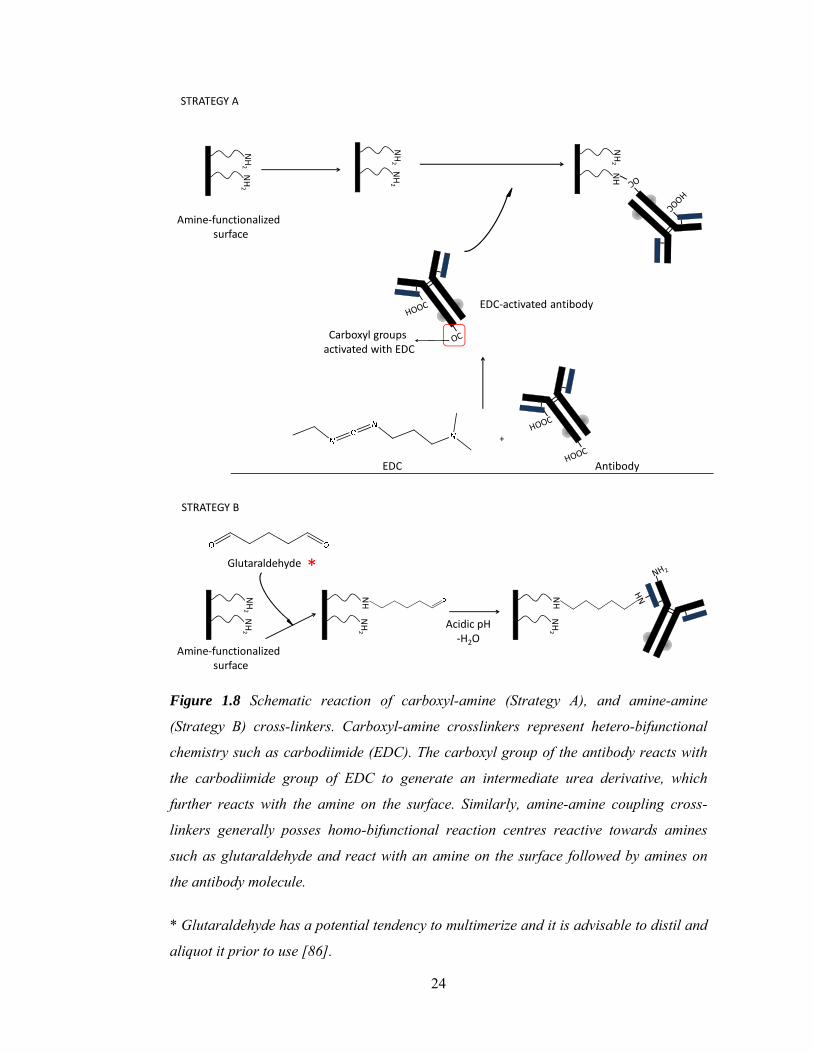
Figure 1.8 Schematic reaction of carboxyl-amine (Strategy A), and amine-amine
(Strategy B) cross-linkers. Carboxyl-amine crosslinkers represent hetero-bifunctional
chemistry such as carbodiimide (EDC). The carboxyl group of the antibody reacts with
the carbodiimide group of EDC to generate an intermediate urea derivative, which
further reacts with the amine on the surface. Similarly, amine-amine coupling cross-
linkers generally posses homo-bifunctional reaction centres reactive towards amines
such as glutaraldehyde and react with an amine on the surface followed by amines on
the antibody molecule.
* Glutaraldehyde has a potential tendency to multimerize and it is advisable to distil and
aliquot it prior to use [86].

25
Amine-targeting linkers possess functional moieties that react with amine groups. Such
reactive centres mainly have esters of NHS/ imide and phenyl hydrazide or aldehyde that
preferably bind to primary amines irrespective of their position [73]. Amine-targeting
strategies may result in the loss of antibody activity [87]. However, the addition of
‘lysine-rich’ pockets at locations on the antibody away from the region associated with
antigen-recognition may be employed for minimizing activity loss [88].
Hetero-bifunctional crosslinkers that target carboxyl groups mainly possess carbodiimide
or azide reactive groups [73] and could be used as alternatives to the amine-targeting
linkers. These linkers are probably the most commonly used in antibody immobilization
over a modified surface because of their robustness in delivering cross-linking on a wide
range of functionalized surfaces with amine, sulfhydryl and epoxy coatings.
Maleimide or vinyl sulfone-containing linkers are used for targeting sulfhydryl groups of
the antibody or the surface [73,85]. These linkers are commercially available in various
combinations and can cross-link sulfhydryl functional groups to amine, carbohydrate or
sulfhydryls. The utility of sulfhydryl-based hetero-bifunctional cross-linkers in protein
coupling is already proven [89]. However, their use in immobilization is fairly restricted
to either amine-functionalized or gold-coated surfaces [89,90]. The major drawback of
this approach is that either the antibody has to be chemically processed to introduce a
thiol group on it or it must be modified genetically to introduce a cysteine. Cysteine, a
sulfhydryl-containing amino acid, is also a widely exploited chemical species for
immobilizing antibody molecules on amine or sulfhydryl-functionalized and gold-coated
surfaces. A cysteine-tagged antibody can be captured on amine and sulfhydryl-
functionalized surfaces with bi-functional cross-linkers, whereas, on gold surfaces it can
be immobilized via co-ordinate bonds formed between the sufhydryl group and gold in a
chemical-free single step. Cysteine-based antibody/fragment immobilization using

26
chemical modifications such as cyanogen bromide-activated surfaces was also reported
[91]. This strategy provides robustness to the approach with minimum antibody leakage
[92]. However, introduction of cysteine in the basic antibody structure could introduce
conformational changes leading to a non-functional antibody. This drawback could
easily be controlled by the introduction of cysteine on the antibody/fragment away from
the antigen recognition domains, using reduction and alkylation chemistries [93], which
may enable the more efficient use of sulfhydryl linkers in solid phase immobilization
[76,94-96] or by genetic approaches.
Photoswitchable linkers, which comprise the most advanced class of linkers, could also
be employed for antibody immobilization [85]. These linkers mainly possess a
photoactivable group at one terminal and a reactive chemical group at the other. The
photoactivable group can be activated to either react with the surface or the antibody.
However, photoactivable chemistry is not selective. Therefore, the linkers are mainly
allowed to react first with the surface via their photoactivable terminal group and,
subsequently, facilitating capture of the antibody through the other end of the cross-
linker that possesses a reactive group e.g. NHS ester or maleimide.
These linker-based chemistries are important because these are easy to perform with
minimum preparation. In addition, the most advantageous use of linkers is that they may
be of variable length and can be used to control the distance of the immobilized
antibodies from the surface. Hence, the linker arm-length could impact on antibody
functionality by controlling the immobilization-associated steric stress on the antigen-
binding site [97]. A spacer/linker of zero-length chemistry (e.g. carbodiimide (EDC)-
succinimide (NHS)) immobilizes antibody on the surface with a rigid bond that
minimizes the freedom of rotation (degrees of freedom) (Figure 1.6, page 20), which
may result in ‘functionally-challenged’ antibody. However, this rigidity could

27
significantly be decreased by increasing the spacer arm-length. Therefore, in many cases
the functional antibody density on the surface may be enhanced.
All the covalent strategies described have been used in the fabrication of different
biosensor formats. The advantages of using covalent immobilization-based strategies are
the higher density and homogeneous loading of the captured antibody on the surface
along with leach-proof immobilization. However, the major drawback is the random
immobilization of antibody as it may drastically affect the antibody functionality.
1.2.3 Site-specific Immobilization of antibody
Site-directed immobilization is where an antibody can be captured specifically through a
desired region. Many approaches are reported claiming strong site-directed antibody
immobilization. These strategies include either the use of affinity biomolecules such as
biotin-avidin, enzyme or domain-specific proteins (e.g. protein A) for antibody capture.
A high degree of orientation can be introduced to the immobilized antibodies using this
strategy and the antigen binding region of the antibody is always correctly positioned for
antigen recognition.
1.2.3.1 Affinity Tag-based site directed antibody capture
Affinity is the strength of interaction between two molecules where the interaction may
be stabilized by multiple bonds. In an antigen-antibody interaction, affinity can be
described as the strength of interaction of an antigen with the single binding site of an
antibody. If a tag has a strong affinity towards a certain molecule and binds with
significantly high specificity, it is referred to as an ‘affinity tag’. These tags are
successfully used for affinity-based protein immobilization [17,98-100]. Tags can also be
incorporated into the antibody either genetically or chemically [17,18]. There is an array
of commercially available affinity tags but those used for the antibody immobilization
are described.

28
In a polyhistidine tag [101] there is a long repeated sequence of histidine residues of
varying numbers (ranging from six to ten). This tag has a strong affinity for divalent
transition metal ions including divalent nickel (Ni+2) and copper (Cu+2) [102-
104](Figure 1.9).
O
OHN
O
O
O Ni
H2O
OH2
O
OH
N
O
O
ONi
H2O
NH2
NHN
ONitrilotriacetic acid
Figure 1.9 Mechanism of ‘His-tag-mediated’ immobilization on a nitrilotriacetic acid
(NTA)-modified surface. The NTA-functionalized surface is chelated with divalent metals
such as Ni+2. The histidine of the tag reacts with the hydrated Ni+2 via the nitrogen of the
imidazole ring, thus immobilizing scFv or recombinant antibodies labelled with the His-
tag.
The transition metal ions are chelated on the support layer, which consists of nitriloacetic
acid (NTA) or iminodiacetic acid (IDA). They provide a matrix for the selective
immobilization of tagged antibodies where this tag is genetically incorporated at either
the N- or C-terminal of the antibody [105]. However, there are many drawbacks
associated with this strategy. Non-homogenous antibody distribution on the NTA-grafted
surface is the foremost [106,107]. The non-homogeneous distribution of antibody is due

29
to the immobilization of a mixture of functional and non-functional antibody on the NTA
matrix. In addition, the selectivity of NTA to distinguish the HIS tagged antibody from
other histidine-rich naturally occurring cellular proteins is very poor [108]. This will
result in competition between tagged antibody and histidine-rich cellular proteins (e.g.
histidine-rich glycoprotein (HRG) and histidine-rich calcium-binding protein (HRC)
[109-111]) for binding with the NTA present on the surface and results in heterogeneous
immobilization on the surface. There are several reports that describe the non-specificity
associated with this system as an attribute of histidine’s relatively low affinity toward
metal ions (Kd in the order of 1-10 μM). However, a better affinity may be achieved
either by increasing the number of histidine residues in the tag or employing two tandem
tags [112,113].
Biotin is a water soluble vitamin known as vitamin B7. It is a fused ring structure
composed of tetrahydroimidazolone and tetrahydrothiophene with valeric acid (C5H10O2)
substitution on the thiophene ring. It is an important biomolecule and plays a critical role
in gluconeogenesis. Biotin can act as an affinity tag if incorporated into the antibodies
either genetically or chemically. These biotinylated antibodies then can easily be
captured on avidin or streptavidin or genetically derived combinations of both proteins,
which are conjugated with functionalized polymeric and metallic [114,115], silicate
[116] and amino-polypyrrole-coated surfaces [117]. Initially, this tag was often used for
histochemical diagnostics [118] but its commercial relevance in immobilization and
purification was realized later. In addition, this approach was successfully employed in
the development of microarrays [119] and optical sensors based on surface plasmon
resonance [120]. However, due to the mulitivalency of avidin, which can accommodate
four biotin molecules per avidin, high densities of the immobilized antibody molecules
may be obtained. Such a high density of antibody may induce interactions between

30
closely bound antibody molecules that may lead to a high degree of steric hindrance
[121]. In addition, endogenous biotin could also compete for the surface-bound avidin or
streptavidin. This non-specific competition may reduce the overall binding efficiency of
the immobilization procedure [122]. The strong affinity (Kd ≈ 10-15 M) of the biotin to
avidin is a major advantage and the interaction is effectively irreversible until a
temperature-dependent regeneration method was developed using a hydro-thermostat
[123]. However, this strong affinity may introduce steric hindrance in the immobilized
antibodies, as an antibody bound through biotin loses freedom of rotation [124,125].
Reducing the affinity of biotin, using genetically-engineered variants of streptavidin or
biomolecules mimicking the biotin-binding event with streptavidin could be used to
minimize steric stress on antibody and improve the regenerative capacity of the bound
antibody [126].
Use of peptide nucleic acids (PNAs) is another approach that was employed for antibody
immobilization. The PNAs are chemically synthesized single stranded nucleic acid
analogs. The backbone repeat unit of the PNA consists of an amino acid derivative of
alkylamide and is shown in Figure 1.10. Nucleotide is attached to the PNA backbone
repeat unit at the 4-oxoethyl position via an aminoethyl-glycine linkage. PNA is then
synthesized by repeated addition of these nucleotide-functionalized amino acids via
peptide bonds replacing the normal phosphodiester backbone [127,128]. Therefore,
PNAs can interact with other nucleic acids in a highly specific manner forming PNA-
PNA, PNA-RNA and PNA-DNA hybridization constructs similar to the hybridization of
complementary DNA strands. In addition, PNAs are non-ionic and achiral molecules.
Their non-ionic nature and achirality makes them resistant to enzymatic hydrolysis.
Achiralilty also provides a high degree of thermal stability. These properties of PNA
could be harnessed in developing efficient immobilization strategies such that the PNA-

31
conjugated antibody could be immobilized on surfaces grafted with the anti-sense
oligonucleotide or PNA by hybridization (Figure 1.10) [129,130]. Subsequently, the
distance between surface and antibody could be controlled by varying the number of
PNA monomers in the backbone [131-134]. Although this strategy is not commonly used
for antibody immobilization, it could be an efficient single-step approach that could
provide high steric freedom to the captured antibodies, with strong site specificity and
orientation. There are few reports pertaining to the use of complementary
oligonucleotide tags[135], which are relatively simpler than PNAs. In addition, the
hybridized oligonucleotide tags can be separated from their complementary strands
grafted on the surface by a variation in either pH or divalent ion strength.
Elastin-like polypeptides (ELP) are a relatively novel category of small thermally
responsive proteins [136,137]. They can easily be tagged on to various antibodies,
antibody fragments or other proteins [138]. ELPs possess ‘biopolymer-like’ properties
such that they polymerize in a temperature range of 4-37 oC. At temperatures above 37
oC these proteins precipitate out of the solution and below 4 oC they remain in solution
phase. Therefore, ELP-tagged antibodies could efficiently be captured as a monolayer on
a specifically tailored surface [139]. ELPs have been successfully used for antibody
immobilization in microarrays and related applications [140].
FLAGR proteins, commercially available from Sigma Aldrich, may also be employed for
immobilization. These are octa-peptides (NH2-DYKDDDDK-COOH) that could be
genetically incorporated into antibodies and antibody fragments (Fabs and scFvs) [141].
Antibodies tagged with FLAG can be captured on the surfaces immobilized with
monoclonal anti-FLAG antibody. Use of the FLAG tag introduces specificity and
orientation for the immobilization [142-144].

32
PNA
2-oxopropyl position for nucleotide base
Backbone repeat unit
Aminoacid-nuclotide monomer of PNAN-(2-(methylamino)ethyl)-N-(2-oxopropyl)propionamide
C-terminal
BN-terminal
Complementary PNA-functionalized surface
Ab-conjugated PNA; Ab can be conjugated to either C- or N-terminals of PNA
Ab-captured on a surface after hybridization of the complementary PNA strand of functionalized
surface with that on the antibody
Ab
Ab
Figure 1.10 Immobilization of an antibody-functionalized with a peptide nucleic acid
(PNA) on a surface grafted with a complementary nucleotide base sequence. Antibody
could be covalently linked to a PNA sequence. This PNA-functionalized antibody can be
immobilized on a second PNA sequence that contains complementary nucleotide
sequences, which easily hybridize directly to it.

33
1.2.3.2 Enzyme-substrate reaction-based site-directed antibody capture
Antibody immobilization using enzyme-mediated coupling is also feasible. There are
many different enzyme-catalyzed reactions. The participating substrates react in the
presence of the enzyme and products are formed. This very fundamental principle of
enzymology can also be used for antibody immobilization. The enzyme-mediated
reaction must be a ‘two substrate component’ system, where the enzyme facilitates the
reaction of one substrate moiety with the other, as shown in Figure 1.11. An antibody
can be conjugated to one substrate of the ‘two substrate component system’. This
antibody-substrate complex can then be immobilized on the surface and grafted with the
second substrate of the ‘two substrate component system’ via an enzyme-catalyzed
reaction.
Antibody could also be conjugated to the active domain of an enzyme that is involved in
catalyzing some reaction. These conjugates can subsequently be captured on the
surfaces, grafted either with the substrate or their analogs. The enzyme’s substrate-
binding domain interacts with the substrate (via interactions such as hydrogen bonding
and electrostatic interactions, along with the formation of covalent bonds). Various
examples of this strategy that are used for antibody immobilization are discussed in this
section.
The immobilization of cutinase-conjugated calmodulin was one of the first
demonstrations of this strategy. Cutinase-conjugated calmodulin was captured on the
phosphonate inhibitor-functionalized thiolated self-assembled monolayer (SAM) on a
gold surface [145]. Although, this report was not specific for antibody immobilization,
its potential implications will be discussed.

34
Figure 1.11 Illustration of enzyme-mediated reaction of two substrates. Substrate 1
binds to the specific site on the enzyme forming enzyme-substrate 1 complex. This
complex then binds to the second substrate. Enzyme catalyzes the reaction between the
two substrates by forming a covalent bond between them. This strategy could be used to
immobilize antibodies tagged with substrate 1 on the surfaces grafted with substrate 2.
Human O6-alkylguanine transferase (hAGT) plays important role in DNA repair during
DNA synthesis. It binds to a DNA molecule at a specific error site and alkylates it by
transferring an alkyl group from its substrate (Figure 1.12). Methyltransferase is one of
the most important enzymes of this category. Immobilization of proteins tagged with this
enzyme has already been demonstrated [146-149]. Similarly, the hAGT-fused antibody
can be covalently captured on surfaces functionalized with benzylguanine. Fused hAGT
enzyme replaces the guanine by covalently binding to a benzyl moiety. Immobilization
of hAGT-fused glutathione S-transferase and tandem repeats of ‘immunoglobulin-type’
domains of a muscle protein, called titin, was demonstrated [150]. This technology is
commercially available under the name SNAP-tag® with New England Biolabs. In
addition, if the enzyme’s substrate is changed from benzylguanine to benzylcytosine then

35
it is known as a CLIP-tag™. These tags can be genetically incorporated in recombinant
antibodies or chemically fused to full length antibodies [151]. There are many
applications of these tags for specific and covalent immobilization of antibodies.
Micropatterning of antibody is also feasible using these strategies [152].
HNO
O
HS
SNAP tag (hAGT enzyme)-tagged antibody (Ab)
Benzylguanine-functionalized surface
Guanine
Benzyl-hAGT covalent complex immobilizing antibody on the surface
S-
Ab
S
Ab
Figure 1.12 Mechanism of SNAP-tag-mediated antibody immobilization. Antibody is
tagged with human O6-alkylguanine transferase (hAGT), which is a DNA repair enzyme.
This enzyme removes guanine and forms covalent bonds with the benzyl group of the
benzylguanine that is grafted on the surface, thus immobilizing the tagged antibody by
displacement catalysis of the benzylguanine group on the surface.
Transglutaminase (TGase) is another enzyme system, which specifically catalyzes the
formation of covalent cross-linking of the peptide-bound glutamine with a primary
amine. This enzyme is specific to a 12 peptide long sequence that is known as
‘glutamine-donor substrate’ sequence and is designated as T26 (HQSYVDPWMLDH).
Flank of (GGGS)3 spacer is incorporated on either side of the T26 sequence prior to

36
molecular tagging onto the N- or C-terminal of the antibody fragment. This strategy was
demonstrated for the immobilization of tagged anti-BSA antibody on aminated surfaces
by inducing the TGase-mediated amine-T26 reaction [153]. The underlying mechanism
of immobilization is shown in Figure 1.13. This technique is efficient as it involves no
chemical modification of the antibody or fragment.
NHH
+
Aminated surface
Transglutaminase
AT26 tag flanked with palindromic glutamine (G)HQSYVDPWMLDH----(GGS)3
NHC=O
Antibody captured on aminated surface via glutamine flank of A26
HQ
SY
VD
PWM
LDH
----
(GG
S) 3
----Ab
QS
YVD
PW
MLD
H---
-(GG
S)3-
---Ab
Figure 1.13 Mechanism of transglutaminase-mediated antibody immobilization on
aminated surfaces. Antibody tagged with T26 and glutamine spacer reacts with the
amine of the surface in TGase-catalyzed acyl-transfer-mediated isopeptide bond
formation by facilitating the reaction between carboxamide of glutamine and the amine
of the side-chain of lysine on the surface.
The use of enzymes on poly-L-lysine (PLL) and polyethylene glycol (PEG)-grafted
surfaces to capture cells and peptides had also been demonstrated. This strategy was also
demonstrated for immobilizing antibodies with preserved functionality [154].

37
CBD-tagged antibody
R = Tyrosine (214),Aspartic acid (142)
NAG (Chitin)
Chitin-grafted surface
Antibody-captured on chitin-grafted surface
Figure 1.14 Schematic representation of immobilization of an antibody conjugated to the
substrate-binding domain of an enzyme on surfaces grafted with the substrate of the
respective enzyme. Antibody tagged with chitin-binding domain (CBD) can interact with
chitin via hydrogen bonds formed between the hydrogen of the aspartic acid (142) of the
CBD and oxygen of the amide of the chitin, which is grafted on surface. Hydrophobic
interactions between the tyrosine (214) of the CBD and N-acetylglucoseamine of chitin
also play a crucial role in this immobilization.
The chitin-binding domain (CBD), which is the hydrolase part of the enzyme chitinase,
catalyses the hydrolysis of chitin polymers. These domains can be incorporated
genetically into recombinant antibodies and can be chemically conjugated to full length
antibodies. Immobilization of a CBD-conjugated antibody on the chitin-grafted surface is
shown in Figure 1.14. An early use of CBD in immobilization was reported by Kuranda
and co-workers [155]. Later, this strategy was employed for whole cell-based assay
development whereby whole cells were immobilized on chitin-grafted surfaces via their

38
surface-expressed CBD [156]. This strategy is primarily used to immobilize antibodies
on the chitin-functionalized chromatographic resins [157]. In addition, several CBD-
antibody constructs were developed [158-160] to capture antibodies on chitin-coated
surfaces for immunoassay-based applications [157,161,162]. Similarly,
polyhydroxyalkanoate (PHA) depolymerase substrate-binding domain (PSBD) [163] was
also reported for use in antibody immobilization-related applications.
1.2.4 Site-specific orientation-based antibody capture
Site specificity can be easily introduced using the tag-based strategies and enzyme-
mediated methods (section 1.2.3, page 27-38). However, the orientation of immobilized
molecules is of major importance for immunoassay development. Strategies based on
domain-recognition proteins (DRPs), such as protein A, are widely used for
immobilizing antibody molecules. These DRPs have high avidity toward specific
domains on the antibody and bind strongly to them introducing strong directed
orientation to the antibodies when immobilized (Figure 1.15). In addition, the covalent
immobilization of these DRPs on a surface could certainly achieve a homogenous
distribution of antibody over the surface [164,165] minimizing leaching and protein
exchange-associated variability in assays [166]. However, DRPs must be correctly
orientated on the surface to provide proper orientation to the antibody molecules. Use of
DRPs with multiple antibody-binding domains, such as protein A with five antibody-
binding domains, may minimize the concerns associated with the orientation of these
DRPs. The applications of these orientated antibodies could be enzyme immunoassays
(ELISA) [166], surface plasmon resonance (SPR), microdevices such as QCM [167,168]
and chromatography [169].

39
Antibody captured an adsorbed prA
Fc-specific protein (prA) Antibody captured an covalently immobilized prA
Figure 1.15 Orientation of an immobilized antibody by capture on a sub-layer of protein
A, which is either adsorbed or covalently captured on the surface.
1.3 Conclusions and Perspective
Adsorption of antibody is very commonly used approach for immobilization. This is a
cost-effective and simple method with no expertise required for its use. However, it has
many drawbacks. These is no control over the (a) amount and packing density of the
antibody, (b) antibody distribution on the surface, (c) site-specificity, orientation and
conformation of the antibody [13,34,48,49], and (d) susceptibility of the antibody to
leaching and protein exchange [24,59]. Introduction of capture stages that include a sub-
layer of Fc-binding proteins or anti-antibody molecules may introduce a higher degree of
orientation with a controlled antibody packing [170]. However, this will significantly
increase the time and cost involved for immunoassay development. Tag-assisted
strategies could be employed to address some of these issues, such as site-specificity and

40
improved control over the active conformation of the immobilized antibody. However,
stability of the interaction of the tag with its affinity ligand is strictly dependent on
various physical and chemical parameters such as temperature, solvent polarity, pH and
salt concentration. Therefore, a slight change in the optimum conditions may introduce
leaching [171,172] and activity loss. In addition, the homogeneous distribution of the
tagged-antibody is dependent on the distribution of the affinity ligand on the surface.
Therefore, a strategy is required that could provide an approach to respond to most of
these drawbacks whilst maintaining the cost-effectiveness of the immunoassay
development. Employing covalent strategies could deal with most of the previously
mentioned drawbacks. Covalently-captured antibodies have least susceptibility for
leaching and protein exchange. In addition, a high degree of site-specificity and
orientation could be introduced to the antibody by employing enzyme-mediated covalent
strategies. However, covalent immobilization may require activation of the antibody
prior to immobilization. This may result in self-conjugation of antibodies forming
multimeric complexes. This multimerization could be a drawback in the case of
antibody-based imaging, whereas in assay development, this multimerization may mask
the active site of antibody along with the introduction of conformational stress. This
multimerization can be restricted by optimizing the molar ratios of antibody to cross-
linker. This could also be addressed by employing direct chemistries. Strong orientation
could also be employed by capturing antibody on a covalently-immobilized sub-layer of
Fc-binding proteins [165]. A summary of all the available techniques, along with their
advantages and drawbacks, is given in Table 1.3.
Theoretically, covalent strategies offer great potential to tag-assisted strategies in terms
of the cost and time involved. Introduction of a tag is performed either by using
recombinant molecular biological approaches or by chemical methods. Therefore, in this

41
thesis, covalent immobilization-based strategies will be employed for developing
immunoassays.
This thesis covers the development and optimization of a mild surface activation and
modification method that could be employed on most of the polymeric surfaces
irrespective of their chemical nature. In addition, the various available chemistries will
be critically analyzed for their immobilization efficiency with the aim of developing
high-sensitivity immunoassays on various bioanalytical platforms including microtiter
plates and nanoparticles.
Table 1.3
Strategy Overview Advantages (+) Disadvantages (-)
Physisorption (Section 1.2.1)
Antibody (Ab) immobilization with physical interactions between Ab and surface. Involves hydrophobic and electrostatic interactions with hydrophobic and charged surfaces, respectively.
(+) i. could be used for any type of
antibody ii. ‘Label-free’ and ‘reagent-free’
(no Ab modification or surface modification required)
(-) i. Highly susceptible to protein-
exchange and leaching ii. Conformational instability of
the adsorbed antibody (denaturation) due to the underlying processes such as molecular spreading or flattening
iii. Random molecular distribution (non-homogenous density of Ab on surface and no control over functional orientation of the molecules)

42
Covalent immobilization (Section 1.2.2)
Linker-mediated chemistries
Linker length plays important role in minimizing the coupling-related conformational stress
EDC-mediated (Figure 1.8 (a)) • For surfaces with amine
or carboxy-functionalization
• A classical example of heterobifunctional cross-linking (carboxy to amine)
The EDC linker activates the carboxyl group followed by transferring the activated carboxyl adduct to primary amine Immobilization on amine-functionalized surfaces: Ab should be activated prior cross-linking Immobilization on carboxyl-functionalized surfaces: the carboxy groups of surface should be activated first
(+) i. No leaching after cross-
linking ii. No Ab modification required
(could be performed with native proteins)
iii. Commercially available from all major suppliers
(-) i. High susceptibility of EDC
toward hydrolysis ii. Random orientation
iii. Activated Ab molecules are susceptible to multi-merization (could be minimized by optimizing the molar ratio of antibody to EDC and using a pH at which only carboxyl groups will interact with EDC)
iv. Ab functionality may be compromised in few cases if amine groups of the antibody are conjugated to the carboxyl groups of the functionalized surfaces
Glutaraldehyde (Figure 1.8 (b)) • For surfaces with amine
functionalization • A classical example of
homobifunctional cross-linking (amine to amine)
Glutaraldehyde activates amine groups irrespective of their position on Ab
(+) i. Strong coupling, hence ‘leach-
proof’ ii. No Ab modification required
iii. Commercially available (-) i. Multimerization of Ab (this
could be compensated by activating the amine-

43
functionalized surface with glutaraldehyde prior to Ab cross-linking)
ii. Ab functionality may be compromised (if the amine-rich regions of antigen recognition portion of the Ab get involved in cross-linking)
iii. No specificity iv. High degree of randomness in
immobilized Ab molecules v. Inappropriate cross-linking
Sulfhydryl-targeting linkers
Primarily contains maleimide or vinyl sulfone as reactive centres which target the sulfhydryl goup These could be either homo- (SH-SH) or heterobifunctional (SH-NH2)
(+) i. Site specific immobilization
in the case of cysteine-tagged Ab
ii. Commercially available (-) i. Generation of thiol groups is
required on the antibody either by converting pendant amino groups of Ab into sulfhydryl (this may change the amines of the antigen recognition domain of the Ab causing compromised Ab functionality) or by reducing disulfide bonds to generate free sulfhydryl groups with chemical agents such as DTT, TCEP etc.
Direct chemistries
Epoxide-mediated (Figure 1.3)
Epoxide-functionalized surface Epoxide ring opens up following the reaction of oxygen species with amine or sulfhydryl
(+) i. ‘Leach-proof’ immobilization
ii. No Ab modification required iii. ‘Reagent-free’, ‘label-free’
single –step immobilization iv. Commercial availablity (-) i. An epoxide functionalization

44
of the surface is required ii. Randomly-orientated Ab
Aldehyde-mediated Aldehyde-functionalized
surface Aldehyde reacts with amino group that results in an imine group between surface and Ab Reduction of imine to amine should be performed in order to stabilize the bond
(+) i. No Ab modification required
(native Ab) ii. Single step strategy
iii. ‘Label-free’ and ‘reagent-free’ (-) i. Reversible imine-bond
formation (require additional step of reduction for stabilization of the imine bond)
ii. No site-specificity iii. Uncontrolled orientation
Amine-mediated Amine-functionalized surface Ab oxidation activates the diol of the sugar moieties on the Fc region, which reacts with amine of the surface to form imine bond
(+) i. Highly site-specific (-) i. Oxidation of Ab is required
(uncontrolled oxidation) ii. Restricted to full-length Ab
which has sugar moiety
Aromatic sulfonate chloride-mediated (Figure 1.4)
Tosyl and trisyl chlorides are widely used Aromatic chloride activates hydroxyl of the surface and conjugates it to the sulfhydryl, primary amine, imidazole and tyrosine hydroxyl of the Ab
(+) i. Covalent attachment
ii. Robust approach (could couple a variety of Ab’s functional groups)
iii. Site-selective iv. Single-step method v. Commercially available (-) i. Non-orientated
immobilization
Diels-Alder cyclo-addition (Figure 1.5)
PEG-functionalized with ligand such as biotin or domain-specific protein A at ω-carbon and cyclopentadiene at α-carbon. PEG grafted through the
(+) i. Covalent attachment with
higher steric freedom (PEG chain length can control the steric freedom of the captured Ab)

45
cyclopentadiene on the N-maleimide-functionalized surface
ii. Orientated capture of Ab on protein A or biotin
iii. Site-specific iv. ‘Label-free’ and ‘reagent-free’ v. Single-step procedure to
generate functionalized surface
Photoactive chemistry (Figure 1.7)
β-cyclodextrin-functionalized surfaces genetically-incorporated p-benzoyl-L-phenylalanine (pBpa) Ab
(+) i. Covalently captured Ab
ii. Site-specific iii. ‘Label-free’ and ‘reagent-free’
single step procedure iv. Commercially available (-) i. Incorporation of pBpa may
introduce conformational changes in Ab fragments such as scFv and Fab
Site-specific immobilization (Section 1.2.3)
Affinity tags
PolyHistidine (His-tag) (Figure 1.9)
Nitrilotriacetic acid (NTA) or iminodiacetic acid (IDA)-functionalized surface Imidazole of histidine forms a metal co-ordination complex
(+) i. Single step methodology
ii. Non-covalent co-ordination complex formation
iii. Site-specific iv. Reusable v. Commercial availability (-) i. Poor ligand affinity (histidine
repeats could be increased to 6 to 10 in number to obtain higher affinity)
ii. Leaching (it could be minimized with multimeric histidine tags)
iii. Low selectivity (metal-

46
functionalized surface does not discriminate tag from the endogenous histidine-rich proteins)
Biotin-Avidin system Avidin/streptavidin-
functionalized surface Chemical conjugation or genetic incorporation of biotin to the Ab
(+) i. Strong specificity and
selectivity (-) i. Conformational stress could
be introduced due to Ab congestion on avidin surface (tetrameric molecule)
Peptide-Nucleic Acid (PNA) tags (Figure 1.10)
Surface-functionalized with complementary nucleic acid sequence PNA could be incorporated to Ab in a selective way either by its carboxyl end or amine end using routine EDC chemistry
(+) i. High stability over a thermal
and pH range ii. Single step, ‘reagent-free’ and
‘label-free’ method iii. High degree of steric freedom
by varying length of the backbone
Elastin-like polypeptides (ELP) Thermally responsive protein
ELP-functionalized surface ELP-fused Ab or fragments
(+) i. Reversible temperature-
dependent immobilization ii. Highly selective (-) i. Immobilized system must be
stored in a temperature range 2-37 oC (below which the ELP depolymerises and above which it precipitates)
Enzyme-mediated O6-alkylguanine transferase (AGT) SNAP-tag (Figure 1.12)
Benzylguanine-functionalized surface AGT-fused Ab
(+) i. Covalent immobilization
ii. Site-specific iii. Commercially available
Transglutaminase (TGase) (Figure 1.13)
T26 tagging on N- or C-terminal of Ab with a flank of (GGGS)3
(+) i. Site-specific and covalent
immobilization

47
Amine-functionalized surface
ii. ‘Reagent-free’ and ‘label-free’ single step immobilization
Site-specific orientation-based (Section 1.2.4)
Enzyme domains
Chitin-binding domain (CBD) (Figure 1.14)
Chitin-functionalized surface CBD-fused Ab
(+) i. High selectivity and site
specificity ii. Covalently-captured Ab
(enzyme domain interact covalently with substrate)
(-) i. Enzyme domain could be
susceptible to conformational change in different physiological conditions
Polyhydroxyalkanoate (PHA) depolymerase substrate-binding domain (PSBD)
PHA-functionalized surface PSBD-fused Ab
(+) iii. High selectivity and site
specificity iv. Covalently-captured Ab
(enzyme domain interact covalently with substrate)
(-) ii. Enzyme domain could be
susceptible to conformational change in different physiological conditions
Proteins specific to Ab domains (Figure 1.15)
Fc-specific
Protein A (prA), protein G (prG) and recombinant
Immobilization of Fc-specific protein on surface
(+) i. Strong orientation

48
protein A/G (prA/G)
ii. Site-specific and high selectivity
iii. Ab captured in its native state iv. Strong binding (-) i. Could only be used on Ab
with Fc domain kL-specific Protein L (prL) prL immobilized on surface (+)
i. Strong orientation ii. Site-specific and high
selectivity iii. Ab captured in its native state iv. Strong binding (-) i. Could only be used on Ab
with kL domain
1.4 References
1. Roux KH: Immunoglobulin structure and function as revealed by electron microscopy.
Int Arch Allergy Immunol 1999, 120(2):85-99.
2. Holliger P, Hudson PJ: Engineered antibody fragments and the rise of single domains.
Nat Biotechnol 2005, 23(9):1126-1136.
3. Blazek D, Celer V: The production and application of single-chain antibody
fragments. Folia Microbiol (Praha) 2003, 48(5):687-698.
4. Ono K, Kamihira M, Kuga Y, Matsumoto H, Hotta A, Itoh T, Nishijima K, Nakamura
N, Matsuda H, Iijima S: Production of anti-prion scFv-Fc fusion proteins by recombinant
animal cells. J Biosci Bioeng 2003, 95(3):231-238.
5. Muyldermans S, Baral TN, Retamozzo VC, Baetselier PD, Genst ED, Kinne J,
Leonhardt H, Magez S, Nguyen VK, Revets H, Rothbauer U, Stijlemans B, Tillib S,

49
Wernery U, Wyns L, Hassanzadeh-Ghassabeh G, Saerens D: Camelid immunoglobulins
and nanobody technology. Vet Immunol Immunopathol 2009, 128(1-3):178.
6. Schramm W, Paek S, Voss G: Strategies for the immobilization of antibodies.
Immunomethods 1993, 3(2):93-103.
7. Saerens D, Ghassabeh GH, Muyldermans S: Antibody technology in proteomics. Brief
Funct Genomic Proteomic 2008, 7(4):275-282.
8. Steinhauer C, Wingren C, Hager AC, Borrebaeck CA: Single framework recombinant
antibody fragments designed for protein chip applications. BioTechniques 2002,
33Sup:38-45.
9. Danczyk R, Krieder B, North A, Webster T, HogenEsch H, Rundell A: Comparison of
antibody functionality using different immobilization methods. Biotechnol Bioeng 2003,
84(2):215-223.
10. Jung Y, Jeong JY, Chung BH: Recent advances in immobilization methods of
antibodies on solid supports. Analyst 2008, 133(6):697-701.
11. Lee CS, Belfort G: Changing activity of ribonuclease A during adsorption: a
molecular explanation. Proc Natl Acad Sci U.S.A. 1989, 86(21):8392-8396.
12. Norde W, Favier JP: Structure of adsorbed and desorbed proteins. Colloids Surf
1992, 64(1):87-93.
13. Haynes CA, Norde W: Structures and stabilities of adsorbed proteins. J Colloid Interf
Sci 1995, 169(2):313-328.

50
14. Kwok KC, Yeung KM, Cheung NH: Adsorption kinetics of bovine serum albumin
on fused silica: population heterogeneities revealed by single-molecule fluorescence
microscopy. Langmuir 2007, 23(4):1948-1952.
15. Rabe M, Verdes D, Zimmermann J, Seeger S: Surface organization and cooperativity
during nonspecific protein adsorption events. J Phys Chem, B 2008, 112(44):13971-
13980.
16. Raffaini G, Ganazzoli F: Protein adsorption on a hydrophobic surface: a molecular
dynamics study of lysozyme on graphite. Langmuir 2010, 26(8):5679-5689.
17. Arnau J, Lauritzen C, Petersen GE, Pedersen J: Current strategies for the use of
affinity tags and tag removal for the purification of recombinant proteins. Protein Expr
Purif 2006, 48(1):1-13.
18. Nogi T, Sangawa T, Tabata S, Nagae M, Tamura-Kawakami K, Beppu A, Hattori M,
Yasui N, Takagi J: Novel affinity tag system using structurally defined antibody-tag
interaction: application to single-step protein purification. Protein Sci 2008,
17(12):2120-2126.
19. Decker RH, Ling C, Overby LR: Hapten conjugated antibody for antibody or antigen
detection. USA Patent 1980, 05/932394(4230683).
20. Holthues H, Pfeifer-Fukumura U, Hartmann I, Baumann W: An immunoassay for
terbutryn using direct hapten linkage to a glutaraldehyde network on the polystyrene
surface of standard microtiter plates. Fresenius J Anal Chem 2001, 371(7):897-902.

51
21. Seurynck-Servoss SL, Baird CL, Miller KD, Pefaur NB, Gonzalez RM, Apiyo DO,
Engelmann HE, Srivastava S, Kagan J, Rodland KD, Zangar RC: Immobilization
strategies for single-chain antibody microarrays. Proteomics 2008, 8(11):2199-2210.
22. Weiping Q, Bin X, Lei W, Chunxiao W, Zengdong S, Danfeng Y, Zuhong L, Yu W:
Orientation of antibodies on a 3-aminopropyltriethoxylsilane-modified silicon wafer
surface. J Inclusion Phenom macrocyclic Chem 1999, 35(1):419-429.
23. de Boer A, Hokke C, Deelder A, Wuhrer M: Serum antibody screening by surface
plasmon resonance using a natural glycan microarray. Glycoconj J 2008, 25(1):75-84.
24. Lvov Y, Mohwald H: Protein Architecture: Interfacing Molecular Assemblies and
Immobilization Biotechnology (ed. 1). Marcel & Dekker, Inc. U.S.A:2000;4-12.
25. Cornish-Bowden A: Enthalpy–entropy compensation: a phantom phenomenon. J
Biosci 2002, 27(2):121-126.
26. Howard IK: H is for enthalpy, thanks to Heike Kamerlingh Onnes and Alfred W.
Porter. J Chem Educ 2002, 79(6):697.
27. Pace CN, Fu H, Fryar KL, Landua J, Trevino SR, Shirley BA, Hendricks MM,
Iimura S, Gajiwala K, Scholtz JM, Grimsley GR: Contribution of hydrophobic
interactions to protein stability. J Mol Biol 2011, 408(3):514.
28. Pace CN: Energetics of protein hydrogen bonds. Nat Struct Mol Biol 2009,
16(7):681-682.
29. Petukhov M, Rychkov G, Firsov L, Serrano L: H-bonding in protein hydration
revisited. Protein Sci 2004, 13(8):2120-2129.

52
30. Scholtz JM, Grimsley GR, Pace CN: Solvent denaturation of proteins and
interpretation of their m-values. Methods Enzymol 2009, 466:549-565.
31. Liao H, Yeh W, Chiang D, Jernigan RL, Lustig B: Protein sequence entropy is
closely related to packing density and hydrophobicity. Protein Eng Des Sel 2005,
18(2):59-64.
32. Schymkowitz JWH, Rousseau F, Serrano L: Surfing on protein folding energy
landscapes. Proc Natl Acad Sci U.S.A. 2002, 99(25):15846-15848.
33. Fernandez-Escamilla AM, Cheung MS, Vega MC, Wilmanns M, Onuchic JN,
Serrano L: Solvation in protein folding analysis: Combination of theoretical and
experimental approaches. Proc Natl Acad Sci U.S.A. 2004, 101(9):2834-2839.
34. Haynes CA, Norde W: Globular proteins at solid/liquid interfaces. Colloids Surf, B
1994, 2(6):517-566.
35. Ball V, Huetz P, Elaissari A, Cazenave JP, Voegel JC, Schaaf P: Kinetics of
exchange processes in the adsorption of proteins on solid surfaces. Proc Natl Acad Sci
U.S.A. 1994, 91(15):7330-7334.
36. Petukhov M, Cregut D, Soares CM, Serrano L: Local water bridges and protein
conformational stability. Protein Sci 1999, 8(10):1982-1989.
37. Sancho J, Serrano L, Fersht AR: Histidine residues at the N- and C-termini of α-
helixes: perturbed pKas and protein stability. Biochemistry (NY) 1992, 31(8):2253-2258.

53
38. Petukhov M, Uegaki K, Yumoto N, Yoshikawa S, Serrano L: Position dependence of
amino acid intrinsic helical propensities II: Non-charged polar residues: Ser, Thr, Asn,
and Gln. Protein Sci 1999, 8(10):2144-2150.
39. Pace G, Venanzi M, Castrucci P, Scarselli M, De Crescenzi M, Palleschi A, Stella L,
Formaggio F, Toniolo C, Marletta G: Static and dynamic features of a helical
hexapeptide chemisorbed on a gold surface. Mater Sci Eng , C 2006, 26(5-7):918-923.
40. Hammer B, Morikawa Y, Nørskov JK: CO chemisorption at metal surfaces and
overlayers. Phys Rev Lett 1996, 76(12):2141-2144.
41. Berkebile S, Ules T, Puschnig P, Romaner L, Koller G, Fleming AJ, Emtsev K,
Seyller T, Ambrosch-Draxl C, Netzer FP, Ramsey MG: A momentum space view of the
surface chemical bond. Phys Chem Chem Phs 2011, 13(9):3604-3611.
42. Liu J, Gustafsson A, Breimer ME, Kussak A, Holgersson J: Anti-pig antibody
adsorption efficacy of α-Gal carrying recombinant P-selectin glycoprotein ligand-
1/immunoglobulin chimeras increases with core 2 β1, 6-N-acetylglucosaminyltransferase
expression. Glycobiology 2005, 15(6):571-583.
43. Dixit CK, Vashist SK, MacCraith BD, O'Kennedy R: Evaluation of apparent non-
specific protein loss due to adsorption on sample tube surfaces and/or altered
immunogenicity. Analyst 2011, 136(7):1406-1411.
44. Wang X, Wang Y, Xu H, Shan H, Lu JR: Dynamic adsorption of monoclonal
antibody layers on hydrophilic silica surface: a combined study by spectroscopic
ellipsometry and AFM. J Colloid Interf Sci 2008, 323(1):18-25.

54
45. Ghatnekar-Nilsson S, Dexlin L, Wingren C, Montelius L, Borrebaeck CA: Design of
atto-vial based recombinant antibody arrays combined with a planar wave-guide
detection system. Proteomics 2007, 7(4):540-547.
46. Hu X, Spada S, White S, Hudson S, Magner E, Wall JG: Adsorption and activity of a
domoic acid binding antibody fragment on mesoporous silicates. J Phys Chem B 2006,
110(37):18703-18709.
47. Bee JS, Chiu D, Sawicki S, Stevenson JL, Chatterjee K, Freund E, Carpenter JF,
Randolph TW: Monoclonal antibody interactions with micro- and nanoparticles:
Adsorption, aggregation, and accelerated stress studies. J Pharm Sci 2009, 98(9):3218-
3238.
48. Butler JE, Ni L, Brown WR, Joshi KS, Chang J, Rosenberg B, Voss EW,Jr: The
immunochemistry of sandwich ELISAs--VI. Greater than 90% of monoclonal and 75%
of polyclonal anti-fluorescyl capture antibodies (CAbs) are denatured by passive
adsorption. Mol Immunol 1993, 30(13):1165-1175.
49. Van Tassel PR, Talbot J, Tarjus G, Viot P: Kinetics of irreversible adsorption with a
particle conformational change: A density expansion approach. Phys Rev E 1996,
53(1):785-798.
50. Fleminger G, Hadas E, Wolf T, Solomon B: Oriented immobilization of periodate-
oxidized monoclonal antibodies on amino and hydrazide derivatives of Eupergit C. Appl
Biochem Biotechnol 1990, 23(2):123-137.

55
51. Xu H, Lu JR, Williams DE: Effect of surface packing density of interfacially
adsorbed monoclonal antibody on the binding of hormonal antigen human chorionic
gonadotrophin. J Phys Chem B 2006, 110(4):1907-1914.
52. Kusnezow W, Jacob A, Walijew A, Diehl F, Hoheisel JD: Antibody microarrays: An
evaluation of production parameters. Proteomics 2003, 3(3):254-264.
53. Kusnezow W, Hoheisel JD: Solid supports for microarray immunoassays. J Mol
Recognit 2003, 16(4):165-176.
54. Guo A, Zhu XY: In functional protein microarrays in drug discovery (ed. 1). Ed.
Predki PF. Boca Raton, CRC Press, Florida, US; 2007:53-71.
55. Ljungberg UK, Jansson B, Niss U, Nilsson R, Sandberg BE, Nilsson B: The
interaction between different domains of staphylococcal protein A and human polyclonal
IgG, IgA, IgM and F(ab')2: separation of affinity from specificity. Mol Immunol 1993,
30(14):1279-1285.
56. Akerstrom B, Bjorck L: A physicochemical study of protein G, a molecule with
unique immunoglobulin G-binding properties. J Biol Chem 1986, 261(22):10240-10247.
57. Kastern W, Sjobring U, Bjorck L: Structure of peptostreptococcal protein L and
identification of a repeated immunoglobulin light chain-binding domain. J Biol Chem
1992, 267(18):12820-12825.
58. Sasakura Y, Kanda K, Yoshimura-Suzuki T, Matsui T, Fukuzono S, Han MH,
Shimizu T: Protein microarray system for detecting protein-protein interactions using an
anti-His-tag antibody and fluorescence scanning: effects of the heme redox state on

56
protein-protein interactions of heme-regulated phosphodiesterase from Escherichia coli.
Anal Chem 2004, 76(22):6521-6527.
59. Huetz P, Ball V, Voegel J, Schaaf P: Exchange kinetics for a heterogeneous protein
system on a solid surface. Langmuir 1995, 11(8):3145-3152.
60. Corso CD, Dickherber A, Hunt WD: An investigation of antibody immobilization
methods employing organosilanes on planar ZnO surfaces for biosensor applications.
Biosens Bioelectron 2008, 24(4):811-817.
61. Das RD, Maji S, Das S, RoyChaudhuri C: Optimization of covalent antibody
immobilization on macroporous silicon solid supports. Appl Surf Sci 2010, 256(20):5867.
62. Dixit CK, Vashist SK, O'Neill FT, O'Reilly B, MacCraith BD, O'Kennedy R:
Development of a high sensitivity rapid sandwich elisa procedure and its comparison
with the conventional approach. Anal Chem 2010, 82(16):7049-7052.
63. Darain F, Gan K, Tjin S: Antibody immobilization onto polystyrene substrate-on-
chip immunoassay for horse IgG based on fluorescence. Biomed Microdevices 2009,
11(3):653-661.
64. Shmanai V, Nikolayeva T, Vinokurova L, Litoshka A: Oriented antibody
immobilization to polystyrene macrocarriers for immunoassay modified with hydrazide
derivatives of poly(meth)acrylic acid. BMC Biotechnol 2001, 1(1):4.
65. Abuknesha RA, Jeganathan F, Wu J, Baalawy Z: Labeling of biotin antibodies with
horseradish peroxidase using cyanuric chloride. Nat Protocols 2009, 4(4):452-460.

57
66. Andersen BR, Abele DC, Vannier WE: Effects of mild periodate oxidation on
antibodies. J Immunol 1966, 97(6):913-924.
67. Wei Z, Feng J, Lin H, Mullapudi S, Bishop E, Tous GI, Casas-Finet J, Hakki F,
Strouse R, Schenerman MA: Identification of a single tryptophan residue as critical for
binding activity in a humanized monoclonal antibody against respiratory syncytial virus.
Anal Chem 2007, 79(7):2797-2805.
68. Zamani L, Andersson, FO, Edebrink P, Yang Y, Jacobsson, SP: Conformational
studies of a monoclonal antibody, IgG1, by chemical oxidation: Structural analysis by
ultrahigh-pressure LC-electrospray ionization time-of-flight MS and multivariate data
analysis. Anal Biochem 2008, 380(2):155-163.
69. Caballero D, Samitier J, Bausells J, Errachid A: Direct patterning of anti-human
serum albumin antibodies on aldehyde-terminated silicon nitride surfaces for hsa protein
detection. Small 2009, 5(13):1531-1534.
70. Shen G, Zhang Y: Highly sensitive electrochemical stripping detection of hepatitis B
surface antigen based on copper-enhanced gold nanoparticle tags and magnetic
nanoparticles. Anal Chim Acta 2010, 674(1):27.
71. Elia G, Silacci M, Scheurer S, Scheuermann J, Neri D: Affinity-capture reagents for
protein arrays. Trends Biotechnol 2002, 20(12):s19-s22.
72. Ehlers J, Rondan NG, Huynh LK, Pham H, Marks M, Truong TN: Theoretical study
on mechanisms of the epoxy-amine curing reaction. Macromolecules 2007, 40(12):4370-
4377.

58
73. Thermo Scientific Pierce Protein Research Products: Cross-linking reagents: A
technical handbook. 2006.
74. Nilsson K, Mosbach K: Immobilization of ligands with organic sulfonyl chlorides.
Methods Enzymol 1986, 104:56-69.
75. Hermanson GT, Mallia AK, Smith PK: Immobilized affinity ligand techniques
(ed.1). Academic Press, San Diego, US; 1992;66-155.
76. Kim HS, Hage DS: Immobilization methods for affinity chromatography. In
Handbook of affinity chromatography (ed,1). Ed. Hage DS. CRC press, Florida, US;
2006:37-78.
77. Uttamchandani M, Wang J, Yao SQ: Protein and small molecule microarrays:
Powerful tool for high-throughput proteomics. Mol Bio Syst 2006, 2:58-68.
78. Sun X, Stabler CL, Cazalis CS, Chaikof EL: Carbohydrate and Protein
Immobilization onto Solid Surfaces by Sequential Diel-Alder and Azide-Alkyne
Cycloadditions. Bioconjug Chem 2006, 17(1):52-57.
79. Sun XL, Yang L, Chaikof EL: Chemoselective immobilization of biomolecules with
aqueous Diel-Alders and PEG chemistry. Tetrahedron Lett 2008, 49(16):2510-2513.
80. Colombo M, Bianchi A: Click chemistry for the synthesis of RGD-containing
integrin ligands. Molecules 2010, 15(1):178-197.
81. Jullian V, Courtois F, Bolbach G, Chassaing G: Carbon-carbon bond ligation
between β-cyclodextrin and peptide by photo-irradiation. Tetrahedron Lett 2003,
44(34):6437-6440.

59
82. Jensen RL, Städe LW, Wimmer R, Stensballe A, Duroux M, Larsen KL, Wingren
C, Duroux L: Direct site-directed photocoupling of proteins onto surfaces coated with β-
cyclodextrins. Langmuir 2010, 26(13):11597-11604.
83. Hino N, Hayashi A, Sakamoto K, Yokoyama S: Site-specific incorporation of non-
natural amino acids into proteins in mammalian cells with an expanded genetic code. Nat
Protocols 2007, 1(6):2957-2962.
84. Wang F, Robbins S, Guo J, Shen W, Schultz PG: Genetic Incorporation of Unnatural
Amino Acids into Proteins in Mycobacterium tuberculosis. PLoS ONE 2010, 5(2):e9354.
85. Wong SS: Conjugation of proteins to solid matrices. In Chemistry of protein
conjugation and cross-linking (ed.1). Ed. Wong SS. Florida, CRC press, US; 1991:295-
318.
86. Migneault I, Dartiguenave C, Bertrand MJ,Waldron KC: Glutaraldehyde: behavior in
aqueous solution, reaction with proteins, and application to enzyme crosslinking.
Biotechniques 2004,37:790–802..
87. Howell S, Kenmore M, Kirkland M, Badley RA: High-density immobilization of an
antibody fragment to a carboxymethylated dextran-linked biosensor surface. J Mol
Recognit 1998, 11(1-6):200-203.
88. Kweon D, Kim S, Han NS, Lee JH, Chung KM, Seo J: Immobilization of Bacillus
macerans cyclodextrin glycosyltransferase fused with poly-lysine using cation
exchanger. Enzyme Microb Technol 2005, 36(4):571-578.

60
89. Bain CD, Troughton EB, Tao YT, Evall J, Whitesides GM, Nuzzo RG: Formation of
monolayer films by the spontaneous assembly of organic thiols from solution onto gold.
J Am Chem Soc 1989, 111(1):321-335.
90. Bhatia SK, Shriver-Lake LC, Prior KJ, Georger JH, Calvert JM, Bredehorst R, Ligler
FS: Use of thiol-terminal silanes and heterobifunctional crosslinkers for immobilization
of antibodies on silica surfaces. Anal Biochem 1989, 178(2):408-413.
91. Morfill J, Blank K, Zahnd C, Luginbuhl B, Kuhner F, Gottschalk KE, Pluckthun A,
Gaub HE: Affinity-matured recombinant antibody fragments analyzed by single-
molecule force spectroscopy. Biophys J 2007, 93(10):3583-3590.
92. Kipriyanov SM, Little M: Affinity purification of tagged recombinant proteins using
immobilized single chain Fv fragments. Anal Biochem 1997, 244(1):189-191.
93. Sun MMC, Beam KS, Cerveny CG, Hamblett KJ, Blackmore RS, Torgov MY,
Handley FGM, Ihle NC, Senter PD, Alley SC: Reduction and alkylation strategies for the
modification of specific monoclonal antibody disulfides. Bioconjug Chem 2005,
16(5):1282-1290.
94. Chen LL, Rosa JJ, Turner S, Pepinsky RB: Production of multimeric forms of CD4
through a sugar-based cross-linking strategy. J Biol Chem 1991, 266(27):18237-18243.
95. Rao SV, Anderson KW, Bachas LG: Oriented immobilization of proteins.
Microchimica Acta 1998, 128(3):127-143.
96. Brogan KL, Wolfe KN, Jones PA, Schoenfisch MH: Direct oriented immobilization
of F(ab′) antibody fragments on gold. Anal Chim Acta 2003, 496(1-2):73-80.

61
97. Wong SS: Reactive groups of proteins and their modifying agents. In Chemistry of
protein conjugation and cross-linking (ed.1). Ed. Wong SS. Florida, CRC press, US;
1991:7-48.
98. Hedhammer M, Graslund T, Hober S: Protein engineering strategies for selective
protein purification. Chem Engineer Technol 2005, 28(11):1315-1325.
99. Rusmini F, Zhong Z, Feijen J: Protein immobilization strategies for protein biochips.
Biomacromolecules 2007, 8(6):1775-1789.
100. Camarero JA: Recent development in the site-specific immobilization of proteins
onto solid supports. Biopolymers 2008, 90(3):450-458.
101. Darain F, Ban C, Shim YB: Development of a new and simple method for the
detection of histidine-tagged proteins. Biosens Bioelectron 2004, 20(4):857-863.
102. Cha T, Guo A, Zhu XY: Enzymatic activity on a chip: the critical role of protein
orientation. Proteomics 2005, 5(2):416-419.
103. Hochuli E: Large-scale chromatography of recombinant proteins. J Chromatog
1988, 444:293-302.
104. Gaberc-Porekar V, Menart V: Potential for using histidine tags in purification of
protein at large scale. Chem Engineer Technol 2005, 28(11):1306-1314.
105. Paramban RI, Bugos RC, Su WW: Engineering green fluorescent protein as a dual
functional tag. Biotechnol Bioengineer 2004, 86(6):687-697.

62
106. Halliwell CM, Morgan G, Ou C, Cass AEG: Introduction of a (poly)histidine tag in
l-lactate dehydrogenase produces a mixture of active and inactive molecules. Anal
Biochem 2001, 295(2):257-261.
107. Lin P, Weinrich D, Waldmann H: Protein biochips: Oriented surface
immobilization of proteins. Macromolecular Chem Phys 2010, 211(2):136-144.
108. Wong LS, Khan F, Micklefield J: Selective covalent protein immobilization:
strategies and applications. Chem Rev 2009, 109(9):4025-4053.
109. Fan G, Gregory KN, Zhao W, Park WJ, Kranias EG: Regulation of myocardial
function by histidine-rich, calcium-binding protein. Am J Physiol Heart Circ Physiol
2004, 287(4):H1705-H1711.
110. Sadanandom A, Findlay K, Doonan JH, Schulze-Lefert P, Shirasu K: CHPA, a
cysteine- and histidine-rich-domain-containing protein, contributes to maintenance of the
diploid state in Aspergillus nidulans. Eukaryotic Cell 2004, 3(4):984-991.
111. Jones AL, Hulett MD, Parish CR: Histidine-rich glycoprotein: A novel adaptor
protein in plasma that modulates the immune, vascular and coagulation systems.
Immunol Cell Biol 2005, 83(2):106-118.
112. Khan F, He M, Taussig MJ: Double-hexahistidine tag with high-affinity binding for
protein immobilization, purification, and detection on Ni+2 nitrilotriacetic acid surfaces.
Anal Chem 2006, 78(9):3072-3079.
113. Steinhauer C, Wingren C, Khan F, He M, Taussig MJ, Borrebaeck CAK: Improved
affinity coupling for antibody microarrays: Engineering of double-(His)6-tagged single
framework recombinant antibody fragments. Proteomics 2006, 6(15):4227-4234.

63
114. Laitinen OH, Nordlund HR, Hytönen VP, Kulomaa MS: Brave new (strept)avidins
in biotechnology. Trends Biotechnol 2007, 25(6):269.
115. Grunwald C: A brief introduction to the streptavidin-biotin system and its usage in
modern surface based assays. Zeitschrift fur Physikalische Chemie 2008, 222(5-6):789-
821.
116. Chalkias NG, Giannelis EP: An avidin-biotin immobilization approach for
horseradish peroxidase and glucose oxidase on layered silicates with high catalytic
activity retention and improved thermal behavior. Industrial Biotechnol 2007, 3(1):82-
88.
117. Torres-Rodríguez LM, Billon M, Roget A, Bidan G: A polypyrrole-biotin based
biosensor: elaboration and characterization. Synth Met 1999, 102(1-3):1328-1329.
118. Jones CJ, Stoddart RW: A post-embedding avidin-biotin peroxidase system to
demonstrate the light and electron microscopic localization of lectin binding sites in rat
kidney tubules. Histochem J 1986, 18(7):371-379.
119. Pavlickova P, Hug H: A streptavidin-biotin-based microarray platform for
immunoassays. Methods Mol Biol 2004, 264:73-83.
120. Pradier CM, Salmain M, Liu Z, Methivier C: Comparison of different procedures of
biotin immobilization on gold for the molecular recognition of avidin: an FT-IRRAS
study. Surf Interface Anal 2002, 34(1):67-71.
121. Kolenko P, Dohnalek J, Duskova J, Skalova T, Collard R, Hasek J: New insights
into intra- and intermolecular interactions of immunoglobulins: crystal structure of
mouse IgG2b-Fc at 2Å-0·1Å resolution. Immunology 2009, 126(3):378-385.

64
122. Burkhart KK, Beard D, Billingsley ML: Enhanced elimination of biotinylated
antibodies by avidin-based hemoperfusion in rats. J Pharmacol Exp Therapeutics 1994,
270(1):356-361.
123. Holmberg A, Blomstergren A, Nord O, Lukacs M, Lundeberg J, Uhlen M: The
biotin-streptavidin interaction can be reversibly broken using water at elevated
temperatures. Electrophoresis 2005, 26(3):501-510.
124. Johnsson B, Lofas S, Lindquist G, Edstrom A, Hillgren RM, Hansson A:
Comparison of methods for immobilization to carboxymethyl dextran sensor surfaces by
analysis of the specific activity of monoclonal antibodies. J Mol Recognit 1995, 8(1-
2):125-131.
125. Evans L, Hughes M, Waters J, Cameron J, Dodsworth N, Tooth D, Greenfield A,
Sleep D: The production, characterisation and enhanced pharmacokinetics of scFv-
albumin fusions expressed in Saccharomyces cerevisiae. Protein Expr Purif 2010,
73(2):113.
126. Piervincenzi RT, Reichert WM, Hellinga HW: Genetic engineering of a single-
chain antibody fragment for surface immobilization in an optical biosensor. Biosens
Bioelectron 1998, 13(3-4):305-312.
127. Nielsen P, Egholm M, Berg R, Buchardt O: Sequence-selective recognition of DNA
by strand displacement with a thymine-substituted polyamide. Science 1991,
254(5037):1497-1500.
128. Egholm M, Buchardt O, Christensen L, Behrens C, Freier SM, Driver DA, Berg
RH, Kim SK, Norden B, Nielsen PE: PNA hybridizes to complementary

65
oligonucleotides obeying the Watson–Crick hydrogen-bonding rules. Nature 1993,
365(6446):566-568.
129. Sheng H, Ye BC: Different strategies of covalent attachment of oligonucleotide
probe onto glass beads and the hybridization properties. Appl Biochem Biotechnol 2009,
152(1):54-65.
130. Silva TAR, Ferreira LF, Souza LM, Goulart LR, Madurro JM, Brito-Madurro AG:
New approach to immobilization and specific-sequence detection of nucleic acids based
on poly(4-hydroxyphenylacetic acid). Mat Sci Eng,C 2009, 29(2):539-545.
131. Hook F, Ray A, Norden B, Kasemo B: Characterization of PNA and DNA
Immobilization and Subsequent Hybridization with DNA Using Acoustic-Shear-Wave
Attenuation Measurements. Langmuir 2001, 17(26):8305-8312.
132. Masuko M: Hybridization of an immobilized PNA probe with its complementary
oligodeoxyribonucleotide on the surface of silica glass. Nucleic Acids Res Suppl 2003,
3:145-146.
133. Lim SY, Chung W, Lee HK, Park MS, Park HG: Direct and nondestructive
verification of PNA immobilization using click chemistry. Biochem Biophys Res
Commun 2008, 376(4):633-636.
134. Ray A, Norden B: Peptide nucleic acid (PNA): its medical and biotechnical
applications and promise for the future. FASEB J 2000, 14(9):1041-1060.
135. Zhong M, Fang J, Wei Y: Site Specific and Reversible Protein Immobilization
Facilitated by A DNA Binding Fusion Tag. Bioconjug Chem 2010, 21(7):1177-1182.

66
136. Meyer DE, Trabbic-Carlson K, Chilkoti A: Protein purification by fusion with an
environmentally responsive elastin-like polypeptide: effect of polypeptide length on the
purification of thioredoxin. Biotechnol Prog 2001, 17(4):720-728.
137. Trabbic-Carlson K, Meyer DE, Liu L, Piervincenzi R, Nath N, LaBean T, Chilkoti
A: Effect of protein fusion on the transition temperature of an environmentally
responsive elastin-like polypeptide: a role for surface hydrophobicity? Protein Eng Des
Sel 2004, 17(1):57-66.
138. Ong SR, Trabbic-Carlson KA, Nettles DL, Lim DW, Chilkoti A, Setton LA:
Epitope tagging for tracking elastin-like polypeptides. Biomaterials 2006, 27(9):1930-
1935.
139. Floss DM, Schallau K, Rose-John S, Conrad U, Scheller J: Elastin-like polypeptides
revolutionize recombinant protein expression and their biomedical application. Trends
Biotechnol 2010; 28:37-45.
140. Gao D, McBean N, Schultz JS, Yan Y, Mulchandani A, Chen W: Fabrication of
antibody arrays using thermally responsive elastin fusion proteins. J Am Chem Soc 2006,
128(3):676-677.
141. Zhang L, Liu Y, Chen T: Label-free amperometric immunosensor based on
antibody immobilized on a positively charged gold nanoparticle/L-cysteine-modified
gold electrode. Microchimica Acta 2009, 164(1):161-166.
142. Hopp TP, Prickett KS, Price VL, Libby RT, March CJ, Pat Cerretti D, Urdal DL,
Conlon PJ: A short polypeptide marker sequence useful for recombinant protein
identification and purification. Nat Biotech 1988, 6(10):1204-1210.

67
143. Nakajima H, Brindle PK, Handa M, Ihle JN: Functional interaction of STAT5 and
nuclear receptor co-repressor SMRT: implications in negative regulation of STAT5-
dependent transcription. EMBO J 2001, 20(23):6836-6844.
144. Butterfield DA, Bhattacharya D: Biofunctional membranes: site specifically
immobilized enzyme arrays. In New insights in membrane science and technology:
polymeric and biofunctional membranes. Edited by Butterfield DA, Bhattacharya D.
Elsevier Science BV, Amsterdam, The Netherlands; 2003:233-240.
145. Hodneland CD, Lee Y, Min D, Mrksich M: Selective immobilization of proteins to
self-assembled monolayers presenting active site-directed capture ligands. Proc Natl
Acad Sci U.S.A. 2002, 99(8):5048-5052.
146. Juillerat A, Gronemeyer T, Keppler A, Gendreizig S, Pick H, Vogel H, Johnsson K:
Directed evolution of o6-alkylguanine-dna alkyltransferase for efficient labeling of fusion
proteins with small molecules in vivo. Chemistry & Biology 2003; 10:313-317.
147. Kindermann M, George N, Johnsson N, Johnsson K: Covalent and selective
immobilization of fusion proteins. J Am Chem Soc 2003, 125(26):7810-7811.
148. Huber W, Perspicace S, Kohler J, Müller F, Schlatter D: SPR-based interaction
studies with small molecular weight ligands using hAGT fusion proteins. Anal Biochem
2004, 333(2):280-288.
149. Engin S, Trouillet V, Franz CM, Welle A, Bruns M, Wedlich D: Benzylguanine
Thiol Self-Assembled Monolayers for the Immobilization of SNAP-tag Proteins on
Microcontact-Printed Surface Structures. Langmuir 2010, 26(9):6097-6101.

68
150. Kufer S, Dietz H, Albrecht C, Blank K, Kardinal A, Rief M, Gaub H: Covalent
immobilization of recombinant fusion proteins with hAGT for single molecule force
spectroscopy. Eur Biophys J 2005, 35(1):72-78.
151. Kampmeier F, Ribbert M, Nachreiner T, Dembski S, Beaufils F, Brecht A, Barth S:
Site-specific, covalent labeling of recombinant antibody fragments via fusion to an
engineered version of 6-o-alkylguanine dna alkyltransferase. Bioconjug Chem 2009,
20(5):1010-1015.
152. Iversen L, Cherouati N, Berthing T, Stamou D, Martinez KL: Templated protein
assembly on micro-contact-printed surface patterns. use of the snap-tag protein
functionality. Langmuir 2008, 24(12):6375-6381.
153. Sugimura Y, Ueda H, Maki M, Hitomi K: Novel site-specific immobilization of a
functional protein using a preferred substrate sequence for transglutaminase 2. J
Biotechnol 2007, 131(2):121-127.
154. Sala A, Ehrbar M, Trentin D, Schoenmakers RG, Voros J, Weber FE: Enzyme
mediated site-specific surface modification. Langmuir 2010, 26(13):11127-11134.
155. Kuranda MJ: Immobilization and purification of fusion proteins using chitin-
binding ability. U.S.A. Patent 303827(5258502).
156. Wang J, Chao Y: Immobilization of cells with surface-displayed chitin-binding
domain. Appl Environ Microbiol 2006, 72(1):927-931.
157. Blank K, Lindner P, Diefenbach B, Plückthun A: Self-immobilizing recombinant
antibody fragments for immunoaffinity chromatography: generic, parallel, and scalable
protein purification. Protein Expr Purif 2002, 24(2):313.

69
158. Lindner P, Blank K, Diefenbach B, Plückthun A: Chitin binding domains for
immobilizing antibody fragments in immunoaffinity chromatography. In Proceedings of
the 5th International Conference of the European Chitin Society. Volume VI. Edited by
K. M. Varum, A. Domard and O. Smirdsrod. Norwegian University of Science and
Technology, Trondheim: The European Chitin Society (EUCHIS); 2002:261-262.
159. Saerens D, Huang L, Bonroy K, Muyldermans S: Antibody fragments as probe in
biosensor development. Sensors 2008, 8(8):4669-4686.
160. Reulen S, van Baal I, Raats J, Merkx M: Efficient, chemoselective synthesis of
immunomicelles using single-domain antibodies with a C-terminal thioester. BMC
Biotechnol 2009, 9(1):66.
161. Tjoelker LW, Gosting L, Frey S, Hunter CL, Trong HL, Steiner B, Brammer H,
Gray PW: Structural and functional definition of the human chitinase chitin-binding
domain. J Biol Chem 2000, 275(1):514-520.
162. Pavlickova P, Schneider EM, Hug H: Advances in recombinant antibody
microarrays. Clinica Chimica Acta 2004, 343(1-2):17-35.
163. Park T, Park J, Lee S, Hong H, Lee S: Polyhydroxyalkanoate chip for the specific
immobilization of recombinant proteins and its applications in immunodiagnostics.
Biotechnol Bioprocess Eng 2006, 11(2):173-177.
164. Kausaite-Minkstimiene A, Ramanaviciene A, Kirlyte J, Ramanavicius A:
Comparative study of random and oriented antibody immobilization techniques on the
binding capacity of immunosensor. Anal Chem 2010, 82(15):6401-6408.

70
165. Vashist SK, Holthofer H, Leister K: A method of immobilising biological
molecules to a support and products thereof. 2009, PCT / IE2008/ 000112 (WO/ 2009/
066275).
166. Schmid AH, Stanca SE, Thakur MS, Thampi KR, Suri CR: Site-directed antibody
immobilization on gold substrate for surface plasmon resonance sensors. Sens Actuat, B:
Chem 2006, 113(1):297.
167. Carrigan SD, Scott G, Tabrizian M: Real-time QCM-D immunoassay through
oriented antibody immobilization using cross-linked hydrogel biointerfaces. Langmuir
2005, 21(13):5966-5973.
168. Yuan Y, He H, Lee LJ: Protein A-based antibody immobilization onto polymeric
microdevices for enhanced sensitivity of enzyme-linked immunosorbent assay.
Biotechnol Bioeng 2009, 102(3):891-901.
169. Beyer NH, Hansen MZ, Schou C, Hojrup P, Heegaard NHH: Optimization of
antibody immobilization for on-line or off-line immunoaffinity chromatography. J Sep
Sci 2009, 32(10):1604.
170. Subramanian A, Velander WH: Effect of antibody orientation on immunosorbent
performance. J Mol Recognit 1996, 9(5-6):528-535.
171. Darby RAJ, Hine AV: LacI-mediated sequence-specific affinity purification of
plasmid DNA for therapeutic applications. FASEB J. 2005, 19(7):801-803.
172. Ueda EKM, Gout PW, Morganti L: Current and prospective applications of metal
ion-protein binding. J Chromat, A 2003, 988(1):1.

71
1.5 Aims The aims of this research were:
1. Development of a generic surface modification strategy which can be used over a
variety of solid supports. This section was dedicated to develop an approach that,
unlike labour-intense chemical methods of surface modification, would be easy to
perform under ambient conditions/ routine laboratory set-ups and would be cost-
effective at the same time. A strategy was developed based on KOH- and O2-plasma-
based surface activation followed by silane-mediated functionalization. This strategy
could be employed on surfaces that are hydrophobic, hydrophilic and chemically
inert.
2. Development of high-sensitivity assays on the aforementioned modified surfaces.
The bioanalytical platforms chosen were of commercial interest and included
polymer-based microtitre plates for enzyme-linked and fluorescence immunoassays
and fluorocap platforms (Bristol Myers Squibb, Syracuse, NY, USA, licensed
platform) for fluorescence immunoassays. The other major analytical strategy
involved custom-functionalized gold surfaces for surface plasmon resonance. This
section was directed towards demonstrating the application of the activated surfaces
following functionalization with silanes and employing them to immobilize
antibodies for immunoassay development in direct (model antigen: IgG) and
sandwich (model antigen: human fetuin A) formats. Fluorocap and chromatographic
resin-based technologies were developed and transferred to Bristol Myers Squibb,
Syracuse, NY, USA.
3. Development of antibody-conjugated nanoprobes using high brightness
nanoparticles, developed within our group, for detection of activation phases of
platelets. This section involved nanoparticle functionalization, optimization and
conjugation to platelet-specific anti-CD41 antibody for surface marker probing.

72
Chapter 2
Development of a modified high sensitivity
rapid sandwich ELIA procedure with
picogram sensitivity
(Published in Analytical Chemistry 2010, 82, 7049-7052)

73
2.1 Abstract
A highly sensitive and rapid sandwich enzyme-linked immunoassay (ELIA) procedure was
developed for the detection of human fetuin A/AHSG (α2-HS-glycoprotein), a specific
biomarker for hepato-cellular carcinoma and atherosclerosis. Anti-human fetuin A antibody
was immobilized on aminopropyltriethoxysilane-mediated amine-functionalized microtitre
plates using 1-ethyl-3-[3-dimethylaminopropyl]carbodiimide hydrochloride and N-
hydroxysulfosuccinimide-based heterobifunctional cross-linking. The analytical sensitivity
of the developed assay was 39 pg/mL, compared to 625 pg/mL for the conventional assay.
The generic nature of the developed procedure was demonstrated by performing human
fetuin A assays on different polymeric matrices i.e. polystyrene, poly(methyl methacrylate)
and polycyclo-olefin (Zeonex), in a modified microtitre plate format. Thus, the newly
developed procedure has considerable advantages over the existing method.

74
2.2 Introduction
Conventional enzyme-linked immunosorbent assay (ELISA) procedures have been followed
for decades for the detection of analytes of importance in industrial, healthcare, and
academic research. However, improvements on existing ELISA technologies are
continuously attempted by many groups [1, 2]. The development of a high-sensitivity
enzyme-linked immunoassay (ELIA)-based assay for human fetuin A (HFA), with lower
detection limits and a higher sensitivity than commercially available assays is reported. The
biological importance of HFA, a member of the cystatin superfamily, which is commonly
present in the cortical plate of the immature cerebral cortex and hemopoietic matrix of bone
marrow, is discussed elsewhere [3-10]. There are many commercially available ELISA kits
for fetuin A, which are listed in Table 2.1. The assay was demonstrated on various
commercially relevant solid supports. The developed ELIA is better than the conventional
procedure in terms of greatly reduced overall assay duration, higher sensitivity, and greater
reproducibility.
HFA was taken as the model analyte system to demonstrate the utility of the developed
ELIA procedure since all the assay components were commercially available in kit form.
This enables the performance of a robust and highly precise comparison of the developed
ELIA with the commercially existing conventional ELISA procedures, as the same assay
components were used under the same conditions. The developed procedure can be
employed on any commercially relevant substrate. Therefore, this approach of immobilizing
antibody on chemically-modified solid supports (Figure 2.1) has potential applications in
many other assays and formats.

75
1% (v/v) BSA incubation, 30 min at 37 oC
Substrate EtOH treatment, 5 min at RT
1% (w/v) KOH, 10 min at RT
O2-Plasma, 3 min
2% (v/v) APTES, 1 hr at 80 oC in vacuum desiccator
Si
NH2
OR
Si
NH2
OR
Si
NH2
OOC/OH OC/OH OC/OH
NH2 NH2 NH2
HN NH2 HN
RH, CO2, H2O
EDC/ SNHS +
Incubate 15 min at RT
C=O
Incubate 1 hr at 37 oC
HN NH2 HN
C=O
Assay procedure, 3 hrs 30 min
Antigen incubation, 1 hr at 37 oC
Detection antibody (enzyme-labeled),1 hr at 37 oC
SA-HRP incubation, 20 min at RT
BSA
COOH
Figure 2.1 Schematic of the developed sandwich ELISA procedure for the HFA assay. Plate
functionalization was performed by treatment with 1% (w/v) KOH, treatment with oxygen
(O2)-plasma and then silanization with 2% (v/v) APTES. KOH oxidizes the organic moiety
of the polymer generating a carbonyl (-CO) or a hydroxyl (-OH) group according to the
nature of the polymer. The alkoxy groups of APTES subsequently react with the carbonyl or
hydroxyl groups of the surface in a hydrolysis-dependent reaction [11, 12]. Anti-HFA/α-
Herman Schmidt glycoprotein (AHSG) antibody was then activated using EDC and SNHS
chemistry on the aminated 96-well plate, where the EDC-SNHS was used in a ratio of 1:100
with the antibody.
2.3 Materials
EDC, SNHS, phosphate buffered saline (PBS, 0.1M, pH 7.4), 2-(N-
morpholino)ethanesulfonic acid (MES, pH 4.7), 3,3',5,5'-Tetramethylbenzidine (TMB)
substrate kit and bovine-serum albumin (BSA) were purchased from Fisher Scientific
(previously Pierce), Ballycoolin, Dublin, Ireland. Potassium hydroxide (KOH), 3-APTES,

76
absolute ethanol and Nunc 96-well polystyrene plates (P7491) were obtained from Sigma-
Aldrich, Blessington, Co. Wicklow, Ireland. The HFA duoset kit, with all HFA assay
components, was procured from RnD Systems, Abingdon, Oxon, UK. The capture antibody
used was mouse anti-HFA/α-Herman Schmidt glycoprotein (AHSG) while detection was
achieved through the use of biotinylated goat anti-HFA/AHSG antibody and streptavidin-
conjugated horseradish peroxidase (HRP).
Table 2.1 A comparative analysis of various commercially available ELISA techniques with their
respective formats and the high sensitivity modified ELISA procedure reported here.
Manufacturer Format of antibody capture
Sensitivity (ng/mL)
Refer to
Covalent assay Chemically cross-linked
0.03 Reported in Chapter 2
Passive assay Passively adsorbed 0.31 Reported in Chapter 2
RnD systems Passively adsorbed 0.37 http://www.rndsystems.com/pdf/DY1184.pdf
Biovendor Passively adsorbed 0.35 http://www.biovendor.com/product/immunoassays/fetuin-a-ahsg-human-elisa
Alpco Diagnostics Passively adsorbed 5.00 http://www.alpco.com/pdfs/43/43-NSEHU-E01.pdf
Immunology Consultants Laboratory, Inc.
Passively adsorbed 6.25 http://www.life-sciences.com.br/pdf/icllab.pdf
Genway Biotech, Inc. Passively adsorbed 6.25 http://www.genwaybio.com/images/gw_tds/elisa_kits/40-374-130036.pdf
Assay Pro Passively adsorbed 6.25 http://www.assaypro.com/datasheet/eg3501_1.pdf

77
All assay components were reconstituted in 0.1M PBS, pH 7.4, with 1% (v/v) BSA. Buffers
and solutions were prepared in Milli-Q deionised water (DIW). The dilutions of all HFA
assay components and BSA were made in 0.1M PBS, pH 7.4, whereas KOH and 3-APTES
were diluted in DIW. EDC and SNHS were reconstituted in 0.1M MES buffer, pH 4.7.
Poly(methyl methacrylate) (PMMA), polystyrene (PS) and zeonexTM slides were purchased
from Microfluidic Chip Shop GmbH, Jena, Germany. Pressure-sensitive adhesive (PSA)
was obtained from Adhesive Research, Limerick, Ireland. Bottomless 96-well microtitre
plates were purchased from Greiner Labortechnik, Germany. The Optec Micro-Master CO2
Laser was procured from Optec S.A., Belgium. The assay temperature was maintained at 37
oC using a thermostat which was obtained from Labnet International Inc., Edison, NJ, USA.
2.4 Experimental Section
2.4.1 Microtitre plate functionalization with amine-silane and anti-human fetuin A
antibody immobilization
Each well of the 96-well plate was treated with 100 µL of absolute ethanol for 5 min at 37
oC and washed five times with 300 µL of deionised water (DIW). Subsequently, each well
was treated with 100 µL of 1.0% (w/v) KOH at 37 oC for 10 min followed by five washings
with 300 µL of DIW. The KOH-treated wells were then treated with O2-plasma and were
functionalized with amino groups by incubation with 100 µL of 2% (v/v)
aminopropyltriethoxysilane (APTES) per well at 80 oC for 1 h inside a vacuum-desiccator,
in order to achieve maximum silanization. It is important to mention here is that KOH-
based chemical oxidation followed by O2-plasma treatment facilitates generation and
stabilization of the surface functional groups. The reaction mechanism following the KOH

78
activation and APTES-based surface functionalization involves mild oxidation followed by
a hydrolytic APTES polymerization on the oxidized surface through its alkoxy groups [11,
12]. The desiccator was equilibrated to room temperature for 20 min. The amine-
functionalized plate was subsequently washed five times with 300 µL of DIW in order to
remove excess unbound 3-APTES from the surface. Afterward, the anti-HFA/AHSG (990
µL of 4 µg/mL) was incubated with 10 µL of a pre-mixed solution of 1-ethyl-3-[3-
dimethylaminopropyl]carbodiimide hydrochloride (EDC) (4 mg/mL) and N-
hydroxysulfosuccinimide (SNHS) (11 mg/mL) for 15 min at 37oC. The resulting EDC cross-
linked anti-HFA/AHSG antibody solution was added to each of the functionalized wells
(100 µL) and incubated for 1 h at 37oC. The anti-HFA/AHSG-coated wells were then
washed five times with 300 µL of PBS. The conventional ELISA was performed with the
protocol supplied by the manufacturer.
2.4.2 ELIA on anti-HFA/AHSG antibody-immobilized microtitre plates
Plates with covalent and passively immobilized anti-HFA antibody were blocked with 1%
(v/v) BSA diluted in 0.1M phosphate buffered saline (PBS), pH 7.4, for 30 min at 37 oC and
subsequently washed five times with 300 µL of PBS. Varying concentrations of
HFA/AHSG (from 4.8 pg/mL to 20 ng/mL) were prepared in 0.1M PBS, pH 7.4, and 100
µL of each of these concentrations were incubated in the antibody-coated plates for 1 h at 37
oC, and, subsequently washed five times with PBS. One hundred microliters of biotinylated
anti-HFA/AHSG detection antibody (200 ng/mL) was added and incubated for 1 hr at 37 oC
followed by five PBS washes. HRP-conjugated streptavidin (100 µL per well), at a dilution
of 1:200, was added and then incubated for 20 min at 37 oC followed by five washes with
PBS. The 3,3’,5,5’-tetramethylbenzidine (TMB) substrate was subsequently added (as per

79
the manufacturer’s recommendations) and the reaction was stopped after 20 min by addition
of 50 µL of 0.5M H2SO4. The absorbance was recorded at a primary wavelength of 450 nm
with a reference wavelength of 540 nm. The dual wavelength system of absorbance
measurement in 96-well plates is designed to eliminate or greatly reduce the optical
imperfections at the well-to-well level.
All the experiments were carried out in triplicate. The control for this study was zero ng/mL
concentration of HFA in 0.1M PBS, pH 7.4, and the absorbance of the control was
subtracted from all the assay values. The respective analytical sensitivity (detection limit) of
the assay were determined where it was calculated using the formula [mean absorbance of
blank + 3σ (standard deviation of the blank)]. Additionally, data sets obtained from the
ELIA and conventional ELISA were analysed using standard curve analysis of Sigma Plot
software, version 11.0.
2.5 Results and Discussion
2.5.1 Developed ELIA and conventional ELISA formats for HFA detection
The range of detection of HFA for the developed ELIA (Figure 2.2) was 9 to 20 pg/mL
(r2=0.99). An analytical sensitivity of 39 pg/mL was recorded for the developed ELIA,
where analytical sensitivity was calculated using the formula [average absorbance of blank
+ 3 (SDblank)]. Assay variability parameters for this high-sensitivity assay for HFA are
described in Table 2.2, where intra-day assay variability was calculated from 5 repeats
performed on a single day and inter-day assay variability from three repeats performed on
three different days in triplicate.
The half-effective concentration (EC50) obtained from the dose-response curve [13], which
is a measure of analyte-ligand interaction, was found to be on an average of 3.3 ng/mL. The

80
EC50 for all the assay repeats was found to be in the range of 3-3.5 ng/mL. A lower EC50
(3.3 ng/mL), which was obtained for the developed ELIA, suggests a strong interaction
between HFA and anti-HFA antibody. This interaction behavior is attributed to the
significant increase in the total amount of immobilized antibody achieved by using the
covalent immobilization strategy, which increased the availability of anti-HFA antibody per
HFA molecule, thus increasing the resultant HFA capture on the well surface.
Table 2.2. Comparison of assay performance parameters (precision and sensitivity) for
conventional ELISA and developed ELIA
Intra-day precision range (CV%), (n=5)
Inter-day precision range (CV%), (n=3)
Sensitivity
Developed ELISA 2.4-10.4 1.7-17.6 39 pg/mL
Conventional ELISA 4.7-17.4 3.6-20.0 312.0 pg/mL
Intra-day precision was calculated from the five repeats (n=5) of the same assay on a single day but
at different times while inter-day precision was calculated from assay repeats on three different days
(n=3). All the assays were carried out in triplicate. Analytical sensitivity was determined using the
formula [average absorbance of blank + 3 (SDblank)].
Conversely, EC50 for the conventional ELISA format was 23 ng/mL, (r2=0.99), which was
significantly higher than the developed ELIA. This suggests a less sensitive conventional
assay (Table 2.3). The most important factor that may be attributed to the enhanced
sensitivity of the modified assay is the covalent immobilization of the anti-HFA/AHSG
capture antibody, since the covalently-crosslinked antibodies should not leach out during the
assay procedure in comparison to the use of passively-adsorbed antibodies, where leaching
may occur more easily.

81
Performance of the developed sandwich ELIA procedure was compared with the
conventional protocols (Figure 2.2), both involving passively adsorbed antibody on normal
unmodified microtitre plates (one performed using the modified protocol and the other as
per the manufacturer’s recommendations). All the assays were performed simultaneously on
the same day under same set of conditions in order to minimize variability, where variability
was reported as percentage coefficient of variation (% CV).
Table 2.3 Comparative analysis of the modified sandwich ELISA procedure which was
developed for HFA/AHSG and the commercially used conventional sandwich ELISA
procedure in terms of timings of various assay steps (Table 2.2). Immunoassay
Steps and Parameters
Conventional HFA ELISA
(passively adsorbed Ab)
Modified HFA ELIA
(covalently captured Ab)
Immobilization of mouse anti-HFA Overnight at room temp (RT) 2 hrs 30 min
Blocking buffer (BSA) 2 hrs at RT 30 min at 37o C
Recombinant HFA 2 hrs at RT 1 hr at 37o C
Incubation of biotinylated goat anti-HFA 2 hrs at RT 1 hr at 37o C
Binding of Streptavidin-HRP 20 min at RT 20 min at 37o C
TMB substrate assay 20 min at RT 20 min at 37o C
Total immunoassay duration ~ 20 hrs ~ 6 hrs
Detection limit 156 pg/mL 9.76 pg/mL
Sensitivity 624 pg/mL 39 pg/mL
Immobilization strategy applicable to many polymeric surfaces
Not possible Possible
The technique employed here for chemically modifying the polymeric surface was
developed and standardized by our group and a standard EDC-based cross-linking strategy
was deployed to immobilize anti-HFA/AHSG antibody. The developed sandwich ELIA
procedure decreased the overall assay duration from 20 h to 6 h, which is more than a 3-fold

82
decrease. Thus, the reported procedure is a rapid ELIA format having the additional benefit
of being generic in nature, i.e. it can be used with sandwich ELISAs on different polymeric
matrices for a range of different analytes. Hence, using the surface modification technique
reported here, it is possible to develop rapid and high sensitivity assays on various categories
of solid supports including those that are chemically inert.
Figure 2.2 Comparative analysis of developed sandwich ELISA (black circles) with
conventional formats. Anti-human fetuin A (anti-HFA) antibody was covalently bound to the
amine-modified surface in the developed ELISA, whereas antibody adsorbed on the
unmodified surface was used in the conventional assay format. The assay was performed
with the manufacturer-recommended protocol (white circles) and the protocol developed in
this study (filled circles). The error bars represents the standard deviations.
As far as can be ascertained, this developed ELIA procedure is the most sensitive assay
reported for the detection of HFA/AHSG (Table 2.2), which is based on the comparison
between different commercially-available ELISA kits for HFA. The sensitivity of the assay
may be attributed to the use of monoclonal antibody at the capture stage, which was

83
covalently immobilized on the surface, and a polyclonal antibody to detect the antigen. The
covalently cross-linked monoclonal antibody that is used as capture antibody in this study
was found to capture significantly higher amounts of HFA, which has improved the
detection limit and the overall assay sensitivity of the developed ELIA procedure in
comparison to other conventional formats. However, the commercially available ELISA
kits, as described in Table 2.1, have different capture and detection antibody combinations
that may be a factor in their reduced sensitivities [14]. The sensitivity and specificity of any
immunoassay is dependent on the antigen capture efficiency of an antibody which is
subsequently governed by the nature of antibody [15, 16] and accounts for the different
assay sensitivities of the commercially-available kits employing different types of antibodies
that are monoclonal or polyclonal at the capture stage in ELISA.
2.5.2 Generic nature of the developed sandwich ELIA procedure on different
substrates
Five different substrates were demonstrated for the use of the developed surface
modification strategy and antibody immobilization followed by immunoassay performance
(Figure 2.3b), whereas, three different types of commercially relevant substrates,
polystyrene (PS) (hydrophobic), polymethyl methacrylate (PMMA) (hydrophilic) and
cyclolefin polymer (zeonex) (inert), were demonstrated for the assay development (Figure
2.3a). The HFA assay was performed on these matrices in the modified microtitre plate
format.
The HFA assays performed on different solid substrates could not be directly compared
because of their different thicknesses. Theoretically, the optical path length contributes

84
toward the absorbance (A). Therefore, a thick solid support increases the path length and
hence significantly changes the absorbance. However, generic nature of the developed
strategy was successfully demonstrated (Figure 2.3).
a b
Figure 2.3 Generic nature of the antibody immobilization procedure that enables
performance of the developed sandwich ELIA for human fetuin A (HFA) on different solid
support matrices. (a) ELIA performance on three categories of solid supports that were
chosen on the basis of their chemical properties, namely polystyrene (PS) which is
hydrophobic (white circles), polymethyl methacrylate (PMMA), a hydrophilic polymer
(black circles) and polycyclolefin polymer, trade name Zeonex that is a neutral matrix
(inverted black triangles). (b) Demonstration of ELIA on a range of various polymeric solid
supports that have been regularly employed in biosensor development. The error bars
represent the standard deviations.
The HFA assay performed on Zeonex support, which is an inert cyclic polyoolefin derivate,
had an analytical sensitivity of 39 pg/mL, while for the assay performed on PS and PMMA,
it was 78 pg/mL. The EC50 for the HFA assay performed on Zeonex and PMMA was
approximately 7 ng/mL (r2=0.98) and for PS it was 9 ng/mL. The analytical sensitivities for
the HFA assays and the dose-response, which determines the slope-dependent linearity

85
range of the assay, of HFA-anti-HFA for all the solid supports used in this study suggests
that this modified protocol increases the overall assay sensitivity and is not confined to the
nature of the solid support used for capturing antibody.
2.6 Conclusions
A rapid sandwich ELISA procedure was developed for the detection of HFA/AHSG. It was
based on the covalent immobilization of anti-HFA capture antibody on a 3-APTES
functionalized microtitre plate and had an analytical sensitivity of 39 pg/mL of HFA/AHSG.
The antibody immobilization procedure is generic in nature for capturing antibody on solid
supports irrespective of their chemical nature of the support. This strategy can also be used
in research for rapid assay development on various commercially relevant substrates and the
screening of substrates for a particular biosensor/diagnostic application.
2.7 References
1. Kaur J, Boro RC, Wangoo N, Singh KR, Suri CR: Direct hapten coated immunoassay
format for the detection of atrazine and 2,4-dichlorophenoxyacetic acid herbicides. Anal
Chim Acta 2008, 607(1):92-99.
2. Jia C, Zhong X, Hua B, Liu M, Jing F, Lou X, Yao S, Xiang J, Jin Q, Zhao J: Nano-
ELISA for highly sensitive protein detection. Biosens Bioelectron 2009, 24(9):2836-2841.
3. Kalabay L, Graf L, Voros K, Jakab L, Benko Z, Telegdy L, Fekete B, Prohaszka Z, Fust
G: Human serum fetuin A/alpha2HS-glycoprotein level is associated with long-term
survival in patients with alcoholic liver cirrhosis, comparison with the Child-Pugh and
MELD scores. BMC Gastroenterol 2007, 7(1):15-23.

86
4. Srinivas P, Wagner A, Reddy L, Deutsch D, Leon M, Goustin A, Grunberger G: Serum
alpha 2-HS-glycoprotein is an inhibitor of the human insulin receptor at the tyrosine kinase
level. Mol Endocrinol 1993, 7(11):1445-1455.
5. Mathews ST, Srinivas PR, Leon MA, Grunberger G: Bovine fetuin is an inhibitor of
insulin receptor tyrosine kinase. Life Sci 1997, 61(16):1583-1592.
6. Kalabay L, Prohaszka Z, Fust G, Benko Z, Telegdy L, Szalay F, Toth K, Graf L, Jakab L,
Pozsonyi T, Arnaud P, Fekete B, Karadi I: Human fetuin/alpha2HS-glycoprotein levels in
sera of patients with liver disease. In Liver Cirrhosis: New Research. Ed. Chen TM. Nova
Science, New York, USA; 2005:63-75.
7. Reynolds JL, Skepper JN, McNair R, Kasama T, Gupta K, Weissberg PL, Jahnen-
Dechent W, Shanahan CM: Multifunctional Roles for Serum Protein Fetuin-A in Inhibition
of Human Vascular Smooth Muscle Cell Calcification. J Am Soc Nephrol 2005,
16(10):2920-2930.
8. Jethwaney D, Lepore T, Hassan S, Mello K, Rangarajan R, Jahnen-Dechent W, Wirth D,
Sultan AA: Fetuin-A, a Hepatocyte-Specific Protein That Binds Plasmodium berghei
Thrombospondin-Related Adhesive Protein: a Potential Role in Infectivity. Infect Immun
2005, 73(9):5883-5891.
9. Ix JH, Shlipak MG, Brandenburg VM, Ali S, Ketteler M, Whooley MA: Association
Between Human Fetuin-A and the Metabolic Syndrome: Data From the Heart and Soul
Study. Circulation 2006, 113(14):1760-1767.

87
10. Hermans MMH, Brandenburg V, Ketteler M, Kooman JP, van der Sande FM,
Boeschoten EW, Leunissen KML, Krediet RT, Dekker FW: Association of serum fetuin-A
levels with mortality in dialysis patients. Kidney Int 2007, 72(2):202-207.
11. Park S, Jung W: Effect of KOH activation on the formation of oxygen structure in
activated carbons synthesized from polymeric precursor. J Colloid Interf Sci 2002,
250(1):93-98.
12. Cass T, Ligler FS: Immobilized biomolecules in analysis: a practical approach (ed.1).
Oxford University Press, New York, USA; 1998.
13. Armbruster D, Schwarzhoff R, Hubster E, Liserio M: Enzyme immunoassay, kinetic
microparticle immunoassay, radioimmunoassay, and fluorescence polarization immunoassay
compared for drugs-of-abuse screening. Clin Chem 1993, 39(10):2137-2146.
14. Wild D: Immunoassay troubleshooting guide. In The Immunoassay Handbook (ed.2).
Ed. Wild D. New York, USA: Nature Publishing Group; 2001:483-518.
15. Byrne B, Stack E, Gilmartin N, O'Kennedy R: Antibody-based sensors: principles,
problems and potential for detection of pathogens and associated toxins. Sensors 2009,
9(6):4407-4445.
16. Lacy A, Dunne L, Fitzpatrick B, Daly S, Keating G, Baxter A, Hearty S, O’Kennedy R:
Rapid analysis of coumarins using Surface Plasmon Resonance. J AOAC Intern 2006,
89(3):884-892.

88
Chapter 3
Evaluation of apparent non-specific protein
loss due to adsorption on sample tube surface
and/ or altered immunogenicity
(Published in the Analyst, 2011, 136(7), 1406-1411)

89
3.1 Abstract
The non-specific loss of protein analytes can have a major effect on assay results particularly
where the concentrations of such analytes are extremely low and the matrix is complex. This
report assesses how the protein incubated in sample tubes may be lost due to adsorption. Use
of proteins, such as bovine serum albumin (BSA), may be used to pre-treat tubes to reduce
such losses. However, such losses may also be associated with structural perturbations
leading to changes in immunogenicity (as a result of alterations in specific epitope-related
conformations). This can lead to erroneous results or lack of comparability with a range of
methodologies such as the bicinchoninic protein assay and immunoassays, or when surface
plasmon resonance (SPR)-based approaches are used. A model system to evaluate these
phenomena is proposed.

90
3.2 Introduction
Analytical techniques such as surface plasmon resonance (SPR) and enzyme-linked
immunosorbent assay (ELISA) may involve sample preparation [1-5] and long incubation
periods of analyte in sample tubes. Thus, there is a possibility of analyte loss due to non-
specific binding to matrix-associated interferents or adsorption of protein molecules onto the
surface of the sampling tubes or onto devices used in the preparation procedure [6-14]. This
may result in a low analyte recovery and/or a considerable decrease in the signal obtained
with specific techniques (e.g. antibody-based methods such as ELISA). Therefore, it is
critically important to prevent such analyte loss, particularly for those antigens that are
present at very low concentrations (such as cardiac troponins, tumor biomarkers or
cytokines) [15]. The use of relevant controls and standards can be used to compensate for
some losses due to this phenomenon. However, it can have very significant effects if the
technique or assay used is no longer able to detect the low levels of the analyte present, due
to the amount lost in the pre-analysis steps.
This report describes analyte losses both as a function of duration of incubation and due to
the chemical nature of the tube material. The model analytes employed for this study were
human fetuin A (HFA) and HRP-labeled mouse IgG. Analyte loss was characterized by
comparative analysis of sampling performed in tubes that were either treated with blocking
proteins such as BSA or were used without any prior treatment (untreated). Known
quantities of the analyte were incubated in BSA-treated and untreated tubes made of
polypropylene (PP) and low protein-adsorbing chrome-coated glass (GT) (customized for
SPR studies) for 0, 0.5 and 12 h. The analyte loss was characterized as a function of

91
decrease in absorbance obtained with the bicinchoninic acid (BCA) assay (which is a direct
method for calculation of total protein concentration in solution and hence, to quantitate
protein recovery). After confirming protein analyte loss with the BCA assay, enzyme-linked
immunoassay (ELIA) and surface plasmon resonance (SPR) were employed to determine
the apparent concentration of the targeted analytes. Results indicated that in immunoassay-
based analyses the reduction in concentration may be due to a combination of losses
associated with non-specific adsorption and to structural changes that may result in altered
epitope conformation, leading to reduced immunogenicity.
3.3 Materials
1-ethyl-3-(3-dimethylaminopropyl)carbodiimide (EDC), sulfo-N-hydroxysuccinimide
(SNHS), phosphate buffered saline (PBS, pH 4.7), tetramethylbenzidine (TMB) substrate
kit, BCA assay kit and bovine serum albumin (BSA) were purchased from Fisher Scientific
(Ireland), Ballycoolin, Dublin, Ireland. HRP-labeled mouse IgG, goat anti-mouse IgG,
potassium hydroxide (KOH), 3-APTES, absolute ethanol, Nunc 96-well polystyrene plates
(P7491) and Eppendorf polypropylene (PP) microtubes were obtained from Sigma-Aldrich,
Blessington, Co. Wicklow, Ireland. SIA gold chips (BR-1004-05) and glass vials (BR-1002-
09) were obtained from GE Healthcare, Buckinghamshire, HP7 9NA, UK. Human fetuin
A/AHSG duoset kit, with all human fetuin A assay components, was procured from R&D
Systems Europe, Abingdon, Oxon, UK. The kit consisted of the capture antihuman fetuin
A/AHSG (anti-HFA) antibody from mouse, human fetuin A/AHSG standard, biotinylated
goat anti-HFA detection antibody and streptavidin–HRP. All the assay components were
reconstituted in 0.15 M PBS, pH 7.4, with 1% (w/v) BSA. All buffers and solutions were
prepared using Milli-Q deionised water (DIW). The dilution of BSA was made in 0.15 M

92
PBS, pH 7.4, whereas KOH and 3-APTES were diluted in DIW. EDC and SNHS were
reconstituted in 0.1 M MES buffer, pH 4.7.
S0 St
S0‐St
% ΔS (Loss)
% ΔSadsorption% ΔSimmunogenicity‐associated
S0: Total signal obtained for sample incubated at 0 h inBSA-treated tubes
St: Signal obtained at given incubation time forcorresponding samples in BSA-treated and untreated tubes
S0-St: Signal loss for the sample incubation at a given time
% ∆S (Loss): Total % signal loss at a given time (refer toeqn (1))
% ∆Sadsorption: Contribution of the adsorption-related loss inthe total signal for the samples incubated for 12 h in BSA-treated and untreated tubes (refer to eqn 4)
S12h, 4o
C: Total signal obtained for sample incubated at 4oCfor 12 h
S12h, RT: Signal obtained for samples incubated at RT for 12h
S 12h, 4o
C - S 12h, RT: Total signal loss corresponding to theanalyte fraction with compromised immunogenicity as aresult of longer incubations (refer to eqn 2)
% ∆Simmunogenicity-associated: Contribution of theimmunogenicity-related loss in the total signal for thesamples incubated for 12 h in BSA-treated and untreatedtubes (refer to eqn 3)
S 12h, 4oC S 12h, RT
S 12h, 4oC ‐ S 12h, RT
Figure 3.1 This model is designed to illustrate that the losses in analyte (e.g. HFA)
concentration, as determined by antibody-based methods, result from a combination of both
adsorption on the tube surfaces and loss of conformation-related immunogenicity and that
these increase over time of incubation and also are temperature dependent.
3.4 Experimental
3.4.1 Tube blocking
Polypropylene (PP) and glass (GT) tubes were treated with 1% (w/v) BSA for 30 min
followed by extensive washing with PBS (0.15 M, pH 7.4). The tubes were dried under
nitrogen (N2).

93
3.4.2 Sampling
For initial experiments, HFA (2 µg/mL) and HRP-labeled mouse IgG (2 µg/mL) were
employed. After performing all the analyses, HFA (0–20 000 pg/mL) and HRP-labeled
mouse IgG (0–1210 ng/mL), reconstituted in 0.15 M PBS, pH 7.4, were employed for the
immunoassay studies. All samples were incubated in BSA-treated and untreated tubes (PP
and GT) for 0, 0.5 and 12 h at room temperature (RT). Additionally, HFA was also
incubated in BSA (1% (w/v))-treated and untreated tubes at 4 oC for analysis essential to
quantify the contribution of sample incubation conditions to the altered immunogenicity.
‘Blank’ controls for this study had zero analyte concentration and were incubated for 0 h in
blocked PP and GT tubes. These controls also facilitate the detection of any BSA leaching
into solution from the tube surfaces following blocking.
3.4.3 Analytical techniques
ELIA was performed over the stated concentration range of analytes for all sets of
sampling, whereas the BCA and SPR assays were performed at concentrations of 10
ng/mL and 300 ng/mL for HFA and HRP-labeled mouse IgG, respectively. The BCA
assay, performed according to the manufacturer’s instructions, was used to determine the
HFA recovery in the samples incubated for different durations. ELIA was performed
with covalently immobilized anti-HFA antibody, as previously described [16]. The
microtiter plate wells were activated using 1% (w/v) KOH. The amine-functionalization
was performed with 3-aminopropyltriethoxy silane (APTES). The anti-HFA antibody
was conjugated to the aminated surface via EDC chemistry.
For SPR (Biacore 3000) analysis each flow-cell of the amine-activated gold chip had
approximately 5000 response units of anti-HFA antibody immobilised, using EDC

94
chemistry. The sample buffer (HEPES, pH 7.0) was passed over all the flow-cells serving as
a blank. Four different concentrations of HFA (1.25–10 ng/mL) in two sets were incubated
in BSA-treated and untreated tubes and were then subjected to SPR analysis. The
corresponding control values were subtracted from each signal value. The absorbance and
RU signals obtained for ELIA and SPR were analysed and reported as the % analyte loss,
where the % signal loss for each set of experiments was calculated as a loss relative to the 0
h sampling performed in blocked (B) tubes and discussed in detail in the Theory and
analysis section. Similarly, goat anti-mouse IgG was immobilized on the chip and HRP-
labeled mouse IgG was assayed at a concentration of 300 ng/mL (concentration just above
the EC50 value). SPR was performed on all samples that were used in the BCA assay and
ELIA.
3.4.4 Theory and analysis
The model developed is an attempt to capture key elements that can impinge on the accuracy
and potential sensitivity of an assay and to suggest approaches that may help to quantitate
and minimise the sample loss. It is also the case that there are inherent limitations in the
analytical methods used but the model attempts to take these into consideration.
The model describing various theoretical considerations is presented in Figure 3.1. The
model follows the sequence of events according to the ‘A–B’ rule, where the contributing
factors are calculated by deducting individual fractions at each step from the total. All the
calculations performed with the results obtained from BCA, ELIA and SPR were based on
an assumption that there was no significant analyte loss for the samples incubated in blocked
tubes for 0 h incubation. The difference in signals obtained for samples incubated for
various durations in blocked and unblocked tubes with respect to those in blocked tubes at 0

95
h quantifies the adsorption-based analyte loss (% ΔS). This is summarized as eqn (1).
% ΔS = [1 – (St / S0hB)] X 100----------------------(1)
where,
St: Signal obtained for the HFA or HRP-labeled mouse IgG samples at a given
incubation time
S0h B: Signal obtained for the HFA or HRP-labeled mouse IgG sample incubated in BSA-
treated tubes at 0 h incubation (equivalent to 100% signal)
The loss was expressed as the relative percentage signal loss (Table 1, marked with *),
where the signal corresponds to the absorbance (for ELIA-based analysis) or response units
(for SPR-based analysis) for each set of sampling performed in either PP or GT tubes.
Similarly, the signal loss contributed by the reduced analyte immunogenicity (%
ΔSimmunogenicity-associated) was calculated as the difference in signals obtained with the samples
incubated for 12 h in blocked tubes at RT with respect to those in blocked tubes for 12 h at 4
oC. This is shown in eqn (2). This assumption was based on the fact that when protein
molecules in solution are adsorbed onto surfaces, their conformation may become more
unstable and a loss of structural integrity can occur [17-21]. There may also be aggregation-
dependent activity loss where the aggregation may either be due to solvent [22, 23] or
temperature effects [24]. Additionally, an increase in protein adsorption occurs in the
temperature range 20-40 oC. However, at lower temperatures there is a significant reduction
in adsorption [25, 26].
For this study, the analyte samples were incubated in the BSA-treated tubes under two
conditions (a) at RT and (b) at 4 oC with the same experimental set-up. The percentage

96
fraction of the signal loss relative to the samples incubated at 4 oC for 12 h was
calculated using the following equation 2,
%ΔSimmunogenicity-associated = [(S0B – S12h RT B) / S12h 4o
C B) – 1 – (S12h RT B / S12h 4o
C B)] X 100---(2)
where,
S12h RT : Signal of the HFA or HRP-labeled mouse IgG samples incubated for 12 h at RT
and,
S12h 4 o
C B : Signal of corresponding sample incubated for 12 h at 4 oC in BSA-treated
tubes (equivalent to the estimate of signal loss contributed mainly by adsorption in
blocked tubes only)
This fraction, as exemplified by equation (2), corresponds to losses associated with
altered immunogenicity in samples due to longer incubations. However, the total analyte
loss (eq. 1) for 12 h incubation at RT consists of two different loss fractions
corresponding to adsorption and altered immunogenicity. Therefore, in order to obtain
how much the comporomized immunogenicity (eq. 2; Table 2 marked with **)
contributes toward the total signal loss equation (3) may be devised.
%(ΔS/ΔSimmunogenicity-associated) = [(Bn) . (An)]--------------(3)
where,
Bn: altered immunogenicity-associated loss (% ΔSimmunogenicity-associated) for ‘n’ h of
incubation at RT,
An: Total signal loss (%ΔS) in the samples for corresponding incubation time (as
calculated using eqn (1)).
Similarly, the adsorption-associated analyte loss can be obtained by deducting the
altered immunogenicity fraction (eqn (3)) from the total analyte loss corresponding to

97
100% loss (eqn (1)). The percentage fraction of total signal loss corresponding to the
loss due to non-specific adsorption (Table 1 marked with **) was calculated using
equation (4),
[(100 – Bn). An] ----------------------(4)
The fractions corresponding to loss due to adsorption and immunogenicity are listed in
Table 1. Moreover, the immunogenicity-associated loss in untreated tubes could be
determined by eqn (5) (Table 2), which is based on assumptions that adsorption is
insignificant in blocked tubes and there is negligible immunogenicity-associated loss of
analyte in blocked tubes incubated at 4oC.
%ΔSimmunogenicity-associated in untreated tubes = [(S0B – S12h RT U)/ S12h 4 o
C B) - ( S12h 4 o
C B – S12h 4 o
C
U )/ (S12h 4 o
C B – S12h RT U)] X 100------------(5)
Where,
[(S0B – S12h RT U)/ S12h 4 o
C B): signal loss of analyte incubated in unblocked at RT relative to
those incubated in blocked tubes at 4 0C,
( S12h 4 o
C B – S12h 4 o
C U )/ (S12h 4 o
C B – S12h RT U): signal loss of analyte incubated in unblocked
tubes at RT relative to those incubated in unblocked tubes at 4 0C eq.
3.5 Results and discussion
3.5.1 Evaluation of protein loss in unblocked PP tubes
The loss of HFA (initial concentration (C0)=10 ng/mL) and HRP-labeled mouse IgG
(initial concentration (C0)=300 ng/mL) over time was quantified by the BCA assay
(Figure 3.2). The relative loss was calculated as a difference between the initial (C0) and

98
recovered (CT) concentrations of the analyte in solution [C0 – CT]. The analyte loss
obtained with the BCA assay was also compared and correlated with SPR and ELIA
(Figure 3.2, 3.3, Table 3.1). However, the analyte loss shown with the BCA assay was
considerably lower than that found with SPR and ELIA.
Incubation time (h)
0B 0U
0.5B
0.5U 12B
12U
12B
4
12U
4
% H
FA lo
ss
(Rel
ativ
e to
0 h
B)
0
10
20
30
40
50
60
70 % δS (Absorbance loss) with ELIA% δS (RU loss) with SPR% δS (HFA loss) with BCA
Figure 3.2 Analysis of protein analyte (HFA) loss due to non-specific adsorption and
conformation-related immunogenicity as a function of percentage signal loss. The BCA
assay was employed initially to quantify HFA loss (initial concentration (C0) of 10 ng/mL) in
the BSA-treated (B) and untreated (U) sample tubes (the numerical value before B and U
correspond to the time of incubation in hours). The findings of the BCA assay were cross-
validated to immunoassay formats with a sandwich ELIA and SPR. The ‘12B 4’ and ‘12U 4’
correspond to the 12 h incubation of HFA performed at 4 oC in BSA-treated and untreated
tubes, respectively. Error bars represent the standard deviation. The % loss was expressed
relative to the signal obtained for samples incubated in blocked tubes at 0 h incubation
(equivalent to 100% signal) and was calculated using equation 1.

99
Concentration of human fetuin A (pg/mL)1 10 100 1000 10000 100000
Abs
orba
nce
at (4
50nm
- 54
0nm
)
0.0
0.5
1.0
1.5
2.0
2.5
3.0
0B
0.5B 12B
Time (S)0 100 200 300 400
Res
pons
e U
nits
(RU
)
18700
18800
18900
19000
19100
19200
19300
19400
19500
0U 0.5U 12U
Time (S)0 100 200 300 400
Res
pons
e U
nits
(RU
)
18700
18800
18900
19000
19100
19200
19300
19400
19500
0B 0.5B 12B
Concentration of human fetuin A (pg/mL)1 10 100 1000 10000 100000
Abs
orba
nce
at (4
50nm
- 54
0nm
)
0.0
0.5
1.0
1.5
2.0
2.5
3.0
0U 0.5U 12U
a c
b d
Figure 3.3 Assay performance for the HFA samples incubated in BSA-treated (B) and
untreated (U) PP tubes. ELIA was performed to analyze the effect of HFA loss on the assay
performance with the sample incubated in untreated (a) and BSA-treated (b) PP tubes. It is
evident that sampling performed in BSA-treated tubes efficiently reduces the adsorption-
associated decline in absorbance. Similar results were obtained with the SPR assay
performed with the samples incubated in BSA-treated (d), and untreated (c) PP tubes. A
significant difference in assay signals obtained for incubation for 0.5h in BSA-treated and
untreated tubes demonstrates the analyte loss as a function of incubation time as
characterized by ELIA and SPR. Error bars in assay points correspond to standard
deviations.

100
This difference in loss calculated using the BCA assay with respect to ELIA and SPR is
due to the nature of the analytical approach. BCA measures the total protein content in
the solution and does not distinguish the conformationally compromised analyte from its
active form. Therefore only the protein loss attributed to adsorption is accountable in the
case of BCA assay. However, ELIA and SPR are highly sensitive approaches and losses
due to both adsorption and the analyte conformation changes are detectable. This results
in a higher % signal loss due to analyte loss. There was no significant loss obtained with
the BCA assay for the samples incubated in untreated tubes for 0h and 0.5 h. Whereas, a
consistent increase in the percentage loss was obtained with ELIA and SPR as a function
of increasing incubation time.
The protein loss was higher in GT tubes in comparison to PP tubes (Figure 3.4). This
differential analyte loss behaviour was attributed to the chemical nature of the tube
material used. The data obtained for HFA was validated with a HRP-labeled mouse IgG-
based two component system (Figure 3.5). Similar results were obtained with the HRP-
labeled mouse IgG and were in accordance with the previous reports on the loss of
immunoglobulin from solution [27]. However, HFA (59 kDa) is a relatively smaller
molecule in comparison to the HRP-labeled mouse IgG molecule (194 kDa). This
significant size difference between HFA and the mouse IgG-HRP conjugate significantly
changes the adsorption behaviour although the loss behaviour follows the same pattern
for both analytes.

101
0B S
PR0U
SPR
0.5B
SPR
0.5U
SPR
12B
SPR
12U
SPR
0B B
CA
0U B
CA
0.5
B BC
A0.
5U B
CA
12B
BCA
12U
BC
A0B
ELI
A0U
ELI
A 0
.5B
ELI
A0.
5U E
LIA
12B
ELI
A12
U E
LIA
% H
FA lo
ssR
elat
ive
to 0
B o
f res
pect
ive
stra
tegy
0
10
20
30
40
50
60
70
Figure 3.4 Assay performance for the HFA samples incubated in BSA-treated (B) and
untreated (U) glass tubes (GT tubes). The BCA assay was performed to analyze the total
protein content available in solution. The protein loss can be quantified by relative analysis
with respect to samples incubated in blocked tubes at 0h (0B). Results for the BCA assay are
designated as ‘0B BCA’ to ‘12U BCA’. ELIA and SPR were performed to analyse the effect
of HFA loss on assay performance with both sandwich and direct immunoassay detection
approaches, respectively. The samples results corresponding to ELIA and SPR are suffixed
as ‘ELIA’ or ‘SPR’ on the X-axis. Since losses due to adsorption and altered
immunogenicity are determined in both the ELIA and SPR-based detection approaches,
therefore, the percentage signal loss obtained for the HFA in all the sets of ELIA and SPR is
higher than that obtained with the BCA assay. Error bars correspond to standard
deviations.

102
0B S
PR0U
SPR
0.5B
SPR
0.5U
SPR
12B
SPR
12U
SPR
0B B
CA
0U B
CA
0.5B
BC
A0.
5U B
CA
12B
BCA
12U
BC
A0B
ELI
A0U
ELI
A0.
5B E
LIA
0.5U
ELI
A12
B EL
IA12
U E
LIA
% lo
ss o
f HR
P-la
bele
d m
ouse
IgG
Rel
ativ
e to
0B
of r
espe
ctiv
e st
rate
gy
0
20
40
60
80
0B S
PR0U
SPR
0.5B
SPR
0.5U
SPR
12B
SPR
12U
SPR
0B B
CA
0U B
CA
0.5B
BC
A0.
5U B
CA
12B
BCA
12U
BC
A0B
ELI
A0U
ELI
A0.
5B E
LIA
0.5U
ELI
A12
B EL
IA12
U E
LIA
% lo
ss o
f HR
P-la
bele
d m
ouse
IgG
Rel
ativ
e to
0B
of r
espe
ctiv
e st
rate
gy
0
20
40
60
80a b
Figure 3.5 Assay performance analysis for HRP-labeled mouse IgG incubated in BSA-
treated (B) and untreated (U) (a) PP and (b) GT tubes. A similar analyte loss pattern was
obtained for PP and GT tubes. This loss was higher with the use of GT tubes, as obtained
with BCA assay. However, higher signal loss obtained in PP tubes relative to GT tubes
corresponding to SPR and ELIA may be attributed to the losses due to impaired
functionality of analyte. The symbols used are as in previous diagrams
3.5.2 Effect of tube-blocking on protein analyte loss
The tube surface was treated with 1% (w/v) bovine serum albumin (BSA), which is a
routine blocking agent for various immunoassays. Tubes were incubated with 1% (w/v)
BSA for 30 min followed by washing with 0.15M PBS, pH 7.4, and dried under N2.
The percentage signal loss (protein analyte loss) in the blocked tubes was analyzed using
the same procedure as described previously for the untreated tubes. A comparative
analysis was made for the analyte loss obtained for test samples incubated in BSA-
treated and untreated tubes over a 12 h incubation period (Figure 3.2). A significant
decrease (p ≤ 0.05) in the amount of protein loss was achieved by blocking the tubes

103
with 1% (w/v) BSA. However, it is advisable to optimize the surface blocking as a
function of BSA concentrations and tube-treatment times for all type of polymers as the
adsorption kinetics of the blocking proteins are dependent on the chemical nature of
surface to be treated.
The tube treatment significantly improved the available antigen concentration in solution
and, hence, various analytical features of the assays (Table 3.2). However, while a
certain amount of analyte loss was observed in the BSA-treated tubes with 12 h
incubation, it was significantly less (p≤0.05), in comparison to the untreated tubes
(Figure 3.3a-d). This signal loss in blocked tubes may be due to the nature of BSA,
which has a tendency to bind reversibly to a variety of potential analytes thus,
developing a dynamic interaction between the BSA adsorbed on the tube surface and the
free analyte in solution [28, 29].
The effect of tube-blocking was also analyzed in a direct assay format by incubating
HRP-labeled mouse IgG in the untreated and BSA-treated tubes, and assaying with
immobilized goat anti-mouse IgG (Figure 3.3.). The results obtained were found to
correspond to those with HFA.
3.5.3 Evaluation of the contribution of HFA fraction with conformation-related
impaired immunogenicity
The loss of protein activity as a result of conformational changes on solid-liquid
interfaces [29-32] or due to molecular flattening/spreading [33] have already been
demonstrated. However, analysis of protein losses due to these phenomena and their
effects on bio/ immunoassay performance have not been reported.

104
Table 3.1. Determination of total HFA loss due to non-specific adsorption and
conformation-related immunogenicity changes
Incubation
time (h) Sample tube Percentage signal loss* relative to
samples of 0h B incubation time Correlation studies
ELIA BCA BCA v/s ELIA 0RT
BSA-treated PP 0 0 BSA-treated PP
r2 = 1.00 (p = 0.76)
Untreated PP r2 = 0.78
(p = 0.48)
Untreated PP
7 5
0.5RT BSA-treated PP 11 2 Untreated PP
23 7
12RT BSA-treated PP 34 (24**) 24 Untreated PP
65 (49**) 30
12 (4 oC) BSA-treated PP 24*** 9 Untreated PP 48*** 24
*The percentage signal loss for each set of HFA samples at a defined incubation time was
calculated using eq. (1). The control used for this study was blocking performed at 0h
incubation.
** The fraction of the total signal loss contributed by adsorption-based non-specific losses
and calculated using eq. (4).
***The percentage signal loss obtained for sampling performed at 12 h in BSA-treated
and untreated tubes (**) after subtracting the fraction corresponding to the immunogenicity
loss (marked with** in Table 3.3) corresponds closely with the percentage signal loss
obtained for the samples incubated in BSA-treated and untreated tubes at 4 oC for 12 h. This
indicates that immunogenicity-related losses in HFA samples were minimized for
incubations at 4 oC.
PP stands for polypropylene tubes.
r2 is the coefficient of determination of the techniques used for cross-validation and the p-
value (at 95% confidence) represents the level of significance between the analysis
performed with both the experimental approaches used. The r2 and p-values for this study
were calculated using SigmaPlot software version 11.0.

105
Table 3.2. Effect of varying incubation time of analyte on the sensitivity of the
immunoassay as observed by the increase in EC50.
Treatment ((Un)blocked)
and Tube composition
*Increase in EC50 values (ng/mL)
over a period of 12 h of incubation
( EC5012h - EC50
0h)
Δ EC50
Analytical error
(% CV)
Unblocked PP 87 24
BSA-blocked PP 31 2
Unblocked GT 85 19
BSA-blocked GT 11 3
EC50 for each set of assay was calculated using SigmaPlot version 11.0.
PP = polypropylene tubes; GT = glass tubes.
SD is standard deviation
% CV corresponds to the coefficient of variation in all the assay repeats and is calculated
as (SD/ Mean of the obtained signal for all repeats) X 100.
*EC50 is a measure of assay performance and is described as the half-maximal effective
concentration.
It was shown that adsorption may be a potential source of conformational stress on
protein molecules, which may result in compromized immunogenicity. Therefore, in this
study an attempt was made to assess the contribution of immunogenicity loss to the loss
of signal in samples incubated for 12 h at room temperature. This fraction of signal loss
was quantified by calculating the difference in the signals obtained for samples
incubated in BSA-treated and untreated tubes at room temperature and comparing them
with samples incubated in BSA-treated tubes at 4 oC using the eqn (2). These

106
calculations were initially based upon previous reports stating that conformation-
associated activity of a protein is retained at lower temperatures in order of 4 oC > RT >
37 oC [34-38]. Therefore, the signal loss contributed by the fraction of HFA with
impaired immunogenicity will be minimal for the samples incubated in BSA-blocked
tubes at 4 oC. There was a substantial signal loss due to this phenomenon, which was
significantly greater in the samples incubated in untreated tubes. As claimed in many
studies the compromised immunogenicity loss may be a result of conformational
stress/change induced either by solid-liquid interface or by molecular spreading of
proteins in physisorbed systems that increases over the period of incubation [29-33].
The adsorption-associated loss (%ΔSadsorption) obtained for samples incubated at RT and
the total loss (%ΔS) in samples incubated at 4 oC was equivalent (p ≤ 0.001) for 12 h
(Table 3.1 and 3.3). These findings justify our assumption that there were significant
reductions in the losses due to the impaired immunogenicity of the HFA samples
incubated at 4 oC for a duration of 12 h. One explanation may be that the kinetics of
proteins in solution reduces significantly at lower temperatures. Therefore, with less
movements in solution, the probability of analyte interactions with the surface is also
reduced.
3.5.4 Evaluation of the signal loss due to the nature of tube material
The dependence of protein loss on the chemical composition of the sample tubes (PP and
glass (GT)) was analyzed. The % signal losses obtained for HFA sampling in BSA-treated
and untreated PP tubes were significantly lower than the corresponding values obtained in
GT tubes even though GT tubes were customized for low protein adsorption.

107
Table 3.3. Determination of immunogenicity-associated loss for HFA samples incubated in
BSA-treated and untreated PP tubes for 12 h at 4 oC
In BSA-treated tubes In untreated tubes eq. (2) eq. (5)
%ΔSimmunogenicity-associated
30*
28*
Immunogenicity-associated
loss as a % of total loss
10**
17**
* The calculations pertaining to immunogenicity-associated analyte loss were performed
using either the eq. (2), as mentioned in the methods section, or modified version of eq. (2)
as mentioned in eq. (5) (Theory & Analysis section)
** Contribution of %ΔSimmunogenicity-associated toward total signal loss (Table 3.1 **) was
calculated using eq. 3.
This protein adsorption behaviour strongly supports the previous reports, where
hydrophilic self-assembled monolayers (SAMs) were found to be more active in terms of
protein adsorption than hydrophobic SAMs [11, 39, 40]. A similar finding was also
reported for silica-based nanoparticulate material [41]. However, the BSA-treatment
significantly reduced the analyte losses due to non-specific adsorption in both types of
tubes (Figure 3.5). Therefore, the use of pre-treated sample tubes significantly reduced
analyte losses and this is of major significance to generate accurate assay results,
minimise the loss of analytes and to yield greater reproducibility in immunoassays based
on different assay/biosensor formats. The results obtained indicate that epitope-
conformation changes and related immunogenicity alterations contribute significantly to
the apparent loss of analytes during sample preparation and processing, and may be
significantly minimized by using BSA-blocked tubes at 4 oC.

108
3.6 Conclusions
In the design of any assay involving protein analytes it is necessary to carefully select the
tubes used and it may be necessary to pre-treat (block) them to minimize losses due to non-
specific adsorption or alterations in the immunogenicity. Careful selection of temperature is
also required. It is advisable to optimize these parameters in relation to each protein studied.
A modeling-based approach was developed to assist in the rationale understanding of the
issues involved.
3.7 References
1. Horie M, Nishio K, Fujita K, Endoh S, Miyauchi A, Saito Y, Iwahashi H, Yamamoto K,
Murayama H, Nakano H, Nanashima N, Niki E, Yoshida Y: Protein adsorption of ultrafine
metal oxide and its influence on cytotoxicity toward cultured cells. Chem Res Toxicol 2009,
22(3):543-553.
2. Krieter DH, Canaud B: High permeability of dialysis membranes: what is the limit of
albumin loss? Nephrol Dial Transplant 2003, 18(4):651-654.
3. Tomisawa N, Yamashita A: Amount of adsorbed albumin loss by dialysis membranes
with protein adsorption. J Artif Organs 2009, 12(3):194-199.
4. Walsh R, Coles M: Binding of IgG and other proteins to microfilters. Clin Chem 1980,
26(3):496-498.
5. Wu CC, Chen GC: Adsorption of proteins onto glass surfaces and its effect on the
intensity of circular dichroism spectra. Anal Biochem 1989, 177(1):178-182.

109
6. Bark SJ, Hook V: Differential recovery of peptides from sample tubes and the
reproducibility of quantitative proteomic data. J Proteome Res 2007, 6(11):4511-4516.
7. Dutta D, Sundaram SK, Teeguarden JG, Riley BJ, Fifield LS, Jacobs JM, Addleman SR,
Kaysen GA, Moudgil BM, Weber TJ: Adsorbed proteins influence the biological activity
and molecular targeting of nanomaterials. Toxicol Sci 2007, 100(1):303-315.
8. Haynes CA, Norde W: Globular proteins at solid/liquid interfaces. Colloids Surf , B 1994,
2(6):517-566.
9. Haynes CA, Norde W: Structures and stabilities of adsorbed proteins. J Colloid Interf Sci
1995, 169(2):313-328.
10. Menaa B, Torres C, Herrero M, Rives V, Gilbert ARW, Eggers DK: Protein adsorption
onto organically modified silica glass leads to a different structure than sol-gel
encapsulation. Biophys J 2008, 95(8):L51-L53.
11. Menaa B, Herrero M, Rives V, Lavrenko M, Eggers DK: Favourable influence of
hydrophobic surfaces on protein structure in porous organically-modified silica glasses.
Biomaterials 2008, 29(18):2710-2718.
12. Nakanishi K, Sakiyama T, Imamura K: On the adsorption of proteins on solid surfaces, a
common but very complicated phenomenon. J Biosci Bioengin 2001, 91(3):233-244.
13. Waanders LF, Chwalek K, Monetti M, Kumar C, Lammert E, Mann M: Quantitative
proteomic analysis of single pancreatic islets. Proc Natl Acad Sci USA 2009,
106(45):18902-18907.

110
14. Weib N, Wente W, Muller P: Note 180: Influence of vessel surface on the recovery rate
of proteins. Eppendorf Applications 2008.
15. McDonnell B, Hearty S, Leonard P, O'Kennedy R: Cardiac biomarkers and the case for
point-of-care testing. Clin Biochem 2009, 42(7-8):549-561.
16. Dixit CK, Vashist SK, O'Neill FT, O'Reilly B, MacCraith BD, O'Kennedy R:
Development of a High Sensitivity rapid sandwich elisa procedure and its comparison with
the conventional approach. Anal Chem 2010, 82(16):7049-7052.
17. Wahlgren M, Arnebrant T: Protein adsorption to solid surfaces. Trends Biotechnol 1991,
9(1):201.
18. Sui S, Wu H, Sheng J, Guo Y: Conformational changes of proteins at an interface
induced by a supported planar phosphatidic acid monolayer. J Biochem 1994, 115(6):1053-
1057.
19. Hlady V, Buijs J: Protein adsorption on solid surfaces. Curr Opin Biotechnol 1996,
7(1):72.
20. Moulin AM, O'Shea SJ, Badley RA, Doyle P, Welland ME: Measuring Surface-Induced
Conformational Changes in Proteins. Langmuir 1999, 15(26):8776-8779.
21. Roach P, Farrar D, Perry CC: Interpretation of Protein Adsorption: Surface-Induced
Conformational Changes. J Am Chem Soc 2005, 127(22):8168-8173.

111
22. Pérez-Rodriguez C, Montano N, Gonzalez K, Griebenow K: Stabilization of [alpha]-
chymotrypsin at the CH2Cl2/water interface and upon water-in-oil-in-water encapsulation in
PLGA microspheres. J Controlled Release 2003, 89(1):71.
23. Goyal K, Walton LJ, Tunnacliffe A: LEA proteins prevent protein aggregation due to
water stress. Biochem J 2005, 388(1):151-157.
24. Capasso P, Aliprandi M, Ossolengo G, Edenhofer F, de Marco A: Monodispersity of
recombinant Cre recombinase correlates with its effectiveness in vivo. BMC Biotechnology
2009, 9(1):80.
25. Satulovsky J, Carignano MA, Szleifer I: Kinetic and thermodynamic control of protein
adsorption. Proc Natl Acad Sci U S A 2000, 97(16):9037-9041.
26. Azzam RMA, Rigby PG, Krueger JA: Kinetics of protein adsorption and immunological
reactions at a liquid/solid interface by ellipsometry. Phys Med Biol 1977, 22(3):422-430.
27. Alves CM, Reis RL, Hunt JA: The competitive adsorption of human proteins onto
natural-based biomaterials. J Royal Soc Interf 2010, 7(50):1367-1377.
28. Jeyachandran YL, Mielczarski JA, Mielczarski E, Rai B: Efficiency of blocking of non-
specific interaction of different proteins by BSA adsorbed on hydrophobic and hydrophilic
surfaces. J Colloid Interf Sci 2010, 341(1):136-142.
29. Kwok KC, Yeung KM, Cheung NH: Adsorption kinetics of bovine serum albumin on
fused silica: population heterogeneities revealed by single-molecule fluorescence
microscopy. Langmuir 2007, 23(4):1948-1952.

112
30. Lee CS, Belfort G: Changing activity of ribonuclease A during adsorption: a molecular
explanation. Proc Natl Acad Sci U S A 1989, 86(21):8392-8396.
31. Norde W, Favier JP: Structure of adsorbed and desorbed proteins. Colloids Surf 1992,
64(1):87-93.
32. Rabe M, Verdes D, Zimmermann J, Seeger S: Surface organization and cooperativity
during nonspecific protein adsorption events. J Phys Chem, B 2008, 112(44):13971-13980.
33. Raffaini G, Ganazzoli F: Protein adsorption on a hydrophobic surface: a molecular
dynamics study of lysozyme on graphite. Langmuir 2010, 26(8):5679-5689.
34. Kiss E: Temperature dependence of bovine serum albumin adsorption onto a
poly(ethylene oxide)-grafted surface. Colloids Surf Physicochem Eng Aspects 1993, 76:135-
140.
35. Kondo A, Fukuda H: Effects of adsorption conditions on kinetics of protein adsorption
and conformational changes at ultrafine silica particles. J Colloid Interf Sci 1998, 198(1):34-
41.
36. Balamurugan S, Ista LK, Yan J, Lopez GP, Fick J, Himmelhaus M, Grunze M:
Reversible protein adsorption and bioadhesion on monolayers terminated with mixtures of
oligo(ethylene glycol) and methyl groups. J Am Chem Soc 2005, 127(42):14548-14549.
37. Cheng X, Canavan HE, Graham DJ, Castner DG, Ratner BD: Temperature dependent
activity and structure of adsorbed proteins on plasma polymerized N-isopropyl acrylamide.
Biointerphases 2006, 1(1):61-72.

113
38. Idenyi NE, Ekuma CE, Umahi AE: Effect of temperature and pH on the spectral and
conformational distribution of adsorbed lysozyme. Biotechnology 2006, 5(4):429-435.
39. Silin V, Weetall H, Vanderah DJ: SPR studies of the nonspecific adsorption kinetics of
human IgG and BSA on gold surfaces modified by self-assembled monolayers (SAMs). J
Colloid Interf Sci 1997, 185(1):94-103.
40. Wang Q, Wang J, Geil PH, Padua GW: Zein Adsorption to hydrophilic and hydrophobic
surfaces investigated by surface plasmon resonance. Biomacromolecules 2004, 5(4):1356-
1361.
41. Menna B, Menna F, Aiolfi-Guimaraes C, Sharts O: Silica-based nanoporous sol-gel
glasses: from bioencapsulation to protein folding studies. Int J Nanotechnol 2010, 7(1):1-
45.

114
Chapter 4
Effect of antibody immobilization strategies
on the analytical performance of a surface
plasmon resonance-based immunoassay
(Published in the Analyst, 2011, 136(21), 4431-4436)

115
4.1 Abstract
Antibody immobilization strategies (random, covalent, orientated and combinations of each)
were examined to determine their performance in a surface plasmon resonance-based
immunoassay using human fetuin A (HFA) as the model antigen system. The random
antibody immobilization strategy selected was based on passive adsorption of anti-HFA
antibody on 3-aminopropyltriethoxysilane (APTES)-functionalized gold (Au) chips. The
covalent strategy employed covalent crosslinking of anti-HFA antibody on APTES-
functionalized chips using 1-ethyl-3-[3-dimethylaminopropyl]carbodiimide (EDC) and
sulfo-N-hydroxysuccinimide (SNHS). The orientation strategy used passive adsorption of
protein A (PrA) on Au chips, with subsequent binding of the anti-HFA antibody in an
orientated fashion via its fragment constant region (Fc) region. In the covalent-orientated
strategy, PrA was first bound covalently, to the surface, which in turn, then binds the anti-
HFA antibody in an orientated manner. Finally, in the most widely used strategy, covalent
binding of anti-HFA antibody to carboxymethyldextran (CM5-dextran) was employed. This
immobilization strategy gave the highest anti-HFA antibody immobilization density,
whereas the highest HFA response was obtained with the covalent-orientated
immobilization strategy. Therefore, the covalent-orientated strategy was the best for SPR-
based HFA immunoassay and can detect 0.6-20.0 ng/mL of HFA in less than 10 min.

116
4.2 Introduction
Immunoassays based on the immobilization of antibodies on a substrate have been widely
used in different biosensing or bioanalytical techniques for the detection of specific antigens.
Surface plasmon resonance (SPR)-based immunosensors are ‘real-time’ and ‘label-free’
rapid assay formats that provide highly useful information on biomolecular interactions [1,
2] and provide highly reproducible immunoassays.
The immobilization of antibodies on sensor chips plays a very important role in the
analytical performance of a SPR-based immunoassay. The affinity, orientation and stability
of the antibodies are often affected by the immobilization strategy employed [3, 4]. In
addition, the size of the antibodies/fragments used and their surface distribution and the size
of the analyte are of significance in relation to the minimization of steric hindrance, which
may affect the immunoassay performance. Antibodies present in the solution phase have
three dimensional freedom of movement and maintain their native structure. Immobilization
of antibodies at an interface restricts their freedom of movement and may impact on their
three dimensional conformation [5]. Therefore, a critical understanding of the interactions
between macromolecular receptor and ligand is important [5].
A large number of chemistries are available for immobilizing antibodies on biosensor
surfaces; however, those introducing orientation [6] and maintaining native antibody
conformation [7] are of major importance. CM5-dextran is often used where the antibody is
linked via its amine groups to the EDC-NHS-activated carboxyl groups of CM5 by
heterobifunctional crosslinking and this chemistry leads to a high density of antibody
immobilization. However, immobilization using this strategy can result in interference with

117
the antigen detection capability if the antibody is inadvertently captured via amine groups of
or adjacent to the antigen binding site [8].
In another format, poly-histidine tagged antibodies are bound to nitriloacetic acid (NTA)-
modified surfaces. However, problems may arise from the strong non-specific adsorption of
other metal-binding proteins to the poly-histidine tag and continuous leaching of the bound
ligand [9, 10]. Biotin-avidin systems have also been used for immobilization in SPR assays.
Streptavidin-coated SPR chips are preferred for the site-directed immobilization of
biotinylated antibodies. This strategy introduces steric freedom along with site-directed
orientation. However, as with other proteins, immobilization of streptavidin on the surface
may introduce conformational-stress that may reduce its binding affinity for biotin. This can
also occur with other physical parameters such as temperature [11]. Conversely, strategies
employing thiols, which can be immobilized on the gold surface with high-affinity and
without any prior surface modification, provide strong site-directed orientation. This
strategy is useful if the antibody naturally has free thiol groups. However, if the thiol group
is introduced chemically, involving reaction with the antibody’s amine groups, then this can
affect activity.
A high degree of orientation for the capture antibody on a sensor surface can be attained
with a sub-layer of proteins such as protein A or protein G [12]. These proteins are specific
for binding the Fc region of the antibodies, which keeps the fragment antigen-binding (Fab)
region free for antigen binding thus obviating the need of any chemical modification of the
antibody. However, randomly immobilized Fc-binding protein molecules may affect the
orientation of antibody to some degree but this is significantly lower in comparison to the
degree of orientation actually achieved. Nevertheless, due to steric considerations, a

118
maximum of two antibodies may only be accommodated by each of these protein binding
molecules [7]. Drawbacks associated with random immobilization and the introduction of
conformational strain will not affect, to a significant degree, the antibody molecules
captured on the sub-layer of Fc-binding proteins. Moreover, Fc-binding protein-antibody
interaction is generally regenerative [13].
The present study is aimed at determining the effect of various antibody immobilization
strategies, i.e. random, orientated, covalent, covalent-orientated and covalent-CM5-dextran
on a SPR-based immunoassay for HFA (Figure 4.1). Recently we reported a multi-substrate
compatible novel antibody immobilization strategy [14], which is based on the APTES
functionalization of the surface and the covalent binding of antibodies to it by EDC-SNHS
heterobifunctional crosslinking. The same strategy was employed for SPR immunoassays
and the schematic representation of the strategy is depicted in Fig. 4.1. An alternative
covalent-orientated strategy was also devised by introducing an additional step in the
covalent strategy, where PrA was initially bound covalently and anti-HFA antibody was
immobilized, thereafter, in an orientated manner. The random strategy employed the passive
adsorption of anti-HFA antibody on APTES-functionalized chips, whereas the orientated
strategy used the passive adsorption of PrA on SIA Au chips followed by the orientated
immobilization of anti-HFA antibody. The covalent-CM5-dextran strategy employed the
covalent binding of amino groups of anti-HFA antibody to the carboxyl groups of CM5-
dextran using EDC-SNHS. Each strategy was compared on the basis of their SPR
immunoassay performance, anti-HFA antibody immobilization density and mass-density of
captured HFA. This study provides an insight as to how antibody orientation strategy may
influence immunoassay performance.

119
Figure 4.1 Schematic representation of APTES functionalization of Au surface and effect of
antibody immobilization strategies on the performance of immunoassays in surface plasmon
resonance format.
4.3 Materials
EDC, SNHS and 2-(N-morpholino)ethane sulfonic acid (MES, pH 4.7) were purchased from
Thermo Scientific. 3-APTES (purity 98%, w/v), 4-(2-hydroxyethyl)-1-
piperazineethanesulfonic acid (HEPES), Tween 20, H2O2 (30%, v/v), H2SO4 (97.5%, v/v),

120
ethylenediaminetetraacetic acid (EDTA), NaCl, HCl and protein A (soluble, Cowan strain,
recombinant, expressed in Escherichia coli) were procured from Sigma-Aldrich, Singapore.
The human Fetuin A/AHSG kit with all the necessary components was obtained from R&D
Systems Inc., USA. All buffers and solutions were prepared with 18MΩ Milli-Q ultrapure
water (UPW) filtered through a 2-µm filter. Oxygen-plasma cleaner was obtained from
Harrick Plasma, Ithaca, New York, USA. UVISEL spectroscopic ellipsometer was obtained
from Jobin Yvon Horiba, ChillyMazarin, France. Surface Plasmon Resonance was
performed on BIAcore 3000 purchased from GE Healthcare, Uppsala, Sweden. The SIA kit
(BR-1004-05) containing SPR Au chips, carboxymethyl dextran (CMD)-functionalized Au
chips, and ethanolamine hydrochloride (1M, pH 8.5) were purchased from GE Healthcare,
London, U.K.
The SPR Au chip was assembled according to the instructions supplied by the manufacturer.
HEPES-buffered saline (HBS) buffer (10mM) pH 7.4, (running buffer for BIAcore) was
made up, filtered through 0.2µm Millipore filter paper and degassed overnight in order to
remove any air bubbles, which may cause interference with SPR analysis. All the sample
dilutions were made in the running buffer (10mM HBS, pH 7.4). The dilutions of HFA were
made in BSA-preblocked glass vials, prepared by incubating with 1% (w/v) BSA for 30
min, to minimize the sample loss due to non-specific adsorption on sample tube surfaces
and/or effects due to altered immunogenicity [15].
4.4 Methods
4.4.1 Surface cleaning of SIA Au chip and APTES functionalization
The Au chip was cleaned with piranha etch [60 µL of H2SO4 (97.5%, v/v): 30 µL of H2O2
(30%, v/v)] for two minutes followed by extensive washing. Piranha treatment oxidizes the

121
gold surface, as evident by the contact angle, which is 9.3o. When the piranha-treated chips
are washed with deionized water, the water molecules get strongly adsorbed on the surface
thereby generating ‘hydroxyl’ groups [16, 17], as evident by the increase in contact angle to
28o-32o.
The chip was then incubated with 100 µL of 2% (v/v) APTES for 1h at room temperature
(RT) in a fume hood followed by five washes with UPW. Following this, different
immobilization strategies were applied.
4.4.2 EDC activation of anti-HFA antibody
Anti-HFA antibody (990µL of 100 µg/ml in HBS) was incubated at room temperature for 15
min with 10 µL of cross-linking solution containing EDC (4 mg/mL) and sulfo-NHS (11
mg/mL) in 0.1 M MES buffer, pH 4.7. The procedure led to the activation of carboxyl
groups on the anti-HFA antibody with EDC. The EDC-activated anti-HFA antibody was
captured on the APTES-functionalized Au chip (previously described) for the covalent
immobilization strategy.
Similarly, PrA was also activated with EDC and captured on the APTES-functionalized Au
chip surface for the orientated strategy.
4.4.3 Immobilization of anti-HFA antibody
For the random antibody non-covalent immobilization strategy, 50 µL of anti-HFA antibody
(100 µg/mL) was injected over all the four flow cells of an APTES-functionalized Au chip
at a flow rate of 10 µL/min and the baseline was allowed to stabilize. BSA (20 µL of 1%
(w/v)) was subsequently used for blocking.

122
For the covalent immobilization strategy, 50 µL of the EDC-activated antibody (100 µg/mL)
was injected over all the four flow cells of an APTES-functionalized Au chip, and BSA was
used for blocking as previously described.
For orientated immobilization strategy, 50 µl of the protein A (100 µg/mL) was injected
over the four flow cells of piranha-treated Au chip at a flow rate of 10 µL/min. Twenty
microliters of 1% (w/v) BSA was then injected for blocking. After baseline stabilization, 50
µL of anti-HFA antibody (100 µg/mL) diluted in 10 mM HBS, pH 7.4, was injected over all
the flow cells at a flow rate of 10 µL/min.
For the covalent-orientated immobilization strategy, 50 µl of EDC-activated PrA was
injected over all four flow cells of an APTES-functionalized Au chip at a flow rate of 10
µL/min. The chip was blocked with BSA, as previously described, followed by the addition
of anti-HFA antibody.
4.4.4 Covalent-CM5-dextran immobilization strategy
A CM5 dextran-functionalized Au chip was docked into BIAcore 3000 and primed. Pre-
concentration studies were performed in order to obtain the optimum pH of the sodium
acetate buffer for use with anti-HFA antibody immobilization (pH range of 4.0-5.0). Further
activation was performed at a pH of 4.2 (optimum). Afterwards, the CM5-dextran chip was
activated by injecting a 50 µL solution containing 200 µg of EDC and 550 µg of SNHS in
0.1M MES buffer, pH 4.7, through all the flow cells at a flow rate of 10 µL/min. Thereafter,
the procedure employed for the random immobilization strategy was used, with the
exception that blocking was initially performed with 20 µL of 1M ethanolamine
hydrochloride, pH 8.5, followed by 20 µL of 1% (w/v) BSA. Ethanolamine hydrochloride

123
(1M), pH 8.5, blocks the unreacted ester groups in the CM5-dextran matrix, while BSA
blocks the non-specific binding sites on the chip surface.
4.4.5 HFA-detection
Fifty microlitres of the dilution buffer (10mM HBS, pH 7.4) was passed through all the flow
cells before HFA capture and the resultant changes in the SPR response units (RU) for each
of the four flow cells were recorded. Fifty microlitres of HFA at six different dilutions (0.6,
1.2, 2.5, 5.0, 10.0 and 20.0 ng/mL) were then passed through the flow cells (sample volume
was chosen arbitrarily). Subsequently, the RU values obtained for the blanks were
subtracted from the RU values obtained for captured HFA of the corresponding flow cells.
All the experiments pertaining to different immobilization strategies were repeated three
times in triplicates and the standard deviation was calculated. The SPR-based HFA detection
curves were plotted with SigmaPlot software, version 11.2 based on a four parameter
logistic. The analytical parameters such as EC50 and Hillslope, generated by the software
analysis report, were then used to evaluate the analytical performance [18] of SPR
immunoassayswith the various antibody immobilization strategies.
4.4.6 Complete regeneration of the APTES-functionalized gold surface
4.4.6.1 Siloxane bond lysis by acid treatment and O2-plasma
The functionalized chips were subjected to acid-lysis of siloxane bonds [19-21] by HCl. The
acid-mediated lysis based regeneration was optimized using various HCl strengths (5, 10, 12
and 15M), and exposure times (5, 10, 20 and 30 min). The O2 plasma etching was also
optimized at three different plasma powers viz. 12, 20 and 29W at different exposure times

124
of 3, 6, 9, 12 and 15 min. Subsequently, the combined HCl and O2 plasma treatment
procedure was optimized for Au surface regeneration.
Thickness of the APTES layer on Au chip was measured using UVISEL spectroscopic
ellipsometer. The measurements of psi (ψ) and delta (Δ) spectra were conducted at an
incident angle of 70o over a wavelength range of 350-1000 nm. The silicon dioxide
dispersion layer fitting model was then followed to determine the effective thicknesses of
Au on glass, and of APTES on Au. This model was based upon fitting of the spectra on a 50
nm thick gold layer deposited on a glass base. The silane layer thickness was then predicted
with this model. Each thickness obtained was an average of 50 measurements of the
thickness at each given point.
Additional surface studies were performed using Rutherford backscattering (RBS) analysis.
The APTES-functionalized SPR Au chip was loaded into the scattering chamber that was
maintained under high vacuum conditions. A collimated beam of 2 MeV He+ ions, generated
by a 3.5 MV HVEE Singletron accelerator, was normally incident onto the sample. The ions
backscattered at 101.25° scattering angle were collected and their energy was measured by
an Ortec Ultra silicon solid-state detector. The resultant spectra were fitted using the
SIMNRA software. The total Si areal density for each sample was extracted from the target
structure that provides the best fit for the respective spectrum.
4.4.6.2 SPR-based HFA immunoassay procedure
Anti-HFA antibody (990 µL of 100 µg/mL reconstituted in HBS) was incubated for 15 min
at RT with 10 µL of the cross-linking solution containing EDC (4 mg/mL) and sulfo-NHS
(11 mg/mL) in 0.1 M, pH 4.7 MES buffer. Later, 100 µL of the resulting EDC-sulfo-NHS

125
activated antibody solution was passed over the silanized Au chip at 10 μL/min to prepare
anti-HFA antibody-coated chip. The anti-HFA antibody immobilized chip was then blocked
using sulfo-NHS acetate, which reacts to free amino groups on the surface. This was
followed by BSA-blocking in order to fill the void space on the chip thus, minimizing non-
specific protein adsorption on the surface. Subsequently, the HFA at 0.6 to 20 ng/mL
dilutions was assayed on the anti-HFA antibody immobilized chips for 30 cycles of
regeneration.
4.4.6.3 SPR-based reproducibility analysis of antibody immobilization on the
regenerated Au chips
The SPR Au chip was regenerated by treatment with 12 M HCl for 10 min followed by the
29 W O2-plasma treatment for 5 min. The regenerated Au chips were re-functionalized with
APTES as described and immunoassay was performed. This cycle of regeneration and
antibody immobilization for immunoassay development was repeated over 30 cycles.
Surface properties such as hydrophilicity, amine-density and silane layer-thickness were
monitored with contact angle, Rutherford back-scattering (RBS) and ellipsometry,
respectively.
4.5 Results and Discussion
4.5.1 Comparison of antibody immobilization strategies as a function of antibody
immobilization density
The development of the APTES-functionalization of Au chips, used for various antibody
immobilization strategies in this study, was optimized for APTES concentration, APTES
functionalization time and APTES density (Figure 4.2).

126
Concentration of human fetuin A (ng/mL)
0.1 1 10 100
Cha
nge
in R
espo
nse
Uni
ts (R
U)
0
50
100
150
200
250
3002% APTES - 15 min2% APTES - 30 min2% APTES - 1 h2% APTES - 2 h2% APTES - 3 h
Concentration of human fetuin A (ng/mL)
0.1 1 10 100
Cha
nge
in R
espo
nse
Uni
ts (R
U)
0
50
100
150
200
250
3000.5% APTES1% APTES2% APTES4% APTES6% APTES8% APTES
APTES concentration (%v/v)
0 2 4 6 8
APTE
S ar
eal d
ensi
ty
(no.
of m
olec
ules
/cm
2 ) x 1
015
0.6
0.8
1.0
1.2
1.4
1.6
1.8
2.0
2.2
2.4
2.6
a b
c
Figure 4.2 Optimization of APTES functionalization of Au surface as a function of
immunoassay performed for HFA detection with the anti-HFA antibodies immobilized on the
surfaces exposed to (a) various concentrations of APTES; (b) Various incubation times of
optimum APTES concentration (2% (v/v)); and, (c) APTES molecule density at different
concentrations using Rutherford backscattering (RBS) method.
The effect of employing various antibody immobilization strategies in a SPR immunoassay
was initially observed by determining the immobilization density of anti-HFA antibody on
the SPR chip. The highest and lowest anti-HFA antibody immobilization densities, as shown
in Table 4.1, were obtained with covalent-CM5-dextran immobilization and random
immobilization strategies, respectively. The anti-HFA antibody immobilization densities
were found in the decreasing order of covalent-CM5-dextran > covalent > covalent-

127
orientated > orientated > random strategies (Figure 4.3a), which were qualitatively analysed
with atomic force microscopy (AFM) (Figure 4.4), qualitatively.
Table 1 Determination of molecular densities of immobilized anti-HFA antibody and
detected amount of HFA when different SPR immunoassay formats, based on various
antibody immobilization strategies, were employed.
Antibody Immobilization Strategy
Immobilization of anti-HFA antibody HFA***
ΔRU Mass density* (ng/cm2)
Molecular Density** (molecules/cm2)
ΔRU Mass density* (ng/cm2)
Molecular Density** (molecules/cm2)
EC50 (ng/mL)
Random 816 .0± 11.7 81.6 ± 1.2 3.3 x10¹¹ 63.0 ± 16 6.3 ± 1.6 (0.9 ± 0.2) x1011 5.7
Covalent 1536.0 ± 10.4 153.6 ± 1.0 6.1 x10¹¹ 149.0 ± 9.6 14.9 ± 1.0 (2.1 ± 0.1) x1011 3.8
Orientated 1112.0 ± 9.6 111.2 ± 1.0 4.4 x10¹¹ 113.0 ± 10.5 11.3 ± 1.0 (1.6 ± 0.1) x1011 4.0
Covalent-orientated 1457.0± 8.8 145.7 ± 1.0 5.8 x10¹¹ 194.0 ± 9.5 19.4 ± 1.0 (2.8 ± 0.1) x1011 3.7
Covalent-CM5-dextran
1728.0 ± 12.4 172.8 ± 1.2 6.9 x10¹¹ 138.0 ± 10.4 13.8 ± 1.0 (1.9 ± 0.1) x1011 4.1
Space here ΔRU: Change in resonance units (RU) caused by binding *Calculated using the commonly used conversion factor i.e. 1000 RU=100 ng/cm2 [13, 14]. **Calculated by [Mass density (ng/cm2) / Molecular weight (in ng)]. Molecular weight of anti-HFA antibody and HFA were 150 kDa and 43.5 kDa, respectively. In order to calculate molecular weight in SI units, the conversion factor 1 kDa = 1000 Da = 1000 g was used. The molecular weight of anti-HFA antibody and HFA are 24.9x10⁻¹¹ng and 7.0x10⁻¹¹ng, respectively. ***Calculations were performed for the detection of 5 ng/mL of HFA i.e. the concentration just above the EC50.
The immobilization densities were calculated using a conversion factor i.e. 1 ng/mm2 of
biomolecule corresponds to a change of 1000 RU [22-24]. The lowest antibody
immobilization density in the random immobilization strategy was attributed to the irregular
and uncontrolled binding of the antibody over the surface mainly by electrostatic and
hydrophobic interactions [23]. Additionally, many other factors, such as molecular
spreading and inter-molecular interactions, may affect the immobilization-density of
adsorbed antibodies at the interface [24, 25], which, in turn, determines the functionality of
adsorbed antibodies [25]. However, the introduction of PrA molecules enables the orientated

128
immobilization of antibodies in a site-directed fashion. The anti-HFA immobilization
density of the orientated immobilization strategy was greater than that of the random
immobilization strategy as PrA binds very strongly to the Au surface, which is the main
reason that it has been used extensively for biosensing applications in various biosensor
formats.
The capture ratio of 1:1 for anti-HFA antibody bound to PrA is in line with previous
findings, which state that one PrA molecule can bind to a maximum of two antibody
molecules based on conformational and intermolecular restrictions [26]. However, the anti-
HFA antibody immobilization density was lower than that of the covalent-orientated
immobilization strategy. This might be because PrA molecules adsorbed in the orientated
format still have a tendency to undergo conformational changes and/or leaching off the
surface. The higher antibody immobilization density of the covalent-orientated strategy may
be due to the fact that covalently bound PrA molecules could resist leaching whilst
maintaining their active conformation for binding.
The antibody immobilization density of the covalent strategy was higher than that of
covalent-orientated strategy and this may be due to the fact that the total number of amino
groups available for antibody crosslinking was significantly higher than the binding sites
available on covalently immobilized PrA molecules. Therefore, there is a higher probability
for an antibody to be directly crosslinked to the amino groups on the surface in comparison
to binding in an orientated fashion to the covalently bound PrA molecules.

129
a b
c
Figure 4.3 Analysis of performance of the antibody immobilization strategies. (a) anti-HFA
antibody capture-density as a function of change in relative response units. Antibody was
captured in an increasing order of random, oriented, cov-oriented, covalent and CM5-
dextran. (b) Capture efficiency analysis of immobilized anti-HFA antibody as a function of
ratio of the amount of HFA captured and amount of antibody immobilized by the respective
strategy. Highest amount of HFA was captured by the antibodies immobilized in cov-
oriented format. (c) Performance of immunoassay for HFA detection (0.6-20 ng/mL) in
each format. ‘Cov’ represents covalent.

130
Figure 4.4 Qualitative analysis of the surface for various steps of silane functionalization
[bare Au (a), piranha activated Au (b), and APTES-coated Au surface (c)]. The surface
analysis of antibody immobilized in various formats [random (d), oriented (e), covalent (f),
and cov-oriented (g)] at a Z-scale of 150 nm in a scan area of 2 µm X 2 µm revealed that
antibody distribution over the surface in oriented formats was highly uniform in comparison
to non-oriented formats. Each section in main panel depicts an upside view numbered a-g
and their corresponding 3D image. ‘Cov’ represents covalent.
On the other hand, the covalent-CM5-dextran immobilization strategy had the highest anti-
HFA antibody density, which may be due to the fact that CM5-dextran has a long
hydrophilic polymer chain with a large number of available carboxyl groups on the side
chains. Moreover, apart from the increased effective surface area provided by the CM5-
dextran, electrostatic interactions of the positively-charged amino groups on anti-HFA
antibody towards the negatively-charged carboxyl functional groups on the surface of CM5
dextran can also occur.

131
Our results are very similar to the findings reported by Kausaite and collegues in 2010 [27],
where it was observed that a carboxymethyl dextran hydrogel-based immobilization strategy
had a very high antibody immobilization density in comparison to random and orientated
immobilization strategies. Here the random immobilization strategy was based on the
passive immobilization of antibodies on a self-assembled monolayer (SAM) formed by 11-
mercaptoundecanoic acid (MUA) whereas, the orientated immobilization strategy employed
the capture of antibodies on the layer of covalently immobilized Protein G on the MUA
SAM-functionalized surface.
4.5.2 Comparison of antibody immobilization strategies as a function of HFA capture
densities
Anti-HFA antibodies bound to SPR Au chips, prepared by various antibody immobilization
strategies, were employed for the detection of different concentrations of HFA (0.6-20.0
ng/mL), as shown in Figure 4.3b. The blank for the assay was the running buffer i.e. 10 mM
HBS, pH 7.4. The RU of the blank in each flow cell was then subtracted from the RU value
corresponding to the detection of a particular concentration of HFA in that flow cell.
All immobilization strategies used were evaluated using an HFA concentration of 5 ng/mL,
which was the HFA concentration just above the EC50 of the random immobilization
strategy. It was highest for the covalent-orientated strategy and lowest for the random
strategy. The mass densities of HFA detected by the various antibody immobilization
strategies were in the decreasing order of covalent-orientated > covalent >covalent-CM5-
dextran > orientated > random. The EC50 of covalent-oriented strategy based anti-HFA
bound SPR chip for HFA detection was also found to be the lowest (Table 4.1), which
indicates that it was the most sensitive immunoassay format. The mass density of HFA

132
detected while using the covalent-orientated strategy was the highest of all the
immobilization strategies employed. Conversely, the covalent-orientated strategy, which had
a lower anti-HFA antibody mass density in comparison to the covalent-CM5-dextran and
covalent strategies, had the highest response units for HFA capture and thus, the highest
mass density. This clearly demonstrates that the total amount of antibody immobilized does
not necessarily equate with maximum antigen (HFA) capture. The antigen-detection ability
of adsorbed antibodies can be significantly compromised [24, 28]. It had been previously
demonstrated that covalently-bound antibodies capture higher amount of antigen than
adsorbed antibodies under the same set of conditions [3]. Therefore, the adsorption of
antibodies can introduce certain conformational changes that may impact their antigen-
recognition behaviour, thereby ultimately resulting in reduced antigen detection. The results
shown in Figure 4.3c demonstrate that the effectiveness of an immunoassay is clearly
dependent on the antibody immobilization strategies.
Our findings show that the covalent-orientated strategy, based on the binding of PrA to the
chip and the orientated immobilization of capture antibodies, is ideal for the SPR-based
immunoassay for HFA. There was no evidence of biofouling even after continuous HFA
detection cycles or extended storage at 4ºC (Figure 4.5).
4.5.3 Regeneration studies of the APTES functionalized Au surface
Various silane-based Au surface modification strategies have been employed for
immunoassays; however, their use for SPR applications is fairly recent [25, 29]. There are
reports claiming the successful regeneration of thiol terminal silane-functionalized Au
surfaces [30]. It was observed that strong acid treatment such as HCl can dissolve the
siloxane bonds and remove upto the second APTES layer as the first APTES layer on the Au

133
surface was very strongly bound through Au- thiol interactions.
Storage duration (in days)
1 2 3 4 5 6 7 8
Cha
nge
in R
espo
nse
Uni
ts (R
U)
0
50
100
150
200
250
300Detection of 5ng/mL HFA
Figure 4.5 Analysis of the biofouling properties of functionalized Au surfaces to determine
the any non-specific protein interaction with the surface over a period of 8 days at 4 oC.
However, there was no critical assessment of reusability and functional characterization of
SPR Au chip. In addition, HCl may also affect the homogeneity of Au coating. Here, a
simple strategy, based on consecutive treatments with HCl and O2-plasma, for the complete
regeneration of Au surface was developed.
The regeneration studies, as depicted in schematic Figure 4.6, were performed on the Au
chips that were (a) functionalized with APTES, and (b) bound to anti-HFA antibody after
APTES-functionalization. The thickness of APTES self-assembled monolayer (SAM) and
the areal density of APTES on the bare, silanized and regenerated Au chip surfaces were
determined by ellipsometry and RBS, respectively (Figure 4.7a).

134
--- 12M HCl 10 min--- UV cleaner 20 min/ O2 plasma 5 min--- DIW wash
Regeneration of the surface (thickness = 0.7 nm)Original APTES thickness with preparatory method 1 = 5 ± 1 nm
Figure 4.6 Schematic representation of complete regeneration of the silane-functionalized
Au surface by acid and/ or plasmolysis of the siloxane bonds. The surface was characterized
with ellipsometry and Rutherford back scattering (RBS) analysis for detection of any
residual APTES on the surface.
Twenty chips were APTES-functionalized and different regions on each chip (four spots in
the corner and one spot in the centre) were analyzed to assess the homogeneity of silane
layer. A SAM thickness of 5 nm ± 1 nm was obtained in all the repeats. The thickness was
calculated with the instrument software, which generates a differential reflection profile
[designated as psi (ψ) and delta (Δ)] for each corresponding layer.
The contact angle of water was obtained for the SAM of APTES on Au surface was 59o ±
1o, which is consistent with previous findings [31]. The contact angle obtained for piranha-
treated bare Au was 25o, which was in agreement with previous reports that claimed the
contact angle of cleaned Au in the range of 0-30o [32]. The regeneration of Au surface by
the developed procedure was confirmed by RBS (Figure 4.7b) based on the absence of
APTES signal on the regenerated SPR chip. XPS is routinely used for characterizing surface
properties but due to poor spatial resolution and inability for absolute quantification of the
species on the surface, the expected error would be less than 10%. Therefore, RBS was
employed, which is capable of quantifying species to levels as low as 5%. XPS is useful

135
with thicker surface layers, but in this work 2-4 nm silane layers were only employed and,
therefore, RBS, even with poor depth resolution was chosen to obtain silane layer
quantification with less error. The areal density of APTES after APTES functionalization on
the regenerated SPR Au chip was similar to that on the fresh SPR chip, which shows the
complete regeneration of Au surface. During 30 consecutive HFA immunoassays, the areal
density of APTES was highly-reproducible; this may imply that the developed regeneration
procedure might not have any corrosive effect on the Au layer, hence maintaining its native
surface properties.
a b
Figure 4.6 (a) Evaluation of Au surface regeneration in SPR Au chips on the basis of
thickness determination by ellipsometry. The data were recorded at 70o in the wavelength
range of 300-1000 nm. A model was defined where gold on glass was used as the control to
define the thickness of silane layer on 50 nm thick Au surface. Thickness values obtained
were an average of 50 measurements at a given point. (b) Evaluation of areal density of
APTES on bare, APTES-functionalized and regenerated Au surfaces by Rutherford back
scattering (RBS). Error bars are standard deviation.
The developed Au surface regeneration procedure was optimized by determining the
appropriate strength and treatment time of HCl and O2-plasma (individually in the initial
136
stages, and in combination thereafter). Initially the efficiency of regeneration was assessed
by treating with 5, 10, 12 and 15 M of HCl for varied duration i.e. 5, 10, 20 and 30 min. The
treatment with 15 M HCl for 30 min was found to be most appropriate for regenerating
down to 1.7 nm thickness (Figure 4.8a). However, the acid treatment does not regenerate
the surface completely due to the role played by the respective anions generated by acid
treatment in the dissolution of siloxane bonds [33]. These anions, such as hydroxyl (OH‾),
sulfate (SO4‾2) and chloride (Cl‾), potentially destabilize the siloxane frame work in the
presence of water. However, the acidic environment enhances the polymerization of silanes,
which is responsible for incomplete regeneration. Therefore, our intial acid-treatment
approach was followed by O2-plasma scrapping/ etching [34].
A plasma is a distinct yet complex state of matter, which is similar to a gas but contains
ionized/charged species such as positive and negative ions and free radicals. Therefore,
plasma can be described as an ionized state of gas where ionization can be achieved by
exposing the gas to a strong electromagnetic field. The presence of considerable number of
charges in plasma makes it suitable for various applications ranging from fusion power to
plasma ashing. In addition, the nature of high energy charged particles and free radicals can
be manipulated by breaking a specific gas such as oxygen, nitrogen and argon in plasma.
Therefore, plasmas have the potential to break the molecular bonds of almost any solid
support such as glass or polymers. Therefore, these plasmas have potential applications in
surface chemistry and can be employed for regenerating the functionalized surfaces
especially with an O2-plasma that contains highly reactive oxygen ions/radicals. The O2-
plasma discharges are known to ionize the siloxane bonds present in the liquid phase hence
breaking the silanol bonds with surface [34]. Therefore, along with HCl treatment, O2-

137
plasma was also incorporated. The O2-plasma scrapping was done at plasma strengths of 12,
20 and 29W for 3, 6, 9, 12 and 15 min such that 29W O2 plasma treatment for 12 min was
most effective and regenerated down to 1.3 nm thickness (Figure 4.8b).
Finally, the surface regeneration was checked with various combinations of HCl and O2
plasma. For the initial HCl treatment step, 12 M and 15 M HCl was used for 10, 20 and 30
min; whereas, for the O2 plasma scrapping, 29W O2 plasma was employed for 5, 10, 12 and
15 min. The complete regeneration of Au surface was obtained by treating it with 12 M HCl
for 10 min followed by O2-plasma scrapping for 5 min, which completely removed the
APTES coating. This was confirmed by the absence of silicon peak due to APTES
functionalization on the Au surface by RBS. A negative ninhydrin test and a contact angle of
28o cross-validated the absence of silane on the surface.
HCl acid strength with exposure time
5M, 5
MIN
5M, 1
0 M
IN5M
, 20
MIN
5M, 3
0 M
IN10
M, 5
MIN
10M
, 10
MIN
10M
, 20
MIN
10M
, 30
MIN
12M
, 5 M
IN12
M, 1
0 M
IN12
M, 2
0 M
IN12
M, 3
0 M
IN15
M, 5
MIN
15M
, 10
MIN
15M
, 20
MIN
15M
, 30
MIN
Thic
knes
s of
the
sila
ne la
yer
afte
r reg
ener
atio
n(in
nm
)
0
1
2
3
4
5a
O2 Plasma strength and time of exposure
12W
, 3 M
IN12
W, 6
MIN
12W
, 9 M
IM12
W, 1
2 M
IN12
W, 1
5 M
IN20
W, 3
MIN
20W
, 6 M
IN20
W, 9
MIM
20W
, 12
MIN
20W
, 15
MIN
29W
, 3 M
IN29
W, 6
MIN
29W
, 9 M
IM29
W, 1
2 M
IN29
W, 1
5 M
IN
Thic
knes
s of
the
sila
ne la
yer
afte
r reg
ener
atio
n(in
nm
)
0
1
2
3
4
5b
Figure 4.8 Optimization of siloxane bond lysis at various acid (HCl) (a) and O2 plasma (b)
strengths as a function of exposure time (min). Thickness for each set was monitored using
ellipsometry. Significant surface regeneration was obtained individually with 12 and 15M
HCl strengths, and 29W plasma strength. However, complete surface regeneration was
achieved with 12M HCl exposure for 10 min in conjunction with 29W plasma for 5 min.

138
The regenerated SPR Au chip was reused 30 times for SPR-based HFA immunoassay
following the same bioanalytical procedure i.e. APTES-functionalization, anti-HFA
antibody immobilization, HFA detection and surface regeneration. The reproducibility of
APTES functionalization in each cycle was confirmed by ellipsometry and RBS, as
discussed previously. The areal density of APTES after APTES functionalization was
consistently uniform in each detection cycle. The immobilization density of anti-HFA
antibody and the subsequent detection of HFA was also highly reproducible in each cycle, as
confirmed by the SPR response (Figure 4.9).
Number of detection cycles
0 5 10 15 20 25 30
Cha
nge
in R
espo
nse
Uni
ts (R
U)
0
200
400
600
800
1000
1200
1400
1600
1800
2000 Anti-HFA antibodyHFA (5 ng/mL)
Figure 4.9 Reproducibility of antibody immobilization and antigen detection on the SPR Au
chip, which was reused 30 times after regenerating the Au surface using the developed
procedure. The black dots correspond to the SPR signal obtained for anti-HFA antibody
binding on the regenerated and re-functionalized Au surface whereas, the white dots
represent the detection of HFA by anti-HFA antibody immobilized on such Au chip.

139
The percentage coefficient of variance in APTES functionalization, anti-HFA
immobilization and HFA detection, during 30 consecutive HFA immunoassays on the same
SPR chip after regeneration, were 3-5, 4-6 and 2-4%, respectively. The developed Au
surface regeneration procedure completely regenerates the Au surface without affecting its
surface properties. It has increased the cost-effectiveness of expensive SPR gold chip as the
same SPR chip can be reused many times without any loss of activity.
4.6 Conclusions
A robust Au surface functionalization method was developed for SPR-based immunoassay
applications. The developed strategy was optimized and employed to study the effect of
various antibody immobilization strategies, i.e. random, covalent, orientated, covalent-
orientated and covalent-CM5-dextran, on the analytical performance of a SPR-based
immunoassay for HFA. The highest anti-HFA antibody immobilization density was obtained
with covalent-CM5-dextran, whereas the highest amount of HFA was captured with the
covalent-orientated strategy. Therefore, the orientation of the antibody plays an important
role in the analytical performance of the SPR immunoassay. The relative molar density of
HFA detected, which corresponds to the functional antibody immobilization density, is a
more reliable indicator of analytical performance than the total antibody immobilization
density. Additionally, a novel strategy, based on the acid and/ or plasmolysis of the siloxane
bonds, was developed to regenerate the APTES-functionalized Au surface completely
allowing the reuse of same Au chip for performing multiple cycles of functionalization and
immunoassay development.
4.7 References

140
1. Skottrup P, Hearty S, Frøkiær H, Leonard P, Hejgaard J, O'Kennedy R, Nicolaisen M,
Justesen AF: Detection of fungal spores using a generic surface plasmon resonance
immunoassay. Biosens Bioelectron 2007, 22(11):2724-2729.
2. Stapleton S, Bradshaw B, O'Kennedy R: Development of a surface plasmon resonance-
based assay for the detection of Corynebacterium pseudotuberculosis infection in sheep.
Anal Chim Acta 2009, 651(1):98-104.
3. Dixit CK, Vashist SK, O'Neill FT, O'Reilly B, MacCraith BD, O'Kennedy R:
Development of a high sensitivity rapid sandwich elisa procedure and its comparison with
the conventional approach. Anal Chem 2010, 82(16):7049-7052.
4. Vashist SK, O'Sullivan SA, O'Neill FT, Holthofer H, O'Reilly B, Dixit CK: Modified
ELISA Plate. 2010, PCT/ IE2009/ 000072 (WO 2010/ 044083 A2).
5. Snopok B, Boltovets P, Rowell F: Effect of the local environment and state of the
immobilized ligand on its reaction with a macromolecular receptor. Theor Exp Chem 2006,
42(4):217-223.
6. Kausaite-Minkstimiene A, Ramanaviciene A, Ramanavicius A: Surface plasmon
resonance biosensor for direct detection of antibodies against human growth hormone.
Analyst 2009, 134(10):2051-2057.
7. Danczyk R, Krieder B, North A, Webster T, HogenEsch H, Rundell A: Comparison of
antibody functionality using different immobilization methods. Biotechnol Bioeng 2003,
84(2):215-223.
141
8. Gedig ET: Surface chemistry in SPR technology. In Handbook of Surface Plasmon
Resonance. Ed. Richard BMS, Anna JT. RSC Publishing, Cambridge, UK; 2008: 173-218.
9. Aslan K, Lakowicz JR, Geddes CD: Plasmon light scattering in biology and medicine:
new sensing approaches, visions and perspectives. Curr Opin Chem Biol 2005, 9(5):538-
544.
10. Yuan Y, He H, Lee LJ: Protein A-based antibody immobilization onto polymeric
microdevices for enhanced sensitivity of enzyme-linked immunosorbent assay. Biotechnol
Bioeng 2009, 102(3):891-901.
11. Holmberg A, Blomstergren A, Nord O, Lukacs M, Lundeberg J, Uhlen M: The biotin-
streptavidin interaction can be reversibly broken using water at elevated temperatures.
Electrophoresis 2005, 26(3):501-510.
12. Svitel J, Boukari H, Ryk DV, Willson RC, Schuck P: Probing the functional
heterogeneity of surface binding sites by analysis of experimental binding traces and the
effect of mass transport limitation. Biophys J 2007, 92(5):1742-1758.
13. Conti M, Falini G, Samorì B: How Strong is the coordination bond between a histidine
tag and Ni+2 - nitrilotriacetate? an experiment of mechanochemistry on single molecules.
Angew Chemie Int Ed 2000, 39(1):215-218.
14. Dixit CK, Vashist SK, MacCraith BD, O'Kennedy R: Multisubstrate-compatible ELISA
procedures for rapid and high-sensitivity immunoassays. Nat Protocols 2011, 6(4):439-445.

142
15. Dixit CK, Vashist SK, MacCraith BD, O'Kennedy R: Evaluation of apparent non-
specific protein loss due to adsorption on sample tube surfaces and/or altered
immunogenicity. Analyst 2011, 136(7):1406-1411.
16. Palasantzas G, Svetovoy VB, van Zwol PJ: Influence of ultrathin water layer on the van
der Waals/Casimir force between gold surfaces. Phys Rev B 2009, 79(23):235434.
17. Pan L, Jung S, Yoon R-: Effect of hydrophobicity on the stability of the wetting films of
water formed on gold surfaces. J Colloid Interface Sci 2011, 361(1):321-330.
18. Kurtinaitiene B, Ambrozaite D, Laurinavicius V, Ramanaviciene A, Ramanavicius A:
Amperometric immunosensor for diagnosis of BLV infection. Biosens Bioelectron 2008,
23(10):1547-1554.
19. Mitsyuk B: Mechanism of silica dissolution and state of silicic acid in hydrothermal
solutions. Theor Exp Chem 1984, 19(5):554-559.
20. Vasant EF, Voort P, Vrancken K: Characterization and chemical modification of the
silica surface: Amsterdam. In Studies in surface science and catalysis Vol.93: Elsevier
Science & Technology; 1995.
21. Cypryk M, Apeloig Y: Mechanism of the acid-catalyzed si-o-si bond cleavage in
siloxanes and siloxanols. a theoretical study. Organometallics 2002, 21(11):2165-2175.
22. Jonsson U, Fagerstam L, Ivarsson B, Johnsson B, Karlsson R, Lundh K, Lofas S,
Persson B, Roos H, Ronnberg I: Real time biospecific interaction analysis using surface
plasmon resonance and a sensor chip technology. Biotechniques 1991, 11(5):620-627.

143
23. Tang Y, Mernaugh R, Zeng X: Nonregeneration protocol for surface plasmon resonance:
study of high-affinity interaction with high-density biosensors. Anal Chem 2006,
78(6):1841-1848.
24. Willard FS, Siderovski DP: Covalent immobilization of histidine-tagged proteins for
surface plasmon resonance. Anal Biochem 2006, 353(1):147-149.
25. Spencer MJS, Nyberg GL: Adsorption of silane and methylsilane on gold surfaces. Surf
Sci 2004, 573(2):151-168.
26. Jung A, Gronewold TMA, Tewes M, Quandt E, Berlin P: Biofunctional structural design
of SAW sensor chip surfaces in a microfluidic sensor system. Sens Act B: Chem 2007,
124(1):46-52.
27. Kausaite-Minkstimiene A, Ramanaviciene A, Kirlyte J, Ramanavicius A: Comparative
study of random and oriented antibody immobilization techniques on the binding capacity of
immunosensor. Anal Chem 2010, 82(15):6401-6408.
28. Snopok BA, Kostyukevich EV: Kinetic studies of protein surface interactions: A two-
stage model of surface-induced protein transitions in adsorbed biofilms. Anal Biochem 2006,
348(2):222-231.
29. Jung A, Gronewold TMA, Tewes M, Quandt E, Berlin P: Biofunctional structural design
of SAW sensor chip surfaces in a microfluidic sensor system. Sens Act B: Chem 2007,
124(1):46-52.

144
30. Corrado DN (Ed): Proceedings of the Sensors and Microsystems: Proceedings of 8Th
Italian Conference, Trento: 2004, World Scientific Publishing Co. Pte Ltd. Singapore. Pp
45-69.
31. Siqueira Petri DF, Wenz G, Schunk P, Schimmel T: An improved method for the
assembly of amino-terminated monolayers on sio2 and the vapor deposition of gold layers.
Langmuir 1999, 15(13):4520-4523.
32. Smith T: The hydrophilic nature of a clean gold surface. J Colloid Interf Sci 1980,
75(1):51.
33. Bai S, Urabe S, Okaue Y, Yokoyama T: Acceleration effect of sulfate ion on the
dissolution of amorphous silica. J Colloid Interf Sci 2009, 331(2):551.
34. Leu GF, Brockhaus A, Engemann J: Diagnostics of a hexamethyldisiloxane/oxygen
deposition plasma. Surf Coatings Technol 2003, 174-175:928.

145
Chapter 5
Development of conjugates of Dye-doped
Silica nanoparticle and anti-CD41 antibody
for efficient platelet probing
(Published in Langmuir, 2010, 26(17), 13741–13746)

146
5.1 Abstract
Aminopropyltriethoxy silane (APTES)-based dye-doped silica nanoparticle (Si NP)-
functionalization strategy was developed that was intended to conjugate anti-CD41 antibody
for imaging platelets. The functionalization of NIR664 dye-doped Si NP was optimized for
duration of functionalization, APTES concentration to obtain maximum NP dispersion and
stability. Various chemical and physical parameters were monitored using techniques like
Fourier Transform infrared spectroscopy (FTIR), Zeta potential measurement, fluorescent
lifetime correlation spectroscopy, transmission-electron, scanning-electron and confocal
microscopy. Afterwards, a microtitre plate-based fluorescent immunoassay (FIA) was
performed to characterize the assay potential of the developed NP-anti-CD41 antibody
conjugates.

147
5.2 Introduction
The use of fluorescence based nanoparticles (NPs) as high-brightness labels in the field of
nanobiophotonics has potential in the broad area of clinical diagnostics [1]. The essential
criterion to construct an efficient high-brightness label is the tuning of primarily two major
properties (i) the brightness and (ii) the target specificity. Silica NPs have advantages such
as high brightness compared to single dye molecule probes, tunability of size, ease of
surface functionalization and relative biocompatibility. These properties are advantageous to
design efficient bioassays with decreased limit of detection (LOD), [2] as well as the use of
the NPs as cellular probes [3]. There are two main approaches to the synthesis of highly
fluorescent silica NPs, the Stöber sol-gel process, [4] and the reverse micro emulsion [5]
approach which is the approach used here. In our system, the near infrared dye, 4,5-Benzo-
5′-(iodoacetaminomethyl)-1′,3,3,3′,3′-pentamethyl-1-(4-sulfobutyl)indodicarbocyanine
(NIR664) has been covalently encapsulated in the silica NPs by the micro-emulsion method.
The cyanine-based dyes are very efficient sensors of chemical environment because of their
strong fluorescence response to the hydrophobicity of the immediate microenvironment [6]
that actually alters the equilibrium between the monomer and aggregate state of the dye,
having distinct absorption peaks at 665 and 615nm, respectively. While encapsulated inside
a nanoparticle, the dye is distributed in two distinct populations and thus any significant
alteration at the immediate micro-environment of the dye doped nanoparticle, can be sensed
by this dynamic equilibrium.
The latter is achieved by functionalizing [7] the NP with appropriate surface chemistry that
could facilitate the conjugation of the relevant ligand such as antibody to develop NP-based
immunosensing [8, 9]. Standard conjugation strategies normally involve covalent linker-

148
based attachment chemistry. Various surface functionalization strategies are available such
as amination, carboxylation, thiolation and epoxylation particularly using silanization
approach [10-12]. The approach used in this study was based upon the amine
functionalization using aminopropyl tri-ethoxy silane (APTES) self-assembled layer (SAL)
strategy [13]. The availability of free hydroxyl groups on the silica surface facilitates the
reaction initiation of alkoxy groups with the hydroxyl of surface (Figure 5.1). Conversely,
specificity of antibody-NP conjugates could be enhanced by monitoring the surface’s
functional morphology and antibody’s leach-proof functional conjugation to NPs. Optimum
conjugation is obtained when the layer containing functional groups on the surface is evenly
distributed over the surface/highly-ordered, [8, 9] which enables the antibody capture with
proper orientation maintaining a functional conformation so as to avoid steric hindrance of
binding to the target antigen.
Techniques like ellipsometry, [14, 15] Contact angle goniometry, [15] X-ray photoelectron
spectroscopy (XPS) [16] are useful to measure the amine layer thickness on a flat surface,
more popularly on the glass surface or silicon wafers [17]. But these techniques fall short for
nanoparticle based systems because of the curved nature of the surface, along with
possibility of NP agglomeration that could be recorded as the thickness. Therefore, Fourier-
transform infrared spectroscopy (FTIR) was used as a basic strategy to determine
functionalization [18, 19]. To elucidate the dynamics of APTES-based amine layer growth
on NPs, FTIR in conjunction with FCS and FLCS were also employed [20].
5.3 Material
NIR664 (1.6 mg), 1-hexanol (99%), Mercaptopropyltri-ethoxy silane (MPTES), Triton X-
100, cyclohexane, hexanol, tetraethoxy silane, NH4OH, absolute ethanol (EtOH),

149
aminopropylotriethoxy silane (APTES) and anti-CD41 antibody were procured from Sigma
Aldrich, Blessington, Co. Wicklow, Ireland. PBS (sachet), 1-ethyl-3-[3-
dimethylaminopropyl]carbodiimide hydrochloride (EDC), N-hydroxysulfosuccinimide
(SNHS) and bovine serum albumin (BSA) were obtained from Thermo Fisher Ireland. The
formvar carbon-coated copper grids for TEM experiments were procured from Agar
Scientific.
Figure 5.1 A schematic representation of the amine-silanization reaction with the TEM
image of the aminated NP having scale bar of 100 and 20nm length for the left and right
image, respectively. Mechanism of APTES-mediated functionalization has been discussed in
Chapter 2 in detail.
5.4. Methods
5.4.1 Synthesis of dye-doped NPs

150
The dye-doped silica nanoparticles were synthesized within our group and details could be
found in the mentioned reference [21].
5.4.2 Amine-silanization of the NPs
The kinetics study was performed in order to study silanization behaviour of APTES on
NPs. Silanization was performed with 2% (v/v) APTES prepared in absolute ethanol in a set
of five different time points viz. 1, 2, 3, 5 and 12 h, considering non-functionalized NP as the
control for this study (0 hr). Each sample set was fed with a starter batch of NP sample at a
concentration of 10 mg/mL. Additionally, a set of NP functionalization with various amine
concentrations was performed in order to find out the optimum working amine
concentration. These NPs were subjected to 0.5, 1, 2, 3 and 4% APTES in absolute ethanol
for the optimized period from the previous set of experiments, again with reference to un-
functionalized NPs as the control (0% (v/v) APTES).
5.4.3 Bioassay performance to determine the efficiency of antibody-NP conjugation
Aminated NPs were conjugated to anti-CD41 antibody produced in mouse by using
conventional carbodiimide linker chemistry. NP (10 mg/mL) were incubated with 2% (v/v)
APTES over five different durations that is 1, 2, 3, 5 and 12 h. The NP-APTES solution was
continuously stirred for corresponding durations. Each set of NP-APTES solution was
subjected to centrifugation (1260 X g) followed by washing with absolute ethanol (five
times) and finally with 0.1M PBS (pH 7.2). The anti-CD41 (900 µL of 15 µg/mL in 0.1M
PBS, pH 7.2) was incubated with a 100 µL pre-mixed solution of EDC (4 mg/mL) and
SNHS (11 mg/mL) for 15 min at 37 oC. The resulting EDC-activated antibody solution was
added to the APTES-functionalized NPs suspended in 0.1M PBS (pH 7.2) and incubated for
1 h at RT. The IgG-conjugated NP solution was washed five times with 0.1M PBS, pH 7.2.

151
The platelet (obtained as a donation from healthy subject at School of Nursing, DCU) was
physisorbed in chemically-activated microtitre plate [22] wells by incubating for 2 h at 37
oC. Afterwards, the plate wells were washed five times with 0.1M PBS (pH 7.2) and blocked
for 30 min with 1% (v/v) BSA in 0.1M PBS, pH 7.2 at 37oC. The plate was washed five
times with PBS. The anti-CD41-conjugated NPs were incubated in the corresponding plate
wells for 1 h at 37 oC followed by five PBS washes. The fluorescence was recorded using
Tecan Safire2 monochromator-based microplate reader (Tecan GmBH, Austria) at
excitation and emission wavelengths of 660 and 700 nm, respectively. Relevant controls
were employed for each study.
5.4.4 Characterization of the aminated NPs
5.4.4.1 Zeta potential for the measurement of interfacial charge
Zeta potential for the aminated samples was measured by a Beckman Coulter Instrument by
using the flow cell. The averages obtained from three measurement sets were analyzed by
the DelsaNano software in the result section where each measurement was the average of 50
individual measurements performed at various positions (10 measurements each in 5
positions) in the flow cell that is set as default. However, size of the NPs could also be
measured using the same instrument but with dynamic light scattering (DLS) settings.
5.4.4.2 Fourier Transform Infra Red spectroscopic studies
Liquid FT-IR (Thermo Nicolet Corporation, Madison, USA) was used to characterize the
chemical nature of the functionalization on the NPs. NPs were re-suspended in two sets,
one in absolute ethanol and the other in toluene at a concentration of 2 mg/ml in both cases.
This was done in order to assess the effect of the solvent-based environment on the FT-IR
spectral analysis. The functionalized samples from each set were subjected to FT-IR and

152
transmittance (%T) was recorded. The measurements were baseline-corrected with
corresponding solvents. All these samples were later analyzed using various peaks in the
transmission spectra.
5.4.4.3 Miscellaneous physical characterization
For UV-visible spectroscopy, a Cary 50 scan UV-visible spectrophotometer (Varian) was
used. For fluorescence spectroscopy, a Fluoromax spectrophotometer was used. Regarding
TEM measurement, a Hitachi 7000 transmission electron microscope was operated at
100kV. Image capture was performed digitally by a Megaview 2 CCD camera. Specimens
were prepared by putting a drop of the ethanolic solutions of the nanoparticles on to a
formvar carbon-coated copper grid. In conjunction with all these techniques, ninhydrin
assay was also employed to determine the amination of the NPs.
5.4.5 Antibody conjugation to the nanoparticle and characterization
Bicinchoninic acid (BCA) assay of protein estimation was employed to characterize the
antibody conjugation to the NPs and to quantify the amount/ number of antibodies on the
NP surface. Fresh BCA solution was prepared according to the manufacturer’s
recommendations. A known weight of antibody-NP conjugate was reconstituted in water
and freshly prepared BCA solution was added to it in recommended ratio and the sample
was incubated at 37 oC for 1 h. Additionally, anti-CD41 that was serially half-diluted from
20 µg/mL to 20 ng/mL was also subjected to the BCA assay under the same conditions. The
solution containing antibody-NP conjugates incubated in BCA was centrifuged (1260 X g)
in order to obtain BCA solution without NPs. Later, conjugates and standard anti-CD41
samples were subjected to spectrophotometry at a wavelength of 562 nm. A calibration
curve was prepared with the standards such that at least five antibody dilutions are in the

153
linear range of the curve. The absorbance obtained for conjugates was then plotted on the
standard curve in order to deduce the exact concentration of antibody that was conjugated to
nanoparticle. The obtained concentration of antibody was then converted into the number of
molecules in the given mass. Additionally, based on the mass density of silica in NPs,
average volume and weight of the NP subjected to BCA assay, the number of Si NP was
calculated. Therefore, an approximate number of anti-CD41 antibody per NP can be
calculated as a ratio of total number of antibody present in the given sample and NPs.
5.5 Results and Discussion
Silica NPs were employed for this study because they are easy to develop. The current Si
NP development methods also allow manipulation of NP morphology, such as size and
shape. In addition, the most important property of these Si NPs is that there is a plethora of
strategies to conjugate high-efficiency dyes to the core or surface of these NPs, which
significantly increases their application.
5.5.1 Zeta potential measurement to measure surface charge
During the amine-silanization reaction of the NPs by APTES, the negative charge of Si NPs
was replaced by a positive charge due to the presence of amine groups. This charge
transition on the nanosurface can effectively be measured as zeta potential. The zeta
potential results are shown in Figure 5.2a. The unmodified nanoparticle shows an average
zeta potential of -35.8 mV. The NP sample collected after 1 h of incubation with APTES
shows a sharp increase of the zeta potential value to -8 mV. It continues to increase until it
reaches saturation at 3 hr with a much higher positive value of over +35 mV signifying the
stable deposition of the amine group all over the surface of the NPs. Additionally, higher the
numerical value of the zeta potential of NPs, higher will their stability in a given solvent.
However, although this technique is straightforward enough to report the effective

154
deposition of the positively charged amine groups on the negatively charged silica
nanosurface, the finer details of the APTES deposition and the formation of the amine
monolayer and cross linked super structures cannot be elucidated. The reason is attributed to
the fact that upon reaching a critical value of the zeta potential (saturation point), any further
conjugation of APTES to the nanosurface is involved in either cross-linking or
intermolecular hydrogen bondings engaging the primary amine groups with -OH groups
(nanosurface silanol group or –OH groups of surface attached APTES molecules) and thus
limiting a further increase of positive zeta potential value.
Figure 5.2. (a) A plot of incubation time for APTES with aminated NP suspension versus
the zeta potential, and (b) FTIR spectra for the same samples where the inset shows peak
shift versus time of amine-silanization.

155
5.5.2. Confirmation of the amine-functionalization of silica nanoparticles by Fourier
Transform Infra Red spectroscopic measurement
FTIR has been performed in two solvents, ethanol and toluene, to investigate two different
aspects of the silica nanosurface. In ethanol, the emphasis is given towards the replacement
kinetics of the silanol (Si-OH) group by the amine group as a result of the siloxane bond
formation and deposition of the protonated amine group on the nanosurface. In the toluene
environment, however, the fine structure of the surface stabilized amine groups has been
studied to make a detailed account of the whole process as there is no interference from the -
OH groups of the ethanol. Firstly, in the presence of ethanol the overall amine-silanization
process was studied by following the gradual disappearance of the surface siloxane (Si-OH)
bond and the corresponding appearance of the amine group for the NPs suspended in
ethanol. Both the hydroxyl and amine groups are closely placed at the far end of the FTIR
spectrum from 3300 to 3600 cm-1 signified by a broad peak in this region. But, there was a
significant kinetic effect to monitor the amine-silanization by assessing the shift of the peak
value of the broad peak from 3350 cm-1 to 3337 cm-1 as observed in Figure 5.2b (and inset).
When this value of the wave number for the corresponding peaks was plotted against the
time of NPs incubation with APTES, time kinetics was obtained showing saturation after 3 h
which is shown in the inset of Figure 5.2b. Hence, like the zeta potential measurement,
FTIR indicates saturation kinetics after 3h of reaction.
FTIR studies performed on the NPs suspended in toluene have shown several fine structures
arising from the amine groups attached to the nanosurface and the attachment between the
silica surface and the APTES moiety. Therefore, all subsequent FTIR characterizations were
performed in toluene. The FTIR spectrum has been divided in two major parts to analyse the
kinetics; the first segment to be discussed is the region between 1200 and 1800 cm-1, which

156
deals with various responses from the amine groups and the second region spans from 900
to 1200 cm-1, which mainly deals with the covalent attachment of the silica surface with the
APTES. Figure 5.3a shows the response of the 900 to 1200 cm-1 region. Its depicted in the
FTIR curve, appearance of the 1110 cm-1 peak indicates that Si-O bonds began to form from
the first hour of reaction. But, the effective conjugation has started from the 2nd h of the
reaction with the appearance of the conjugation peaks like Si-O-Si at 1030 cm-1 and ethoxy
group (-OCH2CH3) at 1080 cm-1. Looking at the reaction steps of the amine-silanization
given in the cartoon in Figure 5.3b, it is evident that the first step is the conjugation of
surface –OH group with the hydrolyzed APTES which is followed by Si-O-Si bond
formation between surface-attached APTES molecules to form the amine monolayer. This
agrees well with the data. It can be seen that the rate of Si-O-Si peak formation is much
higher up to the 3 h. For the NPs with higher APTES incubation time (i.e. 5 and 12 h) there
is a steep increase of the ethoxy group (-OCH2CH3) at 1080 cm-1 signifying incomplete
hydrolysis of APTES and significant increase in siloxane groups (Si-O-Si) at 1110 cm-1
implying cross linking between the surface bound and free APTES leading to multiple
inhomogeneous layers of amine groups on the nanosurface. In addition, the broad shoulder
starting from 1125 cm-1 signifies the polymerized and/or physically adsorbed APTES on the
nanosurface that is very prominent in the 12 h incubation sample. A cartoon based on these
detailed features during the amine-silanization process is presented in Figure 5.3b. In order
to study the detailed amine group kinetics during this amine-silanization reaction the 1200 to
1800 cm-1 spectrum of the aminated NPs was studied in detail and is shown in Figure 5.4.
The characteristic FTIR peaks of the amine groups have been identified in detail. The
asymmetric and symmetric deformation modes of amine groups due to the surface adsorbed
APTES molecules have been observed at 1610 and 1510 cm-1 respectively. These two peaks

157
originate from the interaction between the acidic silanol group on the nanosurface and basic
amino group of the APTES.
Figure 5.3. (a) The FTIR signature of the aminated NP (suspended in toluene) for various
reaction times for the 900-1200 cm-1 region is described to show the conjugation of APTES
on the nanosurface, and (b) based on that, a diagram is presented to depict the APTES
deposition as a function of reaction time.
The deformation modes of amine groups at 1575 and 1493 cm-1, which appear due to the
hydrogen bond formation by the amine group presumably with atmospheric carbon dioxide,
increase with the time of APTES incubation and show significantly higher density in the 12
h aminated NPs, with the 1575 cm-1 band showing a more significant increase. The physical
nature of the interaction is evident from the ω(CH3) mode, i.e. the –CH3 wagging mode of

158
the APTES backbone at 1300cm-1. The two distinct peaks at 1440 and 1390 cm-1 represent
the asymmetric and symmetric deformation mode for the methyl (-CH3) groups from the
ethoxy group (-OCH2CH3) of the APTES indicating the incompleteness of the hydrolysis.
Figure 5.4. The FTIR signature of the aminated NP (suspended in toluene) for various
reaction times for the 1200-1800 cm-1 region is described to show the evolution of various
populations of amine peaks.
5.5.3 Antibody conjugation and bioassay performance
The optimum reaction time for NP surface amine-silanization has been validated by
assaying the antibody conjugation efficiency of the aminated NPs. The anti-CD41-NP

159
conjugates prepared with NPs of reported functionalization durations were incubated with
platelet-adsorbed microtitre plate wells and characterized for their conjugation efficiency as
a function of the fluorescence intensity (of the conjugated dye NIR664) at 694nm upon
excitation at 660nm.
Fluo
resc
ence
Inte
nsity
at 6
94nm
(a.u
.)
Time of amination (hour)
Figure 5.5 A plot of NP fluorescence at 694nm (excited at 660nm) versus amine-silanization
time is shown for a plate based direct binding assay that reports the binding between
surface attached IgG and anti-IgG conjugated with the aminated NP.
The dependence of antibody binding on the ordering of the amine layer deposited on NP is
depicted in Figure 5.5 shows. The plot of fluorescence intensity vs. time of amine-
silanization for the NPs shows an increase of the intensity up to 3 h reaction time followed
by a steady decrease for longer times. Therefore the maximum antibody attachment occurred
on the NPs having the 3 h amine-silanization. This shows the importance of monolayer of
functional groups on a nanosurface for biomolecular conjugation.
For the 5 and 12 h amine-silanization sample, the multiple layering of cross linked APTES
poses a marked inhibition for antibody attachment compared to the 3 h data. Firstly, during
the longer time of amine-silanization reaction it is likely that the primary amine group of the

160
nanosurface bound APTES molecule can interact with the hydroxyl group of unbound
APTES molecule via hydrogen bonding. Furthermore, the reverse interaction, i.e. hydrogen
bond formation between the hydroxyl group of the surface bound APTES molecule and the
primary amine group of a nearby unbound APTES molecule can significantly limit the
number of free primary amine groups in spite of the longer extent of APTES exposure. In
addition to that, the steric hindrance posed by the bulky amine layer composed of a cross-
linking-driven mesh structure is likely to restrict the antibody attachment.
The optimum antibody-NP conjugate that was obtained from various characterization
strategies performed was also characterized by visualizing under SEM (Figure 5.6) and
confocal microscopy (Figure 5.6 d, e) for its specificity of labelling, distribution across the
platelet and imaging performance. The data obtained indicate a highly non-homogeneous
distribution of the conjugates across the platelet surface particularly along the periphery
where the distribution of CD41 is reported to be dense. However, the agglomeration was
found prominent in few instances, suggesting a poly-dispersed conjugate population. Further
work is under investigation in order to obtain better poly-dispersity. On the contrary,
conjugates were found highly specific to platelets as indicated in the confocal micrographs
(Figure 5.6 d, e).
5.6 Conclusion
This work has successfully demonstrated the optimization of functionalization of Si NP and,
most importantly, the use of FL and FCS to monitor the surface modification process of
silica NPs in conjunction with conventional techniques like zeta potentiometry and FTIR
spectroscopy. An optimum reaction time of 3 h was found suitable to functionalize Si NP
with APTES. This could be a generic regimen for all types of silane, as the underlying
chemistry of silanization will remain the same. However, other contributing factors such as

161
the chemistry involved with functional groups of silanes like amine, thiol or carboxy may
have some impact. Further experimentation is required to solve the surface-associated
agglomeration of conjugates in order to improve dispersion of these NP conjugates in
solution.
Control
Ab-Si np
a
b c
d
e
Figure 5.6 Scanning electron micrographs illustrating (a) a platelet; (b) and (c) the
interaction of anti-CD41-NP conjugates of the same stock with the platelet. The strong
agglomeration tendency as depicted in ‘c’ micrographs indicates that further optimization
of the conjugates is required to improve the solubility and dispersion in optimized PBS
strength and pH. (d, e) A confocal micrograph of platelet imaging tendency of the NP- anti-
CD41 antibody conjugates. Horizontally, upper panel depicts the control NPs conjugated to
mouse IgG that is not targeted for CD41, whereas, the lower panel illustrates the anti-
CD41-conjugated NPs, which are specifically labeling the platelet. Vertically, left, central
and right panels correspond to imaging in the red and green component channels in order
to abnegate the auto-fluorescence of platelets.
5.7 References
1. Wang L, Tan W: Multicolor FRET Silica nanoparticles by single wavelength excitation.
Nano Lett 2006, 6(1):84-88.

162
2. Hun X, Zhang Z: Fluoroimmunoassay for tumor necrosis factor-[alpha] in human serum
using Ru(bpy)3Cl2-doped fluorescent silica nanoparticles as labels. Talanta 2007,
73(2):366-371.
3. Bakalova R, Zhelev Z, Aoki I, Masamoto K, Mileva M, Obata T, Higuchi M, Gadjeva V,
Kanno I: Multimodal silica-shelled quantum dots: Direct intracellular delivery,
photosensitization, toxic, and microcirculation effects. Bioconjug Chem 2008, 19(6):1135-
1142.
4. Nakamura M, Shono M, Ishimura K: Synthesis, characterization, and biological
applications of multifluorescent silica nanoparticles. Anal Chem 2007, 79(17):6507-6514.
5. Wu H, Huo Q, Varnum S, Wang J, Liu G, Nie Z, Liu J, Lin Y: Dye-doped silica
nanoparticle labels/protein microarray for detection of protein biomarkers. Analyst 2008,
133(11):1550-1555.
6. West W, Pearce S: The dimeric state of cyanine dyes. J Phys Chem 1965, 69(6):1894-
1903.
7. Giaume D, Poggi M, Casanova D, Mialon G, Lahlil K, Alexandrou A, Gacoin T, Boilot J:
Organic functionalization of luminescent oxide nanoparticles toward their application as
biological probes. Langmuir 2008, 24(19):11018-11026.
8. Pei X, Zhao Y, Zhao X, Xu H, Chen B, Gu Z: Multiplex Chemiluminescent immunoassay
based on silica colloidal crystal beads. J Nanosci Nanotechnol 2009, 9(11):6320-6325.
9. Wang Y, Liu B: Conjugated polymer as a signal amplifier for novel silica nanoparticle-
based fluoroimmunoassay. Biosens Bioelectron 2009, 24(11):3293.

163
10. Shen G, Anand MFG, Levicky R: X-ray photoelectron spectroscopy and infrared
spectroscopy study of maleimide-activated supports for immobilization of
oligodeoxyribonucleotides. Nucleic Acids Res 2004, 32(20):5973-5980.
11. Kommareddy S, Amiji M: Preparation and evaluation of thiol-modified gelatin
nanoparticles for intracellular dna delivery in response to glutathione. Bioconjug Chem
2005, 16(6):1423-1432.
12. Vejayakumaran P, Rahman IA, Sipaut CS, Ismail J, Chee CK: Structural and thermal
characterizations of silica nanoparticles grafted with pendant maleimide and epoxide groups.
J Colloid Interf Sci 2008, 328(1):81-91.
13. Gao F, Luo F, Yin J, Wang L: Preparation of aminated core-shell fluorescent
nanoparticles and their application to the synchronous fluorescence determination of γ-
globulin. Luminescence 2008, 23(6):392-396.
14. Miksa D, Irish ER, Chen D, Composto RJ, Eckmann DM: Dextran Functionalized
surfaces via reductive amination: morphology, wetting, and adhesion. Biomacromolecules
2006, 7(2):557-564.
15. Lee MH, Brass DA, Morris R, Composto RJ, Ducheyne P: The effect of non-specific
interactions on cellular adhesion using model surfaces. Biomaterials 2005, 26(14):1721-
1730.
16. Wang Y, Yuan K, Li Q, Wang L, Gu S, Pei X: Preparation and characterization of
poly(N-isopropylacrylamide) films on a modified glass surface via surface initiated redox
polymerization. Mater Lett 2005, 59(14-15):1736-1740.

164
17. Flink S, van Veggel FCJM, Reinhoudt DN: Functionalization of self-assembled
monolayers on glass and oxidized silicon wafers by surface reactions. J Phys Organ Chem
2001, 14(7):407-415.
18. Chiang C, Ishida H, Koenig JL: The structure of [gamma]-aminopropyltriethoxysilane
on glass surfaces. J Colloid Interf Sci 1980, 74(2):396-404.
19. Pena-Alonso R, Rubio F, Rubio J, Oteo J: Study of the hydrolysis and condensation of
γ-Aminopropyltriethoxysilane by FT-IR spectroscopy. J Mater Sci 2007, 42(2):595-603.
20. Roy S, Dixit CK, Woolley R, MacCraith BD, O'Kennedy R, McDonagh C: Novel
multiparametric approach to elucidate the surface amine-silanization reaction profile on
fluorescent silica nanoparticles. Langmuir 2010, 26(23):18125-18134.
21. Roy S, Woolley R, MacCraith BD, McDonagh C: Fluorescence lifetime analysis and
fluorescence correlation spectroscopy elucidate the internal architecture of fluorescent silica
nanoparticles. Langmuir 2010, 26(17):13741-13746.
22. Dixit CK, Vashist SK, O'Neill FT, O'Reilly B, MacCraith BD, O'Kennedy R:
Development of a high sensitivity rapid sandwich elisa procedure and its comparison with
the conventional approach. Anal Chem 2010, 82(16):7049-7052.

165
Chapter 6
Analysis and reduction of process-induced
aggregation of NIR-664 dye-doped silica
nanoparticles

166
6.1 Abstract
NIR664 dye-doped silica nanoparticles (Si NP) were intended to be employed for probing
platelets under various physiological conditions. However, aggregation was observed in
aminopropyltriethoxy silane (APTES)-functionalized NPs after optimization of silanization
conditions as described in Chapter 5. Therefore, aggregation susceptibility as a function of
zeta potential was studied for unfunctionalized, amine and carboxy-functionalized NPs
under various physico-chemical conditions such as nature of the solvent, ionic strength of
the dispersion medium and pH. In addition, impact of antibody cross-linking with the
functionalized NPs was also analysed. A consistent relation was obtained for salt strength
and pH of the solvent. However, zeta potential of the antibody-cross-linked NPs was highly
variable without any chemical or physical surface blocking. Conversely, no significant
improvement could be registered even after chemical blocking with ethylenediamine (EDA)
followed by physical blocking with BSA.

167
6.2 Introduction
Nanoparticles (NPs) in the form of homogenous solutions have numerous applications in the
field of biomedical diagnostics and therapeutics [1-3]. However, the tendency of
nanoparticles to aggregate has imposed restrictions to their applications in solvent [2, 4].
This aggregation is governed by multiple factors and most critical are the chemical and
physical nature of these NPs. In a given system the stability of bare (unfunctionalized) NPs
is predicted as a function of overall charge (Figure 6.1), which is an inherent property of the
nanoparticle, such that the higher the charge the more stable and dispersed will be the
system [5].
Net negative NP Particle surface
Stern plane
Shear plane
Guoy plane
++++
++++++ ‐
‐ ‐‐‐‐‐
‐‐‐ ‐
‐++ +
+
+++
+
++
+‐
‐
‐‐‐‐ ‐ ‐
‐‐
‐‐
‐‐ ‐
Diffuse layer
ψζ
Bulk solution
Bulk solution
1/x
ζ: zeta potentialΨ: stern potential1/x: Double layer thickness
(reciprocal Debye length)
Figure 6.1 Representation of charge distribution along various planes surrounding the NP
and origin of zeta potential. Adapted from Somasundaram et al. (2009) [5].

168
The chemistry and surface properties of nanoparticles are affected by the surrounding
chemical environment. Therefore, the overall charge-based stability might be compromised
resulting in NP aggregation. Many novel surface modification strategies have been
developed that claimed to minimize the aggregation behavior of NPs [4, 6-9]. Conversely,
there are various interactions that prevail in the functionalized nanoparticles. Examples of
such interactions could be inter-nanoparticle hydrogen bonding, hydrophobicity of the
nanoparticle core, bipolar interface (shear plane; Figure 6.1) and others [10]. We speculate
that the nanoparticle functionalization also introduces multivalency to the existing
interactions by replicating the total number of reactive chemical species on the surface of
nanoparticles. This aggregation model becomes more complicated when proteins are
captured on the functionalized nanoparticles by chemical methods. A native antibody
(model protein), which is not chemically modified, has multiple amine and carboxyl groups
on its surface. Therefore, an antibody activated for either of these functional groups will
have multiple activated sites, which increases the possibility of an antibody interacting with
multiple nanoparticles. This could result in large aggregates of antibody-nanoparticle
conjugates. In addition, this aggregation pattern will be more complicated if the activated
antibodies react with other such antibodies prior to reacting with the functionalized
nanoparticles in a multiple-nanoparticle binding behavior [10].
This study attempts to develop a better understanding of the aggregation behavior of the
NIR664 dye-doped silica nanoparticles having an average size of 95 ± 5 nm with the final
aim of using these nanoparticles for probing platelet surface markers [11]. Bare and
functionalized (carboxy and amine) nanoparticles were subjected to different physical
environments i.e. pH and ionic strengths of phosphate buffer. The effect of antibody

169
crosslinking with the amine and carboxy-functionalized nanoparticles was also analyzed
along with assessing the effect of physical and chemical surface blocking of the antibody
conjugated nanoparticles. Further, an attempt was also made to reduce the heterogeneity in
the size distribution of antibody-nanoparticle conjugates by gradual centrifugation. A
method was attempted for separating out the aggregates from other antibody-conjugated
nanoparticles as a function of centrifugal force and time.
6.3 Materials
NIR664 (1.6 mg), 1-hexanol (99%), Mercaptopropyltri-ethoxy silane (MPTES), Triton X-
100, cyclohexane, hexanol, tetraethoxy silane, ammonium hydroxide (NH4OH), absolute
ethanol (EtOH), aminopropylotriethoxy silane (APTES) and anti-CD41 antibody were
procured from Sigma Aldrich, Blessington, Co. Wicklow, Ireland. PBS (sachet), 1-ethyl-3-
[3-dimethylaminopropyl]carbodiimide hydrochloride (EDC), N-hydroxysulfosuccinimide
(SNHS) and bovine serum albumin (BSA) were obtained from Thermo Fisher Ireland.
6.4 Methods
6.4.1 Defining aggregation tendency in terms of zeta potential
There is no direct measurement of nanoparticle aggregation. However, by measuring the
surface charge properties and the nature of surface functional groups, the stability of the
nanoparticle core could be predicted. This is a highly probabilistic approach that could have
strong variability associated with it.
Therefore, zeta potential was employed as an indicative marker for the stability of the NPs.
Zeta potential is the interfacial voltage or difference in the electrical potential of the solvent
medium in bulk phase (dispersion media) that is interacting/ attached to the surface of

170
dispersed NPs. This is an absolute property associated with a given colloidal system. NP
stability, on the basis of zeta potential, is defined as in Table 6.1 [12].
Table 6.1 Stability characteristics of colloidal particles as a function of zeta potential
Zeta potential (mv) Stability characteristics
± 10 Highly instable (rapid aggregation)
± 10-30 Instable (slow flocculation)
± 30-40 Moderate stability (high solvent dependence)
± 40-60 Stable
Above ± 60 Highly stable
6.4.2 Nanoparticle functionalization
Prior to functionalization, NIR664 dye-doped silica nanoparticles were centrifuged and dried
under a stream of nitrogen to evaporate any traces of solvent (ethanol). Afterwards,
nanoparticles were weighted and a concentration of 20 mg/mL was prepared in the desired
solvent as will be described. The optimization of silane-mediated functionalization of
NIR664 dye-doped silica nanoparticles has already been reported from this laboratory [11].
6.4.2.1 Functionalization with amino-silane
NPs were functionalized according to the protocol described (Chapter 5, section 5.4.2, page
150). NPs, at a concentration described previously, were incubated in a solution of 3-
aminopropyltriethoxy silane (APTES) (2% (v/v) in 90% (v/v) ethanol with deionized water)
for 3 h. The functionalized NPs were washed five times with ethanol by repeated
centrifugation. Functionalized nanoparticles obtained after the last wash were stored in
ethanol for further analysis.
6.4.2.2 Functionalization with carboxy-silane
Nanoparticle stock was dissolved in deionized water because of the insolubility of carboxy
silane in ethanol. Similar to amine functionalization, the dispersed nanoparticles were

171
incubated with carboxyl silane (2% (v/v)) for a period of three hours. These nanoparticles
were also processed accordingly and were stored in ethanol.
6.4.3 Storage stability
6.4.3.1 Effect of solvent
Bare, amine and carboxy-functionalized NPs were incubated in DIW and ethanol along with
three different saline strengths of PBS (viz. 1, 10 and 100mM), pH 7.2. These solvents were
selected on the basis of their use in the preparatory phases of the NPs and antibody-NP
adducts.
6.4.3.2 Effect of NP incubation at different pHs
In order to study the effect of pH, bare and functionalized (amino and carboxy) NPs were
incubated in DIW at pH 4, 7 and 10. The zeta potential was recorded after 24h of sample
incubation.
6.4.3.3 Effect of antibody cross-linking strategy and surface blocking methods
In order to assess the effect of antibody immobilization strategy and surface blocking
method of the antibody-conjugated NPs on their storage stability, two different schemes
namely carboxy silane and amine silane-based functionalization, were performed (Figure
6.2). Functionalized NPs were subjected to antibody conjugation.
6.4.3.3.1 Amine surface blocking strategy for antibody-cross-linked-amine-
functionalized NP adducts
Three different surface blocking strategies were employed for this study. Those were (i)
physical blocking of surface with 1% (w/v) BSA in 1mM PBS, pH 7.2, (ii) chemical
blocking of EDC-activated anti-CD41 antibody with ethylenediamine (EDA), and (iii)
chemical blocking with EDA followed by surface blocking with BSA. The anti-CD41

172
antibody (900 µL of 15 µg/mL diluted in optimized buffer strength of PBS (pH 7.2) was
incubated with a 100 µL pre-mixed solution of EDC (4 mg/mL) and SNHS (11 mg/mL) for
15 min at 37 oC. The resulting EDC-activated antibody solution was added to the APTES-
functionalized NPs (15 mg/mL), suspended in optimized buffer strength of PBS (pH 7.2)
and incubated for 1 h at RT with continuous mild stirring. This antibody-conjugated NP
solution was then washed five times with optimized buffer strength of PBS, pH 7.2, by
repeated centrifugation (750 X g). Afterwards, the antibody-NP adduct was dispersed in
optimized buffer strength of PBS, pH 7.2.
The prepared solution of antibody-conjugated NP was then incubated with 1% (w/v) BSA in
PBS for 2h with continuous mild stirring. This solution was then washed as described and
stored for further study. However, for chemical blocking experiments, after allowing cross-
linking of EDC-activated antibody with NPs for 10 min, 10 µL of EDA diluted in four
different molar concentrations (viz. 50, 10, 0.1 and 0mM) was added to the reaction mixture.
This reaction solution was allowed to further cross-link for 1h with continuous mild stirring
followed by washing (centrifugation at 750 X g). A fraction of EDA-blocked solution was
stored for further analysis; whereas, the other fraction was incubated with 1% (w/v) BSA as
per the description.
6.4.3.3.2 Carboxy surface blocking strategy for antibody-cross-linked-carboxy-
functionalized NP adducts
Similarly, all the three described surface blocking strategies were also employed for
carboxy-functionalized NPs. Those were physical blocking of surface with 1% (w/v) BSA in
optimized buffer strength of PBS, pH 7.2, chemical blocking of EDC-activated carboxy-NPs
with ethylenediamine (EDA), and chemical blocking with EDA followed by surface

173
blocking with BSA. The carboxy groups on the functionalized NPs (1.8 mL of 15 mg/mL)
were activated by incubating with 200 µL EDC (4 mg/mL) and SNHS (11 mg/mL) for 15
min at RT with continuous mild stirring. The EDC-activated nanoparticles were centrifuged
at 750 X g for 15 min in order to remove any unreacted excess EDC. These EDC-activated
NPs were reconstituted in optimized buffer strength of PBS, pH 7.4 and anti-CD41 antibody
(900 µL of 15 µg/mL) was added to it and incubated for 1 h at RT with continuous mild
stirring. This antibody-conjugated NP solution was then washed with PBS (optimized buffer
strength), pH 7.2, for five times by repeated centrifugation (750 X g). Afterwards, the
antibody-NP adduct was dispersed in optimized buffer strength of PBS, pH 7.2.
The prepared solution of antibody-conjugated NP was then incubated with 1% (w/v) BSA in
PBS for 2h with continuous mild stirring. This solution was then washed as described and
stored for further study. However, for chemical blocking experiments, after allowing cross-
linking of EDC-activated NPs with antibody for 10 min, 10 µL of EDA diluted in four
different molar concentrations (viz. 50, 10, 0.1 and 0mM) was added to the reaction mixture.
This reaction solution was allowed to further cross-link for 1h with continuous mild stirring
followed by washing (centrifugation at 750 X g). A fraction of EDA-blocked solution was
stored for further analysis; whereas, the other fraction was incubated with 1% (w/v) BSA as
per the description.
6.4.4 Characterization of NP stability using Surface charge (zeta potential)
measurement
Zeta potential for the aminated samples was measured by a Beckman Coulter Instrument by
using the flow cell. The averages obtained from three measurement sets were analyzed by
the DelsaNano software in the result section where each measurement was the average of 50

174
individual measurements performed at various positions (10 measurements each in 5
positions) in the flow cell that is set as default.
6.5 Results and Discussion
Stability of a colloidal system is described according to the DLVO theory [5]. According to
this theory the stability of a colloidal system is dependent on the extent of attractive van der
Waals and repulsive electrical double layer forces. These two forces predominantly govern
the intermolecular interactions of NPs in Brownian motion. Therefore, the higher the double
layer forces the better will be the dispersion of NPs in the colloid. Unfunctionalized Si NPs
have a net negative charge of -35 to -60 mV, which indicates that strong repulsive double
layer forces are significantly higher in comparison to the van der Waals interaction.
Therefore, the unfunctionalized NPs are highly stable. In addition, size of the NPs is also a
critical parameter in governing their stability. NPs having a smaller size have lower surface
area to charge ratio, which indicates that high amount of charge is distributed over a smaller
double-layer, indicating better dispersion and stability. NPs employed in this study are
moderately sized (~ 95 nm) and therefore these have a moderate stability as also indicated
by their zeta potential (~ -35 mV). However, after functionalizing a NP with carboxy or
amine groups, the surface charge distribution is significantly changed. This causes a
disruption of the pre-existing double layer forces. In this study, the effects of various factors
that NPs are exposed to, during various preparatory phases, are assessed to determine their
stability as a function of their respective zeta potentials.
6.5.1 Effect of solvent
The nature of a solvent governs the strength of double layer forces at the interface of solvent
and NP [13-15]. The zeta potential obtained for NPs incubated in DIW, ethanol and three

175
saline strengths of PBS, pH 7.2 is shown in Figure 6.2. The zeta potential obtained for
unfunctionalized NPs suspended in DIW and ethanol was not significantly different;
whereas, a significant shift toward positive scale was obtained for all the samples incubated
in PBS, regardless of the salt concentration. The rationale to this significant increase in the
zeta potential of the Si NP samples in PBS may be the disruption of the double layer at the
interface due to the presence of additional salt-induced charges [16].
Following the concept of planes (i.e. shear and stern planes) in the measurements of zeta
potential, the charge density around the NP changes the inter-molecular distance. An
exponential decrease in the charge away from the shear plane is the critical parameter which
is important in the NP stability, where the shear plane is the NP environment around it
which interacts with the solvent. Debye double layer thickness (an inverse of the exponential
charge decrease) is the space that holds the ‘shear’ plane. It is a function of available salt
concentration/ ion strength of the given solution, such that with increasing salt concentration
the double layer thickness decreases until the double layer repulsive forces are overcome by
van der Waals attractive forces, resulting in aggregation (salting out effect) [17]. To this
effect, a positive shift in zeta at increasing salt concentrations of PBS could also be justified.
In addition to the double layer interaction behavior of the bare NPs, stability of the
functionalized NPs is governed by the nature of the surface functional groups, as evident
from the results obtained for amine and carboxy-functionalized NPs (Figure 6.2). A
decrease in the zeta was registered for amine-functionalized NPs suspended in the solvents
in the order of DIW, ethanol, PBS 1mM, PBS 10mM and PBS 100mM. However, the
decrease was insignificant for DIW and ethanol dispersed NPs, where ethanol (polarity
index: 5.2) is less polar than water (polarity index: 9) [18]. Functionalization of highly

176
DIW
; SiN
PEt
hano
l; Si
NP
100m
M P
BS, p
H 7.
2; S
iNP
DIW
; SiN
Pam
ine
Etha
nol;
SiNP
amin
e
100m
M P
BS, p
H 7.
2; S
iNPa
min
eDI
W; S
iNPc
arbo
xyEt
hano
l; Si
NPca
rbox
y
100m
M P
BS, p
H 7.
2; S
iNPc
arbo
xy
Zeta
pot
entia
l (m
V)
-60
-40
-20
0
20
40
1mM
PBS
, pH
7.2;
SiN
P
10m
M P
BS, p
H 7.
2; S
iNP
100m
M P
BS, p
H 7.
2; S
iNP
1mM
PBS
, pH
7.2;
SiN
Pam
ine
10m
M P
BS, p
H 7.
2; S
iNPa
min
e
100m
M P
BS, p
H 7.
2; S
iNPa
min
e
1mM
PBS
, pH
7.2;
SiN
Pcar
boxy
10m
M P
BS, p
H 7.
2; S
iNPc
arbo
xy
100m
M P
BS, p
H 7.
2; S
iNPc
arbo
xy
Zeta
pot
entia
l (m
V)
-60
-40
-20
0
20
40a b
Figure 6.2 Zeta potential of unfunctionalized, and carboxy and amine functionalized NPs
as a function of nature of (a) solvent and (b) ionic strength of phosphate buffered saline.
There was no significant effect on zeta potentials of NPs in DIW (polarity index: 9) and
ethanol (polarity index: 5.2); whereas considerable decrease in zeta was registered for
samples incubated in PBS. Zeta potential was found to have an inverse relation with the
ionic strength of the dispersion media. DIW is de-ionized water; SiNP is silica nanoparticle
followed the nature of functionalization as suffix. Amine and carboxy represents the
respective functional groups on SiNP.
negative Si NPs with amino silane induces two basic changes in the composition of the NP.
These are, decreasing the net negative charge of the NP core by engaging most of its
pendant ‘OH’ groups and increasing its size by 5.6Å (for a monolayer). This results in a
decreased ‘stern’ capacity of functionalized NPs where, ‘stern’ capacity is the amount of
charge that can be adsorbed on the negative NP core, which migrates with the particle in the
solvent. In addition, the possible role of hydrogen bonding between the hydrogen of amines

177
and oxygen of the unreacted hydroxyl groups of NPs could not be excluded in aggregation.
Therefore, theoretically, the amine-functionalized NP should be unstable. The results
obtained with zeta potential suggested the same for amine-functionalized NPs and
flocculation could be observed even at a zeta of +29 mV. In addition, it is possible that
functionalized NPs are actually a mixture of functionalized, partially and completely
unfunctionalized NPs. Therefore, it may be speculated that this mixed colloid, which
possesses all the abilities to induce aggregation, might be the plausible reason of NP
instability.
Conversely, a shift in zeta potential further towards the negative scale for the carboxy
functionalized NPs dispersed in DIW, ethanol and PBS (1mM ionic strength) was obtained.
However, this negative shift was insignificant. The most negative zeta obtained with PBS,
pH 7.2 might be the effect of pH (Figure 6.3), which is more basic than DIW and ethanol.
However, the effect of salt concentration on carboxy-functionalized NPs was similar to the
PBS dispersed amine-NPs.
Also, 1mM PBS, pH 7.2 was found to be a suitable replacement as a dispersion medium for
DIW and ethanol for antibody conjugation and storage.
6.5.2 Effect of pH
The zeta potential values of bare Si NP (pI 5.8) samples incubated in DIW at different pH,
i.e. 5, 7 and 10, suggest that negative charge on the unfunctionalized NPs in solution was
increasing with the increasing pH (Figure 6.3).
The rationale for this behaviour could be that with stronger alkaline dispersion medium the
number of hydroxyl groups in the solution and on the surface might increase [19, 20]. These
findings are consistent with the reports claiming to study the effect of various pH on the bare

178
Si NPs [21, 22]. In addition these results suggest that there is an inverse relation between the
charge on NPs and pH of the dispersion medium. However, aminated NP samples with a net
‘+’ charge were found to be moderately stable (slow flocculation zone) at an acidic pH.
SiN
Pam
ine;
DIW
; pH
5Si
NPa
min
e; D
IW; p
H 7
SiN
Pam
ine;
DIW
; pH
10
SiN
Pcar
boxy
; DIW
; pH
5Si
NPc
arbo
xy; D
IW; p
H 7
SiN
Pcar
boxy
; DIW
; pH
10
SiN
P; D
IW; p
H 5
SiN
P; D
IW; p
H 7
SiN
P; D
IW; p
H 1
0
Zeta
pot
entia
l (m
V)
-6 0
-4 0
-2 0
0
2 0
4 0
Figure 6.3 Zeta potential of unfunctionalized, carboxy and amine functionalized NPs as a
function of pH. There was a significant decrease/ negative shift in zeta for unfunctionalized
NPs (SiNPunf) along with the increase in the alkalinity of the dispersion media. Similarly,
carboxy and amine-functionalized NPs followed the decreasing trend with increasing
alkalinity of the medium. Zeta potential was found to have an inverse relation with the alkali
strength in the dispersion media. DIW is de-ionized water; SiNP is silica nanoparticle
followed the nature of functionalization as suffix; amine and carboxy represents the
respective functional groups on SiNP.
The zeta potential of aminated NPs decreases with increasing alkalinity of the solvent. The
rationale is that the number of hydroxyl ions is significantly higher which could efficiently
disrupt the orderly arrangement of ‘+’ surface charge. Conversely, when pH decreases, the

179
number of charged amine species will be higher in accordance of their respective pI values
resulting in an ordered inter-particle repulsion that might contribute to the stability of
aminated Si NPs under acidic conditions. Similarly, the net charge on the carboxy-
functionalized NPs was also found to decrease more toward the basic pH. It could be simply
explained as a function of number of hydroxyl groups present in the solvent. With increasing
alkalinity, the number of hydroxyl groups significantly increases. This results in increased
ionization of carboxy groups to carboxyl ions. This ionization increases the total negative
charge inducing an ordered distribution of negative charge in the system, increasing the
repulsive forces and overall stability of the system. However, there are reports that claim the
possibility of the self-assembly of carboxy and amine silane-functionalized NPs due to
intermolecular interactions of various carboxyl and amine groups [23, 24] implying that
even at moderate zeta potentials these NPs are susceptible to flocculation.
6.5.3 Effect of antibody crosslinking
It was hypothesized that a typical IgG antibody possessing multiple carboxy (approx. 37)
and amine groups (approx. 80) (Entrez protein seq: AAA02914) could react with multiple
antibodies and multiple NPs (Section 6.2) in an uncontrolled manner that could result in big
aggregates of (antibody)n-(NP)n. Therefore, apart from non-covalent interaction-induced
flocculation, the biggest challenge toward generating well dispersed antibody-NP adducts is
to perform the covalent reaction in a controlled manner in order to minimize multimerization
of the single NP and antibodies (individual) covalently bound to its surface. Several
chemical and physical blocking experiments were performed. Chemical blocking was based
upon blocking of EDC-activated carboxy groups on either antibody or NPs with

180
ethylenediamine (EDA). The activated carboxy groups were blocked with different molar
concentrations of EDA with or without subsequent BSA blocking (physical).
The results obtained (Figure 6.4) showed that there was no EDA concentration dependence
in either amine or carboxy functionalized NPs.
In the case of amine functionalized NPs, there was a decrease in the zeta potential (shift
towards negative axis) for all the antibody-conjugated samples with EDA-based blocking
with or without BSA. A plausible reason for this decrease could be the addition of protein
(antibody) that might change the slipping plane of the NPs. In addition, the interaction of
antibody with the NPs in the solution, which is a mixture of functionalized and
unfunctionalized NPs, could have decreased the overall surface charge as their respective
zeta potentials were found to be in the instability zone of the negative axis [8]. However, the
decrease in zeta potential was comparatively more in the case of blocking with EDA in
conjunction with BSA, which is an amine-rich globular protein. There could only be one
plausible explanation for this increase in the negative charge with BSA-blocking. Each
molecule of BSA has 89 primary free amines and 98 free carboxyl groups [25]. Considering
that NPs are positively charged, if BSA blocks the surface with hydrophobic, ionic and van
der Waals interactions then there will be a decrease in overall negative charge due to the
reduction in the number of free carboxyl groups on BSA. However, as evident from Figure
6.4, the overall negative charge has increased following the BSA-blocking. Therefore, it
may be proposed that the BSA coating on the surface might be as a result of non-ionic and
non-covalent interactions, as a result of which there is significantly high number of free
carboxyl groups of BSA on the surface resulting in an overall negative charge [8, 26].

181
DIW
; SiN
PSi
NP a
min
e; 0
EDA,
0B
SiNP
am
ine;
0.1
EDA,
0B
SiNP
am
ine;
10E
DA, 0
B
SiNP
am
ine;
50E
DA, 0
B
SiNP
car
boxy
; 0ED
A, 0
B
SiNP
car
boxy
; 0.1
EDA,
0B
SiNP
car
boxy
; 10E
DA, 0
B
SiNP
car
boxy
; 50E
DA, 0
B
Zeta
pot
entia
l (m
V)
-50
-40
-30
-20
-10
0
10
20
DIW
; SiN
PSi
NP a
min
e; 0
EDA,
1B
SiNP
am
ine;
0.1
EDA,
1B
SiNP
am
ine;
10E
DA, 1
B
SiNP
am
ine;
50E
DA, 1
B
SiNP
car
boxy
; 0ED
A, 1
B
SiNP
car
boxy
; 0.1
EDA,
1B
SiNP
car
boxy
; 10E
DA, 1
B
SiNP
car
boxy
; 50E
DA, 1
B
Zeta
pot
entia
l (m
V)
-60
-40
-20
0
20
40a b
Figure 6.4 Effect of antibody cross-linking and surface blocking strategies on their
respective zeta potentials. Zeta potential registered for (a) EDA-based blocking of the EDC-
activated carboxy groups, and (b) EDA-based blocking in tandem with physical blocking of
the surface with BSA. There was no correlation between antibody conjugation strategies and
their respective zeta potentials. EDA is ethylenediamine and B is BSA. The prefix with EDA
and BSA represents their molar and weight percent concentrations, respectively. SiNP
followed by suffix amine and carboxy represents silica NPs with respective
functionalization.
It was proposed prior to the experiments that addition of EDA should reduce the aggregation
induced by uncontrolled cross-linking. However, for carboxyl NPs the ‘charge reversal’ due
to the blocking of carboxyl groups (negative) with EDA (positive) increased the zeta
potential towards the positive axis. This positive shift of zeta was not significant enough to
induce a stable positive charge. Moreover, this increase in zeta resulted in decreasing the
stability of carboxy NPs. The anomalous charge imparting behaviour obtained for BSA-
based blocking of amine-functionalized NPs was also observed for carboxy NPs. BSA-based

182
blocking of the surface was found to impart a positive shift in zeta unlike in amine NPs
where a negative shift was obtained.
A visible flocculation in all the antibody conjugates was observed. It is important to mention
here is that the aggregation behaviour of protein-conjugated NPs is very complex and it is a
challenging task to design a model for analysing the nature of aggregation and forces that
are playing crucial role in aggregation. There are many variables in antibody-NP conjugates
that increase the possibilities of aggregation in these samples (Figure 6.5). Considering
EDA and BSA blocking as an efficient method for minimizing aggregation due to covalent
cross-linking of many antibody-NPs complexes, the potential participants in these
aggregation events could be non-covalent interaction of proteins with other proteins or
functional groups of NPs and vice versa. In addition, EDA, which is a homobifunctional
blocker, could bind to two antibodies or NPs at the same time, one from each of its ends.
This EDA-mediated cross-linking of antibodies and NPs with each other could result in a
big complex of antibody-NP conjugates. However, EDA is a small molecule and steric
hindrance created by the charge at the interphase and NP size could interfere with EDA-
mediated cross-linking. Conversely, it is also important to mention that EDA is a prominent
chelating agent and a precursor to polymers such as dendrimer polyamidoamine (PAMAM).
Therefore, its potential role in crosslinking-mediated NP aggregation might not be neglected
and further studies are required to understand the behaviour obtained with EDA and BSA-
based surface blocking. In addition, the role of various colloid dispersants should also be
studied.

183
6.6 Conclusions
A stability profile of the functionalized and unfunctionalized Si NPs employed for designing
platelet diagnostics was generated as a function of dispersion media (solvent), salt
++++
++++++
‐‐
‐‐‐
‐‐‐‐‐
‐
‐
‐‐‐
‐‐‐‐‐
‐+
+++
++++++
‐
++++
++++++
‐
Si NP‐‐OH
Si NP‐‐NH2
NH2—(Ab/ BSA)—COOH
NH2—(Ab)—CO
COOH—(Ab)—NH
Peptide bond formation
AntibodyPossible hydrogen bonding
Self‐cross‐linking ofEDC‐activated antibodies
Figure 6.5 A hypothetical model depicting various possible interactions that may have
crucial role in nanoparticle aggregation. However, their individual roles in aggregation are
not known. The main type of interactions that were speculated to play some role are (i) ionic
and hydrophobic interactions along with hydrogen bonding between functional groups of an
antibody (conjugated to a nanoparticle) with other nanoparticle; (ii) binding of
nanoparticles to self-cross-linked antibodies (many antibodies cross-linked during the
process of EDC activation); and, (iii) interaction of functionalized and bare nanoparticles
with each other. The positively charged nanoparticles are amine functionalized; whereas,
negatively charged nanoparticles are bare or partially functionalized.
concentration and pH of the solvent. A correlation was obtained between zeta potential-ionic
strength and zeta potential-pH, as expected. However, for the stability profiling of these NPs
after antibody conjugation and surface blocking with EDA and BSA, a clear relationship
could not be established between zeta and conjugation strategies employed. This could
imply to the complex nature of aggregation and involvement of various type of ionic, non-

184
ionic-non-covalent and covalent interactions in the underlying mechanisms of aggregate
formation. And, it is a scientific challenge to model such a complex physical process. The
choice of chemical blocker was also critical. EDA is reactive from both of its terminals and
possibility of its reaction to two NPs from its each end may not be overlooked. Therefore, in
further studies ethanolamine has been chosen as a chemical blocker. In addition further
complex analysis should be performed in order to obtain a relevant information that could be
employed to devise an efficient NP-based platelet diagnostics.
6.7 References
1. Schiestel T, Brunner H, Tovar GEM: Controlled surface functionalization of silica
nanospheres by covalent conjugation reactions and preparation of high density streptavidin
nanoparticles. J Nanosci Nanotechnol 2004, 4(5):504-511.
2. Bergman L, Rosenholm J, Ost A, Duchanoy A, Kankaanpaa P, Heino J, Linden M: On the
complexity of the electrostatic suspension stabilization of the functionalized silica
nanoparticles for biotargeting and imaging applications. J Nanomater 2008, Article ID
712514.
3. Kneuer C, Sameti M, Haltner EG, Schiestel T, Schirra H, Schmidt H, Lehr C: Silica
nanoparticles modified with aminosilanes as carriers for plasmid DNA. Int J Pharm 2000,
196(2):257-261.
4. Bagwe RP, Hilliard LR, Tan W: Surface modification of silica nanoparticles to reduce
aggregation and nonspecific binding. Langmuir 2006, 22(9):4357-4362.

185
5. Somasundaran P, Mehta S, Yu X, Krishnakumar S: Colloid systems and interfaces
stability of dispersions through polymer and surfactant adsorption. In Handbook of surface
and colloid chemistry (ed. 3). Ed. by Birdi K. CRC Press, Taylor and Francis Group;
Florida, USA: 2009:155-196.
6. Moon JH, Shin JW, Kim SY, Park JW: Formation of uniform aminosilane thin layers: an
imine formation to measure relative surface density of the amine group. Langmuir 1996,
12(20):4621-4624.
7. Rosenholm JM, Linden M: Wet-chemical analysis of surface concentration of accessible
groups on different amino-functionalized mesoporous SBA-15 Silicas. Chem Mater 2007,
19(20):5023-5034.
8. Bharti B, Meissner J, Findenegg GH: Aggregation of silica nanoparticles directed by
adsorption of lysozyme. Langmuir 2011, 27(16):9823-9833.
9. Rosen JE, Gu FX: Surface functionalization of silica nanoparticles with cysteine: a low-
fouling zwitterionic surface. Langmuir 2011, 27(17):10507-10513.
10. Nel AE, Madler L, Velegol D, Xia T, Hoek EMV, Somasundaran P, Klaessig F,
Castranova V, Thompson M: Understanding biophysicochemical interactions at the nano-
bio interface. Nat Mater 2009, 8(7):543-557.
11. Roy S, Dixit CK, Woolley R, MacCraith BD, O'Kennedy R, McDonagh C: Novel
Multiparametric Approach to elucidate the surface amine-silanization reaction profile on
fluorescent silica nanoparticles. Langmuir 2010, 26(23):18125-18134.

186
12. Delgado A, Gonzalez-Caballero F, Hunter R, Koopal L, Lyklema L: Measurement and
interpretation of electrokinetic phenomenon (IUPAC technical report). Pure Appl Chem
2005, 77(10): 1753-1805.
13. Abuin E, Lissi E, Jara P: Effect of the organic solvent on the interfacial micropolarity of
aot-water reverse micelles. J Chilean Chem Soc 2007, 52:1082-1087.
14. Vorobyov I, Allen TW: The electrostatics of solvent and membrane interfaces and the
role of electronic polarizability. J Chem Phys 2010, 132(18):185101.
15. Ioanid A, Mircea R, Ciuceanu TM.: Static dielectric electrolyte permittivity in electric
double layer. Digest J Nanomater Biostruc 2011, 6(3):1427-1434.
16. Matsui J, Yamamoto K, Inokuma N, Orikasa H, Kyotani T, Miyashita T: Fabrication of
densely packed multi-walled carbon nanotube ultrathin films using a liquid-liquid interface.
J Mater Chem. 2007, 17(36):3806-3811.
17. Galindo-Rodriguez S, Allemann E, Fessi H, Doelker E: Physicochemical Parameters
associated with nanoparticle formation in the salting-out, emulsification-diffusion, and
nanoprecipitation methods. Pharm Res 2004, 21(8):1428-1439.
18. Zimmermann Y, El-Sayed M, Prause S, Spange S: The solvent-like nature of silica
particles in organic solvents. Monatshefte fur Chemie/Chemical Monthly 2001,
132(11):1347-1361.
19. Yu JC, Jiang Z, Liu H, Yu J: Influence of solvation interactions on the zeta potential of
titania powders. J Colloid Interf Sci 2003, 262(1):97.

187
20. Carnal F, Stoll S: Adsorption of weak polyelectrolytes on charged nanoparticles. impact
of salt valency, pH, and Nanoparticle charge density. Monte Carlo simulations. J Phys
Chem, B 2011, 115(42):12007-12018.
21. Xiao H, Cezar N: Organo-modified cationic silica nanoparticles/anionic polymer
as flocculants. J Colloid Interf Sci 2003, 267(2):343.
22. Ma X, Lee N, Oh H, Kim J, Rhee C, Park K, Kim S: Surface modification and
characterization of highly dispersed silica nanoparticles by a cationic surfactant. Colloids
Surf Physicochem Eng Aspects 2010, 358(1-3):172.
23. Mahalingam V, Onclin S, Peter M, Ravoo BJ, Huskens J, Reinhoudt DN: Directed self-
assembly of functionalized silica nanoparticles on molecular printboards through multivalent
supramolecular interactions. Langmuir 2004, 20(26):11756-11762.
24. An Y, Chen M, Xue Q, Liu W: Preparation and self-assembly of carboxylic acid-
functionalized silica. J Colloid Interf Sci 2007, 311(2):507.
25. Valencia JV, Weldon SC, Quinn D, Kiers GH, DeGroot J, TeKoppele JM, Hughes TE:
Advanced glycation end product ligands for the receptor for advanced glycation end
products: biochemical characterization and formation kinetics. Anal Biochem 2004,
324(1):68.
26. Chibowski E, Szcze A, Houysz L: Changes of zeta potential and particles size of silica
caused by DPPC adsorption and enzyme phospholipase A2 presence. Adsorption 2010,
16(4):305-312.

188
Chapter 7
Conclusions and future work

189
7.1 Conclusions
A generic strategy was required that could be employed to activate a wide range of solid support
materials to increase stability of oxidized surface with a higher degree of homogeneity.
Therefore, a surface modification strategy was successfully developed and optimized using
potassium hydroxide and oxygen-plasma treatment-based surface activation followed by amino-
terminal silane-based functionalization. The developed method can also be performed using
silanes with carboxy- or epoxy-functionalities because the basic functionalization chemistry of
the silane remains the same irrespective of the terminal functional groups present on it. This
creates many practical applications of the surface activation and functionalization strategy
described in this thesis such as (a) development of high sensitivity immunoassays on various
polymeric and metallic supports as demonstrated in this thesis, and (b) development of
customized biosensor surfaces for potential label-free detection along with microarray and
microfluidic platforms.
This strategy was employed to modify a range of polymeric surfaces, such as polystyrene,
polymethylmethacrylate, cellulose acetate, poly carbonate and polycyclo-olefins. A high
sensitivity immunoassay was developed and optimized using HFA as the model antigen system.
The sensitivity of this immunoassay was 38 pg/mL, which was eight to ten-fold more sensitive
than conventional immunoassays (based on adsorption of the antibody on the surface). The same
strategy was later employed to develop several commercially relevant immunoassays for
proprietary analytes of Bristol Myers Squibb, Syracuse, NY, USA.
The potential of the developed strategy could be realized as the technology was transferred to
Bristol Myers Squibb, and VTT, a leading Finnish research institute. In addition, this strategy

190
was employed for functionalizing other polymer-based bioanalytical platforms for microarray
and fluorescence assay developments with fluorocap.
In the course of this research understanding of several important processes was analyzed and
developed. The most important is determination of the loss of analyte during sample preparation,
which is often overlooked in routine analysis. Minimizing such losses is an important issue
because it could have significant effects on the overall immune/bioassay performance. A model
describing the nature of analyte loss was developed, which was very useful in predicting the
nature of such losses and calculating their amounts. However, this model has certain limitations
and is strictly based upon the model situation examined. Therefore, many additional parameters
may have to be considered. Adsorption and loss of conformation of analyte were predominant
factors contributing to the loss of analyte. BSA-based pre-treatment of sample tubes was
developed to minimize such effects. This pre-treatment step significantly lessens these losses. In
addition, the impact of storing temperature was also established as a contributing factor to such
losses and it was found that storage at 4 oC reduces these losses. Therefore, it is suggested that
sample preparation be performed in pretreated tubes and, when required, they should be stored at
4 oC.
The other major area was the assessment and modelling of the various possibilities of
aggregation of silica nanoparticles following chemical functionalization. However, this model
has significant limitations as it was strictly based upon the assumption that crosslinking might
have a significant role in aggregation of these NPs along with other non-covalent (van der Waals
forces) and ionic interactions. Various factors, such as self-interaction of unfunctionalized and
functionalized silican nanoparticles, self-conjugation of silica nanoparticles after chemical
activation, antibody-antibody interactions in the antibody-conjugated silica nanoparticles, and

191
the tendency of an antibody to conjugate to many nanoparticles were identified as contributing
parameters in silica nanoparticle aggregation. Further studies are ongoing to other contributing
parameters. In addition, with optimum antibody conjugation strategies, these NPs could prove to
be efficient probes for a range of biological systems. Their high probing efficiency (~ 95%) and
specificity (~ 100%) was demonstrated with anti-CD41 antibody-conjugated NPs for detecting
CD41 surface marker on the platelets.
Therefore, this research contributes significantly to understanding the various basic parameters
for enhancing the sensitivities and probing the efficiencies of immobilized antibody systems and
could have tremendous influence on the bioassay performance. In addition, the strategies
developed could be employed to generate commercially relevant immunoassays.
7.2 Future work
Further development of the research described in this thesis should involve the following issues
7.2.1 Sorting NPs as a function of their charge using a self-developed NP sorter
Functionalization of NPs is a method with partial efficiency and only a certain percentage of the
actual mass of NPs is grafted with desired functional groups. It was speculated that a mixed
population of NPs might be the most significant reason for aggregation of the functionalized NPs
(without antibody conjugation), which have a zeta potential in the range of moderate stability.
Therefore, a prototype should be designed for separating out NPs on the basis of their net charge.
This will allow study of the charge-based aggregation behavior of these dye-doped NPs.
However, this sorter will only be useful for amine functionalized NPs, which have a net ‘+’
charge and could be separated using the electric field migration property of NPs.

192
7.2.2 Replacing ethylenediamine (EDA) with ethanolamine (EA)
EDA is a homobifunctional blocker having amine at both the terminals. Initially, EDA was
selected for quenching the activated carboxy-groups due to its high reactivity. However, it was
later realized that due to the presence of two amino groups (bivalency) on EDA this could also
be involved in aggregation as a result of binding two NPs or antibodies in a cascade fashion.
Therefore, EDA will be replaced by EA for future studies despite its low reactivity toward
carboxyl groups in comparison to EDA.
7.2.3 Gradual centrifugation as an aggregate sorting procedure
Aggregation in protein-conjugated NPs is inevitable under the current experimental conditions.
Therefore, a gradual centrifugation model will also be employed in tandem with the above
strategies. This model is based on subjecting the antibody-conjugated NPs as a function of (i)
centrifugal force, and (ii) time of centrifugation. It is anticipated from this model that aggregates
will be removed from the solvent medium according to their sizes and a much more
homogeneous and dispersed solution of antibody-conjugated NPs could be obtained.

193
Appendix I
Publications

Development of a High Sensitivity Rapid SandwichELISA Procedure and Its Comparison with theConventional Approach
Chandra Kumar Dixit,†,‡ Sandeep Kumar Vashist,†,|,# Feidhlim T. O’Neill,† Brian O’Reilly,†
Brian D. MacCraith,†,§ and Richard O’Kennedy*,†,‡,§
Centre for Bioanalytical Sciences (CBAS), National Centre for Sensor Research, Applied Biochemistry Group, Schoolof Biotechnology, and Biomedical Diagnostics Institute (BDI), Dublin City University, Dublin 9, Ireland, andBristol-Myers Squibb (BMS), Swords Laboratories, Watery Lane, Swords, Co. Dublin, Ireland
Anal. Chem. 2010, 82, 7049–7052
10.1021/ac101339q 2010 American Chemical Society 7049Analytical Chemistry, Vol. 82, No. 16, August 15, 2010Published on Web 07/20/2010
This text redacted due to 3rd party copyrightThis text redacted due to 3rd party copyrightThis text redacted due to 3rd party copyrightThis text redacted due to 3rd party copyrightThis text redacted due to 3rd party copyrightThis text redacted due to 3rd party copyrightThis text redacted due to 3rd party copyrightThis text redacted due to 3rd party copyrightThis text redacted due to 3rd party copyrightThis text redacted due to 3rd party copyrightThis text redacted due to 3rd party copyrightThis text redacted due to 3rd party copyrightThis text redacted due to 3rd party copyrightThis text redacted due to 3rd party copyrightThis text redacted due to 3rd party copyrightThis text redacted due to 3rd party copyrightThis text redacted due to 3rd party copyrightThis text redacted due to 3rd party copyrightThis text redacted due to 3rd party copyright

This text redacted due to 3rd party copyrightThis text redacted due to 3rd party copyrightThis text redacted due to 3rd party copyrightThis text redacted due to 3rd party copyrightThis text redacted due to 3rd party copyrightThis text redacted due to 3rd party copyrightThis text redacted due to 3rd party copyrightThis text redacted due to 3rd party copyrightThis text redacted due to 3rd party copyrightThis text redacted due to 3rd party copyrightThis text redacted due to 3rd party copyrightThis text redacted due to 3rd party copyrightThis text redacted due to 3rd party copyrightThis text redacted due to 3rd party copyrightThis text redacted due to 3rd party copyrightThis text redacted due to 3rd party copyrightThis text redacted due to 3rd party copyrightThis text redacted due to 3rd party copyrightThis text redacted due to 3rd party copyrightThis text redacted due to 3rd party copyrightThis text redacted due to 3rd party copyrightThis text redacted due to 3rd party copyrightThis text redacted due to 3rd party copyrightThis text redacted due to 3rd party copyrightThis text redacted due to 3rd party copyrightThis text redacted due to 3rd party copyright

This text redacted due to 3rd party copyrightThis text redacted due to 3rd party copyrightThis text redacted due to 3rd party copyrightThis text redacted due to 3rd party copyrightThis text redacted due to 3rd party copyrightThis text redacted due to 3rd party copyrightThis text redacted due to 3rd party copyrightThis text redacted due to 3rd party copyrightThis text redacted due to 3rd party copyrightThis text redacted due to 3rd party copyrightThis text redacted due to 3rd party copyrightThis text redacted due to 3rd party copyrightThis text redacted due to 3rd party copyrightThis text redacted due to 3rd party copyrightThis text redacted due to 3rd party copyrightThis text redacted due to 3rd party copyrightThis text redacted due to 3rd party copyrightThis text redacted due to 3rd party copyrightThis text redacted due to 3rd party copyrightThis text redacted due to 3rd party copyrightThis text redacted due to 3rd party copyrightThis text redacted due to 3rd party copyrightThis text redacted due to 3rd party copyrightThis text redacted due to 3rd party copyrightThis text redacted due to 3rd party copyrightThis text redacted due to 3rd party copyright

This text redacted due to 3rd party copyrightThis text redacted due to 3rd party copyrightThis text redacted due to 3rd party copyrightThis text redacted due to 3rd party copyrightThis text redacted due to 3rd party copyrightThis text redacted due to 3rd party copyrightThis text redacted due to 3rd party copyrightThis text redacted due to 3rd party copyrightThis text redacted due to 3rd party copyrightThis text redacted due to 3rd party copyrightThis text redacted due to 3rd party copyrightThis text redacted due to 3rd party copyrightThis text redacted due to 3rd party copyrightThis text redacted due to 3rd party copyrightThis text redacted due to 3rd party copyrightThis text redacted due to 3rd party copyrightThis text redacted due to 3rd party copyrightThis text redacted due to 3rd party copyrightThis text redacted due to 3rd party copyrightThis text redacted due to 3rd party copyrightThis text redacted due to 3rd party copyrightThis text redacted due to 3rd party copyrightThis text redacted due to 3rd party copyrightThis text redacted due to 3rd party copyrightThis text redacted due to 3rd party copyrightThis text redacted due to 3rd party copyright

©20
11 N
atu
re A
mer
ica,
Inc.
All
rig
hts
res
erve
d.
protocol
nature protocols | VOL.6 NO.4 | 2011 | 439
Multisubstrate-compatible ELISA procedures for rapid and high-sensitivity immunoassaysChandra Kumar Dixit1,2,6, Sandeep Kumar Vashist1,3,5,6, Brian D MacCraith1,4 & Richard O’Kennedy1,2,4
1Centre for Bioanalytical Sciences, National Centre for Sensor Research, Dublin City University, Dublin, Ireland. 2Applied Biochemistry Group, School of Biotechnology, Dublin City University, Dublin, Ireland. 3Swords Laboratories, Bristol-Myers Squibb, Dublin, Ireland. 4Biomedical Diagnostics Institute, Dublin City University, Dublin, Ireland. 5Present address: NUS Nanoscience and Nanotechnology Initiative Nanocore, National University of Singapore, Singapore. 6These authors contributed equally to this work. Correspondence should be addressed to R.O. ([email protected]).
Published online 10 March 2011; doi:10.1038/nprot.2011.304
This text redacted due to 3rd party copyrightThis text redacted due to 3rd party copyrightThis text redacted due to 3rd party copyrightThis text redacted due to 3rd party copyrightThis text redacted due to 3rd party copyrightThis text redacted due to 3rd party copyrightThis text redacted due to 3rd party copyrightThis text redacted due to 3rd party copyrightThis text redacted due to 3rd party copyrightThis text redacted due to 3rd party copyrightThis text redacted due to 3rd party copyrightThis text redacted due to 3rd party copyrightThis text redacted due to 3rd party copyrightThis text redacted due to 3rd party copyrightThis text redacted due to 3rd party copyrightThis text redacted due to 3rd party copyrightThis text redacted due to 3rd party copyrightThis text redacted due to 3rd party copyrightThis text redacted due to 3rd party copyrightThis text redacted due to 3rd party copyright

©20
11 N
atu
re A
mer
ica,
Inc.
All
rig
hts
res
erve
d.
protocol
440 | VOL.6 NO.4 | 2011 | nature protocols
Nevertheless, silane functionalization can be performed at room temperature (RT; ~25 °C) in a fume hood, thereby obviating the need for use of a vacuum pump and an oven. The use of this alter-native approach will certainly affect various analytical parameters such as limit of detection (LOD) and effective concentration for half-maximum response (EC
50), but the performance may still be
better than the conventional method, particularly when the silani-zation time is increased.
Experimental designSilanization. Silanization requires a vacuum desiccator and oven. The oven should be preset to 80 °C for at least 20 min for tempera-ture equilibration, and vacuum should then be applied. It is advan-tageous to equilibrate the desiccator before generating the vacuum. Various parameters such as APTES concentration and silanization time were also optimized in this study (Supplementary Fig. 1). The characterization of APTES functionalization was monitored with Fourier transform infrared (FTIR) spectroscopy (Supplementary Fig. 2). The silane has a marked tendency for multimerization, as it contains both nucleophilic and electrophilic centers. This makes the duration of silanization a critical parameter, as forma-tion of randomized multilayers of silane can occur over extended time periods. This will eventually result in random antibody capture, which will directly affect overall immunoassay perform-ance. Reports suggest that APTES forms multilayers after 3 h of incubation25,26. Therefore, we fully optimized our protocol, both for the duration of silanization and the concentration of APTES used (Supplementary Fig. 1).
Immunoassay optimization. In immunoassay development, all the components were commercially obtained as lyophilized samples and were checked for quality of performance before use. A number of process controls relevant to the reported procedure were included to optimize the ELISA and are shown in Supplementary Figure 3. Process controls included were also designed to test the cross-reactivity of kit components. HFA (standard positive control) and standards were supplied with the kit. The relevant analyte (fetuin for this study) obtained from various sources and additional con-trol biomolecules that may present cross-reactivity challenges, such as human fetuin B in the assay developed for HFA, must also be used as controls. In this study, 1% (vol/vol) BSA in 0.1 M PBS was used as a blank. Different HFA dilutions were prepared and sub-jected to ELISA in the developed and conventional assay formats in order to define various analytical parameters. In addition, a number of assay dilutions were selected such that the lowest and highest concentrations followed the sigmoidal curve and were required to have at least five concentrations in the linear range on the standard curve. An irrelevant analyte, such as albumin, was routinely used as a negative control.
Result analysis. The EC50
(ref. 26) is the half-maximal effective concentration of antigen/ligand that induces 50% of the maximum detectable signal on the linear working range of an assay. Therefore, EC
50 provides quantitative details of the analyte detection ability of
an assay. Theoretically, it could be calculated by using four- or five-parameter curve fitting equations27. Most of the analysis systems determine the value of EC
50 using four-parameter logistic-based
Substrate Step 1: EtOH treatment, 5 min at RT Step 4: 2% (vol/vol) APTES, 1 h at 80 °C in vacuum desiccator
Step 2: 1% (wt/vol) KOH, 10 min at RT
Step 13: SA-HRP incubation, 20 min at RT
Step 12: Detection of antibody (enzyme labeled), 1 h at 37 °C
Step 11:Antigen incubation,
1 h at 37 °C
Steps 7 and 8: Incubate for 15 min at RT
Step 9: Incubate for1 h at 37 °C
Step 10: 1% (vol/vol) BSA incubation, 30 min at 37 °C
BSA
Assay procedure, 3 h 30 min
EDC/SNHS +
Step 3: O2- plasma, 3 min
OH/CO OH/CO OH/CO
NH2
NH2
COOH
C=O C=O
HN HNNH2
C=O C=O
HN HNNH2
NH2NH2
NH2
SiSi
RH, CO2, H2OOR OR OR
NH2
Si
OR O OROR OO
Figure 1 | Schematic representation of the optimized surface modification and antibody immobilization strategy. The method involves 1% (wt/vol) KOH treatment that generates OH/CO groups on the polymeric surfaces, depending on their chemical nature; this facilitates the reaction of amine-terminal silane (APTES). Antibody was conjugated to the amino groups of the functionalized surface. These steps were followed with a standardized ELISA procedure with optimized blocking, washing and incubation steps. SA-HRP, streptavidin-HRP; SNHS, SulfoNHS.


©20
11 N
atu
re A
mer
ica,
Inc.
All
rig
hts
res
erve
d.
protocol
nature protocols | VOL.6 NO.4 | 2011 | 441
standard curve analysis. We have used SigmaPlot 11.0 software to analyze the various parameters such as EC
50 and Hill slope
(Supplementary Fig. 4).LOD or assay sensitivity is the ability of the assay to detect the
lowest possible analyte concentration present in the solution. There are many stringent ways to calculate LOD28. However, the most common and widely used method to determine LOD is signal of blank ± 3 s.d. The procedure for the calculation of LOD is given in the Supplementary Methods section.
Microtiter plate assembly to develop assays on different poly-meric supports. The design and assembly of microtiter plates with
different solid supports is summarized in Supplementary Figure 5. A patterned, double-sided, pressure-sensitive adhesive was designed and developed such that the hole pattern at the bottom of microtiter plate corresponds to that on the adhesive. This adhesive was applied to the pre-blocked bottomless microtiter plates and the desired polymeric support was then attached. Afterward, pressure was applied with a press. More details about this strategy can be found in Vashist et al.24 and also in Supplementary Figure 5. Microtiter plates prepared using the described method differ from the com-mercially available plates in having a different polymeric solid support. Therefore, these plates can be used in the PROCEDURE in the same manner as that of commercial microtiter plates.
MaterIalsREAGENTS
Anti-HRP antibody (Sigma-Aldrich, cat. no. P2419-2ML)HRP (Sigma-Aldrich, cat. no. P8375)Human fetuin A ELISA kit (R&D Systems, cat. no. DY1184e) ! cautIon Store the reconstituted antibody and antigen at 4 °C only if they are to be used within 2 months, otherwise, aliquot out and store at − 80 °C for up to 6 months. The kit contains mouse anti-HFA capture antibody (720 µg ml − 1); HFA/α-2-HS glycoprotein (AHSG) (90 ng ml − 1); biotin-labeled goat anti-HFA detection antibody; and HRP-conjugated streptavidin ! cautIon Streptavidin is light-sensitive. Store in the dark.Blocker BSA in PBS (10×, pH 7.4, 10% (wt/vol); Thermo Scientific, cat. no. 37525) crItIcal Filter before use to remove microbial contamination. This is critical for surface blocking.Absolute ethanol (Sigma-Aldrich, cat. no. 02856) ! cautIon It is highly flammable. Keep the container tightly closed. Keep away from sources of ignition. Alcohol vapors may be harmful to soft tissues such as the eyes or the respiratory tract. Use in a safety cabinet or a fume cupboard.Sulfuric acid (H
2SO
4; Sigma-Aldrich, cat. no. 339741) ! cautIon It is
strongly corrosive and an irritant. Avoid contact with any part of the body. Wear suitable protective clothing and safety glasses during handling. Handle in a safety cabinet or a fume cupboard. In case of skin contamination, wash immediately with acid neutralizers and seek medical advice immediately.KOH pellets (99.99%, semiconductor grade; Sigma-Aldrich, cat. no. 306568) ! cautIon It can cause severe burns. Avoid contact with skin and eyes. Wear appropriate protection and handle in a safety cabinet. crItIcal The concentration of KOH must be exactly 1% (wt/vol) in autoclaved deionized water (DIW, 18 Ω). It can affect the surface properties and can affect antibody immobilization.3-Aminopropyltriethoxysilane (3-APTES; Sigma-Aldrich, cat. no. A3684) ! cautIon It is a skin and eye irritant. It is potentially toxic to the kidneys. crItIcal Prepare in autoclaved DIW (18 Ω; see REAGENT SETUP).BupH phosphate-buffered saline (PBS) packs (0.1 M sodium phosphate and 0.15 M sodium chloride (pH 7.2); Thermo Scientific, cat. no. 18372) ! cautIon Avoid inhaling the powder. crItIcal Prepare in autoclaved DIW (18 Ω; see REAGENT SETUP).BupH MES-buffered saline packs (0.1 M MES (2-(N-morpholino)ethane sulfonic acid), 0.9% (wt/vol) sodium chloride (pH 4.7); Thermo Scientific, cat. no. 28390) ! cautIon Avoid inhalation. crItIcal Prepare in autoclaved DIW (18 Ω; see REAGENT SETUP).1-Ethyl-3-(3-dimethylaminopropyl)-carbodiimide hydrochloride (EDC; Thermo Scientific, cat. no. 22981) ! cautIon It is an irritant. Handle inside a fume cupboard. EDC is hygroscopic, absorbs moisture and may lose activity. Equilibrate to RT before opening the container crItIcal Store at recommended temperature ( − 20 °C). Reconstitute in 0.1 M MES (pH 4.7; see REAGENT SETUP).Sulfo-N-hydroxysuccinimide (SulfoNHS; Thermo Scientific, cat. no. 24525) crItIcal Store at the recommended temperature (4 °C). Reconstitute in 0.1 M MES (pH 4.7; see REAGENT SETUP).TMB substrate kit (Thermo Scientific, cat. no. 34021). The kit contains TMB solution (0.4 g l − 1) ! cautIon It is a skin, eye and lung irritant. In case
•••
•
•
•
•
•
•
•
•
•
•
of skin contact, wash with plenty of water crItIcal TMB to peroxide ratio is critical for color development. Maintain it in the ratio 1:1; peroxide solution (containing 0.02% (vol/vol) H
2O
2 in citric acid buffer; Thermo
Scientific) ! cautIon It is a strong oxidizing agent. It is harmful if swallowed and also poses severe risk of damage to eyes. Rinse immediately with plenty of water in case of contact and seek medical attention. Wear suitable protection and work in safety cabinet or fume cupboard.Deionized water (18 Ω, DIW)
EQUIPMENTFreezer ( − 70 °C, operating range − 60 to − 80 °C; New Brunswick)Refrigerator (2–8 °C; Future)ELISA plate reader (Tecan)Mini incubator (Labnet)Harrick plasma cleaner (Harrick Plasma; model number PDC-002)PVC fume cupboard (Chemflow range; CSC)Nunc microwell 96-well polystyrene plates (flat bottom (non-treated), sterile; Sigma-Aldrich, cat. no. P7491)PMMA (Poly(methyl methylacrylate)) polycarbonate, cellulose acetate polymer (in the form of 1-mm-thick sheets) and zeonex polymer (1.5-mm-thick slides; Microfluidic ChipShop)Desiccator with vacuum control nozzle (Lennox)Vacuum sourceEppendorf microtubes (1.5 ml; Sigma-Aldrich, cat. no. Z 606340)SigmaPlot software bundle version 11.0 from Systat for detailed assay analysis
REAGENT SETUPPBS Each sachet (BupH phosphate-buffered saline (PBS) packs) contains 0.1 M phosphate and 0.15 M NaCl and yields a volume of 500 ml in the pH range 6.8–7.4. Add a sachet to 100 ml of autoclaved DIW and dissolve. Finally, adjust the volume up to 500 ml. This can be stored at RT for 2 weeks. Check pH before use.MES Each sachet (BupH MES-buffered saline packs) contains 0.1 M MES and 0.9% (wt/wt) NaCl and yields a volume of 500 ml at pH 4.7. Add a sachet to 100 ml of autoclaved DIW and dissolve. Adjust the final volume up to 500 ml. Check the pH and adjust it to 4.7 if necessary. This can be stored at RT for 2 weeks. Check pH before use.APTES The solution is supplied at 99% purity. Reconstitute in autoclaved DIW to produce an effective 2% (vol/vol) solution. Prepare a fresh solution for each functionalization.EDC Each pack contains 25 g EDC. Reconstitute in 0.1 M MES buffer (pH 4.7), at a concentration of 8 mg ml − 1. Mix it with equal volumes of sulfoNHS solution (22 mg ml − 1). Aliquots can be stored effectively for 6 months at − 20 °C.SulfoNHS Each pack contains 5 g sulfoNHS. Reconstitute in 0.1 M MES (pH 4.7), at a concentration of 22 mg ml − 1. Mix with equal volumes of EDC (8 mg ml − 1). Aliquots can be stored effectively for 6 months at − 20 °C.H
2SO
4 The concentrated solution is 37 N. Add 2.7 ml of 37 N H
2SO
4 to
97.3 ml of DIW to produce a 1 N solution. Reaction of acid and water is exothermic; therefore, acid should always be added to water. This solution can be stored indefinitely at RT.
•
•••••••
•
••••

©20
11 N
atu
re A
mer
ica,
Inc.
All
rig
hts
res
erve
d.
protocol
442 | VOL.6 NO.4 | 2011 | nature protocols
proceDuresurface activation and amine functionalization tIMInG ~1 h 30 min1| Incubate the surface of the plate well with 100 µl of absolute ethanol for 5 min and wash extensively with DIW (minimum five times with 300 µl DIW per well). Washing can also be performed with an automatic plate washer or squeeze bottles.
2| Incubate the surface of the plate well with 100 µl of 1% (wt/vol) KOH in DIW for 10 min and wash extensively with DIW (as described in Step 1). crItIcal step KOH treatment should not be longer than the optimized time, because it may cause strong aberrations in the surface and may change the surface properties.
3| Treat the KOH-activated surface with O2 plasma in the plasma cleaner set at a plasma strength of 29.6 W. The total input power of the instrument is 200 W. Vacuum pump with a pump speed of 0.83 ft3 min − 1 was used to generate a pressure of 0.27 mbar. Monitor the process of plasma development in the chamber. crItIcal step Flush the plasma cabinet with O2 to generate an oxidizing environment. Keep the vacuum pump switched on during the whole process. The duration of plasma treatment (3 min) is critical.? trouBlesHootInG
4| Incubate the plasma-oxidized plate well surface with 100 µl of 2% (vol/vol) APTES in DIW. crItIcal step Place the APTES-added plates in the desiccator and create vacuum. Incubate the vacuum desiccator at 80 °C for 1 h.
5| Equilibrate the desiccator to RT after incubation.! cautIon Open the desiccator in a fume cupboard to avoid APTES fumes. Use a fume cupboard and wear protective clothing and safety glasses. crItIcal step Equilibrating the desiccator stops the APTES vaporization and enhances the uniformity of functionalization.? trouBlesHootInG
6| Wash the APTES-modified plate well surface extensively five times with DIW (as described in Step 1).
antibody immobilization tIMInG ~1 h7| Dissolve 0.4 mg of EDC and 1.1 mg of sulfoNHS in 100 µl of 0.1 M MES (pH 4.7). crItIcal step The ratio of EDC and sulfoNHS is important for optimal cross-linking. Use the recommended ratios of EDC and sulfoNHS or optimize this ratio.? trouBlesHootInG
8| Incubate 990 µl of the antibody to be immobilized with 10 µl of EDC-sulfoNHS solution for 15 min at RT.
9| Incubate the activated antibody in the APTES-modified plate well surface for 1 h at 37 °C and wash five times with 300 µl of 0.1 M PBS (pH 7.4).? trouBlesHootInG
elIsa tIMInG ~3 h 30 min (sandwich format)10| To carry out the HFA ELISA (sandwich format), block the antibody-immobilized surface with 300 µl of 1% (vol/vol) BSA for 30 min at 37 °C; follow this by five washings with PBS, as described in Step 9. crItIcal step Use filtered BSA or filter the BSA solution before use to remove any microbial or other contaminants.? trouBlesHootInG
11| Add 100 µl of HFA (range from 4.8 pg ml − 1 to 20 ng ml − 1) in triplicate to BSA-blocked wells and incubate for 1 h at 37 °C; follow with five PBS washings, as described in Step 9.? trouBlesHootInG
12| Incubate 100 µl of biotin-conjugated detection antibody in each of the antigen-captured wells for 1 h at 37 °C, and follow by five PBS washings, as described in Step 9.? trouBlesHootInG
13| Add 100 µl of streptavidin-HRP conjugate and incubate for 20 min; follow with five PBS washings, as described in Step 9.? trouBlesHootInG
14| Add 100 µl of TMB-H2O2 mixture to each of these wells and incubate for 20 min at RT to develop color.? trouBlesHootInG

©20
11 N
atu
re A
mer
ica,
Inc.
All
rig
hts
res
erve
d.
protocol
nature protocols | VOL.6 NO.4 | 2011 | 443
15| Stop the enzyme-substrate reaction with addition of 50 µl of 1 N H2SO4 to each well.
16| Record the absorbance at a primary wavelength of 450 nm and secondary wavelength of 540 nm in the Tecan ELISA plate scanner. crItIcal step Determine the absorbance within 10 min of stopping the enzyme-substrate reaction.
? trouBlesHootInGTroubleshooting advice can be found in table 1.
tIMInGSteps 1–6, Surface activation: ~1 h 30 minSteps 7–9, Antibody immobilization: ~1 hSteps 10–16, Sandwich format: ~3 h 30 min
antIcIpateD resultsThe developed procedure was demonstrated using HFA as the antigen. With this improved method, a 16-fold increase in assay sensitivity was achieved (Fig. 2) in comparison with the conventional assay format. The analytical sensitivity of the developed sandwich ELISA procedure was 39 pg ml − 1, with an EC50 of 3 ng ml − 1, whereas the sensitivity and EC50 of the
taBle 1 | Troubleshooting table.
step problem possible reason solution
3 No violet color High O2 content in the chamber Restart the plasma etching procedure. Flush less O2 into the chamber
5 No color development in the enzyme-substrate reaction of the ELISA procedure, whereas the passively adsorbed control plate developed color
Characterize with FTIR spectroscopy to determine whether there is any characteristic amine peaks. If not, then APTES may be degraded (hydrolyzed). If yes, proceed to next troubleshooting step
Use a different batch of APTES
7 Same as in Step 5 Check as in Step 5; if the problem is not resolved after replacing APTES, then the EDC may be degraded/hydrolyzed
Use a new EDC stock. If the problem still persists, check the next trouble-shooting step
9, 11, 12 No color development in the enzyme-substrate reaction of the ELISA procedure
Capture or detection antibody and/ or antigen may have lost functionality
Proteins, such as antibodies or many antigens, are highly susceptible to temperature or storage conditions. Avoid repeated freeze-thaw cycles and store appropriately
10 High background Check quality of BSA Use different BSA stock and make up fresh solution in PBS
13 No color development in the enzyme-substrate reaction of the ELISA procedure
Degraded streptavidin Streptavidin should be stored in the dark. Reconstitute fresh solution and store in the dark
14 Same as in Step 13 TMB or H2O2 may be degraded Use fresh TMB and/or H2O2 solution
Abs
orba
nce
(450
–540
nm
) 2.5
2.0
1.5
1.0
0.5
0
10 100 1,000 10,000
Concentration of human fetuin A/AHSG (pg ml–1)
Modified ELISA protocol
EC50: 3 ng ml–1
LOD: 39 pg ml–1
% CV: 2.4–10.4EC50: 23 ng ml–1
LOD: 625 pg ml–1
% CV: 4.7–17.4
Passive with manufacturer’sprotocol
100,000
Figure 2 | Assay performance with the developed and conventional ELISA procedure. A concentration range of HFA/AHSG (0–20,000 pg ml − 1) was subjected to ELISA on different plates with either covalently immobilized or passively adsorbed antibody. The results shown are the mean ± s.d. The EC50 represents the concentration of the analyte/ligand that generates 50% of the maximum signal obtained in the linear working range on a standard curve. This is also known as half-maximum effective concentration. The LOD is the minimum amount of the analyte that is detected when the analyte is subjected to an assay. % CV denotes the variability in measuring a specific analyte concentration in a given number of assay repeats.

©20
11 N
atu
re A
mer
ica,
Inc.
All
rig
hts
res
erve
d.
protocol
444 | VOL.6 NO.4 | 2011 | nature protocols
conventional assay were 625 pg ml − 1 and 23 ng ml − 1, respec-tively. The working/dynamic range for the developed sandwich ELISA was 10–20,000 pg ml − 1, whereas for conventional assay it was 150–20,000 pg ml − 1 (ref. 11). This was mainly because of the higher retention rate of the covalently immobilized anti-body, whereas the physisorbed antibody in conventional assays could leach out or be lost because of protein exchanges3–6. Another advantage of this strategy is the long-term storage stability of the capture antibody-coated microtiter plates, which can be useful for the development of commercial diagnostics. The developed protocol also enables the utilization of a number of different solid support matrices, irrespective of their chemical nature, as depicted in Figure 3. The developed procedure was demonstrated by performing ELISA on different polymeric matrices, such as cellulose acetate, polystyrene, PMMA and polycyclo-olefin (Zeonex), in a modified microtiter plate format11,24. Thus, the newly developed procedure has considerable advantages over the existing procedure, which will enable this protocol to find wide applications as an analytical tool in industrial, clinical and research settings.
Abs
orba
nce
(450
–540
nm
)
1.8
1.6
1.4
1.2
1.0
0.8
0.6
0.4
0.2
0
CA
CA-ads PC
PC-ads
PMM
A
PMM
A-ads PS
PS-ads ZnX
ZnX-a
ds
Figure 3 | The performance of the developed procedure on a range of polymeric solid support matrices. The assay was performed at a HFA concentration of 1.25 ng ml − 1 and a comparison was made. CA, PC, PMMA, PS and ZnX correspond to signals obtained for the assays performed on amine-functionalized cellulose acetate, polycarbonate, polymethyl methacrylate, polystyrene and zeonex, respectively. The suffix ‘-ads’ after the polymer abbreviation (on x axis) corresponds to the assay performed with antibody adsorbed on the respective unmodified polymeric matrix, e.g., CA-ads corresponds to signal obtained for the assay performed with antibody adsorbed on unmodified cellulose acetate polymer.
Note: Supplementary information is available via the HTML version of this article.
acKnowleDGMents We acknowledge Bristol-Myers Squibb (BMS), Syracuse, USA and Industrial Development Agency, Ireland for the financial support under the Centre for Bioanalytical Sciences (CBAS) project code 116294. This material is based on work supported in part by the Science Foundation Ireland under grant 05/CE3/B754.
autHor contrIButIons C.K.D., S.K.V. and R.O. conceived, designed and refined the method; C.K.D., S.K.V. and R.O. wrote this manuscript; S.K.V., R.O. and B.D.M. supervised the work.
coMpetInG FInancIal Interests The authors declare no competing financial interests.
1. Jia, C.-P. et al. Nano-ELISA for highly sensitive protein detection. Biosens. Bioelectron. 24, 2836–2841 (2009).
2. Andrade, G., Barbosa-Stancioli, E.F., Mansur, A.A.P., Vasconcelos, W.L. & Mansur, H.S. Design of novel hybrid organic–inorganic nanostructured biomaterials for immunoassay applications. Biomed. Mater. 1, 221–234 (2006).
3. Ball, V., Huetz, P., Elaissari, A., Cazenava, J.P., Voegel, J.C. & Schaaf, P. Kinetics of exchange processes in the adsorption of proteins on solid surfaces. Proc. Natl Acad. Sci. USA 91, 7330–7334 (1994).
4. Huetz, P., Ball, V., Voegel, J.C. & Schaaf, P. Exchange kinetics for a heterogeneous protein system on a solid surface. Langmuir 11, 3145–3152 (1995).
5. Essa, H., Magner, E., Cooney, J. & Hodnett, B.K. Influence of pH and ionic strength on the adsorption, leaching and activity of myoglobin immobilized onto ordered mesoporous silicates. J. Mol. Catal. B 49, 61–68 (2007).
6. Wang, X., Wang, Y., Xu, H., Shan, H. & Lu, J.R. Dynamic adsorption of monoclonal antibody layers on hydrophilic silica surface: a combined study by spectroscopic ellipsometry and AFM. J. Colloid Interface Sci. 323, 18–25 (2008).
7. Kaur, J., Boro, R.C., Wangoo, N., Singh, K.R. & Suri, C.R. Direct hapten-coated immunoassay format for the detection of atrazine and 2,4-dichlorophenoxyacetic acid herbicides. Anal. Chim. Acta. 607, 92–99 (2008).
8. Mansur, H., Palhares, R., Andrade, G., Piscitelli-Mansur, A. & Barbosa-Stancioli, E. Improvement of viral recombinant protein-based
immunoassays using nanostructured hybrids as solid support. J. Mater. Sci. Mater. Med. 20, 513–519 (2009).
9. Gomez-Serrano, V., Acedo-Ramos, M., Valenzuela-Calahorro, C. & Lopez-Peinado, A.J. Regeneration of activated carbon after contact with sulfuric acid solution. J. Chem. Technol. Biotechnol. 75, 835–839 (2000).
10. Vasconcellos, A.S., Oliveira, J.A.P. & Baumhardt-Neto, R. Adhesion of polypropylene treated with nitric and sulfuric acid. Eur. Polym. J. 33, 1731–1734 (1997).
11. Dixit, C.K., Vashist, S.K., O’Neill, F.T., O’Reilly, B., MacCraith, B.D. & O’Kennedy, R. Development of a high sensitivity rapid sandwich ELISA procedure and its comparison with the conventional approach. Anal. Chem. 82, 7049–7052 (2010).
12. Patel, G.N. & Bolikal, D. Single step pre-swelling and etching of plastics for plating. US patent no. 5,049,230 (1991).
13. Patel, G.N. & Patel, S.H. Etching plastics with nitrosyls. US patent no. 5,591,354 (1997).
14. Park, S. & Jung, W. Effect of KOH activation on the formation of oxygen structure in activated carbons synthesized from polymeric precursor. J. Colloid Interface. Sci. 250, 93–98 (2002).
15. Vijayendran, R.A. & Leckband, D.E. A quantitative assessment of heterogeneity for surface-immobilized proteins. Anal. Chem. 73, 471–480 (2001).
16. Park, S., Seo, M., Ma, T. & Lee, D. Effect of chemical treatment of Kevlar fibers on mechanical interfacial properties of composites. J. Colloid Interface Sci. 252, 249–255 (2002).
17. Svarnas, P., Spyrou, N. & Held, B. Polystyrene thin films treatment under DC point-to-plane low-pressure discharge in nitrogen for improving wettability. Eur. Phys. J. Appl. Phys. 28, 105–112 (2004).
18. Laib, S. & MacCraith, B.D. Immobilization of biomolecules on cycloolefin polymer supports. Anal. Chem. 79, 6264–6270 (2007).
19. Raj, J. et al. Surface immobilisation of antibody on cyclic olefin copolymer for sandwich immunoassay. Biosen. Bioelectron. 24, 2654–2658 (2009).
20. Boulares-Pender, A., Prager-Duschke, A., Elsner, C. & Buchmeiser, M.R. Surface-functionalization of plasma-treated polystyrene by hyperbranched polymers and use in biological applications. J. Appl. Polym. Sci. 112, 2701–2709 (2009).
21. North, S.H., Lock, E.H., Cooper, C.J., Franek, J.B., Taitt, C.R. & Walton, S.G. Plasma-based surface modification of polystyrene microtitre plates for covalent immobilization of biomolecules. Appl. Mater. Interface 2, 2884–2891 (2010).

©20
11 N
atu
re A
mer
ica,
Inc.
All
rig
hts
res
erve
d.
protocol
nature protocols | VOL.6 NO.4 | 2011 | 445
22. Kaur, J., Singh, K.V., Raje, M., Varshney, G.C. & Suri, C.R. Strategies for direct attachment of hapten to a polystyrene support for applications in enzyme-linked immunosorbent assay (ELISA). Anal. Chim. Acta. 506, 133–135 (2004).
23. Lacy, A. et al. Rapid analysis of coumarins using surface plasmon resonance. J. AOAC Int. 89, 884–892 (2006).
24. Vashist, S.K., O’Sullivan, S.A., O’Neill, F., Holthofer, H., O’Reilly, B. & Dixit, C.K. A multiwell plate for biological assays. WIPO, Publication no. WO2010/044083 (2010).
25. Song, Y., Hildebrand, H. & Schmuki, P. Optimized monolayer grafting of 3-aminopropyltriethoxysilane onto amorphous, anatase and rutile TiO2. Surf. Sci. 604, 346–353 (2010).
26. Cass, T. & Ligler, F.S. Immobilized Biomolecules in Analysis: A Practical Approach (Oxford University Press, 1998).
27. Armbruster, D.A., Schwarzhoff, R.H., Hubster, E.C. & Liserio, M.K. Enzyme immunoassay, kinetic microparticle immunoassay, radioimmunoassay, and fluorescence polarization immunoassay compared for drugs-of-abuse screening. Clin. Chem. 39, 2137–2146 (1993).
28. Armbruster, D.A., Tillman, M.D. & Hubbs, L.M. Limit of detection (LOD)/limit of quantitation (LOQ): comparison of the empirical and the statistical methods exemplified with GC-MS assays of abused drugs. Clin. Chem. 40, 1233–1238 (1994).

Dynamic Article LinksC<Analyst
Cite this: Analyst, 2011, 136, 1406
www.rsc.org/analyst PAPER
Dow
nloa
ded
by D
ublin
City
Uni
vers
ity o
n 18
Jul
y 20
12Pu
blis
hed
on 2
5 Ja
nuar
y 20
11 o
n ht
tp://
pubs
.rsc
.org
| do
i:10.
1039
/C0A
N00
689K
View Online / Journal Homepage / Table of Contents for this issue
Evaluation of apparent non-specific protein loss due to adsorption on sampletube surfaces and/or altered immunogenicity†
Chandra K. Dixit,‡ab Sandeep K. Vashist,‡xac Brian D. MacCraithad and Richard O’Kennedy*abd
Received 5th September 2010, Accepted 16th December 2010
DOI: 10.1039/c0an00689k
The non-specific loss of protein analytes can have a major effect on assay results particularly where the
concentrations of such analytes are extremely low and the matrix is complex. This report assesses how
the protein incubated in sample tubes may be lost due to adsorption. Use of proteins, such as bovine
serum albumin (BSA), may be used to pre-treat tubes to reduce such losses. However, such losses may
also be associated with structural perturbations leading to changes in immunogenicity (as a result of
alterations in specific epitope-related conformations). This can lead to erroneous results or lack of
comparability with a range of methodologies such as the bicinchoninic protein assay and
immunoassays or when surface plasmon resonance (SPR)-based approaches are used. A model system
to evaluate these phenomena is proposed.
Introduction
Analytical techniques such as surface plasmon resonance (SPR)
and enzyme-linked immunosorbent assay (ELISA) may involve
sample preparation1–5 and long incubation periods of analyte in
sample tubes. Thus, there is a possibility of analyte loss due to
non-specific binding to matrix-associated interferants or
adsorption of protein molecules onto the surface of the tubes or
onto devices used in the preparation procedure.6–14 This may
result in a low analyte recovery and/or a considerable decrease in
the signal obtained with specific techniques (e.g. antibody-based
methods such as ELISA). Therefore, it is critically important to
prevent such analyte loss, particularly for the antigens that are
present at very low concentrations (such as cardiac troponins,
tumor biomarkers or cytokines).15 The use of relevant controls
and standards can be used to compensate for some losses due to
this phenomenon. However, it can have very significant effects if
the technique or assay used is no longer able to detect the low
aCentre for Bioanalytical Sciences (CBAS), National Centre for SensorResearch, Dublin City University, Dublin 9, Ireland. E-mail: [email protected]; Fax: +353 1 700 7810; Tel: +353 1 700 5412bApplied Biochemistry Group, School of Biotechnology, Dublin CityUniversity, Dublin 9, IrelandcBristol-Myers Squibb (BMS), Swords Laboratories, Watery Lane,Swords, Co., Dublin, IrelanddBiomedical Diagnostics Institute (BDI), Dublin City University, Dublin 9,Ireland
† Electronic supplementary information (ESI) available: SupplementaryTheory & Analysis: Details of ELIA (EC50), SPR and BCA assayperformance for analyte incubations. See DOI: 10.1039/c0an00689k
‡ Authors contributed equally.
x Current address: Nanoscience and Nanotechnology Initiative(NUSNNI) Nanocore, National University of Singapore, T-Lab level11, 5A Engineering Drive 1, 117580, Singapore.
1406 | Analyst, 2011, 136, 1406–1411
levels of the analyte present, due to the amount lost in the pre-
analysis steps.
This report describes analyte losses both as a function of
duration of incubation and due to the chemical nature of the tube
material. The model analytes employed for this study were
human fetuin A (HFA) and HRP-labeled mouse IgG. Analyte
loss was characterized by comparative analysis of sampling
performed in tubes that were either treated with blocking
proteins such as BSA or were used without any prior treatment
(untreated). Known quantities of the analyte were incubated in
BSA-treated and untreated tubes made of polypropylene (PP)
and low protein-adsorbing chrome-coated glass (GT) (custom-
ized for SPR studies) for 0, 0.5 and 12 h. The analyte loss was
characterized as a function of decrease in absorbance obtained
with the bicinchoninic acid (BCA) assay (which is a direct
method for calculation of total protein concentration in solution
and hence, to quantitate protein recovery). After confirming
protein analyte loss with the BCA assay, enzyme-linked immu-
noassay (ELIA) and surface plasmon resonance (SPR) were
employed to determine the apparent concentration of the tar-
geted analytes. Our findings indicated that in immunoassay-
based analyses the reduction in concentration may be due to
a combination of losses associated with non-specific adsorption
and structural changes that may result in altered epitope
conformation leading to reduced immunogenicity.
Experimental
Materials
1-Ethyl-3-(3-dimethylaminopropyl)carbodiimide (EDC), sulfo-
N-hydroxysuccinimide (SNHS), phosphate buffered saline (PBS,
0.1 M, pH 7.4), 2-(N-morpholino)ethanesulfonic acid (MES,
This journal is ª The Royal Society of Chemistry 2011

Dow
nloa
ded
by D
ublin
City
Uni
vers
ity o
n 18
Jul
y 20
12Pu
blis
hed
on 2
5 Ja
nuar
y 20
11 o
n ht
tp://
pubs
.rsc
.org
| do
i:10.
1039
/C0A
N00
689K
View Online
pH 4.7), tetramethylbenzidine (TMB) substrate kit, BCA assay
kit and bovine serum albumin (BSA) were purchased from
Fischer Scientific (Ireland), Ballycoolin, Dublin, Ireland. HRP-
labeled mouse IgG, goat anti-mouse IgG, potassium hydroxide
(KOH), 3-APTES, absolute ethanol, Nunc 96-well polystyrene
plates (P7491) and Eppendorf polypropylene (PP) microtubes
were obtained from Sigma-Aldrich, Tallaght, Dublin, Ireland.
SIA gold chips (BR-1004-05) and glass vials (BR-1002-09) were
obtained from GE Healthcare, Buckinghamshire, HP7 9NA,
UK. Human fetuin A/AHSG duoset kit, with all human fetuin A
assay components, was procured from R&D Systems Europe,
Abingdon, Oxon, UK. The kit consisted of the capture anti-
human fetuin A/AHSG (anti-HFA) antibody from mouse,
human fetuin A/AHSG standard, biotinylated goat anti-HFA
detection antibody and streptavidin–HRP. All the assay
components were reconstituted in 0.15 M PBS, pH 7.4, with 1%
(w/v) BSA. All buffers and solutions were prepared using Milli-Q
deionised water (DIW). The dilution of BSA was made in 0.15 M
PBS, pH 7.4, whereas KOH and 3-APTES were diluted in DIW.
EDC and SNHS were reconstituted in 0.1 M MES buffer,
pH 4.7.
Tube-blocking
Polypropylene (PP) and glass (GT) tubes were treated with 1%
(w/v) BSA for 30 min followed by extensive washing with PBS
(0.15 M, pH 7.4; Sigma-Aldrich, Ireland). The tubes were dried
under nitrogen (N2).
Sampling
HFA (0–20 000 pg mL1) and HRP-labeled mouse IgG
(0–1210 ng mL1) were reconstituted in 0.15 M PBS, pH 7.4. All
samples were incubated in BSA-treated and untreated tubes (PP
and GT) for 0, 0.5 and 12 h at room temperature (RT). Addi-
tionally, HFA was also incubated in BSA (1% (w/v))-treated and
untreated tubes at 4 C for analysis essential to quantify the
contribution of sample incubation conditions to the altered
immunogenicity. ‘Blank’ controls for this study had zero analyte
concentration and were incubated for 0 h in blocked PP and GT
tubes. These controls also facilitate the detection of any BSA
leaching into solution from the tube surfaces following blocking.
Analytical
ELIA was performed over the stated concentration range of
analytes for all sets of sampling, whereas the BCA and SPR
assays were performed at concentrations of 10 ng mL1 and
300 ng mL1 for HFA and HRP-labeled mouse IgG, respectively.
The BCA assay, performed according to the manufacturer’s
instructions, was used to determine the HFA recovery in the
samples incubated for different durations. ELIA was performed
with covalently immobilized anti-HFA antibody, as previously
described.16 The microtiter plate wells were activated using 1%
(w/v) KOH. The amine-functionalization was performed with
3-aminopropyltriethoxy silane (APTES). The anti-HFA anti-
body was conjugated to the aminated surface via EDC chemistry.
For SPR (Biacore 3000) analysis each flow-cell of the amine-
activated gold chip had approximately 5000 response units of
anti-HFA antibody immobilised, using EDC chemistry. The
This journal is ª The Royal Society of Chemistry 2011
sample buffer (HEPES, pH 7.0) was passed over all the flow-cells
serving as a blank. Four different concentrations of HFA
(1.25–10 ng mL1) in two sets were incubated in BSA-treated and
untreated tubes and were then subjected to SPR analysis. The
corresponding control values were deducted from each signal
value. The absorbance and RU signals obtained for ELIA and
SPR were analysed and reported as the % analyte loss, where the
% signal loss for each set of experiment was calculated as a loss
relative to the 0 h sampling performed in blocked (B) tubes and
discussed in detail in the Theory and analysis section. Similarly,
goat anti-mouse IgG was immobilized on the chip and
HRP-labeled mouse IgG was assayed at a concentration of
300 ng mL1 (concentration just above the EC50 value). SPR was
performed on all samples that were used in the BCA assay and
ELIA.
Theory and analysis
The model developed is an attempt to capture key elements that
can impinge on the accuracy and potential sensitivity of an assay
and to suggest approaches that may help to quantitate and
minimise the sample loss. It is also the case that there are inherent
limitations in the analytical methods used but the model attempts
to take these into consideration.
The model describing various theoretical considerations is
presented in Fig. 1. The model follows the sequence of events
according to the ‘A–B’ rule, where the contributing factors are
calculated by deducting individual fractions at each step from the
total. All the calculations performed with the results obtained
from BCA, ELIA and SPR were based on an assumption that
there was no significant analyte loss for the samples incubated in
blocked tubes for 0 h incubation. The difference in signals
obtained for samples incubated for various durations in blocked
and unblocked tubes with respect to those in blocked tubes at 0 h
quantifies the adsorption-based analyte loss (%DS). This is
summarized as eqn (1).
%DS ¼ [1 (St/S0h B)] 100 (1)
where St: signal obtained for the HFA or HRP-labeled mouse
IgG samples at a given incubation time and S0h B: signal obtained
for the HFA or HRP-labeled mouse IgG sample incubated in
BSA-treated tubes at 0 h incubation (equivalent to 100% signal).
The loss was expressed as the relative percentage signal loss
(Table 1, marked with *), where the signal corresponds to the
absorbance (for ELIA-based analysis) or response units (for
SPR-based analysis) for each set of sampling performed in either
PP or GT tubes.
Similarly, the signal loss contributed by the reduced analyte
immunogenicity (%DSimmunogenicity-associated) was calculated as
the difference in signals obtained with the samples incubated for
12 h in blocked tubes at RT with respect to those in blocked tubes
for 12 h at 4 C. This is shown in eqn (2). This assumption was
based on the fact that when protein molecules in solution are
adsorbed onto surfaces, their conformation may become more
unstable and a loss of structural integrity can occur.17–21 There
may also be aggregation-dependant activity loss where the
aggregation may either be due to solvent22,23 or temperature
effects.24 Additionally, an increase in protein adsorption occurs
Analyst, 2011, 136, 1406–1411 | 1407

Fig. 1 This model is designed to illustrate that the losses in the analyte (e.g. HFA) concentration, as determined by antibody-based methods, result from
a combination of both adsorption on the tube surfaces and loss of the conformation-related immunogenicity and that these increase over the time of
incubation and also are temperature dependent.
Dow
nloa
ded
by D
ublin
City
Uni
vers
ity o
n 18
Jul
y 20
12Pu
blis
hed
on 2
5 Ja
nuar
y 20
11 o
n ht
tp://
pubs
.rsc
.org
| do
i:10.
1039
/C0A
N00
689K
View Online
in the temperature range 20–40 C. However, at lower temper-
atures there is a significant reduction in adsorption.25,26
For this study, the analyte samples were incubated in the BSA-
treated tubes under two conditions: (a) at RT and (b) at 4 C with
the same experimental set-up. The percentage fraction of the
signal loss relative to the samples incubated at 4 C for 12 h was
calculated using the following eqn (2)
%DSimmunogenicity-associated¼ [1 (S12h RT B/S12h 4 C B)] 100 (2)
where S12h RT: signal of the HFA or HRP-labeled mouse IgG
samples incubated for 12 h at RT and S12h 4 C B: signal of the
Table 1 Determination of the total HFA loss due to non-specific adsorption
Incubation time/h Sample tube
Percentagto sampletime in blo
ELIA
0 RT BSA-treated PP 0Untreated PP 7
0.5 RT BSA-treated PP 11Untreated PP 23
12 RT BSA-treated PP 34 (24**)Untreated PP 65 (49**)
12 (4 C) BSA-treated PP 24***Untreated PP 48***
a * The percentage signal loss for each set of HFA samples at a defined incubaperformed at 0 h incubation. ** The fraction of the total signal loss contributedThe percentage signal loss obtained for sampling performed at 12 h in BSA-trto the immunogenicity loss (marked with ** in Table 2) corresponds closely wtreated and untreated tubes at 4 C for 12 h. This indicates that the immunogeC. b PP stands for polypropylene tubes. c r2 is the coefficient of determinatconfidence) represents the level of significance between the analysis performthis study were calculated using SigmaPlot software version 11.0.
1408 | Analyst, 2011, 136, 1406–1411
corresponding sample incubated for 12 h at 4 C in BSA-treated
tubes (equivalent to the estimate of the signal loss contributed
mainly by adsorption in blocked tubes only).
This fraction, as exemplified by eqn (2), corresponds to losses
associated with the altered immunogenicity in samples due to
longer incubations. However, the total analyte loss (eqn (1)) for
12 h incubation at RT is consisted of two different loss frac-
tions corresponding to adsorption and the altered immunoge-
nicity. Therefore, in order to obtain how much the
compromized immunogenicity (eqn (2); Table 2, marked
with **) contributes toward the total signal loss we have
devised eqn (3).
and the conformation-related immunogenicity changesa
e signal loss* relatives of 0 h incubation
cked tubesCorrelation studies
BCA BCA vs. ELIA
0 BSA-treated PPb, r2c ¼ 1.00 (p ¼0.76) and untreated PP, r2 ¼ 0.78(p ¼ 0.48)
527
24309
24
tion time was calculated using eqn (1). The control for this used blockingby adsorption-based non-specific losses and calculated using eqn (4). ***
eated and untreated tubes (**) after deducting the fraction correspondingith the percentage signal loss obtained for the samples incubated in BSA-nicity-related losses in HFA samples were minimized for incubations at 4ion of the techniques used for cross-validation and the p-value (at 95%ed with both the experimental approaches used. The r2 and p-values for
This journal is ª The Royal Society of Chemistry 2011

Dow
nloa
ded
by D
ublin
City
Uni
vers
ity o
n 18
Jul
y 20
12Pu
blis
hed
on 2
5 Ja
nuar
y 20
11 o
n ht
tp://
pubs
.rsc
.org
| do
i:10.
1039
/C0A
N00
689K
View Online
%(DS/DSimmunogenicity-associated) ¼ [(Bn)(An)] (3)
where Bn: the altered immunogenicity-associated loss
(%DSimmunogenicity-associated) for ‘n’ h of incubation at RT, An: the
total signal loss (%DS) in the samples for the corresponding
incubation time (as calculated using eqn (1)).
Similarly, the adsorption-associated analyte loss can be
obtained by deducting the altered immunogenicity fraction (eqn
(3)) from the total analyte loss corresponding to 100% loss (eqn
(1)). The percentage fraction of the total signal loss corre-
sponding to the loss due to non-specific adsorption (Table 1,
marked with **) was calculated using eqn (4),
[(100 Bn)An] (4)
Furthermore, these equations are explained in detail with an
example in the Theory and analysis section of the ESI†.
Moreover, the immunogenicity-associated loss in untreated
tubes could also be determined by eqn (5) (Table 2). A
comparison of samples incubated in untreated tubes for 12 h at
RT with the samples in untreated tubes for 12 h at 4 C is
informative (eqn (5), Table 2). The fractions corresponding to
loss due to adsorption and immunogenicity are listed in Table 1.
Fig. 2 Analysis of the protein analyte (HFA) loss due to non-specific
adsorption and the conformation-related immunogenicity as a function
of percentage signal loss. The BCA assay was employed initially to
quantify the HFA loss (initial concentration (C0) of 10 ng mL1) in the
BSA-treated (B) and untreated (U) sample tubes (the numerical value
before B and U corresponds to the time of incubation in hours). The
findings of the BCA assay were cross-validated to immunoassay formats
with a sandwich ELIA and SPR. The ‘12B 4’ and ‘12U 4’ correspond to
the 12 h incubation of HFA performed at 4 C in BSA-treated and
untreated tubes, respectively. Error bars represent the standard devia-
tion. The % loss was expressed relative to the signal obtained for samples
incubated in blocked tubes at 0 h incubation (equivalent to 100% signal)
and was calculated using eqn (1).
Results and discussions
Evaluation of the protein loss in unblocked PP tubes
The loss of HFA (initial concentration (C0) ¼ 10 ng mL1) and
HRP-labeled mouse IgG (initial concentration (C0) ¼ 300 ng
mL1) over the time was quantified by the BCA assay (Fig. 2).
The relative loss was calculated as a difference between the initial
(C0) and recovered (CT) concentrations of the analyte in solution
[C0CT]. The analyte loss obtained with the BCA assay was also
compared and correlated with SPR and ELIA (Fig. 2, Table 1
and Fig. S1a and c, ESI†). However, the analyte loss shown with
the BCA assay was considerably lower than that found with SPR
and ELIA.
This difference in loss calculated using the BCA assay with
respect to ELIA and SPR is due to the nature of the analytical
approach. BCA measures the total protein content in the solu-
tion and does not distinguish the conformationally compromised
analyte from its active form. Therefore only the protein loss
attributed to adsorption is accountable in the case of BCA assay.
However, ELIA and SPR are highly sensitive approaches and
Table 2 Determination of the immunogenicity-associated loss for HFA sam
In BSA-treate
%DSimmunogenicity-associated 30*Immunogenicity-associated loss as
a % of the total loss10**
a * The calculations pertaining to the immunogenicity-associated analyte lossmentioned in eqn (5). %DSimmunogenicity-associated in untreated tubes ¼ [1 determines the compromised immunogenicity-associated loss in untreatedduration at 4 C and RT. This comparison will provide the losses due to tunblocked tubes. Fraction ‘[1 (S12h U RT/S12h U 4 C)]’ in eqn (5) correspthe fraction ‘1 (S12h B RT/S12h U RT)’ corresponds to adsorption losses.altered immunogenicity-associated losses in untreated tubes. ** Contributioncalculated using eqn (3).
This journal is ª The Royal Society of Chemistry 2011
losses due to both adsorption and the analyte conformation
changes are detectable. This results in a higher % signal loss due
to analyte loss. There was no significant loss obtained with the
BCA assay for the samples incubated in untreated tubes for 0 h
and 0.5 h. Whereas, a consistent increase in the percentage loss
was obtained with ELIA and SPR as a function of the increasing
incubation time.
The protein loss was higher in GT tubes in comparison to PP
tubes (Fig. S2, ESI†). This differential analyte loss behaviour was
attributed to the chemical nature of the tube material used. The
data obtained for HFA were validated with a HRP-labeled
mouse IgG-based two component system (Fig. S3, ESI†). Similar
results were obtained with the HRP-labeled mouse IgG and were
in accordance with the previous reports on the loss of immuno-
globulin from the solution.27 However, HFA (59 kDa) is
ples incubated in BSA-treated and untreated tubes for 12 h at 4 Ca
d tubes (eqn (2)) In untreated tubes (eqn (5))
28*18**
were performed using either eqn (2), or the modified version of eqn (2), as(S12h U RT/S12h U 4 C)] [1 (S12h B RT/S12h U RT)] 100 eqn (5)tubes by comparing samples incubated in untreated tubes for a 12 hhe altered immunogenicity by deducting adsorption-associated losses in
onds to the loss due to the altered immunogenicity and adsorption butTherefore, by deducting adsorption losses from the earlier will give the
of %DSimmunogenicity-associated toward total signal loss (Table 1, **) was
Analyst, 2011, 136, 1406–1411 | 1409

Dow
nloa
ded
by D
ublin
City
Uni
vers
ity o
n 18
Jul
y 20
12Pu
blis
hed
on 2
5 Ja
nuar
y 20
11 o
n ht
tp://
pubs
.rsc
.org
| do
i:10.
1039
/C0A
N00
689K
View Online
a relatively smaller molecule in comparison to the HRP-labeled
mouse IgG molecule (194 kDa). This significant size difference
between HFA and the mouse IgG–HRP conjugate significantly
changes the adsorption behaviour although the loss behaviour
follows the same pattern for both analytes.
Effect of tube-blocking on the protein analyte loss
The tube surface was treated with 1% (w/v) bovine serum
albumin (BSA), which is a routine blocking agent for various
immunoassays. Tubes were incubated with 1% (w/v) BSA for
30 min followed by washing with 0.15 M PBS, pH 7.4, and dried
under N2.
The percentage signal loss (protein analyte loss) in the blocked
tubes was analyzed using the same procedure as described
previously for the untreated tubes. A comparative analysis was
made for the analyte loss obtained for test samples incubation in
BSA-treated and untreated tubes over a 12 h incubation period
(Fig. 2).
A significant decrease (p # 0.05) in the amount of protein loss
was achieved by blocking the tubes with 1% (w/v) BSA.
However, it is advisable to optimize the surface blocking as
a function of BSA concentrations and tube-treatment times for
all types of polymers as the adsorption kinetics of the blocking
proteins are dependent on the chemical nature of the surface to
be treated.
The tube treatment significantly improved the available
antigen concentration in solution and, hence, various analytical
features of the assays (Table S1, ESI†). However, while a certain
amount of analyte loss was observed in the BSA-treated tubes
with 12 h incubation, it was significantly less (p # 0.05), in
comparison to the untreated tubes (Fig. S1a–d, ESI†). This
signal loss in blocked tubes may be due to the nature of BSA,
which has a tendency to bind reversibly to a variety of potential
analytes, thus developing a dynamic interaction between the
BSA adsorbed on the tube surface and the free analyte in
solution.28,29
The effect of tube-blocking was also analyzed in a direct assay
format by incubating HRP-labeled mouse IgG in the untreated
and BSA-treated tubes, and assaying with immobilized goat anti-
mouse IgG (Fig. S3, ESI†). The results obtained were found to
correspond to those with HFA.
Evaluation of the contribution of the HFA fraction with
conformation-related impaired immunogenicity
The loss of protein activity as a result of conformational changes
on solid–liquid interfaces29–32 or due to molecular flattening/
spreading33 has already been demonstrated. However, analysis of
protein losses due to these phenomena and their effects on the
bio/immunoassay performance have not been reported. It was
shown that adsorption may be a potential source of conforma-
tional stress on protein molecules, which may result in
a compromized immunogenicity. Therefore, in this study an
attempt was made to assess the contribution of the immunoge-
nicity loss to the loss of signal in samples incubated for 12 h at
room temperature. This fraction of signal loss was quantified by
calculating the difference in the signals obtained for samples
incubated in BSA-treated and untreated tubes at room
1410 | Analyst, 2011, 136, 1406–1411
temperature and comparing them with samples incubated in
BSA-treated tubes at 4 C using eqn (2). These calculations were
initially based upon previous reports stating that the conforma-
tion-associated activity of a protein is retained at lower temper-
atures in the order of 4 C > RT > 37 C.34–38 Therefore, the
signal loss contributed by the fraction of HFA with the impaired
immunogenicity will be minimal for the samples incubated in
BSA-blocked tubes at 4 C. There was a substantial signal loss
due to this phenomenon, which was significantly greater in the
samples incubated in untreated tubes. As claimed in many
studies29–33 the compromised immunogenicity loss may be
a result of the conformational stress/change induced either by
solid–liquid interface or by molecular spreading of proteins in
physisorbed systems that increases over the period of incubation.
The adsorption-associated loss (%DSadsorption) obtained for
samples incubated at RT and the total loss (%DS) in samples
incubated at 4 C for 12 h were equivalent (p # 0.001) (Tables 1
and 2). These findings justify our assumption that there were
significant reductions in the losses due to the impaired immu-
nogenicity of the HFA samples incubated at 4 C for a duration
of 12 h. One explanation may be that the kinetics of proteins in
solution reduces significantly at lower temperatures. Therefore,
with less movements in the solution, the probability of analyte
interactions with the surface is also reduced.
The results obtained indicate that epitope-conformation
changes and related immunogenicity alterations contribute
significantly to the apparent loss of analytes during sample
preparation and processing, and may be significantly minimized
by using BSA-blocked tubes at 4 C.
Evaluation of the signal loss due to the nature of tube material
The dependence of the protein loss on the chemical composition
of the sample tubes (PP and glass (GT)) was analyzed. The %
signal losses obtained for HFA sampling in BSA-treated and
untreated PP tubes were significantly lower than the corre-
sponding values obtained in GT tubes even though GT tubes
were customized for low protein adsorption. This protein
adsorption behaviour strongly supports the previous reports,
where hydrophilic self-assembled monolayers (SAMs) were
found to be more active in terms of protein adsorption than
hydrophobic SAMs.10,39,40 A similar finding was also reported for
silica-based nanoparticulate material.41 However, the BSA-
treatment significantly reduced the analyte losses due to non-
specific adsorption in both type of tubes (Fig. S4, ESI†).
Therefore, the use of pre-treated sample tubes significantly
reduced analyte losses and this is of major significance to
generate accurate assay results, minimise the loss of analytes and
leads to a greater reproducibility in immunoassays based on
different assay/biosensor formats.
Conclusions
In the design of any assay involving protein analytes it is
necessary to carefully select the tubes used and it may be neces-
sary to pre-treat (block) them to minimise losses due to non-
specific adsorption or alterations in the immunogenicity. Careful
selection of temperature is also required. It is advisable to opti-
mise these parameters in relation to each protein studied.
This journal is ª The Royal Society of Chemistry 2011

Dow
nloa
ded
by D
ublin
City
Uni
vers
ity o
n 18
Jul
y 20
12Pu
blis
hed
on 2
5 Ja
nuar
y 20
11 o
n ht
tp://
pubs
.rsc
.org
| do
i:10.
1039
/C0A
N00
689K
View Online
A model approach was developed to assist in the rationale
understanding of the issues involved.
Acknowledgements
We acknowledge Bristol-Myers Squibb (BMS), Syracuse, USA
and the Industrial Development Authority, Ireland, for financial
support under the CBAS project code 116294. This material is
based in part on works supported by the Science Foundation
Ireland (Grant 05/CE3/B754), and the Biomedical Diagnostics
Institute (BDI), all of which are gratefully acknowledged.
References
1 M. Horie, K. Nishio, K. Fujita, S. Endoh, A. Miyauchi, Y. Saito,H. Iwahashi, K. Yamamoto, H. Murayama, H. Nakano,N. Nanashima, E. Niki and Y. Yoshida, Chem. Res. Toxicol., 2009,22, 543–553, DOI: 10.1021/tx800289z.
2 D. H. Krieter and B. Canaud, Nephrol., Dial., Transplant., 2003, 18,651–654, DOI: 10.1093/ndt/gfg054.
3 N. Tomisawa and A. Yamashita, J. Artif. Organs, 2009, 12, 194–199,DOI: 10.1007/s10047-009-0469-0.
4 R. Walsh and M. Coles, Clin. Chem. (Washington, D. C.), 1980, 26,496–498.
5 C. C. Wu and G. C. Chen, Anal. Biochem., 1989, 177, 178–182, DOI:10.1016/0003-2697(89)90036-5.
6 S. J. Bark and V. Hook, J. Proteome Res., 2007, 6, 4511–4516, DOI:10.1021/pr070294o.
7 D. Dutta, S. K. Sundaram, J. G. Teeguarden, B. J. Riley, L. S. Fifield,J. M. Jacobs, S. R. Addleman, G. A. Kaysen, B. M. Moudgil andT. J. Weber, Toxicol. Sci., 2007, 100, 303–315, DOI: 10.1093/toxsci/kfm217.
8 C. A. Haynes and W. Norde, Colloids Surf., B, 1994, 2, 517–566.9 C. A. Haynes and W. Norde, J. Colloid Interface Sci., 1995, 169, 313–
328, DOI: 10.1006/jcis.1995.1039.10 B. Menaa, M. Herrero, V. Rives, M. Lavrenko and D. K. Eggers,
Biomaterials, 2008, 29, 2710–2718, DOI: 10.1016/j.biomaterials.2008.02.026.
11 B. Menaa, C. Torres, M. Herrero, V. Rives, A. R. W. Gilbert andD. K. Eggers, Biophys. J., 2008, 95, L51–L53.
12 K. Nakanishi, T. Sakiyama and K. Imamura, J. Biosci. Bioeng., 2001,91, 233–244, DOI: 10.1016/S1389-1723(01)80127-4.
13 L. F. Waanders, K. Chwalek, M. Monetti, C. Kumar, E. Lammertand M. Mann, Proc. Natl. Acad. Sci. U. S. A., 2009, 106, 18902–18907, DOI: 10.1073/pnas.0908351106.
14 N. Weib, W. Wente and P. Muller, Eppendorf Applications, 2008.15 B. McDonnell, S. Hearty, P. Leonard and R. O’Kennedy, Clin.
Biochem., 2009, 42, 549–561, DOI: 10.1016/j.clinbiochem.2009.01.019.
16 C. K. Dixit, S. K. Vashist, F. T. O’Neill, B. O’Reilly, B. D. MacCraithand R. O’Kennedy, Anal. Chem., 2010, 82, 7049–7052, DOI: 10.1021/ac101339q.
This journal is ª The Royal Society of Chemistry 2011
17 M. Wahlgren and T. Arnebrant, Trends Biotechnol., 1991, 9, 201,DOI: 10.1016/0167-7799(91)90064-O.
18 S. Sui, H. Wu, J. Sheng and Y. Guo, J. Biochem. (Tokyo), 1994, 115,1053–1057.
19 V. Hlady and J. Buijs, Curr. Opin. Biotechnol., 1996, 7, 72, DOI:10.1016/S0958-1669(96)80098-X.
20 A. M. Moulin, S. J. O’Shea, R. A. Badley, P. Doyle andM. E. Welland, Langmuir, 1999, 15, 8776–8779, DOI: 10.1021/la990416u.
21 P. Roach, D. Farrar and C. C. Perry, J. Am. Chem. Soc., 2005, 127,8168–8173, DOI: 10.1021/ja042898o.
22 C. Perez-Rodriguez, N. Montano, K. Gonzalez and K. Griebenow,J. Controlled Release, 2003, 89, 71, DOI: 10.1016/S0168-3659(03)00074-9.
23 K. Goyal, L. J. Walton and A. Tunnacliffe, Biochem. J., 2005, 388,151–157, DOI: 10.1042/BJ20041931.
24 P. Capasso, M. Aliprandi, G. Ossolengo, F. Edenhofer and A. deMarco, BMC Biotechnol., 2009, 9, 80, DOI: 10.1186/1472-6750-9-80.
25 J. Satulovsky, M. A. Carignano and I. Szleifer, Proc. Natl. Acad. Sci.U. S. A., 2000, 97, 9037–9041.
26 R. M. A. Azzam, P. G. Rigby and J. A. Krueger, Phys. Med. Biol.,1977, 22, 422–430.
27 C. M. Alves, R. L. Reis and J. A. Hunt, J. R. Soc., Interface, 2010, 7,1367–1377, DOI: 10.1098/rsif.2010.0022.
28 Y. L. Jeyachandran, J. A. Mielczarski, E. Mielczarski and B. Rai, J.Colloid Interface Sci., 2010, 341, 136–142, DOI: 10.1016/j.jcis.2009.09.007.
29 K. C. Kwok, K. M. Yeung and N. H. Cheung, Langmuir, 2007, 23,1948–1952, DOI: 10.1021/la061779e.
30 C. S. Lee and G. Belfort, Proc. Natl. Acad. Sci. U. S. A., 1989, 86,8392–8396.
31 W. Norde and J. P. Favier, Colloids Surf., 1992, 64, 87–93.32 M. Rabe, D. Verdes, J. Zimmermann and S. Seeger, J. Phys. Chem. B,
2008, 112, 13971–13980, DOI: 10.1021/jp804532v.33 G. Raffaini and F. Ganazzoli, Langmuir, 2010, 26, 5679–5689, DOI:
10.1021/la903769c.34 E. Kiss, Colloids Surf., A, 1993, 76, 135, DOI: 10.1016/0927-
7757(93)80071-L.35 A. Kondo and H. Fukuda, J. Colloid Interface Sci., 1998, 198, 34,
DOI: 10.1006/jcis.1997.5278.36 S. Balamurugan, L. K. Ista, J. Yan, G. P. Lopez, J. Fick,
M. Himmelhaus and M. Grunze, J. Am. Chem. Soc., 2005, 127,14548–14549, DOI: 10.1021/ja054156g.
37 X. Cheng, H. E. Canavan, D. J. Graham, D. G. Castner andB. D. Ratner, Biointerphases, 2006, 1, 61–72, DOI: 10.1116/1.2187980.
38 N. E. Idenyi, C. E. Ekuma and A. E. Umahi, Biotechnology, 2006, 5,429–435.
39 V. Silin, H. Weetall and D. J. Vanderah, J. Colloid Interface Sci.,1997, 185, 94–103, DOI: 10.1006/jcis.1996.4586.
40 Q. Wang, J. Wang, P. H. Geil and G. W. Padua, Biomacromolecules,2004, 5, 1356–1361, DOI: 10.1021/bm049965r.
41 B. Menna, F. Menna, C. Aiolfi-Guimaraes and O. Sharts, Int. J.Nanotechnol., 2010, 7, 1–45.
Analyst, 2011, 136, 1406–1411 | 1411

DOI: 10.1021/la103212d 18125Langmuir 2010, 26(23), 18125–18134 Published on Web 11/11/2010
pubs.acs.org/Langmuir
© 2010 American Chemical Society
Novel Multiparametric Approach to Elucidate the SurfaceAmine-Silanization Reaction Profile on Fluorescent Silica Nanoparticles
Shibsekhar Roy,† Chandra K. Dixit,‡ Robert Woolley,† Brian D. MacCraith,†
Richard O’Kennedy,‡ and Colette McDonagh*,†
†National Biophotonics and Imaging Platform, School of Physical Sciences and ‡School of Biotechnology,Dublin City University, Glasnevin, Dublin 9, Ireland
Received August 12, 2010. Revised Manuscript Received October 18, 2010
This Article addresses the important issue of the characterization of surface functional groups for optical bioassayapplications. We use a model system consisting of spherical dye-doped silica nanoparticles (NPs) that have been func-tionalized with amine groups whereby the encapsulated cyanine-based near-infrared dye fluorescence acts as a probe ofthe NP surface environment. This facilitates the identification of the optimum deposition parameters for the formationof a stable ordered amine monolayer and also elucidates the functionalization profile of the amine-silanization process.Specifically, we use a novel approach where the techniques of fluorescence correlation spectroscopy (FCS) and fluores-cence lifetimemeasurement (FL) are used in conjunctionwith themore conventional analytical techniques of zeta poten-tial measurement and Fourier transfer infrared spectroscopy (FTIR). The dynamics of the ordering of the amine layer indifferent stages of the reaction have been characterized by FTIR, FL, and FCS. The results indicate an optimum reac-tion time for the formation of a stable amine layer, which is optimized for further biomolecular conjugation, whereasextended reaction times lead to a disordered cross-linked layer. The results have been validated using an immunoglob-ulin (IgG) plate-based direct binding assay where the maximum number of IgG-conjugated aminated NPs werecaptured by immobilized anti-IgG antibodies for theNP sample corresponding to the optimized amine-silanization con-dition. Importantly, these results point to the potential of FCS and FL as useful analytical tools in diverse fields such ascharacterization of surface functionalization.
1. Introduction
The use of fluorescence-based nanoparticles (NPs) as highbrightness labels in the field of nanobiophotonics has potential inthe broad area of clinical diagnostics.1 The essential criteria toconstruct an efficient high brightness label involves the tuning ofprimarily twomajor properties (i) the brightness and (ii) the targetspecificity. The latter, which is the focus of this Article, is achievedby functionalizing2 the NP with appropriate surface chemistry,which facilitates the conjugation of the relevant biomolecule, forexample, an antibody in the case of NP-based immunosensing.3,4
For this attachment, several surface functionalization strategiesare available such as amine-silanization,5 thiol-silanization,6 andepoxylation.7 Optimum conjugation requires a well-ordered sur-face,8 which enables conjugationof the antibodywith proper func-tional conformation so as to avoid steric hindrance of binding tothe target antigen.
Silica NPs have advantages such as high brightness comparedwith single dye molecule probes, tunability of size, ease of surfacefunctionalization, and relative biocompatibility. These propertiesare advantageous to design efficient bioassays with decreased limit
of detection (LOD)9 as well as the use of the NPs as cellularprobes.10 There are two main approaches to the synthesis ofhighly fluorescent silica NPs, the St€ober sol-gel process,11 andthe reverse microemulsion12 approach, which is the approachused here.
Standard conjugation strategies normally involve covalent linker-based attachment chemistry. The approach used here involves anamine functionalization strategy using aminopropyl triethoxysilane (APTES).13 The availability of free hydroxyl groups on thesilica surface facilitates the surface functionalization of the NP.The ability of silanes such as APTES to form siloxane bonds byreacting with the silanol hydroxyl groups, makes them useful insilica functionalization. In this work, to achieve a homogeneous,ordered amine surface layer, we adopted an APTES-based self-assembly approach.
Techniques like ellipsometry,14,15 contact-angle goniometry,15
andX-ray photoelectron spectroscopy16 are useful tomeasure theamine layer thickness on a flat surface, more popularly on theglass surface15-17 or siliconwafers,17 but these techniques fall short
*Corresponding author. E-mail: [email protected].(1) Wang, L.; Tan, W. Nano Lett. 2006, 6, 84–88.(2) Giaume, D.; Poggi, M.; Casanova, D.; Mialon, G.; Lahlil, K.; Alexandrou,
A.; Gacoin, T.; Boilot, J. Langmuir 2008, 24, 11018–11026.(3) Pei, X.; Zhao, Y.; Zhao, X.; Xu, H.; Chen, B.; Gu, Z. J. Nanosci.
Nanotechnol. 2009, 9, 6320–6325.(4) Wang, Y.; Liu, B. Biosens. Bioelectron. 2009, 24, 3293–3298.(5) Shen, G.; Horgan, A.; Levicky, R. Colloids Surf., B 2004, 35, 59–65.(6) Kommareddy, S.; Amiji, M. Bioconjuate Chem. 2005, 16, 1423–1432.(7) Vejayakumaran, P.; Rahmana, I. A.; Sipaut, C. S.; Ismail, J.; Chee, C. K.
J. Colloid Interface Sci. 2008, 328, 81–91.(8) Tsukruk, V. V.; Rinderspacher, F.; Bliznyuk, V. N. Langmuir 1997, 13,
2171–2176.
(9) Hun, X.; Zhang, Z. Talanta 2007, 73, 366–371.(10) Bakalova, R.; Zhelev, Z.; Aoki, I.; Masamoto, K.; Mileva, M.; Obata, T.;
Higuchi, M.; Gadjeva, V.; Kanno, I. Bioconjugate Chem. 2008, 19, 1135–1142.(11) Nakamura, M.; Shono, M.; Ishimura, K.Anal. Chem. 2007, 79, 6507–6514.(12) Wu, H.; Huo, Q.; Varnum, S.; Wang, J.; Liu, G.; Nie, Z.; Liu, J.; Lin, Y.
Analyst 2008, 133, 1550–1155.(13) Gao, F.; Luo, F.; Yin, J.; Wang, L. Luminescence. 2008, 23, 392–396.(14) Miksa, D.; Irish, E. R.; Chen, D.; Composto, R. J.; Eckmann, D. M.
Biomacromolecules. 2006, 7, 557–564.(15) Lee, M. H.; Brass, D. A.; Morris, R.; Composto, R. J.; Ducheyne, P.
Biomaterials 2005, 26, 1721–1730.(16) Wang, Y.; Yuan, K.; Li, Q.; Wang, L.; Gu, S.; Pei, X.Mater. Lett. 2005, 59,
1736–1740.(17) Flink, S.; van Veggel, F. C. J. M.; Reinhoudt, D. N. J. Phys. Org. Chem.
2001, 14, 407–415.

18126 DOI: 10.1021/la103212d Langmuir 2010, 26(23), 18125–18134
Article Roy et al.
for NP-based systems because of the very small thickness of theamine layer15,17 on theNP surface comparedwith theNPdiameter.This significantly limits the detection by the aforesaid methods.Another important technique, FTIR, has also been used18,19 toelucidate the dynamics of APTES-based amine layer growth on aplanar silica surface, but the dynamics aswell as the time course ofthe reaction on the nanosurface have not yet been reported indetail to our knowledge. In thisArticle, we have not only extendedthe study of the amine-silanization profile to the nanosurface ofdye-doped silica NPs but also introduced fluorescence lifetime(FL)20 and fluorescence correlation spectroscopy (FCS)21,22 tocomplement the conventionally used techniques for NPs like zetapotential measurement and FTIR to construct a complete reac-tion profile for the surface amine-silanization of the NPs. Theadvantage of fluorescence-based techniques lies in high sensitivitydetection. In our system, the near-infrared dye, 4,5-benzo-50-(iodoacetaminomethyl)-10,3,3,30,30-pentamethyl-1-(4-sulfobutyl)-indodicarbocyanine (NIR664), has been covalently encapsulatedin the silica NPs by the microemulsion method. The cyanine-based dyes are very efficient sensors of chemical environmentbecause of their strong fluorescence response to the hydrophobic-ity of the immediate microenvironment23 that actually alters theequilibrium between themonomer and aggregate states of the dye,having distinct absorption peaks at 665 and 615 nm, respectively.While encapsulated inside a NP, the dye is distributed in two dis-tinct populations, and thus any significant alteration at the imme-diate microenvironment of the dye doped NP can be sensed bythis dynamic equilibrium.Hence,wehave applied the fluorescence-based techniques of FCS and FL measurement to probe thebehavior of the dye under the influence of the immediate micro-environment of the aminated NP surface. The dual componentFL behavior of the dye in the NP has recently been reported byour group, which describes smaller (say t1) and larger (say t2)components as representative for the core fraction and a moresolvent accessible, that is, a peripheral fraction.24 This has beenused here as a useful tool for characterizing the surface function-alization.
Regarding FCS, several parameters have been reported to becritical in describing some key biomolecular reactions by variousgroups. Flomenbom et al. have reported on the stretching of fluo-rescence correlation decay to elucidate a protease activity.25 Kohlet al. also used fluorescence cross-correlation to determine pro-tease activity and presented relative cross-correlation as an inter-esting tool to investigate the reaction kinetics.26 Later, Prakashet al. addressed the fluctuation of enzymatic catalysis by using theautocorrelation function (ACF).27 Recently, protein folding dy-namics has been addressed by FCS.28 Of late, the importantapplication of this versatile technique in the field of nanoprobe
characterization has been published by theWiesner group,29 whohave shown the FCS signatures of dye within a NP. We haveextended the application of this technique to probe the surfacefunctionalization of theNP, in particular, to elucidate the detailedstructure of the amine functional layer as a function of APTESexposure.
A key achievement of this work is the use of multiple charac-terization techniques, including somenovel approaches, to achievean optimized ordered amine layer. The model system used is adye-doped silica NP where the dye fluorescence is sensitive to theNP surface environment. This enables the use of novel techniquessuch as FL and FCS, in combination with FTIR and other tech-niques, tomonitor the growth and conformation of amine groupson the surface.
2. Experimental Section
2.1. Synthesis of Dye-Doped NPs.NIR664 (1.6 mg, SigmaAldrich) solubilized in 1-hexanol (Sigma Aldrich) was reactedwith 1 μL of mercaptopropyl triethoxy silane (MPTES, SigmaAldrich) in a nitrogen atmosphere with constant stirring at roomtemperature for 3 to 4 h. The iodoacetamide part drives the reac-tion by interacting with the thiol group to form covalent conjuga-tion. For the particle synthesis, we prepared a microemulsion bymixing 3.78 g Triton X-100 (Sigma Aldrich), 15 mL of cyclohex-ane (SigmaAldrich), 1.6 mL of hexanol (SigmaAldrich), 2 mL ofthe conjugated dye, and 0.96 mL of Milli-Q water by stirring for5 min. After the microemulsion was formed, 200 μL of TEOS(Sigma Aldrich) was added to it and stirred for 30 min. Then,120μLofNH4OH(SigmaAldrich)was added to start the alkalinehydrolysis for 24 h. Then, a further 150 μL of TEOSwas added tothe reaction mixture to make a compact silica shell at the exteriorof theNP. Itwas thenwashed four timeswith ethyl alcohol (SigmaAldrich) in 9000 rpm for 25 min and finally resuspended in ethylalcohol. The TEM picture of the ∼80 nm NP can be found inFigure s1 of the Supporting Information.
2.2. Amine-Silanization of the NPs. The kinetics study wasperformed to study silanizationbehavior ofAPTES (SigmaAldrich,Ireland) onNPs. Silanizationwas performedwith 2% (v/v) APTESprepared in absolute ethanol in a set of five different time pointsviz. 1, 2, 3, 5, and 12 h, considering nonfunctionalized NP as thecontrol for this study (0 h). Each sample set was fed with a starterbatch ofNP sample at a concentration of 6mg/mL.Additionally,a set of NP functionalization with various amine concentrationswas performed to find out the optimum working amine concen-tration. These NPs were subjected to 0.5, 1, 2, and 3%APTES inabsolute ethanol for the optimized period from the previous set ofexperiments again with reference to nonfunctionalizedNPs as thecontrol (0% APTES).
2.3. Thiol-Silanization of the NPs. For comparison, NPsfrom the same batch were modified with 2% (v/v) thiol-terminalmercaptopropyltrimethoxysilane (MPTMS) under similar condi-tions for the same time period at which amine-silanization ofNPswas performed. The extent of thiol-silanizationwas checked spec-trophotometrically by using Ellman’s reagent (Thermo Fischer,Dublin, Ireland; see Figure s2 of Supporting Information fordetails).
Assay Details. Aminated NPs were conjugated to mouse IgGby using conventional linker chemistry for attaching mouse IgG.NPs (10 mg/mL) were incubated with 4% (v/v) APTES over fivedifferent durations, that is, 1, 2, 3, 5, and 12 h. The NP-APTESsolution was continuously stirred for corresponding durations.Each set of NP-APTES solution was subjected to centrifugation(15 000 rpm) wash with absolute ethanol five times and finallywith 0.01M PBS (pH 7.4). The mouse IgG (990 μL of 3 μg/mL in0.1M PBS, pH 7.4; Sigma Aldrich, Ireland) was incubated with a
(18) Chiang, C.; Ishida, H.; Koenig, J. L. J. Colloid Interface Sci. 1980, 74,396–404.(19) Pena-Alonso, R.; Rubio, F.; Rubio, J.; Oteo, J. L. J. Mater. Sci. 2007, 42,
595–603.(20) Berndt, M.; Lorenz, M.; Enderlein, J.; Diez, S. Nano Lett. 2010, 10, 1497–
1500.(21) Digman, M. A.; Gratton, E.Wiley Interdiscip. Rev.: Syst. Biol. Med. 2009,
1, 273–282.(22) Ray, K.; Zhang, J.; Lakowicz, J. R. Anal. Chem. 2008, 80, 7313–7318.(23) West., W.; Pearce., S. J. Phys. Chem. 1965, 69, 1894–1903.(24) Roy, S.; Woolley, R.; MacCraith, B. D.; McDonagh Langmuir 2010, 26,
13741–13746.(25) Flomenbom, O.; Velonia, K.; Loos, D.; Masuo, S.; Cotlet, M.; Engelborghs,
Y.; Hofkens, J.; Rowan, A. E.; Nolte, R. J. M.; Van der Auweraer, M.; de Schryver,F. C.; Klafter, J. Proc. Natl. Acad. Sci. U.S.A. 2005, 102, 2368–2372.(26) Kohl, T.; Haustein, E.; Schwille, P. Biophys. J. 2005, 89, 2770–2782.(27) Prakash, M. K.; Marcus, R. A. Proc. Natl. Acad. Sci. U.S.A. 2007, 104,
15982–15987.(28) Neuweiler, H.; Johnson, C. M.; Fersht, A. R. Proc. Natl. Acad. Sci. U.S.A.
2009, 106, 18569–18574.(29) Larson, D. R.; Ow, H.; Vishwasrao, H. D.; Heikal, A. A.; Wiesner, U.;
Webb, W. W. Chem. Mater. 2008, 20, 2677–2684.

DOI: 10.1021/la103212d 18127Langmuir 2010, 26(23), 18125–18134
Roy et al. Article
10 μL premixed solution of 1-ethyl-3-[3-dimethylaminopropyl]-carbodiimide hydrochloride (EDC; Thermo Fischer, previouslyPierce) (4mg/mL) andN-hydroxysulfosuccinimide (SNHS;ThermoFischer) (11 mg/mL) for 15 min at 37 C. The resulting EDC-activated IgG solution was added to the APTES-functionalizedNPs, suspended in 0.01M PBS (pH 7.4), and incubated for 1 h atRT. The IgG-conjugatedNP solution was washed five times with0.01MPBSpH7.4.The goat anti-IgG (10μg/mL; SigmaAldrich)was physisorbed in the microtiter plate wells by incubating for 2 hat 37 C. Afterward, the plate wells were washed five times with0.1M PBS (pH 7.4) and blocked for 30min with 1% (v/v) bovineserum albumin (BSA; Thermo Fischer) in 0.1M PBS, pH 7.4 at37 C. The plate was subjected to a wash with PBS five times. TheIgG-conjugated NPs were incubated in the corresponding anti-IgG immobilized plate wells and incubated for 1 h at 37 C, fol-lowed by five PBS washes. The fluorescence was recorded withTecan (GmBH, Austria). A 96-well plate was used to incubategoat antimouse IgG on its surface. Unbound anti-IgG waswashed away. IgG-conjugated NPs of equal concentrationswere added to the anti-IgG plate (in triplicate). After 30 min,the unbound NPs were washed away. The plate was excited at660 nm and was read at 690 nm.
2.4. Characterization of the Aminated Nanoparticles.ZetaPotentialMeasurement.The zeta potential for the aminatedsampleswasmeasuredbyaBeckmanCoulter Instrumentbyusingthe flow cell. The average obtained from three measurement setswas analyzed by the DelsaNano software in the Results andDiscussion section, where each measurement was the average of50 individual measurements performed at various positions(10 measurements each in 5 positions) in the flow cell that is set asdefault.
FTIR Study. Liquid FT-IR (Thermo Nicolet Corporation,Madison,WI) was used to characterize the chemical nature of thefunctionalization on NPs. NPs were resuspended in two sets, onein absolute ethanol and the other in toluene at a concentration of2mg/mL for bothof the cases.Thiswas done to assess the effect ofthe solvent-based environment on the FT-IR spectral analysis.The functionalized samples from each set were subjected to FT-IR, and transmittance (%T) was recorded.Themeasurementswerebaseline-corrected with corresponding solvents. All of these sam-ples were later analyzed using various peaks in the transmissionspectra.
Measuring FL and FCS.FLandFCSmeasurementswere per-formed on aMicroTime 200 inverse time-resolved fluorescencemicroscope (MT200, PicoQuant, Berlin, Germany). The micro-scope body comprised amodified Olympus IX71 equipped with a60 water immersion lens (UPlanSApo 60 1.2 water/CC1.48,Olympus). Sample illumination was achieved using a horizontallypolarized 640 nm pulsed diode laser (LDH-P-C640, PicoQuant)operated at 20 MHz. For FL and FCS, a pulsed laser is used toprovide the fluorescence excitation, and the detection was per-formed on two different time scales. The shorter time scale is thepicosecond-to-nanosecond one, where the distance between theexciting laser pulses and the photondetection events is taggedwithtime, whereas the larger time scale of detection is in the range ofhundreds of nanoseconds to seconds. The FL is calculated fromthe decay of the photon intensity. The absolute arrival time re-corded for the detected photons is used for calculating the ACF.However, the ACF function is inversely proportional to the num-ber of particles in the illumination volume. Fluorescence was sep-arated from excitation using a 535/640 nm dichroic filter (z532/635rpc, AHF/Chroma) and passed through a 690/70M emissionfilter (HQ690/70M, AHF/Chroma). NP suspensions were placedon cleanedmicroscope coverslips (Sigma) beforemounting on themicroscope stage. Objective correction collar settings were opti-mized for minimum sample illumination volumes with maximumdetection levels, and all NP lifetime recordings were performed20μmabove the coverslip surfacewith a recording timeof 120 s persample. Emitted light passed through a polarization independent
50/50 beam splitter (Olympus) and focused onto two single-photon avalanche detectors (SPADs, Micro Photon Devices).Illumination was maintained at 0.2 μW at the detectors for allsamples.More than 10000 counts were recorded for each sample,and the resulting lifetime histograms were tail-fit immediately tocorrect for the influence of the machine response function usingdouble exponential fits. Data were recorded in the time-tagged,time-resolved format (TTTR) format that enabled both lifetimeand FCS to be performed on the same data set. All data wereanalyzed using SymPhoTime (PicoQuant).
Theoritical Aspects of the Correlation Study. The fluores-cence ACF is denoted by Ga in eq 1, where Ia is the fluorescenceintensity from channel 1, τ is the time delay, and the brackets(<>) represent the time average (of time t) of the bracketed term.
GaðτÞ ¼< IaðtÞIaðtþ τÞ >< IaðtÞ >< IaðtÞ >
ð1Þ
However, the ACF performed on the solution has been ob-served to include the instrument response function (IRF) at thebeginning of the time scale contributed by both channels 1 and 2.To compensate for the effect of the IRF, the intensities fromchan-nels 1 and 2 have been cross-correlated, and the cross-correlationfunction (CCF), denoted byGab, is defined in eq 2,where Ia and Ibare the fluorescence intensities of channels 1 and 2, respectively,and the other terms are the same as those in eq 1. Significant im-provement of the correlation function is obtained after theACF isreplaced by the CCF, where the CCF acts as a filter for the IRF.For our fluorescence correlation study, CCF has been performedand discussed below.
GabðτÞ ¼< IaðtÞIbðtþ τÞ >< IaðtÞ >< IbðtÞ >
ð2Þ
Another important advantage of CCF in the application fieldlies in its ability to quantify the processmore accurately as the am-plitude of the CCF is proportional to the fluorophore concentra-tion, which can be estimated by applying eq 3, where Veff is theeffective volume. To equilibrate different sets of aminated NPs,we have normalized the fluorophore concentration by dividing theamplitude of CCF by the amplitude of the ACF for the channelhaving a smaller fluorophore count. This is referred to as the rel-ative cross-correlation factor to channel ‘a’ (CCra), as shown ineq 4.26 For theACF calculation, the channel having a lower countis chosen over its high counting counterpart. The choice is sobecause the photon counts, which correlate with the number offluorophores in the confocal volume,may suffer significantly fromfluorescence quenching for a very highconcentrationof the fluoro-phore, leading to inaccurate concentration parameters.
< Cab >¼ Gabð0ÞVeffGað0ÞGbð0Þ
ð3Þ
CCra ¼< Cab >
< Ca >¼ Gabð0Þ
Gað0Þð4Þ
A reaction like surface amine-silanization that manipulates theimmediate microenvironment of the nanosurface significantly af-fects the FCS decay by introducing relaxation to the decay time.In this system of reacting molecules, the reaction system is com-posedof two primary components, theNP surface and hydrolyzedAPTES.During the initial stages of this reaction, the nanosurfaceis considered tobenonhomogeneousbecauseof the incompletenessof the reaction leading to a variety of distributions of surface-attached APTES molecules on the NPs. This heterogeneous dis-tributionof surface-attachedAPTES is sensedas adisorderedmicro-environment by the chemically sensitive monomer-aggregateequilibrium of the encapsulated dye, slowing down the correla-tion decay. It leads to the hypothesis that the decrease in thedecay constant can be utilized to monitor the whole reaction with

18128 DOI: 10.1021/la103212d Langmuir 2010, 26(23), 18125–18134
Article Roy et al.
reference to the initial state (i.e., reaction at time 0) with re-spect to the ordering phenomena of thewhole system. The param-eter Rcor is introduced to correlate the decay constants, as shownin eq 5, where R0 and Rt are the decay constants at time 0 andt (hours), respectively. Hence, the parameter Rcor works as a cor-relator between the state of the reaction at time, t, and the initialstate of a reaction.
Rcor ¼R0 -Rt
R0ð5Þ
Other Characterization Techniques. For UV-visible spec-troscopy, aCary 50 scanUV-visible spectrophotometer (Varian)was used. For fluorescence spectroscopy, a Fluoromax spectro-photometer was used. Regarding TEM measurement, a Hitachi7000 transmission electron microscope was operated at 100 kV.Image capture was performed digitally by a Megaview 2 CCDcamera.Weprepared specimens byputting adropof the ethanolicsolutions of the NPs on a Formvar carbon-coated copper grid(Agar Scientific).
3. Results and Discussion
3.1. Zeta Potential Measurement to Measure SurfaceCharge. During the amine-silanization reaction of the NPs byAPTES, the negatively charged hydroxyl groups are replaced bythe positively charged amine groups (Figure 1). This charge tran-sition on the nanosurface can effectively be measured with zeta
potential measurement. The zeta potential results are shown inFigure 2a. The unmodifiedNP shows an average zeta potential of-35.8 mV. The NP sample collected after 1 h of incubation withAPTES shows a sharp increase of the zeta potential value to-8.1 mV. It continues to increase until it reaches saturation at 3 hwith amuchhigher positive value of>35mV signifying the stabledepositionof the amine groupall over the surface of theNPs.Addi-tionally, the high numerical value of the zeta potential demon-strates the solvent stability of the NPs. However, although thistechnique is straightforward enough to report the effective depo-sition of the positively charged amine groups on the negativelycharged silica nanosurface, the finer details of the APTES depo-sition and the formation of the aminemonolayer and cross-linkedsuper structures cannot be elucidated. The reason for that isattributed to the fact that upon reaching a critical value of the zetapotential (here, the saturation value), any further conjugation ofAPTES to the nanosurface is involved in either cross-linking orintermolecular hydrogen bonding engaging the primary aminegroups with -OH groups (nanosurface silanol group or -OHgroups of surface-attached APTES molecules), thus limiting afurther increase in positive zeta potential value.3.2. FTIRMeasurement Demonstrates Amine Function-
alization. FTIRhas been performed in two solvents, ethanol andtoluene, to investigate two different aspects of the silica nano-surface. In ethanol, the emphasis is given toward the replacementkinetics of the silanol (Si-OH) group by the amine group as a
Figure 1. Cartoondepicting the amine-silanization reactionwith theTEM image of the aminatedNPhaving size bar of 100 and 20nm lengthfor the left and right images, respectively.

DOI: 10.1021/la103212d 18129Langmuir 2010, 26(23), 18125–18134
Roy et al. Article
result of the siloxane bond formation and deposition of theprotonated amine group on the nanosurface. In the toluene envi-ronment, however, the fine structure of the surface-stabilizedamine groups has been studied to make a detailed account of thewhole process because there is no interference from the -OH
groups of the ethanol. First, in the presence of ethanol, the overallamine-silanization process was studied by following the gradualdisappearance of the surface siloxane (Si-OH) bond and the cor-responding appearance of the amine group for theNPs suspendedin ethanol. Both the hydroxyl and amine groups are closely placed
Figure 2. (a) Plot of incubation time for APTES with aminated NP suspension versus the zeta potential. (b) FTIR spectra for the samesamples where the inset shows peak shift versus time of amine-silanization.
Figure 3. (a) FTIR signature of the aminatedNP (suspended in toluene) for various reaction times for the 900-1200 cm-1 region is describedto show the conjugation ofAPTESon the nanosurface. (b) Based on that a cartoon is presented to depict theAPTES deposition as a functionof reaction time.

18130 DOI: 10.1021/la103212d Langmuir 2010, 26(23), 18125–18134
Article Roy et al.
at the far end of the FTIR spectrum from 3300 to 3600 cm-1,signified by a broad peak in this region, but there was a significantkinetic effect to monitor the amine-silanization by assessing theshift of the peak value of the broad peak from 3350 to 3337 cm-1,as observed in Figure 2b. When this value of the wavenumber forthe corresponding peakswas plotted against the time ofNPs incu-bation with APTES, time kinetics was obtained, showing satura-tion after 3 h, which is shown in the inset of Figure 2b. Therefore,like the zeta potential measurement, FTIR indicates saturationkinetics after 3 h of reaction.
FTIR studies performed on theNPs suspended in toluene haveshown several fine structures arising from the amine groups at-tached to the nanosurface and the attachment between the silicasurface and the APTES moiety. The FTIR spectrum has beendivided into two major parts to analyze the kinetics; the first seg-ment to be discussed is the region between 1200 and 1800 cm-1,
which deals with various responses from the amine groups, andthe second region spans from 900 to 1200 cm-1, which mainlydeals with the covalent attachment of the silica surface with theAPTES. Figure 3a shows the response of the 900 to 1200 cm-1
region. It shows that the Si-Obonds began forming from the firsthour of reaction from the 1110 cm-1 peak, but the effective con-jugation has started from the second hour of the reactionwith theappearance of the conjugation peaks like Si-O-Si at 1030 cm-1
and ethoxy group (-OCH2CH3) at 1080 cm-1. Looking at thereaction steps of the amine-silanization given in the cartoon inFigure 3b, it is evident that the first step is the conjugation of sur-face-OH group with the hydrolyzed APTES, which is followedby Si-O-Si bond formation between surface-attached APTESmolecules to form the amine monolayer. This agrees well with thedata. It can be seen that the rate of Si-O-Si peak formation ismuch higher up to the third hour. For theNPswith higherAPTES
Figure 4. FTIRsignature of the aminatedNP (suspended in toluene) for various reaction times for the 1200-1800 cm-1 region isdescribed toshow the evolution of various populations of amine peaks.

DOI: 10.1021/la103212d 18131Langmuir 2010, 26(23), 18125–18134
Roy et al. Article
incubation time (i.e., 5 and 12 h), there is a steep increase in theethoxy group (-OCH2CH3) at 1080 cm-1 signifying incompletehydrolysis of APTES and significant increase in siloxane groups(Si-O-Si) at 1110 cm-1, implying cross-linking between the sur-face bound and free APTES leading to multiple inhomogeneouslayers of amine groups on the nanosurface. In addition, the broadshoulder starting from 1125 cm-1 signifies the polymerized orphysically adsorbedAPTES on the nanosurface that is very prom-inent in the 12 h incubation sample. A cartoon based on thesedetailed features during theamine-silanization process is presentedin Figure 3b.
To study the detailed amine group kinetics during this amine-silanization reaction, we studied the 1200 to 1800 cm-1 spectrumof the aminated NPs in detail, which is shown in Figure 4. Thecharacteristic FTIR peaks of the amine groups have been identi-fied in detail. The asymmetric and symmetric deformation modesof amine groups due to the surface-adsorbed APTES moleculeshave been observed at 1610 and 1510 cm-1, respectively. Thesetwo peaks originate from the interaction between the acidic silanolgroup on the nanosurface and basic amino group of the APTES.The deformation modes of amine groups at 1575 and 1493 cm-1,which appear because of the hydrogen bond formation by theamine group presumably with atmospheric carbon dioxide, in-crease with the time of APTES incubation and show significantlyhigher density in the 12 h aminatedNPs, with the 1575 cm-1 bandshowing a more significant increase. The physical nature of theinteraction is evident from the ω(CH3) mode, that is, the -CH3
wagging mode of the APTES backbone at 1300 cm-1. The twodistinct peaks at 1440 and 1390 cm-1 represent the asymmetricand symmetric deformation mode for the methyl (-CH3) groupsfrom the ethoxy group (-OCH2CH3) of the APTES, indicatingthe incompleteness of the hydrolysis.3.3. Fluorescence Lifetime Studies of the Aminated NP
Surface.NIR664-dopedNPs show two distinct FLs correspond-ing to the solvent-insensitive core fraction of the conjugated dyelocated in the NP interior and the solvent-accessible dye popula-tion located near the nanosurface. The smaller component of theFL (denoted as t1) represents the core fraction, and the large frac-tion (denoted as t2) represents the solvent accessible fraction. Thevalues of the t1 and t2 for the unmodified NPs are 0.534 and0.967 ns, respectively. Figure 5a shows the behavior of both com-ponents of theFLswith the time of amine-silanization. These datashow that both lifetime components reach a maximum at 3 h ofreaction and then drop for longer times (The normalized TCSPCdata can be observed inFigure s3 of the Supporting Information).This behavior is attributed to the efficient sensing of the chemicalmicroenvironment by the entrapped dye.23,24 The intensity decayof the fluorophore is sensitive to the dye environment, in this casethe deposition of the ordered amine layer on the NP surface.The amine monolayer formation that takes place in 3 to 4 hof amine-silanization is correlated to the peak of the FL trendin Figure 5a, whereas the decrease in the FL on the highertime scale denotes the deterioration of layer coherence due tothe secondary cross-linking between the amine monolayer withthe unbound APTES, slowing the intensity decay and resultingin the decrease in the FL.
This FL technique has been extended to evaluate its potentialto estimate the extent of amine-silanization on the nanosurface.NPs were functionalized with different APTES concentrations,as described in the Experimental Section, for 3 h to achieve amonolayer distribution of the amine groups. Then, they weretested byFLmeasurement. The result, demonstrated inFigure 5b,shows the dependence of the FL on the APTES concentration.It clearly shows that both lifetimes (t1 and t2) sense the surface
microenvironment and there is a linear relationship betweenlifetime and APTES concentration.
To test the universality of this technique, we have also tested iton thiolated NPs. NPs with different extents of thiol-silanizationhave shown some dependency of the FL on the MPTES concen-tration (results can be found in the Supporting Information),but the linear fit was not as good as that for the aminated NPs(Figure s4 in Supporting Information). This difference in dye sen-sitivity with various surface functionalization chemistries can beexplained by the chemical stability of the functional group. Thiol(-SH) groups are much more prone to aerial oxidation to formdisulfide linkage (-S-S-) between surface-attached or unboundMPTESmolecules,making the thiolmonolayer irregular and less-ordered comparedwith themuchmore stable aminemonolayer.30
3.4. FCS Study of the Aminated NPs. The cross-corrello-grams of the NPs in the various stages of the amine-silanizationreaction were calculated by cross-correlating the ACFs obtainedfromboth channels, as described in theExperimental Section.TheCCFwas further normalized to nullify the concentration effect,that is, the number of fluorophores present in the confocal volume.
Figure 5. (a) Dependence of FL on the duration of amine-silani-zation reaction and (b) the dependence of the FLs (smaller andhigher component in left and right panel, respectively) of aminatedNPs on the reacting APTES concentration.
(30) Bravo-Osuna, I.; Teutonico, D.; Arpicco, S.; Vauthier, C.; Ponchel, G. Int.J. Pharm. 2007, 340, 173–181.

18132 DOI: 10.1021/la103212d Langmuir 2010, 26(23), 18125–18134
Article Roy et al.
The normalized correllogram for the NPs, shown in Figure 6, iscategorized in two timewindows relating to the different stages ofamine-silanization; Figure 6a shows the correllogram from 0 to3 h, and Figure 6b shows the same for higher reaction times, thatis, 5 and 12 h.HereFigure 6a represents the formation of the aminemonolayer and Figure 6b shows the secondary cross-linking phe-nomenon between surface-attached and unbound APTES. whichwas previously discussed. The decay time of this cross-corrello-gram is strongly correlated with the ordering of the APTESmole-cules on theNP surface. The unboundNP (at time 0 h) is themostordered state characterized by homogenously populated sila-nol (Si-OH) groups throughout the surface. At the start of thereaction, the system gets the maximum disordered configurationafter 1 h when very few APTES molecules are chemically boundto the surface, and some of them are physically adsorbed asunhydrolyzed APTES molecules. There is a 35 times increase intheCCFdecay time from20 to 700ms for the first hour of reaction.This significantly high decay time reflects the nonhomogeneity ofthe immediate surface environment due to the incompleteness ofthe reaction,which is effectively sensed by the dynamicmonomer-aggregate equilibriumof the encapsulateddye.This smaller extentof reactivity during the first hour mark also fits well with theFTIR results discussed earlier. In the second hour of the reac-tion, the CCF decay time decreased to 200 ms, and in the thirdhour, it decreases to 35ms, which is very close to that of the initialstate. Therefore, the maximum stabilization during amine-silanization is achieved during the ordered monolayer formation,which gives a decay time closest to the initial state. On the highertime scale, shown in Figure 6b, the decay time increases again
to as high as 500 ms for both 5 and 12 h. This increment showsthe reappearance of the disordered state signifying the forma-tion of secondary cross-linking between surface-attachedAPTESwith the free APTESmolecule. It is also important to note thatthe maximum of the FL versus reaction time curve (Figure 2c)was observed during the orderedmonolayer formation, where thechemical-ordering-driven alteration of the microenvironment issensed efficiently by the entrapped dye population. While inves-tigating the basic principle behind the sensing method, it has beenfound that the surface functionalization drives the monomer-aggregate equilibrium of NIR664 more toward the aggregatestate, which is evident from the absorption spectra of the NPs(Figure s5 in Supporting Information).
Further analysis of the CCF data can give even more informa-tion about the reaction states of the amine-silanization pathway.The normalizedCCFs (Gn(t)) were fittedwith a single exponentialdecay (in aR2 range of 0.93 to 0.99),whereR is the decay constant.The variation of these decay constants for different fitting con-ditions is given in Table 1. At the start of the reaction, the value ofR decreases sharply in the first hour and returns to a maximum at3 h before further decreasing at longer times. Importantly, thebulk deposition of the amine layer after 12 h is indicated by theemergence of a biexponential fit with both pre-exponential factors(shown in Table 1) having positive values indicating two distinctchemicalmicroenvironments effectively sensed by the fluorophore.However, it canbehypothesized that the ratioof the two exponentsis probably a function of the ratio of the thickness of amine layerand NP radius.
A comparative study between amplitudes of ACF and CCFwas carried out to probe the stabilization of the amine layer on thenanosurface. Figure 7a gives the profiles of ACF and CCF indifferent stages of amine-silanization. The degree of similarity ofthe amplitudes of these two correlations is indicative of the overallorder of the system. The correllogram in the first hour shows alarge difference of the amplitude between the two correlationdecays, indicating a lesser extent of stable monolayer formation.As described in the Experimental Section, this stabilization of theamine layer on the nanosurface has been examined with the var-iation of normalized cross-correlation amplitude (CCra) with timeon a scale where fully unbound and completely stable layers areset as 0 and 1, respectively. The first hour of amine-silanizationshows a very lowvalueofCCra (just over 2%).This value increases
Figure 6. Normalized fluorescence cross correllogram for aminated NPs is shown in two panels, (a) the 0-3 h kinetics and (b) 5-12 hobservations with respect to 0 and 3 h CCFs.
Table 1. Decay Constants of the Normalized CCFs for AminatedNP Samples
time ofamine-silanization
reaction (h)
singleexponential fitGn(t) = ae-Rt
biexponential fitGn(t) = ae-Rt þ be-βt
a R a R b β
0 1.004 0.2647 -1.248 1.392 2.25 0.18751 0.72 0.0048 -0.007 0.006 0.72 0.00482 0.9959 0.0467 1.036 0.0467 -0.04 0.0473 1.006 0.1639 -4.757 0.1639 5.76 0.13795 0.9837 0.0232 0.985 0.0230 -0.0012 0.00712 0.9897 0.0157 0.969 0.0157 0.0198 0.0163

DOI: 10.1021/la103212d 18133Langmuir 2010, 26(23), 18125–18134
Roy et al. Article
significantly in the second hour. The maximum is reached at∼3 hbefore decreasing at higher time scales. This stabilization land-scape, as shown in Figure 7b, denotes the formation of the stablemonolayer of surface-attached APTES molecule up to 3 h, fol-lowed by destabilizing secondary cross-linking by surface-boundand free APTES molecules at longer times.
This monolayer formation and secondary cross-linking-drivenmultipleAPTES layer has been constructed by designing the reac-tion coordinate based on the correlation between the single expo-nential decay constant for different stages of amine-silanizationwith that of the initial reaction stage, as described in eq 5. InFigure 8, a spline was generated on the basis of a hypothesis pre-sented in the Experimental Section with the experimental Rcor
values to form a close approximation to the energy landscape forthe amine-silanization reaction onNP surface.Hence, theminimaand the maxima of the curve represent the most ordered and dis-ordered states of the reaction, respectively. Here the two max-imumpoints, that is, in a very early stage (∼1 h) and in later stages(∼12 h), represent the most disordered states of the reaction be-cause of the heterogeneously distributed nanosurface-attachedAPTESmolecules for the former and formationof secondarybulkyamine layers via cross-linking and H-bonding for the latter. Theminimum of the curve during the reaction occurs after 3 h of reac-tion time denoting the amine monolayer formation, the mostordered state during the reaction having the minimum distancewith the initial stage in the reaction coordinate.
Figure 7. Plots ofACFandCCF for each set of aminatedNPs have been presented (a) and the dependence ofCCra on the amine-silanizationtime is presented to construct the reaction profile (b).

18134 DOI: 10.1021/la103212d Langmuir 2010, 26(23), 18125–18134
Article Roy et al.
3.5. Direct Binding Immunoassay. The optimum reactiontime for NP surface amine-silanization has been validated by as-saying the antibody conjugation efficiency of the aminated NPs.As described in the Experimental Section, the binding betweenanti-IgG-and IgG-conjugatedaminatedNPwas studiedbymeasur-ing the fluorescence intensity (of the conjugated dye NIR664) at700 nmupon excitation at 660 nm.Figure 9 shows the dependenceof antibody binding on the ordering of the amine layer depositedon theNP.The plot of fluorescence intensity versus time of amine-silanization for theNPs shows an increase in the intensity up to 3 hreaction time, followed by a steady decrease for longer times.Therefore, the maximum antibody attachment occurred on the
NPs having the 3 h amine-silanization.This shows the importanceof an ordered monolayer of functional groups on a nanosurfacefor biomolecular conjugation. For the 5 and 12 h amine-silaniza-tion sample, the multiple layering of cross-linked APTES poses amarked inhibition for antibody attachment comparedwith the 3 hdata. First, during the longer time of amine-silanization reaction,it is likely that the primary amine group of the nanosurface-boundAPTES molecule can interact with the hydroxyl group of un-boundAPTESmolecule via hydrogen bonding. Furthermore, thereverse interaction, that is, hydrogen bond formation between thehydroxyl group of the surface bound APTES molecule and theprimary amine group of a nearby unboundAPTESmolecule, cansignificantly limit the number of free primary amine groupsdespite the longer extent of APTES exposure. In addition to that,the steric hindrance posed by the bulky amine layer composedof across-linking driven mesh structure is likely to restrict the anti-body attachment.
4. Conclusions
In this work, we have successfully demonstrated the applica-tion of FL and FCS to monitor the surface-modification processof silica NPs as well as optimized the surface layer structure forefficient conjugation of biomolecules. Conventional techniqueslike zeta potentiometry and FTIR spectroscopy can indicate thebulk amine group deposition and the altered chemical signatureson the nanosurface, respectively. The FTIR analysis identified anoptimum time of 3 h for the formation of anordered aminemono-layer and the formation of a disordered secondary APTES cross-linked layer at longer reaction times. The superior sensitivity offluorescence, in particular, FLs, coupled to the sensitivity of theencapsulated dye to the NP surface environment, facilitated theelucidation of the details of the amine-silanization reaction andthe evolution of the amine layer structure in different stages of thereaction. This is manifested as a variation in FL value as a func-tion of amine-silanization reaction time, which, in agreement withthe FTIRanalysis, indicates an optimum reaction time of 3 h. Thepower of FCS as a novel probe of functional layer structure hasalso been demonstrated. Analysis of both the normalized CCFfunction, Gab, and the relative cross-correlation factor, CCra,indicates an optimum amine-silanization reaction time of 3 h inagreement with the FL analysis. Furthermore, an in-depth anal-ysis of the FCS decay constant variation further validated theabove conclusions. Finally, a direct binding assay was carried outthat indicated that maximum antibody-antigen binding occurson the ordered amine monolayer corresponding to 3 h of reactiontime. Thiswork clearly demonstrates the potential ofFLandFCSto complement and supplement other more conventional char-acterization techniques toward the elucidation of nanosurfacefunctional layer structure and kinetics.
Acknowledgment. This work was supported through the Na-tional Biophotonics and Imaging Platform, Ireland, and fundedby the IrishGovernment’s Programme forResearch inThirdLevelInstitutions, Cycle 4, National Development Plan 2007-2013.
Supporting InformationAvailable: TEMpictureof theNPs,characterizationandFLassayof the thiolatedNPs, normalizedTCSPC results, and UV/visible spectra of silanized NPs. Thismaterial is available free of charge via the Internet at http://pubs.acs.org.
Figure 8. Construction of a reaction profile for the nanosurfaceamine-silanization on the basis of chemical ordering, described bythe correlation parameter Rcor that measures the correlation dis-tance between different states of the reaction and the initial state.
Figure 9. Plot of NP fluorescence at 700 nm (excited at 660 nm)versus amine-silanization time is shown for a plate-based directbinding assay that reports the binding between surface attachedIgG and anti-IgG conjugated with the aminated NP.

Dynamic Article LinksC<Analyst
Cite this: Analyst, 2011, 136, 4431
www.rsc.org/analyst PAPER
Dow
nloa
ded
by D
ublin
City
Uni
vers
ity o
n 18
Jul
y 20
12Pu
blis
hed
on 0
8 Se
ptem
ber
2011
on
http
://pu
bs.r
sc.o
rg |
doi:1
0.10
39/C
1AN
1532
5KView Online / Journal Homepage / Table of Contents for this issue
Effect of antibody immobilization strategies on the analytical performance ofa surface plasmon resonance-based immunoassay†
Sandeep Kumar Vashist,‡xab Chandra Kumar Dixit,xac Brian D. MacCraithad and Richard O’Kennedy*acd
Received 18th April 2011, Accepted 26th July 2011
DOI: 10.1039/c1an15325k
Antibody immobilization strategies (random, covalent, orientated and combinations of each) were
examined to determine their performance in a surface plasmon resonance-based immunoassay using
human fetuin A (HFA) as the model antigen system. The random antibody immobilization strategy
selected was based on passive adsorption of anti-HFA antibody on 3-aminopropyltriethoxysilane
(APTES)-functionalized gold (Au) chips. The covalent strategy employed covalent crosslinking of anti-
HFA antibody on APTES-functionalized chips using 1-ethyl-3-[3-dimethylaminopropyl]carbodiimide
(EDC) and sulfo-N-hydroxysuccinimide (SNHS). The orientation strategy used passive adsorption of
protein A (PrA) on Au chips, with subsequent binding of the anti-HFA antibody in an orientated
fashion via its fragment crystallisable (Fc) region. In the covalent-orientated strategy, PrA was first
bound covalently, to the surface, which in turn, then binds the anti-HFA antibody in an orientated
manner. Finally, in the most widely used strategy, covalent binding of anti-HFA antibody to
carboxymethyldextran (CM5-dextran) was employed. This immobilization strategy gave the highest
anti-HFA antibody immobilization density, whereas the highest HFA response was obtained with the
covalent-orientated immobilization strategy. Therefore, the covalent-orientated strategy was the best
for SPR-based HFA immunoassay and can detect 0.6–20.0 ng/mL of HFA in less than 10 min.
Introduction
Immunoassays based on the immobilization of antibodies on
a substrate have been widely used in different biosensing or
bioanalytical techniques for the detection of specific antigens.
Surface plasmon resonance (SPR)-based immunosensors are
‘real-time’ and ‘label-free’ rapid assay formats that provide
highly useful information on biomolecular interactions1,2 and
provide highly reproducible immunoassays.
The immobilization of antibodies on sensor chips plays a very
important role in the analytical performance of a SPR-based
immunoassay. The affinity, orientation and stability of the
antibodies are often affected by the immobilization strategy
aCentre for Bioanalytical Sciences (CBAS), National Centre for SensorResearch, Dublin City University, Dublin 9, Ireland. E-mail: [email protected]; Fax: +353 1 700 5412; Tel: +353 1 700 7810bBristol-Myers Squibb (BMS), Swords Laboratories, Watery Lane,Swords, Co. Dublin, IrelandcApplied Biochemistry Group, School of Biotechnology, Dublin CityUniversity, Dublin 9, IrelanddBiomedical Diagnostics Institute (BDI), Dublin City University, Dublin 9,Ireland
† Electronic supplementary information (ESI) available. See DOI:10.1039/c1an15325k
‡ Current Address: NUSNNI-NanoCore, National University ofSingapore, T-Lab Level 11, 5A Engineering Drive 1, Singapore 117580
x These authors contributed equally
This journal is ª The Royal Society of Chemistry 2011
employed.3,4 In addition, the size of the antibodies/fragments
used and their surface distribution and the size of the analyte are
of significance in relation to the minimization of steric hindrance,
which may affect the immunoassay performance. Antibodies
present in the solution phase have three dimensional freedom of
movement and maintain their native structure. Immobilization
of antibodies at an interface restricts their freedom of movement
and may impact on their three dimensional conformation.5
Therefore, a critical understanding of the interactions between
macromolecular receptor and ligand is important.5
A large number of chemistries are available for immobilizing
antibodies on biosensor surfaces, however, those introducing
orientation6 and maintaining native antibody conformation7 are
of major importance. CM5-dextran is often used where the
antibody is linked via its amine groups to the EDC-NHS-acti-
vated carboxyl groups of CM5 by heterobifunctional cross-
linking and this chemistry leads to a high density of antibody
immobilization. However, immobilization using this strategy can
result in interference with the antigen detection capability if the
antibody is inadvertently captured via amine groups of or adja-
cent to the antigen binding site.8
In another format, poly-histidine tagged antibodies are bound
to nitriloacetic acid (NTA)-modified surfaces. However, prob-
lems may arise from the strong non-specific adsorption of other
metal-binding proteins to the poly-histidine tag and continuous
leaching of the bound ligand.9,10 Biotin-avidin systems have also
Analyst, 2011, 136, 4431–4436 | 4431

Dow
nloa
ded
by D
ublin
City
Uni
vers
ity o
n 18
Jul
y 20
12Pu
blis
hed
on 0
8 Se
ptem
ber
2011
on
http
://pu
bs.r
sc.o
rg |
doi:1
0.10
39/C
1AN
1532
5K
View Online
been used for immobilization in SPR assays. Streptavidin-coated
SPR chips are preferred for the site-directed immobilization of
biotinylated antibodies. This strategy introduces steric freedom
along with site-directed orientation. However, as with other
proteins, immobilization of streptavidin on the surface may
introduce conformational-stress that may reduce its binding
affinity for biotin. This can also occur with other physical
parameters such as temperature.11 Conversely, strategies
employing thiols, which can be immobilized on the gold surface
with high-affinity and without any prior surface modification,
provide strong site-directed orientation. This strategy is useful if
the antibody naturally has free thiol groups. However, if the thiol
group is introduced chemically, involving reaction with the
antibody’s amine groups, then this can affect activity.
A high degree of orientation for the capture antibody on
a sensor surface can be attained with a sub-layer of proteins such
as protein A or protein G.12 These proteins are specific for
binding the Fc region of the antibodies, which keeps the frag-
ment antigen-binding (Fab) region free for antigen binding thus
obviating the need of any chemical modification of the antibody.
However, randomly immobilized Fc-binding protein molecules
may affect the orientation of antibody to some degree but this is
significantly lower in comparison to the degree of orientation
actually achieved. Nevertheless, due to steric considerations,
a maximum of two antibodies may only be accommodated by
each of these protein binding molecules.7 Drawbacks associated
with random immobilization and the introduction of confor-
mational strain will not affect, to a significant degree, the anti-
body molecules captured on the sub-layer of Fc-binding proteins.
Moreover, Fc-binding protein-antibody interaction is generally
regenerative.13
The present study is aimed at determining the effect of
various antibody immobilization strategies, i.e. random, orien-
tated, covalent, covalent-orientated and covalent-CM5-dextran
on a SPR-based immunoassay for HFA. Recently we reported
a multi-substrate compatible novel antibody immobilization
strategy,14 which is based on the APTES functionalization of
the surface and the covalent binding of antibodies to it by EDC-
SNHS heterobifunctional crosslinking. Various optimization
parameters associated to APTES functionalization of the Au
chip are described in the supplementary section Figure S1 &
S2.† The same strategy was employed for SPR immunoassays
and the schematic representation of the strategy is depicted in
Fig. 1. An alternate covalent-orientated strategy was also
devised by introducing an additional step in the covalent
strategy, where PrA was initially bound covalently and anti-
HFA antibody was immobilized, thereafter, in an orientated
manner. The random strategy employed the passive adsorption
of anti-HFA antibody on APTES-functionalized chips, whereas
the orientated strategy used the passive adsorption of PrA on
SIA Au chips followed by the orientated immobilization of anti-
HFA antibody. The covalent-CM5-dextran strategy employed
the covalent binding of amino groups of anti-HFA antibody to
the carboxyl groups of CM5-dextran using EDC-SNHS. Each
strategy was compared on the basis of their SPR immunoassay
performance, anti-HFA antibody immobilization density and
mass-density of captured HFA. This study provides an insight
as to how antibody orientation strategy may influence immu-
noassay performance.
4432 | Analyst, 2011, 136, 4431–4436
Materials and methods
Reagents and materials
EDC, SNHS and 2-(N-morpholino)ethane sulfonic acid (MES,
pH 4.7) were purchased from Thermo Scientific. 3-APTES (purity
98%, w/v), 4-(2-hydroxyethyl)-1-piperazineethanesulfonic acid
(HEPES), Tween 20, H2O2 (30%, v/v), H2SO4 (97.5%, v/v),
ethylenediaminetetraacetic acid (EDTA), NaCl and protein A
(soluble, Cowan strain, recombinant, expressed in Escherichia
coli) were procured form Sigma-Aldrich, Singapore. The human
Fetuin A/AHSG kit with all the necessary components was
obtained from R&D Systems Inc., USA. All buffers and solutions
were prepared with 18MU Milli-Q ultrapure water (UPW) filtered
through a 2-mm filter. Surface Plasmon Resonance was performed
on BIAcore 3000 purchased from GE Healthcare, Uppsala,
Sweden. The SIA kit (BR-1004-05) containing SPR Au chips,
carboxymethyl dextran (CMD)-functionalized Au chips, and
ethanolamine hydrochloride (1M, pH 8.5) were purchased from
GE Healthcare, London, U.K.
The SPR Au chip was assembled according to the instructions
supplied by the manufacturer. HEPES-buffered saline (HBS)
buffer (10mM) pH 7.4, (running buffer for BIAcore) was made
up, filtered through 0.2mm Millipore filter paper and degassed
overnight in order to remove any air bubbles, which may cause
interference with SPR analysis. All the sample dilutions were
made in the running buffer (10mM HBS, pH 7.4). The dilutions
of HFA were made in BSA-preblocked glass vials, prepared by
incubating with 1% (w/v) BSA for 30 min, to minimize the sample
loss due to non-specific adsorption on sample tube surfaces and/
or effects due to altered immunogenicity.15
Surface cleaning of SIA Au chip and APTES functionalization
The Au chip was cleaned with piranha etch [60 mL of H2SO4
(97.5%, v/v): 30 mL of H2O2 (30%, v/v)] for two minutes followed
by extensive washing. Piranha treatment oxidizes the gold
surface, as evident by the contact angle, which is 9.3. When the
piranha-treated chips are washed with deionized water, the water
molecules get strongly adsorbed on the surface thereby gener-
ating ‘hydroxyl’ groups,16,17 as evident by the increase in contact
angle to 28–32.
The chip was then incubated with 100 mL of 2% (v/v) APTES
for one hr at room temperature (RT) in a fume hood followed by
five washes with UPW. Following this, different immobilization
strategies were applied.
EDC activation of anti-HFA antibody
Anti-HFA antibody (990mL of 100 mg/ml in HBS) was incubated
at room temperature for 15 min with 10 mL of cross-linking
solution containing EDC (4 mg/mL) and sulfo-NHS (11 mg/mL)
in 0.1 M MES buffer, pH 4.7. The procedure led to the activation
of carboxyl groups on the anti-HFA antibody with EDC. The
EDC-activated anti-HFA antibody was captured on the APTES-
functionalized Au chip (previously described) for the covalent
immobilization strategy.
Similarly, PrA was also activated with EDC and captured on
the APTES-functionalized Au chip surface for the orientated
strategy.
This journal is ª The Royal Society of Chemistry 2011

Fig. 1 Schematic representation of various immobilization strategies employed for the SPR-based HFA immunoassay. PrA is protein A.
Dow
nloa
ded
by D
ublin
City
Uni
vers
ity o
n 18
Jul
y 20
12Pu
blis
hed
on 0
8 Se
ptem
ber
2011
on
http
://pu
bs.r
sc.o
rg |
doi:1
0.10
39/C
1AN
1532
5K
View Online
Immobilization of anti-HFA antibody
For the random antibody non-covalent immobilization strategy,
50 mL of anti-HFA antibody (100 mg/mL) was injected over all
the four flow cells of an APTES-functionalized Au chip at a flow
rate of 10 mL/min and the baseline was allowed to stabilize. BSA
(20 mL of 1% (w/v)) was subsequently used for blocking.
For the covalent immobilization strategy, 50 mL of EDC-
activated antibody (100 mg/mL) was injected over all the four
flow cells of an APTES-functionalized Au chip, and BSA was
used for blocking as previously described.
For orientated immobilization strategy, 50 ml of protein A
(100 mg/mL) was injected over the four flow cells of the piranha-
treated Au chip at a flow rate of 10 mL/min. Twenty microliters of
1% (w/v) BSA was then injected for blocking. After baseline
stabilization, 50 mL of anti-HFA antibody (100 mg/mL) diluted in
10 mM HBS, pH 7.4, was injected over all the flow cells at a flow
rate of 10 mL/min.
For the covalent-orientated immobilization strategy, 50 ml of
EDC-activated PrA was injected over all four flow cells of an
APTES-functionalized Au chip at a flow rate of 10 mL/min. The
This journal is ª The Royal Society of Chemistry 2011
chip was blocked with BSA, as previously described, followed by
the addition of anti-HFA antibody.
Covalent-CM5-dextran immobilization strategy
A CM5 dextran-functionalized Au chip was docked into BIA-
core 3000 and primed. Pre-concentration studies were performed
in order to obtain the optimum pH of the sodium acetate buffer
for use with anti-HFA antibody immobilization (pH range of
4.0–5.0). Further activation was performed at a pH of 4.2
(optimum). Afterwards, the CM5-dextran chip was activated by
injecting a 50 mL solution containing 200 mg of EDC and 550 mg
of SNHS in 0.1M MES buffer, pH 4.7, through all the flow cells
at a flow rate of 10 mL/min. Thereafter, the procedure employed
for the random immobilization strategy was used, with the
exception that blocking was initially performed with 20 mL of 1M
ethanolamine hydrochloride, pH 8.5, followed by 20 mL of 1%
(w/v) BSA. Ethanolamine hydrochloride (1M), pH 8.5, blocks
the unreacted ester groups in the CM5-dextran matrix, while
BSA blocks the non-specific binding sites on the chip surface.
Analyst, 2011, 136, 4431–4436 | 4433

Fig. 2 Change in response units caused by anti-HFA antibody immo-
bilization when various antibody immobilization strategies were used for
the SPR-based HFA immunoassay. Error bars represent standard devi-
ation obtained from the nine repeats for each sample. Cov means
covalent.
Dow
nloa
ded
by D
ublin
City
Uni
vers
ity o
n 18
Jul
y 20
12Pu
blis
hed
on 0
8 Se
ptem
ber
2011
on
http
://pu
bs.r
sc.o
rg |
doi:1
0.10
39/C
1AN
1532
5K
View Online
HFA-detection
Fifty microlitres of the dilution buffer (10mM HBS, pH 7.4) was
passed through all the flow cells before HFA capture and the
resultant changes in the SPR response units (RU) for each of the
four flow cells were recorded. Fifty microlitres of HFA at six
different dilutions (0.6, 1.2, 2.5, 5.0, 10.0 and 20.0 ng/mL) were
then passed through the flow cells. Subsequently, the RU values
obtained for the blanks were subtracted from the RU values
obtained for captured HFA of the corresponding flow cells.
All the experiments pertaining to different immobilization
strategies were repeated nine times and the standard deviation
was calculated. The SPR-based HFA detection curves were
plotted with SigmaPlot software, version 11.2 based on a four
parameter logistic (eqn (1)). The analytical parameters such as
EC50 and Hillslope, generated by the software analysis report,
were then used to evaluate the analytical performance18 of SPR
immunoassays with the various antibody immobilization
strategies.
y ¼ min + (max min)/[1 + (x/EC50)(Hillslope)] (1)
Results and discussion
Comparison of antibody immobilization strategies on basis of
anti-HFA antibody immobilization density
The development of the APTES-functionalization of Au chips,
used for various antibody immobilization strategies in this study,
was optimized for APTES concentration, APTES functionali-
zation time and APTES density (Supplementary Fig. S1 and S2).
The effect of employing various antibody immobilization
strategies in a SPR immunoassay was initially observed by
determining the immobilization density of anti-HFA antibody
on the SPR chip. The highest and lowest anti-HFA antibody
immobilization densities, as shown in Table 1, were obtained
with covalent-CM5-dextran immobilization and random
immobilization strategies, respectively. The anti-HFA antibody
immobilization densities were found in the decreasing order of
covalent-CM5-dextran > covalent > covalent-orientated >
orientated > random strategies (Fig. 2). The immobilization
Table 1 Determination of molecular densities of immobilized anti-HFA anformats, based on various antibody immobilization strategies, were employed
AntibodyImmobilizationStrategy
Immobilization of anti-HFA antibody
DRUMass densityb
(ng/cm2)Molecular Den(molecules/cm2)
Random 816.0 11.7 81.6 1.2 3.3 1011
Covalent 1536.0 10.4 153.6 1.0 6.1 1011
Orientated 1112.0 9.6 111.2 1.0 4.4 1011
Covalent-orientated 1457.0 8.8 145.7 1.0 5.8 1011
Covalent-CM5-dextran 1728.0 12.4 172.8 1.2 6.9 1011
a DRU: Change in resonance units (RU) caused by binding. b Calculated usic Calculated by [Mass density (ng/cm2)/Molecular weight (in ng)]. Molecularespectively. In order to calculate molecular weight in SI units, the conversioanti-HFA antibody and HFA are 24.9 1011ng and 7.0 1011ng, respeHFA i.e. the concentration just above the EC50.
4434 | Analyst, 2011, 136, 4431–4436
densities were calculated using a conversion factor i.e. 1 ng/mm2
of biomolecule corresponds to a change of 1000 RU.19–21 The
lowest antibody immobilization density in the random immobi-
lization strategy was attributed to the irregular and uncontrolled
binding of the antibody over the surface mainly by electrostatic
and hydrophobic interactions.21 Additionally, many other
factors, such as molecular spreading and inter-molecular inter-
actions, may affect the immobilization-density of adsorbed
antibodies at the interface,19,22 which, in turn, determines the
functionality of adsorbed antibodies.22 However, the introduc-
tion of PrA molecules enables the orientated immobilization of
antibodies in a site-directed fashion. The anti-HFA immobili-
zation density of the orientated immobilization strategy was
greater than that of the random immobilization strategy as PrA
binds very strongly to the Au surface, which is the main reason
that it has been used extensively for biosensing applications in
various biosensor formats.
The capture ratio of 1 : 1 for anti-HFA antibody bound to
PrA is in line with previous findings, which state that one PrA
molecule can bind to a maximum of two antibody molecules
based on conformational and intermolecular restrictions.23
tibody and detected amount of HFA when different SPR immunoassaya
HFAd
sityc
DRUMass densityb
(ng/cm2)Molecular Densityc
(molecules/cm2)EC50
(ng/mL)
63.0 16 6.3 1.6 (0.9 0.2) 1011 5.7149.0 9.6 14.9 1.0 (2.1 0.1) 1011 3.8113.0 10.5 11.3 1.0 (1.6 0.1) 1011 4.0194.0 9.5 19.4 1.0 (2.8 0.1) 1011 3.7138.0 10.4 13.8 1.0 (1.9 0.1) 1011 4.1
ng the commonly used conversion factor i.e. 1000 RU ¼ 100 ng/cm2.13,14
r weight of anti-HFA antibody and HFA were 150 kDa and 43.5 kDa,n factor 1 kDa ¼ 1000 Da ¼ 1000 g was used. The molecular weight ofctively. d Calculations were performed for the detection of 5 ng/mL of
This journal is ª The Royal Society of Chemistry 2011

Fig. 3 Change in response units caused by HFA detection at 5 ng/mL
when various antibody immobilization strategies were used for the SPR-
based HFA immunoassay. Error bars represent standard deviation
obtained from the nine repeats for each sample. Cov means covalent.
Dow
nloa
ded
by D
ublin
City
Uni
vers
ity o
n 18
Jul
y 20
12Pu
blis
hed
on 0
8 Se
ptem
ber
2011
on
http
://pu
bs.r
sc.o
rg |
doi:1
0.10
39/C
1AN
1532
5K
View Online
However, the anti-HFA antibody immobilization density was
lower than that of the covalent-orientated immobilization
strategy. This might be because PrA molecules adsorbed in the
orientated format still have a tendency to undergo conforma-
tional changes and/or leaching off the surface. The higher anti-
body immobilization density of the covalent-orientated strategy
may be due to the fact that covalently bound PrA molecules
could resist leaching whilst maintaining their active conforma-
tion for binding.
The antibody immobilization density of the covalent strategy
was higher than that of covalent-orientated strategy and this may
be due to the fact that the total number of amino groups avail-
able for antibody crosslinking was significantly higher than the
binding sites available on covalently immobilized PrA molecules.
Therefore, there is a higher probability for an antibody to be
directly crosslinked to the amino groups on the surface in
comparison to binding in an orientated fashion to the covalently
bound PrA molecules.
On the other hand, the covalent-CM5-dextran immobilization
strategy had the highest anti-HFA antibody density, which may
be due to the fact that CM5-dextran has a long hydrophilic
polymer chain with a large number of available carboxyl groups
on the side chains. Moreover, apart from the increased effective
surface area provided by the CM5-dextran, electrostatic inter-
actions of the positively-charged amino groups on anti-HFA
antibody towards the negatively-charged carboxyl functional
groups on the surface of CM5 dextran can also occur.
Our results are very similar to the findings reported recently,24
where it was observed that a carboxymethyl dextran hydrogel-
based immobilization strategy had a very high antibody immo-
bilization density in comparison to random and orientated
immobilization strategies. Here the random immobilization
strategy was based on the passive immobilization of antibodies
on a self-assembled monolayer (SAM) formed by 11-mercap-
toundecanoic acid (MUA) whereas, the orientated immobiliza-
tion strategy employed the capture of antibodies on the layer of
covalently immobilized Protein G on the MUA SAM-function-
alized surface.
Fig. 4 SPR-based immunoassay curves for HFA detection when various
antibody immobilization strategies were employed. Error bars represent
standard deviation obtained from nine assay repeats for each sample
type.
Comparison of antibody immobilization strategies on basis of
HFA capture densities
Anti-HFA antibodies bound to SPR Au chips, prepared by
various antibody immobilization strategies, were employed for
the detection of different concentrations of HFA (0.6–20.0 ng/
mL), as shown in Fig. 3. The blank for the assay was the running
buffer i.e. 10 mM HBS, pH 7.4. The RU of the blank in each flow
cell was then subtracted from the RU value corresponding to the
detection of a particular concentration of HFA in that flow cell.
All immobilization strategies used were evaluated using an
HFA concentration of 5 ng/mL, which was the HFA concen-
tration just above the EC50 of the random immobilization
strategy. It was highest for the covalent-orientated strategy and
lowest for the random strategy. The mass densities of HFA
detected by the various antibody immobilization strategies were
in the decreasing order of covalent-orientated > covalent
>covalent-CM5-dextran > orientated > random. The EC50 of
covalent-oriented strategy based anti-HFA bound SPR chip for
HFA detection was also found to be the lowest (Table 1), which
This journal is ª The Royal Society of Chemistry 2011
indicates that it was the most sensitive immunoassay format. The
mass density of HFA detected while using the covalent-orien-
tated strategy was the highest of all the immobilization strategies
employed. Conversely, the covalent-orientated strategy, which
had a lower anti-HFA antibody mass density in comparison to
the covalent-CM5-dextran and covalent strategies, had the
highest response units for HFA capture and thus, the highest
mass density. This clearly demonstrates that the total amount of
antibody immobilized does not necessarily equate with
maximum antigen (HFA) capture. The antigen-detection ability
of adsorbed antibodies can be significantly compromised.19,25 We
previously demonstrated that covalently-bound antibodies
capture higher amount of antigen than adsorbed antibodies
under the same set of conditions.3 Therefore, the adsorption of
antibodies can introduce certain conformational changes that
may impact their antigen-recognition behaviour, thereby ulti-
mately resulting in reduced antigen detection. The results shown
in Fig. 4 demonstrate that the effectiveness of an immunoassay is
clearly dependent on the antibody immobilization strategies.
Our findings show that the covalent-orientated strategy, based
on the binding of PrA to the chip and the orientated immobili-
zation of capture antibodies, is ideal for the SPR-based
Analyst, 2011, 136, 4431–4436 | 4435

Dow
nloa
ded
by D
ublin
City
Uni
vers
ity o
n 18
Jul
y 20
12Pu
blis
hed
on 0
8 Se
ptem
ber
2011
on
http
://pu
bs.r
sc.o
rg |
doi:1
0.10
39/C
1AN
1532
5K
View Online
immunoassay for HFA. There was no evidence of biofouling
even after continuous HFA detection cycles or extended storage
at 4 C (Supplementary Fig. S2 and S3†).
Conclusions
Various antibody immobilization strategies, i.e. random, cova-
lent, orientated, covalent-orientated and covalent-CM5-dextran,
were employed to determine their effect on the analytical
performance of a SPR-based immunoassay for HFA. The
highest anti-HFA antibody immobilization density was obtained
with covalent-CM5-dextran, whereas the highest amount of
HFA was captured with the covalent-orientated strategy.
Therefore, the orientation of the antibody plays an important
role in the analytical performance of the SPR immunoassay. The
relative molar density of HFA detected, which corresponds to
the functional antibody immobilization density, is a more reli-
able indicator of analytical performance than the total antibody
immobilization density.
Acknowledgements
We acknowledge Bristol Myers Squibb (BMS), Syracuse, USA and
the Industrial Development Authority, Ireland, for the financial
support under the CBAS project code 116294. This material is
based in part on works supported by the Science Foundation Ire-
land (Grant 05/CE3/B754), and the Biomedical Diagnostics Insti-
tute (BDI), all of which are gratefully acknowledged.
References
1 P. Skottrup, S. Hearty, H. Frøkiær, P. Leonard, J. Hejgaard,R. O’Kennedy, M. Nicolaisen and A. F. Justesen, Biosens.Bioelectron., 2007, 22, 2724.
2 S. Stapleton, B. Bradshaw and R. O’Kennedy, Anal. Chim. Acta,2009, 651, 98.
4436 | Analyst, 2011, 136, 4431–4436
3 C. K. Dixit, S. K. Vashist, F. T. O’Neill, B. O’Reilly, B. D. MacCraithand R. O’Kennedy, Anal. Chem., 2010, 82, 7049.
4 S. K. Vashist, S. A. O’Sullivan, F. T. O’Neill, H. Holthofer,B. O’Reilly, C. K. Dixit. WO/2010/044083 (http://www.wipo.int/pctdb/en/wo.jsp?WO¼2010044083).
5 B. A. Snopok, P. N. Boltovets and F. J. Rowell, Theor. Exp. Chem.,2006, 42, 217.
6 A. Kausaite-Minkstimiene, A. Ramanaviciene and A. Ramanavicius,Analyst, 2009, 134, 2051.
7 R. Danczyk, B. Krieder, A. North, T. Webster, H. HogenEsch andA. Rundell, Biotechnol. Bioeng., 2003, 84, 215.
8 B. M. S. Richard, J. T. Anna (2008). Handbook of Surface PlasmonResonance. RSC Publishing, Cambridge.
9 K. Aslan, J. R. Lakowicz and C. Geddes, Curr. Opin. Chem. Biol.,2005, 9, 538.
10 Y. Yuan, H. He and L. J. Lee, Biotechnol. Bioeng., 2009, 102, 891.11 A. Holmberg, A. Blomstergren, O. Nord, M. Lukacs, J. Lundeberg
and M. Uhlen, Electrophoresis, 2005, 26, 501.12 J. Svitel, H. Boukari, D. V. Ryk, R. C. Willson and P. Schuck,
Biophys. J., 2007, 92, 1742.13 M. Conti, G. Falini and B. Samorı, Angew. Chem., 2000, 112, 221.14 C. K. Dixit, S. K. Vashist, B. D. MacCraith and R. O’Kennedy, Nat.
Protoc., 2011, 6, 439.15 C. K. Dixit, S. K. Vashist, B. D. MacCraith and R. O’Kennedy,
Analyst, 2011, 136, 1406.16 G. Palasantzas, V. B. Svetovoy and P. J. V. Zwol, Phys. Rev. B:
Condens. Matter Mater. Phys., 2009, 79, 235434.17 L. Pan, S. Jung and R.-H. Yoon, J. Colloid Int. Sci., 2011, 361, 321.18 B. Kurtinaitiene, D. Ambrozaite, V. Laurinavicius,
A. Ramanaviciene and A. Ramanavicius, Biosens. Bioelectron.,2008, 23, 1547.
19 F. S. Willard and D. P. Siderovski, Anal. Biochem., 2006, 353, 147.20 U. J€onsson, L. F€agerstam, B. Ivarsson, B. Johnsson, R. Karlsson,
K. Lundh, S. L€afas, B. Persson, H. Roos, I. R€onnberg,S. Sj€olander, E. Stenberg, R. Stahlberg, C. Urbaniczky, H. Ostlinand M. Malmqvist, BioTechniques, 1991, 11, 620.
21 Y. Tang, R. Mernaugh and X. Zang, Anal. Biochem., 2006, 78, 1841.22 M. J. S. Spencer and G. L. Nyberg, Surf. Sci., 2004, 573, 151.23 A. Jung, T. M. A. Gronewold, M. Tewesb, E. Quandt and P. Berlin,
Sens. Actuators, B, 2004, 124, 46.24 A. Kausaite-Minkistimiene, A. Ramanaviciene, J. Kirlyte and
A. Ramanavicius, Anal. Chem., 2010, 82, 6401.25 B. A. Snopok and E. V. Kostyukevich, Anal. Biochem., 2006, 348, 222.
This journal is ª The Royal Society of Chemistry 2011