
Surface Enhanced Raman Scattering Properties …nano.iphy.ac.cn/N04/papers/NO4_papers all...
7
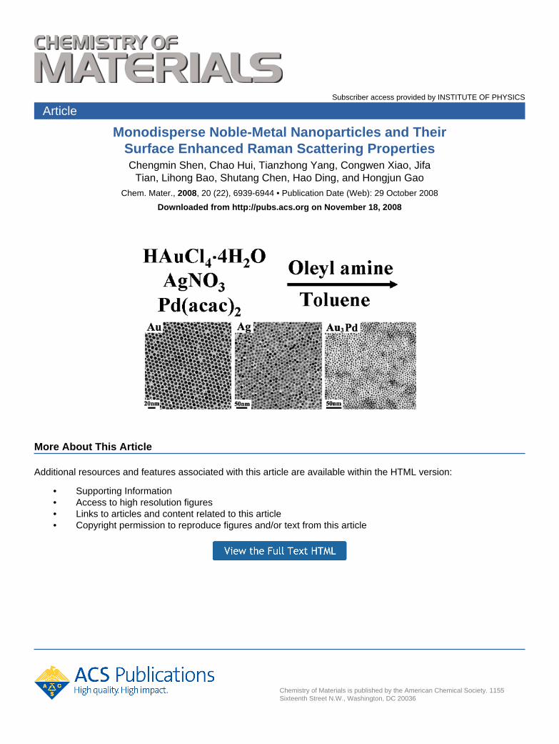
Subscriber access provided by INSTITUTE OF PHYSICS Chemistry of Materials is published by the American Chemical Society. 1155 Sixteenth Street N.W., Washington, DC 20036 Article Monodisperse Noble-Metal Nanoparticles and Their Surface Enhanced Raman Scattering Properties Chengmin Shen, Chao Hui, Tianzhong Yang, Congwen Xiao, Jifa Tian, Lihong Bao, Shutang Chen, Hao Ding, and Hongjun Gao Chem. Mater., 2008, 20 (22), 6939-6944 • Publication Date (Web): 29 October 2008 Downloaded from http://pubs.acs.org on November 18, 2008 More About This Article Additional resources and features associated with this article are available within the HTML version: • Supporting Information • Access to high resolution figures • Links to articles and content related to this article • Copyright permission to reproduce figures and/or text from this article
Transcript of Surface Enhanced Raman Scattering Properties …nano.iphy.ac.cn/N04/papers/NO4_papers all...
Subscriber access provided by INSTITUTE OF PHYSICS
Chemistry of Materials is published by the American Chemical Society. 1155Sixteenth Street N.W., Washington, DC 20036
Article
Monodisperse Noble-Metal Nanoparticles and TheirSurface Enhanced Raman Scattering PropertiesChengmin Shen, Chao Hui, Tianzhong Yang, Congwen Xiao, Jifa
Tian, Lihong Bao, Shutang Chen, Hao Ding, and Hongjun GaoChem. Mater., 2008, 20 (22), 6939-6944 • Publication Date (Web): 29 October 2008
Downloaded from http://pubs.acs.org on November 18, 2008
More About This Article
Additional resources and features associated with this article are available within the HTML version:
• Supporting Information• Access to high resolution figures• Links to articles and content related to this article• Copyright permission to reproduce figures and/or text from this article

Monodisperse Noble-Metal Nanoparticles and Their SurfaceEnhanced Raman Scattering Properties
Chengmin Shen, Chao Hui, Tianzhong Yang, Congwen Xiao, Jifa Tian, Lihong Bao,Shutang Chen, Hao Ding, and Hongjun Gao*
Beijing National Laboratory for Condensed Matter Physics, Institute of Physics, Chinese Academy ofSciences, 100190 Beijing, China
ReceiVed March 27, 2008. ReVised Manuscript ReceiVed September 21, 2008
Monodisperse Au, Ag, and Au3Pd nanoparticles (NPs) with narrow size distribution are prepared bydirect reaction of the related metal salt with oleylamine in toluene. Oleylamine serves as both a reducingagent and a surfactant in the synthesis. The sizes and shape of these NPs are tuned by reaction temperatures.The hydrophobic oleylamine-coated NPs can be made water soluble by replacing oleylamine with3-mercaptopropionic acid. Both surface plasmonic resonance (SPR) and surface enhanced Raman scattering(SERS) observed from the Au and Ag NPs are found to be NP size- and surface-dependent.
Introduction
Au and Ag based noble metal nanoparticles (NPs) haveattracted considerable interest due to their potential applica-tions in catalysis, biolabeling, and photonics.1-10 Their size,size distribution, and morphology control have been the keyto the understanding of the optical and surface properties.Conventionally, Au and Ag NPs are made either by Faradaymethod in aqueous solutions via sodium citrate reduction ofHAuCl4
11-17 or by Brust’s and Schiffrin’s liquid-liquid twophase method in an organic phase by sodium borohydridereduction of AuC14
-1 or AgNO3 in the presence of an
alkanethiol.18-23 To better control the NP size and sizedistribution, the synthesis is also performed in a singleorganic solvent via using strong or mild reducing agent toreduce different gold compound.24-33 Despite all theseefforts made for the synthesis, the quality of the Au and AgNPs is still far from well controlled, and the NPs preparedoften have broad size and shape distribution, unsuitable forconstructing highly ordered superlattice arrays and for deepunderstanding of the physical and chemical properties.34-40
Here, we report a general one-pot organic phase synthesisof monodisperse noble metal NPs via the reaction between
* Corresponding author. E-mail: [email protected].(1) Xia, Y. N.; Halas, N. J. MRS Bull. 2005, 30, 338–348.(2) Sau, T. K.; Murphy, C. J. J. Am. Chem. Soc. 2004, 126, 8648–8649.(3) Hashmi, A. S. K. Chem. ReV. 2007, 107, 3180–3211.(4) Cho, J. K.; Najman, R.; Dean, T. W.; Ichihara, O.; Muller, C.; Bradley,
M. J. Am. Chem. Soc. 2006, 128, 6276–6277.(5) Orendorff, C. J.; Sau, T. K.; Murphy, C. J. Small 2006, 2, 636–639.(6) Merican, Z.; Schiller, T. L.; Hawker, C. J.; Fredericks, P. M.; Blakey,
I. Langmuir 2007, 23, 10539–10545.(7) Mulvaney, S. P.; Musick, M. D.; Keating, C. D.; Natan, M. J. Langmuir
2003, 19, 4784–4790.(8) Zhang, Z. F.; Cui, H.; Lai, C. Z.; Liu, L. J. Anal. Chem. 2005, 77,
3324–3329.(9) Manea, F.; Houillon, F. B.; Pasquato, L.; Scrimin, P. Angew. Chem.,
Int. Ed. 2004, 43, 6165–6169.(10) Hashmi, A. S. K.; Hutchings, G. J. Angew. Chem., Int. Ed. 2006, 45,
7896–7936.(11) Faraday, Philos. Trans. 1857, 145–181.(12) Xiong, Y. J.; Washio, I.; Chen, J. Y.; Sadilek, M.; Xia, Y. N. Angew.
Chem., Int. Ed 2007, 46, 4917–4921.(13) Chen, J. Y.; McLellan, J. M.; Siekkinen, A.; Xiong, Y. J.; Li, Z. Y.;
Xia, Y. N. J. Am. Chem. Soc. 2006, 128, 14776–14777.(14) Datta, K. K. R.; Eswaramoorthy, M.; Rao, C. N. R. J. Mater. Chem.
2007, 17, 613–615.(15) Xiong, Y. J.; Wiley, B.; Chen, J. Y.; Li, Z. Y.; Yin, Y. D.; Xia, Y. N.
Angew. Chem., Int. Ed. 2005, 44, 7913–7917.(16) Njoki, P. N.; Luo, J.; Wang, L. Y.; Maye, M. M.; Quaizar, H.; Zhong,
C. J. Langmuir 2005, 21, 1623–1628.(17) Herricks, T.; Chen, J. Y.; Xia, Y. N. Nano Lett. 2004, 4, 2367–2371.(18) Brust, M.; Walker, M.; Bethell, D.; Schiffrin, D. J.; Whyman, R. Chem.
Commun. 1994, 801–802.(19) (a) Brown, L. O.; Hutchison, J. E. J. Am. Chem. Soc. 1999, 121, 882–
883. (b) Brown, L. O.; Hutchison, J. E. J. Phys. Chem. B 2001, 105,8911–8916.
(20) Chen, S. W.; Huang, K.; Stearns, J. A. Chem. Mater. 2000, 12, 540–547.
(21) (a) Shen, C. M.; Su, Y. K.; Yang, H. T.; Yang, T. Z.; Gao, H.-J.Chem. Phys. Lett. 2003, 373, 39–45. (b) He, S. T.; Yao, J. N.; Jiang,P.; Shi, D. X.; Zhang, H. X.; Xie, S. S.; Pang, S. J.; Gao, H.-J.Langmuir 2001, 17, 1571–1575.
(22) Harfenist, S. A.; Wang, Z. L.; Alvarez, M. M.; Vezmar, I.; Whetten,R. L. J. Phys. Chem. 1996, 100, 13904–13910.
(23) (a) Templeton, A. C.; Wuelfing, M. P.; Murray, R. W. Acc. Chem.Res. 2000, 33, 27–36. (b) Leff, D. V.; Brandt, L.; Heath, J. R. Langmuir1996, 12, 4723–4730.
(24) Schulz-Dobrick, M.; Sarathy, K. V.; Jansen, M. J. Am. Chem. Soc.2005, 127, 12816–12817.
(25) Jana, N. R.; Peng, X. G. J. Am. Chem. Soc. 2003, 125, 14280–14281.(26) Rowe, M. P.; Plass, K. E.; Kim, K.; Kurdak, C.; Zellers, E. T.; Matzger,
A. J. Chem. Mater. 2004, 16, 3513–3517.(27) Zheng, N. F.; Fan, J.; Stucky, G. D. J. Am. Chem. Soc. 2006, 128,
6550–6551.(28) Yee, K.; Jordan, R.; Ulman, A.; White, H.; King, A.; Rafailovich,
M.; Sokolov, J. Langmuir 1999, 15, 3486–3491.(29) Shi, C. S.; Tian, L. F.; Wu, L. L.; Zhu, J. J. Phys. Chem. C 2007,
111, 1243–1247.(30) Ren, J. T.; Tilley, R. D. Small 2007, 3, 1508–1512.(31) Hiramatsu, H.; Osterloh, F. E. Chem. Mater. 2004, 16, 2509–2511.(32) Aslam, M.; Fu, L.; Su, M.; Vijayamohanan, K.; Dravid, V. P. J. Mater.
Chem. 2004, 14, 1795–1797.(33) Liu, X.; Wang, J. H.; Atwater, M.; Dai, Q.; Zou, J. H.; Brennan, J. P.;
Huo, Q. J. Nanosci. Nanotechnol. 2007, 7, 3126–3133.(34) Yamamoto, M.; Nakamoto, M. J. Mater. Chem. 2003, 13, 2064–2065.(35) Kim, S. W.; Park, J.; Jang, Y. J.; Chung, Y. H.; Hwang, S. J.; Hyeon,
T. Nano Lett. 2003, 3, 1289–129.(36) Yang, H. T.; Shen, C. M.; Su, Y. K.; Yang, T. Z.; Gao, H.-J. Appl.
Phys. Lett. 2003, 82, 4729–4731.(37) Rogach, A. L. Angew. Chem., Int. Ed. 2004, 43, 148–149.(38) Xu, Z. C.; Shen, C. M.; Xiao, C. W.; Yang, T. Z.; Zhang, H. R.; Li,
J. Q.; Li, H. L.; Gao, H.-J. Nanotechnology 2007, 18, 115608–115612.(39) Park, J.; Lee, E.; Hwang, N. M.; Kang, M.; Kim, S. C.; Hwang, Y.;
Park, J. G.; Noh, H. J.; Kim, J. Y.; Park, J. H.; Hyeon, T. Angew.Chem., Int. Ed. 2005, 44, 2872–2877.
6939Chem. Mater. 2008, 20, 6939–6944
10.1021/cm800882n CCC: $40.75 2008 American Chemical SocietyPublished on Web 10/29/2008

the related metal salt and oleylamine. Au NPs were preparedby reducing HAuCl4 ·4H2O in the toluene solution ofoleylamine. The sizes of the Au NPs were tuned from 8.5to 23.8 nm by reaction temperatures. Monodisperse Ag orAuPd NPs were also made similarly via the reduction ofAgNO3 or coreduction of HAuCl4 and Pd(acac)2 with theAuPd composition controlled by the molar ratio of these twosalts. The monodipserse Au and Ag NPs self-assembled intothe superlattice arrays and had the surface plasmonicresonance (SPR) band at 523 nm and 407 nm in heptane,respectively. The SPR band of the nanoscale Au was blue-shifted to 7 nm in Au3Pd NPs and red-shifted to 598 nmupon the replacement of oleyalmine around the Au NPs with3-mercaptopropionic acid. The oleylamine-capped Au andAg NPs also exhibited good surface-enhanced Ramanscattering (SERS) effect on the model molecule RhodamineB or 2-naphthalenethiol and the larger Au NPs had higherSERS peak intensity. The work offers a general approach tothe noble metal NPs that may be important for opticalapplications.
Experimental Section
Materials. HAuCl4 ·4H2O and toluene (analysis degree) werepurchased from Beijing Chemical reagents Co. Oleylamine (>70%),2-naphthalenethiol, and 4-mercaptobenzoic acid were obtained fromAldrich. Palladium acetylacetonate (>99%), silver nitrate (>99%),and 3-mercaptopropionic acid were purchased from ACROS.Rhodamine B is obtained from Sigma. All other reagents were usedwithout further purification.
Synthesis of Hydrophobic 8.5 nm Au NPs. A total of 1 mmolof HAuCl4 ·3H2O and 5 mL (10 mmol) of oleylamine were mixedwith 50 mL toluene in a 100 mL flask. Under nitrogen protection,the mixture was heated to 65 °C under magnetic stirring. Thesolution was kept at this temperature for 6 h and cooled down toroom temperature. A total of 50 mL of ethanol was added into thesolution, and the suspension was centrifuged at 6000 rpm for 3min. The supernatant was discarded. The NPs were redispersed inheptane to give a red dispersion.
Synthesis of Hydrophilic 8.5 nm Au NPs. A total of 2 mL ofthe heptane dispersion of the oleylamine-capped Au NPs (17 mg/mL), 10 mL of heptane, and 5 mL of 3-mercaptopropionic acidwere mixed into a 50 mL flask. The resultant solution was stirredovernight at room temperature. The solution was centrifuged at 6000rpm for 3 min, and the precipitation was washed by acetone threetimes. The final product was dissolved in deionized water to givea blue dispersion.
Synthesis of 13 nm Ag NPs. A total of 0.5 mmol of AgNO3
and 2 mL of oleylamine were dissolved in 50 mL of toluene in a100 mL flask. The mixture was heated to 110 °C under nitrogenflow and kept at this temperature for 6 h before it was cooled downto room temperature. A total of 50 mL of ethanol was added intothe solution, and the suspension was centrifuged at 6000 rpm for3 min. The supernatant was discarded. The precipitation wasredissolved in heptane to give a brown-yellow dispersion.
Synthesis of 7 nm Au3Pd NPs. Under a nitrogen flow, 0.66mmol of HAuCl4 ·4H2O, 0.33 mmol of Pd(acac)2, 10 mL ofoleylamine, and 50 mL of toluene were mixed in a 100 mL flask.The mixture was then slowly heated with stirring at 80 °C for 1 h.Then the reaction temperature was raised to 100 °C for 1 h. Thesolution was cooled down room temperature by removing theheating source. A total of 50 mL of enthanol was added, and theproduct was separated by centrifugation (6000 rmp for 5 min). Theproduct was then dispersed in heptane.
Au and Ag NP Assembly for SERS Study. A total of 1.0 mLof heptane dispersion of the hydrophobic Au NPs was dropped onto
(40) Yang, T. Z.; Shen, C. M.; Li, Z. A.; Zhang, H. R.; Xiao, C. W.; Chen,S. T.; Xu, Z. C.; Shi, D. X.; Li, J. Q.; Gao, H.-J. J. Phys. Chem. B2005, 109, 23233–23236.
Figure 1. TEM images and XRD patterns of hydrophobic Au NPs andhydrophilic Au NPs. (a) Large area TEM image of oleylamine-capped AuNPs; (b) superlattice structure of oleylamine-capped Au NPs. (c) TEM imageof 3-mercaptopropionic acid-capped Au NPs. (d) XRD patterns of hydro-phobic and hydrophilic Au NPs. (e) UV-vis spectra of hydrophobic andhydrophilic of Au NPs. (f) Optical photograph of hydrophobic andhydrophilic of Au NP samples dissolved in heptane and water, respectively.
Figure 2. XPS spectra of hydrophobic and hydrophilic Au NPs; (a) Au inhydrophobic; (b) N in hydrophobic; (c) Au in hydrophilic; (d) S inhydrophilic.
6940 Chem. Mater., Vol. 20, No. 22, 2008 Shen et al.
the 1 × 1 cm2 Si(111) wafer and dried under ambient conditions.Then, this Si wafer was put into a 5 mL beaker containing 0.8 mLof 1 × 10⁻³ M Rhodamine B aqueous solution or 1 × 10⁻³ M2-naphthalenethiol enthanol solution. The Au NPs covered Si waferwas washed three times with ethanol and dried in air. Forcomparison, the same procedure was also applied on the Si substratemodified only with Rhodamine B or 2-Naphthalenethiol. Similarto the Au NPs, 1.0 mL of heptane dispersion of oleylamine-cappedAg NPs was dropped on the surface of the Si substrate and putinto 1 × 10⁻³ M 4-mercaptobenzoic acid enthanol solution. TheSi wafer was washed three times with ethanol and dried in air.
Characterization of NPs. X-ray diffraction pattern of the NPsnanoparticle assembly was collected on a Rigaku D/MAX 2400X-ray diffractometer with Cu KR radiation (λ ) 1.5406 Å). TheUV-vis spectrum was recorded at room temperature on a Carry1E ultraviolet-visible spectrometer using 1-cm quartz cuvettes. Thesurface enhanced Raman spectrum was recorded on the Ramansystem YJ-HR800 with confocal microscopy. The solid-state diodelaser (633 nm or 785 nm) was used as an excited source. The laserpower on the samples was kept with 0.9 mW (633 nm) and 0.07mW (785 nm). X-ray photoelectron spectra were obtained on theESCA LAB5 X-ray photoelectron spectrometer with the mono-chromatic Mg X-ray. Transmission electron microcopy (TEM)images were acquired with Tecnai-20 (PHILIPS Cop) at 120 kV.
Results and Discussion
Preparation of Hydrophobic and Hydrophilic AuNPs. Figure 1 presents the TEM images of the Au NPssynthesized in this work. It can be seen that the oleylamine-capped Au NPs have uniform size at 8.5 nm and narrowsize distribution (<5% standard deviation), and self-assemblyof these particles gives hexagonal close packed arrays. Theselective area electron diffraction (SAED) pattern of the 8.5nm Au NPs is shown in the inset of Figure 1A with thediffraction rings corresponding to the (111), (200), (220),and (311) planes from the face-centered-cubic (fcc) Au
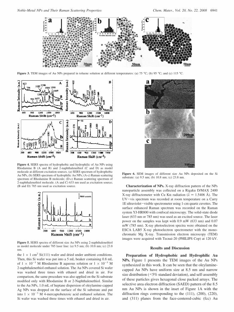
Figure 3. TEM images of Au NPs prepared in toluene solution at different temperatures: (a) 75 °C; (b) 95 °C; and (c) 115 °C.
Figure 4. SERS spectra of hydrophobic and hydrophilic of Au NPs usingRhodamine B (A and B) and 2-naphthalenethiol (C and D) as modelmolecule at different excitation sources. (a) SERS spectrum of hydrophobicAu NPs, (b) SERS spectrum of hydrophilic Au NPs; (A-c) Raman scatteringspectrum of Rhodamine B molecule; (D-c) Raman scattering spectrum of2-naphthalenethiol molecule. (A and C) 633 nm used as excitation source;(B and D) 785 nm used as excitation source.
Figure 5. SERS spectra of different size Au NPs using 2-naphthalenethiolas model molecule under 785 laser line: (a) 9.5 nm; (b) 10.8 nm; (c) 23.8nm.
Figure 6. SEM images of different size Au NPs deposited on the Sisubstrate: (a) 9.5 nm; (b) 10.8 nm; (c) 23.8 nm.
6941Chem. Mater., Vol. 20, No. 22, 2008Noble-Metal NPs and Their Raman Scattering Properties

structure. The oleylamine-capped hydrophobic Au NPs aretransformed into water soluble Au NPs by replacing oley-lamine with 3-mercaptopropionic acid. The -SH group inmercaptopropionic acid binds to Au, and -COOH surroundsthe Au NPs. Figure 1C shows the TEM image of the
hydrophilic Au NPs. Comparing with Figure 1A,C, we cansee that there is no size and morphology change during theligand exchange process. XRD patterns of both hydrophobic
(41) Chen, Z. T.; Gao, L. Mater. Res. Bull. 2007, 42, 1657–1661.
Figure 7. TEM images of 13 nm Ag NPs prepared in toluene solution; (a) monolayer film; (b) superlattices structure; (c) SAED pattern of Ag nanoparticle;and (d) UV-vis spectrum of Ag NPs.
Figure 8. (a) TEM images of 7 nm Au3Pd alloy NPs prepared in toluene solution; (b) XRD pattern of Au3Pd alloy; (c) EDX pattern of Au3Pd alloy; (d)UV-vis spectra of Au3Pd alloy NPs.
6942 Chem. Mater., Vol. 20, No. 22, 2008 Shen et al.

and hydrophilic Au NPs are given in Figure 1D, confirmingthat the Au NPs in both cases have the fcc structure (JCPDSno. 04-0784). The UV-vis spectra of the hydrophobic andhydrophilic Au NPs are shown in the Figure 1E. Thecharacteristic SPR band of the Au NPs capped with differentligands is located at 523 nm and 598 nm (Figure 1E). Theabsorption peak for the hydrophilic Au NPs is red-shifted,and the color of the dispersion turns from red into deep-blue (Figure 1F). This red-shift is likely caused by the partialaggregation of the Au NPs in water as observed in Figure1C. This is consistent with what has been reported.29,31,32
To better understand the surface environment of hydrophobicand hydrophilic Au NPs, we used X-ray photoelectronspectroscopy (XPS) to investigate the interaction of nitrogenand sulfur atoms on the surface of Au particles aftersurfactant exchange. Figure 2 shows the XPS spectra ofhydrophobic and hydrophilic Au NPs. Two peaks areobserved at 87.4 and 83.7 eV in the oleylamine-capped AuNPs, corresponding to the binding energies of 4f5/2 and 4f7/
2, respectively. This is in agreement with the XPS spectraof the other alkylamine-capped Au NPs.23b Compared to thehydrophobic Au NPs, the 4f binding energies of thehydrophilic Au NPs shift to higher values from 87.4 and83.7 eV to 88.1 and 84.4 V. This shift proves that the S-Aubond is stronger than the NH2-Au bond.21
Reaction Temperature Effect on Size of Au NPs.Reaction temperature played a key role in nanoparticle sizecontrol. Figure 3 shows the TEM images of the Au NPsprepared at different temperatures. The size of the Au NPsis 9.5 nm (6.3% std. dev.) at 75 °C, 10.8 nm (12%, std. dev.)at 95 °C, and 23.8 nm (12%, std. dev.) at 115 °C,respectively. The higher reaction temperature results in afaster Au3+ reduction and growth process, producing largesize NPs. 27,41 At lower reaction temperature (65 °C), thereduction is slowed down and the nucleation process overrunsthe growth process, yielding small particles.
SERS Study of the Au NPs. The SERS of the hydro-phobic and hydrophilic Au NPs was investigated using 1 ×10⁻³ M Rhodamine B and 1 × 10⁻³ M 2-naphthalenetiolas model molecules. The excitation sources were 633 nm(laser power 0.9 mW) and 785 nm (laser power 0.07 mW)from a diode laser. Figure 4A,B show the SERS spectra ofRhodamine B adsorbed on hydrophobic and hydrophilic AuNPs at different excitation sources. The Raman peaks ofhydrophobic and hydrophilic Au NPs display evident en-hancement, compared with that from the pure RhodmaineB (Figure 4A-c). The major bands of Rhodamine B moleculeobserved in two samples (Figure 4A) using 633 nm as thelaser exciting line was also found under 785 nm (Figure 4B),indicating that Rhodamine B molecules are adsorbed on thesurface of Au NPs.6 However, the intensity of the SERSspectra is different between the hydrophilic Au NPs andhydrophobic ones under two exciting laser sources. Under633 nm excitation, the SERS from the hydrophilic Au NPsis stronger than that from the hydrophobic ones. This iscaused by the overlap between the exciting laser line andthe SPR band of the hydrophilic particles at 598 nm. Whenthe 785 nm laser exciting line is used, the SERS of the twosamples are also enhanced, but the enhancement from thehydrophilic Au particles is evidently weaker than that fromthe hydrophobic one due to the difference in the excitingline and the SPR absorption of the hydrophilic particles. Wealso measured the SERS of the hydrophobic and hydrophilicAu NPs using 2-naphthalenethiol as a model molecule. TheSERS spectra are shown in Figure 4C,D. Different from thatof the Rhodamine B Au NPs, the SERS signals of the2-naphthalenethiol-hydrophilic Au NPs are weaker in thiscase than that of the hydrophobic. Clearly, this is due to thechemical binding between -SH in 2-naphthalenethiol andthe oleylamine-capped Au NPs, as confirmed from theRaman singlet at about 370 cm-1 (Figure 4C-a,D-a) that canbe designated to one of the vibrational modes of the C-Sbond upon coordination to the Au surface.29 For hydrophilicAu NPs, 2-naphthalenethiol molecule has much less effecton the SERS due to the prebinding of the thiol moleculefrom 3-mercaptopropionic acid.
Size effect of the Au NPs on SERS. The SERS intensityis dependent on the sizes of the Au NPs. Figure 5 shows theSERS spectra of the Au NPs with different sizes (1 × 10⁻³
M 2-naphthalenethiol as model molecule). It can be seenthat the SERS intensity of the 23.8 nm Au NPs is 9 timeshigher than that of the 9.5 nm Au NPs. This increase in SERSsignal with Au NP sizes is in agreement with what is reportedin Au NPs and Au nanohole arrays.42,43 The larger Au NPstend to form the larger area ordered superlattice structureon the Si substrate, as shown in Figure 6. The close packingof the Au NPs in a large area assembly can lead to a stronginterparticle plasmon coupling and therefore the enhancedSERS.44,45
(42) Yu, Q. M.; Guan, P.; Qin, D.; Auen, G.; Wallace, P. M. Nano Lett.2008, 8, 1923–1928.
(43) Kim, K.; Lee, H. B.; Lee, J. W.; Park, H. K.; Shin, K. S. Langmuir2007, 24, 7178–7183.
(44) Sabur, A.; Havel, M.; Gogotsi, Y. J. Raman Spectrosc. 2008, 39, 61–67.
Figure 9. XPS spectra of d Au3Pd alloy NPs prepared in toluene solution:(a) Au; (b) Pd.
6943Chem. Mater., Vol. 20, No. 22, 2008Noble-Metal NPs and Their Raman Scattering Properties

Preparation of Silver and Alloy NPs. The currentsynthesis can be readily extended to the preparation of othernoble metal and alloy NPs. The 13 nm Ag NPs were obtainedusing AgNO3 in oleylamine-toluene solution at 110 °C.Figure 7 shows the TEM images of Ag NPs. These NPs havenarrow size distribution and can self-assemble into monolayer(Figure 7a) and multilayer superlattices (Figure 7b). Theselective area electron diffraction pattern (Figure 7c) showsthat the diffraction rings are from the (111), (200), (220),and (311) planes in the fcc Ag. The SPR band of the 13 nmAg NPs appeared at 407 nm (Figure 7d). The goodsymmetric absorption peaks imply that the size of the NPsis very uniform.21
Using similar reaction conditions, we have also mademonodisperse Au3Pd NPs using 0.33 mmol Pd (acac)2 and0.67 mmol HAuCl4 ·4H2O as precursors. Figure 8a showsthe TEM images of the 7 nm Au3Pd NPs. The 3/1 Au/Pdwas confirmed by EDX (Figure 8b). The HRTEM image ofthe Au3Pd NPs in the inset of Figure 8b reveals goodcrystallinity of the particle. XRD patterns of the Au3Pd NPs(Figure 8c) show the diffraction peaks at 38.8, 44.8, 65.9,and 77.9, corresponding to the (111), (200), (220), and (311)planes of the Au3Pd NPs. These peaks are shifted away fromthose of the standard Au (JCPDS 04-0784) and palladium(JCPDS 46-1043), indicating that Au3Pd alloy is indeedformed. Comparing with that of the Au NPs, the SPR bandof the Au3Pd alloy NPs is blue-shifted, proving that theoptical properties of the NPs can be tuned by controlledcomposition in alloy NPs. The XPS spectra of the Au3PdNPs in the Au 4f and Pd 3d regions are shown in Figure 9.Two peaks locate at 87.1 eV and 83.5 eV corresponded toAu0, while peaks at 340.1 and 334.7 eV are from Pd. The
binding energy in Au3Pd NPs is lower than that of the Au(87.67 eV and 84.0 eV) and Pd (335.1 eV and 340.36 eV).
We measured the SERS spectrum of the 13 nm Ag NPsusing 1 × 10⁻³ M 4-mercaptobenzoic acid as a modelmolecule. The enhancement can be seen in Figure 10a.Similar to that in Au NPs, this enhancement is due to theformation of a large area Ag nanoparticle superlattice[Supporting Information and refs 29 and 42]. Figure 10b isthe SERS spectrum of the Au3Pd NPs. The characteristicRaman peaks from 1 × 10⁻³ M 2-naphthenethiol is detected,indicating that the AuPd alloy NPs may also be used inSERS. Detailed studies in SERS of the Ag NPs and theAu3Pd NPs are underway.
Conclusions
A facile one-pot organic synthesis of Au, Ag, and Au3PdNPs with narrow size distribution is reported. The oleylamineserves as both a surfactant and a reducing agent. The size ofthe NPs is tuned through the reaction temperatures. TheseNPs are SERS active and this activity is dependent on thesurface coating and the size of the particles. Through sizecontrol and surface modification, the optical properties ofthese monodisperse NPs should be readily optimized forpotential highly sensitive optical detections.
Acknowledgment. The work was supported by the NationalNatural Science Foundation of China (Grant 60571045), Na-tional “863” Project of China (Grant 2007AA03Z305), andCAS/ SAFEA international cooperation team.
Supporting Information Available: Preparation of Pd NPs,TEM image of 8.5 nm hydrophilic Au NPs, SEM image of 13 nmAg NPs deposited on the silicon substrate, TEM image of Pd NPs,and XRD of oleylamine-capped Pd NPs (PDF). This material isavailable free of charge via the Internet at http://pubs.acs.org.
CM800882N(45) Clha, M. C.; Kahraman, M.; Tokman, N.; Turkoglu, G. J. Phys. Chem.
C 2008, 112, 10338–10343.
Figure 10. SERS spectra of Ag and Au3Pd alloy NPs prepared in toluene solution: (a) Ag; (b) Au3Pd alloy.
6944 Chem. Mater., Vol. 20, No. 22, 2008 Shen et al.


![Permeation through graphene ripplesnano.iphy.ac.cn/N04/papers/NO4_papers all pdf/2017paper/add25Liang_2017... · at the locally curved surface of graphene [26]. However, ... cylindrical](https://static.fdocuments.us/doc/165x107/5e1c341a7fbf7d53e57eac9c/permeation-through-graphene-all-pdf2017paperadd25liang2017-at-the-locally.jpg)


