Supracondylar Osteotomy of Femur for Management of ... · Management of Deformities around Knee...
6
76 International Journal of Scientific Study | August 2015 | Vol 3 | Issue 5 Supracondylar Osteotomy of Femur for Management of Deformities around Knee Joint: A Camp Experience in Chhattisgarh Antony R Benn 1 , Pankaj Tembhurnikar 2 , Atul Manoharrao Deshkar 3 , K S Bajpai 4 1 Associate Professor, Department of Orthopedics, Government Chhattisgarh Institute of Medical Sciences, Bilaspur, Chhattisgarh, India, 2 Associate Professor, Department of Medicine, Government Chhattisgarh Institute of Medical Sciences, Bilaspur, Chhattisgarh, India, 3 Associate Professor and Head, Department of Physiology and Coordinator Medical Education Unit, Government Chhattisgarh Institute of Medical Sciences, Bilaspur, and Secretary IMA College of General Practitioners Chhattisgarh, India, 4 Consultant Orthopedic Surgeon and Block Medical Officer, Baloda Bazar, Chhattisgarh, India be due to rickets or osteomalacia. An angular correction by wedge corrective osteotomy is the standard treatment. 2 Pediatric femoral shaft fractures are known to produce growth arrest at the proximal tibia, probably caused by compression injuries of the proximal tibial physis, most often in the area of the tibial tubercle, producing late onset genu recurvatum deformities. 3 Genu recurvatum secondary to paralytic poliomyelitis is disabling and difficult to treat. 4 In our country, 10% population is disabled. Deformities around knee joint is a common disability in patients such as flexion contracture of knee joint, genu valgus, genu varus, and genu recurvatum. It INTRODUCTION Genu valgum is a common orthopedic problem in children. The vast majority of cases are physiologic variants, which resolve normally. 1 Frontal plane knee deformity may be congenital, idiopathic, and secondary to trauma or might Original Article Abstract Background: Genu recurvatum secondary to paralytic poliomyelitis is disabling and difficult to treat. In our country, 10% population is disabled. Deformities around the knee joint are common disability in patients such as flexion contracture of knee joint, genu valgus, genu varus, and genu recurvatum. To assess the results of supracondylar osteotomy as corrective surgery for deformities around knee joint with patients of flexion contracture deformity, genu valgus, genu varus, genu recurvatum with different diseases and accidental disorders at free disabled surgical camps. Materials and Methods: In our study, 50 patients were included with 70% male and 30% female, with 10-24 years of age group. Maximum patients had single lower limb knee joint deformity and 8 patients had bilateral lower limb knee joint deformity such as genu valgum and genu varum. Nearly all patients were neglected for long time. All patients were operated in different free disabled surgical camps. Our period of study was 36 weeks (1 st May 2004-31 st April 2007). Improvement in functional ability and locomotion of all operated patients was assessed by physical examination. Results: All patients operated in our study showed significant improvement in functional abilities and locomotion after surgery. All patients who came for subsequent follow-up were maintaining functional ability at follow-up duration of 1-year (12 months). At the end results in our study buttonhole and anterior wedge, osteotomy proved to be with excellent results. Conclusion: Our study shows excellent results that can be achieved in patients with severe neglected knee joint deformities of lower limb with supracondylar osteotomy bony corrective surgeries. It’s the team work of devoted surgeon, paramedical and rehabilitation staff at free disabled surgical camps which facilitates services in remote areas with frequent follow-up and maximum achievements of good results in minimum expenditures. Key words: Genu valgum, Osteotomy, Varus Access this article online www.ijss-sn.com Month of Submission : 07-2015 Month of Peer Review : 07-2015 Month of Acceptance : 07-2015 Month of Publishing : 08-2015 Corresponding Author: Dr. Atul Manoharraao Deshkar, Associate Professor and Head, Department of Physiology, Chhattisgarh Institute of Medical Sciences, Bilaspur, Chhattisgarh, India. Phone: +91-9893008889. E-mail: [email protected] DOI: 10.17354/ijss/2015/351
Transcript of Supracondylar Osteotomy of Femur for Management of ... · Management of Deformities around Knee...
76International Journal of Scientific Study | August 2015 | Vol 3 | Issue 5
Supracondylar Osteotomy of Femur for Management of Deformities around Knee Joint: A Camp Experience in ChhattisgarhAntony R Benn1, Pankaj Tembhurnikar2, Atul Manoharrao Deshkar3, K S Bajpai4
1Associate Professor, Department of Orthopedics, Government Chhattisgarh Institute of Medical Sciences, Bilaspur, Chhattisgarh, India, 2Associate Professor, Department of Medicine, Government Chhattisgarh Institute of Medical Sciences, Bilaspur, Chhattisgarh, India, 3Associate Professor and Head, Department of Physiology and Coordinator Medical Education Unit, Government Chhattisgarh Institute of Medical Sciences, Bilaspur, and Secretary IMA College of General Practitioners Chhattisgarh, India, 4Consultant Orthopedic Surgeon and Block Medical Officer, Baloda Bazar, Chhattisgarh, India
be due to rickets or osteomalacia. An angular correction by wedge corrective osteotomy is the standard treatment.2 Pediatric femoral shaft fractures are known to produce growth arrest at the proximal tibia, probably caused by compression injuries of the proximal tibial physis, most often in the area of the tibial tubercle, producing late onset genu recurvatum deformities.3
Genu recurvatum secondary to paralytic poliomyelitis is disabling and difficult to treat.4 In our country, 10% population is disabled. Deformities around knee joint is a common disability in patients such as flexion contracture of knee joint, genu valgus, genu varus, and genu recurvatum. It
INTRODUCTION
Genu valgum is a common orthopedic problem in children. The vast majority of cases are physiologic variants, which resolve normally.1 Frontal plane knee deformity may be congenital, idiopathic, and secondary to trauma or might
Original Article
AbstractBackground: Genu recurvatum secondary to paralytic poliomyelitis is disabling and difficult to treat. In our country, 10% population is disabled. Deformities around the knee joint are common disability in patients such as flexion contracture of knee joint, genu valgus, genu varus, and genu recurvatum. To assess the results of supracondylar osteotomy as corrective surgery for deformities around knee joint with patients of flexion contracture deformity, genu valgus, genu varus, genu recurvatum with different diseases and accidental disorders at free disabled surgical camps.
Materials and Methods: In our study, 50 patients were included with 70% male and 30% female, with 10-24 years of age group. Maximum patients had single lower limb knee joint deformity and 8 patients had bilateral lower limb knee joint deformity such as genu valgum and genu varum. Nearly all patients were neglected for long time. All patients were operated in different free disabled surgical camps. Our period of study was 36 weeks (1st May 2004-31st April 2007). Improvement in functional ability and locomotion of all operated patients was assessed by physical examination.
Results: All patients operated in our study showed significant improvement in functional abilities and locomotion after surgery. All patients who came for subsequent follow-up were maintaining functional ability at follow-up duration of 1-year (12 months). At the end results in our study buttonhole and anterior wedge, osteotomy proved to be with excellent results.
Conclusion: Our study shows excellent results that can be achieved in patients with severe neglected knee joint deformities of lower limb with supracondylar osteotomy bony corrective surgeries. It’s the team work of devoted surgeon, paramedical and rehabilitation staff at free disabled surgical camps which facilitates services in remote areas with frequent follow-up and maximum achievements of good results in minimum expenditures.
Key words: Genu valgum, Osteotomy, Varus
Access this article online
www.ijss-sn.com
Month of Submission : 07-2015 Month of Peer Review : 07-2015 Month of Acceptance : 07-2015 Month of Publishing : 08-2015
Corresponding Author: Dr. Atul Manoharraao Deshkar, Associate Professor and Head, Department of Physiology, Chhattisgarh Institute of Medical Sciences, Bilaspur, Chhattisgarh, India. Phone: +91-9893008889. E-mail: [email protected]
DOI: 10.17354/ijss/2015/351
Benn, et al.: Supracondylar Osteotomy of Femur
77 International Journal of Scientific Study | August 2015 | Vol 3 | Issue 5
is caused by poliomyelitis, hemiparesis, cerebral palsy, rickets, developmental congenital disorders, and accidental disorders. It’s a big challenge for surgeons to improve their locomotor system and making patient independent. A measurable varus angle or a valgus higher than 11° during this period should be considered abnormal.5 Due to a big load of trauma cases in higher centers in our state, and country these disabled patients are neglected and refused everywhere for the management (corrective surgeries). So small satellite free disabled surgical camps organized by the help of NGOs, Akhil Bharatiya Viklang Chetna Parishad, GOVT agencies and life line express are very helpful for these disabled patients.
Supracondylar osteotomy for the correction of deformities around knee joint not only corrects the deformity but also improves personal hygiene, gait and independent ambulation with or without assistive devices. This treatment should be planned with patient needs. In our study to complete the evaluation of the knee, we have taken different radiographic views, including the standard lateral and the axial views of the patellofemoral joint.6
This retrospective study of 50 patients with deformities around knee joint were operated in free disabled surgical camps at different districts of Chhattisgarh state in institutions with helping NGOs, GOVT agencies, and life line express.
MATERIALS AND METHODS
In our study, out of 50 patients included, 70% were male and 30% were female with 10-24 years in age group. Almost all were neglected for a long time for corrective surgeries, some were operated but not achieved good results.
Patients were admitted and operated at different free disabled camps in different institutions in different districts of Chhattisgarh over the period of 3 years (1st May 2004-31st April 2007). Choice of patients was mild to moderate and severe deformities around the knee joint, standing or walking with some or other gait with or without support. Deformities around knee joint and condition of the lower limb of patients were assessed with a physical and radiological examination at the time of admission. Deformities around knee joint such as flexion contracture, genu valgus, genu varum, genu recurvatum, and gait pattern were noted in all patients clinically.
SCHEDULE OF CAMPS
Day 1• Registration and screening of patients suitable for
surgery.• Preoperative preparation, routine investigations, and
medication of patient and X-rays of the knee joint and lower limbs.
• Meticulous asepsis and arrangement in the operation theater.
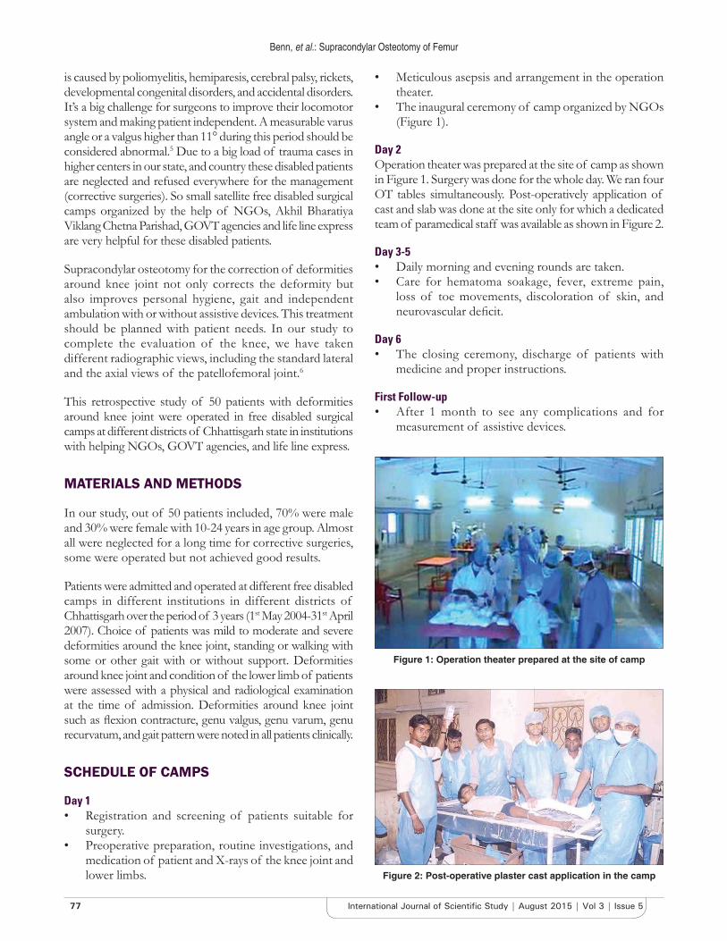
• The inaugural ceremony of camp organized by NGOs (Figure 1).
Day 2Operation theater was prepared at the site of camp as shown in Figure 1. Surgery was done for the whole day. We ran four OT tables simultaneously. Post-operatively application of cast and slab was done at the site only for which a dedicated team of paramedical staff was available as shown in Figure 2.
Day 3-5• Daily morning and evening rounds are taken.• Care for hematoma soakage, fever, extreme pain,
loss of toe movements, discoloration of skin, and neurovascular deficit.
Day 6• The closing ceremony, discharge of patients with
medicine and proper instructions.
First Follow-up• After 1 month to see any complications and for
measurement of assistive devices.
Figure 1: Operation theater prepared at the site of camp
Figure 2: Post-operative plaster cast application in the camp
Benn, et al.: Supracondylar Osteotomy of Femur
78International Journal of Scientific Study | August 2015 | Vol 3 | Issue 5
Second Follow-up• After 2.5-3 months for removal of the pop cast,
stitches, k wires• Calipers and shoes are provided • Parallel bar walking and physiotherapy explained to
patient and relatives• Advised regular follow-up every 2 months for at least
2 years.
SURGICAL PROCEDURES
If correction is not satisfactory after soft tissue release surgery for deformities around the knee joint, the patient needs supracondylar osteotomy of the femur. Supracondylar osteotomy is a big answer to many problems around the knee joint.
TYPES OF OSTEOTOMIES
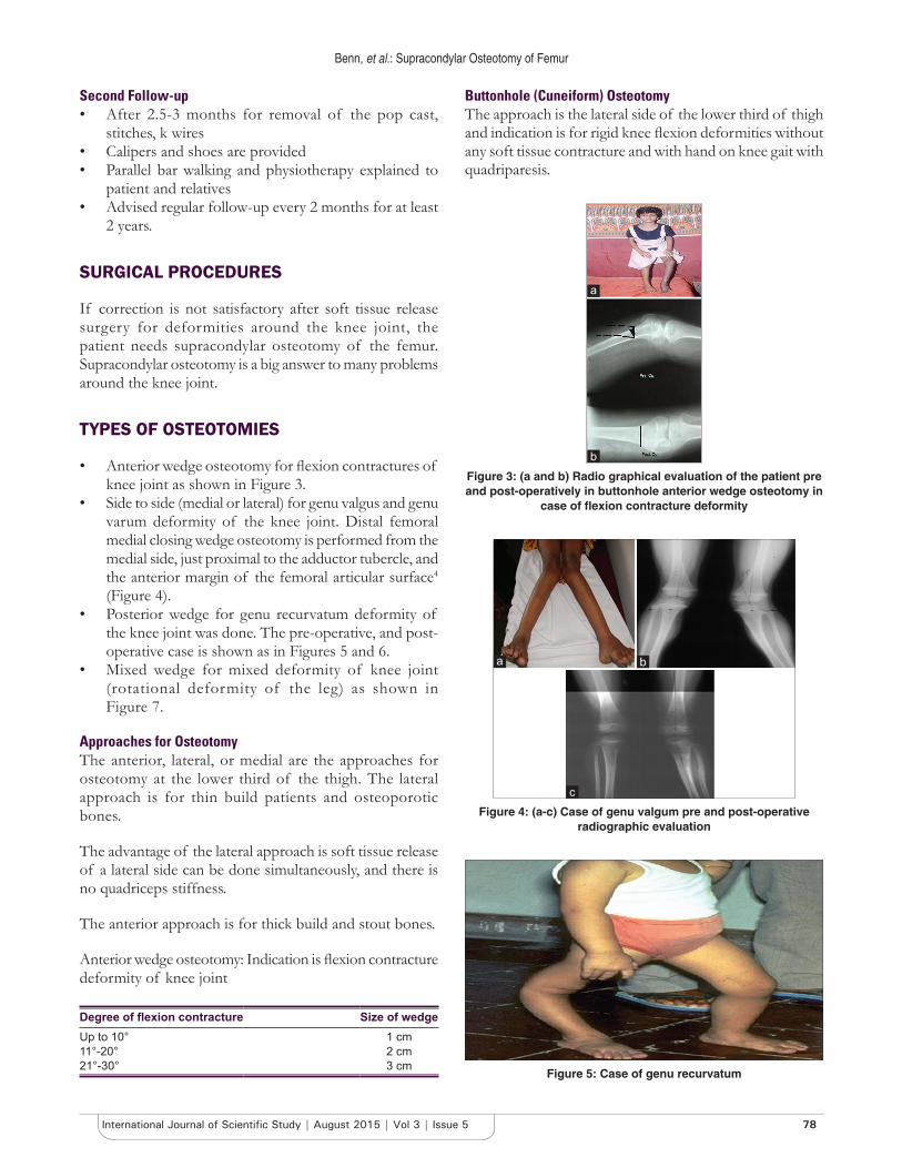
• Anterior wedge osteotomy for flexion contractures of knee joint as shown in Figure 3.
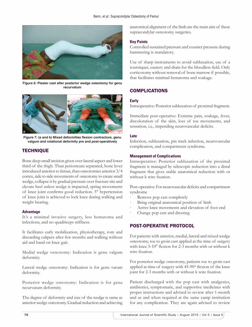
• Side to side (medial or lateral) for genu valgus and genu varum deformity of the knee joint. Distal femoral medial closing wedge osteotomy is performed from the medial side, just proximal to the adductor tubercle, and the anterior margin of the femoral articular surface4 (Figure 4).
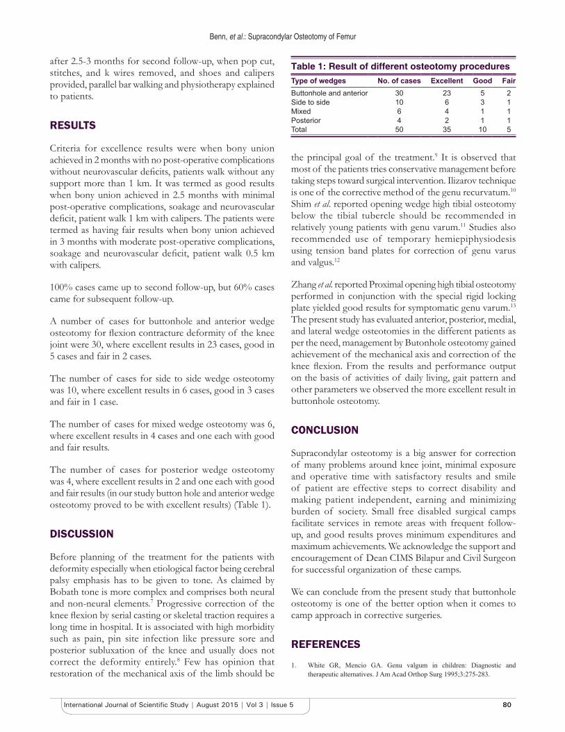
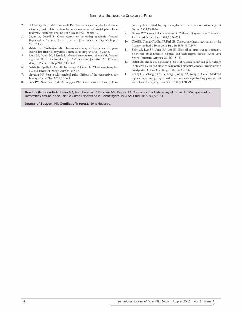
• Posterior wedge for genu recurvatum deformity of the knee joint was done. The pre-operative, and post-operative case is shown as in Figures 5 and 6.
• Mixed wedge for mixed deformity of knee joint (rotational deformity of the leg) as shown in Figure 7.
Approaches for OsteotomyThe anterior, lateral, or medial are the approaches for osteotomy at the lower third of the thigh. The lateral approach is for thin build patients and osteoporotic bones.
The advantage of the lateral approach is soft tissue release of a lateral side can be done simultaneously, and there is no quadriceps stiffness.
The anterior approach is for thick build and stout bones.
Anterior wedge osteotomy: Indication is flexion contracture deformity of knee joint
Degree of flexion contracture Size of wedgeUp to 10° 1 cm11°-20° 2 cm21°-30° 3 cm
Buttonhole (Cuneiform) OsteotomyThe approach is the lateral side of the lower third of thigh and indication is for rigid knee flexion deformities without any soft tissue contracture and with hand on knee gait with quadriparesis.
Figure 5: Case of genu recurvatum
Figure 4: (a-c) Case of genu valgum pre and post-operative radiographic evaluation
b
c
a
Figure 3: (a and b) Radio graphical evaluation of the patient pre and post-operatively in buttonhole anterior wedge osteotomy in
case of flexion contracture deformity
b
a
Benn, et al.: Supracondylar Osteotomy of Femur
79 International Journal of Scientific Study | August 2015 | Vol 3 | Issue 5
TECHNIQUE
Bone deep small incision given over lateral aspect and lower third of the thigh. Than periosteum separated, bone lever introduced anterior to femur, than osteotomies anterior 3/4 cortex, side to side movements of osteotomy to create small wedge, collapse it by gradual pressure over fracture site and elevate heel unless wedge is impacted, spring movements of knee joint confirms good reduction. 5° hypertension of knee joint is achieved to lock knee during walking and weight bearing.
AdvantageIt’s a minimal invasive surgery, less hematoma and infections, and no quadriceps stiffness.
It facilitates early mobilization, physiotherapy, rom and discarding calipers after few months and walking without aid and hand on knee gait.
Medial wedge osteotomy: Indication is genu valgum deformity.
Lateral wedge osteotomy: Indication is for genu varum deformity.
Posterior wedge osteotomy: Indication is for genu recurvatum deformity.
The degree of deformity and size of the wedge is same as anterior wedge osteotomy. Gradual reduction and achieving
anatomical alignment of the limb are the main aim of these supracondylar osteotomy surgeries.
Key PointsControlled sustained pressure and counter pressure during hammering is mandatory.
Use of sharp instruments to avoid subluxation, use of a tourniquet, cautery and drain for the bloodless field. Only corticotomy without removal of bone marrow if possible, that facilitates minimal hematoma and soakage.
COMPLICATIONS
EarlyIntraoperative: Posterior subluxation of proximal fragment.
Immediate post-operative: Extreme pain, soakage, fever, discoloration of the skin, loss of toe movements, and sensation, i.e., impending neurovascular deficits.
LateInfection, subluxation, pin track infection, neurovascular complication, and compartment syndrome.
Management of ComplicationsIntraoperative: Posterior subluxation of the proximal fragment is managed by telescopic reduction into a distal fragment that gives stable anatomical reduction with or without k wire fixation.
Post-operative: For neurovascular deficits and compartment syndrome· Remove pop cast completely· Bring original anatomical position of limb· Active knee movements and elevation of foot end· Change pop cast and dressing
POST-OPERATIVE PROTOCOL
For patients with anterior, medial, lateral and mixed wedge osteotomy, toe to groin cast applied at the time of surgery with knee 5-10° flexion for 2-3 months with or without k wire fixation.
For posterior wedge osteotomy, patients toe to groin cast applied at time of surgery with 45-90* flexion of the knee joint for 2-3 months with or without k wire fixation.
Patient discharged with the pop cast with analgesics, antibiotics, symptomatic, and supportive medicines with proper instructions and advised to review after 1-month and as and when required at the same camp institution for any complication. They are again advised to review
Figure 6: Plaster cast after posterior wedge osteotomy for genu recurvatum
Figure 7: (a and b) Mixed deformities flexion contracture, genu valgum and rotational deformity pre and post-operatively
ba
Benn, et al.: Supracondylar Osteotomy of Femur
80International Journal of Scientific Study | August 2015 | Vol 3 | Issue 5
after 2.5-3 months for second follow-up, when pop cut, stitches, and k wires removed, and shoes and calipers provided, parallel bar walking and physiotherapy explained to patients.
RESULTS
Criteria for excellence results were when bony union achieved in 2 months with no post-operative complications without neurovascular deficits, patients walk without any support more than 1 km. It was termed as good results when bony union achieved in 2.5 months with minimal post-operative complications, soakage and neurovascular deficit, patient walk 1 km with calipers. The patients were termed as having fair results when bony union achieved in 3 months with moderate post-operative complications, soakage and neurovascular deficit, patient walk 0.5 km with calipers.
100% cases came up to second follow-up, but 60% cases came for subsequent follow-up.
A number of cases for buttonhole and anterior wedge osteotomy for flexion contracture deformity of the knee joint were 30, where excellent results in 23 cases, good in 5 cases and fair in 2 cases.
The number of cases for side to side wedge osteotomy was 10, where excellent results in 6 cases, good in 3 cases and fair in 1 case.
The number of cases for mixed wedge osteotomy was 6, where excellent results in 4 cases and one each with good and fair results.
The number of cases for posterior wedge osteotomy was 4, where excellent results in 2 and one each with good and fair results (in our study button hole and anterior wedge osteotomy proved to be with excellent results) (Table 1).
DISCUSSION
Before planning of the treatment for the patients with deformity especially when etiological factor being cerebral palsy emphasis has to be given to tone. As claimed by Bobath tone is more complex and comprises both neural and non-neural elements.7 Progressive correction of the knee flexion by serial casting or skeletal traction requires a long time in hospital. It is associated with high morbidity such as pain, pin site infection like pressure sore and posterior subluxation of the knee and usually does not correct the deformity entirely.8 Few has opinion that restoration of the mechanical axis of the limb should be
the principal goal of the treatment.9 It is observed that most of the patients tries conservative management before taking steps toward surgical intervention. Ilizarov technique is one of the corrective method of the genu recurvatum.10 Shim et al. reported opening wedge high tibial osteotomy below the tibial tubercle should be recommended in relatively young patients with genu varum.11 Studies also recommended use of temporary hemiepiphysiodesis using tension band plates for correction of genu varus and valgus.12
Zhang et al. reported Proximal opening high tibial osteotomy performed in conjunction with the special rigid locking plate yielded good results for symptomatic genu varum.13 The present study has evaluated anterior, posterior, medial, and lateral wedge osteotomies in the different patients as per the need, management by Butonhole osteotomy gained achievement of the mechanical axis and correction of the knee flexion. From the results and performance output on the basis of activities of daily living, gait pattern and other parameters we observed the more excellent result in buttonhole osteotomy.
CONCLUSION
Supracondylar osteotomy is a big answer for correction of many problems around knee joint, minimal exposure and operative time with satisfactory results and smile of patient are effective steps to correct disability and making patient independent, earning and minimizing burden of society. Small free disabled surgical camps facilitate services in remote areas with frequent follow-up, and good results proves minimum expenditures and maximum achievements. We acknowledge the support and encouragement of Dean CIMS Bilapur and Civil Surgeon for successful organization of these camps.
We can conclude from the present study that buttonhole osteotomy is one of the better option when it comes to camp approach in corrective surgeries.
REFERENCES
1. White GR, Mencio GA. Genu valgum in children: Diagnostic and therapeutic alternatives. J Am Acad Orthop Surg 1995;3:275-283.
Table 1: Result of different osteotomy proceduresType of wedges No. of cases Excellent Good FairButtonhole and anterior 30 23 5 2Side to side 10 6 3 1Mixed 6 4 1 1Posterior 4 2 1 1Total 50 35 10 5
Benn, et al.: Supracondylar Osteotomy of Femur
81 International Journal of Scientific Study | August 2015 | Vol 3 | Issue 5
2. El Ghazaly SA, El-Moatasem el-HM. Femoral supracondylar focal dome osteotomy with plate fixation for acute correction of frontal plane knee deformity. Strategies Trauma Limb Reconstr 2015;10:41-7.
3. Cogan A, Donell S. Genu recurvatum following paediatric femoral diaphyseal - fracture: Salter type v injury revisit. Malays Orthop J 2013;7:33-5.
4. Mehta SN, Mukherjee AK. Flexion osteotomy of the femur for genu recurvatum after poliomyelitis. J Bone Joint Surg Br 1991;73:200-2.
5. Arazi M, Ogün TC, Memik R. Normal development of the tibiofemoral angle in children: A clinical study of 590 normal subjects from 3 to 17 years of age. J Pediatr Orthop 2001;21:264-7.
6. Puddu G, Cipolla M, Cerullo G, Franco V, Giannì E. Which osteotomy for a valgus knee? Int Orthop 2010;34:239-47.
7. Maytson MJ. People with cerebral palsy: Effects of the perspectives for therapy. Neural Plast 2001;8:51-69.
8. Fucs PM, Svartman C, de Assumpção RM. Knee flexion deformity from
poliomyelitis treated by supracondylar femoral extension osteotomy. Int Orthop 2005;29:380-4.
9. Brooks WC, Gross RH. Genu Varum in Children: Diagnosis and Treatment. J Am Acad Orthop Surg 1995;3:326-335.
10. Choi IH, Chung CY, Cho TJ, Park SS. Correction of genu recurvatum by the Ilizarov method. J Bone Joint Surg Br 1999;81:769-74.
11. Shim JS, Lee SH, Jung HJ, Lee HI. High tibial open wedge osteotomy below the tibial tubercle: Clinical and radiographic results. Knee Surg Sports Traumatol Arthrosc 2013;21:57-63.
12. Ballal MS, Bruce CE, Nayagam S. Correcting genu varum and genu valgum in children by guided growth: Temporary hemiepiphysiodesis using tension band plates. J Bone Joint Surg Br 2010;92:273-6.
13. Zhang HN, Zhang J, Lv CY, Leng P, Wang YZ, Wang XD, et al. Modified biplanar open-wedge high tibial osteotomy with rigid locking plate to treat varus knee. J Zhejiang Univ Sci B 2009;10:689-95.
How to cite this article: Benn AR, Tembhurnikar P, Deshkar AM, Bajpai KS. Supracondylar Osteotomy of Femur for Management of Deformities around Knee Joint: A Camp Experience in Chhattisgarh. Int J Sci Stud 2015;3(5):76-81.
Source of Support: Nil, Conflict of Interest: None declared.