Suppressors of zyg-1 Caenorhabditis elegans · 3 ABSTRACT In Caenorhabditis elegans, the kinase...
49
1 Suppressors of zyg-1 define regulators of Centrosome Duplication and Nuclear Association in Caenorhabditis elegans Catherine A. Kemp*, Mi Hye Song*, Murali Krishna Addepalli 1 , Ginger Hunter and Kevin O’Connell Laboratory of Biochemistry and Genetics National Institute of Diabetes and Digestive and Kidney Diseases National Institutes of Health Bethesda, MD 20892 1 Present Address: Reliance Life Sciences Pvt. Ltd, Navimumbai, India *These authors contributed equally to this work. Genetics: Published Articles Ahead of Print, published on April 19, 2007 as 10.1534/genetics.107.071803
Transcript of Suppressors of zyg-1 Caenorhabditis elegans · 3 ABSTRACT In Caenorhabditis elegans, the kinase...
1
Suppressors of zyg-1 define regulators of Centrosome Duplication and Nuclear
Association in Caenorhabditis elegans
Catherine A. Kemp*, Mi Hye Song*, Murali Krishna Addepalli1, Ginger Hunter and Kevin
O’Connell
Laboratory of Biochemistry and Genetics
National Institute of Diabetes and Digestive and Kidney Diseases
National Institutes of Health
Bethesda, MD 20892
1Present Address: Reliance Life Sciences Pvt. Ltd, Navimumbai, India
*These authors contributed equally to this work.
Genetics: Published Articles Ahead of Print, published on April 19, 2007 as 10.1534/genetics.107.071803
2
Running Title: Novel regulators of centrosome duplication
Key Words: centrosome, nucleus, C. elegans, cell division, mitotic spindle
Corresponding Author: Kevin F. O'Connell
Laboratory of Biochemistry and Genetics
National Institute of Diabetes and Digestive and Kidney
Diseases
8 Center Drive, Room 2A07
National Institutes of Health
Bethesda, MD 20892
Email: [email protected]
Phone: 301-451-4557
Fax: 301-402-0240
3
ABSTRACT
In Caenorhabditis elegans, the kinase ZYG-1 is required for centrosome duplication. To
identify factors that interact with ZYG-1, we used a classical genetic approach and
identified 21 szy (suppressor of zyg-1) genes that when mutated restore partial viability
to a zyg-1 mutant. None of the suppressors render animals completely independent of
zyg-1 activity and analysis of a subset of the suppressors indicates that all restore the
normal process of centrosome duplication to zyg-1 mutants. Thirteen of these
suppressor mutations confer phenotypes of their own and cytological examination
reveals that these genes function in a variety of cellular processes including cell cycle
timing, microtubule organization, cytokinesis, chromosome segregation, and
centrosome morphology. Interestingly, several of the szy genes play a role in attaching
the centrosome to the nuclear envelope. We have found that one such szy gene is sun-
1, a gene encoding a nuclear envelope component. We further show that the role of
SUN-1 in centrosome duplication is distinct from its role in attachment. Our approach
has thus identified numerous candidate regulators of centrosome duplication and
uncovered an unanticipated regulatory mechanism involving factors that tether the
centrosome to the nucleus.
4
INTRODUCTION
To accomplish its various tasks, the microtubule cytoskeleton is constantly
reorganized throughout the cell cycle. In mitosis, microtubules are assembled into a
bipolar spindle to segregate chromosomes and position the cytokinetic furrow. In
interphase they are organized into a radial array that participates in the trafficking of
material along the cell's central-peripheral axes. This reorganization is largely under
control of the centrosome, the cell’s primary microtubule-organizing center. Through its
capacity to nucleate and anchor microtubules, the centrosome organizes the radial
arrays of interphase cells and the poles of the mitotic spindle (DOXSEY 2001).
The morphology of the animal centrosome varies somewhat among species but
overall its basic structure is conserved (AZIMZADEH and BORNENS 2004). It is comprised
of two parts: an orthogonally-aligned pair of centrioles and an associated matrix of
pericentriolar material (PCM). Centrioles are cylindrical structures composed of a nine-
fold symmetric arrangement of microtubules and are important for maintaining a
discrete domain of PCM (BOBINNEC et al. 1998). Each centrosome contains one
“mother” centriole that is at least one cell cycle old and one “daughter” centriole
synthesized during the last round of centrosome duplication. The PCM is the site of
microtubule nucleation and anchoring. Although its exact molecular composition and
structural organization is not known, it contains a high concentration of coiled-coil
domain proteins that are thought to provide a scaffold for anchoring the γ-tubulin ring
complexes that nucleate microtubules (DOXSEY 2001).
Unlike other organelles, the centrosome is not membrane bound but it does
maintain a close association with the nuclear envelope. Recently, three proteins have
been identified that play a key role in maintaining this close association. Loss of ZYG-
12, a C. elegans member of the Hook family of cytoskeletal linker proteins, results in
detachment of the centrosome from the nucleus (MALONE et al. 2003). Such mutants
exhibit spindle defects, chromosome mis-segregation, and lethality, indicating that at
least in the C. elegans embryo, association of the centrosome and nucleus is essential.
ZYG-12 localizes to both the nuclear envelope and centrosomes and is thought to
5
maintain anchorage through self-association. ZYG-12 also physically interacts with
cytoplasmic dynein (MALONE et al. 2003) and loss of dynein activity results in a
detached centrosome phenotype (GONCZY et al. 1999a; YODER and HAN 2001). Finally,
another conserved component of the nuclear envelope, SUN-1, is required for
centrosome-nuclear association. SUN-1, a member of a family of proteins
characterized by the presence of membrane-spanning region and a C-terminal SUN
domain (STARR and FISCHER 2005), is required for nuclear localization of ZYG-12
(MALONE et al. 2003).
Like DNA, centrosomes duplicate precisely once per cell cycle during S phase.
Ultrastructural studies have resolved duplication into a few discrete steps (SLUDER
2004). The first step involves the separation of mother and daughter centrioles, which
move a short distance apart, losing their orthogonal orientation. Centriole synthesis
then initiates with the formation of a precursor, or procentriole, next to and at a right
angle to each pre-existing centriole. Finally, the procentrioles elongate to form
complete daughter centrioles. The two resulting centriole pairs ultimately migrate apart.
As the cell approaches mitosis, each centrosome “matures” as it accumulates PCM and
acquires increased microtubule-nucleating capacity.
Defects in centrosome duplication can result in spindles with an abnormal
number of poles. For instance, monopolar spindles can result from duplication failure
while multipolar spindles can result from a failure to limit centrosome duplication to one
round per cell cycle. Interestingly, many tumor cells contain more than two
centrosomes suggesting that errors in centrosome duplication contribute to genomic
instability and cancer (SANKARAN and PARVIN 2006). Despite the obvious importance of
centrosome duplication, little is known about the molecular events that comprise this
process. In addition, how duplication is limited to one round per cell cycle and how it is
temporally coordinated with other cell cycle events are still not well understood.
Work in a variety of organisms over the past decade has led to an expanding
inventory of proteins that function in this process. In particular, genetic analysis in C.
elegans has led to the identification of five core components of the duplication
machinery. These include the kinase ZYG-1 (O'CONNELL et al. 2001) and four coiled-
coil domain containing proteins: SPD-2, SAS-4, SAS-5, and SAS-6 (DAMMERMANN et al.
6
2004; DELATTRE et al. 2004; KEMP et al. 2004; KIRKHAM et al. 2003; LEIDEL et al. 2005;
LEIDEL and GONCZY 2003; PELLETIER et al. 2004). Loss of any one of these factors
leads to a complete block whereby mother and daughter centrioles separate but no new
centrioles are formed. In addition, Dammermann et al. (2004) have shown that SPD-5,
another coiled-coil domain protein (HAMILL et al. 2002) and γ-tubulin also are at least
partially required for centriole formation. In the case of SAS-6, a vertebrate ortholog has
been identified and demonstrated to have the same function (LEIDEL et al. 2005).
Potential vertebrate orthologs of SPD-2 and SAS-4 exist as well suggesting that worms
and vertebrates utilize the same basic machinery (LEIDEL and GONCZY 2003; PELLETIER
et al. 2004).
Despite a multitude of mutant hunts (CASSADA et al. 1981; GONCZY et al. 1999b;
HIRSH and VANDERSLICE 1976; KEMPHUES et al. 1988a; KEMPHUES et al. 1988b; MIWA et
al. 1980; O'CONNELL et al. 1998) and exhaustive genome-wide RNAi-based screening
(KAMATH et al. 2003; SIMMER et al. 2003; SONNICHSEN et al. 2005), no additional factors
have been implicated in centrosome duplication. It is thus unlikely that existing forward
and reverse genetic screening strategies will be very effective in identifying many more
genes with important roles in this process. Therefore, a more focused screening
strategy is needed. One such strategy is a genetic modifier screen whereby one
screens for mutations that enhance or suppress the phenotype of an existing mutant.
Genes whose products interact in either a positive or negative manner with the gene of
interest can thus be identified. For lethal mutations, the suppressor screen is particularly
powerful as it allows one to rapidly and efficiently select for mutations that restore some
degree of viability.
Here we have devised a highly sensitive version of the suppressor screen to
identify mutations that restore viability to strains carrying a lethal mutation. The design
of the screen is such that it can be performed on any scale to rapidly and efficiently
identify suppressor mutations of high or low potency, including those suppressor
mutations that are deleterious. We have applied this approach to identify suppressor
mutations that restore centrosome duplication to a strain compromised for zyg-1
function and have identified 40 independent suppressor mutations that define 21 genes.
Many of these genes appear to encode factors with essential functions. Unexpectedly,
7
we identified one of these suppressors as a loss-of-function allele of the sun-1 gene.
RNAi of sun-1 in a zyg-1 mutant strain also restores centrosome duplication whereas
RNAi of zyg-12 does not, indicating that SUN-1 regulates centrosome duplication
independently of its role in centrosome-nuclear attachment. Thus, our approach has
identified a large number of candidate regulators of the centrosome duplication pathway
and has uncovered an unexpected SUN-1 dependent regulatory pathway.
8
MATERIALS AND METHODS
Worm strains and culture conditions: Nematode strains carrying the following
markers were derived from the wild-type Bristol strain N2: LGI: dpy-5(e61) unc-
13(e1091); LGII: lin-31(n301), dpy-25(e817), zyg-1(it25), zyg-1(or409), bli-2(e768),
dpy-10 (e128), unc-4(e120), unc-53(n569); LGIII: unc-93 (e1500), dpy-17(e164), unc-32
(e189), dpy-18(e364), unc-25(e156), unc-64(e246); LGV: dpy-11(e224), sma-1(e30),
unc-76(e911); LGX: daf-3(e1376), lon-2(e678). Genetic analysis was performed using
the following gfp-marked balancer chromosomes: hT2[bli-4(e937) qIs48] (I:III),
mIn1[dpy-10(e128) mIs14] II, and nT1[qIs51] (IV;V). Each balancer chromosome was
marked with the same three fusion constructs: myo-2::gfp, pes-10::gfp, and a gut
enhancer fused to the gfp gene. An integrated egl-15::gfp transgene (ayIS2 IV) was
used to mark suppressor heterozygotes, and the wild-type Hawiian variant CB4856 was
used for single nucleotide polymorphism (SNP) mapping.
Worms were cultured on NGM, MYOB (CHURCH et al. 1995), or high growth
media seeded with Escherichia coli strain OP50. Strains were maintained at 16° or 20°
and tested for suppression at 23.5°, 24°, or 25°. Incubator temperature was checked
periodically with a high precision temperature probe and maintained within 0.2° of the
set point. Tests for suppression were carried out with positive and negative controls
and where possible, all controls carried the same morphological markers as the test
strains.
Suppressor screen: Worms were mutagenized on three separate occasions as
follows. Mixed-stage cultures of zyg-1(it25) II; daf-3(e1376) lon-2(e678) X worms were
washed off plates with M9 buffer and treated in suspension with 40 mM ethyl
methanesulfonate (EMS) as described by Brenner (1974). The inclusion of the daf-3
lon-2 chromosome prevented animals from entering dauer diapause and thus allowed
us to screen at high worm density. Following EMS treatment, 600 Po L4 larvae were
picked, 25 per plate, to 24 100-mm NGM plates, and incubated at 16° until the majority
of F1 individuals became gravid. Each plate (or pool) was then processed individually
as follows. The worms were washed off with water and transferred to a 15-ml conical
9
tube. To determine the number of haploid genomes screened in each pool, a sample of
the worm suspension was removed and the number of gravid F1 hermaphrodites
counted. From this number we estimated the total number of gravid F1 worms and
doubled the number to arrive at the number of haploid genomes. Worms were collected
from the remainder of each F1 worm suspension by centrifugation and a pool of F2 eggs
was then isolated by treating the worm suspension with 1 ml of 1% hypochlorite 0.5 M
NaOH for 5 min at room temperature in a microfuge tube. After all adults and larvae
were dissolved, intact embryos, which are resistant to hypochlorite owing to the
presence of an egg shell, were recovered by centrifugation at 4,000 rpm for 3 minutes.
The embryos were washed 1-2 times with 1 ml M9 buffer then distributed between two
100 mm high growth plates. The embryos were allowed to hatch overnight at 16° and
shifted to 24° the next day. Plates were incubated between three and six weeks and
examined periodically for viable lines. To ensure independence, only one suppressor
was isolated from each pool. All initial isolates were maintained at 20°, and those that
grew reproducibly upon retesting at 23.5 or 24° were selected for further analysis.
Genetic analysis: Before analysis, strains were backcrossed at least twice to the
original zyg-1(it25) line to remove any extraneous mutations produced during EMS
treatment. To quantify suppression, L4 hermaphrodites from each backcrossed line
were picked to individual 35 mm MYOB plates and incubated at either 23.5 or 24°.
Approximately 24 hrs later, hermaphrodites were removed and the plates returned to
24°. Live (larvae) and dead (unhatched eggs) were counted the next day. For each
strain, the progeny of four to ten hermaphrodites were analyzed. In an identical
manner, we tested zyg-1(it25) animals heterozygous for each suppressor. Suppressor
heterozygotes were generated by mating suppressor bearing zyg-1(it25)
hermaphrodites to zyg-1(it25); ayIs2 IV males. Outcross L4 hermaphrodite progeny
(genotype: zyg-1(it25); szy/+; ayIs2/+) were identified based on the presence of the egl-
15::gfp marker. For sun-1(bs12) and szy-20(bs52), we found that embryonic viability
was higher during the second 24-hour period at elevated temperature. We report these
values in Figure 1C and Table 1.
10
To assign each suppressor to a chromosome, we first tested for linkage to zyg-1
(chromosome II) using standard genetic methodology (BRENNER 1974). Suppressors
that did not show linkage to zyg-1 were mapped to one of the other chromosomes using
the snip-SNP mapping technique (Wicks et al. 2001). To SNP map suppressors, we
created OC118, a zyg-1(it25) Hawiian congenic strain by backcrossing the zyg-1(it25)
line to strain CB4856 10 successive times. Analysis of SNPs in OC118 demonstrated
that all chromosomes were of Hawiian origin except for a small region surrounding the
zyg-1 locus. To map, we crossed OC118 males with zyg-1(it25); szy hermaphrodites.
F1 outcross hermaphrodites were allowed to produce an F2 generation and F2 progeny
scored individually for the presence of the suppressor. Equal numbers of Szy-positive
and Szy-negative F2 individuals were then analyzed for a set of 18 SNP markers (three
per chromosome: left arm, right arm, and center) by bulked segregant analysis (WICKS
et al. 2001). Suppressors were further localized to a specific genetic interval on each
chromosome using standard three factor mapping techniques (BRENNER 1974). As
many of the morphological markers used for mapping made scoring for suppression
difficult, we often tested for the presence of a recessive suppressor by backcrossing
marked recombinants to the original zyg-1(it25); szy line and scoring unmarked F1 for
suppression.
In the course of our mapping experiments, we discovered that one of our lines
carried two genetically linked suppressors: sun-1(bs12) and szy-18(bs53). In the zyg-
1(it25) strain, the sun-1(bs12) szy-18(bs53) chromosome conferred robust suppression,
and in an otherwise wild-type background, a temperature-sensitive embryonic lethal
phenotype. At a cytological level, this chromosome conferred two defects: loss of close
association between the centrosome and the nucleus and an S phase delay. Based on
the genetic map position of the suppressor and the detached centrosome phenotype we
decided to sequence the sun-1 locus on this chromosome and identified a single base
substitution. As loss of sun-1 activity does not cause an S phase delay (MALONE et al.
2003), we postulated the existence of a second suppressor. Fortuitously, the sun-
1(bs12) mutation was found to disrupt an AciI site and thus could be followed in crosses
using snip-SNP technology. To separate the two suppressors, we isolated Sma-
nonUnc and Unc-nonSma recombinants from a parental strain of genotype zyg-1(it25)
11
II; sma-1(e30) + + unc-76(e911)/+ sun-1(bs12) szy-18(bs53) +. Analysis of the
recombination data confirmed the existence of two suppressors and revealed that the
szy-18(bs53) mutation segregated with relatively strong suppression and embryonic
lethality marked by the S phase delay while the sun-1(bs12) mutation segregated with
weaker suppression, no embryonic lethality and the detached centrosome phenotype.
Complementation tests were employed to determine if recessive and weakly
semidominant mutations that mapped to a common genetic interval were allelic. In all
tests, males of genotype zyg-1(it25); szy were mated to zyg-1(it25); szy hermaphrodites
carrying a morphological marker that conferred a Dpy, Unc or Egl phenotype. For each
test, four unmarked F1 progeny were picked to a 35 mm plate and incubated at 24° for
two days. Hermaphrodites were removed and the next day the number of live progeny
on the plate were counted. All appropriate zyg-1(it25); szy parental controls were tested
in parallel. Two suppressors were scored as non-complementing when the number of
viable progeny on the test plate was equal to or greater than the number of viable
progeny found on either parental control plate.
To remove the zyg-1(it25) mutation from the suppressor lines, we used one of
the following strategies. For unlinked suppressors, we crossed males heterozygous for
one of the gfp-marked balancer chromosomes with zyg-1(it25); szy hermaphrodites to
create F1 progeny of genotype zyg-1(it25)/zyg-1(+); szy/balancer::gfp. From these
hermaphrodites, two or more zyg-1(+); szy/balancer::gfp lines were identified. Non-
green (szy/szy) progeny were then isolated from each line. To separate suppressors
that mapped to the right of dpy-10 on chromosome II, we crossed zyg-1(it25) szy
hermaphrodites with dpy-10(e128) unc-4(e120)/++ males and selected zyg-1(it25) + szy
+/+dpy-10(e128) + unc-4(e120) F1 progeny. These were picked individually to 35 mm
plates and allowed to produce progeny. From this generation we picked Dpy-nonUnc
recombinants and isolated lines homozygous for the recombinant chromosome
[possible genotypes: dpy-10(e128) or dpy-10(e128) szy]. As the probability of
recovering a line carrying a given szy mutation was less than 100%, we isolated and
analyzed at least six independent recombinant lines per suppressor. Where possible,
suppressor lines were examined for the presence of a number of different phenotypes
at 16°, 20° and 25°. These phenotypes include embryonic lethality, larval lethality, a
12
Him phenotype, as well as anatomical defects. Lines marked with the dpy-10(e128)
mutation could not be scored at 16° due to a partially penetrant cold-sensitive
embryonic lethal phenotype associated with this morphological marker.
To test for allele specificity among the unlinked suppressors, we crossed males
of genotype zyg-1(or409) unc-4(e120)/++; balancer::gfp/+ with szy hermaphrodites.
Several F1 Gfp-positive progeny were picked to individual plates and allowed to produce
progeny. From these, zyg-1(or409) unc-4(e120)/++; szy/balancer::gfp hermaphrodites
were identified and several F2 Gfp-negative Unc progeny [genotype: zyg-1(or409) unc-
4(e120); szy] were picked and used to establish a line. As a control, in parallel we re-
introduced the same suppressor mutations into a zyg-1(it25) unc-4(e120) strain using
the same strategy. To test for suppression, four L4 larvae from each strain were
transferred in parallel to a individual 35 mm plates and the plates incubated at 24° for 2
days. Each plate was then scored for the presence of viable progeny.
Bypass suppression was tested by removing any residual zyg-1 activity in each
zyg-1(it25) szy strain using an RNAi feeding protocol (TIMMONS and FIRE 1998). For
each zyg-1(it25) szy line, L2 larvae were transferred to MYOB plates containing 1 mM
IPTG and 25 mg/ml carbenicillin and seeded with E. coli HT115(DE3) pCK13. These
plates were incubated at 20° until larvae reached the L4 stage at which time the plates
were transferred to 24°. To assess the ability of each szy allele to bypass zyg-1, we
determined the percentage of viable progeny produced during the period of maximal
inactivation of zyg-1 (48-72 hours after initial exposure to dsRNA). Controls not
subjected to RNAi were processed in parallel.
Molecular biology: To create the zyg-1(RNAi) plasmid pCK13, we amplified a 732-bp
fragment of zyg-1 cDNA with the primers 5’- gaagatctaaaggtggattcggcgttgta -3’ and 5’- gaagatctagtgttctctcgagaagattaccgc -3’. The amplified fragment was digested with BglII
and cloned into the BglII site of the RNAi feeding vector L4440 (TIMMONS and FIRE
1998). For RNAi of zyg-12 and sun-1, we used the corresponding clones from the
RNAi feeding library (MRC Gene Service). Amplified genomic DNA was analyzed by
automated fluorescent dye-terminator sequencing on an ABI Prism™ 3730xl sequencer
(Seqwright, Inc.).
13
Antibodies, immunostaining, and microscopy: DM1A, an anti-α-tubulin monoclonal
antibody, was obtained from Sigma. The affinity purified rabbit anti-SPD-2 polyclonal
antibody was described previously (KEMP et al. 2004). The ZYG-1 antibody was raised
in rabbits against a purified fusion protein consisting of ZYG-1 amino acids 201-402
fused to maltose binding protein. ZYG-1 specific antibodies were affinity purified using
a second fusion protein consisting of the same portion of ZYG-1 fused to glutathione S-
transferase. This antibody detects ZYG-1 at centrioles throughout the cell cycle with
highest levels at anaphase in agreement with published results (DAMMERMANN et al.
2004; DELATTRE et al. 2006; O'CONNELL et al. 2001). The specificity of this antibody in
immunofluorescence microscopy experiments was confirmed by staining embryos
depleted of ZYG-1 by RNAi. Such embryos showed a reproducible and significant
reduction in centrosome staining (data not shown). All primary antibodies were used at
either 1:500 or 1:1,000 dilutions. Alexa fluor 488 goat anti-rabbit and 568 goat anti-
mouse secondary antibodies were from Molecular Probes. Each was used at a 1:1000
dilution. Immunostaining, spinning disk confocal microscopy, and four-dimensional
differential interference contrast (4D-DIC) microscopy were as described previously
(KEMP et al. 2004; O'CONNELL 2000; O'CONNELL et al. 2000) and utilized a Plan Apo
100X 1.4 NA lens. For confocal microscopy we used a Nikon Eclipse E800 microscope
equipped with a PerkinElmer UltraVIEW spinning disk unit and a Hamamatsu C9100-12
EM-CCD camera. Confocal images were acquired using Openlab software and gain,
offset, and gamma adjustments were made with Photoshop. For 4D-DIC microscopy,
we used IPLab software to acquire images from a Zeiss Axiovert 200M microscope
equipped with a Hamamatsu ORCA-ER camera.
14
RESULTS Identification of zyg-1 suppressors: In the C. elegans embryo, loss of zyg-1 gene
activity results in a failure of centrosome duplication (O'CONNELL et al. 2001). As a
consequence, bipolar spindles are not assembled, DNA is not properly segregated,
cytokinesis fails, and the embryos die. ZYG-1 is distantly related to vertebrate PLK4, a
kinase which is also required for centriole replication (BETTENCOURT-DIAS et al. 2005;
HABEDANCK et al. 2005). As key regulators of duplication, the activity of these kinases is
likely stringently controlled. To identify factors that interact with zyg-1 to regulate
centrosome duplication we designed a sensitive genetic suppressor screen to identify
mutations that restore normal centrosome duplication to an embryo deficient in zyg-1
activity (Figure 1 A). Animals homozygous for the temperature-sensitive partial loss-of-
function allele zyg-1(it25) appear wild type at 16° but exhibit a fully penetrant embryonic
lethal phenotype at 25° (KEMPHUES et al. 1988a). The mutant form of the protein
encoded by zyg-1(it25) contains a single amino acid substitution (P442L) in the non-
kinase portion of ZYG-1 (Figure 3A), but still localizes to centrosomes (Figure 4A). To
identify suppressors, we used EMS to induce germ-line mutations in a population of
zyg-1(it25) animals, grew this population for two generations at permissive temperature
to allow any suppressor mutations to become homozygous, and then shifted the
population to the restrictive temperature to select for those suppressor-bearing
individuals. There were three key features of our experimental design. First, to make
screening as efficient as possible, we sought to minimize the number of animals
screened without reducing the complexity of the pool. We reasoned that since each F1
mother produces many progeny carrying the same EMS-induced mutation, we only
needed to assay a small fraction of progeny. Thus, we treated the F1 population with
hypochlorite to kill all animals except for the small clutch of F2 eggs present in each
uterus. Second, to make our method as sensitive as possible we screened at the
lowest possible temperature at which 100% of zyg-1(it25) embryos die. This
temperature was found to be 24° (Figure 1B). At this temperature, the mutants still
possess residual zyg-1 activity but this level falls just short of that necessary to sustain
15
viability. Third, we selected for suppressors over an extended period of time (the
equivalent of 6-12 life cycles). We reasoned that this should reduce the number of false
positives as suppressor-bearing lines would be required to survive over multiple
generations. This feature would also allow us a greater chance of identifying
suppressors with a growth defect, due either to weak suppression or to a deleterious
effect caused by the suppressor mutation itself.
In a screen of an estimated 314,000 haploid genomes, we isolated 39
independent mutant lines that could reproducibly grow at 24° despite carrying the zyg-
1(it25) mutation. One of these lines contained two genetically linked suppressor
mutations that we ultimately separated and characterized independently (see Materials
and Methods). Thus we identified a total of 40 independent suppressors. After
backcrossing, each of the suppressor bearing zyg-1(it25) lines was assayed for the
ability to grow at restrictive temperature. We found these mutations differed markedly in
their potency of suppression (Figure 1C and Table 1). A few backcrossed lines
exhibited weak suppression and were assayed at 23.5° (noted in Table 1), but most of
the mutant lines exhibited robust levels of viability (>20%) at 24°, including four (bs30,
bs34, bs44 and bs48) which exhibited wild-type levels of viability. None of the isolated
lines however contained a reversion of the zyg-1(it25) mutation; when challenged to
grow at 25° all of the lines exhibited significant levels of embryonic lethality (unpublished
data). In fact, we were surprised to find that for most suppressors just a one-degree
increase in temperature (from 24° to 25°) resulted in a significant reduction in
suppression. For instance, in the case of zyg-1(it25) animals carrying the bs7
suppressor, about 50% of the offspring survived at 24° while none survived at 25°. We
conclude that the restrictive temperature employed in the screen is a key determinant of
stringency and can profoundly affect the results.
Genetic properties of zyg-1 suppressors: To determine which of our suppressors
are dominant and which are recessive, we determined the percentage of viable zyg-
1(it25) embryos produced by strains heterozygous for each suppressor. Twenty-one of
the 40 suppressors were found to be recessive, although about half of these
heterozygotes do allow an occasional embryo to survive. However, in all cases this

16
amounts to less than 2.0% of the level seen in corresponding homozygote and thus we
deemed this level of suppression insignificant. Of the remaining 19 suppressors, 15
were found to be semidominant; as heterozygotes, these mutations afforded levels of
suppression that range between 4.7% and 38.2% of the corresponding homozygous
levels. Just four of the suppressors—bs18, bs30, bs34, and bs49—were found to be
truly dominant. When heterozygous, these mutations are ≥50% as effective as when
homozygous. Thus, the design of our screen allowed the identification of a genetically
diverse set of suppressors.
Suppressors can work through a variety of mechanisms. Bypass suppressors
work by rewiring the process under study so that the gene being suppressed is no
longer needed. Other suppressors work by restoring activity to the suppressed gene, or
conversely, by lowering the requirement for that gene. With respect to the present
study, bypass suppressors would work in a manner that would render centrosome
duplication (and suppression) completely independent of zyg-1. For example, some
suppressors might activate a centrosome-independent spindle assembly pathway as
described in vertebrates (KHODJAKOV et al. 2000). Alternatively, non-bypass
suppressors would work to restore the normal process of duplication utilizing the
residual zyg-1 activity present in the zyg-1(it25) mutant. To determine by which
mechanism each of our suppressors work, we used RNAi to remove the residual zyg-1
activity present in each suppressor strain and then assayed for suppression. Strikingly,
none of these strains were able to grow when residual zyg-1 activity was eliminated
(Table 1). Thus, none of the suppressors identified in this screen bypass zyg-1. The
most likely explanation for this result is that the ZYG-1-dependent centrosome
duplication pathway is indispensable for proper embryonic cell division. These results
also indicate that all szy suppressors work by increasing the residual activity of the zyg-
1(it25) allele or conversely by reducing the level of zyg-1 needed for successful
centrosome duplication.
zyg-1 activity is regulated by a large number of szy genes: To determine how many
genes were represented by this set of suppressor mutations and to position these
genes for further study, we determined the genetic map position of each of the 40
17
mutations. We found that the suppressor mutations are distributed on four of six C.
elegans chromosomes (Figure 2). Interestingly, we mapped 26 suppressors to
chromosome II within the vicinity of zyg-1, initially leading us to believe that most of the
mutations that we had identified were intragenic suppressors. However, this was not
the case. Additional mapping placed 16 of these suppressors to the right of dpy-10 and
therefore outside of the interval containing the zyg-1 locus. As most of our suppressor
mutations do not map near loci known to be required for centrosome replication (spd-2,
sas-4, etc.), these mutations appear to define genes not previously implicated in this
process.
Through genetic mapping and genomic sequencing, we determined that six of
the 40 suppressors are intragenic suppressors. Each of these mutations—bs8, bs18,
bs30, bs34, bs44, and bs48—were mapped to within less than one map unit of the zyg-
1 locus and all were found to exhibit some degree of dominance as expected for
intragenic mutations. We sequenced the entire zyg-1 locus in each of these mutants
and in every case identified a unique single base-pair substitution (Figure 3A). The
mutation bs30 results in a G-to-A transition three base pairs upstream of the initiator
methionine codon, suggesting that it affects expression of zyg-1. The mutations bs8,
bs18, bs34, bs44, and bs48 each result in a predicted single amino acid substitution
within the C-terminal half of zyg-1 where all previously studied mutations, including it25
and or409, are known to reside. Interestingly, only one of these suppressors, bs44,
affects an amino acid residue conserved between C. elegans and the related species C.
briggsae (Figure 3B). This is in stark contrast to the four loss-of-function mutations all
of which affect conserved residues. This result is not unexpected given that conserved
residues are likely critical for function and that most mutagenic changes in this group of
resides would render the protein non-functional. The positions of suppressor mutations
however appear to be less constrained, with both conserved and non-conserved
residues affected.
To obtain an estimate of the number of genes defined by the 34 extragenic
suppressors, we performed complementation tests on all closely linked recessive and
semidominant mutations (see Materials and Methods). Because the bs49 mutation is
dominant, we were unable to subject it to complementation analysis with closely linked
18
suppressor mutations. As this mutation is unique in that it was the only fully dominant
extragenic suppressor identified, we assigned it as an allele of a distinct gene.
Interestingly, we found that our set of zyg-1 suppressor mutations defines a surprisingly
large number of genes. In total, our data indicates that this set of mutations comprise
21 distinct genes, which we refer to as szy (suppressor of zyg-1) genes. However, as
described below, the bs12 mutation was found to be an allele of the sun-1 gene and
thus we utilize the established nomenclature. Sixteen of these genes are defined by a
single allele. Of the remaining five szy genes, two are defined by two alleles, one by
three alleles, one by five alleles, and one by six alleles. Given that so many of the szy
genes are defined by one allele, we believe the screen is not yet saturated and that
additional genes can be mutated to produce a suppressor phenotype.
Having established map positions, we next addressed whether our collection
contained allele-specific suppressors. Allele-specific suppressors suppress only one
mutant allele of the target gene and are more likely to define genes whose products
physically interact with the targeted gene’s product. Due to the tight linkage between
zyg-1 and 11 of the szy genes, we were unable to test allele specificity in these cases—
we were able to separate most of the linked suppressors from the original zyg-1
mutation but we were not able to design an effective strategy to re-introduce a zyg-1
mutation. We did, however, test at least one allele of each of the ten unlinked szy
genes. Suppressor mutations, once separated from the original zyg-1(it25) mutation,
were crossed back into a zyg-1(it25) background, as well as a zyg-1(or409)
background. The zyg-1(or409) allele confers a temperature-sensitive phenotype similar
in severity to that of the zyg-1(it25) allele (unpublished data), yet at the molecular level,
zyg-1(or409) is distinct from zyg-1(it25), resulting in a ZYG-1 protein with an amino acid
substitution that differs from zyg-1(it25) (Figure 3). Although we did not quantify the
level of suppression in this test, all suppressor mutations tested—szy-1(bs3), szy2-
(bs4), szy-5(bs7), sun-1(bs12), szy-8(bs15), szy-12(bs16), szy-11(bs22), szy-15(bs35),
szy-(bs39), and szy-18(bs53)—allowed both the zyg-1(it25) and zyg-1(or409) mutants
to grow at the restrictive temperature. Thus, alleles of all 10 genes tested do not exhibit
allele specificity, suggesting that the majority of mutations identified in this screen are
capable of suppressing more than one allele of zyg-1.
19
szy mutations suppress the centrosome duplication defect of zyg-1(it25) mutants: We next investigated possible mechanisms whereby the extragenic suppressors
restored viability to zyg-1 mutants. One obvious possibility is that the normal process
of centrosome duplication is restored. However, it is also possible that other
mechanisms are at work. For instance, some szy mutations might allow centrioles to
form de novo rather than through the normal templated pathway (MARSHALL et al. 2001).
As mentioned above, some suppressors could also function by activating a centrosome-
independent spindle assembly mechanism. To address these issues, we analyzed
spindle assembly in wild-type, zyg-1(it25), and zyg-1(it25); szy double mutant embryos.
Using immunofluorescence microscopy, we examined microtubule organization and
ZYG-1 distribution at all stages between first anaphase and second telophase. In wild-
type embryos, ZYG-1 can be detected as a single dot at the poles of the first mitotic
spindle until late anaphase when the two centrioles of a pair separate giving rise to two
dots (Figure 4A). At each spindle pole, one of the dots represents a sperm-derived
centriole while the other dot marks a centriole synthesized during the initial round of
centrosome duplication following fertilization. During the second cell cycle of wild-type
embryos, ZYG-1 can be detected at the center of each centrosome/spindle pole as
either one dot representing a centriole pair prior to anaphase, or later two dots
representing the separated centrioles (Figure 4B).
In zyg-1(it25) mutants, we found that ZYG-1 could still localize to centrioles (n=41
embryos). At most cell cycle stages, we found that there were no discernable
differences between wild-type and zyg-1(it25) mutants in the centriole levels of ZYG-1.
In anaphase however, most wild-type embryos appeared to possess more centriole-
associated ZYG-1 than did the zyg-1(it25) mutants (Figure 4A). This difference
appeared transient as the wild type and mutant invariably possessed similar centriole
levels at telophase. zyg-1(it25) embryos also never contained more than a single dot
of ZYG-1 at each pole of the first mitotic spindle, indicative of a failure to duplicate the
sperm-derived centrioles. In two-cell stage zyg-1(it25) embryos, we continued to detect
a single dot of ZYG-1 at the center of each centrosome, but we never observed more
than one microtubule-organizing center per blastomere (Figure 4B).
20
We next analyzed spindle assembly and ZYG-1 distribution in embryos from
seven zyg-1(it25); szy double mutant lines: szy-1(bs3); zyg-1(it25) (n=28), szy-5(bs7);
zyg-1(it25) (n=26), zyg-1(it25) szy-6(bs9) (n=22), zyg-1(it25) szy-4(bs17) (n=30), zyg-
1(it25) szy-9(bs32) (n=19), zyg-1(it25); szy-17(bs39) (n=19), and zyg-1(it25); szy-
18(bs53) (n=28). For all seven strains, we observed that the first bipolar spindle often
contained poles with two ZYG-1 dots (Figure 4A), indicating that the first round of
centrosome duplication had been executed properly. As in the wild type, the two ZYG-1
dots first became apparent during late anaphase. At the two-cell stage, bipolar spindles
were assembled at a high frequency in all seven lines (Figure 4C). As in the wild type,
ZYG-1 staining indicated that all spindles in the double mutants contained centrioles at
the poles (Figure 4B and unpublished observations). We did not observe any evidence
of acentriolar spindle formation, nor did we observe any indication that centrioles were
arising de novo. The number and position of ZYG-1 positive dots in the zyg-1(it25); szy
double mutants was similar to wild-type during the first (Figure 4A) and second (Figure
4B) cell cycles suggesting that the normal pathway of centriole replication was being
executed. However, in the absence of ultra-structural analysis, we cannot rule out the
possibility of de novo centriole formation.
To determine if any of the szy mutations affect the localization of ZYG-1, we
compared ZYG-1 staining in the double mutant lines with the wild-type and the zyg-
1(it25) line. We found that at all stages examined, all of the zyg-1(it25); szy double
mutants possessed centrosome-associated levels of ZYG-1 that were similar to that of
the parental zyg-1(it25) line. Thus, for this set of szy mutants, we do not find evidence
that suppression occurs as a result of an increase in the centrosome-associated levels
of ZYG-1.
szy genes function in a variety of cellular processes: Some suppressor mutations
confer phenotypes of their own. This is particularly true in cases where the mutation is
in an essential gene. To determine if any of our suppressor mutations confer
phenotypes on their own, we constructed zyg-1(+) derivatives of 30 of the 34 extragenic
suppressors —the szy-4 and szy-13 alleles were too tightly linked to zyg-1 to remove
the zyg-1(it25) mutation. For each suppressor, multiple independent zyg-1(+)
21
derivatives were analyzed for growth defects at 25°, 20° and where possible 16°.
Interestingly, 12 of the 30 suppressors exhibit an observable phenotype (Table 2).
Eight of the suppressors—szy-1(bs3), szy-2(bs4), szy-5(bs7), szy-8(bs15), szy-
10(bs21), szy-15(bs35), szy-20(bs52), and szy-18(bs53)—were found to exhibit an
embryonic lethal phenotype. In addition, the szy-1(bs3) mutant exhibits a partially
penetrant larval arrest phenotype and a high incidence of males (Him) phenotype.
Likewise the szy-5(bs7), szy-11(bs22), szy-17(bs51), and szy-20(bs52) mutants each
possess a Him phenotype. The Him phenotype arises due to meiotic chromosome
segregation defects that result in loss of the sex (X) chromosome and the production of
X/O male progeny. Finally, we found that both alleles of the szy-14 gene—bs31, and
bs38—confers a slow growth phenotype that appears to be due to smaller than normal
brood sizes. Surprisingly, the phenotypes exhibited by most of these mutants are
temperature-sensitive; for szy-2(bs4), szy-5(bs7), szy-8(bs15), szy-15(bs35), szy-
20(bs52), and szy-18(bs53), significantly higher levels of embryonic lethality were
observed at 25° than at lower temperature. Likewise, the Him phenotypes of szy-
11(bs22) and szy-17(bs51) were only observed at 25° and the slow growth phenotypes
of the szy-14 alleles were found to be most severe at 25°. Conditional alleles are
relatively rare and thus it was surprising to identify so many temperature-sensitive
mutations.
It is possible that zyg-1(it25) and one or more of the szy mutations exhibit mutual
suppression. That is, not only would a szy mutation suppress zyg-1(it25) defects, but
the zyg-1(it25) mutation would also suppress szy defects. A comparison of the
numbers reported in Tables 1 and 2 would seem to suggest that animals carrying szy-
5(bs7), szy-10(bs21), szy-15(bs35), or szy-18(bs53) all grow better if the zyg-1(it25)
mutation is also present. However, one should be careful to note that these zyg-1(it25);
szy double mutants were assayed for growth at 23.5° and 24° (Table 1), while the
corresponding szy single mutants were assayed at 25° (Table 2). To assess the ability
of the zyg-1(it25) mutation to suppress a szy mutation the single and double mutants
need to be assayed at the same temperature. Indeed when the szy-5(bs7) mutant is
assayed at 24°, rather than 25°, we find that the level of embryonic lethality drops to
31.4 + 18.2% (n=262). Thus at this temperature, 68.6% of szy-5(bs7) embryos survive.
22
In comparison, only 51.5% of szy-5(bs7); zyg-1(it25) embryos survive at 24° (Table 1).
Therefore at 24° we find no evidence of mutual suppression. Nonetheless, it is still
possible that zyg-1(it25) suppresses one or more of the szy mutations including szy-
5(bs7), but such a determination will require growth comparisons over a range of
temperatures.
The embryonic lethal phenotypes associated with some of the szy mutations
suggest that the corresponding genes are important for normal embryonic development.
To determine what roles these genes play, we cytologically examined lines carrying the
embryonic lethal mutations by immunostaining gonads and embryos for tubulin, DNA
and centrosomes. Most of these suppressor mutants were found to exhibit clearly
observable phenotypes. The most striking phenotype is that associated with the szy-
5(bs7) mutant. In 52% of these embryos (n=27), tubulin staining revealed the presence
of large cytoplasmic structures. In some of the most severely affected szy-5(bs7)
embryos, the robust arrays of microtubules observed in wild-type embryos are
completely absent, with all tubulin appearing in an aggregated form (Figure 5B). These
aggregates were also found throughout the hermaphrodite germ line (Figure 5C). We
also observed that 56% (n=27) of szy-5(bs7) embryos possess cells with an excess of
centrosomes and DNA. It is likely that in many cases this phenotype is due to
cytokinesis failure as a consequence of the tubulin aggregation defect. However we
found several examples of szy-5(bs7) embryos carrying extra centrosomes and DNA
that lacked the tubulin aggregates (Figure 5D). This suggests that szy-5(bs7) mutants
possess two independent defects: tubulin aggregation and supernumerary
centrosomes/chromosomes. The origin of the extra centrosomes in these szy-5(bs7)
embryos is not currently clear. Given that all embryos with an excess number of
centrosomes also contained an excess number of chromosomes, the most plausible
explanation is a defect in cytokinesis. Live imaging of szy-5(bs7) embryos will be
needed to address this issue.
A variety of severe defects were observed in szy-20(bs52) embryos.
Multinucleate cells with supernumerary centrosomes were observed in 32% (n=22) of
szy-20(bs52) embryos (Figure 5E). Live imaging analysis revealed that this defect
arises from cytokinesis failure (unpublished observations). szy-20(bs52) embryos also
23
display defects in other processes (Table 2). A tubulin aggregation phenotype similar to
but less severe than that of szy-5(bs7) embryos was observed in 10% (n=40) of szy-
20(bs52) embryos (Figure 5F). Interestingly, in both szy-5(bs7) and szy-20(bs52)
animals, centriole proteins, such as SPD-2 (Figure 5F) and ZYG-1 (unpublished
observations), also appear in aggregate form. In some instances these aggregates
colocalize with the tubulin aggregates but in other instances they do not. There are a
number of possible explanations for this phenotype. For example, centriole proteins
might be aggregating in response to an inappropriately activated centrosome replication
pathway or centrosomes might be undergoing fragmentation. Additional molecular,
cytological, and genetic analysis should be helpful in distinguishing between the various
possibilities.
Like the szy-5(bs7) and szy-20(bs52) mutants, embryos carrying the szy-
10(bs21) mutation were found to possess multinucleate cells with supernumerary
centrosomes (17%, n=12 embryos) (Figure 5G). These embryos, however, lack the
protein aggregation phenotype seen in the szy-5(bs7) and szy-20(bs52) mutants. As is
the case with szy-5(bs7) and szy-20(bs52)mutants, the excess centrosomes of szy-
10(bs21) embryos are always accompanied by an excess of chromosomes suggesting
a defect in cytokinesis.
The szy-1(bs3), szy-2(bs4), and szy-18(bs53) mutants each possess a defect
that is unique among the szy mutants. In 66% (n=32) of szy-1(bs3) embryos, some
centrosomes were found to exhibit an odd morphological defect. Affected centrosomes
have an elongated appearance (Figure 5H). Many spindles were observed that
possessed one elongated pole and one normal looking pole although some spindles
with two elongated poles were observed. The szy-2(bs4) mutation affects the
separation of chromosomes. In 50% of the mutant embryos (n=14), we observed
blastomeres containing chromatin bridges between the separating sets of anaphase
chromosomes (Figure 5I). There is an interesting developmental aspect to this
phenotype, as the later divisions seem to be affected more than the earlier divisions. In
contrast, the szy-18(bs53) mutation affects cell cycle timing. Wild-type embryos exhibit
asynchronous divisions beginning at the two-cell stage where the anterior blastomere
AB divides about two minutes ahead of its posterior sister P1 (BRAUCHLE et al. 2003).
24
We noticed that in szy-18(bs53) embryos this asynchrony is exaggerated with P1
dividing on average 10.5 minutes (n=6 embryos) after AB. This timing defect results in
embryos with an unusual three-cell configuration (Figure 5J). Interestingly, this
phenotype has been noted to occur when DNA synthesis is inhibited (BRAUCHLE et al.
2003; ENCALADA et al. 2000). In such cases, AB and P1 are delayed in S phase, but P1
is delayed to a much greater extent than AB. Similarly, we found that the underlying
cause of this phenotype is an S phase delay (unpublished data). Further analysis will
be needed to determine if the szy-18(bs53) mutation identifies a pathway regulating S
phase and its associated events such as DNA synthesis and centriole duplication.
Interestingly, six of the suppressor mutations confer a common phenotype: loss
of close association between the centrosome and nuclear envelope. This “detached
centrosome” defect was observed in 54% (n=28) of sun-1(bs12) embryos (Figure 5K), in
64% (n=11) of szy-8(bs15) embryos (Figure 5L), and in 23% (n=22) of szy-5(bs7)
embryos (unpublished observations). Embryos carrying the szy-1(bs3), szy-10(bs21),
or szy-20(bs52) mutations also occasionally exhibit this defect. In sun-1(bs12) mutant
embryos, centrosome detachment was most often observed during the early part of the
cell cycle when the microtubule asters organized by the centrosomes were relatively
small. This detachment appears to be only temporary as prophase-stage blastomeres
typically were found to possess centrosomes and nuclei in close association. Despite
this defect, there is no significant embryonic lethality associated with the sun-1(bs12)
mutation (Table 2), indicating that continuous association between the nucleus and
centrosome is not essential for viability.
The sun-1 gene is a regulator of centrosome duplication: As a mechanistic link
between centrosome duplication and nuclear association had not been previously
established, the identification of genes that participate in linking the centrosome to the
nucleus was an unexpected outcome of our screen. To begin to understand the
mechanisms that tie centrosome-nuclear attachment to duplication, we set out to
molecularly identify one of the suppressors with a detached centrosome phenotype and
discovered that the bs12 mutation is an allele of the sun-1 gene. We accomplished this
by mapping the bs12 mutation to the right arm of chromosome V between the
25
morphological markers sma-1 and unc-76. Within this 3.18 Mbp region of DNA, the only
gene known to have a role in attaching the centrosome to the nucleus is sun-1 (MALONE
et al. 2003). We therefore sequenced the sun-1 genomic region in the bs12 mutant and
found a single G-to-A transition in a conserved residue within the 5’-splice site of the
third intron. Translation of the improperly spliced message would be expected to
produce a truncated protein lacking a putative transmembrane domain and the
conserved C-terminal SUN domain. As noted, sun-1(bs12) is a weak allele with no
effect on embryonic viability (Table 2), and thus this mutation likely reduces, but does
not eliminate, proper splicing of the sun-1 message.
Our results indicate that SUN-1 is a negative regulator of ZYG-1. Yet, the sun-
1(bs12) mutation only marginally affects the viability of the zyg-1(it25) mutant (Figure
1C and Table 1). Given that sun-1(bs12) is a weak allele, the lack of a robust effect
might be due to insufficient inactivation of sun-1. To address this, we used RNAi to
silence expression of sun-1 in the zyg-1(it25) strain and assayed suppression. Under
these conditions we still observed only weak suppression of the embryonic lethal
phenotype (3.5%, n=367). However, under the same conditions, RNAi of sun-1 in wild-
type worms resulted in a high level (87%, n=209) of embryonic lethality. Given that
almost 90% of the embryos die due to loss of sun-1 activity, the highest level of
suppression one could expect to observe would be ~10%. Thus, the high level of
lethality caused by RNAi of sun-1 precluded us from utilizing embryonic viability as an
accurate read out for suppression.
We therefore chose to assay the effect of sun-1(RNAi) directly on centrosome
duplication using two approaches. One approach was to stain fixed embryos for tubulin,
DNA, and ZYG-1. We analyzed 31 one and two-cell zyg-1(it25) embryos that had been
subjected to sun-1(RNAi). Silencing of sun-1 severely impaired the association of
centrosomes and nuclei, and led to chromosome segregation defects and aneuploidy.
Unfortunately, this made it extremely difficult to accurately assign developmental stages
to affected embryos. Nevertheless we found numerous two-cell zyg-1(it25); sun-
1(RNAi) embryos with evidence of centrosome duplication (Figure 6A). In most
instances, we could clearly detect ZYG-1 staining at the center of these centrosomes,
indicating that centrioles were present and that suppression by sun-1(RNAi) involved
26
restoration of centriole replication. In these experiments we also compared the level of
ZYG-1 staining between the 31 zyg-1(it25); sun-1(RNAi) embryos and 26 zyg-1(it25)
embryos that were mock RNAi treated (i.e. those that were grown on bacteria carrying
only the empty RNAi feeding vector L4440). If suppression is due to an increase in
ZYG-1 levels at the centrosome, we might expect that centrioles in zyg-1(it25); sun-
1(RNAi) embryos would stain more intensely than controls. However, we found that
zyg-1(it25) embryos subject to sun-1(RNAi) did not appear to possess any more
centrosome-associated ZYG-1 than controls. In fact, some embryos subject to sun-
1(RNAi) appeared to have less ZYG-1 present at centrioles than control embryos
(Figure 6A). Thus, we do not find evidence that loss of SUN-1 activity results in
elevated levels of ZYG-1 at the centrosome.
We used a second approach to more accurately measure the ability of sun-
1(RNAi) to suppress the centrosome duplication defect of zyg-1(it25) mutants.
Specifically, we used 4D-DIC imaging to follow bipolar spindle formation during the first
several cell cycles in zyg-1(it25) and zyg-1(it25); sun-1(RNAi) embryos. In this assay,
assembly of a bipolar spindle indicates a successful centriole duplication event during
the previous cell cycle, while assembly of a monopolar spindle indicates failure. At 24°,
control zyg-1(it25) embryos exhibit a complete block to centriole duplication (Figure 6B).
In such embryos, the first spindle is always bipolar (n=20 events) owing to the
separation of the sperm donated centrioles. However these centrioles invariably fail to
duplicate, resulting in the formation of monopolar spindles in all blastomeres during the
second and third cell cycles (n=62 events). In zyg-1(it25); sun-1(RNAi) embryos the
first spindle is also invariably bipolar. However, depletion of sun-1 allowed 7 of 32
blastomeres to assemble a bipolar spindle during the second and third cell cycles.
Curiously, the effect of depleting sun-1 on centrosome duplication is more apparent
during the third cell cycle than during the second cell cycle (Figure 6B). This could
indicate a more significant role for SUN-1 in these later cell cycles or else that less zyg-
1 activity is needed later in development. In any event, this result confirms that
suppression arises as a result of loss of sun-1 activity, indicating that SUN-1
antagonizes the activity of ZYG-1.
27
SUN-1 has distinct roles in centrosome duplication and nuclear association: Our
identification of SUN-1 at once suggests the presence of a novel regulatory mechanism
governing centrosome duplication. We envision two alternative models. It is possible
that SUN-1 in concert with at least some other components of the centrosome-nucleus
attachment complex operates in a direct manner to regulate centrosome duplication. In
such a scenario, these proteins would be bifunctional, operating to anchor the
centrosome to the nucleus and independently to regulate duplication. Alternatively,
these proteins might function in an indirect manner to regulate centrosome duplication
simply through their ability to maintain association between the centrosome and
nucleus. The nucleus might be associated with an inhibitor of duplication, and thus by
tethering the centrosome to the nucleus, SUN-1 might maintain contact between the
centrosome and this inhibitory signal.
The indirect model predicts that any condition that detaches the centrosome from
the nucleus will suppress zyg-1 mutations. To test this model we sought to break the
nucleus-centrosome connection via other means. In addition to SUN-1, the hook-
related protein ZYG-12 is known to be required for proper centrosome-nucleus
attachment (MALONE et al. 2003). Interestingly, we did not identify alleles of zyg-12 in
our screen. This might be due to the lack of saturation in our screen or alternatively that
inhibition of zyg-12 does not suppress zyg-1 loss-of-function mutations. To investigate
a potential role for ZYG-12 in centrosome duplication, we performed zyg-12(RNAi) in a
zyg-1(it25) background and assayed for suppression. Since zyg-12 is an essential
gene, we decided to assay the effect of zyg-12(RNAi) on centrosome duplication
directly by analyzing spindle assembly in 4D-DIC recordings of zyg-1(it25) embryos
depleted of zyg-12 by RNAi. In most of the zyg-12(RNAi) zyg-1(it25) embryos
examined (n=18) we observed a dramatic loss of association between the centrosomes
and the nucleus indicating that we had significantly inhibited zyg-12 activity
(unpublished data). However, all 51 spindle-assembly events recorded during the
second and third cell cycles resulted in formation of monopolar spindles (Figure 6B).
Thus, despite significant inhibition of zyg-12, the zyg-1(it25) centrosome duplication
phenotype is not suppressed. This result demonstrates that suppression does not
simply result from freeing centrosomes from the nuclear envelope, indicating that sun-1

28
functions to regulate centrosome duplication independent of its role in centrosome-
nuclear attachment.
29
DISCUSSION
The isolation and characterization of suppressors provides a powerful approach
to uncover the mechanisms regulating centrosome replication. Historically, the
application of suppressor genetics led to one of the most important discoveries in the
centrosome field. γ-tubulin, a central player in microtubule nucleation, was first
identified in the fungus Aspergillus in a screen for mutations that suppress the lethality
of a temperature-sensitive β-tubulin mutation (OAKLEY and OAKLEY 1989; WEIL et al.
1986). In applying this approach to a zyg-1 mutant, we have identified 21 genes with
potentially important roles in regulating centrosome duplication and thus have laid the
foundation for future studies aimed at understanding regulatory inputs that provide
temporal and spatial control of this process.
A number of the suppressor mutations identified in our screen appear to define
essential genes as they are associated with lethal phenotypes. This demonstrates that
the design of our screen successfully cleared a major hurdle, as essential genes can be
particularly difficult to identify via this approach. On the one hand, strong loss-of-
function mutations in essential genes might strongly suppress the zyg-1 duplication
defect but cause such a debilitating growth defect of their own that they would be
missed. On the other hand, weaker alleles might not cause a significant growth defect
but also might not be potent enough to suppress zyg-1 lethality. Based on the results,
our screen appears sensitive enough to identify both strong and weak alleles of
essential genes (Figure 1C and Table 2). For instance, we identified the mutation szy-
1(bs3), which while producing a significant level of embryonic lethality of its own still
modestly suppresses zyg-1 lethality, and we identified a very weak allele of the
essential gene sun-1.
An important issue with suppressor screens is specificity; how many of the
suppressors identify genes that have a functionally relevant interaction with the target
gene? For instance, collagen mutations have been found to suppress temperature-
sensitive glp-1 mutations in C. elegans (MAINE and KIMBLE 1989), and indeed in the
course of our work we found that some collagen mutations also provide modest
suppression of zyg-1 (unpublished data). However, based on map position and
30
phenotypic analysis it appears that our screen filtered out this nonspecific class of
suppressors. A second line of evidence supporting the specificity of our approach is our
cytological analysis, which has demonstrated that many of the szy mutants have
centrosome or microtubule-related defects (Figure 5). Thirdly, we have cloned one of
these szy genes (sun-1) and found it to be a gene with an established centrosome-
related function (MALONE et al. 2003). These three lines of evidence argue that many
of the genes identified here are specifically involved in the process of centrosome
duplication.
Given these arguments for the specificity of our approach, why are there so
many szy genes? In fact, based on the large fraction of szy genes defined by a single
allele (16 of 21), this screen is not yet saturated and thus there must exist more szy
genes. One possible explanation for this surprising result might be that there are
multiple inputs that regulate zyg-1 activity. For instance, zyg-1 activity might be
regulated as a means to coordinate duplication with other cell cycle events, to ensure
centrosomes are not replicated more than once per cell cycle, or to prevent the de novo
formation of centrioles (DELATTRE and GONCZY 2004). Thus the large number of szy
genes might simply reflect the presence of multiple regulatory circuits that fine tune zyg-
1 activity. Alternatively, or in addition, zyg-1 activity might be governed by a large multi-
subunit complex and loss of activity of any one constituent could compromise zyg-1
regulation.
In light of the fact that many of our szy mutants share at least one phenotype in
common—detached centrosomes—it is tempting to speculate that the factors encoded
by these genes assemble into a multifunctional complex that anchors the centrosome to
the nuclear envelope while also regulating duplication. One of these factors is SUN-1, a
member of a conserved family of inner nuclear envelope proteins that play pivotal roles
in linking the nucleus to the microtubule or actin cytoskeletons (STARR and FISCHER
2005). SUN-domain containing proteins target adapter proteins such as ZYG-12
(MALONE et al. 2003) or the actin-binding protein ANC-1 (STARR and HAN 2002) to the
outer nuclear envelope. We found however that SUN-1-dependent regulation of
centrosome duplication does not involve ZYG-12. These results show that the role
played by SUN-1 in centrosome duplication is distinct from its role in providing a
31
physical linkage between the centrosome and nucleus. Recently, Mps3, a homolog of
SUN-1, has been shown to be required for duplication of the yeast spindle pole body, an
organelle analogous to the animal centrosome (JASPERSEN et al. 2006). Our finding
differs, as we define a negative role for SUN-1 in centrosome duplication rather than the
positive role described in this recent work. This might indicate that SUN-domain
containing proteins play a complex role in this process or else that the function of such
proteins has changed over the course of evolution. Further analysis should help clarify
this issue.
Nuclear proteins are emerging as important regulators of the centrosome and
microtubule cytoskeleton. Spindle assembly factors such as TPX2 and NuMA are
sequestered in the nucleus during interphase (HAREL and FORBES 2004), and the
nucleolar protein nucleophosmin associates with centrosomes and inhibits their
duplication (OKUDA et al. 2000). These factors are all under the control of the small
GTPase Ran, a key regulator of nucleocytoplasmic transport (HAREL and FORBES 2004;
WANG et al. 2005). RanGTP functions in this capacity by antagonizing the activity of
importins which bind to and inhibit TPX2 and NuMA. Also, RanGTP in a complex with
the export receptor Crm1 promotes association of nucleophosmin with centrosomes.
Intriguingly, disruption of the Ran/importin regulatory cascade in C. elegans results in
some phenotypes that are similar, though not identical, to those observed in our screen
(ASKJAER et al. 2002). These include detached centrosomes, chromatin bridges, and P1
cell cycle delays. However, the map position of most szy genes does not coincide with
that of Ran or its known effectors and thus if Ran is involved in ZYG-1 regulation, some
szy genes might represent a new class of Ran effectors.
The identification of szy genes has provided us the opportunity to begin to
address the mechanisms that regulate centrosome duplication. Further study of the
szy genes, including molecular analysis, should allow us to quickly dissect the
regulatory networks involved. In some cases we are likely to establish new ties to
existing cellular processes as suggested by the phenotypic similarity of some our
mutants to the defects that arise when other vital processes are perturbed. Equally as
important, some of our mutants have novel phenotypes, such as the striking protein
aggregation phenotype seen in szy-5 and szy-20 mutants, suggesting completely novel
32
forms of regulation. Thus, analysis of the szy genes is likely to provide important
insights into centrosome duplication and to once again validate the power of genetics in
tying together seemingly unrelated cellular processes.
ACKNOWLEDGEMENTS
This research was supported by the Intramural Research Program of the NIH and the
National Institute of Diabetes and Digestive and Kidney Diseases. Some nematode
strains used in this work were provided by the Caenorhabditis Genetics Center, which is
funded by the NIH National Center for Research Resources (NCRR). We thank Kevin
Kopish for technical assistance, Edward Kipreos for helpful suggestions, and Andy
Golden, Kathryn Stein and Nick Miliaras for comments on the manuscript.
33
LITERATURE CITED
ASKJAER, P., V. GALY, E. HANNAK and I. W. MATTAJ, 2002 Ran GTPase cycle and importins alpha and beta are essential for spindle formation and nuclear envelope assembly in living Caenorhabditis elegans embryos. Mol Biol Cell 13: 4355-4370.
AZIMZADEH, J., and M. BORNENS, 2004 The Centrosome IN Evolution, pp. 93-122 in Centrosomes in Development and Disease, edited by E. A. NIGG. Wildy-VCH GmbH & Co., Weinheim.
BETTENCOURT-DIAS, M., A. RODRIGUES-MARTINS, L. CARPENTER, M. RIPARBELLI, L. LEHMANN et al., 2005 SAK/PLK4 is required for centriole duplication and flagella development. Curr Biol 15: 2199-2207.
BOBINNEC, Y., A. KHODJAKOV, L. M. MIR, C. L. RIEDER, B. EDDE et al., 1998 Centriole disassembly in vivo and its effect on centrosome structure and function in vertebrate cells. J Cell Biol 143: 1575-1589.
BRAUCHLE, M., K. BAUMER and P. GONCZY, 2003 Differential activation of the DNA replication checkpoint contributes to asynchrony of cell division in C. elegans embryos. Curr Biol 13: 819-827.
BRENNER, S., 1974 The genetics of Caenorhabditis elegans. Genetics 77: 71-94. CASSADA, R., E. ISNENGHI, M. CULOTTI and G. VON EHRENSTEIN, 1981 Genetic analysis of
temperature-sensitive embryogenesis mutants in Caenorhabditis elegans. Dev Biol 84: 193-205.
CHURCH, D. L., K. L. GUAN and E. J. LAMBIE, 1995 Three genes of the MAP kinase cascade, mek-2, mpk-1/sur-1 and let-60 ras, are required for meiotic cell cycle progression in Caenorhabditis elegans. Development 121: 2525-2535.
DAMMERMANN, A., T. MULLER-REICHERT, L. PELLETIER, B. HABERMANN, A. DESAI et al., 2004 Centriole assembly requires both centriolar and pericentriolar material proteins. Dev Cell 7: 815-829.
DELATTRE, M., C. CANARD and P. GONCZY, 2006 Sequential protein recruitment in C. elegans centriole formation. Curr Biol 16: 1844-1849.
DELATTRE, M., and P. GONCZY, 2004 The arithmetic of centrosome biogenesis. J Cell Sci 117: 1619-1630.
DELATTRE, M., S. LEIDEL, K. WANI, K. BAUMER, J. BAMAT et al., 2004 Centriolar SAS-5 is required for centrosome duplication in C. elegans. Nat Cell Biol 6: 656-664.
DOXSEY, S., 2001 Re-evaluating centrosome function. Nat Rev Mol Cell Biol 2: 688-698. ENCALADA, S. E., P. R. MARTIN, J. B. PHILLIPS, R. LYCZAK, D. R. HAMILL et al., 2000 DNA
replication defects delay cell division and disrupt cell polarity in early Caenorhabditis elegans embryos. Dev Biol 228: 225-238.
GONCZY, P., S. PICHLER, M. KIRKHAM and A. A. HYMAN, 1999a Cytoplasmic dynein is required for distinct aspects of MTOC positioning, including centrosome separation, in the one cell stage Caenorhabditis elegans embryo. J Cell Biol 147: 135-150.
GONCZY, P., H. SCHNABEL, T. KALETTA, A. D. AMORES, T. HYMAN et al., 1999b Dissection of cell division processes in the one cell stage Caenorhabditis elegans embryo by mutational analysis. J Cell Biol 144: 927-946.
34
HABEDANCK, R., Y. D. STIERHOF, C. J. WILKINSON and E. A. NIGG, 2005 The Polo kinase Plk4 functions in centriole duplication. Nat Cell Biol 7: 1140-1146.
HAMILL, D. R., A. F. SEVERSON, J. C. CARTER and B. BOWERMAN, 2002 Centrosome maturation and mitotic spindle assembly in C. elegans require SPD-5, a protein with multiple coiled-coil domains. Dev Cell 3: 673-684.
HAREL, A., and D. J. FORBES, 2004 Importin beta: conducting a much larger cellular symphony. Mol Cell 16: 319-330.
HIRSH, D., and R. VANDERSLICE, 1976 Temperature-sensitive developmental mutants of Caenorhabditis elegans. Dev Biol 49: 220-235.
JASPERSEN, S. L., A. E. MARTIN, G. GLAZKO, T. H. GIDDINGS, JR., G. MORGAN et al., 2006 The Sad1-UNC-84 homology domain in Mps3 interacts with Mps2 to connect the spindle pole body with the nuclear envelope. J Cell Biol 174: 665-675.
KAMATH, R. S., A. G. FRASER, Y. DONG, G. POULIN, R. DURBIN et al., 2003 Systematic functional analysis of the Caenorhabditis elegans genome using RNAi. Nature 421: 231-237.
KEMP, C. A., K. R. KOPISH, P. ZIPPERLEN, J. AHRINGER and K. F. O'CONNELL, 2004 Centrosome Maturation and Duplication in C. elegans Require the Coiled-Coil Protein SPD-2. Dev Cell 6: 511-523.
KEMPHUES, K. J., M. KUSCH and N. WOLF, 1988a Maternal-effect lethal mutations on linkage group II of Caenorhabditis elegans. Genetics 120: 977-986.
KEMPHUES, K. J., J. R. PRIESS, D. G. MORTON and N. S. CHENG, 1988b Identification of genes required for cytoplasmic localization in early C. elegans embryos. Cell 52: 311-320.
KHODJAKOV, A., R. W. COLE, B. R. OAKLEY and C. L. RIEDER, 2000 Centrosome-independent mitotic spindle formation in vertebrates. Curr Biol 10: 59-67.
KIRKHAM, M., T. MULLER-REICHERT, K. OEGEMA, S. GRILL and A. A. HYMAN, 2003 SAS-4 is a C. elegans centriolar protein that controls centrosome size. Cell 112: 575-587.
LEIDEL, S., M. DELATTRE, L. CERUTTI, K. BAUMER and P. GONCZY, 2005 SAS-6 defines a protein family required for centrosome duplication in C. elegans and in human cells. Nat Cell Biol 7: 115-125.
LEIDEL, S., and P. GONCZY, 2003 SAS-4 is essential for centrosome duplication in C elegans and is recruited to daughter centrioles once per cell cycle. Dev Cell 4: 431-439.
MAINE, E. M., and J. KIMBLE, 1989 Identification of genes that interact with glp-1, a gene required for inductive cell interactions in Caenorhabditis elegans. Development 106: 133-143.
MALONE, C. J., L. MISNER, N. LE BOT, M. C. TSAI, J. M. CAMPBELL et al., 2003 The C. elegans hook protein, ZYG-12, mediates the essential attachment between the centrosome and nucleus. Cell 115: 825-836.
MARSHALL, W. F., Y. VUCICA and J. L. ROSENBAUM, 2001 Kinetics and regulation of de novo centriole assembly. Implications for the mechanism of centriole duplication. Curr Biol 11: 308-317.
MIWA, J., E. SCHIERENBERG, S. MIWA and G. VON EHRENSTEIN, 1980 Genetics and mode of expression of temperature-sensitive mutations arresting embryonic development in Caenorhabditis elegans. Dev Biol 76: 160-174.
O'CONNELL, K. F., 2000 The centrosome of the early C. elegans embryo: inheritance, assembly, replication, and developmental roles. Curr Top Dev Biol 49: 365-384.
35
O'CONNELL, K. F., C. CARON, K. R. KOPISH, D. D. HURD, K. J. KEMPHUES et al., 2001 The C. elegans zyg-1 gene encodes a regulator of centrosome duplication with distinct maternal and paternal roles in the embryo. Cell 105: 547-558.
O'CONNELL, K. F., C. M. LEYS and J. G. WHITE, 1998 A genetic screen for temperature-sensitive cell-division mutants of Caenorhabditis elegans. Genetics 149: 1303-1321.
O'CONNELL, K. F., K. N. MAXWELL and J. G. WHITE, 2000 The spd-2 gene is required for polarization of the anteroposterior axis and formation of the sperm asters in the Caenorhabditis elegans zygote [In Process Citation]. Dev Biol 222: 55-70.
OAKLEY, C. E., and B. R. OAKLEY, 1989 Identification of gamma-tubulin, a new member of the tubulin superfamily encoded by mipA gene of Aspergillus nidulans. Nature 338: 662-664.
OKUDA, M., H. F. HORN, P. TARAPORE, Y. TOKUYAMA, A. G. SMULIAN et al., 2000 Nucleophosmin/B23 is a target of CDK2/cyclin E in centrosome duplication. Cell 103: 127-140.
PELLETIER, L., N. OZLU, E. HANNAK, C. COWAN, B. HABERMANN et al., 2004 The Caenorhabditis elegans centrosomal protein SPD-2 is required for both pericentriolar material recruitment and centriole duplication. Curr Biol 14: 863-873.
SANKARAN, S., and J. D. PARVIN, 2006 Centrosome function in normal and tumor cells. J Cell Biochem.
SIMMER, F., C. MOORMAN, A. M. VAN DER LINDEN, E. KUIJK, P. V. VAN DEN BERGHE et al., 2003 Genome-wide RNAi of C. elegans using the hypersensitive rrf-3 strain reveals novel gene functions. PLoS Biol 1: E12.
SLUDER, G., 2004 Centrosome duplication and it regulation in the higher animal cell, pp. 167-189 in Centrosomes in Development and Disease, edited by E. A. NIGG. WILey-VCH Verlag GmbH & Co., Weinheim.
SONNICHSEN, B., L. B. KOSKI, A. WALSH, P. MARSCHALL, B. NEUMANN et al., 2005 Full-genome RNAi profiling of early embryogenesis in Caenorhabditis elegans. Nature 434: 462-469.
STARR, D. A., and J. A. FISCHER, 2005 KASH 'n Karry: the KASH domain family of cargo-specific cytoskeletal adaptor proteins. Bioessays 27: 1136-1146.
STARR, D. A., and M. HAN, 2002 Role of ANC-1 in tethering nuclei to the actin cytoskeleton. Science 298: 406-409.
TIMMONS, L., and A. FIRE, 1998 Specific interference by ingested dsRNA. Nature 395: 854. WANG, W., A. BUDHU, M. FORGUES and X. W. WANG, 2005 Temporal and spatial control of
nucleophosmin by the Ran-Crm1 complex in centrosome duplication. Nat Cell Biol 7: 823-830.
WEIL, C. F., C. E. OAKLEY and B. R. OAKLEY, 1986 Isolation of mip (microtubule-interacting protein) mutations of Aspergillus nidulans. Mol Cell Biol 6: 2963-2968.
WICKS, S. R., R. T. YEH, W. R. GISH, R. H. WATERSTON and R. H. PLASTERK, 2001 Rapid gene mapping in Caenorhabditis elegans using a high density polymorphism map. Nat Genet 28: 160-164.
YODER, J. H., and M. HAN, 2001 Cytoplasmic dynein light intermediate chain is required for discrete aspects of mitosis in Caenorhabditis elegans. Mol Biol Cell 12: 2921-2933.
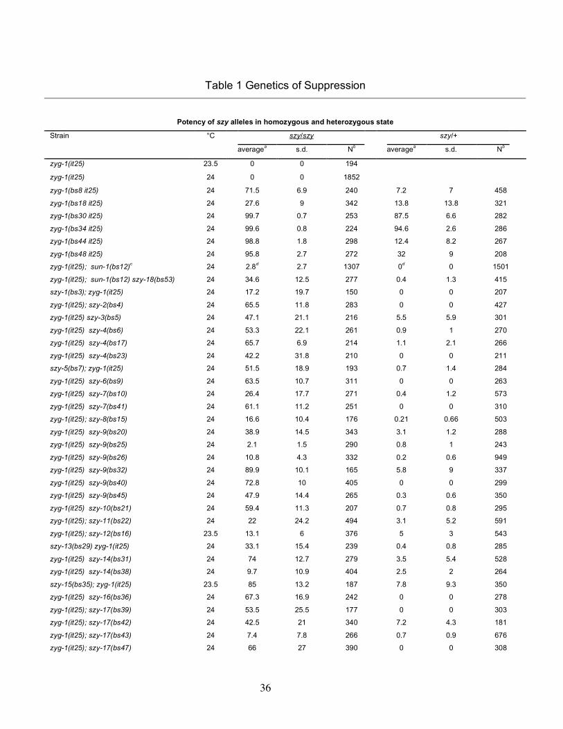
36
Table 1 Genetics of Suppression
Potency of szy alleles in homozygous and heterozygous state
°C szy/szy szy/+ Strain
averagea s.d. Nb averagea s.d. Nb
zyg-1(it25) 23.5 0 0 194
zyg-1(it25) 24 0 0 1852
zyg-1(bs8 it25) 24 71.5 6.9 240 7.2 7 458
zyg-1(bs18 it25) 24 27.6 9 342 13.8 13.8 321
zyg-1(bs30 it25) 24 99.7 0.7 253 87.5 6.6 282
zyg-1(bs34 it25) 24 99.6 0.8 224 94.6 2.6 286
zyg-1(bs44 it25) 24 98.8 1.8 298 12.4 8.2 267
zyg-1(bs48 it25) 24 95.8 2.7 272 32 9 208
zyg-1(it25); sun-1(bs12)c 24 2.8d 2.7 1307 0d 0 1501
zyg-1(it25); sun-1(bs12) szy-18(bs53) 24 34.6 12.5 277 0.4 1.3 415
szy-1(bs3); zyg-1(it25) 24 17.2 19.7 150 0 0 207
zyg-1(it25); szy-2(bs4) 24 65.5 11.8 283 0 0 427
zyg-1(it25) szy-3(bs5) 24 47.1 21.1 216 5.5 5.9 301
zyg-1(it25) szy-4(bs6) 24 53.3 22.1 261 0.9 1 270
zyg-1(it25) szy-4(bs17) 24 65.7 6.9 214 1.1 2.1 266
zyg-1(it25) szy-4(bs23) 24 42.2 31.8 210 0 0 211
szy-5(bs7); zyg-1(it25) 24 51.5 18.9 193 0.7 1.4 284
zyg-1(it25) szy-6(bs9) 24 63.5 10.7 311 0 0 263
zyg-1(it25) szy-7(bs10) 24 26.4 17.7 271 0.4 1.2 573
zyg-1(it25) szy-7(bs41) 24 61.1 11.2 251 0 0 310
zyg-1(it25); szy-8(bs15) 24 16.6 10.4 176 0.21 0.66 503
zyg-1(it25) szy-9(bs20) 24 38.9 14.5 343 3.1 1.2 288
zyg-1(it25) szy-9(bs25) 24 2.1 1.5 290 0.8 1 243
zyg-1(it25) szy-9(bs26) 24 10.8 4.3 332 0.2 0.6 949
zyg-1(it25) szy-9(bs32) 24 89.9 10.1 165 5.8 9 337
zyg-1(it25) szy-9(bs40) 24 72.8 10 405 0 0 299
zyg-1(it25) szy-9(bs45) 24 47.9 14.4 265 0.3 0.6 350
zyg-1(it25) szy-10(bs21) 24 59.4 11.3 207 0.7 0.8 295
zyg-1(it25); szy-11(bs22) 24 22 24.2 494 3.1 5.2 591
zyg-1(it25); szy-12(bs16) 23.5 13.1 6 376 5 3 543
szy-13(bs29) zyg-1(it25) 24 33.1 15.4 239 0.4 0.8 285
zyg-1(it25) szy-14(bs31) 24 74 12.7 279 3.5 5.4 528
zyg-1(it25) szy-14(bs38) 24 9.7 10.9 404 2.5 2 264
szy-15(bs35); zyg-1(it25) 23.5 85 13.2 187 7.8 9.3 350
zyg-1(it25) szy-16(bs36) 24 67.3 16.9 242 0 0 278
zyg-1(it25); szy-17(bs39) 24 53.5 25.5 177 0 0 303
zyg-1(it25); szy-17(bs42) 24 42.5 21 340 7.2 4.3 181
zyg-1(it25); szy-17(bs43) 24 7.4 7.8 266 0.7 0.9 676
zyg-1(it25); szy-17(bs47) 24 66 27 390 0 0 308
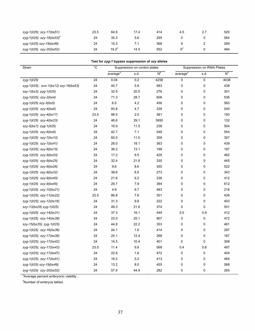
37
zyg-1(it25); szy-17(bs51) 23.5 64.8 17.4 414 4.5 2.7 520
zyg-1(it25); szy-18(bs53)e 24 35.3 5.6 295 0 0 584
zyg-1(it25) szy-19(bs49) 24 15.3 7.1 368 9 2 269
zyg-1(it25) szy-20(bs52) 24 19.2d 14.5 552 0d 0 484
Test for zyg-1 bypass suppression of szy alleles
Strain °C Suppression on control plates Suppression on RNAi Plates
averagea s.d. Nb averagea s.d. Nb
zyg-1(it25) 24 0.04 0.2 4258 0 0 4038
zyg-1(it25); sun-1(bs12) szy-18(bs53) 24 45.7 5.9 583 0 0 438
szy-1(bs3); zyg-1(it25) 24 32.5 20.5 276 0 0 301
zyg-1(it25); szy-2(bs4) 24 71.3 28.7 606 0 0 536
zyg-1(it25) szy-3(bs5) 24 6.0 4.2 456 0 0 565
zyg-1(it25) szy-4(bs6) 24 93.8 4.7 339 0 0 540
zyg-1(it25) szy-4(bs17) 23.5 98.5 2.0 381 0 0 193
zyg-1(it25) szy-4(bs23) 24 46.6 29.1 3950 0 0 132
szy-5(bs7); zyg-1(it25) 24 16.9 11.5 238 0 0 504
zyg-1(it25) szy-6(bs9) 24 42.7 7.1 548 0 0 554
zyg-1(it25) szy-7(bs10) 24 60.0 11.0 358 0 0 307
zyg-1(it25) szy-7(bs41) 24 26.0 18.1 363 0 0 439
zyg-1(it25); szy-8(bs15) 24 36.2 13.1 198 0 0 197
zyg-1(it25) szy-9(bs20) 24 17.2 9.5 429 0 0 482
zyg-1(it25) szy-9(bs25) 24 32.4 21.8 330 0 0 445
zyg-1(it25) szy-9(bs26) 24 9.6 8.6 355 0 0 522
zyg-1(it25) szy-9(bs32) 24 38.6 8.5 273 0 0 343
zyg-1(it25) szy-9(bs40) 24 21.6 6.2 236 0 0 412
zyg-1(it25) szy-9(bs45) 24 29.7 7.9 384 0 0 612
zyg-1(it25) szy-10(bs21) 24 4.9 6.7 483 0 0 218
zyg-1(it25); szy-11(bs22) 23.5 86.8 7.6 301 0 0 436
zyg-1(it25); szy-12(bs16) 24 31.3 9.8 222 0 0 403
szy-13(bs29) zyg-1(it25) 24 49.3 21.6 374 0 0 501
zyg-1(it25) szy-14(bs31) 24 37.3 16.1 449 0.5 0.9 412
zyg-1(it25) szy-14(bs38) 24 23.0 25.1 907 0 0 472
szy-15(bs35); zyg-1(it25) 24 44.8 22.2 353 0 0 481
zyg-1(it25) szy-16(bs36) 24 24.1 1.6 414 0 0 297
zyg-1(it25); szy-17(bs39) 24 24.1 12.4 268 0 0 187
zyg-1(it25); szy-17(bs42) 24 14.5 10.4 401 0 0 308
zyg-1(it25); szy-17(bs43) 23.5 11.4 9.9 568 0.4 0.8 497
zyg-1(it25); szy-17(bs47) 24 22.8 1.8 472 0 0 404
zyg-1(it25); szy-17(bs51) 24 18.2 5.2 413 0 0 484
zyg-1(it25) szy-19(bs49) 24 13.2 8.0 455 0 0 588
zyg-1(it25) szy-20(bs52) 24 57.9 44.9 282 0 0 265 aAverage percent embryonic viability . bNumber of embryos tallied.

38
cComplete genotype of strain tested was zyg-1(it25); sun-1(bs12) szy-18(bs53) +/sun-1(bs12) + unc-76(e911). The congenic strain
zyg-1(it25); sun-1(bs12) szy-18(bs53) unc- 76(e911)/+ + + served as a negative control and produced 0 + 0 viable progeny (n=
1501). dMeasurement made 24-48 hours after shift to restrictive temperature. All other values reported in the upper section of this table
were measured during the first 24 hours after the shift to restrictive temperature. eComplete genotype of strain tested was zyg-1(it25); sma-1(e30) + szy-18(bs53)/+ sun-1(bs12) szy-18(bs53). The strain zyg-
1(it25); sun-1(bs12) szy-18(bs53) unc-76(e911)/+ + + served as a negative control and produced 0 + 0 viable progeny (n= 1501).
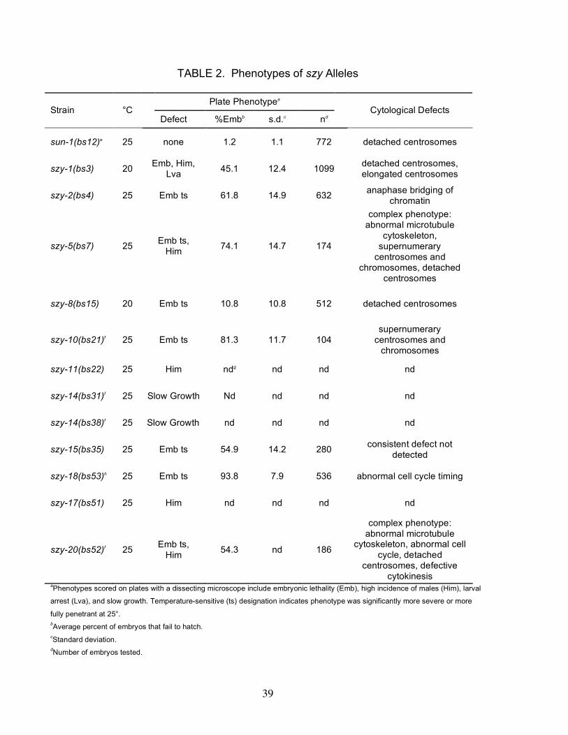
39
TABLE 2. Phenotypes of szy Alleles
Plate Phenotypea Strain °C
Defect %Embb s.d.c nd Cytological Defects
sun-1(bs12)e 25 none 1.2 1.1 772 detached centrosomes
szy-1(bs3) 20 Emb, Him, Lva 45.1 12.4 1099 detached centrosomes,
elongated centrosomes
szy-2(bs4) 25 Emb ts 61.8 14.9 632 anaphase bridging of chromatin
szy-5(bs7) 25 Emb ts, Him 74.1 14.7 174
complex phenotype: abnormal microtubule
cytoskeleton, supernumerary
centrosomes and chromosomes, detached
centrosomes
szy-8(bs15) 20 Emb ts 10.8 10.8 512 detached centrosomes
szy-10(bs21)f 25 Emb ts 81.3 11.7 104 supernumerary
centrosomes and chromosomes
szy-11(bs22) 25 Him ndg nd nd nd
szy-14(bs31)f 25 Slow Growth Nd nd nd nd
szy-14(bs38)f 25 Slow Growth nd nd nd nd
szy-15(bs35) 25 Emb ts 54.9 14.2 280 consistent defect not detected
szy-18(bs53)h 25 Emb ts 93.8 7.9 536 abnormal cell cycle timing
szy-17(bs51) 25 Him nd nd nd nd
szy-20(bs52)f 25 Emb ts, Him 54.3 nd 186
complex phenotype: abnormal microtubule
cytoskeleton, abnormal cell cycle, detached
centrosomes, defective cytokinesis
aPhenotypes scored on plates with a dissecting microscope include embryonic lethality (Emb), high incidence of males (Him), larval
arrest (Lva), and slow growth. Temperature-sensitive (ts) designation indicates phenotype was significantly more severe or more
fully penetrant at 25°. bAverage percent of embryos that fail to hatch. cStandard deviation. dNumber of embryos tested.

40
eComplete strain genotype: zyg-1(it25)/+; sun-1(bs12) + unc-76(e911)/sun-1(bs12) szy-18(bs53) +. fAll contained the dpy-10(e128) marker gnd= not done hComplete strain genotype: zyg-1(it25)/+; sma-1(e30) + szy-18(bs53)/+ sun-1(bs12) szy-18(bs53).
41
Figure Legends Figure 1.—Suppressor Screen. (A) To identify mutations that restore centrosome
duplication and viability to a strain compromised for zyg-1 function, animals carrying the
temperature-sensitive mutation zyg-1(it25) were treated with EMS. At the permissive
temperature, EMS-induced germ-line szy mutations are transmitted to the F1 generation
in the heterozygous state (szy/+) and to the F2 generation in the homozygous state
(szy/szy). To identify szy-bearing individuals, F2 embryos were isolated, allowed to
complete larval development at permissive temperature, and then shifted to restrictive
temperature for three to six weeks. szy-bearing lines give rise to multiple generations
over this time period while all other individuals die off. The columns of green arrows
indicate that individual pools of mutagenized lines were processed in parallel. To
ensure independence, only one suppressor was retained from each pool. (B) Percent
embryonic viability of a zyg-1(it25) strain as a function of temperature. Each data point
represents the average percent viability among the offspring of four to five individuals
shifted as L4 larvae to the indicated temperature for 24 hours. The vertical bars indicate
the standard deviation. Note that the largest standard deviations are observed at the
intermediate temperatures of 21-23°C. (C) Potency of szy alleles. Each szy allele in
the homozygous (red) or heterozygous (blue) state was assayed for suppression of zyg-
1(it25) embryonic lethality. Shown is the average percent of viable offspring. Numerical
values including standard deviations and the number of embryos scored are listed in
Table 1 along with assay conditions and complete strain genotypes
Figure 2.—Genetic map showing location of szy genes. Note that the relative ordering
of some szy genes within the dpy-10-unc-4 interval is not known with certainty.
Figure 3.— Intragenic zyg-1 suppressors. (A) Schematic of the ZYG-1 protein showing
the positions of the zyg-1(it25) and zyg-1(or409) mutations and the six intragenic
suppressors. Five suppressor mutations are within the coding region and the
42
presumptive amino acid substitutions are indicated. The bs30 mutation is a G-to-A
transition 3 bp upstream of the initiator methionine codon. (B) Amino-acid sequence
alignment of the non-kinase portions of ZYG-1 from C. elegans and C. briggsae.
Identical and similar amino acid residues are shaded black and gray respectively. Solid
dots indicate the positions of the four known loss-of-function mutations. Open dots
indicated the positions of the five suppressor mutations that produce an amino acid
substitution.
Figure 4.— Centrosome duplication is restored in zyg-1(it25); szy mutants. (A, B)
Images of wild-type, zyg-1(it25) and zyg-1(it25); szy double mutants stained for tubulin
(red), DNA (blue), and ZYG-1(green). (A) Embryos are in anaphase (left column) or
telophase (right column). Insets show ZYG-1 staining at spindle poles. Note that at
telophase zyg-1(it25) embryos have only a single dot of ZYG-1 at each pole while all
other strains clearly have two, indicating successful centrosome duplication. Bar, 10
µm. (B) Microtubule-organizing activity is similar during the second cell cycle in the wild
type and zyg-1(it25); szy double mutants. Shown are images of wild-type, zyg-1(it25),
and zyg-1(it25); szy embryos at early (left column), mid (center column) and late (right
column) points of the second cell cycle. In wild-type and zyg-1(it25); szy double mutants
microtubule-organizing centers appear similar in size, number, and position indicating
that the normal process of centrosome duplication has been restored. Bar, 10 µm. (C)
Quantification of centrosome duplication in zyg-1(it25) and zyg-1(it25); szy double
mutants. Centrosome duplication was assessed for each strain by scoring two-cell
stage blastomeres in late prophase for the presence of two centrosomes. Blue columns
indicate the percent of successful duplication events. The actual value is shown above
the column with the number of duplication events scored. For comparison, the level of
embryonic viability of each strain is shown in yellow.
Figure 5.—szy genes are required for proper cell division. Confocal (A-I, K & L) or DIC
(J) images of szy mutants. Confocal images are maximal intensity projections of
multiple optical sections of embryos/gonads stained for tubulin (red) and/or DNA (blue).
43
Some embryos are also stained for centrosomes (green) with antibodies to SPD-2 (A,
D, F-I, K, & L). (A) A wild-type embryo at the beginning of the two-cell stage. (B) A szy-
5(bs7) one-cell embryo and (C) proximal gonad showing the tubulin aggregation
phenotype. (D) A one-cell szy-5(bs7) embryo and (E) a one-cell szy-20(bs52) embryo
each with excess centrosomes and DNA. (F) A one-cell szy-20(bs52) embryo with
cytoplasmic aggregates of SPD-2. Arrows indicate centrosomes. (G) A multinucleated
szy-10(bs21) embryo with multiple centrosomes. (H) A two-cell szy-1(bs3) embryo with
an elongated centrosome (arrow). (I) A szy-2(bs4) embryo with anaphase bridging of
chromatin. Inset shows a magnified view of chromatin. (J) A szy-18(bs53) embryo with
an unusual three-cell configuration resulting from delayed division of the P1 blastomere
(asterisk). (K) Two-cell sun-1(bs12) and (L) szy-8(bs15) embryos displaying detached
centrosomes (arrowheads). Bars in A and C are 10 µm.
Figure 6.—sun-1(RNAi), but not zyg-12(RNAi), suppresses the centrosome duplication
defect in zyg-1(it25) mutants. (A) zyg-1(it25) and zyg-1(it25); sun-1(RNAi) embryos
were stained for microtubules (red), DNA (blue), and ZYG-1(green). The zyg-1(it25);
sun-1(RNAi) embryo exhibits evidence of centrosome duplication as well as loss of
contact between a nucleus and centrosome (arrow). Insets show 3-fold magnified
views of ZYG-1 staining at centrosomes. Bar, 10 µm. (B) Spindle assembly in zyg-1
embryos depleted of SUN-1 or ZYG-12. Embryos were isolated from mothers grown at
24° and early development analyzed by 4D-DIC imaging at the same temperature. In
columns two through seven, the number of spindle assembly events resulting in either a
monopolar (•) or bipolar (•–•) spindle is tallied for the first three cell cycles. Note that not
all embryos were scored for the third cell cycle. In the last column, the percentage of
bipolar spindles formed, indicating successful centrosome duplication, is shown.
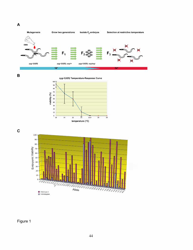
44
Figure 1
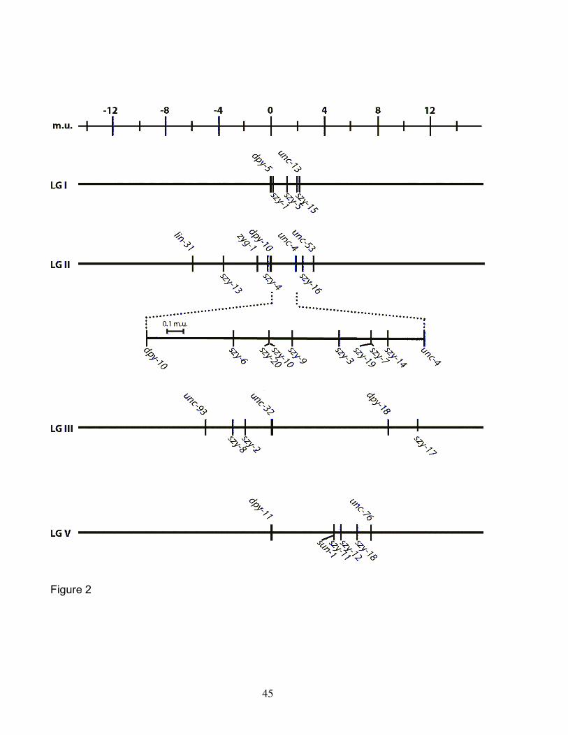
45
Figure 2
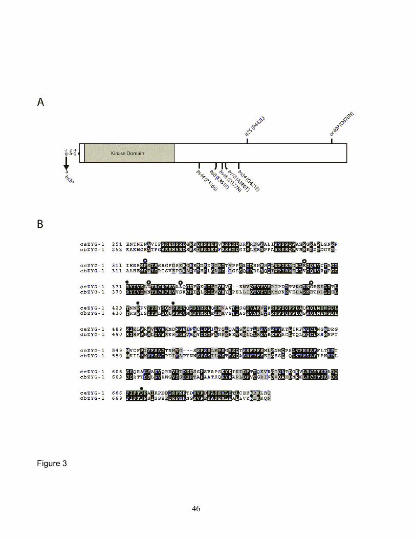
46
Figure 3
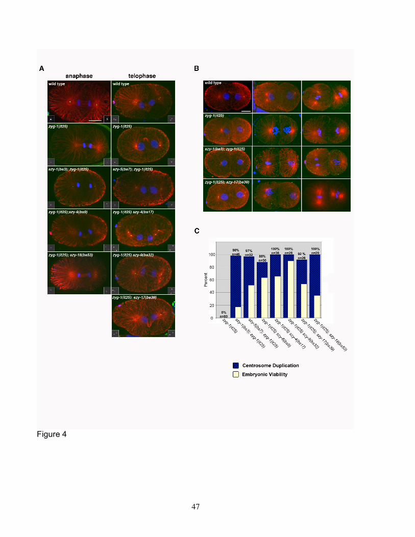
47
Figure 4
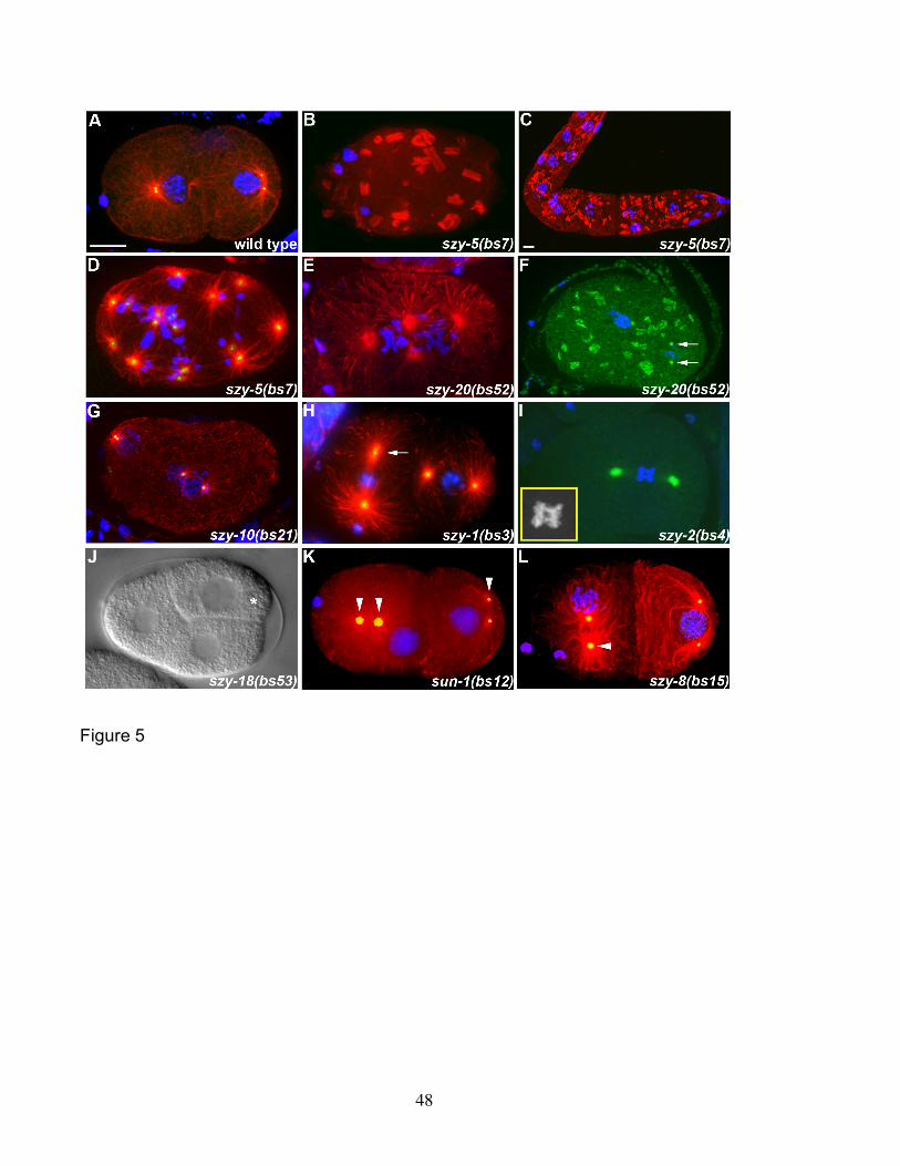
48
Figure 5
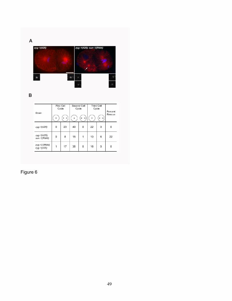
49
Figure 6