Suppression of retinal degeneration in Drosophila by ... of retinal degeneration in Drosophila by...
6
Suppression of retinal degeneration in Drosophila by stimulation of ER-associated degradation Min-Ji Kang and Hyung Don Ryoo 1 Department of Cell Biology, New York University School of Medicine, 550 First Avenue, New York, NY 10016 Edited by David D. Sabatini, New York University School of Medicine, New York, NY, and approved August 18, 2009 (received for review May 19, 2009) Mutations in the rhodopsin gene that disrupt the encoded pro- tein’s folding properties are a major cause of autosomal dominant retinitis pigmentosa (ADRP). This disease is faithfully modeled in Drosophila where similar mutations in the ninaE gene, encoding rhodopsin-1 (Rh-1), cause ER stress and dominantly trigger age- related retinal degeneration. In addition, mutant flies bearing certain ninaE alleles have dramatically reduced Rh-1 protein levels, but the underlying mechanism for this reduction and significance of its contribution to the ADRP phenotype remains unclear. To address this question, we specifically analyzed the role of Dro- sophila genes homologous to the known yeast and animal regu- lators of the ER-associated degradation (ERAD) pathway, a process that reduces levels of misfolded proteins in the ER through pro- teasomal degradation. We found that loss-of-function of these putative ERAD factors resulted in increased levels of Rh-1 in ninaE mutant flies. Conversely, in an ER stress assay where mutant or wild-type Rh-1 were overexpressed in developing imaginal discs beyond the ER protein folding capacity of those cells, co-expression of certain ERAD factors was sufficient to reduce Rh-1 protein levels and to completely suppress ER stress reporter activation. Signifi- cantly, those ERAD factors that specifically reduced misfolded Rh-1 in the imaginal disc assay also delayed age-related retinal degen- eration caused by an endogenous ninaE allele, indicating that ERAD acts as a protective mechanism against retinal degeneration in the Drosophila model for ADRP. These results suggest that manipulation of ERAD may serve as a powerful therapeutic strat- egy against a number of diseases associated with ER stress. apoptosis endoplasmic reticulum rhodopsin unfolded protein response T he endoplasmic reticulum (ER) is an organelle in which membrane and secretory proteins are synthesized and folded into stable conformations. Reflecting the ER’s essential role in protein folding, mutations that either cause misfolding of pro- teins synthesized in the ER or interfere with ER quality control mechanisms are frequent causes of degenerative diseases (1, 2). Among the diseases associated with protein misfolding in the ER are class II autosomal dominant retinitis pigmentosa (ADRP), in which dominant rhodopsin mutations trigger age- related retinal degeneration and blindness (3, 4). The Drosophila genome has several rhodopsin genes, including ninaE that encodes the rhodopsin-1 protein (Rh-1) (5, 6). Exhibiting a striking similarity with human rhodopsin mutants, a number of ninaE alleles dominantly cause age-related retinal degeneration in Drosophila (7, 8). The amino acid substitutions that result from these ninaE mutations are similar to those human rhodopsin mutant proteins that fail to fold properly in cultured cells and underlie ADRP (8). In fact, Drosophila photoreceptors bearing these ninaE mutant alleles activate a specific transcriptional response that helps reduce misfolded proteins in this ER, widely referred to as the unfolded protein response (UPR) (9). More- over, reducing the UPR through a mutation in a key component of this pathway, xbp1, aggravates the course of retinal degener- ation in the Drosophila model (9). Similar observations have been made with the mammalian rhodopsin mutants (10), indi- cating that the pathology underlying ADRP is conserved be- tween Drosophila and mammals. Among a few unexplained features associated with these domi- nant ninaE alleles is a severe reduction of overall Rh-1 protein levels in the afflicted photoreceptors. While heterozygotes with a null ninaE allele have roughly half of the normal Rh-1 protein levels, many disease-causing rhodopsin alleles of humans and Drosophila lead to significantly lower rhodopsin levels under otherwise similar conditions (7, 8). However, whether such a reduction in Rh-1 levels has a functional significance in the retinal degeneration remains unclear. It is possible that the reduction of overall Rh-1 levels may have a beneficial consequence in the ADRP model, as it may protect against toxicity associated with aggregates in the ER. Alternatively, excessive Rh-1 reduction may accelerate retinal degeneration in ADRP, as insufficient levels of Rh-1 protein at the light-sensing compartment can compromise photoreceptor integ- rity and survival (11). To understand the mechanism and function of Rh-1 reduction in this disease model, we investigated the role of putative regulators of Drosophila ER-associated degradation (ERAD). ERAD is reg- ulated by a multiprotein complex that includes proteins involved in the recognition, retrotranslocation, and ubiquitination of misfolded proteins in the ER (12). Studies using Saccharomyces cerevisiae have suggested the existence of three major ERAD subpathways, de- fined by the subcellular location of the lesion that causes protein misfolding (13–15). A protein that misfolds in the ER lumen is thought to be degraded through the ERAD-L pathway. On the other hand, proteins with lesions in transmembrane domains are thought to be substrates of the ERAD-M pathway. Proteins with lesions on the cytoplasmic side of the ER are processed by the ERAD-C pathway. In yeast, each subpathway requires a distinct set of ERAD complex subunits. For example, the two yeast manno- sidases that recognize misfolded glycoproteins, Htm1p, and mem- brane protein complex containing Usa1p and Derlin, appear ded- icated to the ERAD-L pathway, since ERAD-M substrates bypass the requirement of those subunits (13–16). Whether ERAD reg- ulation in metazoans follows the same rules remains unknown. Using genetic tools of Drosophila, here we demonstrate that ERAD regulators help reduce stress and retinal degeneration caused by mutant Rh-1 in the Drosophila ADRP model and serve as a protective mechanism. Specifically, we show that disruption of the ERAD pathway leads to an increase in Rh-1 protein levels in the Drosophila model for ADRP. Conversely, overexpression of certain subunits of the ERAD machinery was sufficient to reduce the levels of ER stress-causing Rh-1 proteins. Individual subunits showed distinct specificity toward their substrates that were some- times inconsistent with what is expected from the ERAD subpath- ways defined in yeast. Most significantly, certain factors were able to suppress late-onset retinal degeneration in a Drosophila model Author contributions: M.-J.K. and H.D.R. designed research; M.-J.K. performed research; M.-J.K. and H.D.R. analyzed data; and M.-J.K. and H.D.R. wrote the paper. The authors declare no conflict of interest. This article is a PNAS Direct Submission. Freely available online through the PNAS open access option. 1 To whom correspondence should be addressed. E-mail: [email protected]. This article contains supporting information online at www.pnas.org/cgi/content/full/ 0905566106/DCSupplemental. www.pnas.orgcgidoi10.1073pnas.0905566106 PNAS October 6, 2009 vol. 106 no. 40 17043–17048 CELL BIOLOGY
Transcript of Suppression of retinal degeneration in Drosophila by ... of retinal degeneration in Drosophila by...

Suppression of retinal degeneration in Drosophilaby stimulation of ER-associated degradationMin-Ji Kang and Hyung Don Ryoo1
Department of Cell Biology, New York University School of Medicine, 550 First Avenue, New York, NY 10016
Edited by David D. Sabatini, New York University School of Medicine, New York, NY, and approved August 18, 2009 (received for review May 19, 2009)
Mutations in the rhodopsin gene that disrupt the encoded pro-tein’s folding properties are a major cause of autosomal dominantretinitis pigmentosa (ADRP). This disease is faithfully modeled inDrosophila where similar mutations in the ninaE gene, encodingrhodopsin-1 (Rh-1), cause ER stress and dominantly trigger age-related retinal degeneration. In addition, mutant flies bearingcertain ninaE alleles have dramatically reduced Rh-1 protein levels,but the underlying mechanism for this reduction and significanceof its contribution to the ADRP phenotype remains unclear. Toaddress this question, we specifically analyzed the role of Dro-sophila genes homologous to the known yeast and animal regu-lators of the ER-associated degradation (ERAD) pathway, a processthat reduces levels of misfolded proteins in the ER through pro-teasomal degradation. We found that loss-of-function of theseputative ERAD factors resulted in increased levels of Rh-1 in ninaEmutant flies. Conversely, in an ER stress assay where mutant orwild-type Rh-1 were overexpressed in developing imaginal discsbeyond the ER protein folding capacity of those cells, co-expressionof certain ERAD factors was sufficient to reduce Rh-1 protein levelsand to completely suppress ER stress reporter activation. Signifi-cantly, those ERAD factors that specifically reduced misfolded Rh-1in the imaginal disc assay also delayed age-related retinal degen-eration caused by an endogenous ninaE allele, indicating thatERAD acts as a protective mechanism against retinal degenerationin the Drosophila model for ADRP. These results suggest thatmanipulation of ERAD may serve as a powerful therapeutic strat-egy against a number of diseases associated with ER stress.
apoptosis � endoplasmic reticulum � rhodopsin � unfolded protein response
The endoplasmic reticulum (ER) is an organelle in whichmembrane and secretory proteins are synthesized and folded
into stable conformations. Reflecting the ER’s essential role inprotein folding, mutations that either cause misfolding of pro-teins synthesized in the ER or interfere with ER quality controlmechanisms are frequent causes of degenerative diseases (1, 2).
Among the diseases associated with protein misfolding in theER are class II autosomal dominant retinitis pigmentosa(ADRP), in which dominant rhodopsin mutations trigger age-related retinal degeneration and blindness (3, 4). The Drosophilagenome has several rhodopsin genes, including ninaE thatencodes the rhodopsin-1 protein (Rh-1) (5, 6). Exhibiting astriking similarity with human rhodopsin mutants, a number ofninaE alleles dominantly cause age-related retinal degenerationin Drosophila (7, 8). The amino acid substitutions that result fromthese ninaE mutations are similar to those human rhodopsinmutant proteins that fail to fold properly in cultured cells andunderlie ADRP (8). In fact, Drosophila photoreceptors bearingthese ninaE mutant alleles activate a specific transcriptionalresponse that helps reduce misfolded proteins in this ER, widelyreferred to as the unfolded protein response (UPR) (9). More-over, reducing the UPR through a mutation in a key componentof this pathway, xbp1, aggravates the course of retinal degener-ation in the Drosophila model (9). Similar observations havebeen made with the mammalian rhodopsin mutants (10), indi-cating that the pathology underlying ADRP is conserved be-tween Drosophila and mammals.
Among a few unexplained features associated with these domi-nant ninaE alleles is a severe reduction of overall Rh-1 protein levelsin the afflicted photoreceptors. While heterozygotes with a nullninaE allele have roughly half of the normal Rh-1 protein levels,many disease-causing rhodopsin alleles of humans and Drosophilalead to significantly lower rhodopsin levels under otherwise similarconditions (7, 8). However, whether such a reduction in Rh-1 levelshas a functional significance in the retinal degeneration remainsunclear. It is possible that the reduction of overall Rh-1 levels mayhave a beneficial consequence in the ADRP model, as it mayprotect against toxicity associated with aggregates in the ER.Alternatively, excessive Rh-1 reduction may accelerate retinaldegeneration in ADRP, as insufficient levels of Rh-1 protein at thelight-sensing compartment can compromise photoreceptor integ-rity and survival (11).
To understand the mechanism and function of Rh-1 reduction inthis disease model, we investigated the role of putative regulatorsof Drosophila ER-associated degradation (ERAD). ERAD is reg-ulated by a multiprotein complex that includes proteins involved inthe recognition, retrotranslocation, and ubiquitination of misfoldedproteins in the ER (12). Studies using Saccharomyces cerevisiae havesuggested the existence of three major ERAD subpathways, de-fined by the subcellular location of the lesion that causes proteinmisfolding (13–15). A protein that misfolds in the ER lumen isthought to be degraded through the ERAD-L pathway. On theother hand, proteins with lesions in transmembrane domains arethought to be substrates of the ERAD-M pathway. Proteins withlesions on the cytoplasmic side of the ER are processed by theERAD-C pathway. In yeast, each subpathway requires a distinct setof ERAD complex subunits. For example, the two yeast manno-sidases that recognize misfolded glycoproteins, Htm1p, and mem-brane protein complex containing Usa1p and Derlin, appear ded-icated to the ERAD-L pathway, since ERAD-M substrates bypassthe requirement of those subunits (13–16). Whether ERAD reg-ulation in metazoans follows the same rules remains unknown.
Using genetic tools of Drosophila, here we demonstrate thatERAD regulators help reduce stress and retinal degenerationcaused by mutant Rh-1 in the Drosophila ADRP model and serveas a protective mechanism. Specifically, we show that disruption ofthe ERAD pathway leads to an increase in Rh-1 protein levels inthe Drosophila model for ADRP. Conversely, overexpression ofcertain subunits of the ERAD machinery was sufficient to reducethe levels of ER stress-causing Rh-1 proteins. Individual subunitsshowed distinct specificity toward their substrates that were some-times inconsistent with what is expected from the ERAD subpath-ways defined in yeast. Most significantly, certain factors were ableto suppress late-onset retinal degeneration in a Drosophila model
Author contributions: M.-J.K. and H.D.R. designed research; M.-J.K. performed research;M.-J.K. and H.D.R. analyzed data; and M.-J.K. and H.D.R. wrote the paper.
The authors declare no conflict of interest.
This article is a PNAS Direct Submission.
Freely available online through the PNAS open access option.
1To whom correspondence should be addressed. E-mail: [email protected].
This article contains supporting information online at www.pnas.org/cgi/content/full/0905566106/DCSupplemental.
www.pnas.org�cgi�doi�10.1073�pnas.0905566106 PNAS � October 6, 2009 � vol. 106 � no. 40 � 17043–17048
CELL
BIO
LOG
Y

for ADRP. These results suggest that specific ERAD mechanismscan be exploited as a therapeutic strategy against conformationaldiseases.
ResultsLoss of ERAD Components Partially Restores the Level of Rhodopsin-1in the ninaEG69D�/� Retina. To study the mechanism and conse-quences of Rh-1 reduction in the Drosophila model for ADRP, weexamined heterozygous flies bearing a ninaE allele with a Glyresidue in a transmembrane domain replaced by Asp (henceforthreferred to as ninaEG69D). Similar to human autosomal dominantretinitis pigmentosa (ADRP), these flies activate the UPR in theirphotoreceptors, which eventually undergo age-related retinal de-generation (7, 8, 11). To determine the role of ERAD in thepathogenesis of ADRP, we modulated the levels of Drosophilahomologs of yeast and human ERAD regulator proteins, with aparticular focus on those integral membrane proteins that interactwith each other (12). One of these Drosophila gene products, knownas septin interacting protein 3, was previously identified in a yeasttwo-hybrid assay (17). We noticed that this Drosophila gene is mosthomologous to yeast and human Hrd1, a ubiquitin ligase of theERAD pathway (18–20), and we will henceforth refer to it as Hrd1.As in humans, Drosophila Hrd1 has six predicted transmembranedomains and a RING domain on the cytoplasmic side (Fig. 1A).Other genes we examined include a candidate retrotranslocation
channel component, Derlin-1 (CG10908), and its associated pro-teins Hrd3 (CG10221) and Herp (CG14536) (Fig. 1A). When theretina of heterozygotic flies bearing the ninaEG69D allele wereimmunolabeled with anti-Rh-1 antibody, we found that the averageanti-Rh-1 labeling intensity was reduced to approximately 28% ofwild-type levels (Fig. 1C), consistent with previous reports (7, 8, 21,22) that have documented the reduction of both wild-type andmutant Rh-1 proteins under these conditions. To determinewhether this reduction is due, at least in part, to enhanced ERAD,we knocked down Hrd1 and Herp in the retina of ninaEG69D�/�flies using inverted repeat transgenic constructs that are designedto produce double stranded (ds) RNA (see Materials and Meth-ods). Knock down of Drosophila Hrd1 in this background restoredthe overall Rh-1 levels to an extent that was statistically indistin-guishable from that of wild-type flies (Fig. 1D; n � 3, P � 0.6).Although to a lesser degree, Herp knock down also restored Rh-1levels to a significant degree (Fig. 1E; n � 3, P � 0.0009). Tocomplement these immunohistochemical analysis, we examinedRh-1 levels through Western blots under similar genetic conditions.The total amount of Rh-1 in ninaEG69D�/� flies estimated throughWestern blots was 7% of the wild-type levels, which was lower thanthat assessed through immunohistochemistry. This is most likelybecause insoluble membrane proteins are frequently lost duringSDS/PAGE sample preparation. However, the changes of Rh-1levels in response to ERAD inhibition was consistent with that
Fig. 1. Loss of ERAD regulators partially restores thelevel of Rh-1 in the retina of ninaEG69D�/� flies. (A)Schematic diagrams of the predicted membrane topol-ogy and domain structure of ERAD components throughEMBnet-CH. (B–E) Horizontal retina cryosections stainedwith anti-Rh-1 antibody in 1- to 3-day-old flies. (B) Acontrol ninaE �/� retina. ninaEG69D�/� retina (C) showlow Rh-1 labeling, while Rh-1 levels recover when Hrd1(D) or Herp (E) are knocked down in this genetic back-ground. In all RNAi experiments, dicer2 (drc2) were co-expressed to enhance the efficiency of RNAi knockdown.(F) The average intensity of anti-Rh-1 labeling (n � 3), asquantifiedthroughtheImageJ.Errorbars,�SEM.(GandI) A Western blot of Drosophila adult head extracts of theindicated genotypes probed for Rh-1 (upper gel) andanti-profilin (as a loading control; lower gel). (H and J)Quantification of the average normalized Rh-1 bandintensity as shown in (G and I) (average of n � 3; Der-1,average of n � 2), with the value from wild-type ofRh-1�/� heads extracts set at 100%. (K) The structure ofthe Herp genomic locus. In the HerpG13463, an EP-elementis inserted within its protein-coding region. Genotypes:Rh1-Gal4;;ninaE�/� (B and B�), Rh1-Gal4;;ninaEG69D/� (Cand C�), Rh1-Gal4;UAS-Dcr2/�;UAS-Hrd1-IR/ninaEG69D (Dand D�), Rh1-Gal4;UAS-Dcr2/�;UAS-Herp-IR/ninaEG69D
(E and E�), (G, lanes 1 and 5) Rh1-Gal4;;ninaE�/�, (G,lanes 2 and 6) Rh1-Gal4;;ninaEG69D/�, (G, lane 3) Rh1-Gal4;UAS-Dcr2/�;UAS-Hrd1-IR/ninaEG69D, (G, lane 4)Rh1-Gal4;UAS-Hrd3-IR/�;UAS-Dcr2/ninaEG69D, (G, lane 7)Rh1-Gal4;UAS-Herp-IR/�;UAS-Dcr2/ninaEG69D, (G, lane8) Rh1-Gal4;UAS-Dcr2/�;UAS-Herp-IR/ninaEG69D, (G, lane9) Rh1-Gal4;UAS-Dcr2/�;UAS-Der-1-IR(#1)/ninaEG69D, (G,lane 10) Rh1-Gal4;UAS-Dcr2/�;UAS-Der-1-IR(#2)/ninaEG69D, (I, lane 1) CantonS, (I, lane 2) ninaEG69D/�,(I, lane 3) HerpG13463/HerpG13463, (I, lane 4)HerpG13463/HerpG13463;ninaEG69D/�.
17044 � www.pnas.org�cgi�doi�10.1073�pnas.0905566106 Kang and Ryoo

observed through immunohistochemistry, with the knock down ofHrd1 or other ERAD regulators partially restoring the Rh-1 levelsin ninaEG69D�/� flies (Fig. 1 G and H). The results were furthervalidated using a Herp mutant allele, HerpG13463, which has an EP-element inserted within its protein coding sequence (Fig. 1K).While the loss-of Herp did not affect the total Rh-1 protein level inthe ninaE� background, it partially restored total Rh-1 levels in theninaEG69D�/� background (p � 0.0003) (Fig. 1 I and J). There wasno statistically significant difference in the steady state Rh-1 levelsbetween the Herp knock down and HerpG13463�/� flies (P � 0.45and 0.1 for comparisons with two independent inverted repeatlines). While these results can be most simply interpreted asevidence of misfolded Rh-1 proteins being degraded by ERAD, analternative scenario is also possible where defective ERAD mayincrease Rh-1 levels by inducing ER chaperons and enhance Rh-1folding. In fact, blocking ERAD in yeast is known to stimulate UPRsignaling and induce ER chaperones (23). However, such anindirect model appears unlikely in the Drosophila retina, as we findthat the Herp mutant flies did not induce the expression of heatshock cognate protein 3 (hsc3), a major ER chaperon that ishomologous to BiP and a well established target of UPR (Fig. S1).These observations support the idea that the examined DrosophilaERAD factors stimulate the degradation of misfolded Rh-1 in theADRP model.
ER Stress Caused by Rh-1 Misexpression Is Strongly Suppressed byDrosophila Hrd1. As the retina of ninaEG69D�/� flies contains amixture of mutant and wild-type Rh-1 proteins that are similar insize, it is difficult to distinguish their individual fates throughWestern blots or immunohistochemistry. To examine the behaviorof particular Rh-1 alleles, we expressed mutant or wild-type Rh-1in the larval eye imaginal discs, a developing tissue that does not yetexpress Rh-1. To assess the level of ER stress caused by such Rh-1misexpression, we used xbp1-EGFP as a reporter designed to detectthe unconventional mRNA splicing of xbp1, which is a key signalingevent for the initiation of UPR. This reporter allows EGFP to beexpressed in frame, only when a 23-nt intron in xbp1 is spliced outby the ER stress-activated endonuclease ire-1, thereby marking withgreen fluorescence those cells suffering significant ER stress (9).While this marker is not activated in response to proteins thatmisfold in the cytoplasm (9), xbp1-EGFP fluorescence was detectedin all examined eye discs in which Rh-1G69D or Rh-1WT wereexpressed (Fig. 2 F and H, n � 10). The observation that Rh-1WT
also activates the UPR in these cells was unexpected, and may bedue to the limited ER capacity of the imaginal disc cells to fold andexport high levels of Rh-1.
The establishment of the above assay allowed us to ask whetheroverexpression of any of the putative ERAD regulator genes could
suppress the ER stress caused by Rh-1 misexpression. We foundthat this was the case, with Drosophila Hrd1 being one the strongestsuppressors among those examined. In larval eye discs, DrosophilaHrd1 lowered Rh-1WT and Rh-1G69D protein levels when these wereco-expressed through the eye-specific gmr-Gal4 driver (Fig. 2 A–D).The effect of Hrd1 on the misexpressed Rh-1 proteins was furthervalidated through an alternative gene expression system that em-ploys tubulin-Gal4 flip-out technology, which generates mosaicclones expressing genes of choice through the tubulin promoter (seeMaterials and Methods). When mosaic clones expressing Drosoph-ila Hrd1 were generated within the population of Rh-1G69D-expressing cells, those clones had distinctively lower levels ofRh-1G69D protein, compared to the neighboring cells that did notexpress Hrd1 (Fig. S2 B and B�; n � 8). Hrd1 appeared to lowermutant and wild-type Rh-1 to levels that virtually eliminated ERstress itself, as evidenced by a near complete suppression of thexbp1-EGFP marker, which was otherwise activated in response tothe misexpression in either wild type or mutant Rh-1 in eye discs(Fig. 2 E–I; n � 10). As an indirect measure of stress, we alsoexamined markers for apoptosis, an active cell death program thatinvolves, among others, proteolytic cleavage and activation ofcaspases. Labeling of imaginal discs with an antibody that detectssuch caspase cleavage event (24) showed that Drosophila Hrd1co-expression suppressed the excessive apoptosis associated withRh-1WT misexpression (Fig. S2 C and D; n � 8). To determinewhether Hrd1 interacts with either Rh-1WT or Rh-1G69D, weperformed co-immunoprecipitation assays. Myc-tagged Hrd1 wascoexpressed in 293T cells along with HA-tagged Rh-1WT, Rh-1G69D
or a control membrane protein, Drob-1 (25). Hrd1 co-precipitatedwith Rh-1WTand Rh-1G69D, but not with Drob-1 (Fig. 2J), estab-lishing that Hrd1 forms a physical complex with mutant andwild-type Rh-1 and strongly suppresses ER stress caused by theseRh-1 proteins.
Overexpression of Putative ERAD Components in Drosophila. Thefindings just described prompted us to test other Drosophila ho-mologs of the ER associated degradation (ERAD) factors, includ-ing Hrd3, Herp, Derlin-1, and Derlin-2 (CG14899). In addition, wecharacterized the role of Drosophila EDEM homologs, CG3810 andCG5682, which we refer to as EDEM1 and EDEM2, respectively.Specifically, we examined their ability to suppress the rough eyephenotype generated by overexpression of Rh-1WT in larval eyediscs, a condition that causes ER stress (Fig. S2). Consistent withthe results obtained with the xbp1-EGFP assay, Hrd1 overexpres-sion in eye imaginal discs almost completely rescued the rough eyephenotype in adults. Herp overexpression also suppressed the eyephenotype, but to a significantly lesser extent. On the other hand,
Fig. 2. ER stress caused by Rh-1 misex-pression is strongly suppressed by Dro-sophila Hrd1. (A–D) Representative im-ages of eye imaginal discs expressingRh-1WT (A) or Rh-1G69D (C) alone, or to-gether with Hrd1 (B and D). Anti-Rh-1antibody labeling is in red. E–I Hrd1co-expression abolished ER stresscaused by wild-type or mutant Rh-1misexpression, as determined by the ERstress marker, xbp1-EGFP (green).Shown are representative discs express-ing xbp1-EGFP alone (E), or togetherwith indicated genes. (J) Co-immuno-precipitation assays between Hrd1 andRh-1 in 293T cells. Hrd1 was tagged withthe myc epitope, while Rh-1 was tagged with HA. HA-Drob-1 is a membrane protein used as a negative control. [Scale bars, 100 �m (A).] Genotypes:gmr-Gal4/�;UAS-Rh-1WT/� (A), gmr-Gal4/UAS-Hrd1;UAS-Rh-1WT/� (B), gmr-Gal4/�;UAS-Rh-1G69D/� (C), gmr-Gal4/UAS-Hrd1;UAS-Rh-1G69D/� (D), gmr-Gal4/�;UAS-xbp1-EGFP/� (E), gmr-Gal4/UAS-lacZ;UAS-Rh-1WT/UAS-xbp1-EGFP (F), gmr-Gal4/UAS-Hrd1;UAS-Rh-1WT/UAS-xbp1-EGFP (G), gmr-Gal4/UAS-lacZ;UAS- Rh-1G69D/UAS-xbp1-EGFP (H), and gmr-Gal4/UAS-Hrd1;UAS-Rh-1G69D/UAS-xbp1-EGFP (I).
Kang and Ryoo PNAS � October 6, 2009 � vol. 106 � no. 40 � 17045
CELL
BIO
LOG
Y
other examined genes failed to rescue the partial eye ablationcaused by Rh-1WT under similar conditions (Fig. S2).
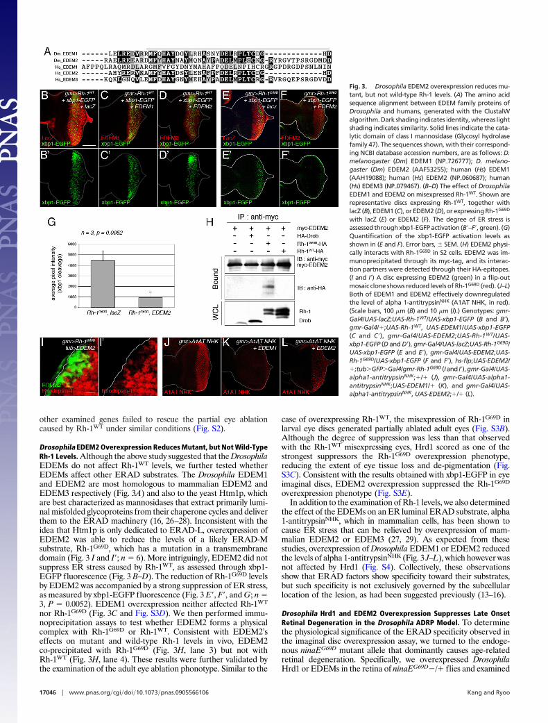
Drosophila EDEM2 Overexpression Reduces Mutant, but Not Wild-TypeRh-1 Levels. Although the above study suggested that the DrosophilaEDEMs do not affect Rh-1WT levels, we further tested whetherEDEMs affect other ERAD substrates. The Drosophila EDEM1and EDEM2 are most homologous to mammalian EDEM2 andEDEM3 respectively (Fig. 3A) and also to the yeast Htm1p, whichare best characterized as mannosidases that extract primarily lumi-nal misfolded glycoproteins from their chaperone cycles and deliverthem to the ERAD machinery (16, 26–28). Inconsistent with theidea that Htm1p is only dedicated to ERAD-L, overexpression ofEDEM2 was able to reduce the levels of a likely ERAD-Msubstrate, Rh-1G69D, which has a mutation in a transmembranedomain (Fig. 3 I and I�; n � 6). More intriguingly, EDEM2 did notsuppress ER stress caused by Rh-1WT, as assessed through xbp1-EGFP fluorescence (Fig. 3 B–D). The reduction of Rh-1G69D levelsby EDEM2 was accompanied by a strong suppression of ER stress,as measured by xbp1-EGFP fluorescence (Fig. 3 E�, F�, and G; n �3, P � 0.0052). EDEM1 overexpression neither affected Rh-1WT
nor Rh-1G69D (Fig. 3C and Fig. S3D). We then performed immu-noprecipitation assays to test whether EDEM2 forms a physicalcomplex with Rh-1G69D or Rh-1WT. Consistent with EDEM2’seffects on mutant and wild-type Rh-1 levels in vivo, EDEM2co-precipitated with Rh-1G69D (Fig. 3H, lane 3) but not withRh-1WT (Fig. 3H, lane 4). These results were further validated bythe examination of the adult eye ablation phonotype. Similar to the
case of overexpressing Rh-1WT, the misexpression of Rh-1G69D inlarval eye discs generated partially ablated adult eyes (Fig. S3B).Although the degree of suppression was less than that observedwith the Rh-1WT misexpressing eyes, Hrd1 scored as one of thestrongest suppressors the Rh-1G69D overexpression phenotype,reducing the extent of eye tissue loss and de-pigmentation (Fig.S3C). Consistent with the results obtained with xbp1-EGFP in eyeimaginal discs, EDEM2 overexpression suppressed the Rh-1G69D
overexpression phenotype (Fig. S3E).In addition to the examination of Rh-1 levels, we also determined
the effect of the EDEMs on an ER luminal ERAD substrate, alpha1-antitrypsinNHK, which in mammalian cells, has been shown tocause ER stress that can be relieved by overexpression of mam-malian EDEM2 or EDEM3 (27, 29). As expected from thesestudies, overexpression of Drosophila EDEM1 or EDEM2 reducedthe levels of alpha 1-antitrypsinNHK (Fig. 3 J–L), which however wasnot affected by Hrd1 (Fig. S4). Collectively, these observationsshow that ERAD factors show specificity toward their substrates,but such specificity is not exclusively governed by the subcellularlocation of the lesion, as had been suggested previously (13–16).
Drosophila Hrd1 and EDEM2 Overexpression Suppresses Late OnsetRetinal Degeneration in the Drosophila ADRP Model. To determinethe physiological significance of the ERAD specificity observed inthe imaginal disc overexpression assay, we turned to the endoge-nous ninaEG69D mutant allele that dominantly causes age-relatedretinal degeneration. Specifically, we overexpressed DrosophilaHrd1 or EDEMs in the retina of ninaEG69D�/� flies and examined
Fig. 3. Drosophila EDEM2 overexpression reduces mu-tant, but not wild-type Rh-1 levels. (A) The amino acidsequence alignment between EDEM family proteins ofDrosophila and humans, generated with the ClustalWalgorithm. Dark shading indicates identity, whereas lightshading indicates similarity. Solid lines indicate the cata-lytic domain of class I mannosidase (Glycosyl hydrolasefamily 47). The sequences shown, with their correspond-ing NCBI database accession numbers, are as follows: D.melanogaster (Dm) EDEM1 (NP�726777); D. melano-gaster (Dm) EDEM2 (AAF53255); human (Hs) EDEM1(AAH19088); human (Hs) EDEM2 (NP�060687); human(Hs) EDEM3 (NP�079467). (B–D) The effect of DrosophilaEDEM1 and EDEM2 on misexpressed Rh-1WT. Shown arerepresentative discs expressing Rh-1WT, together withlacZ (B), EDEM1 (C), or EDEM2 (D), or expressing Rh-1G69D
with lacZ (E) or EDEM2 (F). The degree of ER stress isassessed through xbp1-EGFP activation (B�–F�, green). (G)Quantification of the xbp1-EGFP activation levels asshown in (E and F). Error bars, � SEM. (H) EDEM2 physi-cally interacts with Rh-1G69D in S2 cells. EDEM2 was im-munoprecipitated through its myc-tag, and its interac-tion partners were detected through their HA-epitopes.(I and I�) A disc expressing EDEM2 (green) in a flip-outmosaic clone shows reduced levels of Rh-1G69D (red). (J–L)Both of EDEM1 and EDEM2 effectively downregulatedthe level of alpha 1-antitrypsinNHK (A1AT NHK, in red).(Scale bars, 100 �m (B) and 10 �m (I).) Genotypes: gmr-Gal4/UAS-lacZ;UAS-Rh-1WT/UAS-xbp1-EGFP (B and B�),gmr-Gal4/�;UAS-Rh-1WT, UAS-EDEM1/UAS-xbp1-EGFP(C and C�), gmr-Gal4/UAS-EDEM2;UAS-Rh-1WT/UAS-xbp1-EGFP (D and D�), gmr-Gal4/UAS-lacZ;UAS-Rh-1G69D/UAS-xbp1-EGFP (E and E�), gmr-Gal4/UAS-EDEM2;UAS-Rh-1G69D/UAS-xbp1-EGFP (F and F�), hs-flp;UAS-EDEM2/�;tub�GFP�Gal4/gmr-Rh-1G69D (Iand I�),gmr-Gal4/UAS-alpha1-antitrypsinNHK;�/� (J), gmr-Gal4/UAS-alpha1-antitrypsinNHK;UAS-EDEM1/� (K), and gmr-Gal4/UAS-alpha1-antitrypsinNHK, UAS-EDEM2;�/� (L).
17046 � www.pnas.org�cgi�doi�10.1073�pnas.0905566106 Kang and Ryoo
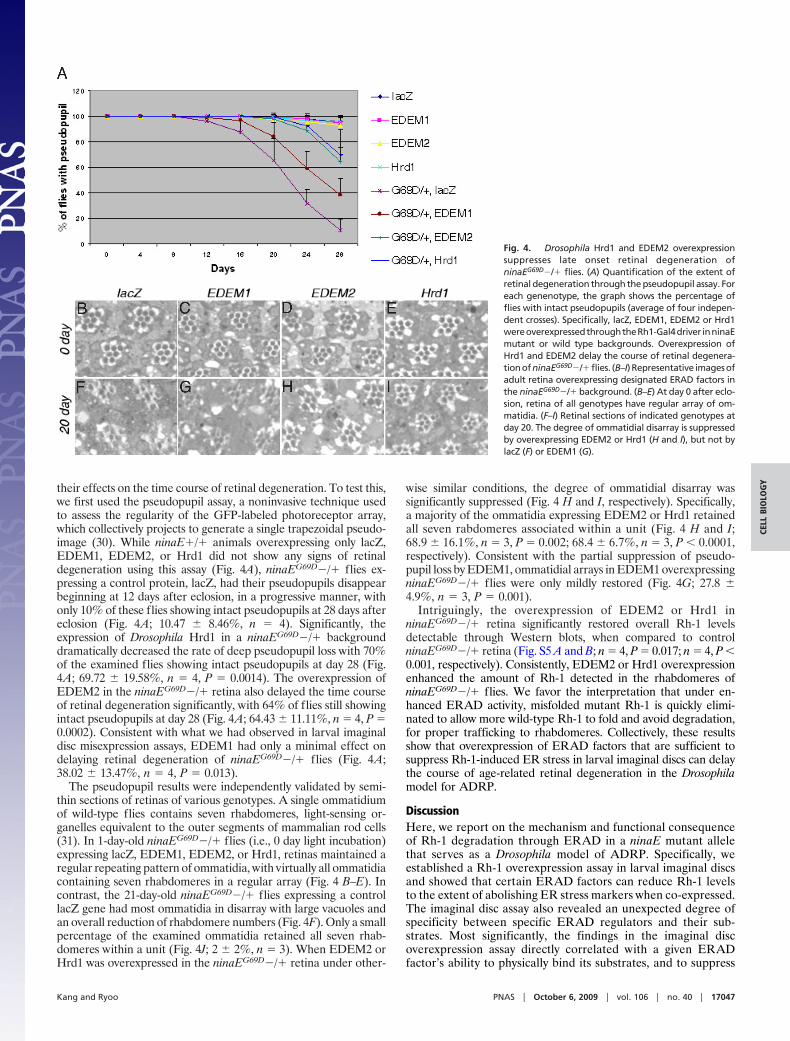
their effects on the time course of retinal degeneration. To test this,we first used the pseudopupil assay, a noninvasive technique usedto assess the regularity of the GFP-labeled photoreceptor array,which collectively projects to generate a single trapezoidal pseudo-image (30). While ninaE�/� animals overexpressing only lacZ,EDEM1, EDEM2, or Hrd1 did not show any signs of retinaldegeneration using this assay (Fig. 4A), ninaEG69D�/� flies ex-pressing a control protein, lacZ, had their pseudopupils disappearbeginning at 12 days after eclosion, in a progressive manner, withonly 10% of these flies showing intact pseudopupils at 28 days aftereclosion (Fig. 4A; 10.47 � 8.46%, n � 4). Significantly, theexpression of Drosophila Hrd1 in a ninaEG69D�/� backgrounddramatically decreased the rate of deep pseudopupil loss with 70%of the examined flies showing intact pseudopupils at day 28 (Fig.4A; 69.72 � 19.58%, n � 4, P � 0.0014). The overexpression ofEDEM2 in the ninaEG69D�/� retina also delayed the time courseof retinal degeneration significantly, with 64% of flies still showingintact pseudopupils at day 28 (Fig. 4A; 64.43 � 11.11%, n � 4, P �0.0002). Consistent with what we had observed in larval imaginaldisc misexpression assays, EDEM1 had only a minimal effect ondelaying retinal degeneration of ninaEG69D�/� flies (Fig. 4A;38.02 � 13.47%, n � 4, P � 0.013).
The pseudopupil results were independently validated by semi-thin sections of retinas of various genotypes. A single ommatidiumof wild-type flies contains seven rhabdomeres, light-sensing or-ganelles equivalent to the outer segments of mammalian rod cells(31). In 1-day-old ninaEG69D�/� flies (i.e., 0 day light incubation)expressing lacZ, EDEM1, EDEM2, or Hrd1, retinas maintained aregular repeating pattern of ommatidia, with virtually all ommatidiacontaining seven rhabdomeres in a regular array (Fig. 4 B–E). Incontrast, the 21-day-old ninaEG69D�/� flies expressing a controllacZ gene had most ommatidia in disarray with large vacuoles andan overall reduction of rhabdomere numbers (Fig. 4F). Only a smallpercentage of the examined ommatidia retained all seven rhab-domeres within a unit (Fig. 4J; 2 � 2%, n � 3). When EDEM2 orHrd1 was overexpressed in the ninaEG69D�/� retina under other-
wise similar conditions, the degree of ommatidial disarray wassignificantly suppressed (Fig. 4 H and I, respectively). Specifically,a majority of the ommatidia expressing EDEM2 or Hrd1 retainedall seven rabdomeres associated within a unit (Fig. 4 H and I;68.9 � 16.1%, n � 3, P � 0.002; 68.4 � 6.7%, n � 3, P � 0.0001,respectively). Consistent with the partial suppression of pseudo-pupil loss by EDEM1, ommatidial arrays in EDEM1 overexpressingninaEG69D�/� flies were only mildly restored (Fig. 4G; 27.8 �4.9%, n � 3, P � 0.001).
Intriguingly, the overexpression of EDEM2 or Hrd1 inninaEG69D�/� retina significantly restored overall Rh-1 levelsdetectable through Western blots, when compared to controlninaEG69D�/� retina (Fig. S5 A and B; n � 4, P � 0.017; n � 4, P �0.001, respectively). Consistently, EDEM2 or Hrd1 overexpressionenhanced the amount of Rh-1 detected in the rhabdomeres ofninaEG69D�/� flies. We favor the interpretation that under en-hanced ERAD activity, misfolded mutant Rh-1 is quickly elimi-nated to allow more wild-type Rh-1 to fold and avoid degradation,for proper trafficking to rhabdomeres. Collectively, these resultsshow that overexpression of ERAD factors that are sufficient tosuppress Rh-1-induced ER stress in larval imaginal discs can delaythe course of age-related retinal degeneration in the Drosophilamodel for ADRP.
DiscussionHere, we report on the mechanism and functional consequenceof Rh-1 degradation through ERAD in a ninaE mutant allelethat serves as a Drosophila model of ADRP. Specifically, weestablished a Rh-1 overexpression assay in larval imaginal discsand showed that certain ERAD factors can reduce Rh-1 levelsto the extent of abolishing ER stress markers when co-expressed.The imaginal disc assay also revealed an unexpected degree ofspecificity between specific ERAD regulators and their sub-strates. Most significantly, the findings in the imaginal discoverexpression assay directly correlated with a given ERADfactor’s ability to physically bind its substrates, and to suppress
Fig. 4. Drosophila Hrd1 and EDEM2 overexpressionsuppresses late onset retinal degeneration ofninaEG69D�/� flies. (A) Quantification of the extent ofretinal degeneration through the pseudopupil assay. Foreach genenotype, the graph shows the percentage offlies with intact pseudopupils (average of four indepen-dent crosses). Specifically, lacZ, EDEM1, EDEM2 or Hrd1wereoverexpressedthroughtheRh1-Gal4driver inninaEmutant or wild type backgrounds. Overexpression ofHrd1 and EDEM2 delay the course of retinal degenera-tionofninaEG69D�/�flies. (B–I)Representative imagesofadult retina overexpressing designated ERAD factors inthe ninaEG69D�/� background. (B–E) At day 0 after eclo-sion, retina of all genotypes have regular array of om-matidia. (F–I) Retinal sections of indicated genotypes atday 20. The degree of ommatidial disarray is suppressedby overexpressing EDEM2 or Hrd1 (H and I), but not bylacZ (F) or EDEM1 (G).
Kang and Ryoo PNAS � October 6, 2009 � vol. 106 � no. 40 � 17047
CELL
BIO
LOG
Y

retinal degeneration in a physiologically relevant disease modelfor ADRP, in which age-related retinal degeneration is causedby the endogenous ninaEG69D allele.
One of the unexpected outcomes of our study is the observedspecificity between given ERAD factors and their misfolded pro-tein substrates. Previous studies conducted in yeast led to theproposal of the presence of three ERAD subpathways, defined bythe subcellular locations of lesions that cause protein misfolding. Inthat view, the yeast homolog of EDEM, Htm1p, and membraneproteins, Derlin and Usa1p were considered specific components ofthe ERAD-L pathway, specializing in ER luminal protein recog-nition and degradation (13–16). By contrast, our study shows thatDrosophila EDEM2, Derlin-1 and Herp are involved in reducingthe levels of a mutant membrane protein, Rh-1G69D. The list ofERAD genes that can reduce Rh-1G69D levels were different fromthose involved in wild-type Rh-1 protein or alpha 1-antitrypsinNHK,which is an established ER luminal substrate. We favor the inter-pretation that EDEM1 initiates an ERAD-L-like pathway in Dro-sophila, in part, based on the fact that EDEM1 only reduced alpha1-antitrypsin but not the Rh-1 proteins. On the other hand, Hrd1overexpression may initiate an ERAD-M-like pathway, as thiscondition only affected Rh-1 proteins, but not alpha 1-antitrypsin.The observation that Rh-1G69D degradation properties are neitheridentical to Rh-1WT nor alpha 1-antitrypsinNHK degradation sug-gests that animals have evolved additional ERAD subpathways notreported in yeast. Such an idea has been suggested previously (32),but awaits further validation.
While the reductionist approach taken in the eye imaginal discassay has allowed us to match specific ERAD regulators withwild-type or mutant Rh-1 alleles, we were not able to follow thefates of wild-type and mutant Rh-1 proteins that must exist as a mixin the ninaEG69D�/� retina. Previous studies have demonstratedthat, under such a condition, the mutant Rh-1 proteins interferewith the proper maturation of the wild-type Rh-1, leading to thedegradation of both species (4, 7, 8, 22). Based on this together withour imaginal disc overexpression assays, we speculate that EDEM2or Hrd1 stimulates the degradation of misfolded Rh-1 proteins,thereby allowing more wild-type Rh-1 to undergo maturation.
Supporting this idea, we found that stimulating ERAD in theninaEG69D�/� retina actually enhanced overall Rh-1 levels and amore efficient Rh-1 trafficking to the rhabdomeres (Fig. S5).Elimination of misfolded Rh-1 that may otherwise cause toxicity,together with enhanced Rh-1 trafficking, most likely contribute tothe suppression of retinal degeneration in this Drosophila model forADRP.
Since the ninaEG69D�/� flies have a mutation that is molecularlysimilar to those mutations found in human patients, leading to asimilar pattern of late-onset retinal degeneration, the role of ERADin this disease progression is likely conserved between the twospecies. We exploited ERAD to delay disease in an animal diseasemodel. As ER stress underlies a wide variety of diseases, manipu-lation of ERAD may be used to therapeutically intervene in avariety of ER stress-related diseases.
Materials and MethodsPlasmids and Fly Stocks. The following flies and DNA have been describedpreviously:ninaEG69D/TM6B (8),UAS-xbp1-EGFP,UAS-Rh-1G69D (9),Rh1-Gal4,Rh1-GFP (30), gmr-Gal4 (33), hs-flp; tubulin�FRT�y�, GFP�FRT�Gal4 flies (9), Drob-1expression plasmid (25) alpha 1-antitrypsinNHK DNA (34). The coding sequencesfor Hrd1, EDEM1, EDEM2, Hrd3, Herp, Der-1, and Der-2 were obtained throughRT-PCR from yw larvae. Myc-tags were added to the N termini of these codingsequences and subcloned into a pUAST (35). pGMR-Rh-1G69D construct was cre-ated by subcloning the corresponding cDNA into the pGMR vector (33).HerpG13463 allele was obtained from BMRC KAIST. For in vivo RNAi, UAS-Hrd1-IR(V6870), UAS-Hrd3-IR (V1161), UAS-Herp-IR (V11724, V11725), and UAS-Der-1-IR(V44210,V44211)wereobtainedfromtheViennaDrosophilaRNAiCenter (http://stockcenter.vdrc.at). To enhance the efficiency of RNAi knockdown, uas-dcr2 wasdriven for co-expression with these inverted repeat lines. Additional methods areavailable in SI Text.
ACKNOWLEDGMENTS. We thank Kazuhiro Nagata (Kyoto University) and Ma-sayuki Miura (Tokyo University) for plasmids; Pedro Domingos, Alexis Gambis,Iwona Gumper, Young Kwon (Johns Hopkins), and Yihong Ye (NIH) for technicaladvice; and Milton Adesnik (NYU), David Ron (NYU), and Peter Shapiro (NYU) forcomments on the manuscript. This project was supported by grants from theNational Institutes of Health (1RO1GM079425) and the Ellison Medical Founda-tion (to H.D.R.).
1. Marciniak SJ, Ron D (2006) Endoplasmic reticulum stress signaling in disease. Physiol Rev86:1133–1149.
2. Lin JH, Walter P, Yen TS (2007) Endoplasmic reticulum stress in disease pathogenesis. AnnuRev Pathol 3:399–425.
3. Dryja T, et al. (1990) A point mutation of the rhodopsin gene in one form of retinitispigmentosa. Nature 343:364–366.
4. Sung CH, et al. (1991) Rhdopsin mutations in autosomal dominant retinitis pigmentosa.Proc Natl Acad Sci USA 88:6481–6485.
5. O’Tousa JE, et al. (1985) The Drosophila ninaE gene encodes an opsin. Cell 40:877–882.6. Zuker CS, Cowman AF, Rubin GM (1985) Isolation and structure of a rhodopsin gene from
D. melanogaster. Cell 40:851–858.7. Kurada P, O’Tousa JE (1995) Retinal degeneration caused by dominant rhodopsin muta-
tions in Drosophila. Neuron 14:571–579.8. Colley NJ, Cassill JA, Baker EK, Zuker CS (1995) Defective intracellular transport is the
molecular basis of rhodopsin-dependent dominant retinal degeneration. Proc Natl AcadSci USA 92:3070–3074.
9. Ryoo HD, Domingos PM, Kang MJ, Steller H (2007) Unfolded protein response in aDrosophila model for retinal degeneration. EMBO J 26:242–252.
10. Lin JH, et al. (2007) IRE1 signaling affects cell fate during the unfolded protein response.Science 318:944–949.
11. Mendes HF, van der Spuy J, Chapple JP, Cheetham ME (2005) Mechanism of cell death inrhodopsin retinitis pigmentosa: Implications for therapy. Trends Mol Med 11:177–185.
12. Vembar SS, Brodsky JL (2008) One step at a time: Endoplasmic reticulum-associateddegradation. Nat Rev Mol Cell Biol 9:944–957.
13. Vashist S, Ng DT (2004) Misfolded proteins are sorted by a sequential checkpoint mecha-nism of ER quality control. J Cell Biol 165:41–52.
14. Carvalho P, Goder V, Rapoport TA (2006) Distinct ubiquitin-ligase complexes defineconvergent pathways for the degradation of ER proteins. Cell 126:361–373.
15. Denic V, Quan EM, Weissman JS (2006) A luminal surveillance complex that selectsmisfolded glycoproteins for ER-associated degradation. Cell 126:349–359.
16. Quan EM, et al. (2008) Defining the glycan destruction signal for endoplasmic reticulum-associated degradation. Mol Cell 32:870–877.
17. Shih HP, Hales KG, Pringle JR, Peifer M (2002) Identification of septin-interacting proteinsand characterization of the Smt3/SUMO-conjugation system in Drosophila. J Cell Sci115:1259–1271.
18. Friedlander R, Jarosch E, Urban J, Volkwein C, Sommer T (2000) A regulatory link betweenER-associated protein degradation and the unfolded-protein response. Nat Cell Biol2:379–384.
19. Neuber O, Jarosch E, Volkwein C, Walter J, Sommer T (2005) Ubx2 links the Cdc48 complexto ER-associated protein degradation. Nat Cell Biol 7:993–998.
20. Gauss R, Jarosch E, Sommer T, Hirsch C (2006) A complex of Yos9p and the HRD ligaseintegratesendoplasmic reticulumqualitycontrol intothedegradationmachinery.NatCellBiol 8:849–854.
21. Leonard DS, Bowman VD, Ready DF, Pak WL (1992) Degeneration of photoreceptors inrhodopsin mutants of Drosophila. J Neurobiol 23:605–626.
22. Rajan RS, Kopito RR (2005) Suppression of wild-type rhodopsin maturation by mutantslinked to autosomal dominant retinitis pigmentosa. J Biol Chem 280:1284–1291.
23. Travers KJ, Patil CK, Wodicka L, Lockhart DJ, Weissman JS, Walter P (2000) Functional andgenomicanalysesrevealanessentialcoordinationbetweentheunfoldedproteinresponseand ER-associated degradation. Cell 101:249–258.
24. Yu SY, et al. (2002) A pathway of signals regulating effector and initiator caspases in thedeveloping Drosophila eye. Development 129:3269–3278.
25. Igaki T, et al. (2000) Drob-1, a Drosophila member of the Bcl-2/CED-9 family that promotescell death. Proc Natl Acad Sci USA 97:662–667.
26. Molinari M, Calanca V, Galli C, Lucca P, Paganetti P (2003) Role of EDEM in the release ofmisfolded glycoproteins from the calnexin cycle. Science 299:1397–1400.
27. Oda Y, Hosokawa N, Wada I, Nagata K (2003) EDEM as an acceptor of terminally misfoldedglycoproteins released from calnexin. Science 299:1394–1397.
28. Clerc S, et al. (2009) Htm1 protein generates the N-glycan signal for glycoprotein degra-dation in the endoplasmic reticulum. J Cell Biol 184:159–172.
29. Oda Y, Okada T, Yoshida H, Kaufman RJ, Nagata K, Mori K (2006) Derlin-2 and Derlin-3 areregulated by the mammalian unfolded protein response and are required for ER-associated degradation. J Cell Biol 172:383–393.
30. Pichaud F, Desplan C (2001) A new visualization approach for identifying mutations thataffect differentiation and organization of the Drosophila ommatidia. Development128:815–826.
31. Ready DF, Ilanson TE, Benzer S (1976) Development of the Drosophila retina, a neuroc-rystalline lattice. Dev Biol 53:217–240.
32. Okuda-Shimizu Y, Hendershot LM (2007) Characterization of an ERAD pathway fornonglycosylated BiP substrates, which require Herp. Mol Cell 28:544–554.
33. Hay BA, Wolff T, Rubin GM (1994) Expression of baculovirus P35 prevents cell death inDrosophila. Development 120:212–219.
34. Hosokawa N, et al. (2001) A novel ER alpha-mannosidase-like protein accelerates ER-associated degradation. EMBO Rep 2:415–422.
35. Brand AH, Perrimon N (1993) Targeted gene expression as a means of altering cell fatesand generating dominant phenotypes. Development 118:401–415.
17048 � www.pnas.org�cgi�doi�10.1073�pnas.0905566106 Kang and Ryoo